Chopinite, [(Mg,Fe) 3 □(PO 4 ) 2 , a new mineral isostructural with sarcopside, from a...
18
Chopinite, [(Mg,Fe) 3 ](PO 4 ) 2 , a new mineral isostructural with sarcopside, from a fluorapatite segregation in granulite-facies paragneiss, Larsemann Hills, Prydz Bay, East Antarctica EDWARD S. GREW 1, *, THOMAS ARMBRUSTER 2 ,OLAF MEDENBACH 3 ,MARTIN G. YATES 1 and CHRISTOPHER J. CARSON 4 1 Department of Earth Sciences, University of Maine, 5790 Bryand Research Center, Orono, Maine 04469-5790, USA *Corresponding author, e-mail: [email protected] 2 Laboratorium für chemische und mineralogische Kristallographie, Universität Bern, Freiestrasse 3, 3012 Bern, Switzerland 3 Institut für Geowissenschaften/Mineralogie, Ruhr-Universität Bochum, 44780 Bochum, Germany 4 Research School of Earth Sciences, Australian National University, Canberra, ACT 0200, Australia Abstract: Chopinite, the Mg-dominant analogue of sarcopside, is a new mineral corresponding to synthetic Mg 3 (PO 4 ) 2 -II, a high- pressure polymorph of the meteoritic mineral farringtonite. A representative electron-microprobe analysis is SiO 2 0.32, P 2 O 5 47.32, Al 2 O 3 0.05, MgO 30.35, MnO 0.15, FeO 20.99, CaO 0.35, F 0.02, Cl 0.01, Sum 99.54 wt %, which gives Ca 0.02 Mg 2.20 Fe 0.86 Mn 0.01 Si 0.02 P 1.95 O 8 . Single-crystal X-ray diffraction gives monoclinic symmetry, P2 1 /c, a = 5.9305(7) Å, b = 4.7583(6) Å, c = 10.2566(10) Å, = 90.663(9)°, V 289.41(6) Å 3 , calculated density 3.34 g/cm 3 , Z = 2. Chopinite is of the olivine structure type, but with ordered vacancies and strongly distorted octahedra due to the valence 5+ for P, which results in marked ordering of Mg at M2, whereas Fe 2+ concentrates at M1, most likely because of its axial symmetry. The strongest lines in the powder pattern [d in Å, (I calc ), (hkl)] are 5.92 (42) (100), 3.84(100) (102), 3.48(52) (111 , 012, 111), 2.51(72) (113 , 113), 2.44 (73) (211 , 211). Chopinite is colorless and transparent, biaxial (–), 1.595(2), 1.648(2), 1.656(2) (589 nm). 2V x (meas.) = 40(2)°, 2V x (calc.) = 41°; X // b,Z^ a ~55°. Chopinite is found as four inclusions isolated in a fluorapatite segregation in a quartz mass in a paragneiss from Brattnevet, Larsemann Hills, East Antarctica. Grains are mostly anhedral and range from 0.1 × 0.3 mm to 0.2 × 0.6 mm in size. Minerals present in the chopinite-bearing specimen include wagnerite-Ma5bc, xenotime-(Y), stornesite-(Y), P-bearing K-feldspar and plagioclase, Ti-rich biotite, sillimanite, orthopyroxene, sapphirine, hercynite, and corundum. It is inferred to have formed as a result of high melt P concentrations by reaction of biotite with an anatectic melt in which P/Ca ratio exceeded that buffered by apatite saturation due to the very slow diffusion of P relative to Ca in anatectic melt. Key-words: phosphate, new mineral, Antarctica, Larsemann Hills, electron microprobe, crystal structure, granulite facies, anatexis. Introduction Sarcopside, (Fe, Mn, Mg) 3 (PO 4 ) 2 , is an uncommon mineral in granite pegmatites and in IIIAB meteorites; there are al- so possible occurrences in metamorphic environments and pallasites. Sarcopside is invariably dominated by Fe, with the maximum X Mg = Mg/(Mg+Fe) = 0.37 and maximum X Mn = Mn/(Mn+Fe) = 0.36. With rare exceptions terrestrial sarcopside forms intergrowths with graftonite or triphylite. The Mg-analogue of sarcopside was first synthesized as a metastable phase by a solid-state exchange process at 660°C and 1 bar by Berthet et al. (1972), and subsequently at 600 °C and 30 kbar, by Annersten & Nord (1980). Bru- net & Vielzeuf (1996) and Brunet et al. (1998) showed that the Mg-analogue of sarcopside is a high-pressure poly- morph of the meteoritic mineral farringtonite and deter- mined the equilibrium reaction to lie at 6.32–8.30 kbar for 566–824 °C. Here we report Mg-dominant analogue of sarcopside as the new mineral chopinite from a terrestrial metamorphic environment: a fluorapatite segregation in granulite-facies (800–860 °C, 6–7 kbar) biotite-quartz-plagioclase para- gneiss at 69° 24.437’ S, 76° 15.057’ E on Brattnevet Penin- sula, Larsemann Hills, Prydz Bay, East Antarctica. The min- eral and name were approved by the Commission on New Minerals and Mineral Names, International Mineralogical Association (2006-004). The name is for Christian Chopin (born 1955) of the Ecole Normale Sup´ erieure, Paris, France, for his major contributions to the mineralogy of phosphates. Holotype material (sample no. 121401E and thin section 121401E4) is deposited in the Mus´ ee de Min´ eralogie, Ecole des Mines de Paris as catalogue number M 73096. Eur. J. Mineral. 2007, 19, 229–245 DOI: 10.1127/0935-1221/2007/0019-1712 0935-1221/07/0019-1712 $ 7.65 2007 E. Schweizerbart’sche Verlagsbuchhandlung, D-70176 Stuttgart
Transcript of Chopinite, [(Mg,Fe) 3 □(PO 4 ) 2 , a new mineral isostructural with sarcopside, from a...
Chopinite, [(Mg,Fe)3�](PO4)2, a new mineral isostructural with sarcopside,from a fluorapatite segregation in granulite-facies paragneiss,
Larsemann Hills, Prydz Bay, East Antarctica
EDWARD S. GREW1,*, THOMAS ARMBRUSTER2, OLAF MEDENBACH3, MARTIN G. YATES1
and CHRISTOPHER J. CARSON4
1Department of Earth Sciences, University of Maine, 5790 Bryand Research Center, Orono, Maine 04469-5790, USA*Corresponding author, e-mail: [email protected]
2Laboratorium für chemische und mineralogische Kristallographie, Universität Bern, Freiestrasse 3,3012 Bern, Switzerland
3Institut für Geowissenschaften/Mineralogie, Ruhr-Universität Bochum, 44780 Bochum, Germany4Research School of Earth Sciences, Australian National University, Canberra, ACT 0200, Australia
Abstract: Chopinite, the Mg-dominant analogue of sarcopside, is a new mineral corresponding to synthetic Mg3(PO4)2-II, a high-pressure polymorph of the meteoritic mineral farringtonite. A representative electron-microprobe analysis is SiO2 0.32, P2O5 47.32,Al2O3 0.05, MgO 30.35, MnO 0.15, FeO 20.99, CaO 0.35, F 0.02, Cl 0.01, Sum 99.54 wt%, which givesCa0.02Mg2.20Fe0.86Mn0.01Si0.02P1.95O8. Single-crystal X-ray diffraction gives monoclinic symmetry, P21/c, a = 5.9305(7) Å, b =4.7583(6) Å, c = 10.2566(10) Å, q = 90.663(9)°, V 289.41(6) Å3, calculated density 3.34 g/cm3, Z = 2. Chopinite is of the olivinestructure type, but with ordered vacancies and strongly distorted octahedra due to the valence 5+ for P, which results in markedordering of Mg at M2, whereas Fe2+ concentrates at M1, most likely because of its axial symmetry. The strongest lines in the powderpattern [d in Å, (Icalc), (hkl)] are 5.92 (42) (100), 3.84(100) (102), 3.48(52) (111, 012, 111), 2.51(72) (113, 113), 2.44 (73) (211, 211).Chopinite is colorless and transparent, biaxial (–), [ 1.595(2), q 1.648(2), * 1.656(2) (589 nm). 2Vx (meas.) = 40(2)°, 2Vx (calc.) =41°; X // b, Z ^ a ~55°. Chopinite is found as four inclusions isolated in a fluorapatite segregation in a quartz mass in a paragneiss fromBrattnevet, Larsemann Hills, East Antarctica. Grains are mostly anhedral and range from 0.1 × 0.3 mm to 0.2 × 0.6 mm in size.Minerals present in the chopinite-bearing specimen include wagnerite-Ma5bc, xenotime-(Y), stornesite-(Y), P-bearing K-feldsparand plagioclase, Ti-rich biotite, sillimanite, orthopyroxene, sapphirine, hercynite, and corundum. It is inferred to have formed as aresult of high melt P concentrations by reaction of biotite with an anatectic melt in which P/Ca ratio exceeded that buffered by apatitesaturation due to the very slow diffusion of P relative to Ca in anatectic melt.
Key-words: phosphate, new mineral, Antarctica, Larsemann Hills, electron microprobe, crystal structure, granulite facies, anatexis.
Introduction
Sarcopside, (Fe, Mn, Mg)3(PO4)2, is an uncommon mineralin granite pegmatites and in IIIAB meteorites; there are al-so possible occurrences in metamorphic environments andpallasites. Sarcopside is invariably dominated by Fe, withthe maximum XMg = Mg/(Mg+Fe) = 0.37 and maximumXMn = Mn/(Mn+Fe) = 0.36. With rare exceptions terrestrialsarcopside forms intergrowths with graftonite or triphylite.The Mg-analogue of sarcopside was first synthesized as ametastable phase by a solid-state exchange process at660°C and 1 bar by Berthet et al. (1972), and subsequentlyat 600 °C and 30 kbar, by Annersten & Nord (1980). Bru-net & Vielzeuf (1996) and Brunet et al. (1998) showed thatthe Mg-analogue of sarcopside is a high-pressure poly-morph of the meteoritic mineral farringtonite and deter-
mined the equilibrium reaction to lie at 6.32–8.30 kbar for566–824 °C.
Here we report Mg-dominant analogue of sarcopside asthe new mineral chopinite from a terrestrial metamorphicenvironment: a fluorapatite segregation in granulite-facies(800–860 °C, 6–7 kbar) biotite-quartz-plagioclase para-gneiss at 69° 24.437’ S, 76° 15.057’ E on Brattnevet Penin-sula, Larsemann Hills, Prydz Bay, East Antarctica. The min-eral and name were approved by the Commission on NewMinerals and Mineral Names, International MineralogicalAssociation (2006-004). The name is for Christian Chopin(born 1955) of the Ecole Normale Superieure, Paris, France,for his major contributions to the mineralogy of phosphates.Holotype material (sample no. 121401E and thin section121401E4) is deposited in the Musee de Mineralogie, Ecoledes Mines de Paris as catalogue number M 73096.
Eur. J. Mineral.2007, 19, 229–245
DOI: 10.1127/0935-1221/2007/0019-17120935-1221/07/0019-1712 $ 7.65
ˇ 2007 E. Schweizerbart’sche Verlagsbuchhandlung, D-70176 Stuttgart

Analytical methods
The optical properties of chopinite grain #3 were measuredat the Ruhr-Universität Bochum by routine immersion pro-cedure using a microrefractometer spindle-stage (Meden-bach, 1985).
X-ray powder-diffraction data were obtained from chopi-nite grain #3 with a 57.3 mm diameter Gandolfi camera andCuK [ radiation at the Ruhr-Universität Bochum (Table 1).
Cell dimensions of chopinite grain #3 (Table 2) were re-fined from reflections at scattering angles 13 < ’ < 20° ob-tained with graphite-monochromated MoK [ radiation on anENRAF NONIUS CAD4 diffractometer equipped with apoint detector at the Universität Bern. CAD4 instrumenta-tion gives higher accuracy albeit similar precision as theCCD instrument.
Single-crystal X-ray intensity data were collected onchopinite grain #3 with a 3-circle SMART BRUKER CCD1K, graphite-monochromated MoK [ radiation at the Uni-versität Bern. Like sarcopside, chopinite is pseudo-ortho-rhombic, but truly monoclinic. We chose the standard set-
Table 1. X-ray powder-diffraction data for chopinite (grain 3).
Iest dmeas (Å) Icalc dcalc (Å) hkl
st 5.92 42 5.9306 1 0 0w 4.31 29 4.3162 0 1 1st 3.84 100 3.8571 1 0 2vw 3.65st 3.48 4 3.4979 1 1 1
11 3.4879 0 1 237 3.4817 1 1 1
w 2.97 25 2.9653 2 0 0m 2.77 46 2.7764 0 1 3st 2.51 59 2.5236 1 1 3
13 2.5055 1 1 3st 2.44 40 2.4497 2 1 1
33 2.4385 2 1 1m 2.26 11 2.2680 2 1 2
17 2.2572 0 1 47 2.2505 2 1 2
w 2.15 7 2.1604 1 2 113 2.1566 1 2 1
w 1.79 8 1.8006 3 1 112 1.7898 1 1 5
m 1.74 17 1.7490 2 2 28 1.7440 0 2 4
16 1.7409 2 2 2vw 1.65 15 1.6696 1 2 4
13 1.6476 1 0 6w 1.47 10 1.4884 2 0 6
14 1.4827 4 0 08 1.4736 2 0 6
w 1.40 6 1.4003 0 1 77 1.3997 1 3 3
vw 1.35 5 1.3511 2 3 25 1.3474 2 3 2
Note: Intensities are visual estimates. Five strongest lines are givenin bold. Calculated data from LAZY PULVERIX (Yvon et al., 1977)based on single-crystal refinement: a = 5.9305(7) Å, b = 4.7583(6)Å, c = 10.2566(10) Å, q = 90.663(9)°
ting P21/c. Twinning (imitating orthorhombic symmetry) isevident from optical inspection and structure refinementleaving the choice between (100) and (001) as twin planes.Due to q = 90.66° (close to 90°) the twin law was not obvi-ous from the single-crystal diffraction pattern. The half-widths of hk0 and 0kl reflections were checked and the hk0reflections were systematically found to be more asymmet-ric and broadened compared to 0kl reflections, which sug-gests that the twin plane is {100}, i.e., identical to the twinplane {001} reported by Hurlbut (1965) for sarcopside inthe P21/a setting. In order to deal with the extensive poly-synthetic twinning, a large window for intensity integrationfor each reflection was chosen. It was then possible to col-lect the intensity of all twin domains. The structure wassolved by direct methods and refined using the SHELXTLVersion 6.12 program package (Sheldrick, 1997). All siteswere refined with neutral atom scattering-factors (Mg, Fe, P,and O) and anisotropic displacement parameters. In addi-tion, variation of Mg and Fe on the octahedral sites M1 andM2 was allowed assuming complete occupancy. The octa-hedral site M1’ at 0,0,1/2, which is occupied in forsterite
Table 2. Parameters for X-ray data collection and crystal-structurerefinement of chopinite.
Diffractometer Siemens Smart CCDX-ray radiation MoK [ (0.71073 Å)X-ray power 50 kV, 40 mATemperature 293 KCrystal size (mm3) 0.1 × 0.1 × 0.03Detector to sample distance 5.4 cmRotation axis KRotation width 0.3°Total number of frames 1362Frame size 512 × 512 pixelsTime per frame 120 secSpace group P21/c (Nr. 14)Cell dimensions a = 5.9305(7) (Å)
b = 4.7583(6) (Å)c = 10.2566(10) (Å)q = 90.663(9)°
V = 289.41(6) Å3
Z = 2Collection mode automated hemisphereReflections collected 1543Maximum 2 ’ 55.43Index range –7 e h e 5
–6 e k e 4–11 e l e 12
Unique reflections 617Reflections > 2 c (I) 567Rint 0.028R c 0.027Number of least squares parameters 64GooF 1.034R1 , I > 2 c (I) 0.024R1 , all data 0.027wR2 (on F2) 0.058+ 2 e (e/Å3) 0.4– 2 e (e/Å3) 0.4
230 E.S. Grew, T. Armbruster, O. Medenbach, M.G. Yates, C.J. Carson

and fayalite, was found to be vacant, or very nearly so. Thetwin contribution was refined to 17.1(2) %. Anisotropic andisotropic displacement parameters, atomic coordinates andoccupancies are given in Tables 3–5.
Chopinite and associated minerals in sections of sample121401E were analyzed with a Cameca SX-100 electronmicroprobe at the University of Maine and with a CamecaSX-50 at the Centre de Microanalyse Camparis, Paris. Ana-lytical conditions for analysis of phosphates at the Universi-ty of Maine were 15 kV accelerating voltage, 10 nA beamcurrent and 20 µm spot diameter, and data were processedusing the X-Phi correction of Merlet (1994). The standardsused for chopinite and wagnerite were fluorapatite (FK [ ),tugtupite (NaK [ ), synthetic Mg3(PO4)2 (MgK [ ), albite(AlK [ ), albite (SiK [ ), synthetic Mg3(PO4)2 (PK [ ), tugtupi-te (ClK [ ), fluorapatite (CaK [ ), rutile (TiK [ ), rhodonite(MnK [ ), almandine (FeK [ ); additionally for apatite, fluor-apatite (PK [ ), barite (SK [ ), celestine (SrL [ ), synthetic Y-Al garnet (YL [ ), synthetic rare-earth element phosphates(REE L [ ), and U metal (UM q ). Three grains of chopinitewere analyzed at twenty spots where each constituent wascounted for 5 seconds, but 4 to 9 analyses were rejected be-cause of impurities and alteration. Fluorine in biotite wasanalyzed using the TAP crystal and a polylithionite stan-dard. Zr content of rutile was counted for a total of 1600 sec-onds using a zirconia standard (SPI-47) and 4 spectrometerssimultaneously to improve the statistics.
Analytical conditions for analysis of silicates and oxidesat the Centre de Microanalyse Camparis were 15 kV accel-erating voltage, 10 nA beam current and 5 µm spot diame-ter, and data were processed using PAP corrections. Thestandards and counting times used for chopinite, wagnerite
Table 3. Anisotropic displacement parameters Uij with standard deviations in parenthesesfor chopinite.
Site U11 U22 U33 U12 U13 U23
Fe1 0.0126(4) 0.0065(4) 0.0103(4) –0.0006(3) 0.0024(3) 0.0001(3)Mg2 0.0093(5) 0.0088(5) 0.0086(4) –0.0001(3) 0.0009(4) 0.0001(3)P 0.0112(3) 0.0078(3) 0.0099(3) 0.0000(3) 0.0005(3) –0.0002(3)O1 0.013(1) 0.009(1) 0.012(1) 0.0009(8) 0.0014(8) –0.0007(7)O2 0.014(1) 0.0115(9) 0.0101(9) –0.0010(8) –0.0003(9) –0.0002(7)O3 0.011(1) 0.011(1) 0.015(1) –0.0008(8) 0.0033(7) –0.0019(8)O4 0.013(1) 0.0096(9) 0.014(1) 0.0000(8) 0.0003(8) –0.0014(7)
Table 4. Atomic coordinates and isotropic displacement parameters, with standard devia-tions in parentheses, for chopinite.
site x/a y/b z/c Beq (Å2) Occupancy
Fe1 1/2 0 1/2 0.77(1) 0.518(6)Mg1 1/2 0 1/2 0.77(1) 0.482Fe2 0.2377(2) 0.0118(1) 0.77910(8) 0.70(1) 0.107(4)Mg2 0.2377(2) 0.0118(1) 0.77910(8) 0.70(1) 0.893P 0.2562(1) –0.4293(2) 0.59884(6) 0.76(1) 1O1 0.2756(3) 0.2519(4) 0.6058(2) 0.89(3) 1O2 0.2552(3) –0.3189(4) 0.4588(2) 0.92(3) 1O3 0.0612(3) –0.3153(4) 0.6789(2) 0.97(3) 1O4 0.5334(3) 0.2781(4) 0.3373(2) 0.95(3) 1
Anisotropically refined atoms are given in the form of the isotropic equivalent displace-ment parameter defined as Beq = (8/3) ‘ 2 7 i( 7 j( Uij ai* aj* ai.aj))
and biotite were Durango fluorapatite or topaz (15 s, FK [ ),albite (10 s, NaK [ ), synthetic Mg3(PO4)2, (10 s, MgK [ ), or-thoclase (15 s, AlK [ ), diopside (15 s, SiK [ ), syntheticMg3(PO4)2 (10 s, PK [ ), scapolite (15 s, ClK [ ), orthoclase(10 s, KK [ ), diopside (10 s, CaK [ ), MnTiO3 (10 s, TiK [ ),MnTiO3 (10 s, MnK [ ), hematite (10 s, FeK [ ), sphalerite(15 s, ZnK [ ), and barite (10 s, BaL [ ).
Identification of minerals not analyzed with WDS wasconfirmed by taking an element scan using energy-disper-sive spectroscopy at the University of Maine.
Crystal structure
Chopinite and sarcopside are of the olivine structure type,but with ordered vacancies and strongly distorted octahe-dra due to the valence 5+ for P (Fig. 1). In olivine each oxy-gen atom is coordinated to 3 M and 1 Si yielding the “ide-al” bond strength of 2 for oxygen (3 x 2/6 + 4/4). In sarcop-side O1, O2, and O3 (our numbering) are bonded 2× to Mand 1× to P yielding 2 × 2/6 + 5/4 = 1.917 indicating slightunderbonding. However, O4 bonds 3 × to M and 1 × to Pyielding 3 × 2/6 + 5/4 = 2.25 indicating significant over-bonding. For this reason all M-O4 and P-O4 bonds arerather long (Table 5), which results in strong distortionsand a marked ordering of Mg at the M2 site (89 % Mg).Fe2+ is ordered at M1 (52% Fe), most likely because of axi-al symmetry of the M1 octahedron, that is, the presence ofthree 180° O-M1-O angles (Table 5). This symmetryseems to be more appropriate for arrangement of d-elec-tron orbitals than the M2 octahedron, which lacks this axialsymmetry (with 152°, 153°, and 169°). The M1 octahedron
Chopinite from the granulite-facies, Prydz Bay, Antarctica 231

Table 5. Bond lengths (Å) and angles (°).
Length Angles
Fe1O1_$1 2.102(2)O1 2.102(2) 180O2_$1 2.139(2) 94.25(7) 85.75(7)O2 2.139(2) 85.75(7) 94.25(7) 180.00(7)O4 2.140(2) 83.41(7) 96.59(7) 69.30(7) 110.70(7)O4_$1 2.141(2) 96.59(7) 83.40(7) 110.70(7) 69.30(7) 180Mean 2.127 O1_$1 O1 O2_$1 O2 O4
VacancyO2 2.187(2)O2_$5 2.187(2) 180.00(7)O1_$5 2.289(2) 92.10(6) 87.90(6)O1 2.289(2) 87.90(6) 92.10(6) 180O3_$5 2.394(2) 112.67(7) 67.33(7) 82.17(6) 97.83(6)O3 2.394(2) 67.33(7) 112.67(7) 97.83(6) 82.17(6) 180Mean 2.290 O2 O2_$5 O1_$5 O1 O3_$5
Mg2O3_$4 2.006(2)O2_$3 2.061(2) 91.50(9)O4_$5 2.100(2) 118.74(9) 85.66(8)O1 2.127(2) 93.52(8) 169.11(9) 83.45(8)O3 2.133(2) 88.45(6) 97.21(8) 152.66(8) 92.59(8)O4_$1 2.282(2) 153.24(9) 100.06(8) 86.38(5) 79.52(7) 66.32(7)Mean 2.118 O3_$4 O2_$3 O4_$5 O1 O3
PO1_$2 1.523(2)O3 1.526(2) 112.74(11)O2 1.529(2) 112.70(11) 112.85(12)O4_$1 1.576(2) 112.11(11) 102.43(10) 103.15(11)Mean 1.539 O1_$2 O3 O2
Symmetry codes. $1: –x+1,–y,–z+1. $2: x, y–1, z. $3: x, –y–1/2, z+1/2. $4: –x, y+1/2,–z+3/2. $5: x, –y+1/2, z+1/2.
is slightly larger (<M1-O> = 2.217 Å) than M2 (<M2-O> =2.118 Å). In addition, M1 is more regular: the differencebetween the largest and shortest M-O bond is 0.038 Å forM1 but 0.274 Å for M2. Lastly, M1 is adjacent to the va-cant octahedral site, thus the M1 octahedron is freer toadopt a favorable coordination for Fe2+.
The largest octahedral site (<M-O> = 2.290 Å) in thestructure is M1’, which is vacant in sarcopside and chopini-te. Local occupation of M1’ would increase the bondstrength of O1, O2, and O3 by additional 2/6, which is ener-getically not favorable. Nevertheless, the highest positivepeak in the final difference-Fourier map of 0.4 e/Å3 wasfound at M1’. This residual density was not considered inthe refinement because the highest negative peak close tothe tetrahedral P sites was also –0.4 e/Å3. One may speculatethat the minor substitution of P5+ by Si4+ determined by elec-tron microbe-analyses (Table 6) is charge balanced by veryminor occupation of M1’ by additional Mg or Fe.
There is a minor difference between XMg obtained fromthe structure refinement (XMg = 0.756) vs. the correspondingvalue (XMg = 0.720) determined by electron microprobeanalyses (Table 6). There are several possible explanationsfor this discrepancy. (1) X-ray single-crystal diffractionyields the 3-dimensional average bulk composition of the
entire crystal, whereas the electron-microprobe beam doesnot penetrate very far below the surface of the section, i.e.,the different results could be due to compositional zoning.(2) Because Fe is the atom with the strongest scatteringpower in the structure, there is also a minor correlation in thestructure refinement between Fe occupancy on one hand,scale factor and displacement parameters on the other. (3)The very minor (0.4 e/Å3) residual density at M1’ was notconsidered. Furthermore, the twinning model with {100} astwin plane maps M1 on M1’ (Fig. 1). Thus there is somecorrelation between M1’ occupancy and the twinning con-tribution. Another possible explanation is the ionization lev-el of oxygen. However, Armbruster et al. (1990) tested theeffect of choosing neutral vs. ionic scattering factors on dis-placement parameters (Beq), which are even more sensitivethan occupancies, and found no significant variation in Beqof T sites in models with Si4+ and Al+3 compared to thosewith neutral Si and Al in albite.
The synthetic analogue Mg3(PO4)2 of chopinite was firstsynthesized by Berthet et al. (1972) as a metastable productof the following solid state reaction at 660°C: 2×LiMg(PO4) (olivine structure-type) + MgSO4 → Mg3(PO4)2 (sar-copside structure-type) + Li2SO4. The above authors deter-mined from X-ray powder data the correct symmetry (P21/b;
232 E.S. Grew, T. Armbruster, O. Medenbach, M.G. Yates, C.J. Carson

Fig. 1. Projection of the chopinite structure along b. M2 octahedra(Mg enriched) form unbroken edge-sharing chains parallel to a,whereas M1 octahedra (Fe enriched) alternate with vacant polyhedra(dark with line and cross pattern). PO4 tetrahedra are marked withcrosses.
their setting: a = 5.912(2), b = 10.21(3), c = 4.73(2), Å,* = 90°60’) and derived in analogy to olivine a correct struc-ture model with ordered octahedral vacancies. At 820°C thismetastable chopinite analogue transformed to Mg3(PO4)2having the farringtonite structure. Farringtonite is not basedon a hexagonal closest-package of oxygen (as the olivineand sarcopside structure-type) and has for this reason also alarger unit-cell volume, 316.6 Å3 (Nord & Kierkegaard,1968) vs. 285.8 Å3. In farringtonite Mg occurs in fivefoldand octahedral coordination.
Recently Henry et al. (2003) refined the sarcopside-likestructure of synthetic Fe2Ni(PO4)2 by neutron powder dif-
fraction and found Fe enriched at M2 and Ni dominating atM1. This Fe order scheme is the opposite of that found inchopinite. However, in Fe2Ni(PO4)2 two transition metalions are competing with each other for the most favorablecoordination. In this case Ni occupies the less distorted site.Previous crystallographic studies of solid solutions ofMe3(PO4)2 with Me = Co2+, Mg, Zn, Mn2+, Fe2+ in sarcopsi-de-like Ni3(PO4)2 yielded the preference Ni > Co >Mg, Zn >Mn >> Fe for M1 over M2 (Nord, 1984). A neutron powderstructure-refinement of synthetic sarcopside Fe3(PO4)2 at59 K (Warner et al., 1992) yielded <M1-O> = 2.147 Åand <M2-O> = 2.159 Å, whereas a single-crystal X-raystructure refinement (Moore, 1972) of natural (Fe0.78Mn0.21Mg0.01)3(PO4)2 sarcopside gave <M1-O> = 2.130 Åand <M2-O> = 2.164 Å. Due to the similarity of Mn and Fescattering factors for X-rays Moore (1972) did not deter-mine whether Fe and Mn order in sarcopside, but instead as-sumed equal distribution of both elements at M1 and M2.However, comparison of <M1-O> and <M2-O> bondlengths with those of Warner et al. (1992) suggests that thelarger Mn2+ will order at M2.
Physical and optical properties of chopinite
Most physical properties cannot be determined because ofthe small grain size and the very limited amount of availablematerial. Two good cleavages are evident in photomicro-graphs and back scattered electron images (see below);these could {001} and {100}, the two good cleavages re-ported for sarcopside (Hurlbut, 1965). Density calculatedwith empirical formula is 3.34 g/cm3.
Chopinite is colorless and transparent, biaxial (–), [1.595(2), q 1.648(2), * 1.656(2) (589 nm). 2Vx (meas.) =40(2)°, 2Vx (calc.) = 41°. Dispersion was not visible, but theinterference figure was poor due to inclusions of brownishmaterial. Assuming that the twin plane is {100}, orientationof the principal vibration directions derived from stereo-graphic projection is X // b, Z /\ a ~55°. Larsen & Berman(1934) reported Z // b, X /\ c = 45° for sarcopside, but theygave the cleavages as perfect in {010} and {100}. Hurlbut(1965) was unable to confirm the Larsen & Berman orienta-tion and reported an apparent optical orientation for sarcop-side characteristic of a triclinic crystal with Y closest to b.Hurlbut (1965) failed to resolve the obvious (to him) incon-sistency with Larsen & Berman (1934), and we have no ex-planation why the orientation for chopinite differs from bothof those reported for sarcopside.
Chemical composition and compatibility indexof chopinite
The three analyzed chopinite grains are fairly homogeneousand virtually identical in composition (Table 6). Chopinite isnearly pure ferromagnesian phosphate containing but minorCa, Mn and Si. We did not analyze Li, which was reported insarcopside (Hurlbut, 1965) and is an essential constituent ofthe structurally related triphylite-lithiophyllite series. Fluo-rine contents obtained at University of Maine are negligible,
Chopinite from the granulite-facies, Prydz Bay, Antarctica 233

Table 6. Analyses of chopinite in section 121401E4-1.
Grain 1 1 2 2 3* 3*Probe UM Cam UM Cam UM SREFNo. spots 12 7 16 5 11
wt%SiO2 0.13(4) 0.10(3) 0.23(6) 0.16(4) 0.32(3)P2O5 48.00(50) 47.80(49) 47.84(53) 47.54(17) 47.32(44) 49.64TiO2 b.d. 0.01(2) b.d. 0.01(2) b.d.Al2O3 0.02(3) 0.02(4) 0.01(2) 0.01(2) 0.05(4)MgO 30.85(25) 30.29(42) 30.87(31) 29.92(50) 30.35(41) 31.96MnO 0.11(4) 0.14(6) 0.14(4) 0.11(4) 0.15(3)FeO 20.75(32) 20.34(42) 21.11(38) 20.53(45) 20.99(38) 18.40ZnO n.a 0.03(6) n.a 0.05(6) n.aNa2O 0.01(4) b.d. 0.01(4) 0.01(2) b.d.K2O n.a 0.01(1) n.a b.d. n.aCaO 0.11(10) 0.02(2) 0.10(10) 0.03(2) 0.35(17)BaO n.a 0.11(18) n.a 0.02(3) n.aF 0.04(6) 0.19(30) 0.01(11) 0.18(33) 0.02(4)Cl b.d. b.d. b.d. 0.01(1) 0.01(2)Sum 99.99 98.88 100.30 98.41 99.54 100.00
Formulae per 8 OxygenSi 0.006 0.005 0.011 0.008 0.016P 1.965 1.977 1.956 1.976 1.952 2.0000Al 0.001 0.001 0.001 0.001 0.003Mg 2.224 2.206 2.223 2.190 2.204 2.2675Mn 0.005 0.006 0.006 0.005 0.006Fe 0.839 0.831 0.853 0.843 0.855 0.7325Zn – 0.001 – 0.002 –Na 0.001 0.000 0.001 0.001 0.000Ca 0.006 0.001 0.005 0.001 0.018Sum 5.046 5.029 5.055 5.028 5.055 5.000XMg 0.726 0.726 0.723 0.722 0.720 0.756
Notes: Probe: UM – University of Maine, Cam – Camparis, SREF – single-crystal structure refinement. n.a.– not analyzed; b.d. – below detection. All Fe as FeO. Numbers in parentheses are 1 c standard deviations oflast 1 or 2 digits in the averages. Totals does not include F, Cl. Formulae do not include Ba, F or Cl. XMg =atomic Mg/(Mg+Fe). *Grain used for optical and crystallographic studies.
whereas those obtained at the Centre de Microanalyse Cam-paris vary with the standard used, ranging from below detec-tion to 0.10 wt% with the topaz standard and from 0.62 to 0.76wt% with the fluorapatite standard. Averages of individualgrains in Table 6 include measurements with both F stan-dards, resulting in high standard deviations for F. Analyses atthe two laboratories gave virtually identical formulae, exceptthe Centre de Microanalyse Camparis analyses gave a closerapproach to ideal stoichiometry in terms of (P+Si):(Mg+Fe+Mn+Ca) ratio and lower Ca contents. Both sets of analy-ses gave a significantly lower Mg:(Fe+Mn) ratio than thecrystal structure refinement (see above).
The Gladstone – Dale relation (Mandarino, 1981) gives acompatibility index 1 – (KP/KC) = 0.001 (superior) for grainno. 3.
Relationship of chopinite to other (Mg, Fe,Mn)3(PO4)2 phases
Only two terrestrial minerals are composed almost exclu-sively of (Mg, Fe, Mn)3(PO4)2: the isostructural phases cho-pinite and sarcopside (Fig. 2). Graftonite (Fe > Mn) and
beusite (Mn > Fe) are (Fe, Mn, Ca, Mg)3(PO4)2 phosphatescontaining significant Ca. Graftonite has a higher Mn/Fe ra-tio and lower Mg/Fe ratio than associated sarcopside. TheBrattnevet chopinite is far more magnesian than any knownsarcopside or graftonite, and unlike most terrestrial sarcop-side, it is not intergrown with either graftonite or triphylite.Experiments suggest that there is no break in solid solutionbetween synthetic end-member chopinite and sarcopside(e.g. Annersten & Nord, 1980; Charalampides et al., 1988).An Mg-dominant analogue of graftonite has not been re-ported in nature or experiment.
Meteoritic Mg-Fe-Mn phosphates include not only graf-tonite and sarcopside, but also farringtonite (Fig. 2). Syn-thetics with the farringtonite structure extend to 60% Fe-end-member (Annersten et al., 1980). Graftonite and sar-copside in IVA and IIIAB irons are Fe-Mn solid solutionscontaining negligible Mg; graftonite is richer in Mn than as-sociated sarcopside; identification by X-ray diffraction waspossible in a few cases (Bild, 1974; Olsen et al., 1999). Incontrast, phosphates in the Graves Nunatak (GRA) 95209lodranite are Mn-poor (Mg, Fe)3(PO4)2 phases that rangefrom Mg-rich to Fe-rich, referred to as “farringtonite” and“Mg-graftonite” or “graftonite/sarcopside”, respectively
234 E.S. Grew, T. Armbruster, O. Medenbach, M.G. Yates, C.J. Carson

0.0
0.2
0.4
0.6
0.8
1.0
0.0 0.2 0.4 0.6 0.8 1.0Fe Fe3(PO4)2Mn3(PO4)2
terrestrialsarcopsideterrestrialgraftonite
chopinite
graftonite in IIIAB irons
sarcopsidein IIIAB irons
FarringtoniteGRA 95209Brahin
Meteoritic phosphatesMg3(PO4)2
Bt 1 Qz
QzAp
12
3
4
Ap
Section 121401E4-1
Bt 2
(Floss, 1999; McCoy et al., 2006). A Fe-Mg phosphate inthe Brahin pallasite also has the stoichiometry of sarcopside(Buseck & Holdsworth, 1977). Although information otherthan chemical was not obtained to identify these ferromag-nesian phosphates, there is reason to suspect that two orthree phases are present in GRA 95209 (see below).
Fig. 2. Plot of natural farringtonite, graftonite, sarcopside, and chopinite compositions in terms of the divalent cations excluding Ca. Thefields for terrestrial graftonite (dashed line) and sarcopside (solid line) are largely from intergrowths (Cerny et al., 1998; Corbella i Cordomı& Melgarejo i Draper, 1990; Fontan & Fransolet, 1986; Fransolet, 1977; Fransolet et al., 1986; Hurlbut, 1965; Huvelin et al. (1971); Lindberg(1950); Livingstone, 1980; Mallo et al., 1995; Palache et al. (1951); Povondra et al., 1987; Roda et al., 2004; Smeds et al.,1998; Stalder &Rozendaal, 2002; Zhang, 1995). Sources of other data: chopinite (this paper); farringtonite (Fuchs et al., 1973; Bild, 1974; Buseck & Holds-worth, 1977); phosphates in meteorite GRA 95209 (Floss, 1999; McCoy et al., 2006); graftonite and sarcopside in IVA and IIIAB iron mete-orites (Bild, 1974; Olsen et al., 1999); phosphate (sarcopside?) in the Brahin pallasite (Buseck & Holdsworth, 1977).
Fig. 3. Photomicrograph of fluorapatite segregation (Ap) containing four grains of chopinite (numbered, Fig. 4) in section 121401E4. Matrixis quartz (Qz); Bt 1 and 2 – biotite grains closest to chopinite (Table 9); only a tip of the second grain is visible in this photograph. Plane polar-ized light.
Occurrence and associated minerals
Chopinite occurs in one (Fig. 3) of many fluorapatite segre-gations in a quartz mass roughly 10 cm thick and 3 m long inbiotite-quartz-plagioclase paragneiss, which also containssegregations of prismatine and cordierite at this locality on
Chopinite from the granulite-facies, Prydz Bay, Antarctica 235

200 m
200 m
QzQz
Qz
Ap
Ap
Cpn
Cpn
(a) (b)
(c) (d)
Qz
Ap
Ap
100 m
Ap
Cpn
Qz
(f )
200 mQz
Ap
Cpn
(e)XntFig. 4. Chopinite (Cpn) enclosed in fluorapatite (Ap) in section 121401E4. (a) Photomicrograph of grain 1. Plane polarized light. (b) Backscattered electron image of this grain. (c) Photomicrograph of grain 3. Plane polarized light. (d) Back scattered electron image of this grain.(e) Back scattered electron image of grain 2. (f) Back scattered electron image of grain 4. Qz – quartz, Xnt – xenotime-(Y). Darker areas inthe photomicrographs are secondary minerals.
Brattnevet Peninsula. The segregation is 8 mm long, vari-able in thickness and highly irregular in outline (only a por-tion is shown in Fig. 3). Overall, 31 sections were cut from22 different slices of sample 121401E, but only one sectioncontained chopinite, four grains in all (section 121401E4).These grains are anhedral, or show some roughly planar sur-faces, and range from 0.1 × 0.3 mm to 0.2 × 0.6 mm (Fig. 4).The mineral is partially altered and its original form is ob-scured; the present grains could have been aggregates of twoor three single crystals. Cores of unaltered material rangefrom less than 0.1 × 0.1 mm to 0.2 × 0.4 mm and constitutesingle crystals, albeit one crystal appears to have been bro-
ken. Alteration of chopinite resulted in a mixture of phos-phates, including secondary fluorapatite, a phase similar tothe isokite-like mineral reported by Grew et al. (2007), andxenotime-(Y), the last in specks (<10 µm) along grainboundaries (Fig. 4e). Other secondary phases appear greenor brown in thin section, but could not be identified. No un-altered chopinite is in contact with quartz, but altered chopi-nite contacts quartz at one spot (Fig. 4f).
Wagnerite-Ma5bc, biotite, albite, pyrite, and monazite-(Ce) are also present in the chopinite-bearing section, butnone contacts chopinite. Overall, a diverse suite of mineralswas found in specimen 121401E, but many very sparingly
236 E.S. Grew, T. Armbruster, O. Medenbach, M.G. Yates, C.J. Carson
0.00
0.01
0.02
0.03
0.04
0.00 0.01 0.02 0.03 0.04 0.05 0.06Na per 12.5 O
Y +
REE
per
12.
5 O
4 Chp 4 Wag1 Sto, WagIdeal
Fluorapatite in 121401E
(Y, REE) =
0.75 Na
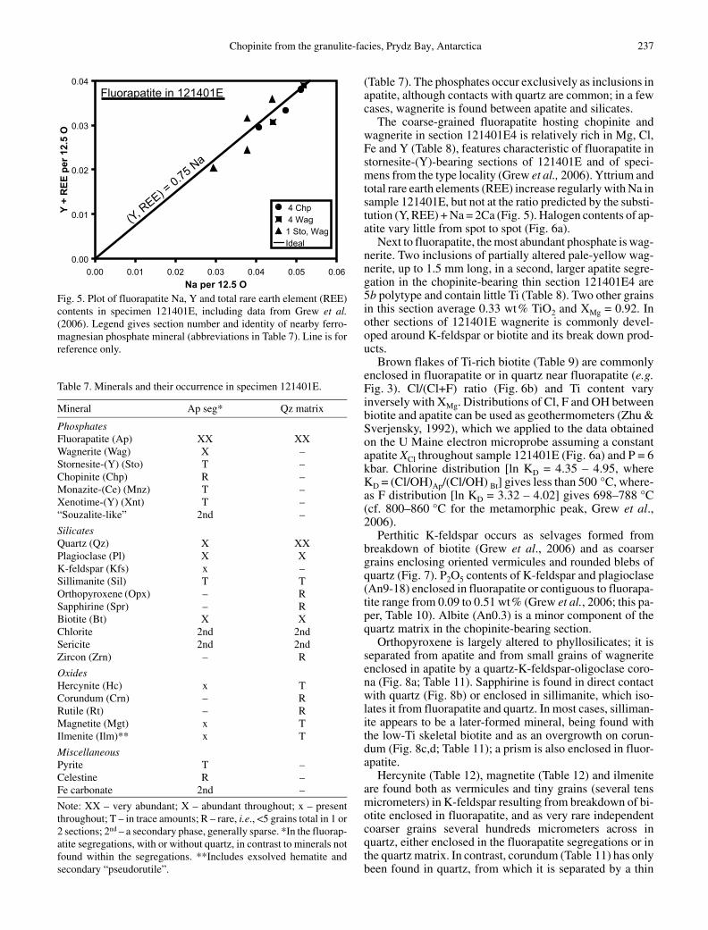
Fig. 5. Plot of fluorapatite Na, Y and total rare earth element (REE)contents in specimen 121401E, including data from Grew et al.(2006). Legend gives section number and identity of nearby ferro-magnesian phosphate mineral (abbreviations in Table 7). Line is forreference only.
Table 7. Minerals and their occurrence in specimen 121401E.
Mineral Ap seg* Qz matrix
PhosphatesFluorapatite (Ap) XX XXWagnerite (Wag) X –Stornesite-(Y) (Sto) T –Chopinite (Chp) R –Monazite-(Ce) (Mnz) T –Xenotime-(Y) (Xnt) T –“Souzalite-like” 2nd –
SilicatesQuartz (Qz) X XXPlagioclase (Pl) X XK-feldspar (Kfs) x –Sillimanite (Sil) T TOrthopyroxene (Opx) – RSapphirine (Spr) – RBiotite (Bt) X XChlorite 2nd 2ndSericite 2nd 2ndZircon (Zrn) – R
OxidesHercynite (Hc) x TCorundum (Crn) – RRutile (Rt) – RMagnetite (Mgt) x TIlmenite (Ilm)** x T
MiscellaneousPyrite T –Celestine R –Fe carbonate 2nd –
Note: XX – very abundant; X – abundant throughout; x – presentthroughout; T – in trace amounts; R – rare, i.e., <5 grains total in 1 or2 sections; 2nd – a secondary phase, generally sparse. *In the fluorap-atite segregations, with or without quartz, in contrast to minerals notfound within the segregations. **Includes exsolved hematite andsecondary “pseudorutile”.
(Table 7). The phosphates occur exclusively as inclusions inapatite, although contacts with quartz are common; in a fewcases, wagnerite is found between apatite and silicates.
The coarse-grained fluorapatite hosting chopinite andwagnerite in section 121401E4 is relatively rich in Mg, Cl,Fe and Y (Table 8), features characteristic of fluorapatite instornesite-(Y)-bearing sections of 121401E and of speci-mens from the type locality (Grew et al., 2006). Yttrium andtotal rare earth elements (REE) increase regularly with Na insample 121401E, but not at the ratio predicted by the substi-tution (Y, REE) + Na = 2Ca (Fig. 5). Halogen contents of ap-atite vary little from spot to spot (Fig. 6a).
Next to fluorapatite, the most abundant phosphate is wag-nerite. Two inclusions of partially altered pale-yellow wag-nerite, up to 1.5 mm long, in a second, larger apatite segre-gation in the chopinite-bearing thin section 121401E4 are5b polytype and contain little Ti (Table 8). Two other grainsin this section average 0.33 wt% TiO2 and XMg = 0.92. Inother sections of 121401E wagnerite is commonly devel-oped around K-feldspar or biotite and its break down prod-ucts.
Brown flakes of Ti-rich biotite (Table 9) are commonlyenclosed in fluorapatite or in quartz near fluorapatite (e.g.Fig. 3). Cl/(Cl+F) ratio (Fig. 6b) and Ti content varyinversely with XMg. Distributions of Cl, F and OH betweenbiotite and apatite can be used as geothermometers (Zhu &Sverjensky, 1992), which we applied to the data obtainedon the U Maine electron microprobe assuming a constantapatite XCl throughout sample 121401E (Fig. 6a) and P = 6kbar. Chlorine distribution [ln KD = 4.35 – 4.95, whereKD = (Cl/OH)Ap/(Cl/OH) Bt] gives less than 500 °C, where-as F distribution [ln KD = 3.32 – 4.02] gives 698–788 °C(cf. 800–860 °C for the metamorphic peak, Grew et al.,2006).
Perthitic K-feldspar occurs as selvages formed frombreakdown of biotite (Grew et al., 2006) and as coarsergrains enclosing oriented vermicules and rounded blebs ofquartz (Fig. 7). P2O5 contents of K-feldspar and plagioclase(An9-18) enclosed in fluorapatite or contiguous to fluorapa-tite range from 0.09 to 0.51 wt% (Grew et al., 2006; this pa-per, Table 10). Albite (An0.3) is a minor component of thequartz matrix in the chopinite-bearing section.
Orthopyroxene is largely altered to phyllosilicates; it isseparated from apatite and from small grains of wagneriteenclosed in apatite by a quartz-K-feldspar-oligoclase coro-na (Fig. 8a; Table 11). Sapphirine is found in direct contactwith quartz (Fig. 8b) or enclosed in sillimanite, which iso-lates it from fluorapatite and quartz. In most cases, silliman-ite appears to be a later-formed mineral, being found withthe low-Ti skeletal biotite and as an overgrowth on corun-dum (Fig. 8c,d; Table 11); a prism is also enclosed in fluor-apatite.
Hercynite (Table 12), magnetite (Table 12) and ilmeniteare found both as vermicules and tiny grains (several tensmicrometers) in K-feldspar resulting from breakdown of bi-otite enclosed in fluorapatite, and as very rare independentcoarser grains several hundreds micrometers across inquartz, either enclosed in the fluorapatite segregations or inthe quartz matrix. In contrast, corundum (Table 11) has onlybeen found in quartz, from which it is separated by a thin
Chopinite from the granulite-facies, Prydz Bay, Antarctica 237

Table 8. Selected analyses of fluorapatite (Ap) and wagnerite-Ma5bc (Wag) in the cho-pinite-bearing section 121401E4-1.
Mineral Ap Ap Ap Wag* Wag**Grain 10 20 50 4 5No. spots 10 17 18 13 20
wt%SiO2 b.d. b.d. b.d. 0.02 0.03P2O5 42.02 41.98 41.44 41.90 42.10SO3 b.d. b.d. 0.01 n.a. n.a.TiO2 b.d. b.d. b.d. 0.08 0.09Al2O3 0.03 b.d. b.d. b.d. 0.01MgO 0.69 0.86 0.61 45.42 45.28MnO 0.22 0.27 0.29 0.12 0.14FeO 2.44 2.74 3.10 6.69 6.68Na2O 0.31 0.29 0.31 b.d. b.d.CaO 50.43 50.27 49.88 0.13 0.11SrO 0.03 0.02 b.d. n.a. n.a.Y2O3 0.56 0.47 0.55 n.a. n.a.La2O3 0.10 0.07 0.10 n.a. n.a.Ce2O3 0.24 0.21 0.29 n.a. n.a.Nd2O3 0.06 0.06 b.d. n.a. n.a.Yb2O3 0.01 0.06 0.06 n.a. n.a.UO2 0.08 0.04 b.d. n.a. n.a.F 2.37 2.28 2.36 10.65 10.75Cl 2.09 2.10 2.07 b.d. b.d.H2O calc 0.10 0.14 0.09 0.32 0.29O=F, Cl –1.47 –1.43 –1.47 –4.48 –4.53Sum 100.32 100.43 99.71 100.87 100.95
FormulaeO (anhyd.) 12.5 12.5 12.5 4.5 4.5Si 0 0 0 0.001 0.001P 3.019 3.014 3.005 0.983 0.986S 0 0 0.001 – –Ti 0.000 0 0.000 0.002 0.002Al 0.003 0 0.000 0 0.000Mg 0.087 0.109 0.078 1.876 1.868Mn 0.016 0.020 0.021 0.003 0.003Fe 0.173 0.194 0.222 0.155 0.155Na 0.051 0.047 0.052 – –Ca 4.585 4.567 4.578 0.004 0.003Sr 0.001 0.001 0.000 – –Y 0.025 0.021 0.025 – –La 0.003 0.002 0.003 – –Ce 0.008 0.007 0.009 – –Nd 0.002 0.002 0.000 – –Yb 0 0.001 0.001 – –U 0.002 0.001 0.000 – –Sum 7.975 7.986 7.997 3.023 3.018
F 0.637 0.613 0.640 0.933 0.940Cl 0.300 0.302 0.300 0 0Hcalc 0.063 0.086 0.059 0.067 0.060Sum 1.000 1.000 1.000 1 1XMg 0.335 0.360 0.259 0.924 0.924
Notes: n.a. – not analyzed; b.d. – below detection. All Fe as FeO. H2O calculated fromstoichiometry: F + Cl + H = 1. XMg = atomic Mg/(Mg+Fe).Analyses done at the University of Maine.0Near to chopinite grains 1 and 2, wagnerite grain 5, respectively*a = 9.670(3), b = 31.74(1), c = 11.924(4) Å, q = 108.25(2)°, V = 3481(2) Å3
**a = 9.681(3), b = 31.685(8), c = 11.919(3) Å, q = 108.14(2)°, V = 3474(2) Å3
238 E.S. Grew, T. Armbruster, O. Medenbach, M.G. Yates, C.J. Carson

selvage of sillimanite (e.g. Fig. 8c,d, Table 11). A rutilegrain (Table 12) occurs with zircon along the boundary ofquartz with a sillimanite aggregate enclosing corundum.Temperatures calculated from the 386 ppm Zr measured inrutile are 751 °C (calibration of Zack et al., 2004) and663 °C (calibration of Watson et al., 2006).
Table 9. Selected analyses of biotite in sample 121401E.
Sect / gr 4-2 / 1 4-3 / 1 19 / 2 4-1 / 1o 4-1 /2o
Probe UM UM UM Cam CamF std Pol Pol Pol Ap ApNo. spots 10 10 7 2 2*
wt%SiO2 37.08 36.64 36.92 37.03 37.22P2O5 0.04 0.07 0.03 0.03 0.04TiO2 5.07 6.19 5.58 5.37 5.43Al2O3 15.18 15.09 14.83 16.07 15.59Cr2O3 0.15 0.09 0.10 n.a. n.a.K2O 9.63 9.58 9.58 9.81 9.67Na2O 0.10 0.08 0.17 0.10 0.12CaO b.d. 0.02 b.d. 0.01 0.01BaO 0.13 0.11 0.07 b.d. b.d.MgO 14.10 12.03 13.09 14.08 14.71MnO 0.02 0.04 b.d. 0.01 0.03FeO 14.63 16.47 16.12 12.78 13.09ZnO n.a. n.a. n.a. b.d. 0.09F 1.32 1.10 1.46 0.92 0.82Cl 0.41 0.52 0.60 0.44 0.37H2O calc 3.32 3.36 3.19 3.51 3.59O=F, Cl –0.65 –0.58 –0.75 –0.48 –0.43Total 100.52 100.83 101.01 99.67 100.34
Formula per 22 O anhydrousSi 5.495 5.464 5.490 5.480 5.477P 0.005 0.009 0.004 0.004 0.005IVAl 2.500 2.527 2.506 2.516 2.518Sum IV 8.000 8.000 8.000 8.000 8.000Ti 0.565 0.694 0.625 0.598 0.601VIAl 0.152 0.125 0.093 0.286 0.186Cr 0.018 0.011 0.012 – –Mg 3.116 2.675 2.902 3.107 3.227Mn 0.002 0.005 0.000 0.001 0.003Fe 1.813 2.054 2.005 1.582 1.610Zn – – – 0.000 0.010Sum VI 5.666 5.564 5.637 5.573 5.637K 1.820 1.823 1.818 1.852 1.815Na 0.028 0.023 0.049 0.029 0.033Ca 0.000 0.004 0.000 0.001 0.001Ba 0.007 0.007 0.004 0.000 0.000Sum XII 1.856 1.857 1.870 1.882 1.849Total 15.522 15.420 15.507 15.455 15.486F 0.619 0.521 0.685 0.429 0.382Cl 0.103 0.132 0.152 0.109 0.091Hcalc 3.278 3.347 3.163 3.461 3.527Sum 4.000 4.000 4.000 4.000 4.000XMg 0.632 0.566 0.591 0.663 0.667
Notes: Sect/gr – section/grain. Probe: UM – University of Maine, Cam – Camparis. n.a. – notanalyzed; b.d. – below detection. F standards: Pol – polylithionite, Ap – fluorapatite. All Fe asFeO. H2O calculated from stoichiometry: F + Cl + H = 4. XMg = atomic Mg/(Mg+Fe). oGrains1 and 2 are the closest biotite grains to chopinite (Fig. 3). *Only one spot for F.
Origin of chopinite
Chopinite is distinct from sarcopside not only in the domi-nance of Mg over Fe, but also in being found as discretegrains enclosed in fluorapatite in a metamorphic assem-blage. There are few terrestrial occurrences of sarcopsidethat is not intergrown with graftonite or triphylite (e.g.
Chopinite from the granulite-facies, Prydz Bay, Antarctica 239

X Cl = -1.1933X Mg + 0.8753R2 = 0.9407
0.08
0.12
0.16
0.20
0.24
0.28
0.50 0.55 0.60 0.65X Mg = Mg/(Mg+Fe)
XC
l = C
l/(C
l+F)
4 -- 24 -- 3193 -- 13 -- 43 -- 6AllLinear (All)
0.31
0.33
0.35
0.25 0.30 0.35 0.40X Mg = Mg/(Mg+Fe)
XC
l = C
l/(C
l+F) 4 C
4 W1Ave
Biotite
Fluorapatite(a)
(b)
Fig. 6. Plots of Cl/(Cl+F) vs. Mg/(Mg+Fe) for primary fluorapatite andbiotite in sample 121401E, including data from Grew et al. (2006). (a)Fluorapatite. Numbers in legend refer to section; C indicates spots nearchopinite, W indicates spots near wagnerite. (b) Numbers in legend re-fer to three sections of sample 121401E (sections 4 – 2 and 4 – 3 arefrom the same slice as the chopinite-bearing section, but do not containchopinite).
Table 10. Analyses of feldspars in sample 121401E.
Sect / gr 19 / 1* 19 / 1a* 10 / 2** 10 / 6Mineral Pl Kfs Pl KfsNo. spots 11 3 10 10
Wt%SiO2 63.92 64.74 65.82 64.73Al2O3 22.77 18.69 21.71 18.46P2O5 0.31 0.33 0.34 0.19Fe2O3 0.32 0.23 0.01 0.19MnO 0.01 0.02 b.d. b.d.MgO b.d. 0.01 b.d. 0.03Na2O 9.75 1.64 10.24 1.00K2O 0.08 14.21 0.54 14.99CaO 3.38 0.11 1.98 b.d.BaO b.d. b.d. 0.02 0.07Sum 100.54 99.96 100.66 99.65
Formulae per 8 OSi 2.806 2.972 2.875 2.988Al 1.178 1.011 1.118 1.004P 0.012 0.013 0.013 0.008Fe 0.010 0.008 0.000 0.007Mn 0.000 0.001 0.000 0.000Mg 0.000 0.000 0.000 0.002Na 0.830 0.146 0.867 0.089K 0.004 0.832 0.030 0.883Ca 0.159 0.005 0.092 0.000Ba 0.000 0.000 0.000 0.001Sum 5.000 4.988 4.996 4.981
Notes: Sect / gr – section / grain. n.a. – not analyzed. All Fe as Fe2O3.Analyses done at the University of Maine. *In corona between or-thopyroxene and fluorapatite (Fig. 8a). **Adjacent to K-feldspar-quartz intergrowth (Fig. 7b).
Zhang, 1995), and only one other metamorphic occurrenceif “mineral A” reported from Lake Quoich, Scotland (Liv-ingstone, 1980) is sarcopside, as suggested by its composi-tion (Cerny et al., 1998). Associations of fluorapatite andsarcopside are not common, and in some cases fluorapatite islater (e.g. Fransolet et al., 1986). The closest analogues to theBrattnevet assemblage of chopinite, wagnerite and stornesi-te-(Y) are assemblages of the Fe-dominant analogues, re-spectively, sarcopside, zwieselite, and johnsomervilleite,as well as the Mn-dominant phosphate graftonite, in granitesand granitic pegmatites (e.g. London et al., 1999; Roda et al.,2004). This analogy supports the suggestion that the originof chopinite and associated ferromagnesian phosphates isclosely tied to granitic melts resulting from anatexis (Grewet al. 2006), a scenario we would like to further develop herebased on new information obtained on the Brattnevet sampleand rethinking after constructive reviews.
Table 11. Selected analyses of sapphirine, corundum, sillimanite andorthopyroxene in sample 121401E.
Sect / gr 4-2 / 1 4-2 / 3 4-2 / 3 19 / 2Mineral Spr Crn* Sil* OpxNo. spot 10 10 10 9
wt%SiO2 11.65 n.a. 36.47 48.91P2O5 b.d. n.a. 0.03 n.a.TiO2 0.04 0.02 0.03 0.20Al2O3 61.56 98.92 61.98 5.67V2O3 0.03 b.d. 0.01 n.a.Cr2O3 0.02 b.d. 0.01 0.07Fe2O3 3.32 1.07 1.00 n.a.FeO 9.99 n.a. n.a. 23.61MnO 0.06 b.d. 0.00 0.19MgO 13.17 b.d. 0.03 20.94CaO b.d. n.a. 0.02 0.06Na2O 0.01 n.a. b.d. 0.02K2O b.d. n.a. b.d. 0.02Sum 99.85 100.02 99.58 99.68
FormulaeO / cations 20 / 14 3 5 6Si 1.420 – 0.992 1.841P 0.000 – 0.001 –Ti 0.003 0.000 0.001 0.006Al 8.846 1.986 1.987 0.251V 0.003 0.000 0.000 –Cr 0.002 0.000 0.000 0.002Fe3+ 0.304 0.014 0.020 –Fe2+ 1.019 – – 0.743Mn 0.006 0.000 0.000 0.006Mg 2.394 0.000 0.001 1.175Ca 0.000 – 0.000 0.002Na 0.002 – 0.000 0.001K 0.000 – 0.000 0.001Sum 14.000 2.000 3.003 4.028XMg 0.702 – – 0.613
Notes: Sect / gr – section / grain. n.a. – not analyzed; b.d. – below de-tection; dash – not calculated. All Fe as FeO or Fe2O3, except sapphi-rine, for which FeO and Fe2O3 were calculated by stoichiometry.XMg = atomic Mg/(Mg+Fe2+). *Fig. 8c,d. Analyses done at the Uni-versity of Maine.
240 E.S. Grew, T. Armbruster, O. Medenbach, M.G. Yates, C.J. Carson

200 m
W Ap
Q
(a)Ap
PlQtz
250 m
(b)
0.2 mmQz
Qz
Spr
HcBt
Hc
Bt
(b)200 m
Crn
Qtz
Qtz
S S
S
S
(d)
Qz
0.2 mm
(c)
100 m
OpxFsp+ Q
W
(a)
Ap
Fig. 7. Back scattered electron images of perthite-quartz intergrowths in section 121401E10. (a) K-feldspar host (light) with quartz blebs (Q,mostly vertical) and plagioclase lamellae (diagonal) surrounded by partially altered wagnerite (W). (b) Mesoperthite (light and dark) withquartz globules (Qtz) and plagioclase (Pl, Table 10). Ap – fluorapatite.
Fig. 8. (a) Back scattered electron image in section 121401E19 of orthopyroxene (Opx) partially altered to phyllosilicates, including biotite,and separated from fluorapatite (Ap) and wagnerite (W) by a corona consisting of K-feldspar (lighter), quartz and oligoclase (both darker).(b) Photomicrograph of sapphirine in direct contact with quartz in section 121401E8. Hercynite (Hc) in grain next to sapphirine is largely al-tered to a transparent material (diaspore?). Plane light. (c) Photomicrograph of corundum in quartz in section 121401E4 – 2 (2nd cut of sliceyielding chopinite). Plane light (d) Back scattered electron image of (c) showing corundum (Crn) mantled by sillimanite (S) in quartz (Qtz).Analyses are given in Table 11. Sillimanite is partially altered to phyllosilicate, resulting in speckled material.
The scenario proposed by Grew et al. (2006) is based onthe premise that the fluorapatite segregations and plagio-clase-bearing quartz masses could be restitic bodies remain-ing after melt had been extracted from P-bearing biotite-pla-gioclase paragneiss. Additional perspective on this scenariois provided by orthopyroxene, sapphirine and corundum, allthree of which were discovered in the Brattnevet specimenafter the Grew et al. (2006) manuscript was in press. Al-though the first two minerals have stabilities with quartz,none has been confirmed for corundum. One possible expla-nation that Harlov & Milke (2002) forwarded for the juxta-
position of corundum and quartz is that the two phasesformed in separate locations and were forced together by de-formation; subsequent reaction between the two was suffi-ciently slowed by low water activities or difficulty of nucle-ation that only a narrow rim of sillimanite developed, armor-ing the corundum.
Failure to attain equilibrium could also explain the ap-pearance of ferromagnesian phosphates in anatectic melts inwhich the phosphorus content was supposedly buffered byapatite. Grew et al. (2006) proposed that melts permeatingthe restitic apatite reacted with it and incorporated P. The
Chopinite from the granulite-facies, Prydz Bay, Antarctica 241
0.0
0.1
0.2
0.3
0.0 0.1 0.2 0.3 0.4 0.5 0.6 0.7 0.8 0.9 1.0
0.0
0.1
0.2
0.3
0.0 0.1 0.2 0.3 0.4 0.5 0.6 0.7 0.8 0.9 1.0Opx
Chp
Bt
Wag
Crd
"Src"Bt
F F
(Mg,Fe)O PO2.5
(a)
Experimental
(b)F F
(Mg,Fe)O PO2.5
Brattnevet
Table 12. Analyses of oxides in sample 121401E.
Sect / gr 19 / 1 4-2 / 2 10 / 4Mineral Mgt Rt Hc*No. spots 10 5** 10
wt%TiO2 0.09 93.49 b.d.ZrO2 n.a. 0.05 b.d.SnO2 b.d. 1.32 b.d.Al2O3 0.17 0.06 57.31V2O3 0.58 0.80 0.29Cr2O3 0.67 b.d. 0.03Fe2O3 67.79 n.a. 4.53MgO 0.01 0.01 5.16MnO 0.01 b.d. 0.01FeO 31.38 1.55 32.48NiO n.a. n.a. 0.14ZnO 0.01 b.d. 0.85Nb2O5 0.02 2.34 b.d.Ta2O5 b.d. 0.11 b.d.WO3 b.d. 0.22 b.d.Total 100.73 99.95 100.79
FormulaeO / cations 4 / 3 2 4 / 3Ti 0.003 0.957 0.000Zr – 0.000 0.000Sn 0.000 0.007 0.000Al 0.008 0.001 1.897V 0.018 0.009 0.006Cr 0.020 0.000 0.001Fe3+ 1.948 – 0.096Mg 0.000 0.000 0.216Mn 0.000 0.000 0.000Fe2+ 1.002 0.018 0.763Ni – – 0.003Zn 0.000 0.000 0.018Nb 0.000 0.014 0.000Ta 0.000 0.000 0.000W 0.000 0.001 0.000Total 3.000 1.007 3.000XMg 0.000 – 0.221
Notes: Sect / gr – section / grain. n.a. – not analyzed; b.d. – below de-tection; dash – not calculated. All Fe is given either as FeO (rutile) orapportioned between FeO and Fe2O3 by assuming stoichiometry(Mgt, Hc). XMg = atomic Mg/(Mg+Fe2+). Analyses done at the Uni-versity of Maine. *Adjacent to quartz. **Except ZrO2 (see text)
distinctive quartz-microperthite intergrowths (Fig. 7) areinferred to be remains of this melt. Under equilibrium con-ditions, P and Ca content of the melt would be buffered byapatite and melt P/Ca ratio would never be great enough forferromagnesian (or manganese) phosphates to be stabilizedin addition to apatite according to the model developed byLondon et al. (1999). However, Wolf & London (1994) re-ported experiments in which the low diffusivity of P in gra-nitic melts resulted in buildup of P adjacent apatite to sever-al times the saturation, whereas Ca diffused away morequickly. These experiments suggest the possibility of high Pconcentrations in small melt bodies surrounded by apatite.If Ca diffused away more rapidly than P, then P/Ca ratiomight have been sufficiently high for ferromagnesian phos-
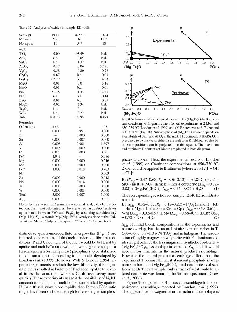
Fig. 9. Schematic relationships of phases in the (Mg,Fe)O-F-PO2.5 sys-tem coexisting with granitic melt for (a) experiments at 2 kbar and650–750 °C (London et al. 1999) and (b) Brattenevet at 6–7 kbar and800–860 °C (Fig. 10). Silicate phase at (Mg,Fe)O corner depends onavailability of SiO2 and Al2O3 in the melt. The component KAlSi3O8 ispresumed to be in excess, either in the melt or in K-feldspar, so that bi-otite compositions can be projected into this system. The maximumand minimum F contents of biotite are plotted in both diagrams.
phates to appear. Thus, the experimental results of Londonet al. (1999) on Ca-absent compositions at 650–750 °C,2 kbar could be applied to Brattnevet [where XF is F/(F + OH+ Cl)]:
Bt (XMg = 0.47–0.68, XF = 0.06–0.12) + Al2SiO5 (melt) +SiO2 (melt) + P2O5 (in melt) = Kfs + cordierite (XMg = 0.72–0.82) + (Mg,Fe)3(PO4)2 (XMg = 0.76–0.85) + H2O (1)
The corresponding reaction for sample 121401E from Bratt-nevet is:Bt (XMg = 0.52–0.67; XF = 0.12–0.22) + P2O5 (in melt) = Kfs+ Hc + Mgt + Ilm ± Spr ± Crn ± Opx (XMg = 0.59–0.61) +Wag (XMg = 0.92–0.93) ± Sto (XMg = 0.68–0.71) ± Chp (XMg= 0.72–0.73) + H2O (2)
XMg of initial biotite compositions in the experiments andnature overlap, but the natural biotite is much richer in Ti(5.0–6.6 vs. 0.9–1.0 wt% TiO2) and in halogens. The associ-ation of highly magnesian wagnerite with Fe-dominant ox-ides might balance the less magnesian synthetic cordierite +(Mg,Fe)3(PO4)2 assemblage in terms of XMg, and Ti wouldaccount for ilmenite in the natural product assemblage.However, the natural product assemblage differs from theexperimental because the most abundant phosphate is wag-nerite rather than (Mg,Fe)3(PO4)2, and cordierite is absentfrom the Brattnevet sample (only a trace of what could be al-tered cordierite was found in the Stornes specimens, Grewet al., 2006).
Figure 9 compares the Brattnevet assemblage to the ex-perimental assemblage reported by London et al. (1999).The appearance of wagnerite in the natural assemblage is
242 E.S. Grew, T. Armbruster, O. Medenbach, M.G. Yates, C.J. Carson

0
1
2
3
4
5
6
7
8
9
500 600 700 800 900Temperature (ºC)
Pres
sure
(kba
r)
Ky
Sil
AndSil
Chp 1
Far 1Chp 0.70
I
IIIII
synthetic (Mg Fe ) (PO )0.8 0.2 3 4 2
Chp 0.40
Chp 0.10
Far 0.84
Far 0.61
Far 0.20Chp 0.4
Larsemann
Hills
most likely due to greater availability of F, which is evi-denced in the higher F contents in the Brattnevet biotite andis consistent with a reaction relating Chp + Bt and Wag +Opx assemblages based on simplified compositions fromthe Brattnevet specimen:
Bt + Chp + 2Qtz + 1.4F = 2Opx + 2Wag + KAlSi3O8 +0.8H2O (3)
However, there is no evidence from fluorapatite and biotitehalogen contents within the Brattnevet specimen (Fig. 6) forvariation in F contents from one part of the specimen to an-other, and thus this reaction would not explain the appear-ance of chopinite in the Brattnevet specimen. Fluorapatiteassociated with chopinite has a higher XMg than other fluor-apatite in the Brattnevet specimen (Fig. 6a), and nearby bio-tite is more magnesian than other biotite in the sample (e.g.Table 9), suggesting that increasing Mg/Fe ratio could favorBt + Chp. However, a local increase in P could be more im-portant than either F or Mg/Fe, i.e., a shift rightward of thebulk composition in the triangles in Fig. 9. If the presence ofwagnerite is due to a non-equilibrium excess of P in the ana-tectic melt over that resulting from fluorapatite saturation,an even greater deviation from equilibrium would be neededto attain the higher P contents necessary for chopinite for-mation. Stornesite-(Y) presumably also results from a local-ized excess of P. It is not as rare as chopinite, implying thatanother condition necessary for chopinite formation is lo-calized depletion of Ca and Na.
Chopinite-farringtonite relations
Brunet & Vielzeuf (1996) and Brunet et al. (1998) showedthat their Mg3(PO4)2-II (chopinite) is a high-pressure poly-morph of the meteoritic mineral farringtonite and predictedthat Fe2+ would stabilize chopinite-sarcopside solid solu-tions to lower pressures. Calculation of isopleths (Fig. 10)requires data on Mg-Fe distribution between farringtoniteand chopinite-sarcopside as well as the volume of reactiondetermined by Brunet & Vielzeuf (1996) and Brunet et al.(1998). Grew et al. (2006) calculated this distribution coef-ficient, KD = (Mg/Fe)Far/(Mg/Fe)Src = 2.30, from the distri-bution coefficients between each of these two minerals andjohnsomervilleite or chladniite calculated from composi-tions in terrestrial and meteoritic minerals (Fig. 11a). We as-sumed that the most magnesian compositions that McCoy etal. (2006) called farringtonite are indeed farringtonite. Moreferroan compositions show different Fe-Mg distributions,i.e., the “Mg-graftonite” of Floss (1999) behaves like sar-copside-chopinite.
The isopleth for 70% chopinite solid solution passesthrough the box indicating peak conditions in the Larse-mann Hills, but the isopleth for 40% chopinite is 2 kbarhigher than the synthesis of this composition by Charalam-pides et al. (1988) at 0.8 kbar, 500 °C (Fig. 10). This dis-crepancy implies the calculated KD is too low, that is, theisopleths are too closely spaced; variation of KD with com-position could also play a role.
London et al. (1999) referred to their (Mg0.8Fe0.2)3(PO4)2phase as “sarcopside” but cited no evidence other than com-
Fig. 10. Schematic pressure-temperature diagram showing isoplethscalculated from the reaction and volume change in the Mg3(PO4)2
system (Chp 1-Far 1, Brunet & Vielzeuf 1996; Brunet et al. 1998) forchopinite-sarcopside solid solution associated with farringtonite sol-id solution and assuming both solid solutions are ideal and a Kd =(Mg/Fe)Far/(Mg/Fe)Chp = 2.30 (Fig. 11a). Charalampides et al.(1988) synthesized sarcopside containing 40% chopinite at 0.8 kbar,500 °C (arrow Chp 0.4). Numbered gray-filled boxes indicate peak(I) and post peak (II, III) metamorphic conditions for the LarsemannHills based on P-T estimates reported for the Larsemann Hills andnearby exposures (Thost et al., 1994; Fitzsimons, 1996; Carson etal., 1997). Unnumbered gray-filled box indicates synthesis condi-tions for (Mg0.8Fe0.2)3(PO4)2 (average composition) by London et al.(1999). Al2SiO5 relations are from Pattison (1992). And – andalu-site, Chp – chopinite, Far – farringtonite, Ky – kyanite, Sil – silli-manite.
position to back this identification. Even allowing for dis-crepancy noted above, Fig. 10 shows that it is highly unlike-ly that their phase is chopinite; more likely, it is farringtoni-te. This identification is consistent with Mg-Fe distribu-tion between biotite and their synthetic phosphate(Mg,Fe)3(PO4)2: KD = (Mg/Fe)P/(Mg/Fe)Chp = 2.28(Fig. 11b), virtually identical for KD for farringtonite-sar-copside (Fig. 11a).
Acknowledgments: We wish to thank the leader, Bob Jones,and other members of the 2003–2004 Australian NationalAntarctic Research Expedition for logistics support during thesummer field season. Assisted by Michel Fialin, ChristianChopin graciously carried out analyses of his namesake, bio-tite, wagnerite, apatite and albite in section 121401E4 at theCentre de Microanalyse Camparis, for which we are deeplygrateful. We thank Fabrice Brunet for fruitful discussions, Da-vid London for comments, and Fabrice Brunet and an anony-mous referee for thoughtful reviews of the manuscript. CJC’sand ESG’s fieldwork in the Larsemann Hills was supported byAntarctic Science Advisory Committee Project no. 2350.ESG’s and MGY’s research was supported by U.S. NationalScience Foundation grants OPP-0228842 and MRI-0116235to the University of Maine.
Chopinite from the granulite-facies, Prydz Bay, Antarctica 243

(Mg/Fe)Chp = 1.3231(Mg/Fe)Bt
R2 = not calc.
(Mg/Fe)P =3.0158(Mg/Fe)Bt
R2 = -0.2
0
1
2
3
4
5
6
7
0.0 0.5 1.0 1.5 2.0 2.5Mg/Fe in biotite
Mg/
Fe in
(Mg,
Fe) 3
(PO
4)2
BrattnevetExperimentLinear (Brattnevet)Linear (Experiment)
(Mg/Fe)Src = 0.588(Mg/Fe)Fil
R2 = 0.93
(Mg/Fe)Far = 1.354(Mg/Fe)Fil
R2 = -1
0
1
2
3
4
5
6
7
0 1 2 3 4 5Mg/Fe in fillowite group
Mg/
Fe in
(Mg,
Fe)
3(PO
4)2
SarcopsideFarringtonite"Mg-graftonite"graftonite/sarcopsideLinear (Sarcopside)Linear (Farringtonite)
(a)
(b)
K = 2.30D
K = 2.28D
Fig. 11. Distribution of Fe and Mg between (Mg, Fe)3(PO4)2 phases,fillowite group minerals and biotite. (a) KD = (Mg/Fe)Far / (Mg/Fe)Scp. Sources of data: Livingstone (1980), Cerny et al. (1998), andCorbella i Cordomı & Melgarejo i Draper (1990) for sarcopside andjohnsomervilleite; Floss (1999) and McCoy et al. (2006) for farring-tonite, “Mg-graftonite”, graftonite/sarcopside and chladniite in lod-ranite GRA 95209. (b) KD = (Mg/Fe)P / (Mg/Fe)Chp, where P is the(Mg, Fe)3(PO4)2 phosphate reported by London et al. (1999) as sar-copside. Sources of data: Camparis analyses of chopinite (averagefrom Table 6), Camparis analyses of biotite in grains 1 and 2 (Table9), and analyses of synthetic (Mg, Fe)3(PO4)2 and biotite from Lon-don et al. (1999).
References
Annersten, H. & Nord, A.G. (1980): A high pressure phase of mag-nesium orthophosphate. Acta Chem. Scand., A34, 389–390.
Annersten, H., Ericsson, T., Nord, A.G. (1980): The cation orderingin iron-containing zinc and magnesium orthophosphates deter-mined from Mössbauer spectroscopy. J. Phys. Chem. Solids, 41,1235–1240.
Armbruster, T., Bürgi, H.B., Kunz, M., Gnos, E., Brönnimann, S.,Lienert, C. (1990): Variation of displacement parameters in struc-ture refinements of low albite. Am. Mineral., 75, 135–140.
Berthet, G., Joubert, J.C., Bertaut, E.F. (1972): Vacancies ordering innew metastable orthophosphates [Co3�]P2O8 and [Mg3�]P2O8
with olivin-related structure. Z. Kristallogr., 136, 98–105.
Bild, R.W. (1974): New occurrences of phosphates in iron meteor-ites. Contrib. Mineral. Petrol, 45, 91–98.
Brunet, F. & Vielzeuf, D. (1996): The farringtonite / Mg3(PO4)2-IItransformation: A new curve for pressure calibration in piston-cylinder apparatus. Eur. J. Mineral., 8, 349–354.
Brunet, F., Chopin, C., Seifert, F. (1998): Phase relations in the MgO–P2O5–H2O system and the stability of phosphoellenbergerite: petro-logical implications. Contrib. Mineral. Petrol., 131, 54–70.
Buseck, P.R. & Holdsworth, E. (1977): Phosphate minerals in palla-site meteorites. Mineral. Mag., 41, 91–102.
Carson, C. J., Powell, R., Wilson, C.J.L., Dirks, P.H.G.M. (1997):Partial melting during tectonic exhumation of a granulite terrane:an example from the Larsemann Hills, East Antarctica. J. Meta-morphic Geol., 15, 105–126.
Cerny, P., Selway, J.P., Ercit, T.S., Breaks, F.W., Anderson, A.J., An-derson, S.D. (1998): Graftonite – beusite in granitic pegmatites ofthe Superior Province: A study in contrasts. Can. Mineral., 36,367–376.
Charalampides, G., Ericsson, T., Nord, A.G., Khangi, F. (1988):Studies of hydrothermally prepared (Fe, M)3(PO4)2-sarcopsides.N. Jahrb. Mineral. Mh., 1988, 324–336.
Corbella i Cordomı, M. & Melgarejo i Draper, J.-C. (1990): Carac-terısticas y distribucion de los fosfatos de las pegmatitas granıti-cas de la penınsula del Cap de Creus (Pirineo oriental catalan).Bol. Soc. Espan. Mineral., 13, 169–182.
Fitzsimons, I.C.W. (1996): Metapelitic migmatites from BrattstrandBluffs, East Antarctica–Metamorphism, melting and exhumationof the mid crust. J. Petrol., 37, 395–414.
Floss, C. (1999): Fe,Mg,Mn-bearing phosphates in the GRA 95209meteorite: Occurrences and mineral chemistry. Am. Mineral., 84,1354–1359.
Fontan, F. & Fransolet, A.-M. (1986): Les phosphates de Fe et Mndes pegmatites de Valmy, Massif des Alberes (Pyrenees Orient-ales), France. Bol. Soc. Espan. Mineral., 9, 391–396.
Fransolet, A.-M. (1977): Intercroissances et inclusions dans les as-sociations graftonite-sarcopside-triphylite. Bull. Soc. Franc.Mineral. Cristallogr., 100, 198–207.
Fransolet, A.-M., Keller, P., Fontan, F. (1986): The phosphate miner-al associations of the Tsaobismund Pegmatite, Namibia. Contrib.Mineral. Petrol., 92, 502–717.
Fuchs, L.H., Olsen, E., Gebert, E. (1973): New X-Ray and composi-tional data for farringtonite, Mg3(PO4)2. Am. Mineral., 58, 949–951.
Grew, E.S., Armbruster, T., Medenbach, O., Yates, M.G., Carson,C.J. (2006): Stornesite-(Y), (Y, Ca)�2Na6(Ca,Na)8(Mg,Fe)43
(PO4)36, the first terrestrial Mg-dominant member of the fillowitegroup, from granulite-facies paragneiss in the Larsemann Hills,Prydz Bay, East Antarctica. Am. Mineral., 91, 1412–1424
Grew, E.S., Armbruster, T., Medenbach, O., Yates, M.G., Carson,C.J. (2007): Tassieite, (Na,�)Ca2(Mg,Fe2+,Fe3+)2(Fe3+,Mg)2
(Fe2+,Mg)2(PO4)6.2H2O, a new hydrothermal wicksite-groupmineral in fluorapatite nodules from granulite-facies paragneissin the Larsemann Hills, Prydz Bay, East Antarctica. Can. Miner-al., 45, in press
Harlov, D.E. & Milke, R. (2002): Stability of corundum + quartz rel-ative to kyanite and sillimanite at high temperature and pressure.Am. Mineral., 87, 424–432.
Henry, P.F., Weller, M.T., Wilson, C.C. (2003): Determination of thecation distribution in Fe2Ni(PO4)2 using isotopic substitution andpowder neutron diffraction. J. Appl. Crystallogr., 36, 1361–1367.
Hurlbut, C.S. (1965): Detailed description of sarcopside from EastAlstead, New Hampshire. Am. Mineral., 50, 1698–1707.
Huvelin, P., Orliac, M., Permingeat, F. (1971): Graftonite et sarcop-
244 E.S. Grew, T. Armbruster, O. Medenbach, M.G. Yates, C.J. Carson

side de Sidi-bou-Othmane (Jebilet, Maroc). Notes Serv. Geol.Maroc, 31, 277–284.
Larsen, E.S. & Berman, H. (1934): The microscopic determinationof the nonopaque minerals. U.S. Geol. Surv. Bull. 848, 266 p.
Lindberg, M.L. (1950): Arrojadite, hühnerkobelite, and graftonite.Am. Mineral., 35, 59–76.
Livingstone, A. (1980): Johnsomervilleite, a new transition-metalphosphate mineral from the Loch Quoich area, Scotland. Miner-al. Mag., 43, 833–836.
London, D., Wolf, M.B., Morgan, G.B., VI, Garrido, M.G. (1999):Experimental silicate – phosphate equilibria in peraluminous gra-nitic magmas, with a case study of the Alburquerque Batholith atTres Arroyos, Badajoz, Spain. J. Petrol., 40, 215–240.
Mallo, A., Fontan, F., Melgarejo, J.-C., Mata, J.M. (1995): The Al-bera zoned pegmatite field, Eastern Pyrenees, France. Mineral.Petrol., 55, 103–116.
Mandarino, J.A. (1981): The Gladstone – Dale relationship: Part IV.The compatibility concept and its application. Can. Mineral., 19,441–450.
McCoy, T.J., Carson, W.D., Nittler, L.R. Stroud, R.M. Bogard, D.D.,Garrison, D.H. (2006): Graves Nunataks 95209: A snapshot ofmetal segregation and core formation. Geochim. Cosmochim. Ac-ta, 70, 516–531.
Medenbach, O. (1985): A new microrefractometer spindle-stage andits application. Fortschr. Mineral. 63, 111–133.
Merlet, C. (1994): An accurate computer correction program forquantitative electron-probe microanalysis. Mikrochimica Acta,114, 363–376.
Moore, P.B. (1972): Sarcopside: its atomic arrangement. Am. Miner-al., 57, 24–35.
Nord, A.G. (1984): Crystallographic studies of olivine-related sar-copside-type solid solutions. Z. Kristallogr., 166, 159–176.
Nord, A.G., & Kierkegaard, P. (1968): The crystal structure ofMg3(PO4)2. Acta Chem. Scand., 22,1466–1474.
Olsen, E.J., Kracher, A., Davis, A.M., Steele, I. M., Hutcheon, I.D.,Bunch, T.E. (1999): The phosphates of IIIAB iron meteorites.Meteoritics & Planetary Science, 34, 285–300.
Palache, C., Berman, H., Frondel, C. (1951): The System of Miner-alogy of James Dwight Dana and Edward Salisbury Dana, YaleUniversity. 7th ed, v. II. Halides, Nitrates, Borates, Carbonates,Sulfates, Phosphates, Arsenates, Tungstates, Molybdates, etc.Wiley, New York, 1124 p.
Pattison, D.R.M. (1992): Stability of andalusite and sillimanite andthe Al2SiO5 triple point: Constraints from the Ballachulish aure-ole, Scotland. J. Geol., 100, 423–446.
Povondra, P., Pivec, E., Cech, F., Lang, M., Novak, F., Prachar, I.,Ulrych, J. (1987): Pribyslavice peraluminous granite. Acta Univ.Carol., Geol., 3, 183–283.
Roda, E., Pesquera, A., Fontan, F., Keller, P. (2004): Phosphate min-eral associations in the Canada pegmatite (Salamanca, Spain):Paragenetic relationships, chemical compositions, and implica-tions for pegmatite evolution. Am. Mineral., 89, 110–125.
Sheldrick, G.M. (1997) SHELXS97 and SHELXL97. University ofGöttingen, Germany..
Smeds, S.-A., Uher, P., Cerny, P., Wise, M.A., Gustafsson, L., Pen-ner, P. (1998): Graftonite – beusite in Sweden: Primary phases,products of exsolution, and distribution in zoned populations ofgranitic pegmatites. Can. Mineral., 36, 377–394.
Stalder, M. & Rozendaal, A. (2002): Graftonite in phosphatic ironformations associated with the mid-Proterozoic Gamsberg Zn-Pbdeposit, Namaqua Province, South Africa. Mineral. Mag., 66,915–927.
Thost, D. E., Hensen, B. J., Motoyoshi, Y. (1994): The geology of arapidly uplifted medium and low pressure granulite facies terraneof Pan-African age: the Bolingen Islands, Prydz Bay, Eastern An-tarctica. Petrology, 2, 293–316.
Warner, J.K., Cheetham, A.K., Nord, A.G., von Dreele, R.B., Yethi-raj, M. (1992): Magnetic structure of iron (II) phosphate, sarcop-side, Fe3(PO4)2. J. Materials Chem., 2 (2), 191–196.
Watson, E.B., Wark, D.A., Thomas, J.B. (2006): Crystallizationthermometers for zircon and rutile. Contrib. Mineral. Petrol.,151, 413–433.
Wolf, M.B. & London, D. (1994): Apatite dissolution into peralumi-nous haplogranitic melts: An experimental study of solubilitiesand mechanisms. Geochim. Cosmochim. Acta, 58, 4175–4145.
Yvon, K., Jeitschko, W., Parthe, E. (1977): LAZY PULVERIX, acomputer-program, for calculating X-ray and neutron-diffractionpowder patterns. J. Appl. Crystallogr., 10, 73–74.
Zack, T., Moraes, R., Kronz, A. (2004): Temperature dependence ofZr in rutile: empirical calibration of a rutile thermometer. Con-trib. Mineral. Petrol., 148, 471–488.
Zhang, R. (1995): Mg-sarcopside, a new variety of sarcopsidegroup. Kuangwu Yanshi = J. Mineral. Petrol., 15, 6–10 (in Chi-nese with English abstract).
Zhu, C. & Sverjensky, D.A. (1992): F-Cl-OH partitioning betweenbiotite and apatite. Geochim. Cosmochim. Acta, 56, 3435–3467.
Received 22 July 2006Modified version received 21 November 2006Accepted 18 December 2006
Chopinite from the granulite-facies, Prydz Bay, Antarctica 245

246 E.S. Grew, T. Armbruster, O. Medenbach, M.G. Yates, C.J. Carson