Isostructural fluorescent and radioactive probes for monitoring neural stem and progenitor cell...
11
Isostructural fluorescent and radioactive probes for monitoring neural stem and progenitor cell transplants Paul Schaffer a , Jacqueline A. Gleave b , Jennifer A. Lemon c , Leslie C. Reid d , Laura K.K. Pacey b , Troy H. Farncombe e , Douglas R. Boreham c , Jon Zubieta f , John W. Babich g , Laurie C. Doering b , John F. Valliant c,d, ⁎ a McMaster Nuclear Reactor, McMaster University, Hamilton, Ontario, Canada L8S 4K1 b Department of Pathology and Molecular Medicine, McMaster University, Hamilton, Ontario, Canada L8N 3Z5 c Department of Medical Physics and Applied Radiation Sciences, McMaster University, Hamilton, Ontario, Canada L8S 4K1 d Department of Chemistry, McMaster University, Hamilton, Ontario, Canada L8S 4M1 e Department of Nuclear Medicine, Hamilton Health Sciences, Hamilton, Ontario, Canada L8N 3Z5 f Department of Chemistry, Syracuse University, Syracuse, NY 13244-4100, USA g Molecular Insight Pharmaceuticals Inc., Cambridge, MA 02142, USA Received 25 July 2007; received in revised form 25 September 2007; accepted 2 November 2007 Abstract A construct for tagging neurospheres and monitoring cell transplantations was developed using a new technology for producing luminescent and radiolabeled probes that have identical structures. The HIV1-Tat basic domain derivatives NAcGRKKRRQRRR(SAACQ)G (SAACQ-1) and [NAcGRKKRRQRRR(Re(CO) 3 SAACQ)G] + (ReSAACQ-1) were prepared in excellent yields using the single amino acid chelate-quinoline (SAACQ) ligand and its Re(I) complex and conventional automated peptide synthesis methods. The distribution of the luminescent Re probe, using epifluorescence microscopy, showed that it localized primarily in the cell nucleus with a significant degree of association on the nuclear envelope. A smaller amount was found to be dispersed in the cytoplasm. The 99m Tc analogue was then prepared in 43±7% (n=12) yield and very high effective specific activity. Following incubation, average uptake of the probe in neurospheres ranged between 10 and 20 Bq/cell. As determined by colorimetric assays, viability for cells labeled with high effective specific activity 99m TcSAACQ-1 was 97±4% at 2 h postlabeling and 85±25% at 24 h postlabeling for incubation activities ranging from 245 to 8900 Bq/cell. DNA analysis showed that at these levels, there was no significant difference between the extent of DNA damage in the treated cells versus control cells. A series of preliminary SPECT/CT studies of transplants in mice were performed, which showed that the strategy is convenient and feasible and that it is possible to routinely assess procedures noninvasively and determine the number of cells transplanted. © 2008 Elsevier Inc. All rights reserved. Keywords: Neurospheres; Imaging; Technetium; Transplants; Peptide 1. Introduction Neural stem cells (NSCs) have the ability to differentiate into neurons, astrocytes and oligodendrocytes in vivo [1] and are therefore under intense investigation as treatments for neurodegenerative diseases [2,3]. Unfortunately, the success of NSC transplantations in animal models and patients has been highly variable, which is due in large part to difficulties in achieving accurate, reproducible and efficient graft place- ments. Despite the development of several methods for long- term tracking of stem cell migration, better in vivo imaging strategies for routine monitoring and recording of implant procedures are needed in order to establish greater experi- mental controls in both preclinical and clinical studies. Here, we report the development of a dual-modality imaging strategy to study labeled neurospheres and monitor cell transplanta- tions, which is based on a new technology for producing Available online at www.sciencedirect.com Nuclear Medicine and Biology 35 (2008) 159 – 169 www.elsevier.com/locate/nucmedbio ⁎ Corresponding author. Department of Chemistry, McMaster Uni- versity, 1280 Main St. West, Hamilton, Ontario, Canada L8S 4M1. Tel.: +1 905 525 9140x22840; fax: +1 905 522 7776. E-mail address: [email protected] (J.F. Valliant). 0969-8051/$ – see front matter © 2008 Elsevier Inc. All rights reserved. doi:10.1016/j.nucmedbio.2007.11.001
Transcript of Isostructural fluorescent and radioactive probes for monitoring neural stem and progenitor cell...

Available online at www.sciencedirect.com
gy 35 (2008) 159–169www.elsevier.com/locate/nucmedbio
Nuclear Medicine and Biolo
Isostructural fluorescent and radioactive probes for monitoring neural stemand progenitor cell transplants
Paul Schaffera, Jacqueline A. Gleaveb, Jennifer A. Lemonc, Leslie C. Reidd,Laura K.K. Paceyb, Troy H. Farncombee, Douglas R. Borehamc, Jon Zubietaf,
John W. Babichg, Laurie C. Doeringb, John F. Valliantc,d,⁎aMcMaster Nuclear Reactor, McMaster University, Hamilton, Ontario, Canada L8S 4K1
bDepartment of Pathology and Molecular Medicine, McMaster University, Hamilton, Ontario, Canada L8N 3Z5cDepartment of Medical Physics and Applied Radiation Sciences, McMaster University, Hamilton, Ontario, Canada L8S 4K1
dDepartment of Chemistry, McMaster University, Hamilton, Ontario, Canada L8S 4M1eDepartment of Nuclear Medicine, Hamilton Health Sciences, Hamilton, Ontario, Canada L8N 3Z5
fDepartment of Chemistry, Syracuse University, Syracuse, NY 13244-4100, USAgMolecular Insight Pharmaceuticals Inc., Cambridge, MA 02142, USA
Received 25 July 2007; received in revised form 25 September 2007; accepted 2 November 2007
Abstract
A construct for tagging neurospheres and monitoring cell transplantations was developed using a new technology for producingluminescent and radiolabeled probes that have identical structures. The HIV1-Tat basic domain derivatives NAcGRKKRRQRRR(SAACQ)G(SAACQ-1) and [NAcGRKKRRQRRR(Re(CO)3SAACQ)G]
+ (ReSAACQ-1) were prepared in excellent yields using the single amino acidchelate-quinoline (SAACQ) ligand and its Re(I) complex and conventional automated peptide synthesis methods. The distribution of theluminescent Re probe, using epifluorescence microscopy, showed that it localized primarily in the cell nucleus with a significant degree ofassociation on the nuclear envelope. A smaller amount was found to be dispersed in the cytoplasm. The 99mTc analogue was then prepared in43±7% (n=12) yield and very high effective specific activity. Following incubation, average uptake of the probe in neurospheres rangedbetween 10 and 20 Bq/cell. As determined by colorimetric assays, viability for cells labeled with high effective specific activity99mTcSAACQ-1 was 97±4% at 2 h postlabeling and 85±25% at 24 h postlabeling for incubation activities ranging from 245 to 8900 Bq/cell.DNA analysis showed that at these levels, there was no significant difference between the extent of DNA damage in the treated cells versuscontrol cells. A series of preliminary SPECT/CT studies of transplants in mice were performed, which showed that the strategy is convenientand feasible and that it is possible to routinely assess procedures noninvasively and determine the number of cells transplanted.© 2008 Elsevier Inc. All rights reserved.
Keywords: Neurospheres; Imaging; Technetium; Transplants; Peptide
1. Introduction
Neural stem cells (NSCs) have the ability to differentiateinto neurons, astrocytes and oligodendrocytes in vivo [1] andare therefore under intense investigation as treatments forneurodegenerative diseases [2,3]. Unfortunately, the success of
⁎ Corresponding author. Department of Chemistry, McMaster Uni-versity, 1280 Main St. West, Hamilton, Ontario, Canada L8S 4M1. Tel.: +1905 525 9140x22840; fax: +1 905 522 7776.
E-mail address: [email protected] (J.F. Valliant).
0969-8051/$ – see front matter © 2008 Elsevier Inc. All rights reserved.doi:10.1016/j.nucmedbio.2007.11.001
NSC transplantations in animal models and patients has beenhighly variable, which is due in large part to difficulties inachieving accurate, reproducible and efficient graft place-ments. Despite the development of several methods for long-term tracking of stem cell migration, better in vivo imagingstrategies for routine monitoring and recording of implantprocedures are needed in order to establish greater experi-mental controls in both preclinical and clinical studies. Here,we report the development of a dual-modality imaging strategyto study labeled neurospheres and monitor cell transplanta-tions, which is based on a new technology for producing

160 P. Schaffer et al. / Nuclear Medicine and Biology 35 (2008) 159–169
luminescent and radiolabeled probes that have identicalstructures. Using this approach, we developed a labeledpeptide that can efficiently and noninvasively tag neurospheresso that transplants can be assessed both qualitatively andquantitatively using small-animal imaging techniques.
Recent studies suggest that cell transplantations areperformed correctly only 50% of the time despite the useof surgical aids such as ultrasound-guided injection [4]. Inthe case of neurological applications, better methods areneeded to assess stem cell transplants in order to eliminatesubject-to-subject and technique-oriented variability and toprovide a permanent record of the initial location and spatialdistribution of transplanted cells. This information, whencorrelated with therapeutic outcomes, can be used to identifythe most effective and reproducible implantation techniques.Dual-modality imaging methods such as SPECT/CT, whichcombine both anatomical and molecular imaging techniques,offer a convenient and accurate means of monitoring stemcell therapies. Once an appropriate cell-labeling system isdeveloped (vide infra), scintigraphic imaging coupled withX-ray CT can provide both qualitative and quantitativeinformation about cell transplants. It is noteworthy that forpreclinical studies, imaging experiments can be performedusing high-resolution small-animal SPECT/CT scanners,where the developed techniques can subsequently be adaptedfor monitoring studies in patients [5].
A variety of strategies for labeling stem cells withradioisotopes have been developed to monitor cell migra-tion by nuclear imaging methods [5–14]. Unfortunately,these methods suffer from limitations that prevent theirroutine use in preclinical research and in clinical trials.These include the use of agents that are expensive or notreadily available and those that have poorly characterizedbiological properties in terms of the impact on cell viabilityor low cell uptake. Poor uptake results in the need to use alarge number of cells to achieve sufficient sensitivity duringimaging studies. This is particularly problematic for small-animal neurological experiments where only a limitednumber of cells can be used so as to avoid host tissuedamage. It is clear that new and more readily accessibleapproaches for labeling stem cells that have well-character-ized biological effects and improved tracer uptake areneeded to routinely monitor implant procedures. Thedevelopment and validation of such a system, based on auniquely labeled permeation peptide, are described herealong with its application in assessing neurospheretransplants in mice using small-animal SPECT/CT.
2. Materials and methods
2.1. Maintenance of animal colonies
All animal experiments were carried out in accordancewith the guidelines set out by the Canadian Council onAnimal Care and were approved by the Animal ResearchEthics Board of McMaster University.
2.2. Chemicals and instrumentation
Unless otherwise stated, all reagents and solvents were ofACS grade or higher and used without further purification.Polystyrene-based N-α-9-fluorenylmethoxycarbonyl(Fmoc)-glycine loaded Wang resin (0.82 mmol g−1, 1%divinylbenzene, 200–400 mesh) was obtained from Nova-Biochem Inc. Fmoc-protected amino acids were purchasedfrom NovaBiochem Inc., Bachem Inc. or AdvancedChemTech Inc. All peptides were prepared on an AdvancedChemTech 348-Ω Peptide Synthesizer using a 40-wellreaction vessel. Peptides were analyzed by electrospraymass spectrometry, performed on a Quattro LC (Micromass,England) triple-quadrupole instrument. IR spectra wereacquired as KBr pellets on a Perkin-Elmer Series 1600 FT-IR spectrometer. Glassware was silanized by pretreating withSigmacote (Aldrich, Canada) followed by air and ovendrying overnight.
2.3. HPLC
Analytical HPLC was performed using a Varian Pro Starmodel 330 PDAdetector, a model 230 solvent delivery systemand a Microsorb C-18 column (4.6×250 mm, 300 Å to 5 μm).The mobile phase consisted of either Method A or Method B.For Method A, Solvent A=H2O containing 0.1% TFA andSolvent B=acetonitrile containing 0.05% TFA. The elutionprotocol consisted of the following: 0–20 min, 90%A to 20%A; 20–22.5 min, 20%A to 0%A; 22.5–25min, 0%A; 25–30min, 0% A to 90% A. For Method B, Solvent A=triethy-lammonium phosphate buffer (pH≅2–2.5) and SolventB=CH3OH. The elution protocol consisted of the following:0–3min, 100%A; 3–6min, 100%A to 75%A; 6–9min, 75%A to 67% A; 9–20 min, 67% A to 0% A; 20–22 min, 0% A;22–25 min, 0% A to 100%A; 25–30 min, 100%A. The flowrate was 1.0 ml min−1, and all runs were monitored at λ=254.Radio-HPLC experiments were performed on a Varian ProStarmodel 230HPLC instrument coupled to a Bio-Rad IN/USγ-detector using a Cobert Xpertek C-18 nucleosil column(4.6×200 mm, 300 Å to 5 μm).
Semipreparative HPLC was performed using a VarianPro Star model 330 PDA detector, a model 230 solventdelivery system and a Phenomenex C-18 Partisil 10-ODS-3 column (9.8×500 mm, 300 Å to 5 μm). The solvents andelution profile were identical to those of Method A(above). The flow rate was 4.0 ml min−1, and runs weremonitored at λ=254.
2.4. Solid-phase peptide synthesis
Fmoc-glycine loaded Wang resin (100 mg, 0.82 mmol/g)was added to the reaction vessel, suspended in DMF (2ml/well)and shaken at 600 rpm for 1 min. The wells were subsequentlyfiltered, suspended in THF (2 ml/well), shaken at 600 rpm for 1min and drained for 90 s. The THF wash was repeated twomore times. The DMFwash was then repeated a final two timesto complete the general wash cycle. This procedure was usedbetween every deprotection and coupling step. Fmoc

161P. Schaffer et al. / Nuclear Medicine and Biology 35 (2008) 159–169
deprotection was brought about through the addition of 20% v/v piperidine–DMF solution to the active vessels (2 ml/well) andshaking for 5 min at 600 rpm. Following filtration, the processwas repeated, shaking for 10 min. The deprotected resin-boundamino acid was washed using the general wash procedure andsubsequently coupled to the next Fmoc-protected amino acidusing a standard HBTU coupling protocol. Coupling reactionsinitially involved adding DMF (200 μl) to the active vesselsfollowed by the addition of a fourfold excess of the protectedamino acid as a 0.5-M solution in DMF. Four equivalents ofHBTU as a 0.5-M solution in DMF was then added, followedby a fourfold excess of DIPEA as a 2.0-M solution in DMF.The reaction block was subsequently shaken for 80 min at 600rpm. Following filtration, the resin was washed using thegeneral washing procedure prior to the start of the next cycle.
Peptides were cleaved from the resin support using a TFAmixture containing EDT (2%), water (2%) and TIS (2%).The cleavage solution was cooled to 0°C and added to theresin. The mixture was allowed to warm to room temperatureand agitated for 24 h. The suspension was filtered into colddiethyl ether, and the resulting precipitate was centrifuged at3000 rpm at 5°C for 30 min. The pellet was subsequentlywashed with cold diethyl ether (3×25 ml), dissolved indistilled deionized water and lyophilized, yielding a whitesolid for NAcGRKKRRQRRR(SAACQ)G (SAACQ-1) andan off-white solid for [NAcGRKKRRQRRR(Re(CO)3-SAACQ)G]+ (ReSAACQ-1).
SAACQ-1 showed the following: FTIR (KBr, cm−1):3309, 3197, 2954, 1668, 1546, 1431, 1204 and 1135; ESMS(m/z, positive ion): 1906 [M+H]+; HPLC: TR=10.7 min(Method A). ReSAACQ-1 showed the following: FTIR(KBr, cm−1): 3312, 3198, 2955, 2035, 1933, 1665, 1546 and1433; ESMS (m/z, positive ion): 2176 [M+H]+; HPLC:TR=13.8 min (Method A).
2.5. Preparation of [99mTc(CO)3(OH2)3]+
A 10-ml multidose vial containing K2[BH3·CO2](8.5 mg, 6.3×10−5 mol), Na2B4O7·10H2O (2.9 mg,7.6×10−6 mol), Na/K-tartrate (15.0 mg, 5.3×10⁻⁵ mol) andNa2CO3 (4.0 mg, 3.8×10⁻⁵ mol) was fitted with a rubberseptum and flushed with N2 (g) for 15 min. 99mTc-generatoreluate (370–1110 MBq, 10–30 mCi) in 900 μl of saline wasadded using a syringe, and the solutionwas heated to 95°C for30 min. After cooling on an ice bath, the pH of the solutionwas adjusted to approximately 7.0–7.5 by the addition of HCl(65 μl of a 2.63-M solution of HCl). Quality control wasperformed by analytical HPLC (Method B): Yield: ≥95%.
2.6. Peptide labeling with 99mTc
An aliquot (100 μl) of AcGRKKRRQRRR(SAACQ)G(SAACQ-1) (1.0 mg, 5.3×10⁻⁷ mol) in 100 μl of distilleddeionized water was added to the solution containing[99mTc(CO)3(OH2)3]
+ using a syringe, and the reaction washeated at 72°C for 60 min. The mixture was subsequentlycooled in an ice bath, and the product was isolated by using
either a C18 Sep-Pak (Waters Inc.) for low specific activityor semipreparative HPLC (Method A) for high specificactivity. Prior to use, the Sep-Pak was conditioned withabsolute ethanol (10 ml), acetonitrile (10 ml), 1:1acetonitrile/10 mM HCl (10 ml) and 10 mM HCl (10 ml).The reaction mixture was subsequently loaded, and the Sep-Pak slowly eluted into 1-ml fractions using 10 mM HCl(7×1 ml), 1:4 acetonitrile/10 mM HCl (2×1 ml), 1:1acetonitrile/10 mM HCl (2×1 ml), 4:1 acetonitrile/10 mMHCl (2×1 ml) and acetonitrile (3×1 ml). The desired producteluted in fractions 9–13 (radiochemical yield=43±7%,radiochemical purity ≥98%).
2.7. Ligand challenge experiments
Ten microliters of a solution of cysteine (7.88 mg in 5 mlof ddH2O) and histidine (7.76 mg in 5 ml of ddH2O) wasadded to two separate vials, each containing 11.1 to 18.5MBq (300 to 500 μCi) of purified 99mTcSAACQ-1 in 250 μlof 100 mM phosphate buffer. Both vials were incubated at37°C and monitored by HPLC (Method A) over 24 h.
2.8. Preparation of neurospheres
CDI or B6.129.FMR1/FvBn mouse pups (1–3 days old)were decapitated, and the brain was removed and transferredto sterile artificial cerebrospinal fluid (aCSF) consisting of0.012 M NaCl, 0.005 M KCl, 0.03 M MgCl2, 0.026 MNaHCO3, 0.01M glucose and 0.097mMCaCl2 in dH2O. Thecerebellum was removed and the hemispheres were cut in themidsagittal plane. Each half was transferred to a tissue culturedish containing 3 ml of aCSF. The tissue was further cut into0.5- to 1-mm2 pieces and transferred to 1 ml of aCSFcontaining 0.13 mg kynurenic acid (Sigma), 0.66 mghyaluronidase (Sigma) and 1.3 mg trypsin (Sigma). Afterincubation at 37°C in a shaking water bath for 1 h, thedigested tissue was centrifuged at 730×g for 5 min and thentransferred to a solution of trypsin inhibitor (Roche) 1 mg/mlin DMEM/F12 (Invitrogen) medium with glucose, HEPESbuffer (Sigma), putrescene (Sigma), progesterone (Sigma),insulin–transferrin–sodium–selenite (Roche), B27 growthsupplement, epidermal growth factor (Sigma), basic fibro-blast growth factor (Sigma) and heparin (Sigma) andincubated for 10 min in a 37°C shaking water bath. Themixture was centrifuged at 730×g for 5 min. The resultingpellet was triturated and reconstituted in 12 ml of serum-freemedium. The cell suspension was plated in two 24-welldishes (Falcon; 500 μl of cell suspension per well) andincubated in 95%O2 and 5%CO2 and passed every 5–7 days.
2.9. Labeling of neurospheres with 99mTcSAACQ-1
Neurospheres were transferred from the 24-well dishesto 15-ml Falcon tubes and centrifuged at 730×g for 5 min.The resulting pellet was incubated in 2 ml of TrypLEExpress (Invitrogen) and incubated at 37°C for 20 min in ashaking water bath. The mixture was centrifuged at 730×gfor 5 min, and the pellet was transferred to 500 μl of

162 P. Schaffer et al. / Nuclear Medicine and Biology 35 (2008) 159–169
DMEM/F12 medium and triturated with a fire-polished,small borehole pipette. Cell counts were determined bymixing 16 μl of the cell suspension with 4 μl of trypan blueand counting phase bright cells with the aid of ahemacytometer. Cells were plated into 60×15 mm tissueculture dishes at a concentration of 10,000 cells/ml; 30×PBS was added to the solution containing 99mTcSAACQ-1to give a 1× PBS solution of 0.01 M and subsequentlyadded to the cells and incubated at 37°C in a 95% O2/5%CO2 incubator for 20 min. The labeled cells weretransferred to a 15-ml siliconized glass centrifuge tube(VWR Canada) and centrifuged at 730×g for 5 min. Thesupernatant was removed, replaced with 2 ml of serum-freemedium and centrifuged for 5 min at 730×g. The wash wasremoved and replaced with 2 ml of serum-free medium.Labeling efficiency was determined using a dose calibratorthat had been calibrated using an NIST traceable standard
Fig. 1. (A) Fmoc-protected single amino acid (bis-quinoline) chelate (SAACQ)AcGRKKRRQRRR(SAACQ)G (SAACQ-1) and (D) [AcGRKKRRQRRR(M(CO
over the range of activities employed. Control cells wereprepared in an identical manner with 0.01 M PBS addedinstead of the labeled peptide.
2.10. Epifluorescence microscopy
Microscopy experiments were conducted with a ZeissAxioskop 2 epifluorescence microscope. Visualization ofReSAACQ-1 was achieved using a commercially availablefilter set consisting of a dichroic 360±40 nm excitation filter,a 400-nm dichroic long-pass filter and a 550-nm long-passemission filter. Image analysis was performed with the ZeissAxioVision 3.1 Imaging software package.
NSCs were isolated and prepared for labeling as describedabove. Specifically, approximately 20,000 cells/ml wereincubated at 37°C for 120 min with 0.0712 or 0.0909 mg/mlof ReSAACQ-1. The cells were collected into 15-ml Falcontubes and centrifuged for 5 min at 730×g. The supernatant
and (B) the corresponding Re complex [Re(CO)3SAACQ]+. Peptides (C)
)3SAACQ)G]+ (M=99mTc, 99mTcSAACQ-1; M=Re, ReSAACQ-1).

163P. Schaffer et al. / Nuclear Medicine and Biology 35 (2008) 159–169
was removed, and cells were fixed in 4% paraformaldehyde(PFA) for 5 min. The 4% PFAwas diluted with 0.01 M PBSand centrifuged for 5 min at 730×g. The supernatant wasremoved, and the cells were washed with 0.01 M PBS. Thecell pellet was suspended in 200 μl of dH2O, and the cellsuspension was placed on a glass slide and allowed to dry.The slides were mounted with DakoCytomation fluorescentmounting medium.
Fig. 2. Automated synthesis of SAACQ-1 and subsequent labelin
2.11. Single-cell gel electrophoresis
Comet assays were performed under alkaline conditions.Using a modified literature protocol [29,30], we diluted thecell culture 1:1 with 1% low melting point agarose (FisherScientific, Middletown, VA), prewarmed to 42°C. Onehundred twenty microliters of this solution was cast into twoindividual wells of a two-well chamber (Nalge Nunc
g with [99mTc(CO)3(OH2)3]+ to generate 99mTcSAACQ-1.

Fig. 3. Epifluorescent images of (A) a single neurosphere, (B) single-cellsuspension at low magnification and (C) single-cell suspension at highermagnification. Cells were incubated with ReSAACQ-1 for 2 h at 37°C.
164 P. Schaffer et al. / Nuclear Medicine and Biology 35 (2008) 159–169
International, Rochester, NY) attached to GelBond film(Mandel Scientific, Guelph, ON). The gels were left to set atroom temperature for 10 min; the two-well chamber formwas then removed, and the gels were placed in lysis buffer[2.5 M NaCl, 100 mM tetrasodium ethylenediaminetetraacetate (EDTA), 10 mM Tris base, 1% N-lauroylsarcosine, 1% Triton-X and 10% DMSO] and incubated at37°C for 4 h. Gels were rinsed two times with 18.2 MΩultrapure water and placed in 4°C DNA unwinding buffer(10 M NaOH, 200 mM EDTA in 18 MΩ ultrapure water,pHN13), and the mixture was incubated at room temperaturefor 30 min. The gels were then placed in submarine agarosegel units (model HE-33, Hoefer, San Francisco, CA)containing 220 ml DNA unwinding buffer and electrophor-esed for 10 min at 25 V. The gels were then rinsed two timesin 1× TAE (40 mM Tris–acetate, 10 mM EDTA, 20 mMglacial acetic acid), dehydrated in absolute ethanol for aminimum of 2 h and air dried overnight. The gels werestained by immersion in 50 ml SYBR Gold nucleic acid stain(1:10,000 dilution in 18 MΩ ultrapure water) for 10 min.The gels were then rinsed in 18.2 MΩ water and mountedonto a microscope slide and covered with a glass coverslip.Cells were analyzed using a fluorescence microscope (ZeissAxioplan 2, Carl Zeiss MicroImaging, Inc., Thornwood,NY) under ×200 magnification with an excitation wave-length of 520 nm and emission of 580 nm. Cells wereclassified as normal, damaged or apoptotic based on theDNA fragmentation pattern observed in the comet tail aspreviously described [15].
2.12. Animal imaging protocols
B6.129.FMR1/FvBn mice (approximately 20 g) wereanesthetized by intraperitoneal injection of 150 mg/kgketamine and 10 mg/kg xylazine. Labeled cells were loadedinto a 10-μl Hamilton syringe and injected into the striatumin a volume of 3.0 μl of lactated Ringer's solution using astereotaxic injection frame. The bilateral striatal injectionwas performed at +0.5 bregma, +1.5 ML, +2.5 DV and +0.5bregma, −1.5 ML, +2.5 DV. The animals were then placed inthe supine position on a standard bed of a GammaMedica X-SPECT preclinical imaging system. Image acquisition wasperformed using a dual-head detector system (125×125×125mm reconstructed field of view). Whole-body SPECTimaging was performed using a low-energy, high-resolutioncollimator over a total of 64 angles around the body (32angles per detector with 30 s per projection). Projection datawere then reconstructed using a filtered back-projectionalgorithm into a three-dimensional representation of radio-tracer distribution. As radiotracer quantitation was notdeemed important at this stage, no corrections for radiotracerdecay or photon attenuation were included.
Following whole-body imaging, high-resolution pinholeSPECT imaging was acquired over the head over severalhours. This imaging utilized a pair of 1.0-mm pinholecollimators and acquired projections over 64 angles around
the head (32 angles per detector). Initial studies involvedsacrificing the animal and imaging the animal over severalhours (16 h total). In this case, radiotracer decay wassignificant; hence, projection data were first decay correctedprior to reconstruction. For acquisitions of less than 2 hduration, projection data were not decay corrected prior toreconstruction. In all cases, the same iterative pinholereconstruction algorithm was used for image reconstruction.As photon attenuation is considered relatively minor in amouse head, no corrections for this effect were included.

165P. Schaffer et al. / Nuclear Medicine and Biology 35 (2008) 159–169
For anatomical localization, an accurately coregisteredvolumetric X-ray CT image was obtained using the samescanner. A total of 256 projection measurements wereacquired into a 1024×1024 array (100 μm pixel size), using80-kVp X-rays and a tube current of 0.24 mA. To minimizeanimal dose, we used a 1.0-mm Al filter in order to removelow-energy X-rays. Projection data were reconstructed usinga modified Feldkamp cone-beam reconstruction algorithminto a 512×512×512 array with a voxel size of 0.156 mm.Reconstructed SPECTand CT images were then coregisteredand interpolated to 256×256×256 matrices covering thesame field of view, so that image fusion could be performed.Image display and analysis were performed using Amide orAmira image visualization software.
3. Results and discussion
99mTc is the most widely used radionuclide in diagnosticmedicine and is an attractive isotope for monitoring celltransplantation procedures. It has a reasonably long half-life(6 h) and is readily available in nearly all hospitals, makingany 99mTc-derived stem-cell-tagging agent accessible to the
Fig. 4. Viability of neurosphere cells labeled with 99mTcSAACQ-1. (A)Results of dye-exclusion assay for cells incubated with 99mTcSAACQ-1,ReSAACQ-1 and unlabeled peptide. All values are normalized to controlcells and reported as mean±S.D. (B) Results of single-cell gel electrophor-esis assays on NSCs at 24 h postincubation with 118.4 MBq of99mTcSAACQ-1 versus cells incubated with only PBS. Values are reportedas mean±S.E.
general research community at a reasonable cost. This is animportant consideration given the number of transplants donein a typical preclinical study. An additional advantage ofusing 99mTc is that it imparts a low dose burden, therebylimiting potential radiation damage to the labeled cells andthe transplant subject. To facilitate labeling of stem cells, weprepared a derivative of a permeation peptide derived fromthe HIV1-Tat basic domain (GRKKRRQRRR48–57) that canbe efficiently radiolabeled with 99mTc. Permeation peptideswere selected because they can concentrate a variety ofmolecular cargo, including nanoparticles [16–19], quantumdots [20] and radionuclides [18,21], into an assortment of celltypes. In addition, they typically localize material intracellu-larly as opposed to concentrating in the lipid bilayer (thecommon target of the majority of existing cell-labelingstrategies), which helps prevent premature loss of the label invivo. The actual sequence selected, which is an N-acetylatedanalogue, has been shown to be a highly efficient deliveryvector that has the necessary stability in vitro and in vivo [22].
To produce a radiolabeled permeation peptide that retainsits biological activity, we employed a new ligand for 99mTcthat can be incorporated into the backbone of peptides as if itwere a natural amino acid. The single amino acid chelate(SAAC) forms a well-defined product upon labeling that isstable, thereby preventing any chance of premature loss ofthe isotope in vivo [23–25]. Because it is an amino acidanalogue, peptides containing the SAAC can be prepared inlarge quantities using a conventional automated peptidesynthesizer. Another advantage of the SAAC system is thatthe nonradioactive Re analogues, which are needed asreference standards for the 99mTc-labeled peptides, can alsobe prepared on the automated synthesizer following the sameprocedures used to prepare the SAAC-peptide derivative.For the purposes of the present study, the SAACQ (SAAC-quinoline) ligand [24] (Fig. 1A) was used because its Recomplex (Fig. 1B), which is structurally identical to the99mTc analogue, is luminescent. This feature, which isunique to this particular ligand system, provided theopportunity to evaluate the cellular uptake and distributionof the target peptide, ReSAACQ-1, in neurospheres at highresolution using epifluorescence microscopy prior to pre-paring the radioactive analogue.
The peptides SAACQ-1 (Fig. 1C) and ReSAACQ-1(Fig. 1D, M=Re) were prepared following conventionalFmoc automated peptide synthesis protocols (Fig. 2). Thepeptides were released from the resin using a standardcleavage cocktail, and the products were purified bypreparative HPLC [26,27]. The purity was confirmed byHPLC, and the nature of the products was verified usingmass spectrometry. The presence of the [Re(CO)3]
+ corein ReSAACQ-1 was also verified by IR spectroscopy,where the distinct CO stretches were observed at 1933and 2035 cm−1.
Using ReSAACQ-1, we determined the uptake anddistribution of the probe in vitro using fluorescence microscopy.Suspensions of dissociated neurospheres were incubated with

166 P. Schaffer et al. / Nuclear Medicine and Biology 35 (2008) 159–169
various concentrations of the probe for different lengths of time,and the microscopy images were acquired (Fig. 3A–C). Theprobe localized primarily in the cell nucleus with a significantdegree of association on the nuclear envelope. A smalleramount was found to be dispersed in the cytoplasm, which issimilar to the results reported for an FITC-labeled HIV-Tatanalogue [18]. The results clearly indicate that the presence ofthe ReSAACQ complex in the backbone of the peptide did nothave a detrimental influence on the cell-penetrating ability ofthe targeting sequence.
Having demonstrated that ReSAACQ-1 was effective atlabeling NSCs, we performed quantitative uptake experimentsusing the 99mTc analogue. To prepare 99mTcSAACQ-1 (Fig. 2),we added SAACQ-1 to a solution of [99mTc(CO)3(OH2)3]
+,which can be synthesized in a single step from the gene-rator product [99mTcO4]
− using an instant kit [28]. The productcan be isolated in both low and high effective specificactivities where the latter was achieved using semipre-parative HPLC, giving the desired peptide in excellent radio-
Fig. 5. SPECT/CT images of bilateral transplants: (A) parallel-hole collimator imagSPECT/CT image using a pinhole collimator of the same animal described in Panel841±43 cells containing 10 Bq/cell (cells were detected in only one transplantatio
chemical purity (N98%) in 43±7% yield (n=12). Followingpurification, the stability of the complex was tested using aligand challenge experiment where there was no evidenceof transchelation when the labeled product was incubated in thepresence of a large excess of either cysteine or histidine for 24 hat 37°C.
Varying amounts of 99mTcSAACQ-1 were subsequentlyincubated with dissociated neurospheres as a function of time.Following copious washing, uptake was determined bymeasuring the amount of activity in the final cell pellet andconservatively assuming the number of cells present to be thesame as at the start of incubation. Uptake levels ranged from 2to 367 Bq of 99mTcSAACQ-1 peptide per cell (1.0×10⁻¹⁹ to1.9×10−17 mol/cell) with average values ranging between 10and 20 Bq/cell, which is significantly higher than for most cell-tagging methods. Cell loading levels were however somewhatvariable, which we propose is the result of difficulties inachieving identical levels of neurosphere dissociation across allexperiments.
e with the CT image of the skull removed (11 Bq/cell); (B) 15 h postmortemA. (C and D) Two-hour pinhole SPECT/CT image of a bilateral transplant ofn site).

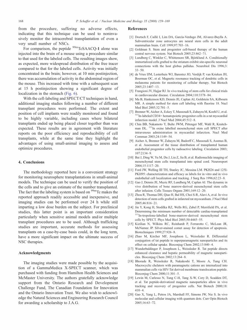
Fig. 6. Static-planar images following (A) bilateral intracranial injection of1.85 MBq of a solution of 99mTcSAACQ-1 and (B) bilateral transplant ofapproximately 5656±446 cells containing 11 Bq/cell of 99mTcSAACQ-1.The peptide-only injection shows that while the majority of the activityremains in the brain, a significant amount does migrate into theabdominal region while the labeled cells reside primarily in the striatumof the mouse.
167P. Schaffer et al. / Nuclear Medicine and Biology 35 (2008) 159–169
Washout studies were also performed in order to assessefflux of 99mTc from the cells following labeling and initialwashing. A loss of activity was detected for the first 20 min;however, after that point, 57±7% (n=12) of the activityremained within the cells, which is consistent with data fromother HIV-Tat-based probes [20]. In spite of the loss, the netuptake was still greater than the values reported for other cellradiolabeling procedures.
As determined by colorimetric cell-exclusion assays,viability for cells labeled with high effective specific activity99mTcSAACQ-1was 97±4% at 2 h postlabeling and 85±25%at 24 h postlabeling (Fig. 4A) for incubation activitiesranging from 245 to 8900 Bq/cell. However, when theincubation amounts per cell were further increased, there wasa substantial drop in cell viability. This is likely due toincreased radiolytic damage and not to the increased amountof ligand since viability at these levels of activity in thepresence of additional amounts of free peptide was notdecreased further.
While colorimetric cell-exclusion assays allow for thedetermination of cell viability in terms of membraneintegrity, they do not provide any information regardingDNA damage, which might arise during incubation or as aresult of intracellular deposition of the radionuclide. In orderto determine whether any DNA damage occurred in thepresence of 99mTcSAACQ-1, we analyzed labeled cells bysingle-cell gel electrophoresis [29,30]. Cells were incubatedwith similar amounts of activity used for the colorimetricviability studies and the extent of DNA damage compared toPBS-treated control cells (Fig. 4B). The experiments showedthat there was no significant difference between the extent ofDNA damage in the treated cells versus control cells,indicating that the presence of 99mTcSAACQ-1 at higheffective specific activity does not induce any additionaldamage to DNAwithin 24 h of incubation. Unfortunately, itwas not possible to compare these results to other studiesinvolving different cell-labeling strategies as this level ofDNA analysis is not typically reported.
For all transplantation experiments, cells were labeledusing the tracer prepared at high effective specific activity.Cells were prepared in 3 μl of lactated Ringer's solution forstereotaxic injection into the striatum of mice. For the firstimaging study, a bilateral transplant of cells (11 Bq/cell afterwashout) into the striatum of a healthy mouse wasperformed. Static-planar and low-resolution SPECT/CTimages were collected using a parallel-hole collimator at20 min posttransplantation. These studies showed that 3-Dimages of cells labeled with 11 Bq/cell could be readilyvisualized (Fig. 5A). Using a calibration curve and selectingthe appropriate region of interest on the planar image, wedetermined that 5656±446 cells containing a total ofapproximately 7.4×10⁴ Bq of activity were successfullytransplanted. Following euthanasia, an extended (15 h) high-resolution image using a pinhole collimator was collected(Fig. 5B). These images show activity residing on the surfaceof the skull, which is an indication of labeled cells residing
outside of the brain. This is likely due to extrusion of thecells upon removal of the transplant needle.
A second study was initiated to determine if the high-resolution images could be obtained in a shorter periodof time using a smaller number of cells and withouthaving to euthanize the subject. A mouse was anesthe-tized and subjected to a bilateral transplant with cellscontaining 10 Bq/cell. Pinhole images were acquired(Fig. 5C and D) for 2 h, after which the mouse wasallowed to recover from the anesthetic. For this particularstudy, it was apparent that only one of two transplantsites contained an adequate number of cells/activity fordetection. This was not totally unexpected given thetendency of such a small number of cells to adhere tothe implant syringe. From a static-planar image, it wasdetermined that 841±43 cells were successfully implantedinto the single injection site. The mouse recovered fully
168 P. Schaffer et al. / Nuclear Medicine and Biology 35 (2008) 159–169
from the procedure, suffering no adverse effects,indicating that this technique can be used to noninva-sively monitor the intracerebral transplantation of even avery small number of NSCs.
For comparison, the peptide 99mTcSAACQ-1 alone wasinjected into the brain of a mouse using a procedure similarto that used for the labeled cells. The resulting images show,as expected, more widespread distribution of the free tracercompared to that for the labeled cells. Activity was initiallyconcentrated in the brain; however, at 10 min postinjection,there was accumulation of activity in the abdominal region ofthe mouse. This increased with time with a subsequent scanat 15 h postinjection showing a significant degree oflocalization in the stomach (Fig. 6).
With the cell-labeling and SPECT/CT techniques in hand,additional imaging studies following a number of differenttransplant procedures were performed. The extent andposition of cell implants were readily monitored and foundto be highly variable, including cases where bilateraltransplants ended up being placed closer together than wasexpected. These results are in agreement with literaturereports on the poor efficiency and reproducibility of celltransplants, while at the same time they highlight theadvantages of using small-animal imaging to assess andoptimize procedures.
4. Conclusions
The methodology reported here is a convenient strategyfor monitoring neurosphere transplantations in small-animalmodels. The technique can be used to verify the position ofthe cells and to give an estimate of the number transplanted.The fact that the labeling system is based on 99mTc makes thereported approach readily accessible and inexpensive, andimaging studies can be performed over 24 h while stillimparting a low dose burden on the subject. For preclinicalstudies, this latter point is an important considerationparticularly when sensitive animal models and/or multipletransplant procedures are to be used. Although traffickingstudies are important, accurate methods for assessingtransplants on a case-by-case basis could, in the long term,have a more significant impact on the ultimate success ofNSC therapies.
Acknowledgments
The imaging studies were made possible by the acquisi-tion of a GammaMedica X-SPECT scanner, which waspurchased with funding from Hamilton Health Sciences andMcMaster University. The authors gratefully acknowledgesupport from the Ontario Research and DevelopmentChallenge Fund, The Canadian Foundation for Innovationand the Ontario Innovation Trust. We also wish to acknowl-edge the Natural Sciences and Engineering Research Councilfor awarding a scholarship to J.A.G.
References
[1] Doetsch F, Caillé I, Lim DA, García-Verdugo JM, Alvarez-Buylla A.Subventricular zone astrocytes are neural stem cells in the adultmammalian brain. Cell 1999;97:703–16.
[2] Goldman S. Stem and progenitor cell-based therapy of the humancentral nervous system. Nat Biotech 2005;23:862–71.
[3] Lundberg C, Winkler C, Whittemore SR, Björklund A. Conditionallyimmortalized cells grafted to the striatum exhibit site-specific neuronalconnections with the host globus pallidus. Neurobiol Dis 1996;3:33–50.
[4] de Vries IJM, Lesterhuis WJ, Barentsz JO, Verdijk P, van Krieken JH,Boerman OC, et al. Magnetic resonance tracking of dendritic cells inmelanoma patients for monitoring of cellular therapy. Nat Biotech2005;23:1407–13.
[5] Frangioni JV, Hajjar RJ. In vivo tracking of stem cells for clinical trialsin cardiovascular disease. Circulation 2004;110:3378–84.
[6] Ma B, Hankenson KD, Dennis JE, Caplan AI, Goldstein SA, KilbournMR. A simple method for stem cell labeling with fluorine 18. NuclMed Biol 2005;32:701–5.
[7] BrennerW, Aicher A, Eckey T, Massoudi S, ZuhayraM, Koehl U, et al.111In-labeled CD34+ hematopoietic progenitor cells in a rat myocardialinfarction model. J Nucl Med 2004;45:512–8.
[8] Chin BB, Nakamoto Y, Bulte JWM, Pittenger MF, Wahl R, Kraitch-man DL. 111In oxine labelled mesenchymal stem cell SPECT afterintravenous administration in myocardial infarction. Nucl MedCommun 2003;24:1149–54.
[9] Aicher A, Brenner W, Zuhayra M, Badorff C, Massoudi S, Assmus B,et al. Assessment of the tissue distribution of transplanted humanendothelial progenitor cells by radioactive labeling. Circulation 2003;107:2134–9.
[10] Bai J, DingW, Yu M, Du J, Liu Z, Jia B, et al. Radionuclide imaging ofmesenchymal stem cells transplanted into spinal cord. Neuroreport2004;15:1117–20.
[11] Ford JW, Welling III TH, Stanley JC, Messina LM. PKH26 and 125I-PKH95: characterization and efficacy as labels for in vitro and in vivoendothelial cell localization and tracking. J Surg Res 1996;62:23–8.
[12] Gao J, Dennis JE, Muzic RF, Lundberg M, Caplan AI. The dynamic invivo distribution of bone marrow-derived mesenchymal stem cellsafter infusion. Cells Tissues Organs 2001;169:12–20.
[13] Zhou R, Thomas DH, Qiao H, Bal HS, Choi S-R, Alavi A, et al. In vivodetection of stem cells grafted in infarcted rat myocardium. J Nucl Med2005;46:816–21.
[14] Jin Y, Kong H, Stodilka RZ, Wells RG, Zabel P, Merrifield PA, et al.Determining the minimum number of detectable cardiac-transplanted111In-tropolone-labelled bone-marrow-derived mesenchymal stemcells by SPECT. Phys Med Biol 2005;50:4445–55.
[15] Kizilian N, Wilkins RC, Reinhardt P, Ferrarotto C, McLean JR,McNamee JP. Silver-stained comet assay for detection of apoptosis.Biotechniques 1999;27:926–8.
[16] Zhao M, Kircher MF, Josephson L, Weissleder R. Differentialconjugation of tat peptide to superparamagnetic nanoparticles and itseffect on cellular uptake. Bioconjug Chem 2002;13:840–4.
[17] Wunderbaldinger P, Josephson L, Weissleder R. Tat peptide directsenhanced clearance and hepatic permeability of magnetic nanoparti-cles. Bioconjug Chem 2002;13:264–8.
[18] Bhorade R, Weissleder R, Nakakoshi T, Moore A, Tung CH.Macrocyclic chelators with paramagnetic cations are internalized intomammalian cells via HIV-Tat derived membrane translocation peptide.Bioconjug Chem 2000;11:301–5.
[19] Lewin M, Carlesso N, Tung C-H, Tang X-W, Cory D, Scadden DT,et al. Tat peptide-derivatized magnetic nanoparticles allow in vivotracking and recovery of progenitor cells. Nat Biotech 2000;18:410–4.
[20] Gao X, Yang L, Petros JA, Marshall FF, Simons JW, Nie S. In vivomolecular and cellular imaging with quantum dots. Curr Opin Biotech2005;16:63–72.

169P. Schaffer et al. / Nuclear Medicine and Biology 35 (2008) 159–169
[21] Polyakov V, Sharma V, Dahlheimer JL, Pica CM, Luker GD, Piwnica-Worms D. Novel Tat-peptide chelates for direct transduction oftechnetium-99m and rhenium into human cells for imaging andradiotherapy. Bioconjug Chem 2000;11:762–71.
[22] Säälik P, Elmquist A, Hansen M, Padari K, Saar K, Viht K, et al.Protein cargo delivery properties of cell-penetrating peptides. Acomparative study. Bioconjugate Chem 2004;15:1246–53.
[23] Banerjee SR, Levadala M, Lazarova N, Wei L, Valliant JF,Stephenson KA, et al. Bifunctional single amino acid chelates forlabeling of biomolecules with the {Tc(CO)3}+ and {Re(CO)3}+cores. Crystal and molecular structures of [ReBr(CO)3(H2NCH2C5-
H4N))], [Re(CO)3{(C5H4NCH2)2NH}]Br, [Re(CO)3{C5H4NCH2)2-NCH2CO2H}]Br, [Re(CO)3{X(Y)NCH2CO2CH2CH3}]Br (X=Y=2-pyridylmethyl; X=2-pyridylmethyl, Y=2-(1-methylimidazolyl)methyl; X=Y=2-(1-methylimidazolyl)methyl), [ReBr(CO)3{(C5H4-
NCH2)NH(CH2C4H3S)}], and Re(CO)3{C5H4NCH2)N(CH2C4H3S)(CH2CO2)}]. Inorg Chem 2002;41:6417–25.
[24] Banerjee SR, Wei L, Levadala MK, Lazarova N, Golub VO, O'ConnorCJ, et al. {ReIIICl3} core complexes with bifunctional single aminoacid chelates. Inorg Chem 2002;41:5795–802.
[25] Stephenson KA, Banerjee SR, Besanger T, Sogbein OO, LevadalaMK,McFarlane N, et al. Bridging the gap between in vitro and in vivoimaging: isostructural Re and 99mTc complexes for correlating fluo-rescence and radioimaging studies. J Am Chem Soc 2004;126:8598–9.
[26] Kiso Y, Satomi M, Ukawa K, Akita T. Efficient deprotection of NG-tosylarginine with thioanisole-trifluoromethanesulphonic acid system.Chem Commun 1980:1063–4.
[27] Atherton E, Sheppard RC, Ward P. Peptide synthesis. Part 7. Solid-phase synthesis of conotoxin G1. J Chem Soc Perkin Trans I 1985:2065–73.
[28] Alberto R, Schibli R, Egli A, Schubiger AP, Abram U, Kadtem TA. Anovel organometallic aqua complex of technetium for the labeling ofbiomolecules: synthesis of [99mTc(OH2)3(CO)3]
+ from [99mTcO4]− in
aqueous solution and its reaction with a bifunctional ligand. J AmChem Soc 1998;120:7987–8.
[29] McNamee JP, McLean JRN, Ferrarotto CL, Bellier PV. Comet assay:rapid processing of multiple samples. Mutat Res 2000;466:63–9.
[30] Ryan LA, Wilkins RC, McFarlane NM, Sung MM, McNamee JP,Boreham DR. Relative biological effectiveness of 280 keV neutronsfor apoptosis in human lymphocytes. Health Phys 2006;91:68–75.