Choanoflagellates (Sarcomastigophora, Protozoa) from the Coastal Waters of Taiwan and Japan. I....
8
J. Euk. Microbiol., 43(2), 1996, pp. 136-143 0 1996 by the Society of Protozoologists Choanoflagellates (Sarcomastigophora, Taiwan and Japan. I. Protozoa) from the Coastal Three New Species Waters of SEIKO HARA,*,’ YUH-LING L. CHEN,* JIA-CHI S H E W and EIJI TAKAHASHI** *Department of Marine Resources, National Sun Yat-sen University,Kaohsiung, Taiwan 804, Republic of China **Department of Biology, Faculty of Science, Yamagata University, Yamagata, Japan ABSTRACT. Three new acanthoecidaean species collected from the coastal waters of Taiwan and Japan are described: Acanthocorbis camarensis n. sp. resembles Acanthocorbis unguiculata in lorica morphology, but differs in having regularly arranged longitudinal and transverse costae at the anterior lorica chamber, and in lacking a nail at the apical end of anterior spine; Diaphanoeca spiralifurca n. sp. is characterized by the spiral arrangement of the costal strips in the posterior half of the lorica chamber and is closely related to Diaphanoeca grandis; Stephanoeca supracostata n. sp. is closely related to Stephanoeca elegans, but differs in having an additional transverse costa at the antenor lorica chamber. Supplementary key words. Acanthocorbis camarensis. Diaphanoeca spiralifirca, Stephanoeca supracostata, taxonomy. HE Choanoflagellida (Sarcomastigophora, Protozoa) are T characterized morphologically by the possession of a fla- gellum surrounded by a tentacle collar [ 131. The Choanoflagel- lida include three families: the Codonosigidae are the naked (without theca or lorica) Choanoflagellida; the Salpingoecidae are species with the protoplasts being surrounded with a thin organic membrane (theca); and the Acanthoecidae are species with the protoplasts being invested in a lorica composed of siliceous costal strips [ 171. Acanthoecidaean taxonomy is based on the lorica morphology (including general form of lorica), the possession of anterior spines, the number of costae, the form of costal strips, and the types of costal strip junctions [27]. During the past two decades enthusiastic investigations using modern techniques have dealt with choanoflagellates from tem- perate and circumpolar waters [ 12, 19, 201. Investigations have also been conducted in the Red Sea [2 11, Galapagos Islands [ 141, Andaman Sea [24-261, and Hawaii [8]. In the north Pacific region, choanoflagellates from subarctic oceanic waters [ 1, 21, temperate coastal waters of Japan [5-7, 181, the North Pacific central gyre [8], and California [3, 281 have been studied. A qualitative survey of choanoflagellates from the typically sub- tropical waters of Taiwan in the western Pacific has great im- portance in regard to worldwide choanoflagellate distribution. The objective of this investigation was to describe the new taxa collected from the coastal waters of Taiwan. MATERIALS AND METHODS Surface water samples were collected between September 1992 and July 1993 from the coastal waters of Kaohsiung (south- western Taiwan), Nan-Wan (southern Taiwan), and five other localities (Fang-Liao, Tung-Ho, Hua-Lien, Sun-Chiao-Chiao and Yeh-Liu) around Taiwan (Fig. 1). Supplementary water samples were collected from Japan (Osaka Bay [6] and Lake Saroma, August 1982), Thailand (KO Lan Island, August 1983) and the USA (Hawaii, December 198 1). Samples were collected using a Van Dorn water sampler, fixed with neutralized formaldehyde (final concentration 1 O/o v/v) immediately after sampling, and stored at 1” C-4” C before preparation. All the samples were prepared within 1 wk., most within less than 1 d. Portions of the seawater samples (0.5-1 L) were cultured crudely at room temperature for up to 2 wk. Details of concentration and prep- aration of samples for electron microscopical observations have been described [6]. JEOL lOOB and 100s were used for the transmission electron microscopic observation. In this paper, the anterior projection [27] is considered as a I To whom correspondence should be addressed. Present address: Miyazaki International College, 1405 Kano, Kiyotake-cho, Miyazaki 889-16, Japan. lorica structure independent of the longitudinal costa because, in certain species (e.g. Calliacantha natans and Acanthoeca spec- tabilis) the anterior projection and the longitudinal costae are discontinuous and are formed by costal strips that are very different in structure. The number of costal strips constructing the longitudinal costa, consequently, does not include the costal strip number forming the anterior projection. RESULTS AND DISCUSSION Acanthocorbis camarensis Hara, n. sp. (Fig. 2-4) Diagnosis. Cells solitary. Protoplast when dried 2.5-3.5 pm x 1.5-2.0 pm, flagellum ca. 8 pm long surrounded by a collar. Lorica obpyriform with waist, consists of anterior projections and lorica chamber, 12-1 6 pm in length. Diameter of the lorica is maximum (6.3-7.3 pm) at the anterior projection, ca. 5.3- 6.0 pm at the first transverse costa and 3.5-4.0 pm at the waist. Each of the 10-14 anterior projections consists of two costal strips which connect with the longitudinal costa which is con- structed of two costal strips. Lorica chamber consists of anterior and posterior chambers. The anterior chamber is constructed with regularly arranged longitudinal costae and the first trans- verse costa, which is located at the anterior end of the longi- tudinal costae. Costal strips overlap greatly at the first transverse costa. Posteri, ‘r chamber is constructed of more or less disar- ranged longitudinal costae and many supplemental costal strips. Protoplast is located at the posterior chamber. A waist exists at the boundary of the anterior and the posterior chambers. All the costal strips are narrow rods. Type specimen. Collected from Hawaii (December 198 1) (Fig. 3 ), deposited in the Herbarium of Miyazaki International College, Miyazaki, Japan, No. 6 140. Distribution. Coastal waters of Taiwan, Hawaii, and Thai- land. Etymology. The specific name is derived from the Latin ca- marensis meaning “chamber.” This new species closely resembles Acanthocorbis unguiculata [20] and Acanthocorbis apoda [9, 221. Morphological details of the lorica that distinguish these three species are as follows: (1) Regularly arranged longitudinal and transverse costae form open windows at the anterior part of the lorica chambers in A. apoda and A. camarensis n. sp. (Fig. 2, 3), versus a uniform chamber constructed of more or less disarranged and closely arranged supplemental costal strips, without any clear longitudinal nor transverse costae in A. unguiculata; (2) A. apoda and A. ca- marensis n. sp. have anterior projections with round apical tips (Fig. 4), while ‘4. unguiculata has claw-formed tips; (3) the num- ber of transverse costae in the posterior chamber is more than three in A. unguiculata and A. camarensis n. sp., while A. apoda has one to three transverse costae; and (4) the first and the second 136
Transcript of Choanoflagellates (Sarcomastigophora, Protozoa) from the Coastal Waters of Taiwan and Japan. I....

J. Euk. Microbiol., 43(2), 1996, pp. 136-143 0 1996 by the Society of Protozoologists
Choanoflagellates (Sarcomastigophora, Taiwan and Japan. I.
Protozoa) from the Coastal Three New Species
Waters of
SEIKO HARA,*,’ YUH-LING L. CHEN,* JIA-CHI SHEW and EIJI TAKAHASHI** *Department of Marine Resources, National Sun Yat-sen University, Kaohsiung, Taiwan 804, Republic of China
**Department of Biology, Faculty of Science, Yamagata University, Yamagata, Japan
ABSTRACT. Three new acanthoecidaean species collected from the coastal waters of Taiwan and Japan are described: Acanthocorbis camarensis n. sp. resembles Acanthocorbis unguiculata in lorica morphology, but differs in having regularly arranged longitudinal and transverse costae at the anterior lorica chamber, and in lacking a nail at the apical end of anterior spine; Diaphanoeca spiralifurca n. sp. is characterized by the spiral arrangement of the costal strips in the posterior half of the lorica chamber and is closely related to Diaphanoeca grandis; Stephanoeca supracostata n. sp. is closely related to Stephanoeca elegans, but differs in having an additional transverse costa at the antenor lorica chamber.
Supplementary key words. Acanthocorbis camarensis. Diaphanoeca spiralifirca, Stephanoeca supracostata, taxonomy.
HE Choanoflagellida (Sarcomastigophora, Protozoa) are T characterized morphologically by the possession of a fla- gellum surrounded by a tentacle collar [ 131. The Choanoflagel- lida include three families: the Codonosigidae are the naked (without theca or lorica) Choanoflagellida; the Salpingoecidae are species with the protoplasts being surrounded with a thin organic membrane (theca); and the Acanthoecidae are species with the protoplasts being invested in a lorica composed of siliceous costal strips [ 171. Acanthoecidaean taxonomy is based on the lorica morphology (including general form of lorica), the possession of anterior spines, the number of costae, the form of costal strips, and the types of costal strip junctions [27].
During the past two decades enthusiastic investigations using modern techniques have dealt with choanoflagellates from tem- perate and circumpolar waters [ 12, 19, 201. Investigations have also been conducted in the Red Sea [2 11, Galapagos Islands [ 141, Andaman Sea [24-261, and Hawaii [8]. In the north Pacific region, choanoflagellates from subarctic oceanic waters [ 1 , 21, temperate coastal waters of Japan [5-7, 181, the North Pacific central gyre [8], and California [3, 281 have been studied. A qualitative survey of choanoflagellates from the typically sub- tropical waters of Taiwan in the western Pacific has great im- portance in regard to worldwide choanoflagellate distribution.
The objective of this investigation was to describe the new taxa collected from the coastal waters of Taiwan.
MATERIALS AND METHODS Surface water samples were collected between September 1992
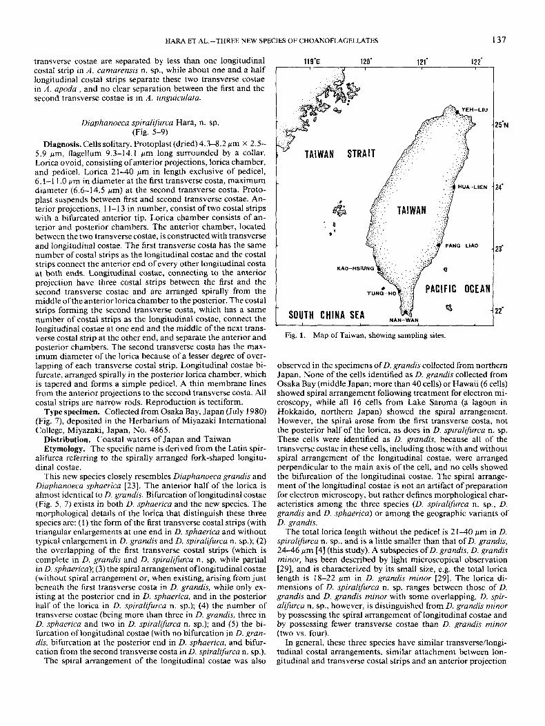
and July 1993 from the coastal waters of Kaohsiung (south- western Taiwan), Nan-Wan (southern Taiwan), and five other localities (Fang-Liao, Tung-Ho, Hua-Lien, Sun-Chiao-Chiao and Yeh-Liu) around Taiwan (Fig. 1). Supplementary water samples were collected from Japan (Osaka Bay [6] and Lake Saroma, August 1982), Thailand (KO Lan Island, August 1983) and the USA (Hawaii, December 198 1). Samples were collected using a Van Dorn water sampler, fixed with neutralized formaldehyde (final concentration 1 O/o v/v) immediately after sampling, and stored at 1” C-4” C before preparation. All the samples were prepared within 1 wk., most within less than 1 d. Portions of the seawater samples (0.5-1 L) were cultured crudely at room temperature for up to 2 wk. Details of concentration and prep- aration of samples for electron microscopical observations have been described [6]. JEOL lOOB and 100s were used for the transmission electron microscopic observation.
In this paper, the anterior projection [27] is considered as a
I To whom correspondence should be addressed. Present address: Miyazaki International College, 1405 Kano, Kiyotake-cho, Miyazaki 889-16, Japan.
lorica structure independent of the longitudinal costa because, in certain species (e.g. Calliacantha natans and Acanthoeca spec- tabilis) the anterior projection and the longitudinal costae are discontinuous and are formed by costal strips that are very different in structure. The number of costal strips constructing the longitudinal costa, consequently, does not include the costal strip number forming the anterior projection.
RESULTS AND DISCUSSION Acanthocorbis camarensis Hara, n. sp.
(Fig. 2-4) Diagnosis. Cells solitary. Protoplast when dried 2.5-3.5 pm
x 1.5-2.0 pm, flagellum ca. 8 pm long surrounded by a collar. Lorica obpyriform with waist, consists of anterior projections and lorica chamber, 12-1 6 pm in length. Diameter of the lorica is maximum (6.3-7.3 pm) at the anterior projection, ca. 5.3- 6.0 pm at the first transverse costa and 3.5-4.0 pm at the waist. Each of the 10-14 anterior projections consists of two costal strips which connect with the longitudinal costa which is con- structed of two costal strips. Lorica chamber consists of anterior and posterior chambers. The anterior chamber is constructed with regularly arranged longitudinal costae and the first trans- verse costa, which is located at the anterior end of the longi- tudinal costae. Costal strips overlap greatly at the first transverse costa. Posteri, ‘r chamber is constructed of more or less disar- ranged longitudinal costae and many supplemental costal strips. Protoplast is located at the posterior chamber. A waist exists at the boundary of the anterior and the posterior chambers. All the costal strips are narrow rods.
Type specimen. Collected from Hawaii (December 198 1) (Fig. 3 ), deposited in the Herbarium of Miyazaki International College, Miyazaki, Japan, No. 6 140.
Distribution. Coastal waters of Taiwan, Hawaii, and Thai- land.
Etymology. The specific name is derived from the Latin ca- marensis meaning “chamber.”
This new species closely resembles Acanthocorbis unguiculata [20] and Acanthocorbis apoda [9, 221. Morphological details of the lorica that distinguish these three species are as follows: (1) Regularly arranged longitudinal and transverse costae form open windows at the anterior part of the lorica chambers in A. apoda and A. camarensis n. sp. (Fig. 2, 3), versus a uniform chamber constructed of more or less disarranged and closely arranged supplemental costal strips, without any clear longitudinal nor transverse costae in A. unguiculata; (2) A. apoda and A. ca- marensis n. sp. have anterior projections with round apical tips (Fig. 4), while ‘4. unguiculata has claw-formed tips; (3) the num- ber of transverse costae in the posterior chamber is more than three in A. unguiculata and A. camarensis n. sp., while A. apoda has one to three transverse costae; and (4) the first and the second
136
H A M ET AL.-THREE NEW SPECIES OF CHOANOFLAGELLATES 137
transverse costae are separated by less than one longitudinal costal strip in A . camarensis n. sp., while about one and a half longitudinal costal strips separate these two transverse costae in A. apoda , and no clear separation between the first and the second transverse costae is in A . unguiculata.
Diaphanoeca spiralifurca Hara, n. sp. (Fig. 5-9)
Diagnosis. Cells solitary. Protoplast (dried) 4.3-8.2 wm x 2.5- 5.9 pm, flagellum 9.3-14.1 pm long surrounded by a collar. Lorica ovoid, consisting of anterior projections, lorica chamber, and pedicel. Lorica 21-40 pm in length exclusive of pedicel, 6.1-1 1 .O pm in diameter at the first transverse costa, maximum diameter (6.6-14.5 Mm) at the second transverse costa. Proto- plast suspends between first and second transverse costae. An- terior projections, 1 1-1 3 in number, consist of two costal strips with a bifurcated anterior tip. Lorica chamber consists of an- terior and posterior chambers. The anterior chamber, located between the two transverse costae, is constructed with transverse and longitudinal costae. The first transverse costa has the same number of costal strips as the longitudinal costae and the costal strips connect the anterior end of every other longitudinal costa at both ends. Longitudinal costae, connecting to the anterior projection have three costal strips between the first and the second transverse costae and are arranged spirally from the middle ofthe anterior lorica chamber to the posterior. The costal strips forming the second transverse costa, which has a same number of costal strips as the longitudinal costae, connect the longitudinal costae at one end and the middle of the next trans- verse costal strip at the other end, and separate the anterior and posterior chambers. The second transverse costa has the max- imum diameter of the lorica because of a lesser degree of over- lapping of each transverse costal strip. Longitudinal costae bi- furcate, arranged spirally in the posterior lorica chamber, which is tapered and forms a simple pedicel. A thin membrane lines from the anterior projections to the second transverse costa. All costal strips are narrow rods. Reproduction is tectiform.
Type specimen. Collected from Osaka Bay, Japan (July 1980) (Fig. 7), deposited in the Herbarium of Miyazaki International College, Miyazaki, Japan, No. 4865.
Distribution. Coastal waters of Japan and Taiwan Etymology. The specific name is derived from the Latin spir-
alifurca referring to the spirally arranged fork-shaped longitu- dinal costae.
This new species closely resembles Diuphanoeca grandis and Diaphanoeca sphaerica [23]. The anterior half of the lorica is almost identical to D. grandis. Bifurcation of longitudinal costae (Fig. 5, 7) exists in both D. sphaerica and the new species. The morphological details of the lorica that distinguish these three species are: (1) the form of the first transverse costal strips (with triangular enlargements at one end in D. sphaerica and without typical enlargement in D. grandis and D. spiralifurca n. sp.); (2) the overlapping of the first transverse costal strips (which is complete in D. grandis and D. spiralifurca n. sp. while partial in D. sphaerica); (3) the spiral arrangement oflongitudinal costae (without spiral arrangement or, when existing, arising from just beneath the first transverse costa in D. grandis, while only ex- isting at the posterior end in D. sphaerica, and in the posterior half of the lorica in D. spiralifurca n. sp.); (4) the number of transverse costae (being more than three in D. grandis, three in D. sphaerica and two in D. spiralifurca n. sp.); and (5) the bi- furcation of longitudinal costae (with no bifurcation in D. gran- dis, bifurcation at the posterior end in D. sphaerica, and bifur- cation from the second transverse costa in D. spiralifurca n. sp.).
The spiral arrangement of the longitudinal costae was also
11 9-E 120' 121' 122' I
25"
24'
23'
2 2'
Fig. 1. Map of Taiwan, showing sampling sites.
observed in the specimens of D. grandis collected from northern Japan. None of the cells identified as D. grandis collected from Osaka Bay (middle Japan; more than 40 cells) or Hawaii (6 cells) showed spiral arrangement following treatment for electron mi- croscopy, while all 16 cells from Lake Saroma (a lagoon in Hokkaido, northern Japan) showed the spiral arrangement. However, the spiral arose from the first transverse costa, not the posterior half of the lorica, as does in D. spiralifurca n. sp. These cells were identified as D. grandis, because all of the transverse costae in these cells, including those with and without spiral arrangement of the longitudinal costae, were arranged perpendicular to the main axis of the cell, and no cells showed the bifurcation of the longitudinal costae. The spiral arrange- ment of the longitudinal costae is not an artifact of preparation for electron microscopy, but rather defines morphological char- acteristics among the three species (D. spiralifurca n. sp., D. grandis and D. sphaerica) or among the geographic variants of D. grandis.
The total lorica length without the pedicel is 21-40 pm in D. spiralifurca n. sp., and is a little smaller than that of D. grandis, 24-46 pm [4] (this study). A subspecies of D. grandis, D. grandis minor, has been described by light microscopical observation [29], and is characterized by its small size, e.g. the total lorica length is 18-22 pm in D. grandis minor [29]. The lorica di- mensions of D. spiralzjiurca n. sp. ranges between those of D. grandis and D. grandis minor with some overlapping. D. spir- alifurca n. sp., however, is distinguished from D. grandis minor by possessing the spiral arrangement of longitudinal costae and by possessing fewer transverse costae than D. grandis minor (two vs. four).
In general, these three species have similar transverse/longi- tudinal costal arrangements, similar attachment between lon- gitudinal and transverse costal strips and an anterior projection

138 J. EUK. MICROBIOL., VOL. 43, NO. 2, MARCH-APRIL 1996
HARA ET AL.-THREE NEW SPECIES OF CHOANOFLAGELLATES 139
with two costal strips. In addition, similarity is also found be- tween each of two of the three species, such as overlapping of the transverse costal strips and spiral arrangement of longitu- dinal costae. It is thus evident that these three species form a well-circumscribed group in the genus Diaphanoeca and are closely related to each other.
The lorica lining membrane of D. spiralifurca n. sp., covering the lorica from the anterior projection (Fig. 8) to the second transverse costa (Fig. 9), closely resembles that of D. grandis [ 151, but consists not only of fibrils but also of continuous mem- branous matrices. The fibrils are woven more or less perpen- dicularly in D. grandis, more randomly in D. spiralifurca n. sp. Figure 9 shows the clear shadows of the particles (arrowheads) on the formvar membrane around the lorica and also inside of the lorica without the membrane lining. Particles under the membrane lining area, however, have no clear shadow. Because the clear shadows of the thin fibrils are visible, if a bare particle exists in the membranous area, the particle should have a clear shadow. It is, thus, likely that the particles under the membra- nous area lose their shadows because the membrane matrices cover them smoothly, much like a snowfall covering the rough surface of the land. The existence of the continuous membrane also can be elucidated by the different texture of the formvar matrix and differences in electron density between the area in the lorica with or without lining membrane.
Cells of D. spiralifurca n. sp. were observed in a natural water sample collected from Osaka Bay, Japan (August 1980; water temperature 23.7" C). Other cells were observed from crude cultures that were originally collected from surface coastal wa- ters in Osaka Bay, Japan, and Nan-Wan, Taiwan.
Stephanoeca supracostata Hara, n. sp. (Fig. 10-15)
Diagnosis. Cells solitary. Protoplast when dried 2.4-7.9 pm x 1.7-3.6 pm, flagellum ca. 9-pm long surrounded by a collar. Lorica obpyriform with waist, consists of anterior and posterior lorica chambers, 9-18 pm in length. Diameter of the lorica is maximum (4.5-10.0 pm) at the level of second transverse costa, ca. 3.1-6.5 pm at the first transverse costa, and 2.2-4.4 Mm at the waist. The anterior chamber is constructed of regularly ar- ranged 12-18 longitudinal costae, each of which is composed oftwo costal strips, and three transverse costae which are located at the anterior end of the chamber, near the middle level of the second longitudinal costal strips, and the posterior end of the chamber, respectively. Posterior chamber is constructed of 7- 20 longitudinal costae, each of which is composed of one costal strip, and a transverse costa which is located at the anterior end of the chamber. Protoplast is located at the posterior chamber. A waist exists at the boundary of the anterior and the posterior chambers. All the costal strips are narrow rods. Reproduction is tectiform.
Type specimen. Collected from Nan-Wan, Taiwan (Decem-
ber 1992) (Fig. 10) , deposited in the Herbarium of Miyazaki International College, Miyazaki, Japan, No. 192 14.
Distribution. Coastal waters of Japan and Taiwan Etymology. The specific name is derived from the Latin su-
pracostata referring to the additional transverse costa above the waist.
Stephanoeca supracostata n. sp. is closely related to Stephan- oeca elegans (Norris) Throndsen. The original type of S. elegans (Norris) Throndsen sensu stricto has no transverse costa be- tween the anterior end and the waist, while all specimens col- lected from the Taiwanese waters possess an additional trans- verse costa at the two-thirds level from the anterior end of the anterior lorica chamber where the maximum diameter of the lorica is located. Stephanoeca elegans sensu stricto has been reported from temperate Atlantic and northwest Pacific coastal, Mediterranean and Japanese waters [9, 10, 17,291 (present study), whereas S. supracostata n. sp. is distributed in Japanese and Taiwanese waters. A cell possessing an additional transverse costa between the anterior end and the waist has been reported as S. elegans based on the specimen from California [28].
Cells of S. elegans and S. supracostata n. sp. coexisted in water samples collected from Osaka Bay in Japan. This makes these Japanese samples the most suitable for examining the morphological differences of these two species. The total length of lorica and the diameter of the first transverse costa of the two species overlap (Table I). The diameter of the waist of S. elegans, however, is slightly larger than that of S. supracostata n. sp. The clear waist of the lorica in S. supracostata n. sp., at the level of the posterior transverse costae, occurs because there is a greater degree of overlap of the transverse costal strips in the posterior transverse costa than in the second transverse costa. Except for the existence or absence of the additional transverse costa, the most conspicuous morphological difference in the populations collected from Osaka Bay is the number of longitudinal costae: 20-25 in S. elegans vs. 14-18 in S. supracostata n. sp. No intermediate form between these two morphological types has been observed from this locality (i.e. Osaka Bay), thus indicating that the two species identified were not artifacts of same con- tinuous morphological variation in S. elegans, but were genet- ically distinct.
Except for the number of the longitudinal costae; morpho- logical, dimensional, and numerical characteristics of S. elegans collected from Japan coincided well with the type material (Ta- ble 1). The type specimen ofS. elegans has 18 longitudinal costae [ 171, while the specimens collected from Osaka Bay possessed 20-25 longitudinal costae. The numerical variation in longi- tudinal costae in this species has been reported by Leadbeater as 16-18 from Norway [9] and as 13-15 from Jugoslavia [lo]. It appears clear that S. elegans has a very wide range of variation in the number of the longitudinal costae, from 13 to 25.
The S. supracostata n. sp. specimens collected from Japan and Taiwan showed variation in several of the lorica dimen- sions. The range of variation in these dimensions differed in the
t
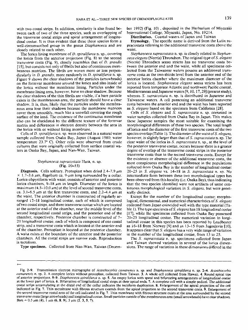
Fig. 2-9. Transmission electron micrographs of Acanthocorbis cumarensis n. sp. and Diaphanoeca spiralifurca n. sp. 2-4. Acanthocorbis cumarensis n. sp. 2. A complete lorica without protoplast, collected from Taiwan. 3. A whole cell collected from Hawaii. 4. Round apical tips of anterior projections. 5-9. Diuphanoeca spiralijiirca n. sp. 5 . An empty lorica with spiral and bifurcating arrangements of longitudinal costae at the lower part of lorica. 6 . Bifurcation of longitudinal costal strips at their apical ends. 7. A complete cell with a simple pedicel. The additional costal strips accumulating at the distal end of the collar indicates the tectiform duplication. 8. Enlargement of the apical projection of the cell indicated in Fig. 7. Thin membrane with fibrous structure extends from the apical projection to the second transverse costa. 9. Enlargement of the second transverse costal area of the cell indicated in Fig. 7. Thin membrane with fibrous structure exists at the area surrounded by the second transverse costa (large arrowheads) and longitudinal costae. Small particles outside of the membranous area (small arrowheads) have clear shadows. Bars = 0.5 pm (4), 1 pm (6, 8, 9), 5 pm (2, 3, 5, 7).

140 J. EUK. MJCROBJOL., VOL. 43, NO. 2, MARCH-APRIL 1996
Fig. 10-13. Transmission electron micrographs of Stephanueca supracustata n. sp. 10. A complete lonca with a transverse costa at the level about two-thirds from the anterior end of the anterior lorica chamber. One end of longitudinal costal strip is spatulate. 11. Supplemental costal strips around the collar region indicating the tectiform cell division. 12. A lorica. One of the two transverse costa at the waist connects the posterior end of the anterior lorica chamber, the other transverse costa connects the anterior end of the posterior lorica chamber. 13. A complete cell with long slender tentacle emerged from the posterior end of the protoplast. Bars = 5 gm.
HARA ET AL.-THREE NEW SPECIES OF CHOANOFLAGELLATES 141
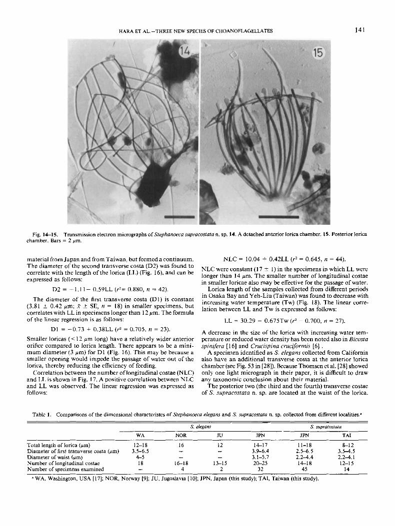
Fig. 14-15. Transmission electron micrographs of Stephanoem supracostata n. sp. 14. A detached anterior lorica chamber. 15. Posterior lonca chamber. Bars = 2 pm.
material from Japan and from Taiwan, but formed a continuum. NLC = 10.04 + 0.42LL (r2 = 0.645, n = 44). The diameter ofthe second transverse costa (D2) was found to correlate with the length of the lorica (LL) (Fig. 16), and can be expressed as follows:
NLC were constant 1) in the specimens in which LL were longer than 14 pm. The smaller number of longitudinal costae in smaller loricae also may be effective for the passage of water.
D2 = - 1.11 + 0.59LL (r2= 0.880. n = 42). Lorica length of the samdes collected fromhifferent Deriods ~
in Osaka Baiand Yeh-Liu iTaiwan) was found to decrease with
lation between LL and Tw is expressed as follows: The diameter Of the first transverse costa (D1) is 'Onstant increasing water temperature (Tw) (Fig. 18). The linear (3.81 * 0.42 pm; ,T 2 SE, n = 18) in smaller specimens, but
correlates with LL in specimens longer than 12 pm. The formula of the linear regression is as follows:
D1 = -0.73 + 0.38LL ( r2 = 0.705, n = 23). Smaller loricas (< 12 pm long) have a relatively wider anterior orifice compared to lorica length. There appears to be a mini- mum diameter (3 pm) for DI (Fig. 16). This may be because a smaller opening would impede the passage of water out of the lorica, thereby reducing the efficiency of feeding.
Correlation between the number of longitudinal costae (NLC) and LL is shown in Fig. 17. A positive correlation between NLC and LL was observed. The linear regression was expressed as follows:
LL = 30.29 - 0.675Tw (r2 = 0.700, n = 27).
A decrease in the size of the lorica with increasing water tem- perature or reduced water density has been noted also in Bicosta spinifera [ 161 and Crucispina cruciforrnis [6] .
A specimen identified as S. eleguns collected from California also have an additional transverse costa at the anterior lorica chamber (see Fig. 53 in [28]). Because Thomsen et al. [28] showed only one light micrograph in their paper, it is difficult to draw any taxonomic conclusion about their material.
The posterior two (the third and the fourth) transverse costae of S. supracostata n. sp. are located at the waist of the lorica.
Table 1. Comparisons of the dimensional characteristcs of Stephanoem e/egans and S. supracostafa n. sp. collected from different localities."
S. elegans S. supracostata
WA NOR JU JPN JPN TAI
Total length of lorica (pm) 12-18 16 12 14-17 11-18 8-1 2 Diameter of first transverse costa (pm) 3.5-6.5 - - 3.9-6.4 2.5-6.5 3.54 .5 Diameter of waist (pm) 4-5 - - 3.1-5.7 2.2-4.4 2 .24 .1 Number of longitudinal costae 18 16-18 13-15 20-25 14-18 12-15 Number of sDecirnens examined - 4 2 32 45 14
a WA, Washington, USA [ 171; NOR, Noway [9]; JU, Jugoslavia [ 101; JPN, Japan (this study); TAI, Taiwan (this study).

142 J. EUK. MICROBIOL., VOL. 43, NO. 2, MARCH-APRIL 1996
17-
16- .&
Y
' 2 1 5 -
2 14- ru 0
Q) L 13-
$12-
g 11-
10 18.0
9 17.0
16.0
E 15.0 5 5 14.0
8 13.0
h
E 3 8
z 7 s g 6
% 5
z 4 .1
* 5 3 4 e, 10.0 9 2 0 First transverse costa
9.0
8.0
h v cd
W
e,
3 5 2 12.0
8 11.0 0
18.0 20.0 22.0 24.0 26.0 28.0 30.0 Water temperature ("C)
0 8.0 9.0 10.0 11.0 12.0 13.0 14.0 15.0 16.0 17.0 18.0
Lorica length (pm) Fig. 18. The lorica length vs. the water temperature in Stephanoeca Fig. 16. Relationship between the diameter of the first and the sec-
ond transverse costae and the lonca length in Stephanoeca supracostata supracostata n. sp.
n. sp.
The third transverse costa binds the posterior ends of the second longitudinal costal strips to construct the anterior lorica cham- ber. The anterior ends of the third longitudinal costal strips are connected by the fourth transverse costa (Fig. 10, 12). The sec- ond and third longitudinal costal strips are not well matched (Fig. 10, 12), and usually do not connect with each other. The longitudinal costae are not continuous from the top to the bot- tom of the lorica but are separated into two parts at the waist. The anterior and the posterior chambers are connected by link- age between some of the posterior ends of the second longitu- dinal costal strips with the fourth transverse costa; and some of the anterior ends of the third longitudinal costal strips link to the third transverse costa (Fig. 10, 12). Because the anterior and posterior chambers may not be firmly connected, separations between them are occasionally observed (Fig. 14, 15). This in- dependence of the lorica chambers in s. supracostata n. sp.
-" I
$ 18 1 9 1
m e
e-e
m e
10 1 , I > I , , ! ! \ , I I 8 I ( 1 , I ! 8 1 8 , I I I I I , , 1 8 1 1 I 1 1 , I 8 " ' ' ' 1 8.0 9.0 10.011.012.013.014.015.016.017.018.0
Lorica length (pm) Fig. 17. Relationship between the number of longitudinal costae
and the lonca length in Stephanoeca SUp!XZCOSfQta n. sp.
suggests different construction and function of these lorica chambers for the protoplast. The posterior lorica chamber may protect the protoplast from direct attachment of outside solids. The anterior chamber surrounding the feeding organelles, a fla- gellum, and a collar, may control water current produced by the flagellum and sieve food particles of suitable sizes.
One cell collected from Osaka Bay and stored at room tem- perature for 1 d was found to possess a long (ca. 30 pm) slender tentacle developed from the distal end of a posterior labial pe- duncle (Fig. 13). Cells of Codonosigidae usually have posterior tentacles when they are floating. In Acanthoecidae, posterior tentacles are observed in naked daughter cells in Acanthoeca spectabilis [ 1 I]. After the lorica construction is complete, ten- tacles develop inside the lorica and some connect the protoplast with the lorica to suspend the protoplast within the lorica in a fixed position, such as in Diaphanoeca grandis [ 151. This is the first report of the posterior tentacle extending outwardly from the lorica in an Acanthoecidae species. Many cells of S. supra- costata n. sp. are observed to attach to substrates, such as de- tritus. The major purposes for the tentacles in choanoflagellates are to attach the cell to a substrate (posterior tentacles) and to capture food particles (collar tentacles). The exceptionally long posterior tentacle observed in the free-floating cells of S. supra- costata n. sp. are probably used for the purpose of attachment.
The range of water temperature in which cells of S. supra- costata n. sp. were collected was 18.8" C-26.8" C in Osaka Bay and 28.8" C in Yeh-Liu (Taiwan). Almost all of the observed cells collected from Osaka Bay appeared from just before the highest water temperature period (August) until the early mixing period (September to November ) in 1979 through 1982. The water temperature at the sampling point in Osaka Bay ranged from 7.5" C to 27.3" C. This species may be thermophilic. Even though the same water temperature existed before and after the temperature peak, this species appeared just before the peak and persisted to the early mixing period, whereas this species has hardly been observed in the stratified period. The water tem- perature may not be the only environmental factor, but other physical, chemical and/or biological factors may define the dis- tribution of this species. Because this species began to appear in the surface water before the mixing period, it could not simply be supposed that the species had migrated from the subsurface

HARA ET AL.-THREE NEW SPECIES OF CHOANOFLAGELLATES 143
layer. It is unclear from where this species migrated to the surface water in Osaka Bay. It is also not clear whether this species is a native species or an immigrant species in Osaka Bay.
ACKNOWLEDGMENTS The authors thank L. Stone for reading the manuscript. This
study was supported in part by a grant (NSC 82-021 l-B-110- 001) from the National Science Council of the Republic of Chi- na.
LITERATURE CITED 1. Booth, B. C. 1990. Choanoflagellates from the subarctic North
Pacific Ocean, with description of two new species. Can. J. Zool., 68:
2. Booth, B. C., Lewin, J. & N o h , R. E. 1982. Nanoplankton species predominant in the subarctic Pacific in May and June 1978. Deep-sea Rex, 29: 185-200.
3. Buck, K. R., Chavez, F. P. & Thomsen, H. A. 1991. Choano- flagellates of the central California waters: abundance and distribution. Ophelia. 33:179-186.
4. Ellis, W. N. 1929. Recent researches on the Choanoflagellata (Craspedomonadines) (freshwater and marine). Ann. Soc. Zool. Belg.,
5. Hara, S. & Takahashi, E. 1984. Re-investigation of Polyoeca dichotoma and Acanthoeca spectabilis (Acanthoecidae: Choanoflagel- lida). J. Mar. Biol. Ass. (UK), 64:s 19-827.
6. Hara, S. & Takahashi, E. 1987a. An investigation with electron microscope of marine choanoflagellates (Protozoa: Choanoflagellida) from Osaka Bay, Japan. I. Re-investigation of Bicosta spinifera, B. minor and Crucispina cruciformis. Bull. Plankton SOC. Japan, 34: 1-1 3.
7 . Hara, S. & Takahashi E. 1987b. An investigation with electron microscope of marine choanoflagellates (Protozoa: Choanoflagellida) from Osaka Bay, Japan. 11. Two new genera and a new species of Acan- thoecidae. Bull. Plankton Soc. Japan, 34: 15-23.
8. Hoepffner, N. & Haas, L. W. 1990. Electron microscopy of na- noplankton from the North Pacific Central Gyre. J. Phycol., 26:421- 439.
9. Leadbeater, B. S. C. 1972. Fine-structural observations on some marine choanoflagellates from the coast of Norway. J. Mar. Biol. Ass.
10. Leadbeater, B. S. C. 1973. External morphology ofsome marine choanoflagellates from the coast of Jugoslavia. Arch. Protistenk., 115:
11. Leadbeater, B. S. C. 1979. Developmental and ultrastructural observations on two stalked marine choanoflagellates, Acanthoecopszs spiculifera Noms and Acanthoeca spectabilis Ellis. Proc. R. Soc. Lond.
12. Leadbeater, B. S. C. 1980. Four new species of loricate choan- oflagellates from South Brittany, France. Cahiers Biol. Mar., 21:345- 353.
13. Levine, N. D., Corliss, J. O., Cox, F. E. G., Deroux, G., Grain, J., Honigberg, B. M., Leedale, G. F., Loeblich, A. R., 111, Lom, J., Lynn, D., Merinfeld, E. G., Page, F. C., Poljansky, G., Sprague, V., Vavra, J. & Wallace, F. G. 1980. A newly revised classification of the Protozoa. J. Protozool., 27:37-58.
2393-2402.
60:49-8 8.
(UK), 52~67-79.
2 34-2 52.
B, 204~57-66.
14. Manton, I. & Bremer, G. 198 1 . Observation on lorica structure and aspects of replication in the Pleurasiga sphyrelata Thomsen com- plex (Polyfibula spp., gen. n.) (Choanoflagellata). Zool. Scripta, 10:273- 291.
15. Manton, I. , Bremer, G. & Oates, K. 198 1. Problems of structure and biology in a large collared flagellate (Diaphanoeca grandis Ellis) from arctic seas. Proc. R. SOC. Lond. B, 213: 15-26.
16. Manton, I., Sutherland, J. & Oates K. 1980. A reinvestigation of collared flagellates in the genus Bicosta Leadbeater with special ref- erence to correlations with climate. Phil. Trans. R. Soc. Lond., B, 290: 431447.
17. Noms, R. E. 1965. Neustonic marine Craspedomonadales (Choanoflagellates) from Washington and California. J. Protozool., 12:
18. Takahashi, E. 198 la. Floristic study of ice algae in the sea ice of a lagoon, Lake Saroma, Hokkaido. Mem. Natl. Inst. Polar Res. Ser.
19. Takahashi, E. 1981b. Loricate and scale-bearing protists from Lutzow-Holm Bay, Antarctica. I. Species of the Acanthoecidae and the Centrohelida found at a site selected on the fast ice. Antarctic Rec., 73: 1-22.
20. Thomsen, H. A. 1973. Studies on marine choanoflagellates. I. silicified choanoflagellates of the Isefjord (Denmark). Ophelia. 12: 1-26.
21. Thomsen, H. A. 1978. Nanoplankton from the Gulf of Elat (= Gulf of Aqaba), with particular emphasis on choanoflagellates. Israel J. Zool., 27:3444.
22. Thomsen, H. A. 1979. Electron microscopical observations on brackish-water nannoplankton from the Tvarminne area, SW coast of Finland. Acta Bot. Fennica, 110: 11-37.
23. Thomsen, H. A. 1982. Planktonic choanoflagellates from Disko Bugt, West Greenland, with a survey of the marine nanoplankton of the area. Meddr Gronland, Biosci., 8: 1-35.
24. Thomsen, H. A. & Boonruang, P. 1983a. A microscopical study of marine collared flagellates (Choanoflagellida) from the Andaman Sea, SW Thailand: species of Stephanacantha gen. nov. and Platypleura gen. nov. Protistologica, 19: 193-214.
25. Thomsen, H. A. & Boonruang, P. 1983b. Ultrastructural ob- servation on marine choanoflagellates (Choanoflagellida, Acanthoeci- dae) from the coast of Thailand: species of Apheloecion gen. nov. J. Plankton Rex, 5739-753.
26. Thomsen, H. A. &. Boonruang, P. 1984. A light and electron microscopical investigation of loricate choanoflagellates (Choanoflagel- lida, Acanthoecidae) from the Andaman Sea, SW Thailand and Den- mark: Species of Cosmoeca gen. n. Zool. Scripta, 13:165-181.
27. Thomsen, H. A. & Buck, K. R. 199 1. Choanoflagellate diversity with particular emphasis on the Acanthoecidae. In: Patterson, D. J. & Larsen, J. (ed.), The Biology of Free-Living Heterotrophic Flagellates, Systematics Association, Special Vol. 45. Clarendon Press, Oxford. Pp.
28. Thomsen, H. A., Buck, K. R. & Chavez, F. P. 1991. Choan- oflagellates of the central California waters: taxonomy, morphology and species assemblages. Ophelza,33: I 3 1-164.
29. Throndsen, J. 1974. Planktonic choanoflagellates from North Atlantic waters. Sarsia, 56:95-122.
589-602.
E, 34149-56.
259-284.
Received 3- 17-95, 8- 1- 95, I I - 14- 95; accepted 1 I - 16- 95