Children with new-onset epilepsy exhibit diffusion abnormalities in cerebral white matter in the...
14
Children with New Onset Epilepsy Exhibit Diffusion Abnormalities in Cerebral White Matter in the Absence of Volumetric Differences Elizabeth Hutchinson 1,4,* , Dalin Pulsipher 2 , Kevin Dabbs 3 , Adan Myers y Gutierrez 3 , Raj Sheth 3 , Jana Jones 3 , Michael Seidenberg 2 , Elizabeth Meyerand 1,4 , and Bruce Hermann 3 1 Neuroscience Training Program, University of Wisconsin7225 Medical Sciences Center, 1300 University Avenue, Madison, WI 53706 2 Department of Psychology, Rosalind Franklin University of Medicine and Science 3333 Green Bay Rd, North Chicago, IL 60064 3 Department of Neurology, University of Wisconsin School of Medicine and Public Health 600 Highland Ave, Madison, WI 53792 4 Department of Medical Physics, University of WisconsinWisconsin Institutes for Medical Research, 1111 Highland Avenue, Room 1005 Madison, WI 53705 SUMMARY The purpose of this investigation was to examine the diffusion properties of cerebral white matter in children with recent onset epilepsy (n=19) compared to healthy controls (n=11). Subjects underwent DTI with quantification of mean diffusion (MD), fractional anisotropy (FA), axial diffusivity (D ax ) and radial diffusivity (D rad ) for regions of interest including anterior and posterior corpus callosum, fornix, cingulum, and internal and external capsules. Quantitative volumetrics were also performed for the corpus callosum and its subregions (anterior, midbody and posterior) and total lobar white and gray matter for the frontal, parietal, temporal and occipital lobes. The results demonstrated no group differences in total lobar gray or white matter volumes or volume of the corpus callosum and its subregions, but did show reduced FA and increased D rad in the posterior corpus callosum and cingulum. These results provide the earliest indication of microstructural abnormality in cerebral white matter among children with idiopathic epilepsies. This abnormality occurs in the context of normal volumetrics and suggests disruption in myelination processes. Keywords DTI; volumetrics; MRI; epilepsy 1. INTRODUCTION Diffusion tensor imaging (DTI) studies have characterized abnormalities in the microstructral integrity of cerebral white matter in patients with chronic epilepsy including temporal lobe epilepsy (Duncan, 2008; Yogarajah et al., 2008; Yogarajah and Duncan, 2008) and other epilepsy syndromes such as juvenile myoclonic epilepsy (Deppe et al., 2008). Among patients *Correspondence to: Elizabeth Hutchinson, [email protected], Fax – (608) 265-9840. Publisher's Disclaimer: This is a PDF file of an unedited manuscript that has been accepted for publication. As a service to our customers we are providing this early version of the manuscript. The manuscript will undergo copyediting, typesetting, and review of the resulting proof before it is published in its final citable form. Please note that during the production process errors may be discovered which could affect the content, and all legal disclaimers that apply to the journal pertain. NIH Public Access Author Manuscript Epilepsy Res. Author manuscript; available in PMC 2011 February 1. Published in final edited form as: Epilepsy Res. 2010 February ; 88(2-3): 208–214. doi:10.1016/j.eplepsyres.2009.11.011. NIH-PA Author Manuscript NIH-PA Author Manuscript NIH-PA Author Manuscript
-
Upload
independent -
Category
Documents
-
view
1 -
download
0
Transcript of Children with new-onset epilepsy exhibit diffusion abnormalities in cerebral white matter in the...
Children with New Onset Epilepsy Exhibit Diffusion Abnormalitiesin Cerebral White Matter in the Absence of Volumetric Differences
Elizabeth Hutchinson1,4,*, Dalin Pulsipher2, Kevin Dabbs3, Adan Myers y Gutierrez3, RajSheth3, Jana Jones3, Michael Seidenberg2, Elizabeth Meyerand1,4, and Bruce Hermann31Neuroscience Training Program, University of Wisconsin7225 Medical Sciences Center, 1300University Avenue, Madison, WI 537062Department of Psychology, Rosalind Franklin University of Medicine and Science 3333 Green BayRd, North Chicago, IL 600643Department of Neurology, University of Wisconsin School of Medicine and Public Health 600Highland Ave, Madison, WI 537924Department of Medical Physics, University of WisconsinWisconsin Institutes for Medical Research,1111 Highland Avenue, Room 1005 Madison, WI 53705
SUMMARYThe purpose of this investigation was to examine the diffusion properties of cerebral white matter inchildren with recent onset epilepsy (n=19) compared to healthy controls (n=11). Subjects underwentDTI with quantification of mean diffusion (MD), fractional anisotropy (FA), axial diffusivity (Dax)and radial diffusivity (Drad) for regions of interest including anterior and posterior corpus callosum,fornix, cingulum, and internal and external capsules. Quantitative volumetrics were also performedfor the corpus callosum and its subregions (anterior, midbody and posterior) and total lobar whiteand gray matter for the frontal, parietal, temporal and occipital lobes. The results demonstrated nogroup differences in total lobar gray or white matter volumes or volume of the corpus callosum andits subregions, but did show reduced FA and increased Drad in the posterior corpus callosum andcingulum. These results provide the earliest indication of microstructural abnormality in cerebralwhite matter among children with idiopathic epilepsies. This abnormality occurs in the context ofnormal volumetrics and suggests disruption in myelination processes.
KeywordsDTI; volumetrics; MRI; epilepsy
1. INTRODUCTIONDiffusion tensor imaging (DTI) studies have characterized abnormalities in the microstructralintegrity of cerebral white matter in patients with chronic epilepsy including temporal lobeepilepsy (Duncan, 2008; Yogarajah et al., 2008; Yogarajah and Duncan, 2008) and otherepilepsy syndromes such as juvenile myoclonic epilepsy (Deppe et al., 2008). Among patients
*Correspondence to: Elizabeth Hutchinson, [email protected], Fax – (608) 265-9840.Publisher's Disclaimer: This is a PDF file of an unedited manuscript that has been accepted for publication. As a service to our customerswe are providing this early version of the manuscript. The manuscript will undergo copyediting, typesetting, and review of the resultingproof before it is published in its final citable form. Please note that during the production process errors may be discovered which couldaffect the content, and all legal disclaimers that apply to the journal pertain.
NIH Public AccessAuthor ManuscriptEpilepsy Res. Author manuscript; available in PMC 2011 February 1.
Published in final edited form as:Epilepsy Res. 2010 February ; 88(2-3): 208–214. doi:10.1016/j.eplepsyres.2009.11.011.
NIH
-PA Author Manuscript
NIH
-PA Author Manuscript
NIH
-PA Author Manuscript
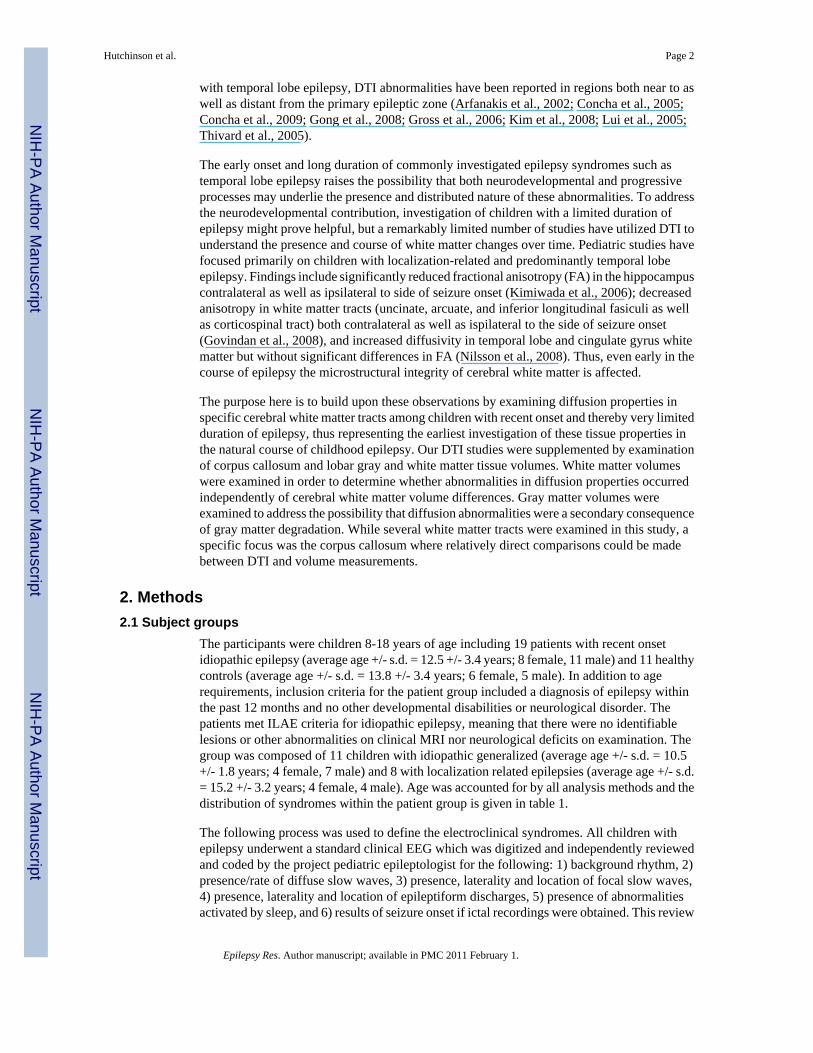
with temporal lobe epilepsy, DTI abnormalities have been reported in regions both near to aswell as distant from the primary epileptic zone (Arfanakis et al., 2002; Concha et al., 2005;Concha et al., 2009; Gong et al., 2008; Gross et al., 2006; Kim et al., 2008; Lui et al., 2005;Thivard et al., 2005).
The early onset and long duration of commonly investigated epilepsy syndromes such astemporal lobe epilepsy raises the possibility that both neurodevelopmental and progressiveprocesses may underlie the presence and distributed nature of these abnormalities. To addressthe neurodevelopmental contribution, investigation of children with a limited duration ofepilepsy might prove helpful, but a remarkably limited number of studies have utilized DTI tounderstand the presence and course of white matter changes over time. Pediatric studies havefocused primarily on children with localization-related and predominantly temporal lobeepilepsy. Findings include significantly reduced fractional anisotropy (FA) in the hippocampuscontralateral as well as ipsilateral to side of seizure onset (Kimiwada et al., 2006); decreasedanisotropy in white matter tracts (uncinate, arcuate, and inferior longitudinal fasiculi as wellas corticospinal tract) both contralateral as well as ispilateral to the side of seizure onset(Govindan et al., 2008), and increased diffusivity in temporal lobe and cingulate gyrus whitematter but without significant differences in FA (Nilsson et al., 2008). Thus, even early in thecourse of epilepsy the microstructural integrity of cerebral white matter is affected.
The purpose here is to build upon these observations by examining diffusion properties inspecific cerebral white matter tracts among children with recent onset and thereby very limitedduration of epilepsy, thus representing the earliest investigation of these tissue properties inthe natural course of childhood epilepsy. Our DTI studies were supplemented by examinationof corpus callosum and lobar gray and white matter tissue volumes. White matter volumeswere examined in order to determine whether abnormalities in diffusion properties occurredindependently of cerebral white matter volume differences. Gray matter volumes wereexamined to address the possibility that diffusion abnormalities were a secondary consequenceof gray matter degradation. While several white matter tracts were examined in this study, aspecific focus was the corpus callosum where relatively direct comparisons could be madebetween DTI and volume measurements.
2. Methods2.1 Subject groups
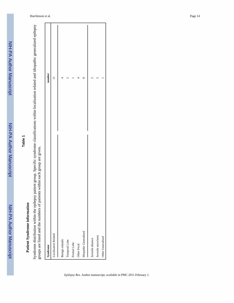
The participants were children 8-18 years of age including 19 patients with recent onsetidiopathic epilepsy (average age +/- s.d. = 12.5 +/- 3.4 years; 8 female, 11 male) and 11 healthycontrols (average age +/- s.d. = 13.8 +/- 3.4 years; 6 female, 5 male). In addition to agerequirements, inclusion criteria for the patient group included a diagnosis of epilepsy withinthe past 12 months and no other developmental disabilities or neurological disorder. Thepatients met ILAE criteria for idiopathic epilepsy, meaning that there were no identifiablelesions or other abnormalities on clinical MRI nor neurological deficits on examination. Thegroup was composed of 11 children with idiopathic generalized (average age +/- s.d. = 10.5+/- 1.8 years; 4 female, 7 male) and 8 with localization related epilepsies (average age +/- s.d.= 15.2 +/- 3.2 years; 4 female, 4 male). Age was accounted for by all analysis methods and thedistribution of syndromes within the patient group is given in table 1.
The following process was used to define the electroclinical syndromes. All children withepilepsy underwent a standard clinical EEG which was digitized and independently reviewedand coded by the project pediatric epileptologist for the following: 1) background rhythm, 2)presence/rate of diffuse slow waves, 3) presence, laterality and location of focal slow waves,4) presence, laterality and location of epileptiform discharges, 5) presence of abnormalitiesactivated by sleep, and 6) results of seizure onset if ictal recordings were obtained. This review
Hutchinson et al. Page 2
Epilepsy Res. Author manuscript; available in PMC 2011 February 1.
NIH
-PA Author Manuscript
NIH
-PA Author Manuscript
NIH
-PA Author Manuscript
was conducted while blinded to all MRI, cognitive, and psychiatric data. This information wasused with clinical characterization of seizures and history to define electroclinical syndromesin consensus conference.
Controls were first-degree cousins who were age and gender matched with no history ofseizures, early initial precipitating injuries (e.g., febrile convulsions), no other developmentalor neurological disease, or loss of consciousness greater than five minutes. Participants in bothgroups attended regular school. Further details regarding subject selection criteria are availablein (Hermann et al., 2006). There were no significant differences between the epilepsy andcontrol groups in age (12.6 vs. 13.6, p=.43), gender (53% male vs. 45% male, p=.70), or FullScale IQ (96.6 vs. 97.1, p=.89).
2.2 Imaging parametersImages were obtained on a 1.5 Tesla GE Signa MR scanner. Sequences acquired for eachparticipant included: (i) T1-weighted, three-dimensional SPGR acquired with the followingparameters: TE = 5ms, TR = 24ms, flip angle = 40deg, NEX = 1, slice thickness = 1.5 mm,slices = 124, plane = coronal, FOV = 20 × 20 cm2, matrix = 256X256; (ii) proton density (PD)and (iii) T2-weighted images acquired with the following parameters: TE = 36ms (for PD) or96 msec (for T2), TR = 3000 ms, NEX = 1, slice thickness = 3.0 mm, slices = 64, slice plane= coronal, FOV = 20 × 20 cm2, matrix = 256X256; (iv) EPI DTI data series of one referenceimage (b=0 s/mm2) and 13 diffusion weighted images (b=1000 s/mm2, non-colinear gradientorientations) with the following parameters: TE = 76.6 msec, TR = 4000 msec, NEX = 4, slicethickness = 4 mm and gap thickness = 4 mm, slices=15, slice plane = axial, FOV = 24 × 24cm2, matrix = 256 × 256 (reconstructed).
2.3 Volumetric postacquisition processingMRIs were processed using a semi-automated software package, i.e., Brain Research: Analysisof Images, Networks, and Systems (BRAINS2) (Andreasen et al., 1996; Harris et al., 1999).Neuroimaging analyses were conducted blinded to group status and both clinical anddemographic characteristics of the subjects. The T1-weighted images were spatiallynormalized so that the anterior-posterior axis of the brain was realigned parallel to the anteriorcommissure - posterior commissure (ACPC) line, and the interhemispheric fissure was alignedon the other two axes. A six-point linear transformation was used to warp the standard Talairachatlas space onto the resampled image. Images from the three pulse sequences were thencoregistered using a local adaptation of automated image registration software. Followingalignment of the image sets, all images were resampled into 1 mm cubic voxels, followingwhich an automated algorithm classified each voxel as gray matter, white matter, CSF, blood,or “other.” Manual inspection and correction of the output of the neural network tracing wasconducted. The brain images were then volume rendered using local utilities, producing tissuevolumes for regions of interest within the brain. The six outer boundaries of the brain form abounding box that along with the AC-PC line produces a volumetric grid based on the Talairachdivisions. Each lobar volume measured is defined in this proportional coordinate system. Thus,all measurements are obtained in the image space of the subject without morphing the imageto fit a standard brain atlas. Dependent measures were white and gray matter volumes of theleft and right frontal, parietal, temporal and occipital lobes. All volumes were adjusted for ageand total intracranial volume (ICV).
2.4 Corpus callosum guidelinesManual trace of the corpus callosum began in the midsagittal plane. Using a previouslyestablished tracing protocol (Hermann et al., 2003b), three co-registered image sets were usedsimultaneously to delineate the corpus callosum. Boundary identification was based on a pre-manipulated trimodal image (a composite of the T1, T2, and PD images) as the primary image
Hutchinson et al. Page 3
Epilepsy Res. Author manuscript; available in PMC 2011 February 1.
NIH
-PA Author Manuscript
NIH
-PA Author Manuscript
NIH
-PA Author Manuscript
set, with reference to the continuously segmented image, and a discretely segmented image inareas of poorly defined borders (such as the confluence of the fornix and the ventral callosum).Additionally, voxel signal intensities were used to separate white matter voxels fromneighboring voxels of cerebral spinal fluid, blood vessel wall, and gray matter. In addition tothe midsagittal slice, two additional slices extending laterally from the midsagittal view weretraced. The anterior and posterior extreme of the corpus callosum trace were kept consistentwhen extending the trace to parasagittal slices. Although the lateral extent of the corpuscallosum extends beyond the central five sagittal slices, these regions were excluded from thetrace due to difficulty in boundary determination. In addition, a BRAINS2 script was appliedto automatically partition the CC into seven regions defined by Witelson (Witelson, 1989).This technique used the inner convexity of the genu as a landmark in conjunction with therostral-caudal axis length to subdivide the callosum. Witelson regions were then summed tocreate anterior (1-3), midbody (4-5), and posterior (6-7) regions (Hermann et al., 2003a). Aninterrater agreement (kappa) of .99 was achieved. Dependent measures were the volumes ofthe total corpus callosum as well as the anterior, midbody and posterior regions of the corpuscallosum. In order to account for head size volumetric differences, all callosal volumes wereadjusted for age and total intracranial volume (ICV).
2.5 DTI postacquisition processingOffline processing of the DTI datasets was completed using the diffusion II toolbox for SPM5in Matlab. The reference and diffusion weighted images were first corrected for motion andfield inhomogeneity artifacts by corregistration of the image volumes. Next, the diffusiontensor was calculated at each voxel and maps for the mean diffusion (MD), fractionalanisotropy (FA), axial diffusivity (Dax) and radial diffusivity (Drad) were calculated for eachsubject.
In order to quantify diffusion indices of six specific white matter structures, the anterior andposterior corpus callosum, fornix, cingulum, internal capsule and external capsule, binarymasks of regions of interest (ROIs) were created using FSL software (Smith et al., 2004) andaccording to anatomical landmarks on axial slices of the FA map. All ROI masks were bilateral.For the anterior and posterior corpus callosum a single axial slice was selected for which thestructure was the most apparent and ROIs were constrained to the medial 13.5 mm of eachstructure for consistency with volumetric data. For the other white matter structures, all slicescontaining a given structure were included in creating the ROI mask. Initially, masks werecoarsely drawn to include structure edges and surrounding gray matter, but to exclude all otherwhite matter. Next, an FA threshold was applied to refine the rough masks to include onlywhite matter voxels. This FA threshold was set at 0.5 for the corpus callosum regions and 0.45for the other regions. For the anterior and posterior corpus callosum masks, the anterior andposterior edges were further eroded by two voxels in order to reduce partial volume effectsnear the structure edges. ROI masks were applied to DTI index maps to calculate mean valuesfor each structure for each subject. The FA map for a representative subject is shown in figure1 with the ROI masks overlaid. Analysis of covariance with age as a covariate was used to testfor group differences after accounting for age effects.
3. Results3.1 Lobar gray and white matter volumes
There were no significant differences between the epilepsy and control groups in age and ICVadjusted gray and white matter volumes. Specifically, there were no significant univariateeffects for lobar frontal (p=.94), parietal (p=.46), temporal (p=.56), or occipital (p=.98) whitematter volumes. There were also no significant univariate effects for lobar frontal (.73), parietal(p=.38), temporal (p=.46) or occipital (p=.44) gray matter volumes (see figure 2).
Hutchinson et al. Page 4
Epilepsy Res. Author manuscript; available in PMC 2011 February 1.
NIH
-PA Author Manuscript
NIH
-PA Author Manuscript
NIH
-PA Author Manuscript
3.2 Corpus callosum volumesThere were no significant differences between the epilepsy and control groups in age and ICVcorrected volumes of the total corpus callosum (p=.69) as well as the anterior (p=.60), midbody(p=.98), and posterior (p=.15) regions (see figure 3).
3.3 Diffusion indicesDTI indices of FA, MD, Dax and Drad were analyzed for the anterior and posterior corpuscallosum, fornix, cingulum, internal capsule and external capsule and compared betweenepilepsy patients and controls. Analysis of covariance with age as a covariate showed a groupdifference of reduced FA for the posterior corpus callosum (p=0.04) and cingulum (p=0.04)in the epilepsy group. Additionally, there was an increase in MD for the posterior corpuscallosum (p=0.03). There were no significant group differences for Dax in any region. However,Drad was significantly increased in the cingulum (p=0.03) and posterior corpus callosum (p=.03) (see figure 4).
When the epilepsy group was divided into localization related (LRE) and idiopathic generalized(IGE) groups, non-significant trends were observed to follow the findings of the pooled groupfor both the posterior corpus callosum and cingulum. Additionally, there was significantlyincreased MD and Drad (p= 0.039 and 0.043 respectively) in the fornix of the LRE group, butnot the IGE group compared to controls (see supplemental table 1).
4. DiscussionThe primary finding of this study is the presence of DTI abnormalities in the white matter ofchildren with very recent onset idiopathic epilepsies compared to control children. Thesediffusion abnormalities occur in the context of comparable volumes of lobar gray and whitematter in the frontal, parietal, temporal and occipital lobes. Volumetric analysis of the corpuscallosum also shows no difference for the total corpus callosum as well as for the anterior,posterior or middle subregions in particular. Taken together, these findings imply an earlyvulnerability of the structural integrity of cerebral white matter in new-onset epilepsy to whichDTI, but not volumetric analysis, is sensitive. Our results extend previous DTI investigationsof children with epilepsy (Govindan et al., 2008; Kimiwada et al., 2006; Nilsson et al., 2008)by demonstrating that not only are these abnormalities evident in children with a relativelyshort duration of disorder, but that they can be seen near to the time of diagnosis of epilepsy,and occur in other epilepsy syndromes.
The corpus callosum has been previously implicated in adults with chronic temporal lobeepilepsy and to incur both DTI (Arfanakis et al., 2002; Kim et al., 2008; Thivard et al., 2005)and volumetric (Hermann et al., 2003b; Weber et al., 2007) abnormalities. In the present studythe posterior corpus callosum exhibits reduced FA, increased Drad and increased MD inepilepsy patients compared to controls. Interestingly, the finding of abnormal DTI took placein the absence of volumetric change in the same callosal region. This finding is in direct supportof the idea that DTI is able to detect abnormalities not evident using conventional MRItechniques (Duncan, 2002). Other MRI studies have shown DTI abnormalities in the presenceof volumetric differences (Gong et al., 2008), however to our knowledge this is the first studyof epilepsy patients to exhibit a structure specific abnormality in diffusion but not volume.
Structural studies of the normal development of white matter in children suggest that theanterior corpus callosum matures before the mid or posterior regions (Giedd et al., 1999; Hasanet al., 2009). Our finding of DTI abnormality in the posterior but not anterior corpus callosummay indicate a preferential damage to later myelinating callosal regions. Furthermore, radialbut not axial diffusivity was altered in the posterior corpus callosum. Although there is no
Hutchinson et al. Page 5
Epilepsy Res. Author manuscript; available in PMC 2011 February 1.
NIH
-PA Author Manuscript
NIH
-PA Author Manuscript
NIH
-PA Author Manuscript

definitive microstructural change certain to underlie this pattern, there are a growing numberof DTI studies relating altered Drad to myelination abnormalities (Budde et al., 2007; Song etal., 2005) and showing a reduction of Drad during normal white matter development in humansthought to correspond to compacting fibers and myelination (Alexander et al., 2007; Hasan etal., 2009; Snook et al., 2005). It is possible that the myelination of axons in the patients isslowed by the epileptogenic process or that there is seizure-related damage to the posteriorcorpus callosum myelin sheath surrounding the onset of epilepsy. In support of the view ofmyelin as a primary target, there is no observed group difference in gray matter volume in anylobar region to suggest axonal degeneration secondary to neuronal loss, although the limitationsof volumetric measurements are recognized as specific layers of the neuropil may be affectedbut not detected by these volumetric measurements.
The other region that displays abnormal DTI values is the cingulum, in which FA is reducedand Drad is increased. This finding is in agreement with several studies of chronic temporallobe epilepsy populations (Concha et al., 2005; Concha et al., 2009; McDonald et al., 2008)and suggests a vulnerability of the cingulum that may accompany the onset of epilepsy. Theinternal capsule, external capsule and fornix were not significantly different from controlvalues. It is possible that there is no change in these structures; however, they should not beruled out of future studies without first addressing the limitations of this study.
Future studies will be needed to more fully characterize the structural state of the newlyepileptic brain. This study provides an important initial characterization of DTI indices in thisgroup, however there are several limitations that must be addressed. These include the smallsample size and mixed epilepsy syndromes of the patient group and the imaging limitations ofresolution and acquisition gap thickness, which was large and precluded the use of tractographyor analysis of small white matter structures as well as measurement of complete white matterstructures.
The finding of white matter vulnerability in this study is in conceptual agreement with previousMRI studies of patients with epilepsy. Diffuse abnormalities in cerebral white matter in focalepilepsy have been reported using both traditional volumetrics and voxel based morphometry(Hermann et al., 2003b; McMillan et al., 2004; Mueller et al., 2006; Seidenberg et al., 2005;Yu et al., 2008). Vulnerability of frontal-temporal connections including the uncinate andarcuate fasciculi have been described in temporal lobe epilepsy (Lin et al., 2008; Rodrigo etal., 2007) with verification of this abnormal connectivity and demonstration of their importantlinkages to well known cognitive abnormalities (Diehl et al., 2008; Flugel et al., 2006; Lui etal., 2005; McDonald et al., 2008; Powell et al., 2007; Yogarajah et al., 2008). Understandingthe cause, course, and consequences of microstructural abnormalities of cerebral white mattershould help to understand the etiology of neurobehavioral comorbidities of epilepsy.
Supplementary MaterialRefer to Web version on PubMed Central for supplementary material.
AcknowledgmentsThe authors would like to thank the UW Institute for Clinical and Translational Research (ICTR) and support fromthe following sources: NIH RO144351, T90DK070079 and R90DK071515.
ReferencesAlexander AL, Lee JE, Lazar M, Boudos R, DuBray MB, Oakes TR, Miller JN, Lu J, Jeong EK, McMahon
WM, Bigler ED, Lainhart JE. Diffusion tensor imaging of the corpus callosum in Autism. Neuroimage2007;34:61–73. [PubMed: 17023185]
Hutchinson et al. Page 6
Epilepsy Res. Author manuscript; available in PMC 2011 February 1.
NIH
-PA Author Manuscript
NIH
-PA Author Manuscript
NIH
-PA Author Manuscript
Andreasen NC, Rajarethinam R, Cizadlo T, Arndt S, Swayze VW 2nd, Flashman LA, O’Leary DS,Ehrhardt JC, Yuh WT. Automatic atlas-based volume estimation of human brain regions from MRimages. J Comput Assist Tomogr 1996;20:98–106. [PubMed: 8576490]
Arfanakis K, Hermann BP, Rogers BP, Carew JD, Seidenberg M, Meyerand ME. Diffusion tensor MRIin temporal lobe epilepsy. Magn Reson Imaging 2002;20:511–519. [PubMed: 12413596]
Budde MD, Kim JH, Liang HF, Schmidt RE, Russell JH, Cross AH, Song SK. Toward accurate diagnosisof white matter pathology using diffusion tensor imaging. Magn Reson Med 2007;57:688–695.[PubMed: 17390365]
Concha L, Beaulieu C, Gross DW. Bilateral limbic diffusion abnormalities in unilateral temporal lobeepilepsy. Ann Neurol 2005;57:188–196. [PubMed: 15562425]
Concha L, Beaulieu C, Collins DL, Gross DW. White-matter diffusion abnormalities in temporal-lobeepilepsy with and without mesial temporal sclerosis. J Neurol Neurosurg Psychiatry 2009;80:312–319. [PubMed: 18977826]
Deppe M, Kellinghaus C, Duning T, Moddel G, Mohammadi S, Deppe K, Schiffbauer H, Kugel H, KellerSS, Ringelstein EB, Knecht S. Nerve fiber impairment of anterior thalamocortical circuitry in juvenilemyoclonic epilepsy. Neurology 2008;71:1981–1985. [PubMed: 19064879]
Diehl B, Busch RM, Duncan JS, Piao Z, Tkach J, Luders HO. Abnormalities in diffusion tensor imagingof the uncinate fasciculus relate to reduced memory in temporal lobe epilepsy. Epilepsia2008;49:1409–1418. [PubMed: 18397294]
Duncan JS. Neuroimaging methods to evaluate the etiology and consequences of epilepsy. Epilepsy Res2002;50:131–140. [PubMed: 12151124]
Duncan JS. Imaging the Brain’s Highways-Diffusion Tensor Imaging in Epilepsy. Epilepsy Curr2008;8:85–89. [PubMed: 18596882]
Flugel D, Cercignani M, Symms MR, O’Toole A, Thompson PJ, Koepp MJ, Foong J. Diffusion tensorimaging findings and their correlation with neuropsychological deficits in patients with temporallobe epilepsy and interictal psychosis. Epilepsia 2006;47:941–944. [PubMed: 16686662]
Giedd JN, Blumenthal J, Jeffries NO, Rajapakse JC, Vaituzis AC, Liu H, Berry YC, Tobin M, Nelson J,Castellanos FX. Development of the human corpus callosum during childhood and adolescence: alongitudinal MRI study. Prog Neuropsychopharmacol Biol Psychiatry 1999;23:571–588. [PubMed:10390717]
Gong G, Concha L, Beaulieu C, Gross DW. Thalamic diffusion and volumetry in temporal lobe epilepsywith and without mesial temporal sclerosis. Epilepsy Res 2008;80:184–193. [PubMed: 18490143]
Govindan RM, Makki MI, Sundaram SK, Juhasz C, Chugani HT. Diffusion tensor analysis of temporaland extra-temporal lobe tracts in temporal lobe epilepsy. Epilepsy Res 2008;80:30–41. [PubMed:18436432]
Gross DW, Concha L, Beaulieu C. Extratemporal white matter abnormalities in mesial temporal lobeepilepsy demonstrated with diffusion tensor imaging. Epilepsia 2006;47:1360–1363. [PubMed:16922882]
Harris G, Andreasen NC, Cizadlo T, Bailey JM, Bockholt HJ, Magnotta VA, Arndt S. Improving tissueclassification in MRI: a three-dimensional multispectral discriminant analysis method withautomated training class selection. J Comput Assist Tomogr 1999;23:144–154. [PubMed: 10050826]
Hasan KM, Kamali A, Iftikhar A, Kramer LA, Papanicolaou AC, Fletcher JM, Ewing-Cobbs L. Diffusiontensor tractography quantification of the human corpus callosum fiber pathways across the lifespan.Brain Res 2009;1249:91–100. [PubMed: 18996095]
Hermann B, Hansen R, Seidenberg M, Magnotta V, O’Leary D. Neurodevelopmental vulnerability ofthe corpus callosum to childhood onset localization-related epilepsy. Neuroimage 2003a;18:284–292. [PubMed: 12595183]
Hermann B, Jones J, Sheth R, Dow C, Koehn M, Seidenberg M. Children with new-onset epilepsy:neuropsychological status and brain structure. Brain 2006;129:2609–2619. [PubMed: 16928696]
Hermann BP, Seidenberg M, Bell B, Rutecki P, Sheth R, Sutula T, Wendt G, O’Leary D, Magnotta V.Extratemporal quantitative MRI volumetrics and neuropsychological function in temporal lobeepilepsy. J Int Neuropsychol Soc 2003b;9:353–362. [PubMed: 12666760]
Hutchinson et al. Page 7
Epilepsy Res. Author manuscript; available in PMC 2011 February 1.
NIH
-PA Author Manuscript
NIH
-PA Author Manuscript
NIH
-PA Author Manuscript
Kim H, Piao Z, Liu P, Bingaman W, Diehl B. Secondary white matter degeneration of the corpus callosumin patients with intractable temporal lobe epilepsy: a diffusion tensor imaging study. Epilepsy Res2008;81:136–142. [PubMed: 18572387]
Kimiwada T, Juhasz C, Makki M, Muzik O, Chugani DC, Asano E, Chugani HT. Hippocampal andthalamic diffusion abnormalities in children with temporal lobe epilepsy. Epilepsia 2006;47:167–175. [PubMed: 16417545]
Lin JJ, Riley JD, Juranek J, Cramer SC. Vulnerability of the frontal-temporal connections in temporallobe epilepsy. Epilepsy Res 2008;82:162–170. [PubMed: 18829258]
Lui YW, Nusbaum AO, Barr WB, Johnson G, Babb JS, Orbach D, Kim A, Laliotis G, Devinsky O.Correlation of apparent diffusion coefficient with neuropsychological testing in temporal lobeepilepsy. AJNR Am J Neuroradiol 2005;26:1832–1839. [PubMed: 16091538]
McDonald CR, Ahmadi ME, Hagler DJ, Tecoma ES, Iragui VJ, Gharapetian L, Dale AM, Halgren E.Diffusion tensor imaging correlates of memory and language impairments in temporal lobe epilepsy.Neurology 2008;71:1869–1876. [PubMed: 18946001]
McMillan AB, Hermann BP, Johnson SC, Hansen RR, Seidenberg M, Meyerand ME. Voxel-basedmorphometry of unilateral temporal lobe epilepsy reveals abnormalities in cerebral white matter.Neuroimage 2004;23:167–174. [PubMed: 15325363]
Mueller SG, Laxer KD, Cashdollar N, Buckley S, Paul C, Weiner MW. Voxel-based optimizedmorphometry (VBM) of gray and white matter in temporal lobe epilepsy (TLE) with and withoutmesial temporal sclerosis. Epilepsia 2006;47:900–907. [PubMed: 16686655]
Nilsson D, Go C, Rutka JT, Rydenhag B, Mabbott DJ, Snead OC 3rd, Raybaud CR, Widjaja E. Bilateraldiffusion tensor abnormalities of temporal lobe and cingulate gyrus white matter in children withtemporal lobe epilepsy. Epilepsy Res 2008;81:128–135. [PubMed: 18595664]
Powell HW, Parker GJ, Alexander DC, Symms MR, Boulby PA, Wheeler-Kingshott CA, Barker GJ,Koepp MJ, Duncan JS. Abnormalities of language networks in temporal lobe epilepsy. Neuroimage2007;36:209–221. [PubMed: 17400477]
Rodrigo S, Oppenheim C, Chassoux F, Golestani N, Cointepas Y, Poupon C, Semah F, Mangin JF, LeBihan D, Meder JF. Uncinate fasciculus fiber tracking in mesial temporal lobe epilepsy. Initialfindings. Eur Radiol 2007;17:1663–1668. [PubMed: 17219141]
Seidenberg M, Kelly KG, Parrish J, Geary E, Dow C, Rutecki P, Hermann B. Ipsilateral and contralateralMRI volumetric abnormalities in chronic unilateral temporal lobe epilepsy and their clinicalcorrelates. Epilepsia 2005;46:420–430. [PubMed: 15730540]
Smith SM, Jenkinson M, Woolrich MW, Beckmann CF, Behrens TE, Johansen-Berg H, Bannister PR,De Luca M, Drobnjak I, Flitney DE, Niazy RK, Saunders J, Vickers J, Zhang Y, De Stefano N, BradyJM, Matthews PM. Advances in functional and structural MR image analysis and implementation asFSL. Neuroimage 2004;23:S208–219. [PubMed: 15501092]
Snook L, Paulson LA, Roy D, Phillips L, Beaulieu C. Diffusion tensor imaging of neurodevelopment inchildren and young adults. Neuroimage 2005;26:1164–1173. [PubMed: 15961051]
Song SK, Yoshino J, Le TQ, Lin SJ, Sun SW, Cross AH, Armstrong RC. Demyelination increases radialdiffusivity in corpus callosum of mouse brain. Neuroimage 2005;26:132–140. [PubMed: 15862213]
Thivard L, Lehericy S, Krainik A, Adam C, Dormont D, Chiras J, Baulac M, Dupont S. Diffusion tensorimaging in medial temporal lobe epilepsy with hippocampal sclerosis. Neuroimage 2005;28:682–690. [PubMed: 16084113]
Weber B, Luders E, Faber J, Richter S, Quesada CM, Urbach H, Thompson PM, Toga AW, Elger CE,Helmstaedter C. Distinct regional atrophy in the corpus callosum of patients with temporal lobeepilepsy. Brain 2007;130:3149–3154. [PubMed: 17728360]
Witelson SF. Hand and sex differences in the isthmus and genu of the human corpus callosum. Apostmortem morphological study. Brain 1989;112(Pt 3):799–835. [PubMed: 2731030]
Yogarajah M, Powell HW, Parker GJ, Alexander DC, Thompson PJ, Symms MR, Boulby P, Wheeler-Kingshott CA, Barker GJ, Koepp MJ, Duncan JS. Tractography of the parahippocampal gyrus andmaterial specific memory impairment in unilateral temporal lobe epilepsy. Neuroimage2008;40:1755–1764. [PubMed: 18314352]
Yogarajah M, Duncan JS. Diffusion-based magnetic resonance imaging and tractography in epilepsy.Epilepsia 2008;49:189–200. [PubMed: 17941849]
Hutchinson et al. Page 8
Epilepsy Res. Author manuscript; available in PMC 2011 February 1.
NIH
-PA Author Manuscript
NIH
-PA Author Manuscript
NIH
-PA Author Manuscript
Yu A, Li K, Li L, Shan B, Wang Y, Xue S. Whole-brain voxel-based morphometry of white matter inmedial temporal lobe epilepsy. Eur J Radiol 2008;65:86–90. [PubMed: 17553646]
Hutchinson et al. Page 9
Epilepsy Res. Author manuscript; available in PMC 2011 February 1.
NIH
-PA Author Manuscript
NIH
-PA Author Manuscript
NIH
-PA Author Manuscript
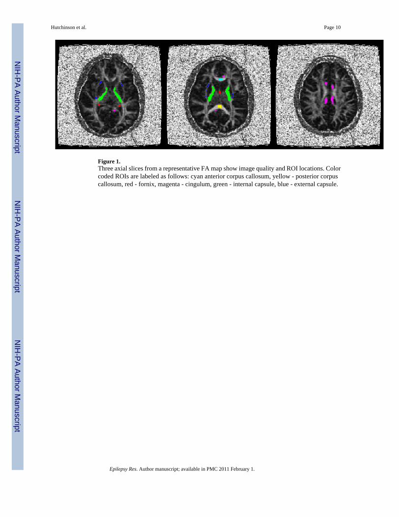
Figure 1.Three axial slices from a representative FA map show image quality and ROI locations. Colorcoded ROIs are labeled as follows: cyan anterior corpus callosum, yellow - posterior corpuscallosum, red - fornix, magenta - cingulum, green - internal capsule, blue - external capsule.
Hutchinson et al. Page 10
Epilepsy Res. Author manuscript; available in PMC 2011 February 1.
NIH
-PA Author Manuscript
NIH
-PA Author Manuscript
NIH
-PA Author Manuscript

Figure 2.Mean total white (left panel) and gray (right panel) matter volumes for frontal, parietal,temporal and occipital lobes in children with epilepsy compared to healthy controls. Therewere no group differences across any of these regions of interest.
Hutchinson et al. Page 11
Epilepsy Res. Author manuscript; available in PMC 2011 February 1.
NIH
-PA Author Manuscript
NIH
-PA Author Manuscript
NIH
-PA Author Manuscript

Figure 3.Mean volumes for anterior, midbody, posterior and total corpus callosum in children withepilepsy compared to healthy controls. There were no group differences across any of theseregions of interest.
Hutchinson et al. Page 12
Epilepsy Res. Author manuscript; available in PMC 2011 February 1.
NIH
-PA Author Manuscript
NIH
-PA Author Manuscript
NIH
-PA Author Manuscript

Figure 4.Mean DTI values (FA, MD, Dax and Drad) of six white matter structures (anterior and posteriorcorpus callosum, fornix, cingulum, internal capsule and external capsule) for epilepsy andcontrol groups. * indicates p<0.05 for group difference by analysis of covariance and errorbars indicate standard error.
Hutchinson et al. Page 13
Epilepsy Res. Author manuscript; available in PMC 2011 February 1.
NIH
-PA Author Manuscript
NIH
-PA Author Manuscript
NIH
-PA Author Manuscript
NIH
-PA Author Manuscript
NIH
-PA Author Manuscript
NIH
-PA Author Manuscript
Hutchinson et al. Page 14
Tabl
e 1
Patie
nt S
yndr
ome
info
rmat
ion
Synd
rom
e di
strib
utio
n w
ithin
the
epile
psy
patie
nt g
roup
. Spe
cific
synd
rom
e cl
assi
ficat
ions
with
in lo
caliz
atio
n re
late
d an
d id
iopa
thic
gen
eral
ized
epi
leps
ygr
oups
are
list
ed a
nd th
e nu
mbe
rs o
f pat
ient
s with
in e
ach
grou
p ar
e gi
ven.
Synd
rom
enu
mbe
r
Loca
lizat
ion
Rel
ated
11
Ben
ign
rola
ndic
4
Tem
pora
l Lob
e2
Fron
tal L
obe
1
Oth
er F
ocal
4
Idio
path
ic G
ener
aliz
ed8
Juve
nile
abs
ence
2
Juve
nile
myo
clon
ic5
Oth
er G
ener
aliz
ed1
Epilepsy Res. Author manuscript; available in PMC 2011 February 1.