ChemInform Abstract: A Benzil and an Isoflavone from Iris tenuifolia
12
HETEROCYCLES, Vol. 82, No. 1, 2010, pp. 813 - 824. © The Japan Institute of Heterocyclic Chemistry Received, 23rd January, 2010, Accepted, 12th April, 2010, Published online, 13th April, 2010 DOI: 10.3987/COM-10-S(E)6 NEW AND KNOWN CONSTITUENTS FROM IRIS UNGUICULARIS AND THEIR ANTIOXIDANT ACTIVITY Atta-ur-Rahman, a * Sumaira Hareem, a M. Iqbal Choudhary, a Bilge Sener, b Ahmed Abbaskhan, a Hina Siddiqui, a Shazia Anjum, c Ilkay Orhan, b Ilhan Gurbuz, b and Filiz Ayanoglu d a H.E.J. Research Institute of Chemistry, International Center for Chemical and Biological Sciences, University of Karachi, Karachi-75270, Pakistan b Department of Pharmacognosy, Faculty of Pharmacy, Gazi University, Ankara 06330, Turkey c University of Saskatchewan, Department of Chemistry, Saskatoon, S7N 5C9, Canada d Department of Field Crops, Faculty of Agriculture, Mustafa Kemal University, Hatay 31034, Turkey * Tel: +92-21-4824924-5; Fax: (+92)-21-4819018-9; E-mail: [email protected] Abstract _ A new compound, 1,3-O-diferuloylsucrose (1), and a synthetically known compound, 5,7-dihydroxy-6-methoxychromone (2), along with several known natural products, irilone (3), 4ƍ,5,7-trihydroxy-6-methoxyflavanone (4), tectorigenin (5), kaempferol (6), 4ƍ,5,7-trihydroxy-3ƍ,8-dimethoxyflavanone (7), 8-methoxyeriodictyol (8), hispidulin (9), and mangiferin (10) were isolated from the rhizomes of Iris unguicularis. Compounds 1, 6, 8 and 10 showed a considerable DPPH radicals scavenging activity. Their structures have been deduced through different spectroscopic techniques. The structure of compound 2 was also confirmed by single-crystal X-ray diffraction techniques as well. Dedicated to Professor Dr. Albert Eschenmoser, ETH Zürich, on his 85 th birthday. The family Iridaceae comprises some 92 genera and has more than 1,800 species, 1 mostly perennial herbs with rhizomes (bulbs). The genus Iris includes about 300 species, some of the species being ornamental. HETEROCYCLES, Vol. 82, No. 1, 2010 813
-
Upload
independent -
Category
Documents
-
view
0 -
download
0
Transcript of ChemInform Abstract: A Benzil and an Isoflavone from Iris tenuifolia

HETEROCYCLES, Vol. 82, No. 1, 2010, pp. 813 - 824. © The Japan Institute of Heterocyclic Chemistry Received, 23rd January, 2010, Accepted, 12th April, 2010, Published online, 13th April, 2010 DOI: 10.3987/COM-10-S(E)6
NEW AND KNOWN CONSTITUENTS FROM IRIS UNGUICULARIS AND THEIR ANTIOXIDANT ACTIVITY
Atta-ur-Rahman,a* Sumaira Hareem,a M. Iqbal Choudhary,a Bilge Sener,b Ahmed Abbaskhan,a Hina Siddiqui,a Shazia Anjum,c Ilkay Orhan,b Ilhan Gurbuz,b and Filiz Ayanoglud
aH.E.J. Research Institute of Chemistry, International Center for Chemical and
Biological Sciences, University of Karachi, Karachi-75270, Pakistan bDepartment of Pharmacognosy, Faculty of Pharmacy, Gazi University, Ankara
06330, Turkey cUniversity of Saskatchewan, Department of Chemistry, Saskatoon, S7N 5C9,
Canada dDepartment of Field Crops, Faculty of Agriculture, Mustafa Kemal University,
Hatay 31034, Turkey
* Tel: +92-21-4824924-5; Fax: (+92)-21-4819018-9;
E-mail: [email protected]
Abstract _ A new compound, 1,3-O-diferuloylsucrose (1), and a synthetically
known compound, 5,7-dihydroxy-6-methoxychromone (2), along with several
known natural products, irilone (3), 4 ,5,7-trihydroxy-6-methoxyflavanone (4),
tectorigenin (5), kaempferol (6), 4 ,5,7-trihydroxy-3 ,8-dimethoxyflavanone (7),
8-methoxyeriodictyol (8), hispidulin (9), and mangiferin (10) were isolated from
the rhizomes of Iris unguicularis. Compounds 1, 6, 8 and 10 showed a
considerable DPPH radicals scavenging activity. Their structures have been
deduced through different spectroscopic techniques. The structure of compound 2
was also confirmed by single-crystal X-ray diffraction techniques as well.
Dedicated to Professor Dr. Albert Eschenmoser, ETH Zürich, on his 85th birthday.
The family Iridaceae comprises some 92 genera and has more than 1,800 species,1 mostly perennial herbs
with rhizomes (bulbs). The genus Iris includes about 300 species, some of the species being ornamental.
HETEROCYCLES, Vol. 82, No. 1, 2010 813

In Pakistan, approximately 16 species are found1,2 average about 40 species are found in Turkey.3 Except
for the coldest regions, they are found all around the world and especially in South Africa and in tropical
America. The roots of Iris are used in flavoring dentifrices and perfumes. Phytochemical investigations
on various species of Iris have resulted in the isolation of more than 250 compounds, which include
flavonoids, flavanoids, isoflavonoids and their glycosides, benzoquinones and triterpenoids.4-8 The leaves
of Iris contain ascorbic acid. The rhizomes contain terpenes, organic acids (undecilene acid, miristic acid,
tridecilic acid), and glycosides such as iridin. Iris species have been reported to contain piscicidal,
antineoplastic, antiplasmodial, antioxidant and antituberculosis properties.9-12 The flavonoid constituents
of red wine and teas have been extensively studied for their antioxidant properties. It was noted that the
substitution pattern of flavonoids affects their antioxidant properties.13
Iris unguicularis Poir. is closely related to I. cretensis and I. lazica. It is commonly called the �“winter
Iris�” or the �“Algerian Iris�”. It is native to the Eastern Mediterranean regions, with scattered populations in
Algeria up through Turkey and the Greek Peloponnese. During the current study, we have isolated a new
compound, 1,3-O-diferuloylsucrose (1), and a synthetically known compound, 5,7-dihydroxy-6-
methoxychromone (2) along with eight other known compounds 3-10. The structures of all the
compounds were obtained from the spectroscopic analysis, especially 1D and 2D NMR spectral data and
comparing them with the reported data. DPPH Radical scavenging activity of new and known compounds
was also evaluated. Compound 2 was also subjected to single-crystal X-ray diffraction analysis.
A subfraction of the EtOAc extract was subjected to preparative recycling HPLC using water-methanol
(1 : 1) as solvent system to afford compound 1 (10.2 mg) as a yellowish gummy material. Strong IR
absorptions at 3401 (OH), 1696 (conjugated ester C=O) and 1598 (C=C) cm-1, and UV bands at 325, 235,
218 and 202 nm indicated conjugated aromatic system. The HRFAB-MS (-ve) of compound 1 exhibited
an [M-H]- at m/z 693.2019 (C32H37O17, calcd. 693.2030).
The 1H-NMR spectrum suggested that compound 1 contained two feruloyl moieties, represented by two
sets of signals for the trans olefinic protons [ H 7.62 (1H, d, J7 ,8 = 15.9 Hz), 6.46 (1H, d, J8 ,7 = 15.9
Hz); 7.72 (1H, d, J7 ,8 = 15.9 Hz), 6.38 (1H, d, J8 ,7 = 15.9 Hz)], two sets of 1,3,4-trisubstituted
aromatic ring protons [ H 7.14 (1H, brs), 6.73 (1H, d, J5 ,6 = 8.5 Hz), 7.05 (1H, dd, J6 ,5 = 8.1 Hz, J6 ,2 =
1.2 Hz); 7.17 (1H, brs), 6.79 (1H, d, J5 ,6 = 8.5 Hz), 7.08 (1H, dd, J6 ,5 = 8.1 Hz, J6 ,2 = 1.2 Hz)], and
protons of the two methoxy groups [ H 3.86 (3H, s); 3.82 (3H, s)]. A doublet at H 5.49 (J1 ,2 = 3.7 Hz)
indicated the presence of an anomeric proton of an -linked sugar (Table 1). The broad-band decoupled 13C-NMR spectrum of compound 1 showed resonances for thirty two carbon atoms, including two methyl,
three methylene, eighteen methine and nine quaternary carbons (Table 1). Signals at C 93.7, 72.9, 74.9,
814 HETEROCYCLES, Vol. 82, No. 1, 2010

71.9, 74.5, 62.4; 66.3, 103.3, 79.5, 73.1, 83.9 and 62.9 suggested the presence of a disaccharide moiety. A
characteristic doublet with a smaller coupling constant at H 5.49 (1H, d, J1 ,2 = 3.7 Hz) was attributed to
the anomeric proton of the -glucopyranose unit.14,15 This also supported the presence of a sucrose unit in
1. Two downfield signals at C 56.5 and 56.4 were due to the methoxy carbons of two feruloyl groups.
The linkage of the two feruloyl groups, located at C-1 and C-3 of the fructose unit of sucrose, was
deduced from the HMBC interactions of the C-3 methine proton ( H 5.58) with the C-9 carbonyl carbon
( C 168.4). The C-1 methylene protons ( H 4.26, 4.38) showed interactions with the C-9 carbonyl carbon
( C 168.5) (Figure 1). On the basis of this spectroscopic data, the structure of compound 1 was deduced as
1,3-O-diferuloylsucrose. Interestingly 5 -methoxy derivative of compound 1 was reported earlier.16
OO
HOOMe
O
O
OH
OMe
H
H
H
H
H
H
H
HH
H
1
5''4'' 3''
2''
1''
7''8''
9''
7'''
6'''5''' 4'''
3'''2'''1'''
8'''9'''
6''
O
OOH
2
3 4
5 6
H
H OH
H H
1'6'
5'4'
3'
2'
O
HOHO
OH
HHO
Figure 1. Key HMBC interactions in Compound 1.
HETEROCYCLES, Vol. 82, No. 1, 2010 815
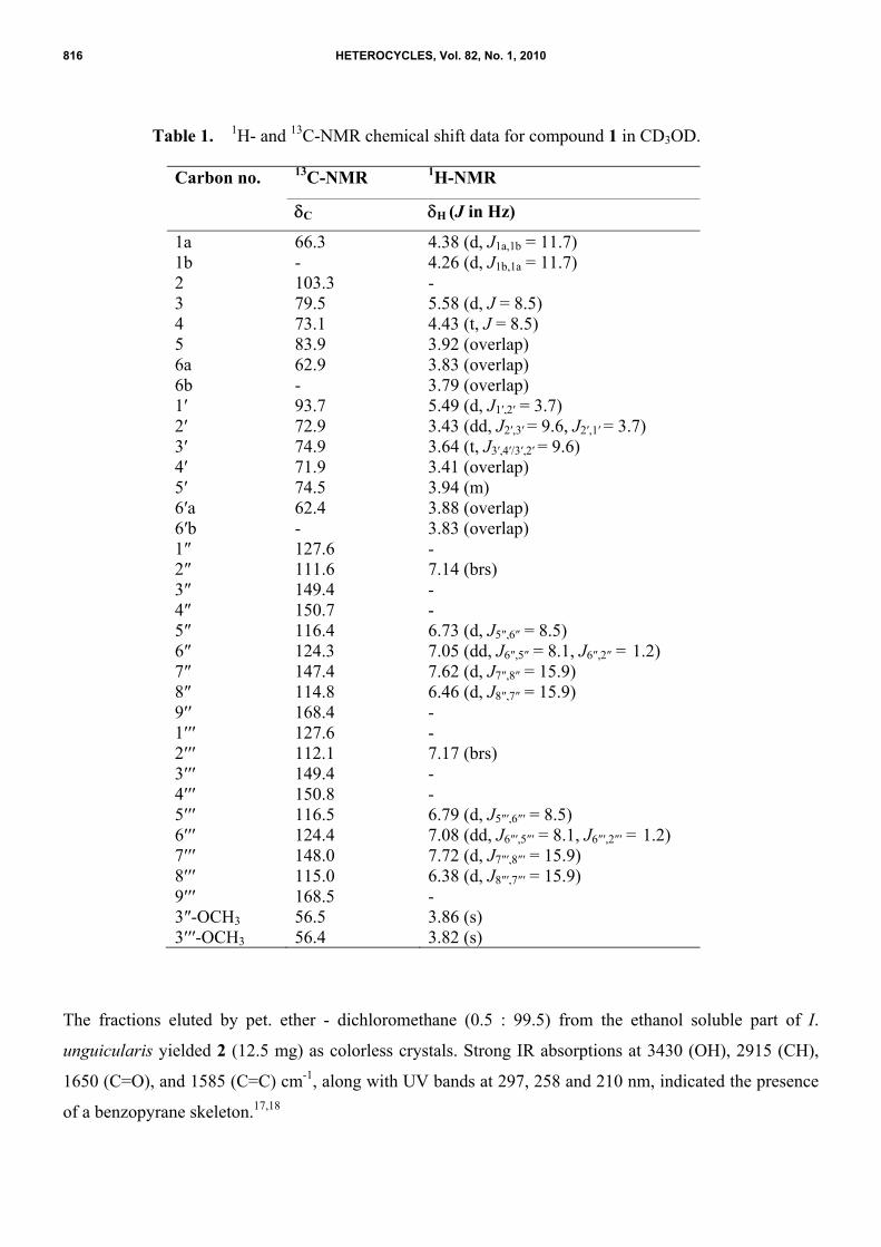
Table 1. 1H- and 13C-NMR chemical shift data for compound 1 in CD3OD.
Carbon no. 13C-NMR 1H-NMR
C
H (J in Hz)
1a 66.3 4.38 (d, J1a,1b = 11.7) 1b - 4.26 (d, J1b,1a = 11.7) 2 103.3 - 3 79.5 5.58 (d, J = 8.5) 4 73.1 4.43 (t, J = 8.5) 5 83.9 3.92 (overlap) 6a 62.9 3.83 (overlap) 6b - 3.79 (overlap) 1 93.7 5.49 (d, J1 ,2 = 3.7) 2 72.9 3.43 (dd, J2 ,3 = 9.6, J2 ,1 = 3.7) 3 74.9 3.64 (t, J3 ,4 /3 ,2 = 9.6) 4 71.9 3.41 (overlap) 5 74.5 3.94 (m) 6 a 62.4 3.88 (overlap) 6 b - 3.83 (overlap) 1 127.6 - 2 111.6 7.14 (brs) 3 149.4 - 4 150.7 - 5 116.4 6.73 (d, J5 ,6 = 8.5) 6 124.3 7.05 (dd, J6 ,5 = 8.1, J6 ,2 = 1.2) 7 147.4 7.62 (d, J7 ,8 = 15.9) 8 114.8 6.46 (d, J8 ,7 = 15.9) 9 168.4 - 1 127.6 - 2 112.1 7.17 (brs) 3 149.4 - 4 150.8 - 5 116.5 6.79 (d, J5 ,6 = 8.5) 6 124.4 7.08 (dd, J6 ,5 = 8.1, J6 ,2 = 1.2) 7 148.0 7.72 (d, J7 ,8 = 15.9) 8 115.0 6.38 (d, J8 ,7 = 15.9) 9 168.5 - 3 -OCH3 56.5 3.86 (s) 3 -OCH3 56.4 3.82 (s)
The fractions eluted by pet. ether - dichloromethane (0.5 : 99.5) from the ethanol soluble part of I.
unguicularis yielded 2 (12.5 mg) as colorless crystals. Strong IR absorptions at 3430 (OH), 2915 (CH),
1650 (C=O), and 1585 (C=C) cm-1, along with UV bands at 297, 258 and 210 nm, indicated the presence
of a benzopyrane skeleton.17,18
816 HETEROCYCLES, Vol. 82, No. 1, 2010

The molecular formula of compound 2 was inferred from the HREI-MS which exhibited an M+ as the
base peak at m/z 208.0380 (C10H8O5, calcd. 208.0371) with nine degrees of unsaturation. A peak at m/z
193 indicated the loss of a methyl group from C-6 position of M+,19 while another peak at m/z 165 was
due to the loss of a carbonyl group from the M+-CH3.20
The 1H-NMR spectrum of compound 2 showed signals for two olefinic and one aromatic protons (Table
2). A singlet at H 6.25 corresponded to the aromatic H-8. Two olefinic protons, resonating at H 7.63 and
5.99 (J2,3/3,2 = 5.9 Hz), were assigned to the ortho coupled H-2 and H-3. A 3H singlet at H 3.70 was due
to the O-methyl protons, substituted at the C-6 of the aromatic ring.18 The broad-band (BB) decoupled 13C-NMR spectrum of compound 2 showed resonances for ten carbons, including one methyl, three
methine and six quaternary carbons (Table 2). Two olefinic carbons, resonating at C 155.8 and 110.1,
were assigned to C-2 and C-3, respectively.21 The methoxy carbon signal resonated at C 60.3. The most
downfield signal in the 13C-NMR spectrum at C 182.2 was assigned to the C-4 ketonic carbonyl carbon
which indicated the presence of a hydroxyl group at the C-5 position.22 Other carbon signals appeared at
C 152.4 (C-5), 131.2 (C-6), 156.8 (C-7), 94.2 (C-8), 153.5 (C-9), and 106.1 (C-10).
Table 2. 1H- and 13C-NMR chemical shift data for compound 2 in CD3OD.
The NMR assignments were made on the basis of HMQC, HMBC and COSY-45° techniques. In the
COSY-45° spectrum, the olefinic protons [H-2 and H-3] showed couplings with each other. Direct
one-bond 1H/13C interactions were deduced by the HMQC spectrum. The olefinic and aromatic protons
Carbon no. 13C-NMR 1H-NMR
C
H (J in Hz)
2 155.8 7.63 (d, J2,3 = 5.9)
3 110.1 5.99 (d, J3,2 = 5.9)
4 182.2 -
5 152.4 -
6 131.2 -
7 156.8 -
8 94.2 6.25 (s)
9 153.5 -
10 106.1 -
OCH3 60.3 3.70 (s)
HETEROCYCLES, Vol. 82, No. 1, 2010 817

i.e., H-2 ( H 7.63), H-3 ( H 5.99) and H-8 ( H 6.25), showed connectivities with C-2 ( C 155.8), C-3 ( C
110.1), and C-8 ( C 94.2), respectively. The HMBC spectrum showed long-range interactions of H-2 ( H
7.63) with C-3 ( C 110.1), C-4 ( C 182.2) and C-9 ( C 153.5). The other HMBC interactions were of H-3
( H 5.99) with C-2 ( C 155.8), C-4 ( C 182.2) and C-10 ( C 106.1), and of H-8 ( H 6.25) with C-6 ( C
131.2), C-7 ( C 156.8), C-9 ( C 153.5), and C-10 ( C 106.1). The O-methyl protons ( H 3.70), were
coupled with the aromatic C-6 ( C 131.2) (Figure 2). Finally, the structure was unambiguously established
by X-ray diffraction analysis (Figure 3). The data indicated that compound 2 is a new natural product
with the structure corresponding to 5,7-dihydroxy-6-methoxychromone. This compound was previously
reported as a synthetic product.23
O
OOH
MeO
HO
H
H
H
12
34
5
6
79
8
10
Figure 2. Key HMBC interactions in compound 2.
Compound 1 (1,3-O-diferuloylsucrose) is a new compound while the compound 2 (5,7-dihydroxy-6-
methoxychromone) has not been reported so far from any natural source.23 In addition to this, several
known compounds have also been isolated, and identified as, irilone (3), 4 ,5,7-trihydroxy-6-
methoxyflavanone (4), tectorigenin (5), kaempferol (6), 4 ,5,7-trihydroxy-3 ,8-dimethoxyflavanone (7),
8-methoxyeriodictyol (8), hispidulin (9), and mangiferin (10).24-30 This is the first report of the isolation
of compounds 3-10 from I. unguicularis.
The compounds of I. unguicularis exhibited a dose-dependent anti-radical activity by reducing the stable
DPPH radicals to the yellow colored diphenylpicrylhydrazine derivative. Mangiferin (10), kaempferol (6),
8-methoxyeriodictyol (8) and 1,3-O-diferuloylsucrose (1) were found to be active in this assay.
Compound 10 showed potent anti-radical activity with IC50 value 22.45 ± 0.35 µM, which is lesser than
both the standards used during the studies and exhibited greater antiradical potential then standards (BHT,
IC50 44.2 ± 0.20 µM and nPG IC50 30.0 0.27 µM). Compound 6 also exhibited higher antiradical
Figure 3. Computer-generated ORTEP diagram of the final X-ray model of 2.
818 HETEROCYCLES, Vol. 82, No. 1, 2010

potential with IC50 value 40.26 ± 1.04 µM, which is still lesser than one of the standard used (BHT. IC50
44.2 ± 0.20 µM). Compounds 1 and 8 also exhibited a good anti-radical activity in this assay.
Sturcture-Activity Relationship:
O
O
1
5
6
7
82
3 1'2'
3'
4'
5'6'
A C
B
Isoflavone
O
O
A
B
C
1
456
7
8
1'
2'
3'
4'
5'
6'
2
3
Flavone
The flavonoids have basic skeleton of diphenylpropanes with different oxidation of the central pyran ring.
The analyzed compounds belonged to different sub classes of flavonoids, such as flavones, flavanones
and isoflavones.
The difference in antiradical activity of these compounds seems to be due to the structural differences in
hydroxylation and methoxylation on the basic skeleton. The presence of ortho-dihydroxyl groups was
potentially found to be responsible of the higher antiradical activity of these molecules. The tested
compounds showed a variety of radical scavenging activities, some of the compounds showed a very
strong activity, while others were completely inactive. On the basis of preliminary analysis, it can be
concluded that the radical scavenging activities of flavonoids is based on the number of phenolic groups
present in the molecules.
Presence of the 3-OH in the C-ring, and the 2,3-double bond, in conjugation with 4-oxo function
(carbonyl group) in the C-ring, as in compound 6, seems to be responsible of the higher activity of this
compound. Absence of this 3-OH in the C-ring, as in compounds 9 and 4, resulted in a loss of activity.
Presence of ortho-dihydroxyl groups (catechol structure) in the B-ring, as in compound 8, is potentially
responsible of its higher activity, replacement of the -OH with -OCH3 at C-3 position in ring B, results in
a loss of activity many folds, as in compound 7. Table 3 presents the antioxidant properties of these
compounds in DPPH radical scavenging assay.
HETEROCYCLES, Vol. 82, No. 1, 2010 819

Table 3. Antioxidant (Radical Scavenging) activity of compounds 1-10.
�“-�” Inactive BHT = Butylated Hydroxytoluene (Standard) nPG = n-Propyl Gallate (Standard) SEM = Standard Error of Mean of Three Experiments. RSA = Radical Scavenging Activity
EXPERIMENTAL
General Experimental Procedures. Ultraviolet (UV) spectra were recorded in methanol on Hitachi
UV-3200 spectrophotometer and UV absorption ( max) values are given in nm. Infrared (IR) spectra were
measured in CHCl3 or as KBr discs on Shimadzu FTIR-8900/Bruker Vector 22 spectrophotometer and
presented in cm-1. The 1H-NMR spectra were measured at 300 MHz and 600 MHz while the 13C-NMR
spectra were measured at 75 MHz and 150 MHz in deuterated solvents (CDCl3 or in CD3OD) on Bruker
AVANCE spectrometers. Chemical shifts are measured in ppm ( ) relative to SiMe4 as internal standard,
and the coupling constants (J) are given in Hz. CDCl3 ( 7.25) and CD3OD ( 3.33 and 4.80) were used
as solvents and as internal standards.
Mass spectra (EI-MS) was measured in an electron impact mode on Varian MAT 312 double focusing
spectrometer or on a JMS-600 H (Jeol, Japan) spectrometers and ions are given in m/z (%).
High-resolution electron impact mass measurements (HREI-MS) were performed on Jeol HX 110 and/
MAT 95 XP mass spectrometers Thermo Finnigan. Chromatography was carried out on silica gel (E.
Merck, type 60, 70-230 mesh), Sephadex LH-20 (Pharmacia, Uppsala, Sweden), precoated silica gel
GF-254 preparative TLC plates (20 x 20, 0.5 mm thick, E. Merck) and by recycling preparative HPLC
(Japan Analytical Industry Co., Ltd.). H-80 C18 (ODS) column was used for HPLC separation. Purity of
Compounds IC50 (µM ± SEM) 1,3-O-Diferuloylsucrose (1) 110.21 ± 2.57 5,7-Dihydroxy-6-methoxychromone (2) - Irilone (3) - 4 ,5,7-Trihydroxy-6-methoxyflavanone (4) >500 ± 0.42 Tectorigenin (5) >500 ± 1.72 Kaempferol (6) 40.26 ± 1.04 4 ,5,7-Trihydroxy-3 ,8-dimethoxyflavanone (7) <500 ± 1.23 8-Methoxyeriodictyol (8) 56.84 ± 0.02 Hispidulin (9) >500 Mangiferin (10) 22.45 ± 0.35 Standard BHT 44.2 ± 0.20 Standard nPG 30.0 0.27
820 HETEROCYCLES, Vol. 82, No. 1, 2010

the samples was checked on precoated TLC plates (silica gel) and by HPLC on Shimadzu LC-6A
instrument by using RP-18 column. Melting points were determined on a Gallenkemp apparatus and are
uncorrected. X-Ray data was collected on a Bruker Smart Apex I, CCD 4-K area detector diffractometer.
Plant Material. The rhizomes of Iris unguicularis Poir. were collected from Antakya, Turkey, in June
2004 and air dried (1 kg). A voucher specimen (GUE 8964) was deposited in the Herbarium of Faculty of
Pharmacy, Gazi University, Ankara, Turkey. The plant was identified by Prof. Dr. Filiz Ayanoglu.
Extraction and Isolation. The dried rhizomes (1 kg) were chopped, and soaked in EtOH (10 L) for two
weeks at room temperature, and the resulting extract was concentrated to a gum (250.3 gm). The
ethanolic extract (250.3 gm) was dissolved in distilled water (3 L) and defatted with pet. ether (5 x 3 L),
to obtain 10.4 gm of pet. ether extract. The defatted aqueous extract was further fractionated with CH2Cl2
(5 x 3 L), EtOAc (5 x 3 L) and BuOH (5 x 3 L). On evaporation of the organic solvents, 42.3, 45.5 and
12.4 gm of extracts were obtained, respectively. Five compounds 2, and 3-6 were isolated from the
CH2Cl2 extract, while five compounds 1, and 7-10 were obtained from EtOAc extract by using repeated
column chromatography (silica gel) and preparative recycling HPLC (ODS column).
The CH2Cl2 extract was chromatographed on a column (silica gel) by using pet. ether - dichloromethane -
methanol in increasing order of polarity to obtain four major fractions (Fr-1 to Fr-4). These fractions were
further subjected to repeated column chromatography on silica gel by using various mixtures of pet. ether
- dichloromethane - methanol to afford compounds 2 (12.5 mg), 3 (6.2 mg), 4 (10.1 mg), 5 (9.8 mg), and
6 (12.4 mg).
The EtOAc extract was subjected to column chromatography (silica gel) and eluted with increasing
polarities of pet. ether - dichloromethane - methanol mixtures to obtain five major fractions (Fr-1 to Fr-5).
These fractions were subjected to further column chromatography and eluted with mixtures of pet. ether �–
CH2Cl2 - MeOH to obtain 1 (10.2 mg), 7 (15.2 mg), 8 (12.4 mg), and 9 (20.1 mg). Compounds 1 and 10
were purified from fraction Fr-5. The fraction Fr-5 were subjected to recycling HPLC by using
water-MeOH (1 : 1) as solvent system on H-80 ODS column with 3 mL/min flow rate (Rt 28 and 26 min)
to obtain compounds 1 (10.2 mg) and 10 (54.2 mg).
The known compounds 3-10 were characterized through comparison of their physical and spectral data
with those reported in the literature.24-30 These known compounds were isolated for the first time from
this plant species.
1,3-O-Diferuloylsucrose (1): IR (KBr) max cm-1: 3401 (OH), 1696 (conjugated ester C=O), 1598 (C=C).
UV max (MeOH) (log ) nm: 325, 235, 218, 202, 197. UV min (MeOH) (log ) nm: 263, 228, 210.
FAB-MS (-ve) m/z: 693 (20) [M-H]-, 517 (18), 337 (30). HRFAB-MS (-ve) m/z: 693.2019 [M-H]- (calcd.
HETEROCYCLES, Vol. 82, No. 1, 2010 821

for C32H37O17: 693.2030), EI-MS (rel. int., %) m/z: 370 (10), 338 (21), 194 (79), 177 (100), 150 (54), 135
(36), 77 (40), 1H-NMR and 13C-NMR: Table 1. HMBC: See Figure 1.
5,7-Dihydroxy-6-methoxychromone (2): Mp 77-79 °C. IR (KBr) max cm-1: 3430 (OH), 2915 (CH),
1650 (C=O) and 1585 (C=C). UV max (MeOH) (log ) nm: 297, 258 and 210. UV min (MeOH) (log )
nm: 276, 243. EI-MS m/z: 208 (M+, 100), 193 (M+-CH3, 40), 190 (34), 178 (8), 165 (M+-C=O, 25).
HREI-MS m/z: 208.0380 (calcd. for C10H8O5: 208.0371), 1H-NMR and 13C-NMR: Table 2. HMBC: See
Figure 2.
Single-crystal X-ray diffraction data of 5,7-dihydroxy-6-methoxychromone (2): A block-shaped
colorless crystal of compound 2 with dimension 0.31 x 0.17 x 0.15 mm was selected for X-ray diffraction
studies. C10H8O5: Mr 208.16; monoclinic; a = 7.5164 (3), b = 16.3891 (7), c = 7.1054 (3) Å; = =
90o, = 91.0910 (10)o; V = 875.13 (6) Å3, space group = P21/c, Z = 4, Dcalc. = 1.580 g/cm3, F(000) =
432.0, Mo-K ( 0.71073 Å). Intensity data of compound 2 was collected on a Bruker Smart Apex I,
CCD 4-K area detector diffractometer, attached with a KRYO-FLEX low temperature device. Data
reductions were performed by using SAINT program.31 The structure was solved by direct methods32 and
refined by full-matrix least squares on F2 using the SHELXTL-PC package. The intensity data within the
range 2.49-28.30 were collected at 173 (2) K. A total of 11,780 reflections were recorded, of which
2,168 reflections were judged on the basis of I > 2 s (1). The final R and Rw values were 0.0383 and
0.1146, respectively. Figure 3 was plotted with the aid of ORTEP.33 Anisotropic thermal displacement
parameters were allowed for hydrogen and non-hydrogen atoms. The Uiso values were constrained to be
1.5 Ueq of the carrier atom for methyl H atoms, and 1.2 Ueq for the remaining H atoms. In the absence of
significant anomalous dispersion effects, Friedel pairs were merged before the final refinement.
Crystallographic data for compound 2 has been deposited at the Cambridge Crystallographic Data Center
(CCDC-655285) 12 Union Road, Cambridge, CB2 1EZ, UK via Internet at
http://www.ccdc.cam.ac.uk/data_request/cif.
DPPH Free Radical Scavenging Assay. For quantitative determination of electron donating ability of
any compound, stable radicals have the advantage and their concentrations can be readily and directly
measurable.34 Free radical scavenging potential of these compounds were determined by measuring the
changes in absorbance of DPPH (l, l-Diphenyl-2-picrylhydrazyl radical) spectrophotometrically, as
described by S. K. Lee.35
1,1-Diphenyl-2-picrylhydrazyl radical (DPPH) is a stable free radical in EtOH. In solution form, it has a
deep violet color which absorbs strongly at 515 nm. As the free radical is converted into a neutral
822 HETEROCYCLES, Vol. 82, No. 1, 2010

molecule after accepting an electron, the absorption vanishes into a decolorized hydrazine form. The
antioxidant activity obtained from DPPH method is comparable to other methods reported, so far.
During this study, test samples were solubilized in DMSO and allowed to react with
1,1-diphenyl-2-picrylhydrazyl (DPPH) radical in EtOH for 30 min at 37 ºC. The DPPH concentration was
kept 300 M. After incubation, decrease in absorption was measured at 515 nm by using a multiplate
reader (Spectra MAX-340, Molecular Devices, CA, USA). Percent radical scavenging activity of samples
was determined by using the following formula, in comparison to a DMSO treated control group.
% RSA = 100 �– {(Optical Density test compound / Optical density control) x 100}
ACKNOWLEDGMENTS
We are grateful to the Government of Pakistan for the financial support for Pak-Turkish Joint Research
Project entitled, �“Development of Phytomedicines with Hepatoprotective Activity from Pakistan and
Turkish Plants�”. One of us (Sumaira Hareem) acknowledges the role of the Higher Education
Commission, Islamabad, Pakistan, for providing the financial support to conduct this research work
through the �“Merit Scholarship Scheme for Ph.D. Studies in Science and Technology�”.
REFRENCES AND NOTES
1. S. I. Ali and B. Mathew, (ed. by S. I. Ali and M. Qaiser), �“Flora of Pakistan�”, Department of Botany,
University of Karachi, Pakistan, 2000, 202, p. 1, 4-35.
2. M. S. Krishnan, �“The Wealth of India�”, A Dictionary of Raw Material and Industrial Products.
Council of Scientific and Industrial Research, New Delhi, 2001, 2, 89.
3. T. Baytop and B. Mathew, �“The Bulbous Plants of Turkey�”, B. T., Batsford Ltd., London, 1984.
4. A. K. Kala, M. K. Bhan, and K. L. Dhar, Phytochemistry, 1978, 17, 1441.
5. Atta-ur-Rahman, M. I. Choudhary, M. Nur-e-Alam, P. O. Ndognii, and T. Badarchiin, Chem. Pharm.
Bull., 2000, 48, 738.
6. M. I. Choudhary, M. Nur-e-Alam, F. Akhtar, S. Ahmad, I. Baig, P. Ondognh, P. Gombosurengyin,
and Atta-ur-Rahman, Chem. Pharm. Bull., 2001, 49, 1295.
7. Atta-ur-Rahman, S. Nasim, I. Baig, J. I. Ara, B. Sener, I. Orhan, and M. I. Choudhary, Chem.
Pharm. Bull., 2002, 50, 1100.
8. Atta-ur-Rahman, S. Nasim, I. Baig, I. Orhan, B. Sener, F. Ayanoglu, and M. I. Choudhary, Helv.
Chim. Acta, 2003, 86, 3354.
9. F. Hanawa, S. Tahara, and J. Mizutani, Phytochemistry, 1991, 30, 157.
10. I. Hideyuki, Y. Miyake, and T. Yoshida, Chem. Pharm. Bull., 1995, 43, 1260.
HETEROCYCLES, Vol. 82, No. 1, 2010 823
11. Y. Miyake, I. Hideyuki and T. Yashida, Can. J. Chem., 1997, 75, 734.
12. O. Papendrof, G. M. König and A. D. Wright, Phytochemistry, 1998, 49, 2383.
13. L. Zechmeister, �“Progress in the Chemistry of Organic Natural Products�”, Springer Verlag, 1957, 17,
19.
14. W. W. Binkley, D. Horton, and N. S. Bhacca, Carbohydr. Res., 1969, 10, 245.
15. M. Linscheid, D. Wendisch, and D. Strack, Z. Naturforsch., 1980, 35c, 907.
16. M. Hamburger and K. Hostettmann, Phytochemistry, 1985, 24, 1793.
17. D. T. A. Youssef, M. A. Ramadan, and A. A. Khalifa, Phytochemistry, 1998, 49, 2579.
18. A. Simon, A. J. Chulia, M. Kaouadji, and C. Delage, Phytochemistry, 1994, 36, 1043.
19. J. B. Harborne, T. J. Mabry, and H. Mabry, �“The Flavonoids�”, Academic Press, New York, 1975, 93.
20. F. R. van Heerden, E. V. Brandt, and D. G. Roux, Tetrahedron Lett., 1979, 20, 4507.
21. M. J. Vasconcelos, M. S. Silva, and A. S. Cavaleiro, Phytochemistry, 1998, 49, 1421.
22. P. K. Agrawal and C. B. Mahesh, (ed. by P. K. Agrawal), �“13C-NMR of Flavonoids�”, Elsevier
Science Publishers, Amsterdam, 1987, p. 193.
23. L. Farkas, A. Gottsegen, M. Nogradi, and J. Strelisky, Tetrahedron, 1971, 27, 5049.
24. D. E. Pegnyemb, B. B. Messanga, R. T. Ghogomu, and B. L. Sondengam, Fitoterapia, 1998, 69, 551.
25. T. A. Stepanova, V. I. Sheichenko, L. P. Smirnova, and V. I. Glyzin, Chem. Nat. Compd., 1981, 17,
520.
26. B. B. Gupta, A. Bhattacharyya, S. R. Mitra, and C. N. Adityachaudhury, Phytochemistry, 1983, 22,
1306.
27. K. R. Markham, B. Ternai, R. Stanley, H. Geiger, and T. J. Mabry, Tetrahedron, 1978, 34, 1389.
28. P. Proksch, U. Politt, E. Wollenweber, V. Wray, and C. Clark, Planta Med., 1988, 20, 542.
29. T. Iwashina, K. Kamenosono, and T. Ueno, Phytochemistry, 1999, 51, 1109.
30. T. Tanaka, T. Sueyasu, G. Nonaka, and I. Nishioka, Chem. Pharm. Bull., 1984, 32, 2676.
31. Siemens, SMART and SAINT. Siemens Analytical X-Ray Instruments Inc., Madison, Wisconsin,
USA, 1996.
32. G. M. Sheldrick, SHELXTL-PC (Version 5.1), Siemens Analytical Instruments, Inc., Madison, WI,
1997.
33. C. K. Johnson, �‘ORTEPII�’ Report ORNL-5138. Oak Ridge National Laboratory, Tennessee, USA,
1976.
34. M. Nishizawa, M. Kohno, M. Nishimura, A. Kitagawa, and Y. Niwano, Chem. Pharm. Bull., 2005,
53, 714.
35. S. K. Lee, Z. H. Mbwambo, H. Chung, L. Luyengi, E. J. Gamez, R. G. Mehta, A. D. Kinghorn, and J.
M. Pezzuto, Comb. Chem. High Throughput Screen., 1998, 1, 35.
824 HETEROCYCLES, Vol. 82, No. 1, 2010