Chemical Science Review and Letters Interaction of Nano HA-Biomolecules makes Thermally Stable...
10
Chemical Science Review and Letters ISSN 2278-6783 Che Sci Rev Lett 2012, 1(2), 53–62 Article CS26204207 53 Research Article Interaction of Nano HA-Biomolecules makes Thermally Stable Biomolecules for Bone Regeneration Arun Karthick S, Gobi N*, Nisha S Department of Textile Technology, Anna University, Chennai-600 025, Tamil Nadu, India. Abstract A simple method of synthesizing nano hydroxyapatite (HA) has been developed by biomimetic wet chemical precipitation method. Powder X-ray diffraction analyses (XRD), Transmission electron Microscopy (TEM) were employed to characterize the formation of HA and its size where the biomolecules Bovine Serum Albumin (BSA), Methionine (MET), and Glycine (GLY) are used as HA growth medium. The results, FTIR showed the interaction between biomolecules-HA is through the C=O (carboxylic) group present in biomolecules and the flurometric studies confirms the stability of the biomolecule at 1000ºC. Such stability of biomolecule has a potential application in biocompatibility, biological response of host by cell attachment & activation, and also provides drug delivery in resorbable bone implants. *Correspondence Gobi N Email: [email protected] Tel No: +91-9884845999 Key words: Hydroxyapatite, biomimetic, thermal stability, biocompatibility, biological response. Introduction Bone is a unique composite containing 10% of water, 20 (wt) % of collagen, 69 (wt) % of mineral phase and other organic minerals in small quantities. The main function of collagen is elasticity and structural supports were HA in the form of needles and plates arranged parallel to collagen provides stiffness to bone tissue. Of this 69% of mineral phase, 95% is of HA [Ca 10 (PO 4 ) 6 (OH) 2 ] hence synthetic HA with as excellent properties like biocompatibility, bioactivity, nontoxicity, noninflamatory, and most important osteoconductivity (i.e. slowly reabsorbed by the host bone after implantation) has been made for extensive use in repair and reconstruction of hard tissue [1-4]. The application of HA depends on the crystal shape, size, crystallinity, thermal stability and solubility criteria’s, which are all strongly influenced by the biomineralization process (as present in natural bone & teeth) [5]. Understanding the natural mechanism of biomineralization for the development of new materials is called “biomimetics” [6]. Biomolecules present in human blood participates in biomineralization process to form new bones. Human Serum albumin (HSA) is the most abundant protein in blood plasma. Albumins of many different mammalian shows similarities in there pysico-chemical properties with HAS among those Bovine Serum Albumin (BSA) shows 76% sequential identity with HSA [7]. BSA is a serum albumin protein comes from Ox cattle’s, hence the name “bovine”. The full length BSA precursor protein has 607 amino acids which are further cleaved to yield mature BSA with 585 amino acids. The glycine and methionine content in BSA are lower than in the average protein [8]. It is well known that HA shows high affinity for biomolecule [9-11] which helps to remove pathogens from blood or act as carrier for drug delivery. Surface interaction of biomolecule with materials plays two important roles such as determination of the final success of the implanted material by biological response and establishes cell attachment and activation [12-13]. In some cases these interactions produce adverse effects. In the present scenario therefore a detailed understanding of the formation mechanism in natural mineralization process and biomolecule- material interaction is essential to tailor the surface of the materials for its final success. In order to address such issue and to improve our knowledge our present study is focused on HA-BSA (i.e. material-biomolecule) interaction, its stability at higher temperature and also to make a comparative study between HA-BSA with HA-MET, HA-GLY as BSA contains both Glycine and Methionine as its amino acid block. Later the samples has been structurally characterized and investigated as well.
-
Upload
independent -
Category
Documents
-
view
1 -
download
0
Transcript of Chemical Science Review and Letters Interaction of Nano HA-Biomolecules makes Thermally Stable...

Chemical Science Review and Letters ISSN 2278-6783
Che Sci Rev Lett 2012, 1(2), 53–62 Article CS26204207 53
Research Article
Interaction of Nano HA-Biomolecules makes Thermally Stable Biomolecules for Bone Regeneration
Arun Karthick S, Gobi N*, Nisha S
Department of Textile Technology, Anna University, Chennai-600 025, Tamil Nadu, India.
Abstract A simple method of synthesizing nano hydroxyapatite (HA) has been developed by biomimetic wet chemical precipitation method. Powder X-ray diffraction analyses (XRD), Transmission electron Microscopy (TEM) were employed to characterize the formation of HA and its size where the biomolecules Bovine Serum Albumin (BSA), Methionine (MET), and Glycine (GLY) are used as HA growth medium. The results, FTIR showed the interaction between biomolecules-HA is through the C=O
(carboxylic) group present in biomolecules and the flurometric studies confirms the stability of the biomolecule at 1000ºC. Such stability of biomolecule has a potential application in biocompatibility, biological response of host by cell attachment & activation, and also provides drug delivery in resorbable bone implants. *Correspondence Gobi N Email: [email protected] Tel No: +91-9884845999
Key words: Hydroxyapatite, biomimetic, thermal stability, biocompatibility, biological response.
Introduction Bone is a unique composite containing 10% of water, 20 (wt) % of collagen, 69 (wt) % of mineral phase and other organic minerals in small quantities. The main function of collagen is elasticity and structural supports were HA in the form of needles and plates arranged parallel to collagen provides stiffness to bone tissue. Of this 69% of mineral phase, 95% is of HA [Ca10 (PO4)6 (OH)2] hence synthetic HA with as excellent properties like biocompatibility, bioactivity, nontoxicity, noninflamatory, and most important osteoconductivity (i.e. slowly reabsorbed by the host bone after implantation) has been made for extensive use in repair and reconstruction of hard tissue [1-4].
The application of HA depends on the crystal shape, size, crystallinity, thermal stability and solubility criteria’s, which are all strongly influenced by the biomineralization process (as present in natural bone & teeth) [5]. Understanding the natural mechanism of biomineralization for the development of new materials is called “biomimetics” [6]. Biomolecules present in human blood participates in biomineralization process to form new bones. Human Serum albumin (HSA) is the most abundant protein in blood plasma. Albumins of many different mammalian shows similarities in there pysico-chemical properties with HAS among those Bovine Serum Albumin (BSA)
shows 76% sequential identity with HSA [7]. BSA is a serum albumin protein comes from Ox cattle’s, hence the name “bovine”. The full length BSA precursor protein has 607 amino acids which are further cleaved to yield mature BSA with 585 amino acids. The glycine and methionine content in BSA are lower than in the average protein [8].
It is well known that HA shows high affinity for biomolecule [9-11] which helps to remove pathogens from blood or act as carrier for drug delivery. Surface interaction of biomolecule with materials plays two important roles such as determination of the final success of the implanted material by biological response and establishes cell attachment and activation [12-13]. In some cases these interactions produce adverse effects. In the present scenario therefore a detailed understanding of the formation mechanism in natural mineralization process and biomolecule-material interaction is essential to tailor the surface of the materials for its final success. In order to address such issue and to improve our knowledge our present study is focused on HA-BSA (i.e. material-biomolecule) interaction, its stability at higher temperature and also to make a comparative study between HA-BSA with HA-MET, HA-GLY as BSA contains both Glycine and Methionine as its amino acid block. Later the samples has been structurally characterized and investigated as well.

Chemical Science Review and Letters ISSN 2278-6783
Che Sci Rev Lett 2012, 1(2), 53–62 Article CS26204207 54
Experimental
Materials and methods
All chemicals used in this study namely calcium nitrate tetrahydrate (Ca(NO3)2.4H2O), diammonium hydrogen phosphate ((NH4)2HPO4), BSA, Methionine (MET), Glycine (GLY) and ammonia was analytical grade reagents.
To carry out a comparative study, HA samples were synthesized separately with BSA, MET & GLY at 0.05% which involves two steps as shown in Figure 1. Three freshly prepared calcium nitrate tetrahydrate
solution of strength 0.4M was made alkaline by using ammonia: water in 1:2 ratio and the pH was adjusted to 9-11. 50ml of 0.05% BSA, MET and GLY was made in three different reactors. The above Ca(NO3)2 4H2O solution was mixed separately with BSA,MET & GLY and gently stirred for 15min to ensure uniform distribution. Then it was incubated at room temperature for 24h. After 24h, freshly prepared 0.156M diammonium hydrogen phosphate solution was made alkaline using 1:1 ratio of ammonia: water and mixed with the incubated solution under stirring condition along the sidewalls of the beaker.
Figure 1 Flow chart for the synthesis of HA with BSA/MET/GLY
Chemical Science Review and Letters ISSN 2278-6783
Che Sci Rev Lett 2012, 1(2), 53–62 Article CS26204207 55
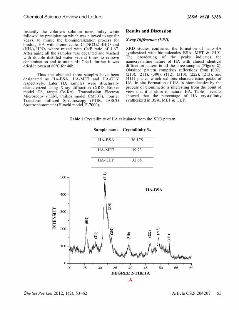
Instantly the colorless solution turns milky white followed by precipitation which was allowed to age for 7days, to mimic the biomineralization process for binding HA with biomolecule. Ca(NO3)2 4H2O and (NH4)2.HPO4 where mixed with Ca/P ratio of 1.67. After aging all the samples was decanted and washed with double distilled water several times to remove contamination and to attain pH 7.4±1, further it was dried in oven at 80ºC for 48h.
Thus the obtained three samples have been designated as HA-BSA, HA-MET and HA-GLY respectively. Later HA samples were structurally characterized using X-ray diffraction (XRD, Bruker model D8, target Co-Kα), Transmission Electron Microscopy (TEM, Philips model CM30T), Fourier Transform Infrared Spectroscopy (FTIR, JASCO Spectrophotometer (Hitachi model, F-7000).
Results and Discussion X-ray Diffraction (XRD) XRD studies confirmed the formation of nano-HA synthesized with biomolecules BSA, MET & GLY. The broadening of the peaks indicates the nanocrystalline nature of HA with almost identical diffraction pattern in all the three samples (Figure 2). Obtained pattern comprises reflections from (002), (210), (211), (300), (112), (310), (222), (213), and (411) planes which exhibits characteristics peaks of HA. In situ Formation of HA in biomolecules by the process of biomimetic is interesting from the point of view that it is close to natural HA. Table 1 results showed that the percentage of HA crystallinity synthesized in BSA, MET & GLY.
Table 1 Crystallinity of HA calculated from the XRD pattern
Sample name Crystallinity %
HA-BSA 36.175
HA-MET 39.73
HA-GLY 32.68

Chemical Science Review and Letters ISSN 2278-6783
Che Sci Rev Lett 2012, 1(2), 53–62 Article CS26204207 56
Figure 2 XRD pattern of (A) HA-BSA, (B) HA-MET and (C) HA-GLY

Chemical Science Review and Letters ISSN 2278-6783
Che Sci Rev Lett 2012, 1(2), 53–62 Article CS26204207 57
Chemical Science Review and Letters ISSN 2278-6783
Che Sci Rev Lett 2012, 1(2), 53–62 Article CS26204207 58
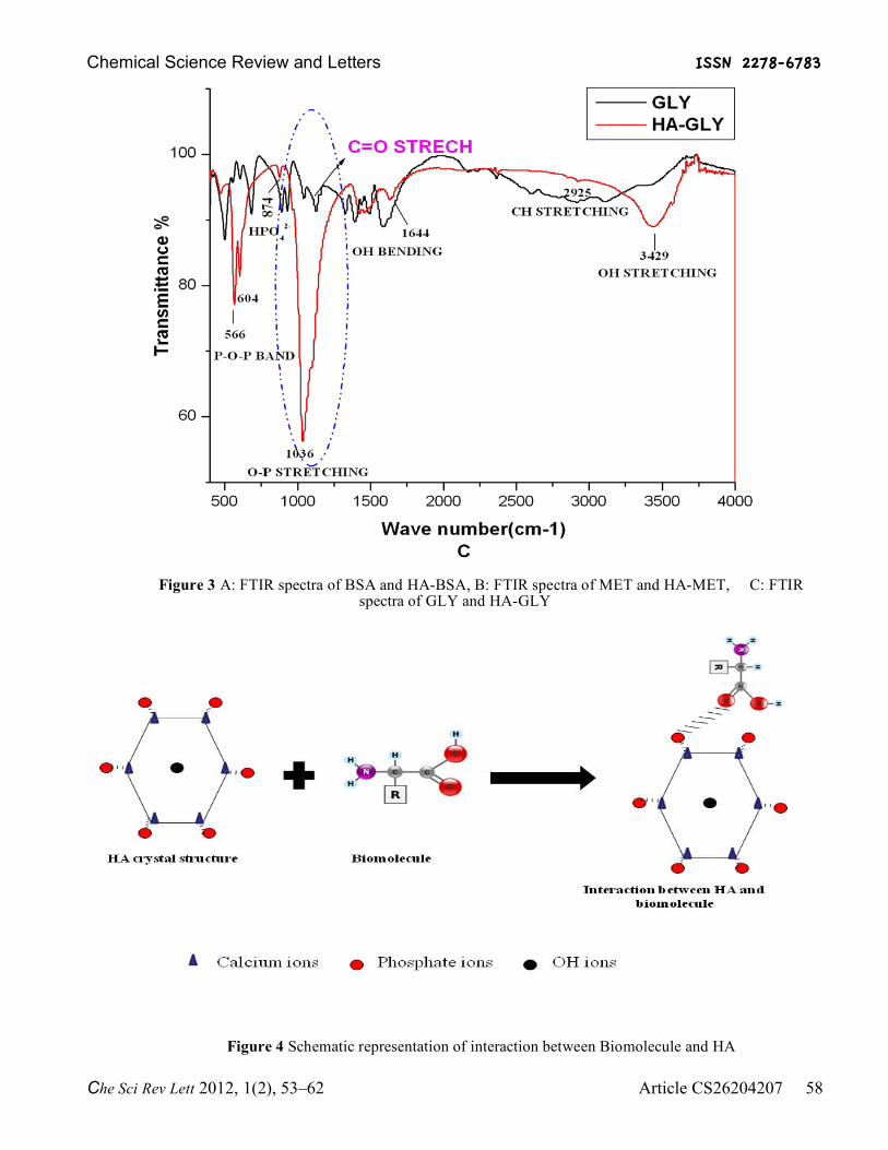
Figure 3 A: FTIR spectra of BSA and HA-BSA, B: FTIR spectra of MET and HA-MET, C: FTIR spectra of GLY and HA-GLY
Figure 4 Schematic representation of interaction between Biomolecule and HA

Chemical Science Review and Letters ISSN 2278-6783
Che Sci Rev Lett 2012, 1(2), 53–62 Article CS26204207 59
Along with XRD, FTIR also confirmed the formation of nano HA in all three samples. All three HA samples revealed almost similar FTIR spectrum, except small change in the absorbance bands associated with HA (Figure 3). The absorbance band obtained at 3429 cm-1 corresponds to OH stretching band. Band at 2925 and 2855 is due to –CH2- stretching. The peak at 1644 cm-1 was attributed due to the OH bending. The broad and high intensity band at 1036 cm-1 derives from O-P stretching band, while at 874 cm-1 indicates HPO4
2-
stretching. Similarly peaks at 566 & 604 were assigned to P-O-P band formation.
It is clearly observed in the encircled area of Figure 3 A, B & C, that the band corresponds to C=O stretching in all three FTIR spectra’s of BSA, Methionine, and Glycine shows a drastic variation after interaction with HA. Obtained results manifest that HA binds through C=O group of BSA, MET & GLY. By the formation of interaction between HA with BSA,
MET & GLY it prevents the C=O stretching by the overlap of O-P band stretch. Figure 4 shows the schematic representation of interaction between Biomolecule and HA
Transmission Electron Microscopy (TEM) Transmission electron microscopy was performed with the aim of analyzing the size and morphology of synthesized HA. The process of biomineralization (i.e. the biomolecules in host), generally controls the mineralization kinetics of hard tissue at different skeletal structures. TEM images are represented in the Figure 5. All three TEM images show HA synthesized mostly have rod-like morphology and relatively homogeneous in terms of thickness and highly regular in shape with an average particle size of 5-15nm breath and length of 30-40nm in dimension. Among the three synthesized HA-MET shows smaller particles followed HA-GLY and HA-BSA.
A) B)
A)
B)

Chemical Science Review and Letters ISSN 2278-6783
Che Sci Rev Lett 2012, 1(2), 53–62 Article CS26204207 60
Figure 5 A: TEM image of HA-BSA, B: TEM image of HA-MET, and C: TEM image of HA-GLY.
Fluorescent Spectroscopy (FS):
C)

Chemical Science Review and Letters ISSN 2278-6783
Che Sci Rev Lett 2012, 1(2), 53–62 Article CS26204207 61
Figure 6 A: fluorescent spectra of BSA & HA-BSA, B: fluorescent spectra of MET & HA-MET, and C:
fluorescent spectra of GLY & HA-GLY
Fluorimetric studies confirms the presence of biomolecules remain even after sintering the HA nanocomposite in a tubular furnace at 1000° C for 2hrs. The fluorescent spectral images are represented in the Figure 6. The emission wavelengths obtained for pure
BSA, MET, & GLY without heat treatment are at 330nm, 308.61nm, & 310.16nm respectively. When treated at 80ºC gives emission wavelength at 326.15nm, 309.04nm, & 309.30nm respectively, further treatment at 1000˚C for 2hrs made all three samples to get

Chemical Science Review and Letters ISSN 2278-6783
Che Sci Rev Lett 2012, 1(2), 53–62 Article CS26204207 62
evaporate. Where as HA precipitate synthesized with BSA, MET & GLY dried at 80ºC for 48h gives emission wavelength at 322.4nm for HA-BSA, 309.64nm for HA-MET and 309.73nm for HA-GLY. Similarly all three HA samples treated at 1000ºC for 2hrs gives emission wavelength at 309.31nm, 319.50nm and 319.29nm respectively which is well with in the range of emission. Therefore the stabiltity of biomolecule at 1000˚C is confirmed which is attained due to the interaction formed between nano-HA and biomolecule.
It is true that as of now, the present development is a proof of concept to understand HA-biomolecule interaction and its stability at higher temperature rather than a total solution. However, it needs further investigation to make it clear and we are analyzing its impact and aim to report the same soon. Conclusion In summary, this investigation has resulted in brief description of biomimetic synthesis process of HA with better understanding of HA-biomolecules interaction and its stability at higher temperature. The most attractive part of this study, FTIR which shows that HA binds through the C=O (carboxylic) group present in BSA, MET & GLY and the fluorimetric studies manifests the stability of biomolecule treated at 1000ºC for 2h which is attained by HA interaction. By the binding of biomolecule-HA and its stability at higher temperature plays a potential application in biocompatibility, biological response of host by cell attachment & activation, and also provides drug delivery in resorbable bone implants. References [1] Donglu Shi. Introduction to Biomaterials.
Tsinghua university press, ISBN 7-302-10807-2. [2] Xu, Carey HKH, Takagi LE, Chow LC (2007)
premixed calcium phosphate cements: synthesis, physical properties, and cell cytotoxicity. Dent. Mater. 23: 433-441.
[3] Kalita, Bhardwaj SJ, Bhatt A (2007) Nanocrystalline calcium phosphate ceramics in biomedical engineering. Sci. Eng C. 27: 441-449.
[4] Legeros R (2002) Properties of osteoconductive biomaterials: calcium phosphate. Clin. Orthop. Relat. Res. 395: 81-98.
[5] Pena J, Regi MV (2003) Hydroxyapatite, tricalcium phosphate and biphasic materials prepared by a liquid mix method. J. Eur. Ceram. Soc. 23: 1687-1696.
[6] Arvind Sinha, Avijit Guha (2008) Biomimetic pattering of polymer hydrogels with hydroxyapatite nanoparticles. Materials Science and Engineering C. 29: 1330-1333.
[7] Elena Mavropoulos, Andrea M. Costa, Lilian T. Costa, Carlos A. Achete, Alexander Mello, Jose M. Granjeiro, Alexandre M. Rossi (2011)
Adsorption and bioactivity studies of albumin onto hydroxyapatite surface. Colloids and Surface B: Biointerfaces. 83: 1-9.
[8] Theodore Peters (1950) Studies on Serum Albumin Production in vitro. Harvard University. 151-155.
[9] Tanaka T, Hirose M, Kotobuki N, Ohgushi H, Furuzono T, Sato J (2007) Conformational modification of serum albumins adsorbed on different kinds of biomimetic hydroxyapatite nanocrystals. Mater. Sci. Eng. C. 27: 817-823.
[10] Paul W, Sharma CP (1999) Development of porous spherical hydroxyapatite granules: application towards protein delivery. J. Mater. Sci. Mater. Med. 10: 383-388.
[11] Liu TY, Liu SY, Liu DM, Liou SC (2005) on the study of BSA loaded calcium deficient hydroxyapatite nano-carriers for controlled drug delivery. J. Control. Release. 107: 112-121.
[12] Gary JJ (2004) The interaction of protein with solid surface. Curr. Opin. Struct. Biol. 14: 110-115.
[13] Bajpai A (2005) Preparation and characterization of spongy cryogel of poly(vinyl alcohol)- casein system: water sorption and blood compatibility study. Polym. Int. 54: 304-315.
[14] Tubio G, Nerli B, Pico V (2004) Relationship between protein surface hydrophobicity and its partitioning behavior in aqueous two-phase system of poly ethylene glycol-dextran. J. Chromatogr. B 799: 293-301.
[15] Suprabha Nayar, Sinha MK, Basu D, Arvind Sinha (2006) Synthesis and sintering of biomimetic hydroxyapatite nanoparticles for biomedical application. J Mater Sci: Mater Med. 17: 1063–1068.
[16] Arvind Sinha, Swapan Kumar Das, Rao V, Ramachandrarao P (2001) Biomimetic route to produce nanosized inorganic crystals. Scripta mater. 44: 1933–1936.
[17] Sadjadi MS, Meskinfam M, Jazdarreh H (2010) Hydroxyapatite- starch nanobiocomposite synthesis and characterization. Int. J. Nano. Dim. 1(1): 57-63.
[18] Naruporn Monmaturapoj (2008) Nano-size hydroxyapatite powders preparation by wet chemical precipitation route. Journal of Metals, Materials and Minerals. 18(1): 15-20.
© 2012, by the Authors. The articles published from this journal are distributed to the public under “Creative Commons Attribution License” (http://creativecommons.org/licenses/by/3.0/). Therefore, upon proper citation of the original work, all the articles can be used without any restriction or can be distributed in any medium in any form.
Received : 26th July, 2012 Revised : 14th August, 2012 Accepted : 15th August, 2012 Online : 19th August, 2012