Chloroplast DNA variation of white oaks in the alpine region
Upload
khangminh22Category
view
0download
0
Characterization of new proteins involved in chloroplast biogenesis from Arabidopsis thaliana
Dissertation
der Fakultät für Biologie
der Ludwig-Maximilians-Universität München
vorgelegt von
Roberto Andres Espinoza Corral
München
März 2019

Diese Dissertation wurde angefertigt unter der Leitung von Prof. Dr. Jürgen Soll an der Fakultät für Biologie der Ludwig-Maximilians-Universität München. Erstgutachter: Prof. Dr. Jürgen Soll Zweitgutachter: Prof. Dr. Wolfgang Frank Tag der Abgabe: 29.01.2019 Tag der mündlichen Prüfung: 21.03.2019
Printed with the support of the German Academic Exchange Service.
Table of Contents
SUMMARY ............................................................................................................... 1 ZUSAMMENFASSUNG ........................................................................................... 2 ABBREVIATIONS .................................................................................................... 3 1 INTRODUCTION ................................................................................................... 6
1.1 ORIGIN OF PLASTIDS .......................................................................................... 6 1.2 PROTEIN TARGETING AND IMPORT INTO THE PLASTID ............................................ 6 1.3 CHLOROPLAST BIOGENESIS AND MATURATION ..................................................... 8 1.4 THYLAKOID BIOGENESIS ................................................................................... 10 1.5 THE FUNCTION OF PLASTOGLOBULI .................................................................. 13 1.6 AIM OF THIS WORK ........................................................................................... 16
2 MATERIAL AND METHODS .............................................................................. 17 2.1 MATERIAL ....................................................................................................... 17
2.1.1 Chemicals ............................................................................................. 17 2.1.2 Molecular weight and size markers .................................................... 17 2.1.3 Oligonucleotides .................................................................................. 17 2.1.4 Vectors .................................................................................................. 19 2.1.5 Enzymes ................................................................................................ 20 2.1.6 Bacterial strains ................................................................................... 20 2.1.7 Membranes ........................................................................................... 20 2.1.8 Antisera ................................................................................................. 20 2.1.9 Accession numbers ............................................................................. 21 2.1.10 Computational analyses .................................................................... 21
2.2 MOLECULAR BIOLOGICAL METHODS .................................................................. 22 2.2.1 Cloning strategies ................................................................................ 22 2.2.2 Polymerase chain reaction (PCR) ....................................................... 23 2.2.3 Isolation of plasmid DNA from E. coli ................................................ 23 2.2.4 Sequencing ........................................................................................... 23 2.2.5 Isolation of genomic DNA from A. thaliana for genotyping PCR ..... 23 2.2.6 Isolation of RNA from A. thaliana ....................................................... 24 2.2.7 cDNA synthesis .................................................................................... 24 2.2.8 Protein precipitation with trichloroacetic acid (TCA) ....................... 24
2.3 BIOCHEMICAL METHODS ................................................................................... 24 2.3.1 Overexpression of recombinant proteins .......................................... 24 2.3.2 Purification of soluble proteins .......................................................... 25 2.3.3 Purification of proteins out of inclusion bodies ................................ 25 2.3.4 Isolation of proteins from A. thaliana ................................................. 26 2.3.5 Determination of protein concentration ............................................. 26 2.3.6 SDS polyacrylamide gel electrophoresis (SDS-PAGE) ..................... 26 2.3.7 Semi-dry electro blot and immunodetection of proteins .................. 27 2.3.8 Detection of radiolabeled proteins ..................................................... 27 2.3.9 Blue Native PAGE (BN-PAGE) ............................................................. 27 2.3.10 Silver staining of SDS gels ................................................................ 28
2.3.11 Mass spectrometry ............................................................................. 28 2.3.12 Transformation of A. tumefacium ..................................................... 28 2.3.13 Pigment analysis ................................................................................ 28 2.3.14 In vitro transcription .......................................................................... 29 2.3.15 In vitro translation .............................................................................. 29 2.3.16 Agarose gel electrophoresis ............................................................. 30 2.3.17 Electrophoretic mobility shift assay (EMSA) ................................... 30 2.3.18 RNAse assay ....................................................................................... 30 2.3.19 Northern Blot ...................................................................................... 31
2.4 CELL BIOLOGICAL METHODS ............................................................................. 32 2.4.1 Isolation of intact chloroplasts from P. sativum ............................... 32 2.4.2 Isolation of inner and outer chloroplast envelope from P. sativum 32 2.4.3 Isolation of plastoglobuli from P. sativum ......................................... 33 2.4.4 Isolation of thylakoid membranes ...................................................... 33 2.4.5 Solubilization of thylakoid membranes .............................................. 34 2.4.6 Thylakoid membrane wash from A. thaliana ..................................... 34 2.4.7 Trypsin digestion of thylakoid membranes from A. thaliana ........... 34 2.4.8 In vitro import into chloroplasts from P. sativum ............................. 35 2.4.9 In vivo translation of proteins in Arabidopsis seedlings ................. 35 2.4.10 RNAse A treatment ............................................................................. 36
2.5 PLANT BIOLOGICAL METHODS ........................................................................... 36 2.5.1 Plant growth conditions ...................................................................... 36 2.5.2 Stable transformation of A. thaliana with A. tumefacium ................. 36 2.5.3 Transient transformation of N. benthamiana ..................................... 37 2.5.4 Isolation of protoplast from N. benthamiana for GFP localization .. 37 2.5.5 Photosynthetic performance by pulse amplitude modulation analysis (PAM) ............................................................................................... 38
2.6 MICROSCOPY .................................................................................................. 39 2.6.1 Analysis of chloroplast ultrastructure ............................................... 39
3 RESULTS ............................................................................................................ 40 3.1 RV-1 .............................................................................................................. 40
3.1.1 Phylogenetic conservation of RV-1 .................................................... 40 3.1.2 RV-1 localization in chloroplasts ........................................................ 42 3.1.3 Isolation of rv-1 mutant lines .............................................................. 44 3.1.4 Translation is impaired in rv-1 ............................................................ 48 3.1.5 Analysis of a putative RNAse activity of RV-1 .................................. 50 3.1.6 Characterization of the RV-1-like gene ............................................... 55
3.2 PG18 ............................................................................................................. 58 3.2.1 Characterization of PG18 ..................................................................... 58 3.2.2 Characterization of pg18 mutant lines ............................................... 63 3.2.3 Arabidopsis pg18 mutant plants show symptoms of light stress ... 67 3.2.4 Photosynthetic performance is affected in pg18 mutant plants ...... 70 3.2.5 Loss of PG18 has an impact on the accumulation of thylakoid membrane complexes ................................................................................... 74
4 DISCUSSION ...................................................................................................... 76
4.1 RV-1 .............................................................................................................. 77 4.2 PG18 ............................................................................................................. 79
5 LITERATURE ...................................................................................................... 84 CURRICULUM VITAE ............................................................................................ 95 VERÖFFENTLICHUNGEN .................................................................................... 96 EIDESSTATTLICHE VERSICHERUNG ................................................................ 97 DANKSAGUNG ..................................................................................................... 98
1
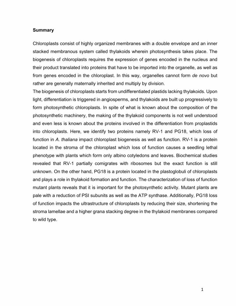
Summary
Chloroplasts consist of highly organized membranes with a double envelope and an inner
stacked membranous system called thylakoids wherein photosynthesis takes place. The
biogenesis of chloroplasts requires the expression of genes encoded in the nucleus and
their product translated into proteins that have to be imported into the organelle, as well as
from genes encoded in the chloroplast. In this way, organelles cannot form de novo but
rather are generally maternally inherited and multiply by division.
The biogenesis of chloroplasts starts from undifferentiated plastids lacking thylakoids. Upon
light, differentiation is triggered in angiosperms, and thylakoids are built up progressively to
form photosynthetic chloroplasts. In spite of what is known about the composition of the
photosynthetic machinery, the making of the thylakoid components is not well understood
and even less is known about the proteins involved in the differentiation from proplastids
into chloroplasts. Here, we identify two proteins namely RV-1 and PG18, which loss of
function in A. thaliana impact chloroplast biogenesis as well as function. RV-1 is a protein
located in the stroma of the chloroplast which loss of function causes a seedling lethal
phenotype with plants which form only albino cotyledons and leaves. Biochemical studies
revealed that RV-1 partially comigrates with ribosomes but the exact function is still
unknown. On the other hand, PG18 is a protein located in the plastoglobuli of chloroplasts
and plays a role in thylakoid formation and function. The characterization of loss of function
mutant plants reveals that it is important for the photosynthetic activity. Mutant plants are
pale with a reduction of PSI subunits as well as the ATP synthase. Additionally, PG18 loss
of function impacts the ultrastructure of chloroplasts by reducing their size, shortening the
stroma lamellae and a higher grana stacking degree in the thylakoid membranes compared
to wild type.
2
Zusammenfassung
Chloroplasten bestehen aus hochorganisierten Membranen mit einer doppelten Hülle und
einem inneren, gestapelten Membransystem, den Thylakoiden, an denen die
Photosynthese stattfindet. Die Biogenese von Chloroplasten erfordert die Expression von
Genen, welche direkt in den Plastiden kodiert sind. Darüberhinaus werden Proteine
benötigt, deren Gene im Kern kodiert sind. Diese Proteine müssen post-translational in den
Chloroplasten importiert werden. Daher können Organellen nicht de novo gebildet werden,
sondern werden in der Regel mütterlich vererbt und durch Teilung vermehrt.
Die Biogenese von Chloroplasten beginnt mit undifferenzierten Plastiden ohne Thylakoide,
den sogenannten Proplastiden. Durch Licht wird in Angiospermen eine Differenzierung
ausgelöst und die Thylakoide werden nach und nach zu photosynthetischen Einheiten
aufgebaut. Trotz des Wissens über die Zusammensetzung der Photosynthesemaschine ist
die Entstehung der Thylakoidkomponenten noch ungeklärt. Gleiches gilt für die Proteine,
die an der Differenzierung von Proplastiden zu Chloroplasten beteiligt sind. Hier
identifizieren wir zwei Proteine, nämlich RV-1 und PG18, deren Funktionsverluste bei A.
thaliana sowohl die Chloroplastenbiogenese als auch die Funktion beeinflussen. RV-1 ist
ein Protein, das sich im Stroma des Chloroplasten befindet, dessen Funktionsverlust einen
keimlingsletalen Phänotyp verursacht, wobei die Pflanzen nur albinotische Kotyledonen
und -blätter erzeugen. Biochemische Studien haben gezeigt, dass RV-1 teilweise mit
Ribosomen komigriert, obwohl die genaue Funktion noch unbekannt ist. Auf der anderen
Seite ist PG18 ein Protein, das sich in den Plastoglobuli der Chloroplasten befindet und
eine Rolle bei der Bildung und Funktion von Thylakoiden spielt. Die Charakterisierung des
Funktionsverlustes von Mutantenpflanzen zeigt, dass PG18 für die photosynthetische
Aktivität wichtig ist. Mutantenpflanzen sind blass mit einer Reduktion von PSI-
Untereinheiten sowie der ATP-Synthase. Zudem beeinflusst der PG18-Funktionsverlust die
Ultrastruktur von Chloroplasten, indem er deren Größe reduziert, die Stromalamellen
verkürzt und einen höheren Grad von Granastapel in den Thylakoidmembranen im
Vergleich zum Wildtyp aufweist.
3
Abbreviations
ATP Adenosine triphosphate
BCA Bicinchoninic acid
BLAST Basic local alignment search tool
BN-PAGE Blue native polyacrylamide gel electrophoresis
BSA Bovine serum albumin
CDS Coding sequence
Col-0 Arabidopsis thaliana ecotype Columbia
cpSEC Chloroplast Secretory pathway
cpTAT Chloroplast Twin Arginine Translocase
Cyt Cytochrome
DGDG Digalactosyl diacylglycerol
DMF Dimethyl formamide
DNA Deoxyribonucleic acid DTT Dithiothreitol
ECL Enhanced chemiluminescence
EDTA Ethylene-diamine-tetra-acetic acid
EGTA Ethylene glycol bis (aminoethyl ether) -N, N, N ', N'-tetraacetic acid
EMSA Electrophoretic Mobility Shift Assay
ETR Electron Transfer Rate
ER Endoplasmic reticulum
FBN Fibrillin
gDNA Genomic DNA
GFP Green fluorescent protein
GTP Guanosine triphosphate
H Homozygous
h Heterozygous
HEPES 2- (4- (2-hydroxyethyl) -1-piperazinyl) ethanesulfonic acid
His tag Hexa or deca histidine tag
Hsp Heat shock protein
4
IE Inner envelope of the chloroplast
IL Increased light
IPTG Isopropyl-β-D-thiogalactopyranoside
kDa Kilodalton
LDS Lithium dodecyl sulfate
LHC Light harvesting complex
MBP Maltose binding protein
MDGD Mono galactosyl diacylglycerol
MES 2- (N-Morpholino) ethanesulfonic acid
MOPS 3- (N-Morpholino) propanesulfonic acid
mRNA Messenger RNA
mt Mutant
NADPH Nicotinamide adenine dinucleotide phosphate
Ni-NTA Nickel-nitrilotriacetic acid
NL Normal light
NPQ Non-photochemical quenching
PAGE Polyacrylamid gel electrophoresis
PAM Pulse Amplitude Modulation
pb Pair base
PCR Polymerase chain reaction
PG Platoglobuli
PLBs Prolamellar bodies
PMSF Phenylmethylsulfonyl fluoride
PS Photosystem
PVDF Polyvinylidene fluoride
PVP Polyvinylpyrrolidone
RNA Ribonucleic acid
rRNA Ribosomal RNA
RT Room temperature
ROS Reactive oxygen species
RSH RelA and SpoT homolog
5
OE Outer envelope of the chloroplast
SDS Sodium dodecyl sulfate
SGDG Sulfoquinovosyl diacylglycerol
SPP Stromal processing peptidase
T-DNA Transferred DNA
TCA Trichloroacetic acid
TBST Tris-buffered saline and Polysorbate 20
TEM Transmission electron microscopy
TIC Translocase of the inner membrane of chloroplasts
tRNA Transfer RNA
TOC Translocase of the outer membrane of chloroplasts
TP Transit peptide
TPP Thylakoidal processing peptidase
WT Wildtype
Y Quantum yield of photosystem
β-DM β-dodecyl maltoside
6
1 Introduction
1.1 Origin of plastids Life as we know it today is the result of successful adaptation and evolution
throughout billions of years. In the beginning of this process, our planet had a toxic
and warmer atmosphere deprived of oxygen with abundant greenhouse gases and
dramatically different ocean chemistries (Kerr, 2005). In this scenario, life appeared
3,5 billions of years ago as self-replicable organisms with the fundamental
components such as RNAs, DNAs, amino acids, and membranes (Brasier et al.,
2015; Lake et al., 2018). Among all the components that were changing on early
earth, one constant that shaped the rest of life’s story is the sun. About 2.5 billions
of years ago emerged the first organism able to convert solar radiation into chemical
energy and releasing oxygen to the atmosphere, a process called oxygenic
photosynthesis (Shih, 2015). This event dramatically changed the earth’s fate by
producing oxygen from water splitting and using its electrons to drive biosynthetic
metabolism (De Marais, 2000). In order to do so, this photosynthetic ancestor had
to develop systems of protein complexes to split water and transport electrons to
oxidized acceptors (Cardona, 2018; Ponce-Toledo et al., 2017). It was not until 1.5
billions of years ago when a heterotrophic protist engulfed an ancestral
photosynthetic cyanobacterium marking the birth of modern plastids such as
chloroplasts (de Vries & Archibald, 2017; Douzery et al., 2004).
1.2 Protein targeting and import into the plastid The endosymbiotic event resulted in a massive transfer of genetic material to the
host cell leading the cyanobacterial guest into a semiautonomous state (Stiller,
2007). In spite of this, plastids retained their own genome, today encoding for just a
small number of their proteins. Modern plastids contain between 2100 to 4800
proteins, for which plastid genomes encode for between 60 to 200 proteins in various
linages (Richly & Leister, 2004; Timmis et al., 2004). Ever since, plastids necessitate
a system for trafficking of translated proteins encoded by nuclear genes through the
organelle membranes which gave rise to the translocons of the outer and inner
7
envelope membrane of the modern chloroplast (TOC and TIC respectively) (Gross
& Bhattacharya, 2009). The right sorting of proteins destined to the plastid and other
organelles was accomplished by sequence information to facilitate their correct
trafficking within the cell. Although in most cases this information resides in a
cleavable N-terminal sequence the different organelle-targeting sequences have
distinct properties (Kim & Hwang, 2013). The N-terminal sequences for chloroplast
localization is highly heterologous and have a net positive charge due to lack of
acidic residues (Schwenkert et al., 2011).
The proteins encoded by nuclear genes that are localized to the plastid are
translated in the cytosol and recognized at the surface of the organelle in order to
proceed with their import. At this point, the pre-proteins are assisted by chaperones
in the cytosol to prevent their aggregation (Schwenkert et al., 2011). Once the
complex of pre-protein and chaperones reaches the plastid surface, the N-terminal
transit peptide (TP) is recognized by members of the TOC apparatus. Its core
components are Toc159, Toc34 and Toc75. Depending on the chaperones bound
to the pre-protein, the TP makes first contact with either Toc34 or Toc159 receptors
in a GTP depending manner (Demarsy et al., 2014; Hirsch et al., 1994; Kessler et
al., 1994). Afterwards the import across the outer envelope continues through Toc75,
which forms a β-barrel protein acting as a channel embedded in the membrane
(Hinnah et al., 2002). Once the pre-protein passes across the outer envelope, it
reaches the TIC apparatus composed of Tic110, Tic40 and Hsp93 as the minimal
functional unit and Tic32, Tic55 and Tic62, which form the redox regulon. At the inner
membrane, the first point of contact is assumed to be Tic110, which was identified
decades ago (Schnell et al., 1994). This has been thought to be the major channel
of the TIC apparatus (Balsera et al., 2009; Heins et al., 2002; Kovacheva et al.,
2005), however Tic20 has also been proposed as a channel at the inner envelope
(Kikuchi et al., 2009; Kouranov et al., 1998). Tic40 and Hsp93 assist the import of
the pre-protein through the inner envelope along with the consumption of ATP (Chou
et al., 2006; Kovacheva et al., 2005). This process has been reported to be regulated
by the NADP+/NADPH ratio in the interaction of Tic32 and Tic62 with Tic110 (Benz
et al., 2009; Chigri et al., 2006). Additionally, Tic55 has been identified as a potential
8
thioredoxin target (Bartsch et al., 2008). Once the pre-protein crosses the inner and
outer envelopes, its TP is cleaved off by the stromal processing peptidase (SPP)
(Trosch & Jarvis, 2011) and the mature protein is folded with the assistance of Hsp70
(Flores-Perez & Jarvis, 2013; Yalovsky et al., 1992).
Imported proteins can still continue their transit to the thylakoid membranes or the
lumen inside the thylakoids. These proteins bear a bipartite TP, wherein its first part
is used for translocation across the envelopes and the second part guides the
intermediate to thylakoids where it is processed by a thylakoidal processing
peptidase (TPP) (Chaal et al., 1998; Schackleton & Robinson, 1991). Then the
protein in transit can follow one of four pathways; for membrane proteins
spontaneous insertion or the Signal Recognition Particle (SRP) dependent pathway,
and for lumenal proteins the Secretory (cpSec) or the Twin Arginine Translocase
(cpTat) pathways (Albiniak et al., 2012; Schunemann, 2007).
1.3 Chloroplast biogenesis and maturation Plastids, while functionally and structurally distinct, are present in all plant cells. They
originate from the differentiation of proplastids, requiring thylakoid membrane
formation and protein synthesis (Barsan et al., 2012; Enami et al., 2011; Rottet et
al., 2015). Proplastids are colorless round organelles deprived of internal
membranes which in the presence of light undergo differentiation into chloroplasts
in angiosperms by building up consecutive phases of internal membrane systems, a
process during which often vesicles or small saccular structures have been observed
(Muhlethaler, 1959; Vothknecht & Westhoff, 2001). However, in absence of light,
plastids turn into etioplasts, which contain few internal membranes but a
characteristic prolamellar body, rich in Mg-tetrapyrrole protochlorophyllide and
NADPH dependent protochlorophyllide oxidoreductase (Sperling et al., 1998). Upon
illumination, etioplasts can continue their differentiation into chloroplasts (Solymosi
& Schoefs, 2010).
Plastids are not synthetized de-novo but rather originate from division - similar to
prokaryotes. Plastid division happens in developing tissues such as new leaves and
meristems, i.e. chloroplasts as well as proplastids can undergo division

9
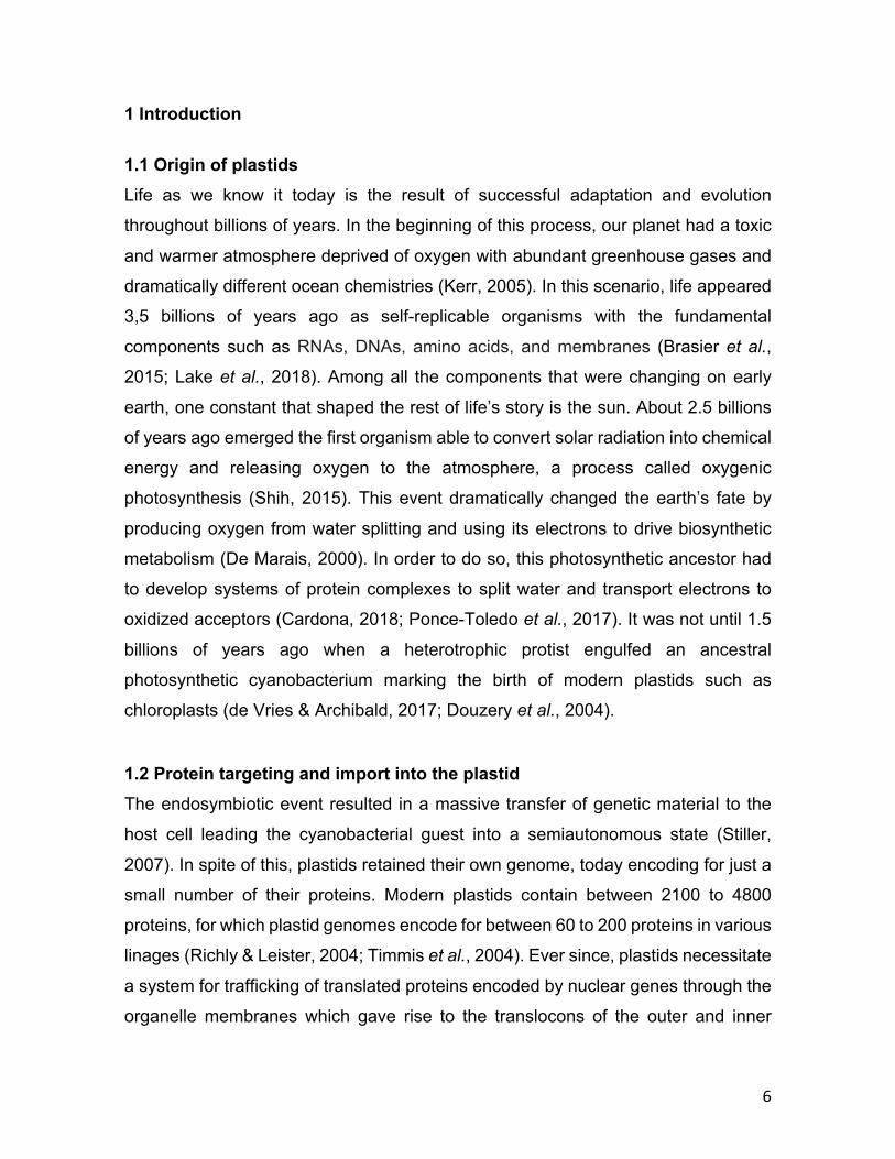
(Miyagishima, 2011). Depending on the tissue and developmental stage, proplastids
differentiate into multiple organelles with distinct functions (Figure 1). Among these
we find chromoplasts, which are non-photosynthetic carotenoid accumulating
plastids abundant in flowers, fruits and roots (Egea et al., 2010). Alternatively,
proplastids can differentiate into amyloplasts, located in root cells and specialized in
starch accumulation. Recently, these plastids have been found to play an important
role in gravitropism of roots (Toyota et al., 2013). Finally, proplastids can differentiate
into chloroplast. Their maturation starts with the elongation of a first lamella, which
ultimately forms stacks known as grana (Westphal et al., 2003). The specific steps
that lead to the initial thylakoid formation are still unknown. Nevertheless the
observation of vesicles in early stages of chloroplast maturation suggests that
vesicle trafficking is involved in thylakoid biogenesis (Morre et al., 1991). Moreover,
in some plants such as maize and sorghum vesicles have also been observed in
fully mature chloroplasts (Rosado-Alberio et al., 1968).
Figure 1. Proplastid differentiation fates. In absence of light, proplastids differentiate into etioplasts
which then turn into chloroplasts upon light exposure. Additionally, proplastids can differentiate into
amyloplast in tissues such as roots. Depending on the developmental stage, chloroplast can further
differentiate into plastids rich in carotenoids known as chromoplast which are present in tissues such
as fruits and flowers. Modified from Kato and Sakamoto (2010).
10
In addition to membrane re-modeling and protein import, chloroplast maturation
requires the coordination of other processes happening inside the organelle such as
transcription and translation. Plastid genes are highly regulated at RNA level by RNA
splicing, editing and processing (Mullet, 1993; Suzuki et al., 2003). Besides, it has
been shown that plastid rRNAs undergo multiple maturation steps for ribosome
assembly by various nuclear encoded RNases. Chloroplast RNases have been
shown to play a role also in the maturation of tRNAs and other transcripts as well as
for RNA decay. The importance of these RNases has been confirmed by their
deletions which often leads to lethal phenotypes (Stoppel & Meurer, 2012). Protein
synthesis inside the chloroplast is performed by bacterial-type 70S ribosomes,
composed of a large subunit (50S) and a small subunit (30S) containing one (16S
rRNA) and three rRNAs species (23S rRNA, 5S rRNA, and 4.5S rRNA), respectively
(Zoschke & Bock, 2018). Just recently, the structure of the chloroplast ribosome has
been resolved which reveals plastid specific extensions remodeling the mRNA entry
and exit sites (Bieri et al., 2017).
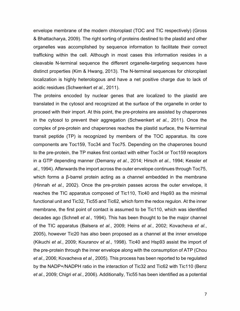
1.4 Thylakoid biogenesis Thylakoids house the photosystems (PSs), wherein light energy is used to split water
and use its electrons to produce NADPH and the chemical potential for ATP
synthesis to fuel anabolic metabolism. These membranes are organized in two
recognizable regions namely grana stacks and stroma lamellae (Figure 2). These
are not just structurally different but also differ in their protein composition wherein
PSII and the Cyt b6f complex are enriched in the grana stacks and PSI and the ATP
synthase in the stroma lamellae (Danielsson et al., 2004; Dumas et al., 2016). The
synthesis of these complexes is achieved by the coordinated expression of their
respective nuclear and plastid encoded subunits. The proper assembly and
stoichiometry of thylakoid complexes is ensured by a number of auxiliary assembly
factor such as in the case of the retrograde signaling regulation of nuclear encoded
genes expression (Chan et al., 2016). The de novo synthesis of thylakoid complexes
is highly dependent on the developmental stage of the tissue in higher plants
(Roberts et al., 1987), with a half-life that can be up to one week for Cyt b6f complex
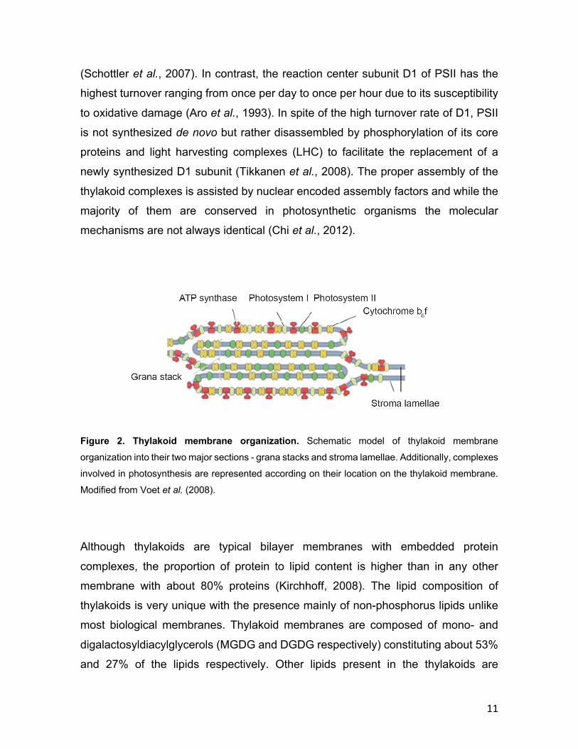
11
(Schottler et al., 2007). In contrast, the reaction center subunit D1 of PSII has the
highest turnover ranging from once per day to once per hour due to its susceptibility
to oxidative damage (Aro et al., 1993). In spite of the high turnover rate of D1, PSII
is not synthesized de novo but rather disassembled by phosphorylation of its core
proteins and light harvesting complexes (LHC) to facilitate the replacement of a
newly synthesized D1 subunit (Tikkanen et al., 2008). The proper assembly of the
thylakoid complexes is assisted by nuclear encoded assembly factors and while the
majority of them are conserved in photosynthetic organisms the molecular
mechanisms are not always identical (Chi et al., 2012).
Figure 2. Thylakoid membrane organization. Schematic model of thylakoid membrane
organization into their two major sections - grana stacks and stroma lamellae. Additionally, complexes
involved in photosynthesis are represented according on their location on the thylakoid membrane.
Modified from Voet et al. (2008).
Although thylakoids are typical bilayer membranes with embedded protein
complexes, the proportion of protein to lipid content is higher than in any other
membrane with about 80% proteins (Kirchhoff, 2008). The lipid composition of
thylakoids is very unique with the presence mainly of non-phosphorus lipids unlike
most biological membranes. Thylakoid membranes are composed of mono- and
digalactosyldiacylglycerols (MGDG and DGDG respectively) constituting about 53%
and 27% of the lipids respectively. Other lipids present in the thylakoids are

12
sulfoquinovosyldiacylglycerol (SQDG) and phosphatidylglycerol (PG), which are
anionic lipids with a negative charge in their head groups (Kobayashi, 2016). The
biosynthesis of thylakoid lipids takes place at the inner envelope where the MGDG
synthase is located (Kobayashi et al., 2007), and also at the outer envelope where
the DGDG synthase transfers a second galactose to MGDG (Froehlich et al., 2001).
Since lipids cannot diffuse freely from one membrane to another without assistance,
i.e. from the inner envelope to the thylakoid membranes, it has been theorized that
chloroplast could use vesicle trafficking to mobilize lipids for thylakoid membrane
formation (Benning et al., 2006). This hypothesis has also been formulated by the
observation of fatty acid desaturase mutants in Arabidopsis, which are localized in
the endoplasmic reticulum (ER). These mutants showed a strong reduction on
MGDG species suggesting the necessity of lipid transport from the ER to the
thylakoid membrane (Miquel & Browse, 1992; Slack et al., 1977).
Besides the fatty acid and protein content of the thylakoid membrane, several
pigments play a crucial role in thylakoid function. Chlorophyll is the most abundant
pigment in thylakoids and is associated with proteins such as LHCs and both PSI
and PSII, thus allowing the harvest of light to power photosynthesis (Eckhardt et al.,
2004). Among other relevant pigments that play a role in photosynthesis, there are
compounds related to photo protection such as the members of the xanthophyll
cycle. When light intensity is not higher than the capacity of the photosystems, the
Δ pH across the thylakoid membrane leads to the lumen being more acidic than the
stroma. However, during higher light intensities, photosystems reach their maximum
capacity to transfer electrons, thus prolonging the chlorophyll excitation state
lifetime, which in turn leads to the production of reactive oxygen species (ROS)
(Johnson et al., 2012). In addition to the latter, the lumen pH drops due to the
limitation of the ATP synthase to relax the Δ pH. This causes the de-epoxidation of
violaxanthin to antheraxanthin further de-epoxidation of the latter to the photo
protective pigment zeaxanthin (Rockholm & Yamamoto, 1996). Thus, zeaxanthin
attached to the LHC quenches the excited state of chlorophyll preventing the
production of ROS by a process called Non Photochemical Quenching (NPQ) (Jahns
& Holzwarth, 2012).

13
1.5 The function of Plastoglobuli Beside vesicles, that might transport lipid and protein components to the thylakoids,
lipid bodies persistently attached to the thylakoid membrane have been observed,
which are called plastoglobuli (PGs). Early descriptions of these compartments were
performed during the sixties and they were first described as osmiophilic globules
due to their ability to get stained with osmium tetroxide during the preparation of TEM
pictures. These early observations led to the study of their composition and they
were initially described as lipid bodies containing plastoquinone and galactolipids
(Greenwood et al., 1963). Later it was found that PGs are present throughout the
plant’s life and propagate in distinct developmental stages and tissues. For instance,
PGs propagate and are bigger in size in chromoplasts especially in flowers and fruits
(Hansmann & Sitte, 1982). This holds true for several plant species. Further
investigation of the composition of PGs in chromoplasts revealed that they are rich
in carotenoids. Moreover, this led to the identification of fibrillin (FBN) as the core
protein of PGs (Deruere, Bouvier, et al., 1994; Deruere, Romer, et al., 1994).
Additionally, it was observed that during senescence the number and size of PGs
increased in chloroplasts and that they play a role in the accumulation of break down
intermediates of chlorophyll degradation such as phytol (Tevini & Steinmuller, 1985;
Vom Dorp et al., 2015). In this stage, plastid metabolism is solely catabolic losing
stroma and storing thylakoid breakdown content into PGs. Moreover, PGs are also
accumulating under stress conditions such as drought (Langenkamper et al., 2001),
high light stress (Zhang et al., 2010) and nitrogen limiting conditions (Gaude et al.,
2007).
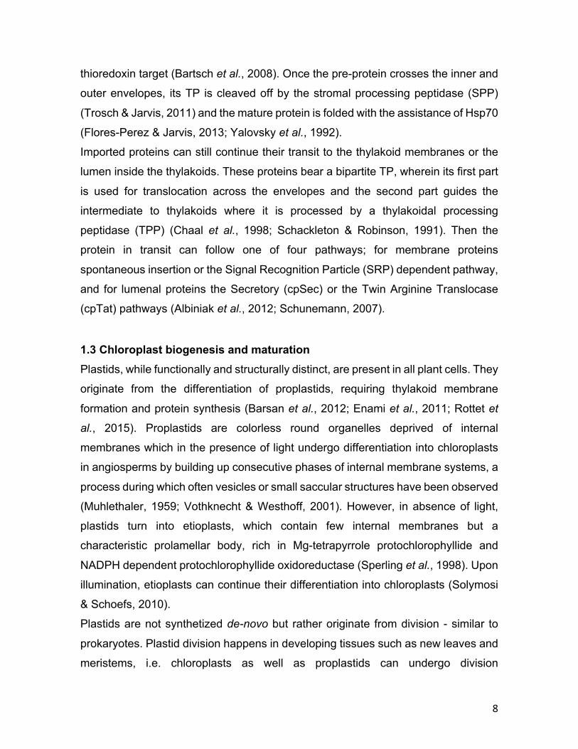
Electron tomography studies on the structure of PGs have revealed that they are
composed of a neutral lipid core and a lipid monolayer, which is physically attached
and continuous with the stroma-side leaflet of the thylakoid membrane (Figure 3).

14
Figure 3. Plastoglobuli organization. Scheme representing a PG in relation with the thylakoid
membrane. Proteins are represented as shapes on the monolayer of the PG. Modified from Austin et
al. (2006).
Besides, it was shown that even though PGs propagate they always maintain at least
one contact site with another PG or the thylakoid membrane, i.e. PGs are not found
as individual components (Austin et al., 2006). Further analyses on the composition
of PGs have shown that certain variations in their lipid composition can occur
depending on the developmental stage of the plant or the plant tissue. Under non-
stress conditions, PGs are composed of high levels of tocopherols and quinones as
well as neutral lipids such as triacylglycerol and free fatty acids (Steinmuller & Tevini,
1985; Vidi et al., 2006; Zbierzak et al., 2009). Other less abundant components are
galactolipids, carotenoids and phytol esters, which increase in senescent
chloroplasts (Gaude et al., 2007; Tevini & Steinmuller, 1985) or during chromoplast
differentiation, i.e. carotenoids (Deruere, Romer, et al., 1994; Hansmann & Sitte,
1982). The proteome of PGs was identified by mass-spectrometry of isolated PGs
from chromoplasts and chloroplasts (Vidi et al., 2006; Ytterberg et al., 2006). This
revealed that in normal conditions PGs are composed of 30 proteins, the most
abundant of which are FBNs (Lundquist et al., 2012). This family of proteins is well
conserved from plants to cyanobacteria (Cunningham et al., 2010; Singh & McNellis,
2011). Initially, they were called fibrillins due of their ability to form fibers in the
15
presence of carotenoids and polar lipids, a phenomenon that was also observed in
chromoplasts (Deruere, Romer, et al., 1994). Furthermore, the second most
abundant family of proteins in PGs is the ABC1K kinase family two of which were
shown to play a role in vitamin E synthesis (Martinis et al., 2013; Martinis et al.,
2014). Other proteins present in PGs are related to isoprenoid and neutral lipid
metabolism (Lundquist et al., 2012).
Analyses of mutant knockout plants for many of the PG’s proteins lead to the
conclusion that the function PGs is related to stress conditions or specific
developmental stages. (Fatihi et al., 2015; Singh et al., 2010; Youssef et al., 2010).
Thus, PGs are assumed not to be essential for plant survival but rather relevant in
their adaptation to different conditions and successful tissue differentiation.

16
1.6 Aim of this work The central question to be answered in this thesis was: which are the crucial factors
required for chloroplast biogenesis that are still unknown?
To date, the process by which proplastids undergo thylakoid formation and arrange
their membranes along with the different embedded photosynthetic complexes
embedded is still obscure. In order to tackle this question, a chloroplast proteome
analysis from young plants should be utilized to identify proteins without known
function. This should provide insights into proteins that might be involved in either
early development of thylakoid formation (and therefore differentiation from
proplastids to chloroplasts) or chloroplast biogenesis in general. To understand
those processes the study of proteins with either unknown domains or function is
crucial. In this work two proteins with unknown function, RV-1 and PG18, were
selected to pursue a molecular characterization and investigate their role in plant
development using knockout mutant plants in Arabidopsis.

17
2 Material and methods
2.1 Material 2.1.1 Chemicals If not noted otherwise, all used chemicals were received from Sigma Aldrich
(Taufkirchen, Germany), Roth (Karlsruhe, Germany), Merck (Darmstadt, Germany),
Thermo Fisher Scientific (Braunschweig, Germany) or Serva (Heidelberg,
Germany).
2.1.2 Molecular weight and size markers For SDS-PAGE peqGOLD protein marker I (VWR, Ismaning, Germany) was used.
EcoRI and HindIII digested lambda phage DNA (Thermo Fisher Scientific) was used
as a marker for agarose gel electrophoresis. HMW Calibration Kit for Native
Electrophoresis (GE Healthcare, Munich, Germany) was used for BN-PAGE.
2.1.3 Oligonucleotides DNA oligonucleotides were ordered from Metabion (Martinsried, Germany) and are
listed in Table 1.
Table 1. Oligonucleotides used for this work.
Oligonucleotide 5'-3' oligonucleotide sequence Purpose Gabi Kat F ATATTGACCATCATACTCATTGC Genotyping
AT1G36320 GT F GGGGACAAGTTTGTACAAAAAAGCAGGCTTCGAAGGAGATAGAACC
ATGGTTTCAGTGTT
pK7FWG2
AT1G36320 GT R -stop GGGGACCACTTTGTACAAGAAAGCTGGGTCTCCACCTCCGGACATA
TACTTCTTTT
pK7FWG2
AT4g37920 F EcoRV -TP GATATCGCGGAAGTAAAAAGCTC pMAL-c5x
AT4g37920 R EcoRI Histag -stop GAATTCGTGATGGTGATGGTGATGGTTCAAAAAAT
pMAL-c5x
18
AT4g37920 GT F GGGGACAAGTTTGTACAAAAAAGCAGGCTTCGAAGGAGATAGAACC
ATGGCGAATTTACTG
gateway vectors, Genotyping
AT4g37920 GT R -stop GGGGACCACTTTGTACAAGAAAGCTGGGTCTCCACCTCCGGAGTTC
AAAAAATCTTCA
pK7FWG2
AT4g37920 GT R +stop GGGGACCACTTTGTACAAGAAAGCTGGGTCTCCACCTCCGGATCAG
TTCAAAAAATC
pDest14, pDest17, Genotyping
AT4g37920 GT F -TP GGGGACAAGTTTGTACAAAAAAGCAGGCTTCCTGGAAGTTCTGTTTCAGGGCCCGGCGGAAGTAAAAA
GCTC
pDest17
AT4g37920 R cDNA AGTCGTAATTGCAATTATAGCTGA Genotyping
AT4g37920 RNAi F GGGGACAAGTTTGTACAAAAAAGCAGGCTTTAGTTTTAACATTTT
pOpOff2
AT4g37920 RNAi R GGGGACCACTTTGTACAAGAAAGCTGGGTCTTTGTAAGTGACAGTG
pOpOff2
ATCG00920 rRNA 16S F TCTCATGGAGAGTTCGATCCT pSP64
ATCG00920 rRNA 16S R AAAGGAGGTGATCCAGCCG pSP64
ATCG00950 rRNA 23S F TTCAAACGAGGAAAGGCTT pSP64
ATCG00950 rRNA 23S R AGGAGAGCACTCATCTTGG pSP64
AT1G36320 F TGATCACTAAAGCTTGGTCG Genotyping
AT1G36320 R TCACATATACTTCTTTTCAA Genotyping
AT4G13200 GT F GGGGACAAGTTTGTACAAAAAAGCAGGCTTCGAAGGAGATAGAACC
ATGAGTAGCTTCACGA
gateway vectors, Genotyping
AT4G13200 GT F -TP GGGGACAAGTTTGTACAAAAAAGCAGGCTTCCTGGAAGTTCTGTTT
CAGGGCCCGGAGTCTCGAAG
pDest17
AT4G13200 GT R -stop GGGGACCACTTTGTACAAGAAAGCTGGGTCTCCACCTCCGGATCAG
TCTTCATCACT
pK7FWG2
19
AT4G13200 GT R +stop 23S F NW 23S R NW 16S F NW 16S R NW
GGGGACCACTTTGTACAAGAAAGCTGGGTCTCCACCTCCGGATCAG
TCTTCATCA
TTCAAACGAGGAAAGGCTTA
AGGAGAGCACTCATCTTG
GTAAAGCGTCTGTAGGTG
GCCTAGTATCCATCGTTT
pDest17, Genotyping
Northern Blot
Northern Blot
Northern Blot
Northern Blot
2.1.4 Vectors
To overproduce proteins fused to an N-terminal His-tag pDest17 vector was used
(Thermo Fisher Scientific). The vector pMAL-c5x (New England Biolabs) was used
to overproduce proteins fused to MBP at their N-terminal. pDest14 (Thermo Fisher
Scientific) and pSP64 (Promega) vectors were used for in vitro transcription and
translation. For plant transformation the following binary vectors were used:
pK7FWG2 for expression under 35S promoter and pOpOff2 for expression of
interference RNA (both Plant Systems Biology, Zwijnaarde, Belgium). Cloning into
binary vectors as well as pDest14 and pDest17, was performed using the Gateway
system (Thermo Fisher Scientific) via pDONR207 vector. Restriction site cloning was
performed by using the entry vector Zero blunt (Thermo Fisher Scientific). All
plasmids used for this thesis are listed in Table 2.
Table 2. List of plasmids used for this work.
Gene Description vector Restriction sites purpose AT4g37920 - stop pDONR207 - localization AT4g37920 + stop pDONR207 - in-vitro translation AT4g37920 - TP / + stop pDONR207 - over-expression AT4g37920 - stop pK7FWG2 - localization AT4g37920 - TP / + stop pDest17 - over-expression AT4g37920 + stop pDest14 - in-vitro translation AT4g37920 Histag / - TP / - stop pCR-Blunt EcoRV / EcoRI over-expression AT4g37920 Histag / - TP / - stop pMAL-c5x EcoRV / EcoRI over-expression AT4g37920 - pDONR207 - RNAi AT4g37920 - pOpOff2 - RNAi AT1G20020 + stop pF3A - in-vitro translation
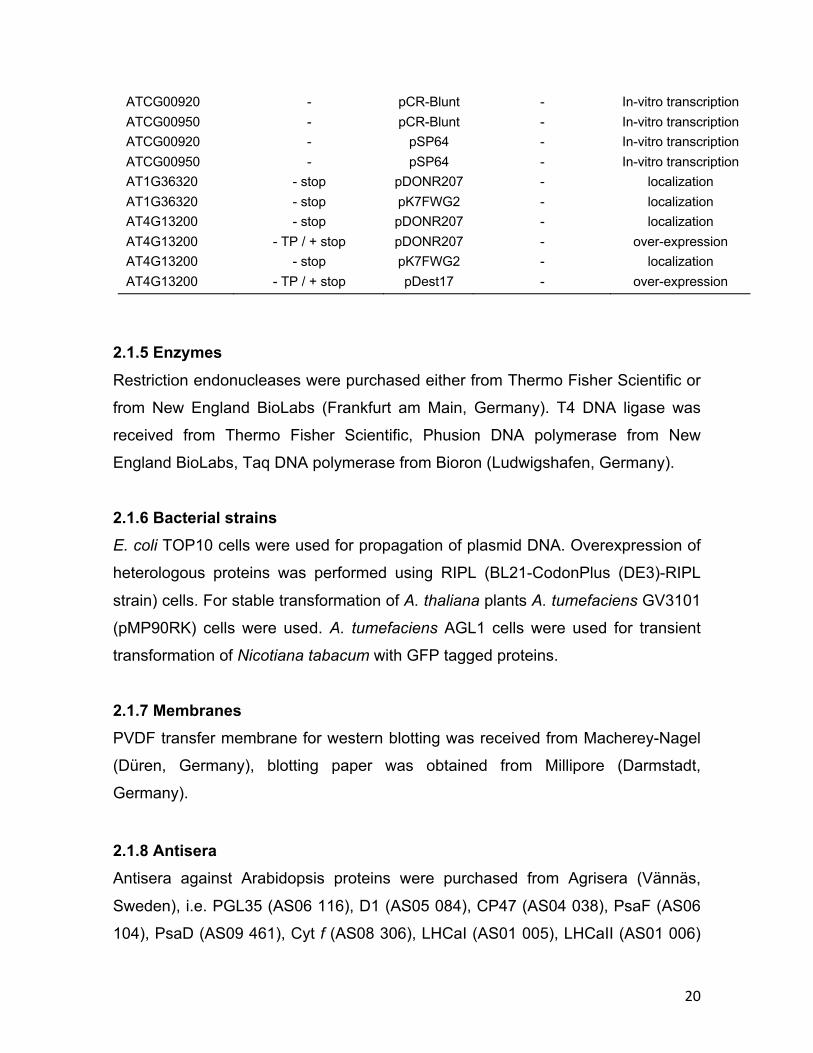
20
ATCG00920 - pCR-Blunt - In-vitro transcription ATCG00950 - pCR-Blunt - In-vitro transcription ATCG00920 - pSP64 - In-vitro transcription ATCG00950 - pSP64 - In-vitro transcription AT1G36320 - stop pDONR207 - localization AT1G36320 - stop pK7FWG2 - localization AT4G13200 - stop pDONR207 - localization AT4G13200 - TP / + stop pDONR207 - over-expression AT4G13200 - stop pK7FWG2 - localization AT4G13200 - TP / + stop pDest17 - over-expression
2.1.5 Enzymes Restriction endonucleases were purchased either from Thermo Fisher Scientific or
from New England BioLabs (Frankfurt am Main, Germany). T4 DNA ligase was
received from Thermo Fisher Scientific, Phusion DNA polymerase from New
England BioLabs, Taq DNA polymerase from Bioron (Ludwigshafen, Germany). 2.1.6 Bacterial strains E. coli TOP10 cells were used for propagation of plasmid DNA. Overexpression of
heterologous proteins was performed using RIPL (BL21-CodonPlus (DE3)-RIPL
strain) cells. For stable transformation of A. thaliana plants A. tumefaciens GV3101
(pMP90RK) cells were used. A. tumefaciens AGL1 cells were used for transient
transformation of Nicotiana tabacum with GFP tagged proteins.
2.1.7 Membranes PVDF transfer membrane for western blotting was received from Macherey-Nagel
(Düren, Germany), blotting paper was obtained from Millipore (Darmstadt,
Germany).
2.1.8 Antisera Antisera against Arabidopsis proteins were purchased from Agrisera (Vännäs,
Sweden), i.e. PGL35 (AS06 116), D1 (AS05 084), CP47 (AS04 038), PsaF (AS06
104), PsaD (AS09 461), Cyt f (AS08 306), LHCaI (AS01 005), LHCaII (AS01 006)

21
and LHCbII (AS01 003). Phycocyanin antiserum was purchased from ABBIOTEC
(250488). ATP synthase, D1 and OE33 antisera were kindly provided by Stephan
Greiner, TIC110 and FBPase antisera from Bettina Bölter and PsaG antiserum from
Jörg Meurer. RV-1 and PG18 antisera were produced by Biogenes (Berlin,
Germany).
2.1.9 Accession numbers The gene accession numbers of the proteins involved in this work can be seen in
Table 3.
Table 3. Gene accession numbers of proteins involved in this work.
Gene name Accession number RV-1 AT4g37920
RV-1-like AT1G36320 PG18 AT4G13200
rRNA 16S ATCG00920 rRNA 23S ATCG00950
FNR AT1G20020
2.1.10 Computational analyses Sequences for RV-1, RV-1-like PG18 from Arabidopsis were obtained from TAIR
(https://www.arabidopsis.org). Homologs of RV-1, RV-1-like PG18 from other
species were collected from NCBI/BLAST (http://blast.ncbi.nlm.nih.gov/Blast.cgi)
and Phytozome (https://phytozome.jgi.doe.gov/pz/portal.html). Phylogenetic trees
were generated by using the MEGA7 software (Kumar et al., 2016). The accessions
for each sequence used for phylogenetic trees are indicated in table 4. Alignments
were generated by using the algorithm provided by CLC Main Workbench
(developed by QIAGEN Aarhus).
Graphs and statistical analyses were generated by using GraphPad Prism version
6.0, GraphPad Software, La Jolla, California, USA (www.graphpad.com). Image
analyses were done by using the software ImageJ.

22
Table 4. Gene accession numbers of proteins used for phylogenetic analyses.
Species RV-1 RV-1-like PG18 Arabidopsis
thaliana AT4g37920 AT1G36320 AT4G13200
Physcomitrella patens
- - Pp3c16_19690V3.2
Citrus sinensis orange1.1g013676m orange1.1g048140m A0A067FRT7_CITSI Fragaria vesca mrna05566.1-v1.0-
hybrid mrna31319.1-v1.0-
hybrid mrna17524.1-v1.0-hybrid
Solanum tuberosum PGSC0003DMT400078670
PGSC0003DMT400070893
PGSC0003DMT400071989
Solanum lycopersicum
Solyc02g091640.2.1 Solyc04g072400.2.1 Solyc09g061440.2.1
Sorghum bicolor Sobic.010G157700.1 Sobic.001G530900.1 Sobic.006G145200.1 Zea mays B6TPX9_MAIZE C0PEL6_MAIZE B6TBJ2_MAIZE
Oryza sativa LOC_Os01g20110.1 A3A5Q9_ORYSJ LOC_Os04g43350.1 Sphagnum fallax - - Sphfalx0010s0144.1
Micromonas pusilla - - 189112 Calothrix sp. 336/3 - - WP_035149421 Synechocystis sp.
PCC 6803 - - WP_010872303
Anabaena sp. WA113
- - WP_066381343
2.2 Molecular biological methods General methods not listed below were performed according to Sambrook and
Russell (2001). Competent cells for DNA transformation were prepared according to
Hanahan (1985).
2.2.1 Cloning strategies For over-expression of proteins fused to an N-terminal MBP tag pMAL-c5x vector
and in vitro transcription of rRNAs, PCR product created with primes containing the
appropriate restriction site were digested with EcorV and EcoRI for RV-1 and with
XbaI and BamHI for rRNAs. Ligation was carried out for 1 h at RT using T4 ligase
into the vector pCR-Blunt. Afterwards, the corresponding plasmids were digested
with EcorV and EcoRI, as well as the pMAL-c5x vector. Additionally, the
corresponding plasmids with rRNA were digested with XbaI and BamHI as well as
the vector pSP64 for its ligation as described above. For stable plant transformation,
23
over-expression of proteins and in vitro transcription and translation, Gateway
system (Thermo Fisher Scientific) was used to clone constructs via homologous
recombination from pDONR207 into binary vectors pk7FWG2, pDest14, pDest17 or
pOpOff2. Cloning was performed according to the manufacturer’s instructions.
2.2.2 Polymerase chain reaction (PCR) PCR was performed with gDNA, cDNA or plasmid DNA as templates. For
subsequent cloning proof-reading Phusion polymerase was used. For genotyping
and colony PCR Taq polymerase was chosen. Annealing temperature and
elongation time were adapted concerning oligonucleotides and length of constructs.
PCR products for cloning were excised from 1% agarose gels run in TAE buffer (40
mM Tris, 2.5 mM EDTA, 1% acetic acid) and purified using NucleoSpin Gel and PCR
Clean-up kit (Macherey-Nagel).
2.2.3 Isolation of plasmid DNA from E. coli Plasmid DNA was isolated from 2 ml overnight E. coli culture using the NucleoSpin
Plasmid EasyPure kit (Macherey-Nagel) according to the manufacturer’s
instructions.
2.2.4 Sequencing Each plasmid was confirmed by sequencing which was performed by the sequencing
service of the Faculty of Biology (Ludwig-Maximilians-Universität München,
Germany) using 100 – 200 ng of vector with appropriate primer.
2.2.5 Isolation of genomic DNA from A. thaliana for genotyping PCR
One leaf was homogenized in 500 µl of extraction buffer (1 M Tris-HCl pH 7.5, 50
mM NaCl, 50 mM EDTA, 1% (w/v) PVP 40) by a mixing mill for 3 min in a tube with
a tungsten carbide 3 mm ball. Afterwards, 66 µl of 10% (w/v) SDS were added and
the samples were mixed by inverting them several times followed by the addition of
166 µl of 5 M potassium acetate. The sample was then centrifuged for 15 min at
15000 gRT. The supernatant was rescued and mixed with 0.7 vol of isopropanol and

24
incubated for 15 min at -20 ºC. The sample was then centrifuged for 15 min at 15000
g and 4 ºC and the supernatant was discarded. The pellet was washed with 500 µl
of 70% (v/v) ethanol and mixed by inverting the tube and then centrifuged for 5 min
at 15000 g and 4 ºC. The pellet was dried at RT and dissolved in 50 µl of H2O.
2.2.6 Isolation of RNA from A. thaliana
RNA from A. thaliana leaves was isolated using the Rneasy Plant Mini kit (Qiagen)
according to the manufacturer’s instructions. Digestion with Dnase was either
performed during RNA isolation (DnaseI, Roche, Mannheim, Germany).
2.2.7 cDNA synthesis cDNA was synthesized in 10 μl reaction volume from 1 μg RNA using M-MLV reverse
transcriptase (Promega) according to the manufacturer’s instructions.
2.2.8 Protein precipitation with trichloroacetic acid (TCA) Proteins were precipitated by adding TCA (final percentage 10% [v/v]) and
incubating the samples for 30 min at -20 ºC. Afterwards, proteins were centrifuged
for 15 min at 16000 g and 4ºC and the supernatant was discarded. The pellet was
washed twice by adding 1 ml of 100% ethanol and centrifuging for 5 min at 16000 g
and 4 ºC. The pellet was dried at Rtfor 15 min and resuspended in loading buffer
(100 mM Tris pH 7.5, 2% SDS, 10% glycerol, 5% β-mercaptoethanol, 0.004%
bromphenol blue).
2.3 Biochemical methods 2.3.1 Overexpression of recombinant proteins Transformed E. coli bacteria were grown in LB medium (1% peptone from casein,
0.5% yeast extract, 171 mM NaCl) at 37°C to an OD600 of 0.6 – 0.8. Overexpression
was induced by the addition of 1 mM isopropyl β-D-1-thiogalactopyranoside. E. coli
strains and conditions for overexpression were depending on the construct and are

25
listed in Table 5.
Table 5. Conditions for overexpression and way of purification of recombinant proteins
Construct Vector E. coli strain Temperature Time Purification RV-1 pDest17 RIPL 37 ºC overnight inclusion bodies RV-1 pMAL-c5x RIPL 12 ºC overnight soluble PG18 pDest17 RIPL 37 ºC overnight inclusion bodies
2.3.2 Purification of soluble proteins Pelleted bacteria from 1 l overexpression of constructs with His-tags were
resuspended in 25 ml lysis buffer (20 mM Tris pH 7.5, 200 mM NaCl, 15 mM
imidazole). After cell disruption by a microfluidizer (Microfluidics, Westwood, USA)
the solution was centrifuged at 20000 g, 4°C for 30 min and the supernatant was
rotated with 250 μl Ni-Sepharose at 4°C for 2 h. The beads were washed three times
with 5 ml wash buffer (20 mM Tris pH 7.5, 200 mM NaCl, 15 mM imidazole).
Recombinant proteins were eluted in 200 – 400 μl fractions with elution buffer (20
mM Tris pH 7.5, 200 mM NaCl, 500 mM imidazole). Samples were then dialyzed to
remove imidazole in bags with a molecular weight cut-off of 3.5 kDa (Spectra/Por 3)
against 2 l of lysis buffer without imidazole overnight at 4 ºC with agitation.
2.3.3 Purification of proteins out of inclusion bodies Insoluble proteins were purified out of inclusion bodies. Pelleted bacteria from 0.5 –
1 l overexpression were resuspended in 25 ml resuspension buffer (50 mM Tris pH
8.0, 200 mM NaCl, 5 mM β-mercaptoethanol). Cells were disrupted and centrifuged
as described in 2.3.4. The pellet was washed one time with 20 ml detergent buffer
(20 mM Tris pH 7.5, 200 mM NaCl, 1% deoxycholic acid, 1% nonidet P-40, 10 mM
β-mercaptoethanol), two times with Triton buffer (20 mM Tris pH 7.5, 0.5% Triton X-
100, 5 mM β-mercaptoethanol) and two times with Tris buffer (20 mM Tris pH 8.0,
10 mM DTT). Centrifugation was done at 12000 g, 4°C for 10 min. Finally, the pellet
was resuspended in 5 ml urea buffer (50 mM Tris pH 8.0, 100 mM NaCl, 7 M urea)
and rotated for overnight at 4 ºC. After centrifugation at 20000 g, 4 ºC for 15 min
denaturated proteins were present in the supernatant. In order to get rid of urea from

26
the buffer, samples were dialyzed as described in section 2.3.2.
2.3.4 Isolation of proteins from A. thaliana A. thaliana leaves were homogenized in 300 μl homogenization medium (50 mM Tris
pH 8.0, 10 mM EDTA, 2 mM EGTA, 10 mM DTT) using an electronic micropestle.
The suspension was incubated for 10 min in the dark on ice then filtered and
centrifuged at 9300 g for 10 min and 4 ºC. Supernatant contained soluble proteins,
pellet resuspended in homogenization medium contained membrane proteins. For
total protein extraction, leaves were grinded with liquid nitrogen and the powder was
then transferred into a tube and resuspended by adding equal volume of extraction
buffer (50 mM Tris-HCl pH 8, 2% [w/v] LDS, 0.1 mM PMSF). The sample was
vortexed and then incubated on ice for 30 min and centrifuged for 15 min at 16000
g and 4 ºC. The supernatant was rescued, and protein concentration was measured
by BCA test. Finally, EDTA and DTT were added immediately to a final concentration
of 50 mM and 10 mM respectively.
2.3.5 Determination of protein concentration Concentration of proteins was determined using Bradford reagent (Bio-rad). 10 μl
protein was mixed with 1:5 diluted Bradford reagent and absorption was measured
against buffer at 595 nm. When indicated, protein concentrations were also
quantified by using the kit Pierce™ BCA protein assay according to the
manufacturer’s instructions (Thermo Fisher Scientific).
2.3.6 SDS polyacrylamide gel electrophoresis (SDS-PAGE) Proteins were separated by SDS-PAGE using discontinuous gels according to
(Laemmli, 1970) consisting of a stacking gel (5% polyacrylamide) and a running gel
(10 – 15% polyacrylamide). Samples were loaded with SDS loading buffer (62.5 mM
Tris pH 6.8, 2% SDS, 10% glycerol, 5% β-mercaptoethanol, 0.004% bromphenol
blue). Gels were run in SDS running buffer (25 mM Tris, 192 mM glycine, 0.1% SDS)
and afterwards either stained with 26oomassie (45% methanol, 9% acetic acid, 0.2%
26oomassie brilliant blue R-250) or used for western blotting (see 2.3.7).
27
2.3.7 Semi-dry electro blot and immunodetection of proteins Proteins were electrotransferred out of an SDS gel onto a PVDF membrane using a
semi-dry blotting apparatus. The blot was assembled as follows on the anode: three
blotting papers in anode I buffer (20% methanol, 300 mM Tris), two blotting papers
in anode II buffer (20% methanol, 25 mM Tris), activated membrane, gel, three
blotting papers in cathode buffer (20% methanol, 40 mM aminocaproic acid).
Transfer was carried out for 1 h at 0.8 mA/cm2, proteins on membrane were stained
with ponceau solution (5% acetic acid, 0.3% ponceau S). For immunodetection of
proteins membrane was blocked for 30 min with 5% skimmed milk in TBST (20 mM
Tris pH 7.6, 137 mM NaCl, 0.075% Tween). Incubation with primary antibody was
performed over night at 4°C. After three times 10 min washing in TBST membrane
was incubated for 2 h at RT with horse radish peroxidase conjugated secondary
antibody. After three times 10 min washing in TBST membrane was incubated in
equal volumes of development solution I (100 mM Tris pH 8.5, 1% luminol, 0.44%
coumaric acid) and II (100 mM Tris pH 8.5, 0.018% H2O2) and signal was detected
with enhanced chemiluminescence using Image Quant LAS 400 (GE Healthcare).
2.3.8 Detection of radiolabeled proteins To visualize radiolabeled proteins dried SDS gels were exposed overnight to BAS-
MS phosphor imaging plates (FUJIFILM) which were analyzed using a Typhoon
scanner (GE healthcare).
2.3.9 Blue Native PAGE (BN-PAGE) Solubilized samples (thylakoid membranes) were separated on native acrylamide
gradient Bis-Tris gel (5 – 12% polyacrylamide). Samples were loaded with BN
loading buffer (750 mM aminocapronic acid, 5% Serva-G 250), gel was run with
cathode buffer (50 mM tricine, 15 mM Bis-Tris pH 7.0, 0.2% Serva-G 250) and anode
buffer (50 mM Bis-Tris pH 7.0). For second dimension one lane of the BN gel was
placed on top of an SDS gel containing 4 M urea which was either silver stained to
visualize proteins.
28
2.3.10 Silver staining of SDS gels Gels were incubated for 1 h in fixation solution (50% [v/v] ethanol, 12% [v/v] acetic
acid, 0.05% [v/v] formaldehyde) then washed three times for 30 min in 50% (v/v)
ethanol. After 90 sec pre-impregnation in 0.02% (v/v) sodium thiosulfate and three
times 30 sec washing in water impregnation of the gel was performed for 30 min in
darkness using 0.2% (w/v) silver nitrate and 0.075% (v/v) formaldehyde. Gels were
washed again in water then stained with development solution (6% [w/v] Na2CO3,
0.05% [v/v] formaldehyde, 0.0004% [v/v] sodium thiosulfate). After protein signals
became visible reaction was stopped with stopping solution (50% [v/v] ethanol, 12%
[v/v] acetic acid). 2.3.11 Mass spectrometry Mass spectrometric analyses were performed at the MSBioLMU core facility
(Deparment Biology I, Ludwig-Maximilians-Universität München).
2.3.12 Transformation of A. tumefacium
1 – 2 µg plasmid was added to either to competent GV2101 or AGL1 cells which
were incubated 5 min on ice then for 5 min in liquid nitrogen. Heat shock was
performed for 5 min at 37 ºC then 800 µl LB was added and cells were incubated
shaking for 3 h at 28 ºC before plated on LB plates with appropriate antibiotics. Cells
were grown for 2 days at 28 ºC.
2.3.13 Pigment analysis Pigment measurements were done by Prof. Dr Peter Jahns (Plant Biochemistry,
Heinrich-Heine-University Düsseldorf, Düsseldorf). Leaf samples were frozen in
liquid N2 and either used directly for pigment extraction or stored at -80°C for up to
2 weeks until further use. Pigments were extracted by grinding frozen leaf material
in a mortar after addition of 1 ml 100% acetone. After short centrifugation, the
supernatant was filtered through a 0.2-μm membrane filter (GE Healthcare, Little
Chalfont, Buckinghamshire, UK) and then subjected to HPLC analysis. Separation
29
and quantification of pigments was done by reversed-phase chromatography as
described in Farber et al. (1997).
2.3.14 In vitro transcription Vectors including either a T7 or SP6 promoter sequence were used for in vitro
transcription of various genes (see Table 2). For the transcription reaction, 1 μg
plasmid, 0.05% BSA, 2 mM DTT, 0.25 mM m7G(5’)ppp(5’)G Cap analog (Ambion),
0.4 mM ACU (Roche), 50 U RibolockRI (Thermo Fisher Scientific), 30 U T7 or SP6
RNA polymerase (Thermo Fisher Scientific), 1x transcription buffer (Thermo Fisher
Scientific) were added in a total volume of 50 μl and incubated at 37°C for 15 min
for RNA capping. To this capped RNA, 1.2 mM GTP was added and reaction was
incubated at 37 ºC for 120 min for final mRNA generation. For transcription and
simultaneous radiolabeling, the transcription reaction was set up as follows: 1 μg
plasmid, 0.5 mM AGU (Roche), 12 μM CTP (Roche), 40 U RibolockRI (Thermo
Fisher Scientific), 20 U SP6 RNA polymerase (Thermo Fisher Scientific), 1x
transcription buffer (Thermo Fisher Scientific), 2.5 μCi/μl [a 32P] CTP (sp. act. 800
Ci/mmol) (Perkin Elmer, Walluf, Germany) in a total volume of 20 μl. The reaction
was performed at 37 ºC for 120 min and then 2 U of Dnase I (Roche) were added
and incubated for extra 15 min 37 ºC. The reaction was stopped by adding 230 μl of
buffer G50 (20 mM Tris-HCl pH 7.5, 300 mM sodium acetate, 2 mM EDTA, 0.25%
[w/v] SDS).500 μl of 25:24:1 phenol/chloroform/isoamylalcohol and the solution was
vortexed. The sample was then centrifuged for 5 min at 14000 g at4 ºC and the upper
aqueous phase was transferred into a new tube. 600 μl of 100% ethanol were added
to the sample and then centrifuged for 10 min at 14000 g and 4ºC discarding the
supernatant and the pellet was dried for 10 min at RT. The pellet was resuspended
in 10 μl of distilled, deionized H2O.
2.3.15 In vitro translation
In vitro translation of radiolabeled proteins was done using reticulocyte lysate
(Promega). 1 μl in vitro transcription product was used for 10 μl translation reaction
with 30 μCi 35S methionine (Perkin Elmer, Walluf, Germany), 80 μM amino acid
30
mixture without methionine, 66% reticulocyte lysate and 70 mM KCl. Translation was
carried out at 30°C for 50 min.
2.3.16 Agarose gel electrophoresis
DNA samples were separated on 1% agarose gels containing 0.5 μg/ml ethidium
bromide in TAE 1X buffer (40 mM Tris, 20 mM acetate, 1 mM EDTA). Samples were
loaded into the gel by adding loading buffer (5% (v/v) glycerol. 0.042% (w/v)
bromophenol blue 0.042% (w/v) xylene cyanol FF).
RNA samples were separated on 1.2% agarose gels in buffer MOPS (20 mM MOPS
pH 7.5 mM sodium acetate, 1 mM EDTA) and 1X MOPS running buffer. The RNA
was prepared by incubating the sample for 10 min at 60ºC in 3.5% DMSO (v/v), 0.4%
glyoxal, @ 10 μg RNA in 1X MOPS buffer (total volume of 10 μl) and then 5 μl of
loading buffer were added as mentioned above. EMSA samples were separated on
a 1% agarose gels without ethidium bromide in buffer TBE 1X (89 mM Tris pH 8, 89
mM boric acid, 2 mM EDTA). Samples were loaded using the loading buffer indicated
above and the gels were run in buffer TBE.
2.3.17 Electrophoretic mobility shift assay (EMSA) RNA synthesized as described in section 2.3.14 was used along with soluble purified
proteins as described in section 2.3.2. Binding was performed by preparing a 15 μl
sample containing 0-10 μg of protein and 0.125 μCi RNA [a 32P] in binding buffer (40
mM Tris-HCl pH 8, 30 mM KCl, 1 mM MgCl2, 1 mM DTT, 0.01% [v/v] NP40). The
reaction was performed for 30 min at 20ºC and samples were further separated on
agarose gels as described in section 2.3.15.
2.3.18 RNAse assay Total RNA extracted from plants as described in section 2.2.6 or in vitro transcribed
RNA as described in section 2.3.14 was used to performed RNAse assays. Soluble
proteins purified as described in section 2.3.2 were used for preparing 20 μl of
reaction mix containing 1 μg of RNA, 5 μg of protein in RNAse buffer (50 mM HEPES
31
pH 7, 10 mM MgCl2). The reaction was incubated for 30 min at RT and separated
on an agarose gel as described in section 2.3.15.
2.3.19 Northern Blot EMSA samples that were run on an agarose gel were transferred onto a nylon
membrane (Biodyne, Pall). The agarose gel was rinsed twice in buffer 20X SSC (3
M NaCl, 300 mM sodium citrate pH 7) and placed onto wet whatman paper
connected to a beaker with 20X SSC buffer. Transfer was done by putting the nylon
membrane onto the agarose gel and whatman paper, letting it transfer by capillarity
overnight at RT. Afterwards, the membrane was exposed overnight to BAS-MS
phosphor imaging plates (FUJIFILM).
In case of transferring RNA, the samples isolated as described in section 2.2.6 were
separated in agarose gels as described in section 2.3.16 containing 0.925%
formaldehyde. The gels were then washed twice for 15 min in 20X SSC buffer and
transferred onto a nylon membrane as described above. The membrane was then
expose to UV light (0.12 J, Stratalinker). The RNA onto the membrane was
hybridized with probes obtained by PCR (section 2.2.2) using Digoxigenin-11-UDP
(Roche) and specific primers for the 16S and 23S rRNAs. Hybridization mix was
prepared with probes incubated for 30 min at 68 ºC at a concentration of 2.5 ng
probe/ml in prehybridization mix (0.25 M Na2HPO4 pH 7.2, 1 mM EDTA, 20% SDS,
0.5% Blocking reagent [10 g Blocking (Roche) in 0.1 M maleic acid, pH 8, 0.15 M
NaCl]). The membrane was transferred into a tube containing pre-heated
hybridization mix for 1 h at 68 ºC. Denaturation of DIG-labeled DNA probes were
done by adding 25 ng of each probe to 100 μL of water in a clean microreaction tube
and incubate for 10 min at 99°C, immediately the probe was placed on ice for at least
1 min. These probes were then added into the membrane with hybridization mix
incubating it overnight at 68 ºC. Later the membrane was washed for 20 min at 65
ºC three times with pre-heated hybridization wash buffer (20 mM Na2HPO4, 1 mM
EDTA, 1% SDS). The membrane was brought to RT and washed with buffer I (0.1
M maleic acid, pH 8.0, 3 M NaCl, 0.3% Tween 20) and then transferred into blocking
buffer II (19 mL wash buffer I plus 1 ml 20 x blocking) and incubated for 1 h at RT.
32
For immunodetection of the probes onto the membrane, conjugate buffer III was
prepared by diluting α-DIG-AP 1: 20000 with blocking buffer II (0.5 μL antibody / 10
mL). The membrane was incubated for 1 h in conjugate buffer containing α-DIG-AP
and then washed four times for 10 min with buffer I. Then the membrane was
incubated for 5 min in substrate buffer IV (0.1 M Tris-HCl pH 9.5, 0.1 M NaCl, 50 mM
MgCl2) and transfered from tube onto a plastic foil, pipet substrate buffer V (0.12 mM
CDP* [1:100, Roche] 50 μL/ 5 mL substrate buffer IV) onto membrane and incubate
for 5 minutes and the membrane expose in ECL reader. 2.4 Cell biological methods
2.4.1 Isolation of intact chloroplasts from P. sativum
Approximately 200 g leaf material of 9 – 14 days old peas was mixed in isolation
buffer (330 mM sorbitol, 20 mM MOPS, 13 mM Tris pH 7.6, 3 mM MgCl2, 0.1% BSA)
filtered and centrifuged for 1 min at 1900 g, 4°C. Intact chloroplasts were isolated
out of the pellet via a discontinuous percoll gradient of 12 ml 40% percoll solution
(330 mM sorbitol, 50 mM HEPES pH 7.6, 40% percoll) and 8 ml 80% percoll solution
(330 mM sorbitol, 50 mM HEPES pH 7.6, 80% percoll) for 5 min at 8000 g, 4°C and
washed twice with washing buffer (330 mM sorbitol, 25 mM HEPES pH 7.6, 3 mM
MgCl2). The chlorophyll concentration was determined by measuring the absorption
of 1 μl chloroplast solution in 1 ml 80% acetone and calculated with the following
formula:
mg chlorophyll / ml = 8.02 x (E663 – E750) + 20.2 x (E645 – E750)
2.4.2 Isolation of inner and outer chloroplast envelope from P. sativum Plant material from 20 trays of 9-11 days old pea seedlings was harvested in dark
and homogenized in 5-7 l of isolation medium (330 mM sorbitol, 20 mM MOPS, 13
mM Tris, 0.1 mM MgCl2, 0.02% (w/v) BSA). The suspension was filtered through
four layers of mull and one layer of gauze (30 μm pore size) and centrifuged for 5
min at 1500 g. Using a soft brush, the pellet was gently resuspended in small volume
of isolation medium and overlaid onto discontinuous Percoll gradient (as described
33
in section 2.4.1). Intact chloroplasts separated at the interface were transferred to
250 ml beakers and washed twice with wash media (330 mM sorbitol, Tris-base pH
7.6). The chloroplasts were burst by incubation in a hypotonic 0.7 M sucrose buffer
for 10 min on ice in darkness and subsequent pottering in down’s homogenizer. The
suspension was further treated according to the modification of Waegemann et al.
(1992) of the previously described method by Keegstra and Yousif (1986).
Afterwards, the suspension was centrifugated for 1 h at 100000 g and 4 ºC and the
pellet carefully resuspended in buffer Tricine (10 mM Tricine pH 7.9, 1 mM EDTA).
This was then loaded onto a discontinuous sucrose gradient (composed by three
phases of 0.996 M, 0.8 M and 0.465 M sucrose) and centrifuged for 3 h at 100000 g
and 4 ºC. The gradients were fractionated and further precipitated by using TCA as
described in section 2.2.8.
2.4.3 Isolation of plastoglobuli from P. sativum Intact chloroplasts from 3-week-old peas were isolated as described in section 2.4.2.
The chloroplasts were then separated in a discontinuous sucrose gradient according
to Vidi et al. (2006). Intact chloroplasts were resuspended in osmotic buffer (10 mM
Tricine pH 7.9, 0.6 M sucrose, 1 mM EDTA) and incubated for 30 min on ice and
then centrifugated for 1 h at 100000 g and 4 ºC. The pellet was resuspended in TED
buffer plus sucrose (50 mM Tricine pH 7.9, 2 mM DTT, 2 mM EDTA, 48% sucrose)
and subsequently pottered in down’s homogenizer. The solution was further treated
with three sonication pulses of 1 min separated by intervals of 5 min. The final
solution was used as the bottom solution for the discontinuous gradient by adding
four extra phases (38%, 20%, 15% and 5% sucrose with 1 mM EDTA pH 7.9). The
gradient was then centrifuged over night at 100000 g and 4ºC and then fractionated
by taking 1 ml samples. The proteins in each fraction were precipitated with
trichloroacetic acid as described in section 2.2.8.
2.4.4 Isolation of thylakoid membranes Approximately 1 g leaf material of 21 days old Arabidopsis plants grown on soil was
mixed in 25 ml isolation medium (330 mM sorbitol, 50 mM HEPES pH 7.5, 2 mM

34
EDTA, 1 mM MgCl2, 5 mM ascorbic acid) using a polytron homogenizer. After
filtration the homogenate was centrifuged at 760 g at 4 min, 4°C. The pellet was
resuspended in washing buffer (5 mM sorbitol, 50 mM HEPES pH 7.5) and
centrifuged again. The pellet was resuspended in TMK buffer (100 mM sorbitol, 50
mM HEPES pH 7.5, 5 mM MgCl2) and the sample was incubated 10 min on ice,
centrifuged and resuspended in a small volume of TMK buffer. Chlorophyll content
was measured.
2.4.5 Solubilization of thylakoid membranes For analysis of photosynthetic protein complexes via BN-PAGE thylakoid
membranes according to 30 μg chlorophyll were pelleted at 3300 g, 3 min, 4°C, then
solubilized in 70 μl ACA buffer with n-dodecyl β-D-maltoside (β-DM) (1.1% final
concentration) for 10 min on ice. After 10 min centrifugation at 16000 g, 4°C
supernatant was loaded on BN-PAGE.
2.4.6 Thylakoid membrane wash from A. thaliana Thylakoids isolated as described in section 2.4.2, were sonicated on ice with three
1 min pulses applied at 5 min intervals. Immediately, broken thylakoids were mixed
with different salt solutions (final concentration of 1 M NaCl, 100 mM Na2CO3, 3 mM
urea or 1% LDS, respectively) and incubated for 30 min on ice. The samples were
further centrifuged for 10 min at 10000 g and 4ºC to separate the soluble and
membrane fraction of the thylakoids.
2.4.7 Trypsin digestion of thylakoid membranes from A. thaliana
Intact thylakoids isolated as described in section 2.4.2 were diluted to a final
concentration of 1 mg/ml of chlorophyll in 1 ml in digestion buffer (50 mM HEPES
pH 8, 5 mM MgCl2) and incubated for 15 min on ice. Afterwards, 20 μl of trypsin at 1
mg/ml was added to 1 ml of thylakoids (final trypsin concentration 20 μg/ml) and
incubated on ice for 5, 10 and 15 min. The reaction was stopped by adding SDS-
loading buffer.
35
2.4.8 In vitro import into chloroplasts from P. sativum 10 μg chlorophyll was used in a final reaction volume of 100 μl import buffer (330
mM sorbitol, 50 mM HEPES pH 7.6, 3 mM MgCl2, 10 mM methionine, 10 mM
cysteine, 0.2% BSA, 3 mM ATP) together with 4 μl 35S labeled, reticulocyte lysate
translated preprotein. Import was performed for indicated times at 25°C. Sample was
loaded on 300 μl of 40% percoll solution to reisolate intact chloroplasts by
centrifugation at 4500 g, 5 min, 4°C. Pellets were washed twice in 100 μl washing
buffer (1100 g, 1 min, 4°C) then resuspended in SDS loading buffer, heated for 3
min at 95°C and loaded onto a SDS gel. Radioactive signals were detected by
exposure overnight to BAS-MS phosphor imaging plates (FUJIFILM).
2.4.9 In vivo translation of proteins in Arabidopsis seedlings Arabidopsis seedlings grown for 10 days on MS medium with sugar were taken and
immersed into 50 μl of 1 mM KH2PO4. To this mix 30 μCi 35S methionine (Perkin
Elmer, Walluf, Germany) was added and vacuum infiltration was applied. Afterwards,
samples were incubated at 25ºC for the indicated time points with illumination (125
100 μmol / m2s). Subsequently, the buffer was discarded, and the seedlings were
washed twice with 500 μl of Na2CO3. Finally, the seedlings were homogenized in100
μl isolation buffer (330 mM sorbitol, 50 mM HEPES pH 7.5, 2 mM EDTA, 1 mM
MgCl2, 5 mM ascorbic acid) by using a pistil. The homogenate was then centrifuged
for 3 min at 6000 g and 4 ºC. The pellet was washed twice with washing buffer (5
mM sorbitol, 50 mM HEPES pH 7.5) by centrifuging the samples for 3 min at 6000 g
and 4 ºC. Finally, samples were used for photosynthetic protein complexes analysis
via BN-PAGE as it is described in section 2.4.5. To separate soluble and membrane
proteins, infiltrated seedlings were homogenized in60 μl of 100 mM Na2CO3 by using
a pistil and centrifuged for 15 min at 16000 g and 4 ºC. The supernatant containing
the soluble proteins was stored and the pellet was further washed with 100 μl of 20
mM Na2CO3 by centrifuging the samples for 15 min at 16000 g and 4 ºC. The pellet
was then resuspended in50 μl of 100 mM Na2CO3. Soluble and membrane proteins
were further separated by SDS-PAGE as described in section 2.3.6.
36
2.4.10 RNAse A treatment Soluble proteins from A. thaliana were isolated as described in section 2.3.4. The
protein concentration was adjusted to 1.5 mg/ml in 5 ml. For RNAse A treatment,
200 μl of RNAse A (0.4 mg/ml) were added to 2 ml of the soluble proteins and the
reaction was incubated for 1 hon ice. In parallel, 2 ml of soluble proteins were
incubated with the addition of 50 U RibolockRI (Thermo Fisher Scientific). The
proteins were further separated by loading 2 ml of the samples onto continuous
sucrose gradients (5% -40%), which were centrifuged for 3 h at 100000 g and 4ºC.
The gradients were fractionated into 1 ml fractions and the proteins were precipitated
by using TCA as described in section 2.2.8.
2.5 Plant biological methods
2.5.1 Plant growth conditions Pea (Pisum sativum) was grown under long day conditions (14 h light / 10 h dark).
A. thaliana WT Columbia ecotype (Col-0) and the mutant plants were grown either
on soil or on half-strength MS (Murashige and Skoog) medium supplied with 1%
sucrose and 1.2% agar-agar under controlled conditions in a growth chamber (16 h
light / 8 h dark, 22°C, 100 μmol / m2s in fluorescent light conditions). For plants grown
on soil; Seeds were sown and vernalized at 4°C in the dark for 2 days to synchronize
germination. For sowing seed on sterile plates, seeds were surface-sterilized by
washing once with 70%ethanol with 0.1% Tween-20 for 15 min and then thrice with
100% ethanol, dried before sowing. The plates were sealed and vernalized at 4°C
for 2 days. For phenotyping analysis plants were grown on soil in long day condition
(16 h light / 8 h dark, 22°C, 100 μmol / m2s in fluorescent light conditions).
2.5.2 Stable transformation of A. thaliana with A. tumefacium 400 ml LB medium was inoculated with preculture of transformed A. tumefacium
strain GV3101 and grown over night. Cells were harvested by 20 min centrifugation
at 1900 g, resuspended in Silwet medium (5% sucrose, 0.05% silwet L-77) and
37
adjusted to an OD600 of 0.8. Flowering A. thaliana plants were dipped for 5 sec in
cell suspension. Seeds from transformed plants were selected on MS medium with
50 mg/l kanamycin or 15 mg/l hygromycin.
2.5.3 Transient transformation of N. benthamiana
Subcellular localization of proteins was analyzed by transient expression of GFP-
fusion proteins in 3 - 4 weeks old Nicotiana benthamiana plants. Agrobacterium
tumefaciens (AGL1) transformed with plasmid of interest was grown in LB medium
with appropriate antibiotics. When cell density of 0.5 OD600 was reached, cells were
pelleted (centrifugation: 15 min at 4000 rpm) and resuspended in infiltration medium
(10 mM MgCl2, 10 mM MES / KOH pH 5.6, 150 μM Acetosyringone) such that OD600
was 1. This cell suspension was incubated in dark for 2 h (rotating) and then used
to infiltrate the abaxial surface of Nicotiana benthamiana leaves. Infiltrated plants
were kept for two-three days before isolation of protoplast for observing the
expression of GFP-fusion protein.
2.5.4 Isolation of protoplast from N. benthamiana for GFP localization Nicotiana benthamiana leaves transiently expressing GFP-fusion proteins were
used for isolation of protoplasts. The leaves cut approximately into 0.2 - 0.4 cm wide
and 1 cm long stripes were incubated in 10 ml enzyme solution (1% Cellulase R10
and 0.3% Macerozyme R10) made in F-PIN medium (MS medium PC-vitamins [200
mg/l Myoinositol, 1 mg/l thiamin-HCl, 2 mg/l Ca-panthotenate, 2 mg/l nicotinic acid,
2 mg/l pyridoxin-HCl, 0.02 mg/l biotin, 1 mg/l 6-benzylaminopurin (BAP), 0.1 mg/l α-
naphtaleneacetic acid (NAA)], 2 mM MES pH 5.8, 5 mM KNO3, 1.5 mM CaCl2, 0.75
mM MgSO4, 0.625 mM KH2PO4, 20 mM NH4-succinat, 80 g/l glucose [550 Osm]) for
2 h rotating in dark at 40 rpm on bench top shaker. After a short 1 min rotation at 80
rpm, the suspension was filtered through 100 μM Nylon-membrane and overlaid with
2 ml F-PCN medium (F-PIN, except instead of glucose, sucrose was added as the
osmoticum). The gradient was centrifuged for 10 min at 70 g (slow deceleration) to
separate intact protoplasts at the interface between the F-PIN and F-PCN media.
The protoplasts were transferred to new tubes and washed with 10 ml W5 buffer
38
(centrifugation: 10 min at 50 g) and resuspended with 200 μl of W5 (2 mM MES ph
5.8, 125 mM CaCl2, 5 mM KCl, 550 Osm glucose).To stain mitochondria, 5 μl of
Mitotracker orange (Thermo Fisher Scientific) were added and incubated in dark for
30 min. Protoplasts were then centrifuged at 100 g for 1 min at RT and resuspended
in W5 buffer. GFP as well as Mitotracker orange fluorescence was observed with a
TCS-SP5 confocal laser scanning microscope (Leica, Wetzlar, Germany).
2.5.5 Photosynthetic performance by pulse amplitude modulation analysis (PAM) The kinetics of induction of chlorophyll a fluorescence in WT and mutant leaves was
measured using a pulse-modulated fluorimeter (DUAL-PAM100; Walz). Leaves,
dark adapted for at least 15 min, were used to analyze minimal (F0) and maximal
(Fm) fluorescence yields, the latter being determined by application of a saturating
light pulse (1-s duration, 500 μmol photons m−2 s−1). The potential maximum
quantum yield of PSII was measured as (Fm − F0)/Fm = Fv/Fm (Schreiber et al., 1998).
PSI yield in leaves was measured as absorption changes at 820 nm induced by
saturating pulses and far-red light (12 W m−2 as measured with a YSI Kettering
model 65 A radiometer) in the absence or presence of actinic light (650 nm, 20 and
250 μmol m−2 s−1) using the DUAL-PAM100 (Klughammer & Schreiber, 1994). Other
parameters were calculated using the algorithms provided in the DUAL-PAM100
software (Walz).
39
2.6 Microscopy
2.6.1 Analysis of chloroplast ultrastructure Sample preparation and imaging was performed by Prof. Dr. Andreas Klingl
(Department of Biology I, Ludwig-Maximillian’s-University Munich). Plant leaves
were cut into small pieces (1 x 1 x 1 mm or smaller) in 75 mM cacodylate buffer
containing 2 mM MgCl2 and 2.5% glutaraldehyde. After over-pressure/normal
pressure infiltration with the fixation buffer and storage of the samples overnight at
4°C, the samples were post-fixed with 1% osmium tetroxide for 1 h. This step was
followed by dehydration in a graded acetone series: samples were successively
incubated in 10% acetone for 15 min, 20% acetone supplemented with 1% uranyl
acetate for 30 min and in 40, 60 and 80% acetone for 20 min each. Finally, the
samples were put into 100% acetone at least twice (for 5 min, then overnight).
Afterwards, the plant tissue was infiltrated with Spurr’s resin and polymerized at 63°C
for at least 16 h. After thin sectioning, the material was examined on a Zeiss EM 912
with an integrated OMEGA filter for transmission electron microscopy (TEM). The
acceleration voltage was set to 80 kV and the microscope was operated in the zero-
loss mode. Images were acquired using a 2k x 2k slow-scan CCD camera (TRS
Tröndle Restlichtverstärkersysteme, Moorenweis, Germany).
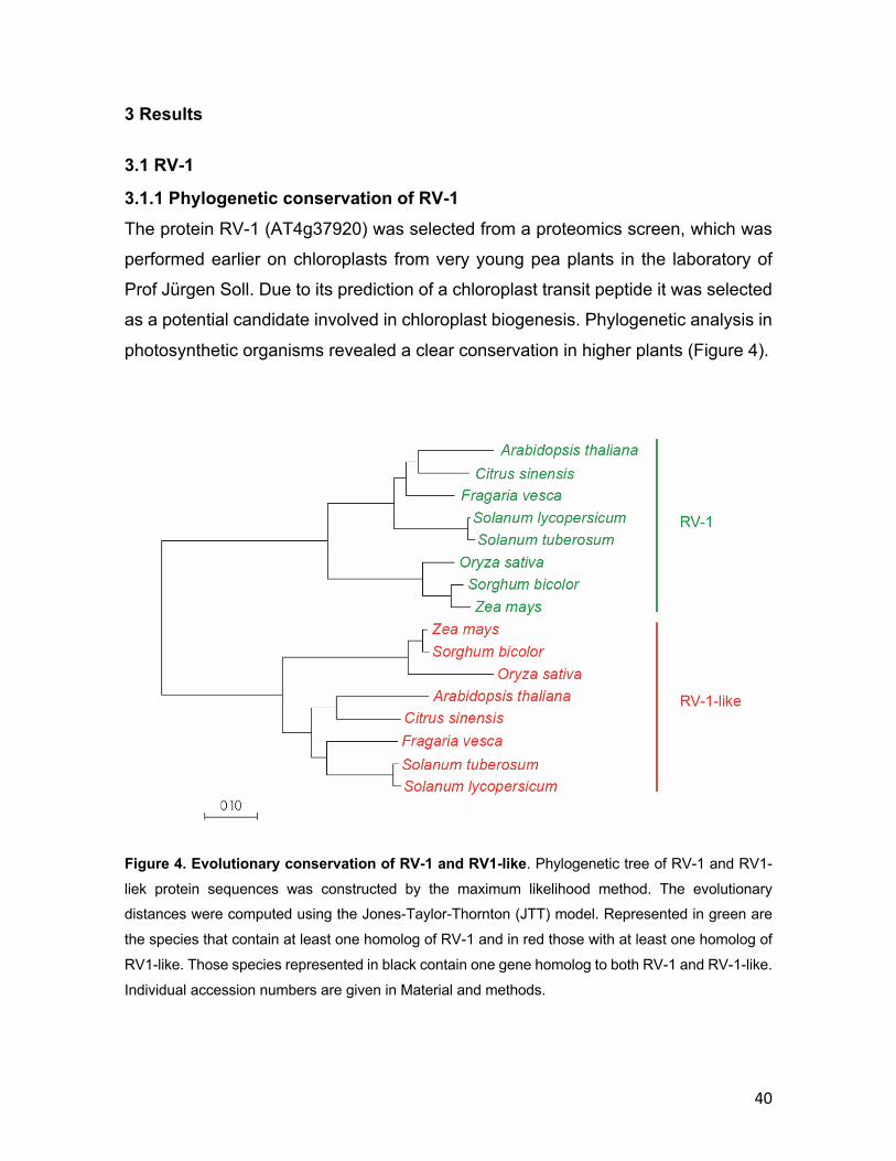
40
3 Results
3.1 RV-1
3.1.1 Phylogenetic conservation of RV-1 The protein RV-1 (AT4g37920) was selected from a proteomics screen, which was
performed earlier on chloroplasts from very young pea plants in the laboratory of
Prof Jürgen Soll. Due to its prediction of a chloroplast transit peptide it was selected
as a potential candidate involved in chloroplast biogenesis. Phylogenetic analysis in
photosynthetic organisms revealed a clear conservation in higher plants (Figure 4).
Figure 4. Evolutionary conservation of RV-1 and RV1-like. Phylogenetic tree of RV-1 and RV1-
liek protein sequences was constructed by the maximum likelihood method. The evolutionary distances were computed using the Jones-Taylor-Thornton (JTT) model. Represented in green are
the species that contain at least one homolog of RV-1 and in red those with at least one homolog of
RV1-like. Those species represented in black contain one gene homolog to both RV-1 and RV-1-like.
Individual accession numbers are given in Material and methods.
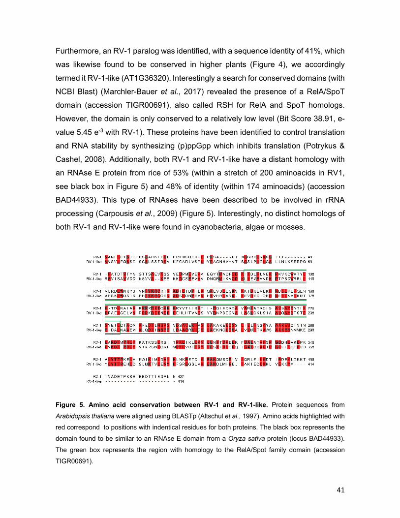
41
Furthermore, an RV-1 paralog was identified, with a sequence identity of 41%, which
was likewise found to be conserved in higher plants (Figure 4), we accordingly
termed it RV-1-like (AT1G36320). Interestingly a search for conserved domains (with
NCBI Blast) (Marchler-Bauer et al., 2017) revealed the presence of a RelA/SpoT
domain (accession TIGR00691), also called RSH for RelA and SpoT homologs.
However, the domain is only conserved to a relatively low level (Bit Score 38.91, e-
value 5.45 e-3 with RV-1). These proteins have been identified to control translation
and RNA stability by synthesizing (p)ppGpp which inhibits translation (Potrykus &
Cashel, 2008). Additionally, both RV-1 and RV-1-like have a distant homology with
an RNAse E protein from rice of 53% (within a stretch of 200 aminoacids in RV1,
see black box in Figure 5) and 48% of identity (within 174 aminoacids) (accession
BAD44933). This type of RNAses have been described to be involved in rRNA
processing (Carpousis et al., 2009) (Figure 5). Interestingly, no distinct homologs of
both RV-1 and RV-1-like were found in cyanobacteria, algae or mosses.
Figure 5. Amino acid conservation between RV-1 and RV-1-like. Protein sequences from
Arabidopsis thaliana were aligned using BLASTp (Altschul et al., 1997). Amino acids highlighted with red correspond to positions with indentical residues for both proteins. The black box represents the
domain found to be similar to an RNAse E domain from a Oryza sativa protein (locus BAD44933).
The green box represents the region with homology to the RelA/Spot family domain (accession
TIGR00691).
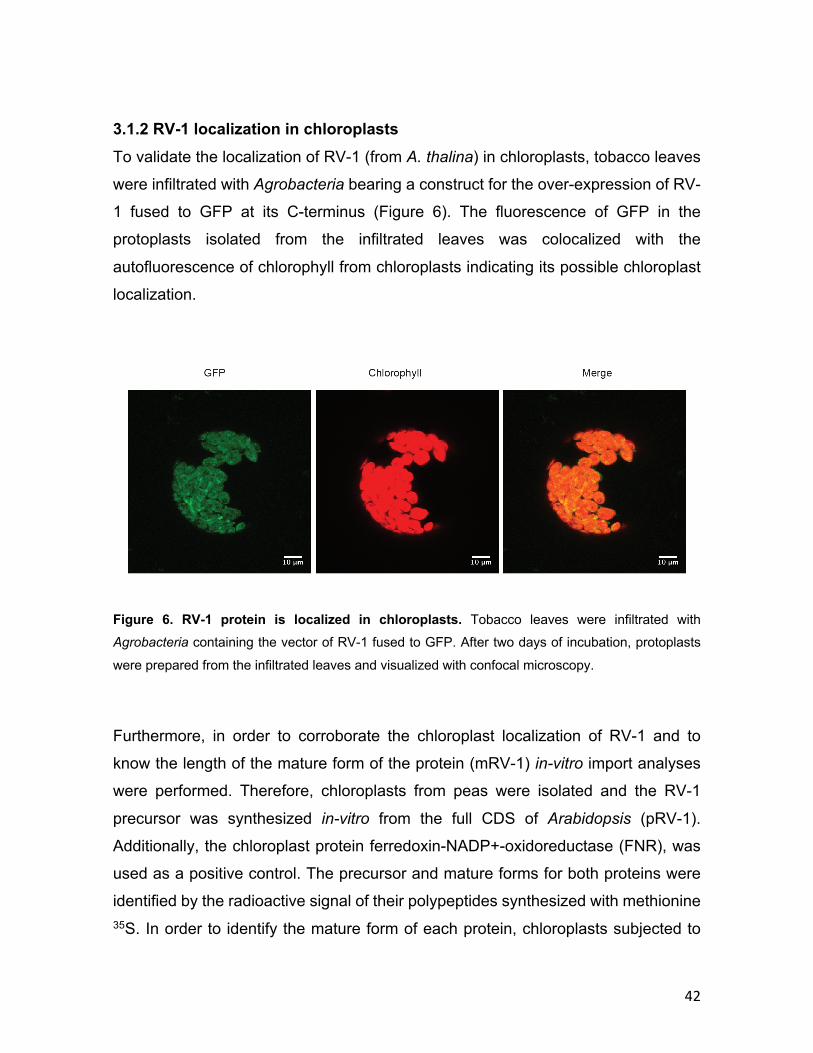
42
3.1.2 RV-1 localization in chloroplasts
To validate the localization of RV-1 (from A. thalina) in chloroplasts, tobacco leaves
were infiltrated with Agrobacteria bearing a construct for the over-expression of RV-
1 fused to GFP at its C-terminus (Figure 6). The fluorescence of GFP in the
protoplasts isolated from the infiltrated leaves was colocalized with the
autofluorescence of chlorophyll from chloroplasts indicating its possible chloroplast
localization.
Figure 6. RV-1 protein is localized in chloroplasts. Tobacco leaves were infiltrated with Agrobacteria containing the vector of RV-1 fused to GFP. After two days of incubation, protoplasts
were prepared from the infiltrated leaves and visualized with confocal microscopy.
Furthermore, in order to corroborate the chloroplast localization of RV-1 and to
know the length of the mature form of the protein (mRV-1) in-vitro import analyses
were performed. Therefore, chloroplasts from peas were isolated and the RV-1
precursor was synthesized in-vitro from the full CDS of Arabidopsis (pRV-1).
Additionally, the chloroplast protein ferredoxin-NADP+-oxidoreductase (FNR), was
used as a positive control. The precursor and mature forms for both proteins were
identified by the radioactive signal of their polypeptides synthesized with methionine 35S. In order to identify the mature form of each protein, chloroplasts subjected to
43
import were further treated with thermolysin for the digestion of non-imported
precursors. As a result, the FNR protein showed a size shift corresponding to the
cleavage of its TP to produce its mature form and validating the competence of the
chloroplast to perform import. For RV-1, the import showed also a size shift from its
precursor into a mature form of 45 kDa (Figure 7A). In silico analysis for RV-1
predicted a TP of 6.8 kDa (analysis done using ChloroP software) (Emanuelsson et
al., 1999), however the experimental data revealed that the true TP consists of about
the first 34 aminoacids (with a size of 3.8 kDa). This result supports the in-vivo
localization observed by the fluorescence of GFP fused to RV-1 indicating that it is
indeed localized in the chloroplast.
Additionally, the mature form of RV-1 was heterologously over-expressed in E. coli
and purified, from which antibodies were raised. Chloroplast sub-compartments
were separated in order to identify RV-1 localization within the chloroplast. As
expected, due to lack of hydrophobic regions on its sequence, RV-1 was found to be
located in the stroma of chloroplasts (Figure 7B). The band obtained in the
immunoblot matches with the size of the mature form of RV-1 of 45 kDa observed in
the in in-vitro import, thus corroborating the correct size of the protein. The
distribution of RV-1 in the different subcompartments was observed by its
accumulation in the stroma as it also does the protein fructose-1,6-
bisphosphate (FBPase) which was used as a marker for stroma proteins. On the
other hand, marker proteins for thylakoids as LHCbII, inner envelope TIC110 and
outer envelope OEP37 showed the purity of the fractions indicating the absence of
RV-1 in these
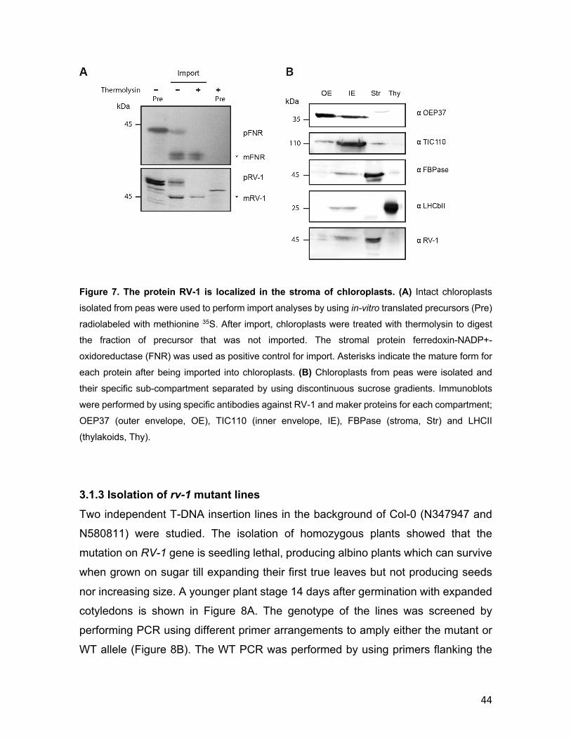
44
Figure 7. The protein RV-1 is localized in the stroma of chloroplasts. (A) Intact chloroplasts
isolated from peas were used to perform import analyses by using in-vitro translated precursors (Pre)
radiolabeled with methionine 35S. After import, chloroplasts were treated with thermolysin to digest the fraction of precursor that was not imported. The stromal protein ferredoxin-NADP+-
oxidoreductase (FNR) was used as positive control for import. Asterisks indicate the mature form for
each protein after being imported into chloroplasts. (B) Chloroplasts from peas were isolated and
their specific sub-compartment separated by using discontinuous sucrose gradients. Immunoblots
were performed by using specific antibodies against RV-1 and maker proteins for each compartment;
OEP37 (outer envelope, OE), TIC110 (inner envelope, IE), FBPase (stroma, Str) and LHCII
(thylakoids, Thy).
3.1.3 Isolation of rv-1 mutant lines Two independent T-DNA insertion lines in the background of Col-0 (N347947 and
N580811) were studied. The isolation of homozygous plants showed that the
mutation on RV-1 gene is seedling lethal, producing albino plants which can survive
when grown on sugar till expanding their first true leaves but not producing seeds
nor increasing size. A younger plant stage 14 days after germination with expanded
cotyledons is shown in Figure 8A. The genotype of the lines was screened by
performing PCR using different primer arrangements to amply either the mutant or
WT allele (Figure 8B). The WT PCR was performed by using primers flanking the
45
CDS region of the gene, whereas for the mutant PCR the insertion primer from GABI-
Kat was used with either forward CDS primer (for the line N347947) or reverse CDS
primer (for the line N580811) (Figure 8C upper panel). Furthermore, the line
N347947 was complemented by transforming heterozygous plants with Agrobacteria
bearing a construct for the over-expression of RV-1 under the control of the 35S
promotor. The complementation line was checked by PCR for its homozygous
mutant genotype along with the presence of the transgene (Figure 8C, lower panel).
The PCR arrangement done by amplifying the mutant allele as explained above but
in this case the WT PCR using CDS forward and reverse primers were used to
amplify the transgene which was 35S::RV-1. In order to identify if the plants had
heterozygous or homozygous genotype from the insertion, the WT PCR was
repeated by using the forward CDS primer with a reverse primer annealing in the 3’
UTR region which ensures amplification just in case the full sequence is present and
differs in size from the previous PCR because of the additional intron sequences.
The phenotype was completely restored in the complemented line (data not shown).

46
Figure 8. Homozygous rv-1 mutant present pale phenotype. (A) Heterozygous seeds from the
exon insertional line N347947 were sowed on MS plus 1% sugar plates for 2 weeks. The genotype
of the plants was checked by PCR confirming the pale phenotype for the homozygous plants
(N347947, rv-1). (B) Gene scheme represents the position of the insertion lines N347947 and
N580811. The position of the primers used for genotyping are represented as arrows. (C) Genotyping was performed by using gDNA from lines N347947 and N580811 (upper panel). For line N347947,
the mutant allele PCR (m) was performed by using the primers WF and GF on (A). The line N580811
was genotyped performing PCR for the mutant allele using WR and GF primers. The asterisk
indicates an unspecific amplicon. WT PCR was performed using WF and WR primers (W). On the
lower panel is shown the genotyping for the complementation of the line N347947. The WT PCR (W2)
was performed by using WF and WR2 whereas the mutant PCR was performed as mentioned above.
From this point the line N347947 (henceforth referred to as rv-1) was used for further
studies since both lines showed the same phenotype in homozygous mutants (data
not shown). Ultrastructure images of chloroplasts on the homozygous rv-1 showed
an aberrant chloroplast morphology with the absence of thylakoid membranes.
Overall aberrant chloroplasts in rv-1 were strongly reduced in size in comparison to

47
WT (Figure 9). Accumulation of large vesicle was observed in every chloroplast and
their sizes were also very different from one another.
Figure 9. Homozygous rv-1 mutant affected in chloroplast morphology. (D) Heterozygous seeds
were sowed on MS plus 1% sugar for 3 weeks and true leaves from WT and homozygous plants
(N347947, rv-1) were excised for electron microscopy analyses. Pictures taken by Prof Andreas
Klingl.

48
3.1.4 Translation is impaired in rv-1 Since the homozygous mutant plants showed undeveloped chloroplasts and were
albino, the ability to perform translation was assessed. Therefore, 21 days old
seedlings were infiltrated with radiolabeled methionine 35S and then incubated under
light exposure for 20 min. Proteins were extracted from the seedlings and separated
into soluble and membrane proteins. In the WT, translation of the large subunit of
RubisCO was very prominent in the soluble fraction as well for the D1 subunit of PSII
in the membrane fraction. Surprisingly, the mutant rv-1 showed barely any
translation in comparison to WT seedlings (Figure 10A). Interestingly, it could be
observed in the CBB gel even a high reduction in RubisCO content compared with
the total soluble proteins in the rv-1 mutant as well for LHCs in the membrane fraction
in comparison to WT. This observation posed the question about the absence or
presence of plastid ribosomes. This was investigated by isolating total proteins from
WT and rv-1 mutant seedlings and performing immunoblots using specific antibodies
against members of the 50S subunit of plastid ribosomes, RPL2 and RPL4.
Interestingly, this showed the absence of these in the mutant rv-1 seedlings
compared to WT (Figure 10B). Since absence of thylakoid membranes was
observed from ultrastructure pictures, the protein TIC110 present in the inner
envelope of the chloroplast was used as a loading control and to monitor the
presence of plastid ribosomes, two proteins of the large ribosomal subunit were for
immunoblot analyses.
Furthermore, since ribosomes subunits are complexes of protein and rRNA, the
presence of the rRNA was analyzed by isolating total RNA from WT and rv-1 mutant
seedlings. As expected, rv-1 showed high reduction of the chloroplast rRNAs, both
of the 30S and 50S ribosomal subunit, 16S and 23S rRNA, respectively. In
comparison to WT levels (Figure 10C). However, the 25S and the 18S were even
slightly more abundant in the mutant compared to WT indicating that the problem of
translation or ribosomal stability relays on the plastid ribosomes.
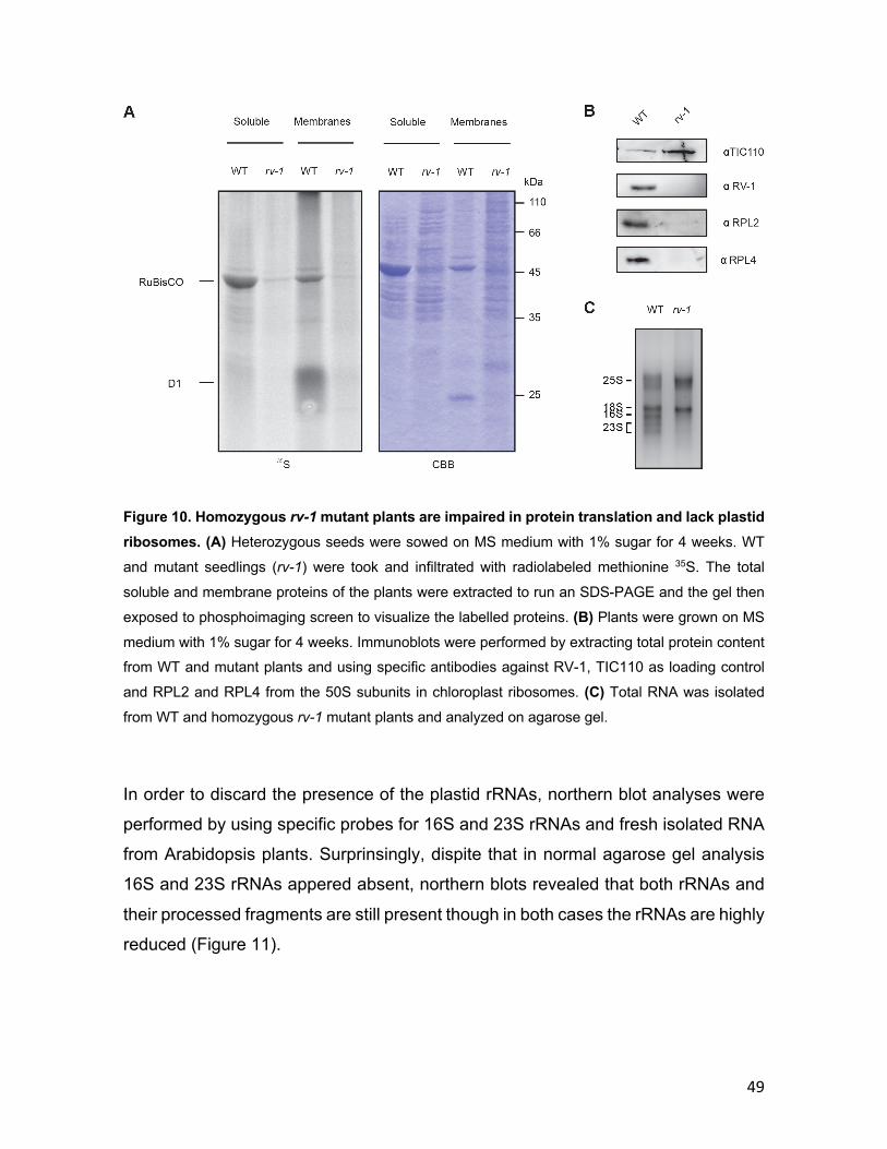
49
Figure 10. Homozygous rv-1 mutant plants are impaired in protein translation and lack plastid ribosomes. (A) Heterozygous seeds were sowed on MS medium with 1% sugar for 4 weeks. WT
and mutant seedlings (rv-1) were took and infiltrated with radiolabeled methionine 35S. The total
soluble and membrane proteins of the plants were extracted to run an SDS-PAGE and the gel then
exposed to phosphoimaging screen to visualize the labelled proteins. (B) Plants were grown on MS
medium with 1% sugar for 4 weeks. Immunoblots were performed by extracting total protein content from WT and mutant plants and using specific antibodies against RV-1, TIC110 as loading control
and RPL2 and RPL4 from the 50S subunits in chloroplast ribosomes. (C) Total RNA was isolated
from WT and homozygous rv-1 mutant plants and analyzed on agarose gel.
In order to discard the presence of the plastid rRNAs, northern blot analyses were
performed by using specific probes for 16S and 23S rRNAs and fresh isolated RNA
from Arabidopsis plants. Surprinsingly, dispite that in normal agarose gel analysis
16S and 23S rRNAs appered absent, northern blots revealed that both rRNAs and
their processed fragments are still present though in both cases the rRNAs are highly
reduced (Figure 11).
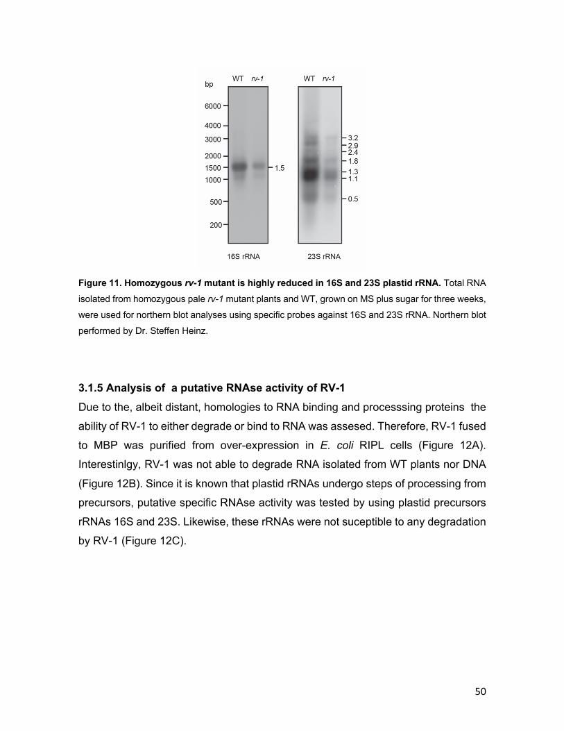
50
Figure 11. Homozygous rv-1 mutant is highly reduced in 16S and 23S plastid rRNA. Total RNA
isolated from homozygous pale rv-1 mutant plants and WT, grown on MS plus sugar for three weeks,
were used for northern blot analyses using specific probes against 16S and 23S rRNA. Northern blot
performed by Dr. Steffen Heinz.
3.1.5 Analysis of a putative RNAse activity of RV-1 Due to the, albeit distant, homologies to RNA binding and processsing proteins the
ability of RV-1 to either degrade or bind to RNA was assesed. Therefore, RV-1 fused
to MBP was purified from over-expression in E. coli RIPL cells (Figure 12A).
Interestinlgy, RV-1 was not able to degrade RNA isolated from WT plants nor DNA
(Figure 12B). Since it is known that plastid rRNAs undergo steps of processing from
precursors, putative specific RNAse activity was tested by using plastid precursors
rRNAs 16S and 23S. Likewise, these rRNAs were not suceptible to any degradation
by RV-1 (Figure 12C).
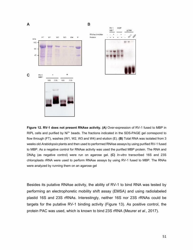
51
Figure 12. RV-1 does not present RNAse activity. (A) Over-expression of RV-1 fused to MBP in
RIPL cells and purified by Ni2+ beads. The fractions indicated in the SDS-PAGE gel correspond to
flow through (FT), washes (W1, W2, W3 and W4) and elution (E). (B) Total RNA was isolated from 3
weeks old Arabidopsis plants and then used to performed RNAse assays by using purified RV-1 fused
to MBP. As a negative control for RNAse activity was used the purified MBP protein. The RNA and
DNAg (as negative control) were run on agarose gel. (C) In-vitro transcribed 16S and 23S chloroplasts rRNA were used to perform RNAse assays by using RV-1 fused to MBP. The RNAs
were analyzed by running them on an agarose gel
Besides its putative RNAse activity, the ability of RV-1 to bind RNA was tested by
performing an electrophoretic mobility shift assay (EMSA) and using radiolabeled
plastid 16S and 23S rRNAs. Interestingly, neither 16S nor 23S rRNAs could be
targets for the putative RV-1 binding activity (Figure 13). As positive control, the
protein PAC was used, which is known to bind 23S rRNA (Meurer et al., 2017).
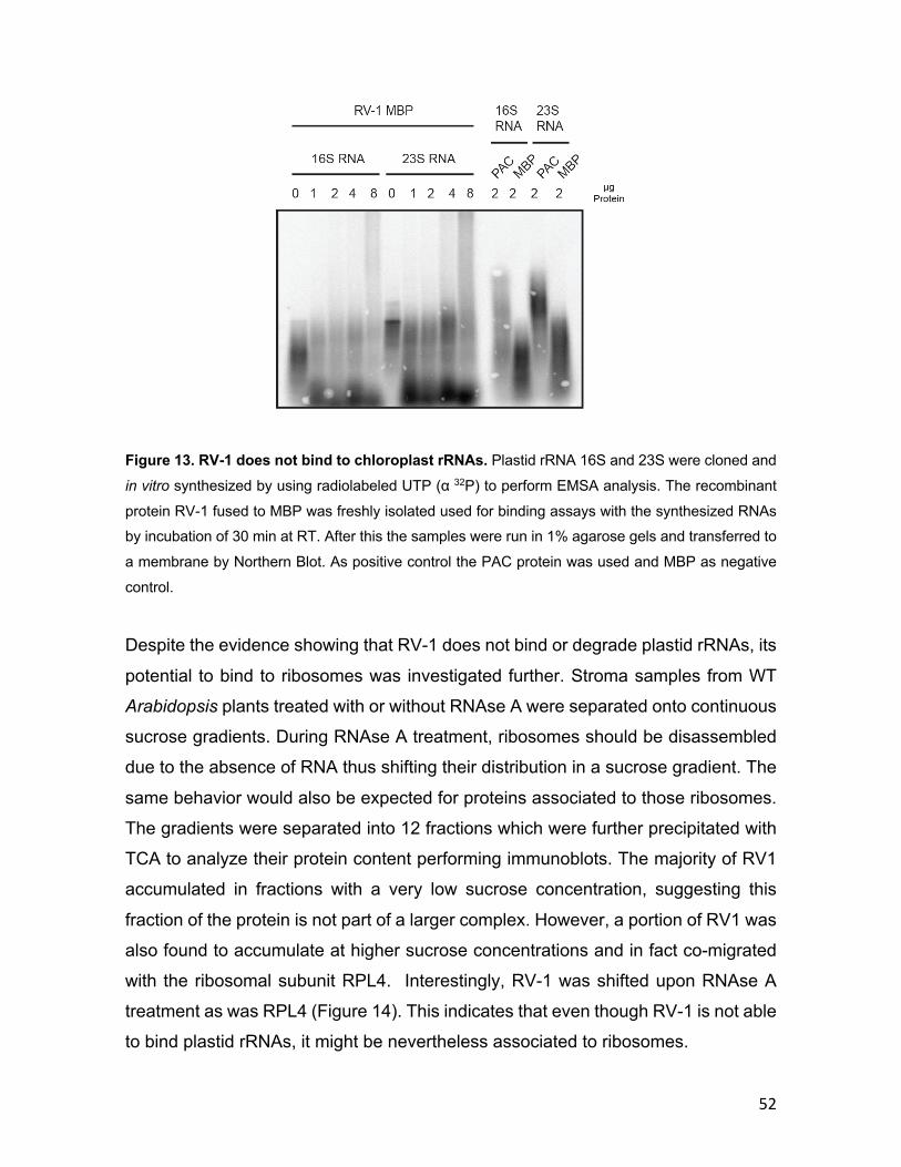
52
Figure 13. RV-1 does not bind to chloroplast rRNAs. Plastid rRNA 16S and 23S were cloned and
in vitro synthesized by using radiolabeled UTP (α 32P) to perform EMSA analysis. The recombinant
protein RV-1 fused to MBP was freshly isolated used for binding assays with the synthesized RNAs by incubation of 30 min at RT. After this the samples were run in 1% agarose gels and transferred to
a membrane by Northern Blot. As positive control the PAC protein was used and MBP as negative
control.
Despite the evidence showing that RV-1 does not bind or degrade plastid rRNAs, its
potential to bind to ribosomes was investigated further. Stroma samples from WT
Arabidopsis plants treated with or without RNAse A were separated onto continuous
sucrose gradients. During RNAse A treatment, ribosomes should be disassembled
due to the absence of RNA thus shifting their distribution in a sucrose gradient. The
same behavior would also be expected for proteins associated to those ribosomes.
The gradients were separated into 12 fractions which were further precipitated with
TCA to analyze their protein content performing immunoblots. The majority of RV1
accumulated in fractions with a very low sucrose concentration, suggesting this
fraction of the protein is not part of a larger complex. However, a portion of RV1 was
also found to accumulate at higher sucrose concentrations and in fact co-migrated
with the ribosomal subunit RPL4. Interestingly, RV-1 was shifted upon RNAse A
treatment as was RPL4 (Figure 14). This indicates that even though RV-1 is not able
to bind plastid rRNAs, it might be nevertheless associated to ribosomes.

53
Figure 14. RV-1 forms RNAase A sensitive complexes that co-migrate with ribosomes. Stroma
fractions from peas chloroplast were isolated and treated with RNAse A or RNAse inhibitor. These samples were separated in sucrose gradients for immunoblot analyses of the different fractions using
specific antibodies against RV-1 and the 50S subunit of chloroplasts ribosomes RPL4.
In order to analyze the function of RV-1 in more detail, RNA interference (RNAi) lines
for the RV-1 gene were generated in Col-0 background. As expected, over-
expressing RNAi lines were affected in greening of leaves as well as overall fitness
of the plants. However, they reached adulthood and were able to produce seeds
(Figure 15A). In order to verify that the phenotype correlates with a decrease in RV-
1 levels, the down-regulation of RV-1 expression due to RNAi was tested by
performing immunoblots. As expected, RV-1 is highly reduced in the RNAi mutant
line in comparison to the WT, and therefore it validates the affected phenotype in the
RNAi line (Figure 15B). Interestingly, when analyzing the ultrastructure of chloroplast
of the RNAi line 1 in comparison to WT it is possible to observed severly affected
chloroplast similar to Figure 9. Nevertheless, chloroplast containing thylakoid
membranes are also observed though also affected with reduced grana stacks in
comparison to WT (Figure 15C). These lines will be useful for further analysis to
monitor their chloroplast function and content since they can grow on soil being able
to produce enough plant material.

54
Figure 15. Down-regulation of RV-1 affects Arabidopsis plant development at all stages. (A) WT plants were stably transformed with a construction for down-regulation of RV1 expression by
RNAi in the vector pOpOff2. Stable lines affected in their development were selected. Plants were
grown on soil for 3 weeks and just two out of three survived to produced seeds. (B) Immunoblots
were done by isolating total protein from the RNAi line 1 and WT plants. Specific antibodies were
used to detect RV-1 and as loading control TIC110. (C) Arabidopsis plants from the RV-1 RNAi line
1 and WT were grown on soil for 3 weeks under long day conditions and their leaves were excised
for electron microscopy analyses. The scale bars represent for each case 500 nm. Pictures taken by
Prof Andreas Klingl.
55
3.1.6 Characterization of the RV-1-like gene As mentioned above, RV-1 has a paralog, termed RV-1-like. In-silico analysis for its
localization using the online server TargetP 1.1 showed chloroplast predicted TP
(Emanuelsson et al., 2000). Therefore, we proceeded to investigate the localization
of RV-1-like experimentally. The expression of a RV-1-like-GFP fusion protein in
tobacco leaves was analyzed. In addition to infiltrating leaves RV-1-like-GFP
construct, protoplast extracted from those leaves were further treated with
Mitotracker orange to visualize mitochondria on the microscope. Surprisingly, the
GFP signal obtained did not colocalize with chlorophyll but with the mitotracker signal
suggesting that RV-1-like is localized in the plant mitochondria (Figure 16).
Figure 16. RV-1-like is localized in plant mitochondria. Tobacco leaves were infiltrated with
Agrobacteria containing the vector of RV-1-like fused to GFP. After two days of incubation, protoplasts
were prepared from the infiltrated leaves. Protoplast were incubated with Mitotracker and then
visualized with confocal microscopy.
In order to understand the relevance of RV-1-like for plant growth, a T-DNA mutant
line, with the T-DNA located in the last exon of RV-1-like gene, was used for further
studies (GK844F05, henceforth referred to as rv-1-like) (Figure 17A). Genotype
analyses was performed with primers located at the fourth exon as forward and at
the last exon as reverse for the WT PCR. On the other hand, for the mutant PCR it
was used the forward GABI-kat primer with the forward primer at the fourth exon of
RV-1-like gene. This showed that the heterozygous plants did not produce
56
homozygous individuals but rather a distribution of 50% WT and 50% heterozygous
plants (analyzing around 50 individual plants). Therefore, the siliques of the
heterozygous plants were analyzed in order to identify aborted embryos which might
explain the absence of homozygous plants in the segregation of the heterozygous
rv-1-like line. Indeed, the siliques from heterozygous rv-1-like plants showed empty
spaces (Figure 17C). Furthermore, heterozygous rv-1-like showed a reduced
amount of seeds per silique and an increased number of empty spaces or aborted
embryos per silique (Table 6).
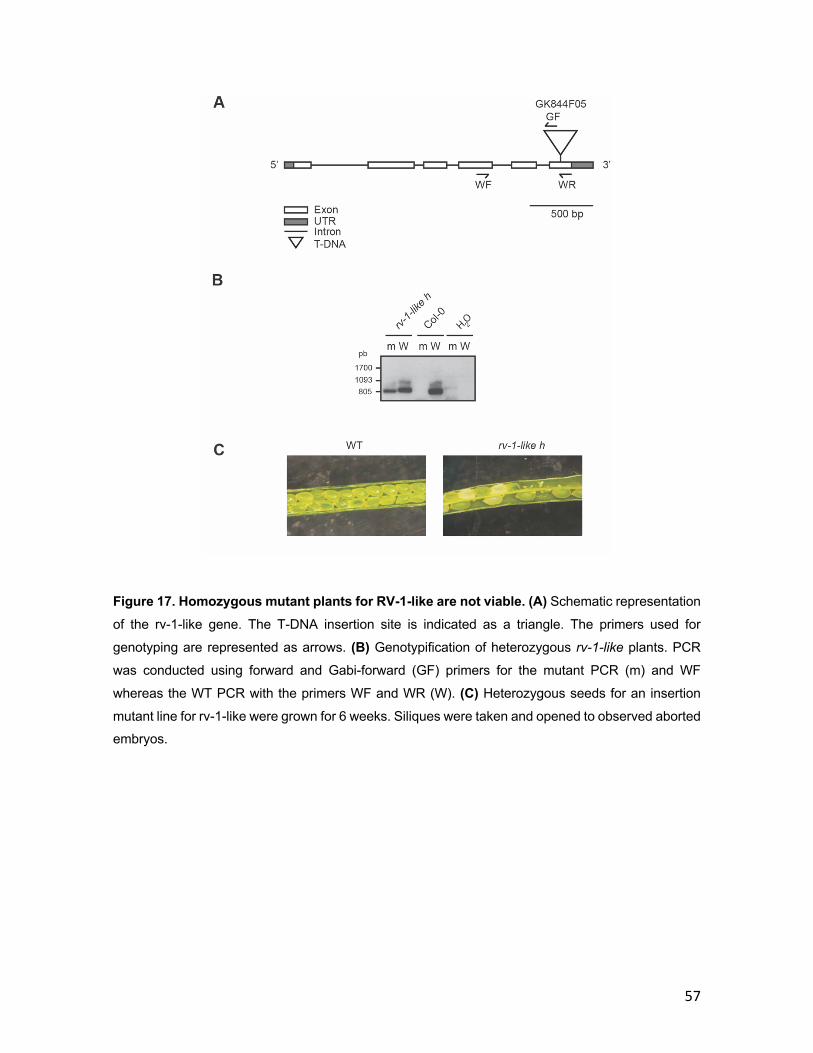
57
Figure 17. Homozygous mutant plants for RV-1-like are not viable. (A) Schematic representation
of the rv-1-like gene. The T-DNA insertion site is indicated as a triangle. The primers used for
genotyping are represented as arrows. (B) Genotypification of heterozygous rv-1-like plants. PCR
was conducted using forward and Gabi-forward (GF) primers for the mutant PCR (m) and WF
whereas the WT PCR with the primers WF and WR (W). (C) Heterozygous seeds for an insertion
mutant line for rv-1-like were grown for 6 weeks. Siliques were taken and opened to observed aborted embryos.

58
Genotype
Nº of seeds / Silique
Aborted embryos / Silique
WT 44.5 ± 1.2 0
rv-1-like h 20.5 ± 2 * 10 ± 0.6 *
Table 6. Heterozygous rv1-like mutant plants present aborted embryos and reduced seeds in their siliques. Seeds from the heterozygous mutant line (rv-1-like h) were sowed and plants grew for
6 weeks on soil. Heterozygous and WT plants were identified by genotyping them with PCR and the siliques were used to count their number of seeds as well as aborted embryos per silique. Asterisks
represent significant differences (P < 0.05) in comparison to the WT based on the t-test. The mean
of seeds and aborted embryos from 10 siliques taken from 3 independent plants are represented
along with the SEMs.
The phenotype for RV-1 and RV-1-like demonstrated that both genes are essential
for viability of the plant. Loss of function of RV-1 alters not just chloroplast
morphology but also the plastid ribosomes stability or assembly which provides
insight on its possible function. Whereas RV-1-like is most likely localized in the plant
mitochondria but still impacting plant viability in its loss of function with the inability
of producing homozygous individuals. However, the exact molecular function of both
proteins remains to be invesigated.
3.2 PG18
3.2.1 Characterization of PG18 Similar to RV1, PG18 (AT4G13200) was selected from the prevoisly performed
proteomic study. Interestingly, this protein was also found in a proteome analysis
from PG of Arabidiopsis (Lundquist et al., 2012). It has no kown protein domains and
phylogenetic analyis showed that PG18 is conserved from higher plants to
cyanobacteria (Figure 18).
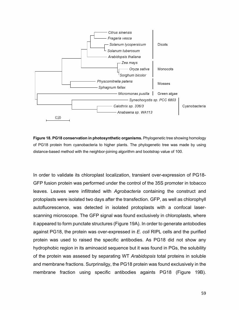
59
Figure 18. PG18 conservation in photosynthetic organisms. Phylogenetic tree showing homology
of PG18 protein from cyanobacteria to higher plants. The phylogenetic tree was made by using
distance-based method with the neighbor-joining algorithm and bootstrap value of 100.
In order to validate its chloroplast localization, transient over-expression of PG18-
GFP fusion protein was performed under the control of the 35S promoter in tobacco
leaves. Leaves were infiltrated with Agrobacteria containing the construct and
protoplasts were isolated two days after the transfection. GFP, as well as chlorophyll
autofluorescence, was detected in isolated protoplasts with a confocal laser-
scanning microscope. The GFP signal was found exclusively in chloroplasts, where
it appeared to form punctate structures (Figure 19A). In order to generate antobodies
against PG18, the protein was over-expressed in E. coli RIPL cells and the purified
protein was used to raised the specific antibodies. As PG18 did not show any
hydrophobic region in its aminoacid sequence but it was found in PGs, the solubility
of the protein was assesed by separating WT Arabidopsis total proteins in soluble
and membrane fractions. Surprinsilgy, the PG18 protein was found exclusively in the
membrane fraction using specific antibodies againts PG18 (Figure 19B).
60
Furthermore, in order understand the assosiation of PG18 with membranes,
thylakoids were isolated and washed with several buffers. Then the membrane
fraction was treated with 1 M NaCl, 0.1 M Na2CO3, and 3 M urea to ascertain whether
PG18 is attached to the membrane by hydrophobic or electrostatic interactions and
could therefore be removed by any of these agents (Figure 19C). Incubation with
buffer (HEPES/KOH 5 mM, EDTA 5 mM, pH 7.5) served as a negative control and
disruption of the membrane with 1% LDS was applied to achieve total solubilization
of the membrane. Only the treatment with 1% LDS resulted in complete solubilization
of PG18. However, a small amount of PG18 was found in the supernatant after
treatment with 3 M urea (Figure 19C). To assess the efficiency of these treatments,
the blot was probed with OE33 antiserum. OE33 is a peripheral lumenal protein and
is at least partially extracted by each of the agents mentioned above, as they all
damage the integrity of the thylakoid membrane (Figure 19C) (Bhuiyan et al., 2015).
Cytochrome f (Cyt f) was used as an example of an integral membrane protein and,
as expected, it is solubilized only by treatment with 1% LDS (Figure 19C). Several
proteomics studies have identified PG18 in PGs (Friso et al., 2004; Peltier et al.,
2004; Ytterberg et al., 2006) and therefore it was set out to test this further. To this
end, pea chloroplasts were fractionated into PGs and thylakoid membranes. Pea
plants were chosen instead of Arabidopsis for this experiment because they provide
larger amounts of leaf material. Thylakoid membranes and PGs were separated on
a sucrose density gradient, and selected fractions were subjected to SDS-PAGE and
subsequently probed with antisera against LHC1a as a thylakoid marker,
plastoglobulin 35 (PGL35) as a PG marker and PG18 (Figure 19D). As revealed by
the distribution of the PG marker protein PGL35, the PG fraction was not
contaminated with thylakoids, and the majority of PG18 was also detected in this
fraction. Since PGs are known to be associated with the thylakoid membrane (Austin
et al., 2006), it is not surprising that small amounts of both PG18 and PGL35 are
detected in the thylakoid fraction. Additionally, in order to demonstrate that PG18 is
located at the surface of the PGs, intact thylakoids and their associated PGs were
treated with trypsin (Figure 19E). As expected, PG18 was completely digested – as
was the alpha subunit of ATP synthase, which is exposed on the stroma side of the
61
thylakoids – while the lumenal OE33 protein used as a control for thylakoid integrity
remained intact. Taken together, the observed punctate structures of PG18-GFP,
the sub-fractionation data, and the fact that PG18 has been assigned to PGs by
mass spectrometric analyses (Lundquist et al., 2012) led to the conclusion that PG18
is indeed a PG-localized protein. Considering that none of the PG proteins identified
so far harbor transmembrane domains but are nevertheless most likely to be
integrated into the lipid monolayer surface of PGs, the observation that PG18
behaves similarly to an integral membrane protein upon treatment with various
chaotropic agents is compatible with the protein’s localization to PGs.

62

63
Figure 19. PG18 is a membrane-associated protein in PGs. (A) Tobacco leaves were infiltrated
with Agrobacteria containing a vector expressing PG18 fused to GFP (see Materials and methods).
Protoplasts were prepared from the infiltrated leaves and visualized with confocal microscopy. (B) A total protein extract from Arabidopsis was separated into a membrane (M) and a soluble fraction (S).
PG18 was detected on immunoblots using specific antibodies. Antibodies raised against TIC110 (a
membrane protein marker) and FBPase (a soluble protein marker) were used as controls. (C) Disrupted Arabidopsis thylakoids were extracted with salt-containing buffers and separated into
supernatant (S) and pellet (P) fractions by centrifugation. The samples were fractionated by SDS-
PAGE and analyzed immunologically using antibodies specific for PG18, Cyt f and OE33,
respectively. As a control for the specificity of the anti-PG18 antibody, isolated chloroplasts from the
mutant line pg18 were also loaded onto the gel. (D) Chloroplast membranes were isolated from 3-
week-old peas. The samples were then further fractionated on a discontinuous sucrose gradient (the
number of the fraction is indicated above each lane). Immunoblotting was performed and the blot was
probed for the presence of the marker proteins for thylakoids (LHCA1) and plastoglobuli (PGL35). (E) Arabidopsis thylakoids were isolated from plants grown on soil for three weeks. Trypsin digestion of
thylakoids was performed on ice for 10 min and the samples were analyzed after immunoblotting. An
antiserum directed against the ATP synthase α subunit was used to ensure that the stromal side of
the thylakoid was accessible to the enzyme, whereas the lumen protein OE33 served as a marker for
the integrity of the thylakoids. PGL35 was used as a PG control.
3.2.2 Characterization of pg18 mutant lines Two independent T-DNA insertion lines in the background Col-0 (N442085 and
N459729) were obtained (Figure 20A). The genotype of the line N442085 was
screened by performing PCR (Figure 20B) and was then used for further studies
(henceforth referred to as pg18). The WT PCR was performed by using forward and
reverse primers flanking the full CDS sequence of PG18. Whereas for the mutant
PCR it was assessed two combination of primers which was the GABI-kat forward
primer with either the reverse or forward primer for the CDS sequence of PG18.

64
Figure 20. pg18 mutant lines isolation. (A) Schematic representation of the pg18 gene. The T-DNA
insertion site is indicated as a triangle. The primers used for genotyping are represented as arrows.
(B) Genotypification of pg18 mutant plants (line N442085). WT PCR was conducted using the
primers WF and WR (W). On the other hand, mutant PCR was done by using Gabi-forward primers
(GF) with either WF (m1) or WR (m2) primers. The asterisk indicates an unspecific amplicon and the H indicates homozygous and h to heterozygous plant for the line N442085.
The mutant was viable on soil but displayed a pale green phenotype under normal
lighting (NL) conditions (21°C, 120 µmol / m2s light, 16 / 8 h light / dark). Since it has
been observed that PGs become more abundant under light stress (Zhang et al.,
2010), mutant plants were also exposed to increased light intensities (IL, 250
µmol/m2s) for 3.5 days, following 17.5 days growth under NL conditions (Figure 21A).
To ensure that the phenotype correlated with the disruption of the PG18 gene, the
mutant pg18 line was complemented with a PG18-GFP fusion construct expressed
under the control of a 35S promoter. A representative example of a complemented
line is shown in Figure 21A. The knock-out of PG18 was verified at the protein level
using the specific antiserum (Figure 21B). Total protein extracts of WT and pg18
were probed with the PG18 antiserum, which detected a specific band at 18 kDa in
the WT that was absent in the mutant. An antiserum raised against the chloroplast
protein TIC110 was used as a loading control. In addition, proteins were extracted
65
from the complemented mutant line and likewise probed with PG18 antiserum. A
protein of 45 kDa was detected, corresponding to the expected size of PG18-GFP.
These results demonstrate the specificity of the generated antiserum, as well as
successful complementation of the mutant. It has been reported that PG proteins
are strongly expressed under increased light (IL) – with the exception of PG18
(Ytterberg et al., 2006). To verify this, levels of PG18 in plants were monitored under
NL and IL conditions. As expected, P18 is equally abundant in NL and IL plants,
while expression of the core PG protein PGL35 increases upon exposure to high
light levels (Figure 21C).
Since the pale phenotype indicated a defect in chloroplasts, the phenotype was
analyzed at the ultrastructural level (Figure 21D). Interestingly, when examined at
higher magnification, the stroma lamellae in pg18 mutant chloroplasts are shorter
and appear to be stacked, rather than present in single layers. Moreover, the stroma
lamellae exhibit less branching between grana stacks, while the latter display a more
compact organization and generally consist of more layers in the mutant than in the
WT or the complemented line. This effect is observed under both NL and IL
conditions, although it is more pronounced in IL (Figure 21D). In comparison to the
WT, the cross-sectional area of pg18 chloroplasts is significantly smaller under both
conditions (Figure 21E). Interestingly, under both light conditions, PG abundance
per chloroplast is also reduced in the mutant chloroplast in comparison to WT. The
number of PGs increases under IL in the WT by 100%. Strikingly, this effect of
exposure to IL is also observed in the mutant, in spite of the overall reduction in the
numbers of PGs (Figure 21F).

66

67
Figure 21. PG18 is required for normal development of A. thaliana. (A) WT, homozygous mutant
(pg18) and complemented mutant plants (35S::PG18-GFP) were grown in long-day conditions for
three weeks (normal light, NL) and for 17.5 days under 120 µmol photons m-2 s-1 followed by 3.5 days
under 250 µmol photons m-2 s-1 (increased light, IL). (B) Total protein extracts from WT and pg18
mutant plants were gel-fractionated and immunoblots were prepared. The PG18 protein was detected
with specific antibodies. TIC110, a protein found in the inner envelope of the chloroplast was used as a loading control. The immunolabeled PG18-GFP is highlighted with an asterisk. (C) Total protein
was extracted from plants grown under NL and IL conditions, and subjected to immunoblot analysis.
The PG18 protein was detected using the specific antibodies mentioned above. The alpha subunit of
the chloroplast ATP synthase was used as a loading control. Antibodies against PGL35 were used
to show PG accumulation under IL relative to NL conditions. (D) Plants were grown in long-day
conditions for three weeks under NL and IL as explained in (A), and the leaves were excised for
electron microscopy. Scale bars represent 500 nm. Thylakoid ultrastructure zooms in plants grown
on the panels bellow under NL or IL conditions. The scale bar corresponds to 200 nm. Pictures taken by Prof Andreas Klingl. (E) Quantification of chloroplast area (in µm2). The bars represent standard
error (SEM) and asterisks represent significant differences (P < 0.05) in comparison to the WT
(Student’s t-test). (F) Numbers of PGs per chloroplast. The bars represent the standard error (SEM)
and asterisks represent significant differences (P < 0.05) relative to the WT based on the t-test.
3.2.3 Arabidopsis pg18 mutant plants show symptoms of light stress To assess differences in thylakoid pigments that might be related to the mutant
phenotype, the pigment content of pg18 leaves was analyzed. Chlorophylls a and b
were indeed reduced in the mutant, in agreement with its pale green phenotype (20%
and 36% reduction for chlorophylls b and a respectively); however, only the
difference in chlorophyll a relative to the WT was significant (Figure 22A).
Analysis of carotenoid content revealed that β-carotene was significantly reduced in
the pg18 mutant compared to WT, irrespective of the lighting conditions during
growth. In particular, the level of the protective carotenoid lutein was reduced in the
mutant under IL conditions, whereas the neoxanthin content remained unchanged
(Figure 22B). Further pigments related to photoprotection, specifically constituents
of the xanthophyll cycle, were also analyzed. In WT plants under NL conditions,
violaxanthin (Vx), which binds to light-harvesting complexes (LHCs), is by far the
most abundant of these, but upon light stress this compound undergoes two de-
68
epoxidation steps to produce the intermediate antheraxanthin (Ax) and finally
zeaxanthin (Zx). On the return of NL, this process is reversed, with epoxidation of Zx
regenerating Vx. Interestingly, antheraxanthin (Ax) and zeaxanthin (Zx) levels were
clearly increased in the mutant, suggesting a light stress response, although the pool
of xanthophyll cycle pigments as a whole (the VAZ pool) was not markedly altered,
except under IL conditions, where the mutant pg18 showed a higher overall pigment
content (Figure 22C). However, the ratio of the xanthophyll intermediates, which
takes the de-epoxidation reactions into account reveals that both lighting conditions
are associated with significantly higher levels of de-epoxidation in the pg18 mutant
than in WT (Figure 22C), suggesting that the mutant is more susceptible to light
stress.
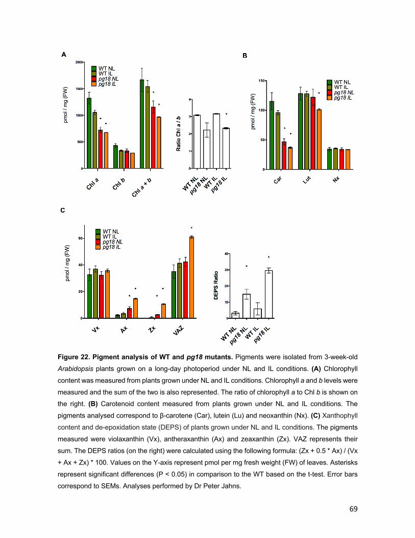
69
Figure 22. Pigment analysis of WT and pg18 mutants. Pigments were isolated from 3-week-old
Arabidopsis plants grown on a long-day photoperiod under NL and IL conditions. (A) Chlorophyll
content was measured from plants grown under NL and IL conditions. Chlorophyll a and b levels were
measured and the sum of the two is also represented. The ratio of chlorophyll a to Chl b is shown on the right. (B) Carotenoid content measured from plants grown under NL and IL conditions. The
pigments analysed correspond to β-carotene (Car), lutein (Lu) and neoxanthin (Nx). (C) Xanthophyll
content and de-epoxidation state (DEPS) of plants grown under NL and IL conditions. The pigments
measured were violaxanthin (Vx), antheraxanthin (Ax) and zeaxanthin (Zx). VAZ represents their
sum. The DEPS ratios (on the right) were calculated using the following formula: (Zx + 0.5 * Ax) / (Vx
+ Ax + Zx) * 100. Values on the Y-axis represent pmol per mg fresh weight (FW) of leaves. Asterisks
represent significant differences (P < 0.05) in comparison to the WT based on the t-test. Error bars
correspond to SEMs. Analyses performed by Dr Peter Jahns.

70
3.2.4 Photosynthetic performance is affected in pg18 mutant plants Since the ultrastructure of the chloroplast is altered in pg18 mutant plants, their
photosynthetic performance was analyzed by measuring the quantum yields of both
photosystems in 3-week-old plants grown under NL and IL conditions using a pulse-
amplitude modulated (PAM) fluorimeter. Both parameters are reduced in the pg18
mutant, especially under high light intensities (Figure 23A and 23B). Moreover, the
electron transport rate (ETR) was dramatically reduced by more than threefold in NL
and about fivefold in NL in the mutant compared to the WT, which is an indication of
much less efficient electron transport from PSII to PSI (Figure 23C). Interestingly,
however, levels of nonphotochemical quenching (NPQ) in the pg18 mutant were
twice as high as in the WT, which is a symptom of light stress (Figure 23D).
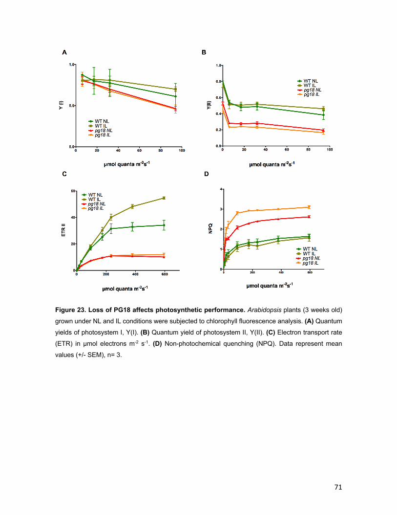
71
Figure 23. Loss of PG18 affects photosynthetic performance. Arabidopsis plants (3 weeks old)
grown under NL and IL conditions were subjected to chlorophyll fluorescence analysis. (A) Quantum
yields of photosystem I, Y(I). (B) Quantum yield of photosystem II, Y(II). (C) Electron transport rate
(ETR) in μmol electrons m-2 s-1. (D) Non-photochemical quenching (NPQ). Data represent mean
values (+/- SEM), n= 3.

72
Since the differences between WT and pg18 plants follow the same trend regardless
of whether they are grown and evaluated under NL or IL conditions their recovery
from photo-inhibition of PSII was analyzed by measuring their Fv / Fm values.
Interestingly, the degree of photo-inhibition after a 2-h exposure to 1000 µE m2 s−1
was the same for WT and pg18 plants coming from NL as well for IL conditions.
Likewise, their Fv / Fm values recovered with the same kinetics on return to normal
light levels (Figure 24A). This indicates again that PSII is equally susceptible to
photo-damage in both lines. Additionally, the translation efficiency was assessed to
discard any effect on biogenesis of the different subunits of the photosynthetic
complexes. In-vivo translation was performed by infiltrating young WT and mutant
pg18 plants with methionine 35S and then incubating it for 20 min. Afterwards,
thylakoids were isolated, and the photosynthetic complexes were separated in a
Blue native gel. In line with the recovery experiment results, the translation rate for
WT and pg18 mutant did not show high differences indicating that the defects in
photosynthetic parameters could come from the accumulation of the complexes
(Figure 24B). Since the effect on ETR or low quantum yield for both photosystems
might relate to stoichiometry or assembly of the different complexes in the thylakoid
membrane, they were considered to be investigated in more detail.
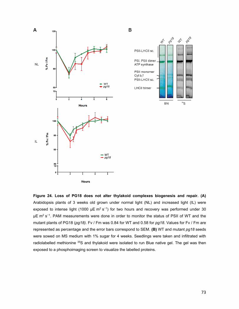
73
Figure 24. Loss of PG18 does not alter thylakoid complexes biogenesis and repair. (A) Arabidopsis plants of 3 weeks old grown under normal light (NL) and increased light (IL) were
exposed to intense light (1000 µE m2 s−1) for two hours and recovery was performed under 30
µE m2 s−1. PAM measurements were done in order to monitor the status of PSII of WT and the
mutant plants of PG18 (pg18). Fv / Fm was 0.84 for WT and 0.58 for pg18. Values for Fv / Fm are represented as percentage and the error bars correspond to SEM. (B) WT and mutant pg18 seeds
were sowed on MS medium with 1% sugar for 4 weeks. Seedlings were taken and infiltrated with
radiolabelled methionine 35S and thylakoid were isolated to run Blue native gel. The gel was then
exposed to a phosphoimaging screen to visualize the labelled proteins.
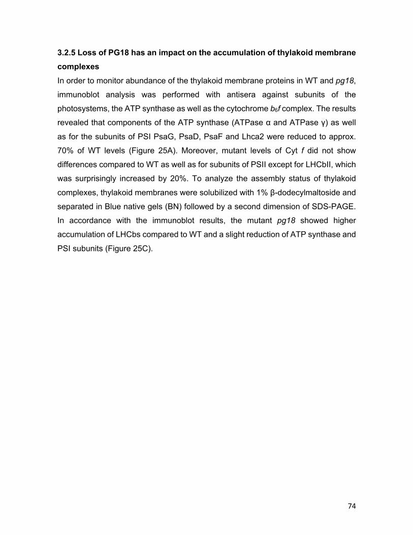
74
3.2.5 Loss of PG18 has an impact on the accumulation of thylakoid membrane complexes In order to monitor abundance of the thylakoid membrane proteins in WT and pg18,
immunoblot analysis was performed with antisera against subunits of the
photosystems, the ATP synthase as well as the cytochrome b6f complex. The results
revealed that components of the ATP synthase (ATPase α and ATPase γ) as well
as for the subunits of PSI PsaG, PsaD, PsaF and Lhca2 were reduced to approx.
70% of WT levels (Figure 25A). Moreover, mutant levels of Cyt f did not show
differences compared to WT as well as for subunits of PSII except for LHCbII, which
was surprisingly increased by 20%. To analyze the assembly status of thylakoid
complexes, thylakoid membranes were solubilized with 1% β-dodecylmaltoside and
separated in Blue native gels (BN) followed by a second dimension of SDS-PAGE.
In accordance with the immunoblot results, the mutant pg18 showed higher
accumulation of LHCbs compared to WT and a slight reduction of ATP synthase and
PSI subunits (Figure 25C).

75
Figure 25. Loss of PG18 has an impact on the accumulation of thylakoid membrane complexes. (A) Thylakoids were isolated from plants grown on soil of three weeks old under long
day conditions. Proteins in the thylakoid membranes were immunodetected by using antibodies against subunits of PSI, PSII, ATP synthase and the Cyt b6f complex. Loading of 100% corresponds
to 20 μg protein and the same amount was loaded for the mutant. (B) Immunoblotting quantification
using ImageJ. Each column represents percentage in relation to WT with mean value of three
replicates for each case and the error bars represent SEM with n = 3. (C) Thylakoid membranes were
isolated from Arabidopsis plants grown on MS sugar plates, and then solubilized with 1% (w/v) DM.
BN-PAGE (left panel) was followed by separation of protein complexes in the second dimension by
SDS/PAGE and silver staining (right panel).
76
4 Discussion
With the development of molecular biology techniques, there has been a rapid
advance in gaining knowledge about the chemistry and the biology of
photosynthesis. The last years have seen the development of techniques such as
X-ray emission spectroscopy to understand the very steps that electrons follow when
water is split by PSII (Gul et al., 2015). In spite of the deep knowledge that has been
collected around the photosynthetic complexes, undoubtedly there is still a lot to
discover since mimicking photosynthesis has turned out to be very challenging
(Rutherford & Moore, 2008). Furthermore, the biosynthesis of various complexes are
quite different from one organism to another showing that despite their common
principle they differ dramatically in assembly and therefore compatibility (Nickelsen
& Rengstl, 2013). For instance, recently has been elucidated that a thylakoid region
in cyanobacteria could be dedicated for PSII biogenesis and assembly (Nickelsen &
Zerges, 2013). Additionally, in higher plants it has been identified that for the
biosynthesis and assembly of the thylakoid complexes alone there are multiple
nuclear encoded proteins required (Chi et al., 2012; Schottler et al., 2011; Schottler
et al., 2015). However, the focus over the past years has been on the protein level
of the complexes neglecting how important the actual organization of these are and
how they become organized and segregated in different portions of the thylakoid
membranes. Attempts to understand the processes that are not just involved protein
complexes assembly but also in the biogenesis of thylakoids as a whole have started
with the observation of vesicles (Westphal et al., 2001). In line with this idea, in-silico
analyses have been done in order to identify candidates with homology to proteins
known to participate in vesicle trafficking (Khan et al., 2013). Unfortunately, there is
still no protein clearly identified which plays a role specifically in vesicle trafficking
inside the chloroplast making the process even more difficult to tackle. Whether or
not vesicles play a role in the biogenesis of thylakoids, there are still unknown
processes that require other ways to be approached for they thorough
understanding.
77
4.1 RV-1 The unknown protein named RV-1 was successfully localized to chloroplasts and its
mature form was identified by import experiments. Despite of not having close
homologs with known domains, RV-1 is conserved in higher plants highlighting its
relevance. Other green linages like mosses and algae present distant homologs to
RV-1 but not to RV-1-like. This suggests that in those linages RV-1 might be dual
targeted to chloroplast and mitochondria. When analyzing the mutant rv-1 lines, the
seedling lethal phenotype observed revealed its fundamental role in chloroplast
biogenesis. In addition of not being able to produce seeds, homozygous rv-1 plants
were pale and present aberrant chloroplast morphology which indicates that the loss
of RV-1 might be affecting a very initial step in chloroplast biogenesis. The lethal
phenotype of homozygous rv-1 plants was very surprising, taking into account that
its paralog is as conserved as RV-1 is and therefore functional redundancy was
initially expected. Interestingly, however, the cellular localization of RV-1-like
showed that it is localized in the mitochondria. This observation revealed that
probably RV-1 and RV-1-like are derived from an ancestral gene which got
duplicated and evolutionary distributed into both organelles. The absence of
homologs in cyanobacteria or even non-photosynthetic prokaryotes posed the
question about their ancestry from a conserved gene in endosymbiotic organisms or
from a horizontal gene transfer followed by duplication. Furthermore, the rv-1-like
mutant line also showed that RV-1-like might play a fundamental role in plant
physiology since mutant siliques exhibited embryos abortion which is a typical
phenotype for genes essential for plant mitochondria (Leon et al., 2007; Yamaoka &
Leaver, 2008). Thus, these observations suggest that both paralogs might have
similar functions that are essential and shared between chloroplast and
mitochondria.
In order to provide insights on the function of RV-1 which turned out to be essential
for chloroplast biogenesis, the drastic reduction on plastid rRNAs exhibited by the
rv-1 mutant was investigated in detail. Since proteins like PAC known to participate
in plastid rRNA maturation also present a similar phenotype with reduction or
absence of processed rRNA (Meurer et al., 2017), the distant homology of RV-1 to
78
an RNAse E from rice was taken into account. RNAse E proteins were first identified
in E. coli playing a role in the processing of 5S rRNA (Ghora & Apirion, 1978). Later
it was found that this family of proteins play similar role in other organisms such as
Arabidopsis and Synechocystis (Horie et al., 2007; Mudd et al., 2008). It is known
that RNAse E proteins play a role not just in the maturation of RNA but also in the
degradation of mRNA (Carpousis, 2007; Carpousis et al., 2009). Thus, the putative
RNAse E activity of RV-1 was assessed with the unmatured form of plastid rRNAs
namely 16S and 23S. However, no RNAse activity was observed even when using
total RNA extracted from young plants. Therefore, the ability to bind rRNA was tested
again using as targets the plastid 16S and 23S rRNAs. EMSA analyses revealed
that RV-1 does not have the ability to bind to any of these two rRNAs, whereas the
protein PAC binds specifically 23S.
Moreover, RV-1 has a distant homology to RelA and SpoT proteins (RSHs). These
proteins were characterized to cause inhibition of stable RNA accumulation
provoked by amino acid starvation in E. coli (Cashel & Gallant, 1969). In prokaryotes
it has been shown that RelA is responsible for the accumulation of the (p)ppGpp
which causes failure of stable RNA accumulation (Alfoldi et al., 1962; Potrykus &
Cashel, 2008). Later it was shown that a RelA homolog SpoT was able to synthesize
and degrade (p)ppGpp (Battesti & Bouveret, 2006; Seyfzadeh et al., 1993). In
Arabidopsis, RSHs have been already identified playing a role in chloroplast function
and gene expression (Sugliani et al., 2016). However, similar to the effect of
(p)ppGpp in translation (English et al., 2011), rv-1 mutant seedling exhibited drastic
reduction in de-novo translation of proteins. Furthermore, it has been shown that
(p)ppGpp inhibits the activity of chloroplast translation in-vitro which could give a hint
on RV-1 putative function (Nomura et al., 2012). However, the distant homology to
the RelA/Spot domain and the absence of homology to other RSH proteins makes
the relation of RV-1 with RelA/Spot probably more evolutive than functional.
Since RV-1 could neither bind or process any of the plastid rRNA, it was
hypothesized that it could bind to the ribosomes. The rv-1 mutant plants showed
absence of plastid ribosome proteins posing the question whether RV-1 could be
related to the stability or assembly of these ribosomes. Surprisingly, experiments
79
performed by treating stroma with and without RNAse shifted the distribution of RV-
1 in a sucrose gradient separation which was also observed for one subunit of the
plastid ribosome. This might imply that RV-1 could bind ribosomes similar to proteins
such as PAC. In order to validate this hypothesis, RNA sequencing of pulled down
RV-1 might provide its targets. Additionally, since the transit peptide of RV-1 is
already known it could be assessed the chloroplast localization of the paralog RV-1-
like protein and test its ability to rescue the rv-1 mutant phenotype. Thus, the
successful complementation of rv-1 mutant by chloroplast RV-1-like could
demonstrate their functional homology in chloroplast and plant mitochondria.
4.2 PG18 Several lines of evidence indicate that PGs play an important role in thylakoid
development. Their number decreases during de-etiolation, while mutants with
defects in thylakoid formation often accumulate larger numbers of PGs (Babiychuk
et al., 2008; Kroll et al., 2001). Their function may consist in providing metabolites,
such as carotenoids and prenylquinones. Additionally, they may be responsible for
providing triglycerides for membrane lipid synthesis.
The protein with unknown domain identity named PG18 was investigated for its role
in chloroplast biogenesis. PG18 is a protein of unknown function that was identified
in PGs of higher plants by mass spectrometry (Lundquist et al., 2012). With the help
of a PG18-specific antiserum, it was confirmed that PG18 is indeed localized to PGs.
Surprisingly, despite the absence of obvious hydrophobic regions in its sequence,
PG18 behaves as a membrane protein. A previous study on the fibrillin PGL34,
which likewise does not contain typical transmembrane domains, revealed that most
deletion variants tested failed to localize to PGs when transiently expressed as GFP
fusions (Vidi et al., 2006). This suggests that the association of PG proteins with the
PG monolayer might rely on their folded conformation rather than a particular
hydrophobic domain.
The characterization of a loss-of-function mutant of PG18 clearly demonstrates that
the lack of this protein has a severe impact on plant fitness throughout development.
Mutant plants show a light green phenotype and are smaller than WT. Since PGs
80
have been shown to accumulate under light stress (Zhang et al., 2010), it was
assessed whether the mutant line exhibits a stronger phenotype under such
conditions. Mutants for other PG proteins typically show enhanced phenotypes when
subjected to light stress (Fatihi et al., 2015; Porfirova et al., 2002; Youssef et al.,
2010). However, the phenotype of pg18 remained essentially the same under NL
and IL conditions. This indicates that PG18 plays a more general role in chloroplast
biogenesis. This notion is supported by the fact that pg18 plants have smaller
chloroplasts and fewer PGs per chloroplast than WT. Interestingly, the number of
PGs was increased under IL conditions in both WT and pg18 chloroplasts, although
the latter always contained fewer PGs per chloroplast. This indicates that PG18 is
not involved in promoting PG accumulation under light stress.
The pale green leaves of pg18 plants point to a reduction in chlorophyll content. This
assumption was verified by analyzing the chlorophyll content of leaves from plants
grown under NL and IL conditions, and the most significant reduction was noted in
chlorophyll a. The carotenoid fraction revealed a strong reduction in β-carotene in
the pg18 mutant compared to WT under both NL and IL conditions, whereas the
reduction in lutein was statistically significant only under IL. PGs are known to play
a role in carotenoid accumulation, for example in the chromoplasts of fruits and
flowers (Steinmuller & Tevini, 1985), which are enriched in biosynthetic enzymes
such as zeaxanthin epoxidase (ZDS) and lycopene-β cyclase (LYC-β) (van Wijk &
Kessler, 2017). Therefore, a reduction in carotenoid content could be a reflection of
PG dysfunction linked to changes in the activity of such enzymes.
Analysis of the pigments of the xanthopyll cycle in the pg18 mutant showed that
levels of the photoprotective pigments Ax and Zx were increased relative to WT, and
further enhanced under IL conditions. These pigments are synthesized from Vx
under light stress and serve as photoprotectants for PSII, binding to its LHC proteins
and contributing to energy dissipation as heat (nonphotochemical quenching; NPQ)
(Gilmore et al., 1995). Thus, pg18 mutant plants show symptoms of light stress even
under normal light conditions, which probably explains the accumulation of larger
grana stacks in their chloroplasts.
81
Chlorophyll fluorescence analyses showed that photosynthetic performance in
general was affected, with both photosystems showing lower quantum yields than
WT, and these deficits became more pronounced under increased light intensities.
Taking into account the fact that levels of ATP synthase and PSI were reduced in
the mutant plants, the reduction observed in ETR can be explained by an early
saturation of the electron transport chain, since the Δ pH cannot be fully relaxed.
Therefore, the early reduction and saturation of PSII and PSI might cause damage,
which could also explain a further reduction in the abundances of both photosystems
(Gilmore et al., 1995). In accordance with an expected rise in lumen pH, NPQ levels
were higher in the mutant than in the WT, which dissipates energy mainly from PSII
(Horton et al., 1996). This observation correlates with the high levels of Zx and Ax
found in the mutant plants relative to WT. These pigments are known to modulate
NPQ by deactivating excited states in the PSII antenna, and also acting as
antioxidants in the thylakoid membrane (Havaux & Niyogi, 1999; Nilkens et al.,
2010). Complementation of the mutant line rescued both the photosynthetic activity
and reduced pigment accumulation to WT levels (data no shown).
Moreover, to investigate the light sensitivity of PSII and the ability to repair it via de
novo synthesis and incorporation of D1 under IL conditions, it was performed light
stress recovery analysis. Strikingly, both WT and pg18 were equally affected by light
stress and both showed the same ability to recover. Thus, the pleiotropic effects on
photosynthetic performance in pg18 chloroplasts probably do not result from a PSII
that is more sensitive to light, but rather from initial assembly defects or altered
stoichiometry of the photosynthetic complexes. In line with this, analysis of the
accumulation of thylakoid proteins revealed a reduction in the ATP synthase and PSI
complexes. Concordantly with the reduction in PSI, levels of chlorophyll a were
sharply reduced in the pg18 mutant compared to the WT, which is also reflected in
the fact that the ratio of chlorophyll a to b remains essentially the same under NL
and IL conditions. Furthermore, our finding that the stroma lamellae are shorter and
less branched in the pg18 mutant than in WT correlates with the reduction in levels
of PSI and ATP synthase, as less membrane area is available for their integration.
Furthermore, the overall assembly of the complexes in the thylakoid membrane is

82
affected in the mutant pg18. Interestingly, there is a clear accumulation of LHC
proteins in the mutant line which is compatible with the fact that pg18 plants
accumulate more photoprotective LHC-binding pigments like Zx (Johnson et al.,
2007). This effect can also be seen in the high level of NPQ seen in the pg18 mutant,
which is further enhanced under IL, in accordance with the increased levels of Zx in
the mutant relative to WT under both NL and IL conditions. Hence, PG18 seems not
to intervene in the assembly of any particular complex in the thylakoid membrane,
but affects the composition of some of them, possibly by modulating the structural
organization of the thylakoid membrane during its biogenesis.
Despite the lack of identifiable functional domains, which suggests that PG18 is not
itself an enzyme, loss of PG18 has a significant impact on the composition and
architecture of the thylakoid membrane. In light of the indications that PGs play an
important role in mobilizing lipids for incorporation into the thylakoid membrane
(Deruere, Romer, et al., 1994; Simkin et al., 2007), an alteration in PGs could be
expected to affect thylakoid complexes, as has been observed when the lipid content
of the thylakoid membrane is altered (Kansy et al., 2014; Zhou et al., 2009). Levels
of the PG18 protein were not found to be specifically upregulated under stress
conditions, nor does it accumulate to a greater extent in PGs isolated after high light
treatment (Ytterberg et al., 2006). These findings indicate that it is a constitutive
component of PGs, possibly fulfilling a general role in PG maintenance or interacting
with other PG proteins. This inference is supported by the observation that knockout
mutants for other PG proteins do not show a phenotype under normal conditions.
Phenotypes only become manifest when PG mutants are exposed to stresses, such
as high light intensities (Avendano-Vazquez et al., 2014; Fatihi et al., 2015; Martinis
et al., 2013; Porfirova et al., 2002; Singh et al., 2010; Youssef et al., 2010). Moreover,
PG18 was also found in chromoplasts of red pepper (Ytterberg et al., 2006), also
suggesting that its function is not restricted to chloroplasts. Interestingly however,
PG18 is phosphorylated, which might lead to conformational changes or otherwise
have an impact on its activity, possibly depending on different developmental stages
or stress conditions (Wang et al., 2013).
83
In summary, investigation of the pg18 mutant underlines the importance of PGs in
the formation of thylakoid membranes. Elucidating its exact role will be a challenging
task to address in the future.
84
5 Literature
Albiniak,A.M.,Baglieri,J.,&Robinson,C.(2012).Targetingoflumenalproteinsacrossthethylakoidmembrane.JExpBot,63(4),1689-1698.doi:10.1093/jxb/err444
Alfoldi,L.,Stent,G.S.,&Clowes,R.C.(1962).ThechromosomalsiteoftheRNAcontrol(RC)locusinEscherichiacoli.JMolBiol,5,348-355.
Altschul,S.F.,Madden,T.L.,Schaffer,A.A.,Zhang,J.,Zhang,Z.,Miller,W.,&Lipman,D.J.(1997).GappedBLASTandPSI-BLAST:anewgenerationofproteindatabasesearchprograms.NucleicAcidsRes,25(17),3389-3402.
Aro, E. M., McCaffery, S., & Anderson, J. M. (1993). Photoinhibition and D1 ProteinDegradationinPeasAcclimatedtoDifferentGrowthIrradiances.PlantPhysiol,103(3),835-843.
Austin,J.R.,2nd,Frost,E.,Vidi,P.A.,Kessler,F.,&Staehelin,L.A.(2006).Plastoglobulesare lipoprotein subcompartments of the chloroplast that are permanentlycoupledtothylakoidmembranesandcontainbiosyntheticenzymes.PlantCell,18(7),1693-1703.doi:10.1105/tpc.105.039859
Avendano-Vazquez,A.O.,Cordoba,E.,Llamas,E.,SanRoman,C.,Nisar,N.,DelaTorre,S., . . . Leon, P. (2014). An Uncharacterized Apocarotenoid-Derived SignalGenerated in zeta-CaroteneDesaturaseMutants Regulates Leaf DevelopmentandtheExpressionofChloroplastandNuclearGenesinArabidopsis.PlantCell,26(6),2524-2537.doi:10.1105/tpc.114.123349
Babiychuk,E.,Bouvier-Nave,P.,Compagnon,V.,Suzuki,M.,Muranaka,T.,VanMontagu,M., . . . Schaller, H. (2008). Allelicmutant series reveal distinct functions forArabidopsiscycloartenolsynthase1incellviabilityandplastidbiogenesis.ProcNatlAcadSciUSA,105(8),3163-3168.doi:10.1073/pnas.0712190105
Balsera,M.,Goetze,T.A.,Kovacs-Bogdan,E.,Schurmann,P.,Wagner,R.,Buchanan,B.B.,. . .Bolter,B.(2009).CharacterizationofTic110,achannel-formingproteinattheinnerenvelopemembraneofchloroplasts,unveilsaresponsetoCa(2+)anda stromal regulatory disulfide bridge. J Biol Chem, 284(5), 2603-2616.doi:10.1074/jbc.M807134200
Barsan,C.,Zouine,M.,Maza,E.,Bian,W.,Egea,I.,Rossignol,M., . . .Pech,J.C.(2012).Proteomicanalysisofchloroplast-to-chromoplasttransitionintomatorevealsmetabolic shifts coupledwith disrupted thylakoid biogenesismachinery andelevated energy-production components. Plant Physiol, 160(2), 708-725.doi:10.1104/pp.112.203679
Bartsch,S.,Monnet,J.,Selbach,K.,Quigley,F.,Gray,J.,vonWettstein,D.,...Reinbothe,C. (2008). Three thioredoxin targets in the inner envelope membrane ofchloroplastsfunctioninproteinimportandchlorophyllmetabolism.ProcNatlAcadSciUSA,105(12),4933-4938.doi:10.1073/pnas.0800378105
Battesti,A.,&Bouveret,E. (2006).Acyl carrierprotein/SpoT interaction, the switchlinkingSpoT-dependentstressresponsetofattyacidmetabolism.MolMicrobiol,62(4),1048-1063.doi:10.1111/j.1365-2958.2006.05442.x
85
Benning, C., Xu, C., & Awai, K. (2006). Non-vesicular and vesicular lipid traffickinginvolving plastids. Curr Opin Plant Biol, 9(3), 241-247.doi:10.1016/j.pbi.2006.03.012
Benz,J.P.,Stengel,A.,Lintala,M.,Lee,Y.H.,Weber,A.,Philippar,K.,...Bolter,B.(2009).Arabidopsis Tic62 and ferredoxin-NADP(H) oxidoreductase form light-regulatedcomplexesthatareintegratedintothechloroplastredoxpoise.PlantCell,21(12),3965-3983.doi:10.1105/tpc.109.069815
Bhuiyan,N.H.,Friso,G.,Poliakov,A.,Ponnala,L.,&vanWijk,K. J. (2015).MET1isathylakoid-associated TPR protein involved in photosystem II supercomplexformation and repair in Arabidopsis. Plant Cell, 27(1), 262-285.doi:10.1105/tpc.114.132787
Bieri, P., Leibundgut,M., Saurer,M., Boehringer,D.,&Ban,N. (2017). The completestructureofthechloroplast70SribosomeincomplexwithtranslationfactorpY.EMBOJ,36(4),475-486.doi:10.15252/embj.201695959
Brasier,M.D.,Antcliffe, J.,Saunders,M.,&Wacey,D.(2015).ChangingthepictureofEarth'searliestfossils(3.5-1.9Ga)withnewapproachesandnewdiscoveries.ProcNatlAcadSciUSA,112(16),4859-4864.doi:10.1073/pnas.1405338111
Cardona,T.(2018).EarlyArcheanoriginofheterodimericPhotosystemI.Heliyon,4(3),e00548.doi:10.1016/j.heliyon.2018.e00548
Carpousis,A.J.(2007).TheRNAdegradosomeofEscherichiacoli:anmRNA-degradingmachine assembled on RNase E. Annu Rev Microbiol, 61, 71-87.doi:10.1146/annurev.micro.61.080706.093440
Carpousis,A.J.,Luisi,B.F.,&McDowall,K.J.(2009).EndonucleolyticinitiationofmRNAdecay in Escherichia coli. Prog Mol Biol Transl Sci, 85, 91-135.doi:10.1016/S0079-6603(08)00803-9
Cashel,M.,&Gallant, J. (1969).TwocompoundsimplicatedinthefunctionoftheRCgeneofEscherichiacoli.Nature,221(5183),838-841.
Chaal, B. K., Mould, R. M., Barbrook, A. C., Gray, J. C., & Howe, C. J. (1998).CharacterizationofacDNAencodingthethylakoidalprocessingpeptidasefromArabidopsisthaliana.Implicationsfortheoriginandcatalyticmechanismoftheenzyme.JBiolChem,273(2),689-692.
Chan, K. X., Phua, S. Y., Crisp, P., McQuinn, R., & Pogson, B. J. (2016). Learning theLanguagesoftheChloroplast:RetrogradeSignalingandBeyond.AnnuRevPlantBiol,67,25-53.doi:10.1146/annurev-arplant-043015-111854
Chi,W.,Ma, J., & Zhang, L. (2012). Regulatory factors for the assembly of thylakoidmembraneprotein complexes.PhilosTransR Soc LondBBiol Sci, 367(1608),3420-3429.doi:10.1098/rstb.2012.0065
Chigri,F.,Hormann,F.,Stamp,A.,Stammers,D.K.,Bolter,B.,Soll,J.,&Vothknecht,U.C.(2006).CalciumregulationofchloroplastproteintranslocationismediatedbycalmodulinbindingtoTic32.ProcNatlAcadSciUSA,103(43),16051-16056.doi:10.1073/pnas.0607150103
Chou,M.L.,Chu,C.C.,Chen,L. J.,Akita,M.,&Li,H.M.(2006).Stimulationoftransit-peptidereleaseandATPhydrolysisbyacochaperoneduringproteinimportintochloroplasts.JCellBiol,175(6),893-900.doi:10.1083/jcb.200609172
Cunningham, F. X., Jr., Tice,A.B., Pham,C.,&Gantt, E. (2010). Inactivationof genesencodingplastoglobuli-likeproteins inSynechocystis sp.PCC6803 leads toa

86
light-sensitive phenotype. J Bacteriol, 192(6), 1700-1709.doi:10.1128/JB.01434-09
Danielsson,R.,Albertsson,P.A.,Mamedov,F.,&Styring, S. (2004).QuantificationofphotosystemIandIIindifferentpartsofthethylakoidmembranefromspinach.BiochimBiophysActa,1608(1),53-61.
DeMarais,D.J.(2000).Evolution.WhendidphotosynthesisemergeonEarth?Science,289(5485),1703-1705.
de Vries, J., & Archibald, J. M. (2017). Endosymbiosis: Did Plastids Evolve from aFreshwater Cyanobacterium? Curr Biol, 27(3), R103-R105.doi:10.1016/j.cub.2016.12.006
Demarsy, E., Lakshmanan, A.M., & Kessler, F. (2014). Border control: selectivity ofchloroplastproteinimportandregulationattheTOC-complex.FrontPlantSci,5,483.doi:10.3389/fpls.2014.00483
Deruere,J.,Bouvier,F.,Steppuhn,J.,Klein,A.,Camara,B.,&Kuntz,M.(1994).Structureand expression of two plant genes encoding chromoplast-specific proteins:occurrence of partially spliced transcripts. Biochem Biophys Res Commun,199(3),1144-1150.doi:10.1006/bbrc.1994.1350
Deruere,J.,Romer,S.,d'Harlingue,A.,Backhaus,R.A.,Kuntz,M.,&Camara,B.(1994).Fibrilassemblyandcarotenoidoveraccumulationinchromoplasts:amodelforsupramolecular lipoprotein structures. Plant Cell, 6(1), 119-133.doi:10.1105/tpc.6.1.119
Douzery,E.J.,Snell,E.A.,Bapteste,E.,Delsuc,F.,&Philippe,H.(2004).Thetimingofeukaryotic evolution: does a relaxed molecular clock reconcile proteins andfossils? Proc Natl Acad Sci U S A, 101(43), 15386-15391.doi:10.1073/pnas.0403984101
Dumas, L., Chazaux,M., Peltier, G., Johnson, X., & Alric, J. (2016). Cytochrome b 6 ffunction and localization, phosphorylation state of thylakoid membraneproteinsandconsequencesoncyclicelectronflow.PhotosynthRes,129(3),307-320.doi:10.1007/s11120-016-0298-y
Eckhardt, U., Grimm, B., &Hortensteiner, S. (2004). Recent advances in chlorophyllbiosynthesis and breakdown in higher plants. Plant Mol Biol, 56(1), 1-14.doi:10.1007/s11103-004-2331-3
Egea,I.,Barsan,C.,Bian,W.,Purgatto,E.,Latche,A.,Chervin,C., . . .Pech,J.C.(2010).Chromoplastdifferentiation:currentstatusandperspectives.PlantCellPhysiol,51(10),1601-1611.doi:10.1093/pcp/pcq136
Emanuelsson,O.,Nielsen,H.,Brunak,S.,&vonHeijne,G.(2000).Predictingsubcellularlocalizationofproteinsbasedon theirN-terminalaminoacidsequence. JMolBiol,300(4),1005-1016.doi:10.1006/jmbi.2000.3903
Emanuelsson,O.,Nielsen,H.,&vonHeijne,G.(1999).ChloroP,aneuralnetwork-basedmethod for predicting chloroplast transit peptides and their cleavage sites.ProteinSci,8(5),978-984.doi:10.1110/ps.8.5.978
Enami,K.,Ozawa,T.,Motohashi,N.,Nakamura,M.,Tanaka,K.,&Hanaoka,M.(2011).Plastid-to-nucleusretrogradesignalsareessentialfortheexpressionofnuclearstarch biosynthesis genes during amyloplast differentiation in tobacco BY-2culturedcells.PlantPhysiol,157(1),518-530.doi:10.1104/pp.111.178897

87
English,B. P.,Hauryliuk,V., Sanamrad,A., Tankov, S.,Dekker,N.H.,&Elf, J. (2011).Single-molecule investigations of the stringent response machinery in livingbacterial cells. Proc Natl Acad Sci U S A, 108(31), E365-373.doi:10.1073/pnas.1102255108
Farber, A., Young, A. J., Ruban, A. V., Horton, P., & Jahns, P. (1997). Dynamics ofXanthophyll-Cycle Activity in Different Antenna Subcomplexes in thePhotosynthetic Membranes of Higher Plants (The Relationship betweenZeaxanthinConversionandNonphotochemicalFluorescenceQuenching).PlantPhysiol,115(4),1609-1618.
Fatihi,A.,Latimer,S.,Schmollinger,S.,Block,A.,Dussault,P.H.,Vermaas,W.F.,...Basset,G. J. (2015). A Dedicated Type II NADPH Dehydrogenase Performs thePenultimate Step in the Biosynthesis of Vitamin K1 in Synechocystis andArabidopsis.PlantCell,27(6),1730-1741.doi:10.1105/tpc.15.00103
Flores-Perez,U.,&Jarvis,P.(2013).Molecularchaperoneinvolvementinchloroplastprotein import. Biochim Biophys Acta, 1833(2), 332-340.doi:10.1016/j.bbamcr.2012.03.019
Friso,G.,Giacomelli, L.,Ytterberg,A. J.,Peltier, J.B.,Rudella,A., Sun,Q.,&Wijk,K. J.(2004).In-depthanalysisofthethylakoidmembraneproteomeofArabidopsisthaliana chloroplasts: new proteins, new functions, and a plastid proteomedatabase.PlantCell,16(2),478-499.doi:10.1105/tpc.017814
Froehlich, J. E., Benning, C., & Dormann, P. (2001). The digalactosyldiacylglycerol(DGDG) synthase DGD1 is inserted into the outer envelope membrane ofchloroplastsinamannerindependentofthegeneralimportpathwayanddoesnot dependondirect interactionwithmonogalactosyldiacylglycerol synthasefor DGDG biosynthesis. J Biol Chem, 276(34), 31806-31812.doi:10.1074/jbc.M104652200
Gaude, N., Brehelin, C., Tischendorf, G., Kessler, F., & Dormann, P. (2007). NitrogendeficiencyinArabidopsisaffectsgalactolipidcompositionandgeneexpressionandresultsinaccumulationoffattyacidphytylesters.PlantJ,49(4),729-739.doi:10.1111/j.1365-313X.2006.02992.x
Ghora,B.K.,&Apirion,D.(1978).Structuralanalysisandinvitroprocessingtop5rRNAofa9SRNAmoleculeisolatedfromanrnemutantofE.coli.Cell,15(3),1055-1066.
Gilmore, A. M., Hazlett, T. L., & Govindjee. (1995). Xanthophyll cycle-dependentquenching of photosystem II chlorophyll a fluorescence: formation of aquenchingcomplexwithashortfluorescencelifetime.ProcNatlAcadSciUSA,92(6),2273-2277.
Greenwood,A.D., Leech,R.M.,&Williams, J. P. (1963).Theosmiophilic globulesofchloroplasts:I.Osmiophilicglobulesasanormalcomponentofchloroplastsandtheir isolationandcomposition inVicia fabaL.BiochimicaetBiophysicaActa,78(1),148-162.doi:doi.org/10.1016/0006-3002(63)91620-2
Gross,J.,&Bhattacharya,D.(2009).RevaluatingtheevolutionoftheTocandTicproteintranslocons.TrendsPlantSci,14(1),13-20.doi:10.1016/j.tplants.2008.10.003
Gul,S.,Ng,J.W.,Alonso-Mori,R.,Kern,J.,Sokaras,D.,Anzenberg,E.,...Yano,J.(2015).Simultaneousdetectionofelectronicstructurechangesfromtwoelementsofabifunctionalcatalystusingwavelength-dispersiveX-rayemissionspectroscopy
88
and in situ electrochemistry. Phys Chem Chem Phys, 17(14), 8901-8912.doi:10.1039/c5cp01023c
Hanahan,N.(1985).TechniquesfortransformationofE.coli.DNAcloning.1,109-135.Hansmann,P.,&Sitte,P.(1982).Compositionandmolecularstructureofchromoplast
globules of Viola tricolor. Plant Cell Rep, 1(3), 111-114.doi:10.1007/BF00272366
Havaux, M., & Niyogi, K. K. (1999). The violaxanthin cycle protects plants fromphotooxidativedamagebymorethanonemechanism.ProcNatlAcadSciUSA,96(15),8762-8767.
Heins,L.,Mehrle,A.,Hemmler,R.,Wagner,R.,Kuchler,M.,Hormann,F.,...Soll,J.(2002).Thepreproteinconductingchannelattheinnerenvelopemembraneofplastids.EMBOJ,21(11),2616-2625.doi:10.1093/emboj/21.11.2616
Hinnah,S.C.,Wagner,R.,Sveshnikova,N.,Harrer,R.,&Soll,J.(2002).Thechloroplastprotein import channel Toc75: pore properties and interaction with transitpeptides.BiophysJ,83(2),899-911.doi:10.1016/S0006-3495(02)75216-8
Hirsch, S., Muckel, E., Heemeyer, F., von Heijne, G., & Soll, J. (1994). A receptorcomponent of the chloroplast protein translocation machinery. Science,266(5193),1989-1992.
Horie,Y.,Ito,Y.,Ono,M.,Moriwaki,N.,Kato,H.,Hamakubo,Y.,...Asayama,M.(2007).Dark-induced mRNA instability involves RNase E/G-type endoribonucleasecleavageattheAU-boxandSDsequencesincyanobacteria.MolGenetGenomics,278(3),331-346.doi:10.1007/s00438-007-0254-9
Horton,P.,Ruban,A.V.,&Walters,R.G.(1996).RegulationofLightHarvestinginGreenPlants. Annu Rev Plant Physiol Plant Mol Biol, 47, 655-684.doi:10.1146/annurev.arplant.47.1.655
Jahns,P.,&Holzwarth,A.R.(2012).Theroleofthexanthophyllcycleandofluteininphotoprotection of photosystem II. Biochim Biophys Acta, 1817(1), 182-193.doi:10.1016/j.bbabio.2011.04.012
Johnson,M. P., Havaux,M., Triantaphylides, C., Ksas, B., Pascal, A. A., Robert, B., . . .Horton,P.(2007).ElevatedzeaxanthinboundtooligomericLHCIIenhancestheresistance of Arabidopsis to photooxidative stress by a lipid-protective,antioxidant mechanism. J Biol Chem, 282(31), 22605-22618.doi:10.1074/jbc.M702831200
Johnson, M. P., Zia, A., & Ruban, A. V. (2012). Elevated DeltapH restores rapidlyreversible photoprotective energy dissipation in Arabidopsis chloroplastsdeficient in lutein and xanthophyll cycle activity. Planta, 235(1), 193-204.doi:10.1007/s00425-011-1502-0
Kansy,M.,Wilhelm,C.,&Goss,R.(2014).InfluenceofthylakoidmembranelipidsonthestructureandfunctionoftheplantphotosystemIIcorecomplex.Planta,240(4),781-796.doi:10.1007/s00425-014-2130-2
Kato,Y.,&Sakamoto,W.(2010).Newinsightsintothetypesandfunctionofproteasesin plastids. Int Rev Cell Mol Biol, 280, 185-218. doi:10.1016/S1937-6448(10)80004-8
Keegstra, K., & Yousif, A. E. (1986). Isolation and characterization of chloroplastenvelope membranes. In Methods in Enzymology (Vol. 118, pp. 316-325):AcademicPress.

89
Kerr, R. A. (2005). Earth science. The story of O2. Science, 308(5729), 1730-1732.doi:10.1126/science.308.5729.1730
Kessler, F., Blobel, G., Patel, H. A., & Schnell, D. J. (1994). Identification of twoGTP-binding proteins in the chloroplast protein import machinery. Science,266(5187),1035-1039.
Khan, N. Z., Lindquist, E., & Aronsson, H. (2013). New putative chloroplast vesicletransport components and cargo proteins revealed using a bioinformaticsapproach: an Arabidopsis model. PLoS One, 8(4), e59898.doi:10.1371/journal.pone.0059898
Kikuchi, S.,Oishi,M.,Hirabayashi, Y., Lee,D.W.,Hwang, I.,&Nakai,M. (2009).A1-megadalton translocation complex containing Tic20 and Tic21 mediateschloroplastproteinimportattheinnerenvelopemembrane.PlantCell,21(6),1781-1797.doi:10.1105/tpc.108.063552
Kim, D. H., & Hwang, I. (2013). Direct targeting of proteins from the cytosol toorganelles: the ER versus endosymbiotic organelles. Traffic, 14(6), 613-621.doi:10.1111/tra.12043
Kirchhoff, H. (2008). Molecular crowding and order in photosynthetic membranes.TrendsPlantSci,13(5),201-207.doi:10.1016/j.tplants.2008.03.001
Klughammer,C.,&Schreiber,U. (1994).An improvedmethod,usingsaturating lightpulses, for the determination of photosystem I quantum yield via P700<sup>+</sup>-absorbancechangesat830nm.Planta,192(2),261-268.
Kobayashi, K. (2016). Role of membrane glycerolipids in photosynthesis, thylakoidbiogenesis and chloroplast development. J Plant Res, 129(4), 565-580.doi:10.1007/s10265-016-0827-y
Kobayashi, K., Kondo,M., Fukuda,H., Nishimura,M., &Ohta,H. (2007). Galactolipidsynthesis in chloroplast inner envelope is essential for proper thylakoidbiogenesis, photosynthesis, and embryogenesis. Proc Natl Acad Sci U S A,104(43),17216-17221.doi:10.1073/pnas.0704680104
Kouranov, A., Chen, X., Fuks, B., & Schnell, D. J. (1998). Tic20 and Tic22 are newcomponentsoftheproteinimportapparatusatthechloroplastinnerenvelopemembrane.JCellBiol,143(4),991-1002.
Kovacheva,S.,Bedard,J.,Patel,R.,Dudley,P.,Twell,D.,Rios,G.,...Jarvis,P.(2005).InvivostudiesontherolesofTic110,Tic40andHsp93duringchloroplastproteinimport.PlantJ,41(3),412-428.doi:10.1111/j.1365-313X.2004.02307.x
Kroll,D.,Meierhoff,K.,Bechtold,N.,Kinoshita,M.,Westphal,S.,Vothknecht,U.C., . . .Westhoff,P.(2001).VIPP1,anucleargeneofArabidopsisthalianaessentialforthylakoidmembrane formation.Proc Natl Acad Sci U S A, 98(7), 4238-4242.doi:10.1073/pnas.061500998
Kumar,S.,Stecher,G.,&Tamura,K.(2016).MEGA7:MolecularEvolutionaryGeneticsAnalysis Version 7.0 for Bigger Datasets. Mol Biol Evol, 33(7), 1870-1874.doi:10.1093/molbev/msw054
Laemmli,U.K.(1970).CleavageofstructuralproteinsduringtheassemblyoftheheadofbacteriophageT4.Nature,227(5259),680-685.
Lake, J.A.,Larsen, J.,Tran,D.T.,&Sinsheimer, J.S. (2018).Uncovering theGenomicOriginsofLife.GenomeBiolEvol,10(7),1705-1714.doi:10.1093/gbe/evy129
90
Langenkamper, G.,Manac'h,N., Broin,M., Cuine, S., Becuwe,N., Kuntz,M.,&Rey, P.(2001). Accumulation of plastid lipid-associated proteins (fibrillin/CDSP34)uponoxidativestress,ageingandbioticstressinSolanaceaeandinresponsetodroughtinotherspecies.JExpBot,52(360),1545-1554.
Leon,G.,Holuigue,L.,& Jordana,X. (2007).Mitochondrialcomplex II Isessential forgametophyte development in Arabidopsis. Plant Physiol, 143(4), 1534-1546.doi:10.1104/pp.106.095158
Lundquist,P.K.,Poliakov,A.,Bhuiyan,N.H.,Zybailov,B.,Sun,Q.,&vanWijk,K.J.(2012).The functional network of the Arabidopsis plastoglobule proteome based onquantitativeproteomicsandgenome-widecoexpressionanalysis.PlantPhysiol,158(3),1172-1192.doi:10.1104/pp.111.193144
Marchler-Bauer,A.,Bo,Y.,Han,L.,He,J.,Lanczycki,C.J.,Lu,S.,...Bryant,S.H.(2017).CDD/SPARCLE: functional classification of proteins via subfamily domainarchitectures.NucleicAcidsRes,45(D1),D200-D203.doi:10.1093/nar/gkw1129
Martinis,J.,Glauser,G.,Valimareanu,S.,&Kessler,F.(2013).AchloroplastABC1-likekinase regulates vitamin Emetabolism in Arabidopsis. Plant Physiol, 162(2),652-662.doi:10.1104/pp.113.218644
Martinis, J.,Glauser,G.,Valimareanu,S.,Stettler,M.,Zeeman,S.C.,Yamamoto,H., . . .Kessler, F. (2014). ABC1K1/PGR6 kinase: a regulatory link betweenphotosynthetic activity and chloroplast metabolism. Plant J, 77(2), 269-283.doi:10.1111/tpj.12385
Meurer,J.,Schmid,L.M.,Stoppel,R.,Leister,D.,Brachmann,A.,&Manavski,N.(2017).PALE CRESS binds to plastid RNAs and facilitates the biogenesis of the 50Sribosomalsubunit.PlantJ,92(3),400-413.doi:10.1111/tpj.13662
Miquel,M.,&Browse,J.(1992).Arabidopsismutantsdeficientinpolyunsaturatedfattyacid synthesis. Biochemical and genetic characterization of a plant oleoyl-phosphatidylcholinedesaturase.JBiolChem,267(3),1502-1509.
Miyagishima, S. Y. (2011). Mechanism of plastid division: from a bacterium to anorganelle.PlantPhysiol,155(4),1533-1544.doi:10.1104/pp.110.170688
Morre,D.J.,Sellden,G.,Sundqvist,C.,&Sandelius,A.S.(1991).Stromallowtemperaturecompartment derived from the innermembrane of the chloroplast envelope.PlantPhysiol,97(4),1558-1564.
Mudd,E.A.,Sullivan,S.,Gisby,M.F.,Mironov,A.,Kwon,C.S.,Chung,W. I.,&Day,A.(2008).A125kDaRNaseE/G-likeproteinispresentinplastidsandisessentialforchloroplastdevelopmentandautotrophicgrowthinArabidopsis.JExpBot,59(10),2597-2610.doi:10.1093/jxb/ern126
Muhlethaler,K.(1959).EntwicklungundStrukturderProplastiden.TheJournalofCellBiology,6(3),507-512.doi:10.1083/jcb.6.3.507
Mullet, J. E. (1993). Dynamic regulation of chloroplast transcription. Plant Physiol,103(2),309-313.
Nickelsen, J., & Rengstl, B. (2013). Photosystem II assembly: from cyanobacteria toplants.AnnuRevPlantBiol,64,609-635.doi:10.1146/annurev-arplant-050312-120124
Nickelsen, J., & Zerges, W. (2013). Thylakoid biogenesis has joined the new era ofbacterialcellbiology.FrontPlantSci,4,458.doi:10.3389/fpls.2013.00458
91
Nilkens,M.,Kress,E.,Lambrev,P.,Miloslavina,Y.,Muller,M.,Holzwarth,A.R.,&Jahns,P.(2010).Identificationofaslowlyinduciblezeaxanthin-dependentcomponentofnon-photochemicalquenchingofchlorophyll fluorescencegeneratedundersteady-stateconditionsinArabidopsis.BiochimBiophysActa,1797(4),466-475.doi:10.1016/j.bbabio.2010.01.001
Nomura,Y.,Takabayashi,T.,Kuroda,H.,Yukawa,Y.,Sattasuk,K.,Akita,M.,...Tozawa,Y. (2012). ppGpp inhibits peptide elongation cycle of chloroplast translationsystem in vitro. Plant Mol Biol, 78(1-2), 185-196. doi:10.1007/s11103-011-9858-x
Peltier, J. B., Ytterberg, A. J., Sun, Q., & vanWijk, K. J. (2004). New functions of thethylakoidmembrane proteome of Arabidopsis thaliana revealed by a simple,fast, and versatile fractionation strategy. J Biol Chem, 279(47), 49367-49383.doi:10.1074/jbc.M406763200
Ponce-Toledo, R. I., Deschamps, P., Lopez-Garcia, P., Zivanovic, Y., Benzerara, K., &Moreira, D. (2017). An Early-Branching Freshwater Cyanobacterium at theOriginofPlastids.CurrBiol,27(3),386-391.doi:10.1016/j.cub.2016.11.056
Porfirova,S.,Bergmuller,E.,Tropf,S.,Lemke,R.,&Dormann,P.(2002).IsolationofanArabidopsismutantlackingvitaminEandidentificationofacyclaseessentialforall tocopherol biosynthesis. Proc Natl Acad Sci U S A, 99(19), 12495-12500.doi:10.1073/pnas.182330899
Potrykus,K.,&Cashel,M.(2008).(p)ppGpp:stillmagical?AnnuRevMicrobiol,62,35-51.doi:10.1146/annurev.micro.62.081307.162903
Richly,E.,&Leister,D.(2004).AnimprovedpredictionofchloroplastproteinsrevealsdiversitiesandcommonalitiesinthechloroplastproteomesofArabidopsisandrice.Gene,329,11-16.doi:10.1016/j.gene.2004.01.008
Roberts,D.R., Thompson, J. E.,Dumbroff, E. B., Gepstein, S.,&Mattoo,A.K. (1987).Differential changes in the synthesis and steady-state levels of thylakoidproteins during bean leaf senescence. Plant Mol Biol, 9(4), 343-353.doi:10.1007/BF00014909
Rockholm,D.C.,&Yamamoto,H.Y.(1996).Violaxanthinde-epoxidase.PlantPhysiol,110(2),697-703.
Rosado-Alberio,J.,Weier,T.R.,&Stocking,C.R.(1968).Continuityofthechloroplastmembranesystemsinzeamays1968.PlantPhysiol,43(9),1325-1331.
Rottet,S.,Besagni,C.,&Kessler,F.(2015).Theroleofplastoglobulesinthylakoidlipidremodelingduringplantdevelopment.BiochimBiophysActa,1847(9),889-899.doi:10.1016/j.bbabio.2015.02.002
Rutherford,A.W.,&Moore,T.A.(2008).Mimickingphotosynthesis,butjustthebestbits.Nature,453,449.doi:10.1038/453449b
Sambrook,J.,&Russell,D.W.(2001).Molecularcloning:alaboratorymanual(3rded.).ColdSpringHarbor,N.Y.:ColdSpringHarborLaboratoryPress.
Schackleton,J.B.,&Robinson,C.(1991).Transportofproteinsintochloroplasts.Thethylakoidal processing peptidase is a signal-type peptidase with stringentsubstrate requirements at the -3 and -1 positions. The Journal of BiologicalChemistry,266(5),2152-12156.
Schnell,D.J.,Kessler,F.,&Blobel,G.(1994).Isolationofcomponentsofthechloroplastproteinimportmachinery.Science,266(5187),1007-1012.
92
Schottler, M. A., Albus, C. A., & Bock, R. (2011). Photosystem I: its biogenesis andfunction in higher plants. J Plant Physiol, 168(12), 1452-1461.doi:10.1016/j.jplph.2010.12.009
Schottler, M. A., Flugel, C., Thiele, W., & Bock, R. (2007). Knock-out of the plastid-encoded PetL subunit results in reduced stability and accelerated leaf age-dependent lossof thecytochromeb6fcomplex. JBiolChem,282(2),976-985.doi:10.1074/jbc.M606436200
Schottler,M.A.,Toth,S.Z.,Boulouis,A.,&Kahlau,S.(2015).Photosyntheticcomplexstoichiometrydynamicsinhigherplants:biogenesis,function,andturnoverofATP synthase and the cytochromeb6f complex. J ExpBot, 66(9), 2373-2400.doi:10.1093/jxb/eru495
Schreiber,U.,Bilger,W.,Hormann,H.,&Neubauer,C.(1998).Chlorophyllfluorescenceasadiagnostictool:Basicsandsomeaspectsofpracticalrelevance.
Schunemann,D.(2007).Mechanismsofproteinimportintothylakoidsofchloroplasts.BiolChem,388(9),907-915.doi:10.1515/BC.2007.111
Schwenkert, S., Soll, J., & Bolter, B. (2011). Protein import into chloroplasts--howchaperones feature into the game. Biochim Biophys Acta, 1808(3), 901-911.doi:10.1016/j.bbamem.2010.07.021
Seyfzadeh, M., Keener, J., & Nomura, M. (1993). spoT-dependent accumulation ofguanosinetetraphosphateinresponsetofattyacidstarvationinEscherichiacoli.ProcNatlAcadSciUSA,90(23),11004-11008.
Shih, P. M. (2015). Photosynthesis and early Earth. Curr Biol, 25(19), R855-859.doi:10.1016/j.cub.2015.04.046
Simkin,A.J.,Gaffe,J.,Alcaraz,J.P.,Carde,J.P.,Bramley,P.M.,Fraser,P.D.,&Kuntz,M.(2007). Fibrillin influence on plastid ultrastructure and pigment content intomato fruit. Phytochemistry, 68(11), 1545-1556.doi:10.1016/j.phytochem.2007.03.014
Singh,D.K.,Maximova,S.N.,Jensen,P.J.,Lehman,B.L.,Ngugi,H.K.,&McNellis,T.W.(2010). FIBRILLIN4 is required for plastoglobule development and stressresistance in apple and Arabidopsis. Plant Physiol, 154(3), 1281-1293.doi:10.1104/pp.110.164095
Singh,D.K.,&McNellis,T.W.(2011).Fibrillinproteinfunction:thetipoftheiceberg?TrendsPlantSci,16(8),432-441.doi:10.1016/j.tplants.2011.03.014
Slack,C.R.,Roughan,P.G.,&Balasingham,N.(1977).Labellingstudiesinvivoonthemetabolism of the acyl and glycerol moieties of the glycerolipids in thedevelopingmaizeleaf.BiochemJ,162(2),289-296.
Solymosi, K., & Schoefs, B. (2010). Etioplast and etio-chloroplast formation undernatural conditions: the dark side of chlorophyll biosynthesis in angiosperms.PhotosynthRes,105(2),143-166.doi:10.1007/s11120-010-9568-2
Sperling, U., Franck, F., van Cleve, B., Frick, G., Apel, K., & Armstrong, G. A. (1998).Etioplast differentiation in arabidopsis: both PORA and PORB restore theprolamellar body and photoactive protochlorophyllide-F655 to the cop1photomorphogenicmutant.PlantCell,10(2),283-296.
Steinmuller, D., & Tevini, M. (1985). Composition and function of plastoglobuli : I.Isolationandpurificationfromchloroplastsandchromoplasts.Planta,163(2),201-207.doi:10.1007/BF00393507

93
Stiller,J.W.(2007).Plastidendosymbiosis,genomeevolutionandtheoriginofgreenplants.TrendsPlantSci,12(9),391-396.doi:10.1016/j.tplants.2007.08.002
Stoppel,R.,&Meurer,J.(2012).Thecuttingcrew-ribonucleasesarekeyplayersinthecontrol of plastid gene expression. J Exp Bot, 63(4), 1663-1673.doi:10.1093/jxb/err401
Sugliani,M.,Abdelkefi,H.,Ke,H.,Bouveret,E.,Robaglia,C.,Caffarri,S.,&Field,B.(2016).An Ancient Bacterial Signaling Pathway Regulates Chloroplast Function toInfluenceGrowthandDevelopmentinArabidopsis.PlantCell,28(3),661-679.doi:10.1105/tpc.16.00045
Suzuki,J.Y.,Sriraman,P.,Svab,Z.,&Maliga,P.(2003).Uniquearchitectureoftheplastidribosomal RNA operon promoter recognized by the multisubunit RNApolymeraseintobaccoandotherhigherplants.PlantCell,15(1),195-205.
Tevini,M.,&Steinmuller,D.(1985).Compositionandfunctionofplastoglobuli:II.Lipidcompositionof leavesandplastoglobuliduringbeech leaf senescence.Planta,163(1),91-96.doi:10.1007/BF00395902
Tikkanen, M., Nurmi, M., Kangasjarvi, S., & Aro, E. M. (2008). Core proteinphosphorylationfacilitatestherepairofphotodamagedphotosystemIIathighlight. Biochim Biophys Acta, 1777(11), 1432-1437.doi:10.1016/j.bbabio.2008.08.004
Timmis, J. N., Ayliffe, M. A., Huang, C. Y., &Martin,W. (2004). Endosymbiotic genetransfer:organellegenomesforgeeukaryoticchromosomes.NatRevGenet,5(2),123-135.doi:10.1038/nrg1271
Toyota,M., Ikeda,N.,Sawai-Toyota,S.,Kato,T.,Gilroy,S.,Tasaka,M.,&Morita,M.T.(2013).Amyloplastdisplacement isnecessaryforgravisensinginArabidopsisshoots as revealed by a centrifuge microscope. Plant J, 76(4), 648-660.doi:10.1111/tpj.12324
Trosch, R., & Jarvis, P. (2011). The stromal processing peptidase of chloroplasts isessentialinArabidopsis,withknockoutmutationscausingembryoarrestafterthe16-cellstage.PLoSOne,6(8),e23039.doi:10.1371/journal.pone.0023039
vanWijk, K. J., & Kessler, F. (2017). Plastoglobuli: PlastidMicrocompartmentswithIntegrated Functions in Metabolism, Plastid Developmental Transitions, andEnvironmental Adaptation. Annu Rev Plant Biol, 68, 253-289.doi:10.1146/annurev-arplant-043015-111737
Vidi,P.A.,Kanwischer,M.,Baginsky,S.,Austin,J.R.,Csucs,G.,Dormann,P.,...Brehelin,C.(2006).Tocopherolcyclase(VTE1)localizationandvitaminEaccumulationinchloroplast plastoglobule lipoprotein particles. J Biol Chem, 281(16), 11225-11234.doi:10.1074/jbc.M511939200
Voet, D., Voet, J. G., & Pratt, C.W. (2008).Fundamentals of biochemistry : life at themolecularlevel(3rded.).Hoboken,NJ:Wiley.
Vom Dorp, K., Holzl, G., Plohmann, C., Eisenhut, M., Abraham, M., Weber, A. P., . . .Dormann,P.(2015).RemobilizationofPhytolfromChlorophyllDegradationIsEssentialforTocopherolSynthesisandGrowthofArabidopsis.PlantCell,27(10),2846-2859.doi:10.1105/tpc.15.00395
Vothknecht,U.C.,&Westhoff,P.(2001).Biogenesisandoriginofthylakoidmembranes.BiochimBiophysActa,1541(1-2),91-101.

94
Waegemann, K., Eichacker, S., & Soll, J. (1992). Outer envelope membranes fromchloroplasts are isolated as right-side-out vesicles. Planta, 187(1), 89-94.doi:10.1007/BF00201628
Wang,P.,Xue,L.,Batelli,G.,Lee,S.,Hou,Y. J.,VanOosten,M. J., . . .Zhu, J.K. (2013).Quantitative phosphoproteomics identifies SnRK2 protein kinase substratesandrevealstheeffectorsofabscisicacidaction.ProcNatlAcadSciUSA,110(27),11205-11210.doi:10.1073/pnas.1308974110
Westphal, S., Soll, J., & Vothknecht, U. C. (2001). A vesicle transport system insidechloroplasts.FEBSLett,506(3),257-261.
Westphal, S., Soll, J., & Vothknecht, U. C. (2003). Evolution of chloroplast vesicletransport.PlantCellPhysiol,44(2),217-222.
Yalovsky,S.,Paulsen,H.,Michaeli,D.,Chitnis,P.R.,&Nechushtai,R.(1992).InvolvementofachloroplastHSP70heatshockproteinintheintegrationofaprotein(light-harvestingcomplexproteinprecursor)intothethylakoidmembrane.ProcNatlAcadSciUSA,89(12),5616-5619.
Yamaoka,S.,&Leaver,C.J.(2008).EMB2473/MIRO1,anArabidopsisMiroGTPase,isrequiredforembryogenesisandinfluencesmitochondrialmorphologyinpollen.PlantCell,20(3),589-601.doi:10.1105/tpc.107.055756
Youssef,A.,Laizet,Y.,Block,M.A.,Marechal,E.,Alcaraz,J.P.,Larson,T.R.,...Kuntz,M.(2010).Plantlipid-associatedfibrillinproteinsconditionjasmonateproductionunder photosynthetic stress. Plant J, 61(3), 436-445. doi:10.1111/j.1365-313X.2009.04067.x
Ytterberg,A.J.,Peltier,J.B.,&vanWijk,K.J.(2006).Proteinprofilingofplastoglobulesinchloroplastsandchromoplasts.Asurprisingsitefordifferentialaccumulationof metabolic enzymes. Plant Physiol, 140(3), 984-997.doi:10.1104/pp.105.076083
Zbierzak, A.M., Kanwischer,M.,Wille, C., Vidi, P. A., Giavalisco, P., Lohmann, A., . . .Dormann,P.(2009).Intersectionofthetocopherolandplastoquinolmetabolicpathways at the plastoglobule. Biochem J, 425(2), 389-399.doi:10.1042/BJ20090704
Zhang,R.,Wise,R.R., Struck,K.R.,&Sharkey,T.D. (2010).Moderateheat stressofArabidopsis thaliana leaves causes chloroplast swelling and plastoglobuleformation.PhotosynthRes,105(2),123-134.doi:10.1007/s11120-010-9572-6
Zhou, F., Liu, S., Hu, Z., Kuang, T., Paulsen, H., & Yang, C. (2009). Effect ofmonogalactosyldiacylglycerolonthe interactionbetweenphotosystemIIcorecomplexanditsantennacomplexesinliposomesofthylakoidlipids.PhotosynthRes,99(3),185-193.doi:10.1007/s11120-008-9388-9
Zoschke, R., & Bock, R. (2018). Chloroplast Translation: Structural and FunctionalOrganization,OperationalControl, andRegulation.PlantCell, 30(4), 745-770.doi:10.1105/tpc.18.00016
95
CURRICULUM VITAE
PERSONAL INFORMATION Given name Roberto Andres Family name Espinoza Corral Date of Birth 24/05/1988 Nationality Chile EDUCATION
April 2015 - to date PhD student Project “Protein import in chloroplasts”,
Supervisor Prof. Dr. Soll, Fakultät für Biologie, L Ludwig-Maximilians-Universität München.
October 2010 – August 2014 Professional degree of Biochemist (Valparaiso, Chile) Pontificia Universidad Católica de Valparaiso March 2006 – October 2010 Degree in Biochemistry (Valparaiso, Chile) Pontificia Universidad Católica de Valparaiso 2002 – 2005 Secondary education (Iquique, Chile) Cambridge Academy school (Iquique, Chile) 1994 – 2001 Primary education (Iquique, Chile) American College school (Iquique, Chile) SCIENTIFIC CONTRIBUTIONS
24-28th June 2018 First European Congress on Photosynthesis (Uppsala, Sweden) research, ePS-1. 3-7th September 2017 XVIII Annual meeting of the international (Konstanz, Germany) society of endocytobiology German section (ISE-G). 12-16th May 2013 8th International Conference for Plant (Rosario, Santa Fé, Argentina) Mitochondrial, Biology (ICPMB). PUBLICATIONS Restovic F., Espinoza-Corral R., Gomez I., Vicente-Carbajosa J., Jordana X. (2017) An active mitochondrial complex II present in mature seeds contains an embryo-specific iron–sulfur subunit regulated by ABA and bZIP53 and is involved in germination and seedling establishment, Frontiers in Plant Science, 8, art. no. 277.
CONFERENCES

96
Veröffentlichungen
Teile dieser Arbeit sind ebenfalls zur Veröffentlichung vorgesehen. Folgendes Manuskript ist eingereicht: Espinoza-Corral R., Heinz S., Klingl A,, Jahns P., Lehmann M., Meurer J., Nickelsen J., Soll J., Schwenkert S. (2019) Plastoglobular protein 18 is involved in chloroplast function and thylakoid formation.

97
Eidesstattliche Versicherung Ich versichere hiermit an Eides statt, dass die vorgelegte Dissertation von mir selbstständig und ohne unerlaubte Hilfe angefertigt wurde. Ich habe weder anderweitig versucht, eine Dissertation einzureichen oder eine Doktorprüfung durchzuführen, noch habe ich diese Dissertation oder Teile derselben einer anderen Prüfungskommission vorgelegt. München, den 26.03.2019
Roberto Espinoza-Corral

98
Danksagung
Este trabajo, como también las enseñanzas que han guiado mi vida por altos y bajos se las debo a mi familia, a mi cultura, a Dios y a mis hermanos y hermanas de la vida. Creo que nadie pensó entre nosotros que algo como esto pasaría, donde tras generaciones de trabajo duro y muchas veces represión y discriminación, el tiempo en que nuestro trabajo y esfuerzo se recompense con el debido reconocimiento llegue en nación extranjera. Lo anteriormente dicho no es un reconocimiento al esfuerzo del que escribe, porque es poco o nada comparado con el sacrificio de mis ancestros a los que les debo todo, sea a ustedes los que aun viven mi admiración y agradecimiento eterno, a mi abuelo Héctor y mis abuelas Nelly y Carmen. Mi profundo y eterno agradecimiento a mis padres Roberto y Elisa por enseñarme, cuidarme y darme aquellos valores que nos han forjado a luchar donde sea que tengamos que trabajar. A mi hermana Cristina por su apoyo incondicional que me ha dado fuerzas para soportar lo que nos ha llegado de la vida. A mis hermanos y hermanas de la vida, son tantos que con esto espero me disculpen, pero los llevo siempre en mi corazón, a cada uno de ustedes y espero sus hijos sean luego aquellos quienes demuestren al mundo la riqueza de nuestro continente y nuestra cultura. My family has expanded since I went abroad, for all those brothers and sisters who started this journey with me, I wish you all the success. To the family I meet in Germany and please excuse me if I cannot name all of you, I want to give thanks for your support and for making me feel once again at home. That also applies for the laboratory I worked in, for all my brothers and sisters who gave me support and a minute to exchange not just science but real and honest conversations and our life, I would like to express my gratitude. To my brothers Ahmed, Daniel, sisters Li, Tamy, Renuka, Annabel, Franka, Manali, etc., please excuse me if I am not mentioning all of you. Real scientific development and understanding does not go anywhere but backwards without freedom, that is why I want to deeply give thanks to Serena for her supervision, support and constructive criticism, if I am a better scientist later a lot is because of her. I would like to also give thanks for the opportunity that Prof. Soll gave me to pursue my scientific carrier in his lab. Moreover, I would like to apologize to every single person I have annoyed and for the mistakes I have made, to those who felt I was rude or said wrong words, please forgive me for those moments, for all the headaches I have meant and for my faults. Finally, I would just express my gratitude for this opportunity in life and those who made it possible. “In this modern day, when material goals and selfish aims dominate the scene of human effort, this high professional ideal of self-sacrifice and self-less devotion to one’s fellowmen may appear too remote, its demands too severe. But man is not meant to live for himself alone. He exists with others and for others, and it is this sense of social consciousness which distinguishes him from all other beings. And this goal can and will be attained by those who realize the tremendous potential of spiritual strength and their stay in striving ceaselessly for the attainment of this high objective” Negus Nagast, Haile Selassie I, July the 11th, 1958, Addis Ababa.
Copyright © 2022 FDOKUMEN