Measurement of length distribution of beta-lactoglobulin fibrils ...

A novel class of secreted hydrophobicproteins is involved in aerial hyphaeformation in Streptomyces coelicolorby forming amyloid-like fibrilsDennis Claessen,1 Rick Rink,1,3 Wouter de Jong,1 Jeroen Siebring,1 Peter de Vreugd,1
F.G. Hidde Boersma,1 Lubbert Dijkhuizen,1,4 and Han A.B. Wosten2
1Groningen Biomolecular Sciences and Biotechnology Institute (GBB), Department of Microbiology, Universityof Groningen, Kerklaan 30, 9751 NN Haren, The Netherlands; 2University of Utrecht, Microbiology, Padualaan 8,3584 CH Utrecht, The Netherlands
Streptomycetes exhibit a complex morphological differentiation. After a submerged mycelium has beenformed, filaments grow into the air to septate into spores. A class of eight hydrophobic secreted proteins,ChpA–H, was shown to be instrumental in the development of Streptomyces coelicolor. Mature forms ofChpD–H are up to 63 amino acids in length, and those of ChpA–C are larger (±225 amino acids). ChpA–Ccontain two domains similar to ChpD–H, as well as a cell-wall sorting signal. The chp genes were expressedin submerged mycelium (chpE and chpH) as well as in aerial hyphae (chpA–H). Formation of aerial hyphaewas strongly affected in a strain in which six chp genes were deleted (�chpABCDEH). A mixture of ChpD–Hpurified from cell walls of aerial hyphae complemented the �chpABCDEH strain extracellularly, and itaccelerated development in the wild-type strain. The protein mixture was highly surface active, and itself-assembled into amyloid-like fibrils at the water–air interface. The fibrils resembled those of a surface layerof aerial hyphae. We thus conclude that the amyloid-like fibrils of ChpD–H lower the water surface tension toallow aerial growth and cover aerial structures, rendering them hydrophobic. ChpA–C possibly bind ChpD–Hto the cell wall.
[Keywords: Streptomycetes; aerial hyphae; surface protein; chaplins; amyloid; surface activity]
Received March 6, 2003; revised version accepted May 16, 2003.
Streptomyces coelicolor is a representative of the groupof soil-dwelling, filamentous bacteria responsible forproducing most natural antibiotics used today in medi-cine. It has become a model organism to study bacterialdifferentiation with the great advantage of the availabil-ity of the complete genome sequence (Bentley et al.2002). Its life cycle resembles that of filamentous fungi.After a feeding submerged mycelium is established,aerial hyphae are formed that eventually septate intochains of spores. These spores are dispersed by wind orinsects to give rise to a new vegetative mycelium. Sur-face characteristics change dramatically when sub-merged hyphae grow into the air. Surfaces of submergedhyphae are hydrophilic, whereas those of aerial hyphaeand spores are hydrophobic. This change in surface hy-drophobicity may prevent backgrowth of hyphae into the
moist substrate, facilitate dispersal of spores, and medi-ate attachment to hydrophobic surfaces such as that of ahost.Aerial growth and differentiation in S. coelicolor has
been defined genetically by the isolation of bld mutantsthat lack aerial hyphae and, therefore, have a “bald” ap-pearance (for reviews, see Chater 1998, 2001; Kelemenand Buttner 1998; Wosten and Willey 2000). This phe-notype correlates with the failure to produce and secreteSapB (Willey et al. 1991, 1993). SapB is a small peptide of18 amino acids with a high surface activity. By loweringthe water surface tension of the aqueous environmentfrom 72 to 32 mJ/m2, SapB enables hyphae to overcomethe physical barrier posed by the medium–air interface(Tillotson et al. 1998). SapB is produced on complex agarmedia but not on minimal media supplemented withmannitol or arabinose (Willey et al. 1991). On the lattermedia, other molecules are expected to fulfill a role simi-lar to that of SapB.Application of purified SapB to growing colonies of S.
coelicolor bld mutants restores the capacity to formaerial hyphae that, however, do not sporulate (Tillotson
3Present address: BioMaDe, Nijenborgh 4, 9747 AG Groningen, TheNetherlands.4Corresponding author.E-MAIL [email protected]; FAX 31-50-3632154.Article published online ahead of print. Article and publication date areat http://www.genesdev.org/cgi/doi/10.1101/gad.264303.
1714 GENES & DEVELOPMENT 17:1714–1726 © 2003 by Cold Spring Harbor Laboratory Press ISSN 0890-9369/03 $5.00; www.genesdev.org
on May 24, 2007 www.genesdev.orgDownloaded from

et al. 1998). The vegetative nature of these aerial hyphaeindicates that SapB enables hyphae to grow into the airbut has no role as a signaling molecule in differentiation.A function of SapB, once hyphae have escaped the aque-ous environment, is also not envisaged because SapBcould only be detected in the medium but not at surfacesof aerial hyphae and spores (Wosten and Willey 2000).Thus, the change in surface characteristics accompany-ing aerial growth should be attributed to other mol-ecules.Aerial hyphae and spores of streptomycetes possess a
surface layer, called the rodlet layer, with a typical ul-trastructure of a mosaic of 8–10-nm-wide parallel rods(Wildermuth et al. 1971; Smucker and Pfister 1978;Claessen et al. 2002). Two homologous proteins, the rod-lins RdlA and RdlB, are necessary for formation of thislayer (Claessen et al. 2002). RdlA and RdlB are secretedinto the cell wall of growing aerial hyphae, where theyform a highly stable insoluble surface layer. Deletion ofboth rdl genes resulted in loss of the rodlet layer (Claes-sen et al. 2002). Interestingly, the water-repellent natureof aerial hyphae and spores was not affected, implyingthe existence of other hydrophobic compounds. In thisstudy we describe the identification and characterizationof these molecules that comprise a novel class of pro-teins, called chaplins. Chaplins lower the water surfacetension of the aqueous environment, thus enabling hy-phae to grow into the air, and coat aerial structures, mak-ing them hydrophobic. They function by self-assemblyinto amyloid-like fibrils.
Results
Identification and characterization of the chaplinsof S. coelicolor
Recently, RdlA and RdlB of S. coelicolor were identifiedas proteins extractable with trifluoroacetic acid (TFA)that form an SDS-insoluble layer at the outer surface ofaerial hyphae. Deleting the rdlA and rdlB genes resulted
in the absence of the outer rodlet layer but did not affectsurface hydrophobicity (Claessen et al. 2002). To isolatethe molecules that are involved in the surface hydropho-bicity of aerial structures, we performed a TFA extrac-tion on SDS-extracted cell walls from a 5-day-old sporu-lating culture of an S. coelicolor strain in which bothrdlA and rdlBwere deleted (M145�rdlAB). SDS-PAGE ofthe TFA extract, followed by staining with CoomassieBrilliant Blue (CBB), revealed a faint smear between 5and 10 kD (Fig. 1A). The intensity of staining slightlyincreased by staining with silver (data not shown). Asimilar smear was observed in wild-type extracts thatcontained the rodlins RdlA and RdlB as the major pro-teins (Fig. 1A). Masses of the proteins in the TFA extractsof the SDS-extracted cell walls of S. coelicolor wild-typeand �rdlAB strains were determined by matrix-assistedlaser desorption/ionization-time of flight (MALDI-TOF)mass spectroscopy. Five discrete peaks could be identi-fied in extracts of the �rdlAB strain with molecularmasses ranging from 5071 to 5994 (Fig. 1B). Peptideswith identical masses were identified in the wild-typeextract that also contained proteins with masses corre-sponding to those of RdlA and RdlB (data not shown).The 5-kD peptides were not observed in cell walls of1-day-old cultures growing submerged only. They ap-peared at day 2, coinciding with formation of aerial hy-phae and were shown to be still present in 7-day-oldcultures that completed sporulation. Amounts of theseproteins in cell walls of differentiating cultures wereconsiderable, comprising ∼1% of the dry weight.N-terminal sequencing of the proteins running at 5–10
kD in SDS-PAGE revealed one to three amino acids ateach of the first six positions [(D/T)(A/D/S)(G)(A/G)(A/E/Q)(A/G/H)]. This information was used for a proteinmotif search in the genome sequence of S. coelicolor(http://www.sanger.ac.uk/Projects/S_coelicolor). Amongthe 118 hits, four proteins were identified with a mo-lecular weight between 5 and 10 kD. These protein se-quences were blasted against the complete genome se-quence of S. coelicolor, resulting in the identification of
Figure 1. (A) SDS-PAGE analysis of TFA extracts of SDS-treated cell walls of sporulating cultures of S. coelicolorM145�rdlAB (lane1) and wild-type strain M145 (lane 2). (B) A faint smear is observed between 5 and 10 kD, consisting of a mixture of five small proteins,called ChpD–H, with masses ranging from 5071 to 5994 Da as determined by MALDI-TOF mass spectrometry.
Aerial hyphae formation in S. coelicolor
GENES & DEVELOPMENT 1715
on May 24, 2007 www.genesdev.orgDownloaded from

eight homologous genes, which we called chaplins, forcoelicolor hydrophobic aerial proteins, with gene desig-nations chpA–H. The genes, except for chpB, are locatedin the core region of the linear chromosome of S. co-elicolor. Genes chpC and chpH, as well as chpA andchpD, are located next to each other, the latter genesbeing adjacent to the rodlin genes rdlA and rdlB. Thechp genes encode proteins (Table 1, Fig. 2) with putativesignal sequences for secretion, as determined withSignalP (http://www.cbs.dtu.dk/services/SignalP-2.0). Ex-perimental masses and N-terminal sequences of the pro-teins in the TFA extract of the �rdlAB strain agreed withthose predicted for ChpD–H, assuming cleavage of theirputative signal sequences (Table 1). Thus, ChpA–C werenot identified in the cell wall extract. These chaplins arepredicted to be larger than those that were extracted. Infact, they consist of two sequences very similar to thoseof the extractable chaplins. These sequences are sepa-rated by a stretch of 27–30 amino acids and are followedby a hydrophilic region (Fig. 2A). The C terminus ofChpA–C contains an LAXTG sequence, followed by amembrane-spanning domain and a charged C-terminaltail. This is a typical motif for anchoring surface proteinsto the peptido-glycan in the cell wall (Mazmanian et al.1999; Pallen et al. 2001). The similarity between the ma-ture forms of the small chaplins and the correspondingsequences in the large chaplins is high: 33% of the aminoacids are identical with an additional similarity of 18%when all homologous sequences are compared (Fig. 2B).Hydropathy patterns show that these sequences arequite hydrophobic (Fig. 2A). Genomic DNA of Strepto-myces tendae, Streptomyces griseus, and the potatopathogen Streptomyces scabies hybridized with the cod-ing sequence of chpD, indicating the presence of chpgenes in other streptomycetes as well (data not shown).
Chaplins are differentially expressed
Total RNA from cultures at different stages of morpho-logical differentiation was hybridized with the codingsequences of the chp genes (Fig. 3). Genes chpE and chpHwere highly expressed in 24-hour-old solid cultures
growing submerged only. Expression remained high dur-ing formation of aerial hyphae (between 36 and 72 h) butdecreased when aerial growth ceased. In contrast, mRNAof chpD (data not shown), chpF (Fig. 3), and chpG (datanot shown) did not accumulate during submergedgrowth. However, these mRNA accumulated to high lev-els during formation of aerial hyphae. Expression ofchpA (Fig. 3), chpB, and chpC (data not shown) also co-incided with formation of aerial hyphae, but mRNA lev-els were 10- to 25-fold lower compared with those of thesmall chaplins. Thus, expression of chpA, chpB, chpC,chpD, chpF, and chpG is strictly correlated with forma-tion of aerial hyphae, whereas chpE and chpH are ex-pressed in submerged hyphae as well.To monitor expression of chp genes in colonies, we
cloned promoter regions of chpA, chpC, chpG and chpHin vector pIJ8630 in front of the coding sequence of theeGFP gene (Sun et al. 1999). The resulting constructswere introduced in S. coelicolor. Cross-sections of colo-nies of transformants and the wild-type strain grown onagar plates were analyzed by confocal laser scanning mi-croscopy (Fig. 4). Strong fluorescence was observed inaerial hyphae of strains with eGFP expression regulatedby the chpG and chpH promoter, whereas fluorescencewas weak in the case of chpA and chpC. Fluorescence inwild-type aerial hyphae was not observed. Fluorescenceof eGFP in submerged hyphae of transformants and thewild-type strain could not be detected throughoutgrowth. Similar results were obtained in liquid standingcultures (data not shown).
Chaplins are involved in formation of aerial hyphae
Up to six of the chp genes were deleted in S. coelicolorstrain M145 using the Redirect technology (Gust et al.2002, 2003). Because chpA and chpD as well as chpC andchpH are adjacent to each other on the bacterial chro-mosome, these genes were deleted pairwise. In contrastto the �chpAD and the �chpB strains, formation of aerialhyphae was strongly delayed in mutants lacking chpCand chpH (�chpCH, �chpACDH, and �chpABCDHstrains) on all solid media tested (Fig. 5A). Besides the
Table 1. Chaplins encoded in the genome of S. coelicolor
Name
Number of aminoacids (with/withoutsignal sequence)
Predicted N-terminalsequence of themature protein
Predicted molecularmass of the matureprotein (Dalton)
Determined molecularmass of the matureprotein (Dalton)
ChpA (SCO2716) 252/232 DSHADG 19,876a NDChpB (SCO7257) 237/208 SDGAGA 16,595a NDChpC (SCO1674) 259/231 DSGAHG 18,863a NDChpD (SCO2717) 75/52 DAGAEG 5071 5071ChpE (SCO1800) 82/55 TDGGAH 5274 5279ChpF (SCO2705) 79/52 DSGAQA 5182 5182ChpG (SCO2699) 90/63 DAGAAG 5994 5994ChpH (SCO1675) 77/52 DSGAQG 5121 5122
aMolecular masses were calculated based on the assumption that the peptides are cleaved between the threonine and glycine containedin the LAXTG motif (Mazmanian et al. 1999; Pallen et al. 2001).ND, not detected.
Claessen et al.
1716 GENES & DEVELOPMENT
on May 24, 2007 www.genesdev.orgDownloaded from

delay in onset of development, the number of aerial hy-phae formed was also strongly reduced (Fig. 5B,C). How-ever, the aerial hyphae formed were not affected insporulation. Viability of spores after lysozyme treat-ment, desiccation, or heat exposure was unaffected (datanot shown).
When chpE was deleted in the �chpABCDH strain,aerial hyphae formation was further delayed and evenmore affected (Fig. 5A). Colonies were bald on all mini-mal media tested, and only few aerial hyphae were ob-served on rich media, provided that colonies were al-lowed to grow for more than 1 wk (data not shown). The
Figure 2. (A) The hydropathy patterns of ChpC (solid line) and ChpG (broken line) illustrate the hydrophobic nature of the large(ChpA–C) and small (ChpD–H) chaplins. All chaplins have a signal sequence for secretion (SS). In ChpA–C, the two regions thatconform to the sequences of the small chaplins (dark boxes) are followed by a hydrophilic region and a putative cell wall anchorcontaining an LAXTGmotif and a stretch of hydrophobic amino acids. (B) Alignment of the homologous sequences present in the eightchaplins. The consensus sequence shows residues that are present in >50% of the sequences. The conserved amino acids, present inall sequences, are indicated in uppercase.
Aerial hyphae formation in S. coelicolor
GENES & DEVELOPMENT 1717
on May 24, 2007 www.genesdev.orgDownloaded from

few aerial hyphae formed did differentiate into sporesand possessed the characteristic rodlet layer (data notshown).The chp deletion strains were also grown in liquid
standing medium. Results were similar to those ob-served on solid medium. Wild-type colonies at the wat-er–air interface formed a confluent layer of aerial hyphaeafter 4 d of growth (Fig. 5D). However, formation of aerialhyphae was delayed and reduced in the �chpABCDHstrain (Fig. 5E), and the �chpABCDEH strain remainedbald even after prolonged incubation (Fig. 5F). The rigidmembrane-fixing colonies at the surface of liquid stand-ing cultures (van Keulen et al. 2003) was observed in allcases.
Extracellular complementation of the chp mutantsby purified chaplins
To see whether the chpmutant strains could be comple-mented extracellularly with the small chaplins, we ap-
plied aqueous solutions of TFA extracts of SDS-treatedcell walls of 3-day-old cultures of the �rdlAB strainforming aerial hyphae at the colony surface after 24 h.This restored aerial hyphae formation in the �chpABCDH(data not shown) and the �chpABCDEH strains (Fig. 5G)at the spot where the chaplins were applied. Formationof aerial hyphae started only 4 h after application, al-though the number of aerial hyphae did not reach wild-type levels. Complementation was observed with 0.1–20 µg of chaplin. Mutant colonies were neither comple-mented with an extract isolated from 3-day-old culturesof the �chpABCDEH strain (Fig. 5H) nor when the wild-type strain was grown in close proximity (data notshown).Development of the wild-type strain was accelerated
at spots where chaplins were applied. Also, in this case,aerial hyphae were formed 4 h after applying the proteinmixture to a 1-day-old culture. In contrast, none of avariety of bald mutants (bldA, bldD, bldH, and bldJ)tested could be complemented.
Chaplins are highly surface active and form functionalamyloid-like fibrils
The secondary structure of the mixture of chaplins asobtained from cell walls of 3-day-old cultures of S. coe-licolor strain M145�rdlAB was studied using circular di-chroism. The spectrum of an aqueous solution of themixture of small chaplins was indicative for randomcoiled proteins (Fig. 6A). When the aqueous solution wasvortexed, thus creating a large surface of air–water inter-face, the protein mixture adopted a conformation rich in�-sheet. This change in conformation coincided with theformation of a white precipitate. A similar change into a�-sheet-rich state was observed when the aqueous solu-tion of chaplins was allowed to dry in the cuvette (datanot shown). In contrast, proteins in the extract of 3-day-old cultures of the �chpABCDEH strain did not changetheir conformation on vortexing (data not shown).In order to investigate whether the change to the
�-sheet-rich state was accompanied with a change in ul-trastructure, we dried the protein mixture down onFormvar-coated grids followed by shadowing. A mosaicof 4–6-nm-wide parallel fibrils was observed (Fig. 7A). Asimilar mosaic was observed when an aqueous solution
Figure 4. Spatial expression of chpC (A) and chpH (B) in S. coelicolor grown on solid medium for 2 d and visualized using eGFP asa reporter. GFP expression was observed in aerial hyphae (AH) of transformants but not in submerged hyphae (SH). Fluorescence dueto the chpC promoter was relatively low compared with that of chpH. (C) Wild-type hyphae served as a control. Bar, 10 µm.
Figure 3. Temporal expression of the chp genes in S. coelicolorgrown on solid NMMP medium as determined by Northernanalysis. Genes chpE and chpH were expressed after 24 h ofgrowth, when cultures still grew submerged, and during forma-tion of aerial hyphae. In contrast, chpA and chpFwere expressedduring formation of aerial hyphae only. Expression of chpA was10- to 25-fold lower compared with that of chpE, chpF, andchpH. Blots were rehybridized with 16S rDNA to confirm thatlanes contained equal amounts of RNA.
Claessen et al.
1718 GENES & DEVELOPMENT
on May 24, 2007 www.genesdev.orgDownloaded from

of a TFA extract of SDS-treated cell walls of the wild-type strain was used (data not shown). Thus, rodlins donot affect formation of chaplin fibrils. The fibrils formedby the chaplins were different from the rodlets observedat wild-type aerial hyphae and spores (Fig. 7C) but re-sembled those observed at aerial structures of theM145�rdlAB strain (Fig. 7B). From this, we conclude
that chaplins assemble into fibrils at surfaces of aerialstructures.Formation of �-sheet rich fibrils at the water–air inter-
face suggested that amyloid-like structures are formed(Wosten and de Vocht 2000). Therefore, the interactionof chaplins with the amyloid-specific fluorescent dyethioflavin T (ThT) was studied. Chaplins in the �-sheet
Figure 5. Chaplins are involved in formation of aerial hyphae. (A) After 3 d of growth on solid minimal medium, the wild-type strainformed a confluent layer of white aerial hyphae, whereas the �chpABCDH and the �chpABCDEH were strongly affected in aerialgrowth. (B) The �chpABCDH strain formed only few aerial hyphae on solid complete R5 medium even after 8 d of growth, thewild-type strain serving as a control (C). Colonies of the wild-type strain at the water–air interface of liquid standing minimal mediumshowed a confluent layer of aerial hyphae after 4 d of growth (D), in contrast to the �chpABCDH strain (E) and the �chpABCDEH strain(F). Formation of aerial hyphae, as indicated by a white fluffy layer, could be restored by applying a mixture of chaplins extracted froma 3-day-old culture of the �rdlAB strain at the surface of a colony of the �chpABCDEH strain grown on solid medium (G), an extractof the �chpABCDEH strain serving as a control (H). Bars: B,C,G,H, 1 mm; D–F, 400 µm.
Aerial hyphae formation in S. coelicolor
GENES & DEVELOPMENT 1719
on May 24, 2007 www.genesdev.orgDownloaded from

state increased fluorescence of ThT significantly (Fig.6B). At 482 nm, a 10-fold increase of ThT fluorescencewas observed. In contrast, no increase was observed withextracts of the �chpABCDEH strain (data not shown).This shows that chaplins form amyloid-like structures.Surface activity of the mixture of small chaplins (200–
300 µg/mL) was studied using the pendant droplet tech-nique. At the concentration used, the water surface ten-sion decreased within 15 min from 72 (±1) to 26 (±2)mJ/m2. This was accompanied by the appearance of alight-reflecting film at the water surface and a dramaticchange of the shape of the water droplet (Fig. 8). Fromthis we conclude that the conformational change ofchaplins at the water–air interface coincides with a tre-mendous drop in surface tension.
Discussion
Formation of aerial (reproductive) structures in both fila-mentous bacteria and filamentous fungi was described asa two-step process (Wosten et al. 1999; Wosten and Wil-ley 2000). This model is an oversimplification, given thegenetic complexity of the differentiation process (Chater2001), but provides a first basis to understand the mecha-nisms involved in aerial growth. In the first step, hyphaebreach the water–air interface to escape the aqueous en-vironment to grow into the air. In the second step, aerialhyphae are covered with one or more surface layers, ren-dering hydrophobicity and possibly providing rigidityand protection. In contrast to filamentous fungi (Wosten2001), little is known about the molecules that allowaerial growth in streptomycetes. We here report for thefirst time a class of proteins, called chaplins, that areessential for aerial growth in S. coelicolor on all mediatested. These proteins are involved in escape of hyphaefrom the aqueous environment into the air but also pro-
vide aerial hyphae with a hydrophobic coating. Chaplinsfunction as amyloid-like proteins, and are the first ex-ample of functional amyloid-like fibrils in Gram-posi-tive bacteria, and only the second example in the bacte-rial domain (Chapman et al. 2002).To identify proteins that render aerial hyphae hydro-
phobic, we isolated proteins from cell walls of differen-tiating cultures of a S. coelicolor wild-type strain and a�rdlAB strain. We adopted a procedure previously usedto isolate such proteins from filamentous fungi (de Vrieset al. 1993; Wosten et al. 1993). In this way, five proteinswere identified. They had masses of 5071–5994 Da andhardly stained with CBB or silver in SDS-PAGE, whichexplains why they were not reported by Claessen et al.(2002). Yet, considerable amounts of these proteins areproduced. They represent about 1% of the mass of thecell wall. Masses and N-terminal sequences of the pep-tides corresponded to mature forms of deduced hydro-phobic secreted proteins encoded by five genes in the S.coelicolor genome (Bentley et al. 2002). These geneswere called chpD–H. Three additional homologs, chpA–C, encoding secreted proteins of about 16–20 kD, wereidentified in the genome. Their mature forms containtwo domains highly similar to those of the extractablechaplins. These domains are followed by a hydrophilicregion of sufficient length to traverse the cell wall. TheC-terminal part of the large nonextractable chaplinsChpA–C contain an LAXTG sequence followed by amembrane-spanning domain and a charged C-terminaltail. Proteins with this motif, such as protein A ofStaphylococcus aureus (Ton-That et al. 1997; Mazma-nian et al. 1999), are often cell-surface proteins anchoredto the peptidoglycan by sortase-like proteins (Mazma-nian et al. 1999; Pallen et al. 2001). A covalent linkage ofChpA–C to the cell wall would explain why they havenot been physically isolated using the TFA extraction.
Figure 6. Conformational changes of a mixture of chaplins (ChpD–H) isolated from cell walls of the M145�rdlAB strain. (A) Thecircular dichroism spectrum of an aqueous solution of the mixture of small chaplins was indicative for random coiled proteins (brokenline), and the protein mixture adopted a conformation rich in �-sheet on vortexing (solid line). (B) Chaplins in the �-sheet rich stateinteract with the fluorescent dye thioflavin T (solid line), increasing its fluorescence 10-fold compared with water that served as acontrol (broken line).
Claessen et al.
1720 GENES & DEVELOPMENT
on May 24, 2007 www.genesdev.orgDownloaded from
The chp genes were deleted one by one or pairwise.The �chpCH, the �chpACDH, and the �chpABCDH
strain showed a delay of several days in onset of devel-opment on all media tested. Furthermore, the number ofaerial hyphae that eventually formed was dramaticallyreduced. Formation of aerial hyphae on minimal mediawas almost completely abolished when chpE was alsodeleted (�chpABCDEH strain). Similar results were ob-tained in liquid standing cultures. Although the�chpABCDEH strain formed colonies at the liquid–airinterface, it did not form aerial hyphae. All deletionstrains formed the rigid film fixing colonies at the water–air interface (van Keulen et al. 2003). This shows thatthis film of unknown nature is not sufficient to allowaerial growth.Deletion studies indicated the importance of chpE and
chpH. These genes were highly expressed after 24 h ofgrowth on solid media, which is in agreement with arraydata presented by Elliot et al. (2003). At this time, only asubmerged mycelium was formed. These chaplins, butnot the other members of this class of proteins, wereidentified in the medium of liquid shaken cultures (D.Claessen, unpubl.).Mixtures of ChpD–H isolated from cell walls of aerial
hyphae were shown to be highly surface active. Within afew minutes the surface tension was lowered from 72 to26 mJ/m2. This was accompanied by a conformationalchange in a state rich in �-sheet, resulting in the forma-tion of a light-reflecting film at the water surface. Theultrastructure of the film was composed of a mosaic ofparallel 4–6-nm-wide fibrils. These fibrils increased fluo-rescence of the amyloid-specific dye ThT, showing thatchaplins lower the water surface tension by formingamyloid-like fibrils. From these data, we conclude thatChpE and ChpH lower the water surface tension to enablehyphae to breach the medium–air interface to grow intothe air. Indeed, development was fully restored when achaplin mixture was dried down at surfaces of 1-day-oldcolonies of the �chpABCDH and the �chpABCDEHstrains. The formation of a nondiffusible film of highmolecular weight at the water–air interface explains whyaerial hyphae only developed at the spot of applicationand why wild-type strains grown in close vicinity couldnot complement the mutants. After prolonged incuba-tion, some aerial hyphae developed in the multipleknockout strains. This may be due to other surface-ac-tive molecules that are secreted in the culture mediumsuch as SapB (Willey et al. 1991, 1993). Interestingly,chaplins applied at the surface of the wild-type strainaccelerated development for at least 8–16 h. In contrast,bld mutants could not be complemented for reasonsunknown.Once escaped into the air, hyphae of streptomycetes
are covered with several surface layers. The most outerlayer is the rodlet layer. The rodlins RdlA and RdlB areinvolved in the formation of this structure (Claessen etal. 2002). In the �rdlAB strain, the rodlet layer is absentand thus the underlying layer becomes exposed. It iscomposed of a mosaic of parallel 4–6-nm-wide fibrils.These fibrils resemble those formed by the chaplins atthe water–air interface, indicating that chaplins form asurface layer just beneath the rodlet layer. The fact that
Figure 7. (A) Four- to six-nanometer-wide fibrils are formedafter drying down an aqueous solution of chaplins. The fibrilsresemble those present at the outer surface of the M145�rdlABstrain (B) and are different from the rodlets present on aerialhyphae and spores of the wild-type strain (C). Arrow indicatesdirection of shadowing. Bar, 200 µm.
Aerial hyphae formation in S. coelicolor
GENES & DEVELOPMENT 1721
on May 24, 2007 www.genesdev.orgDownloaded from

all chaplins are moderately (chpA–C) to highly (chpD–H)expressed in aerial hyphae, as was shown by GFP re-porter studies and confirmed by Northerns, supports thishypothesis. The chaplin layer underneath the rodletlayer confers surface hydrophobicity in the absence ofthe latter layer and may interact with it.From these and other data, we present a model for the
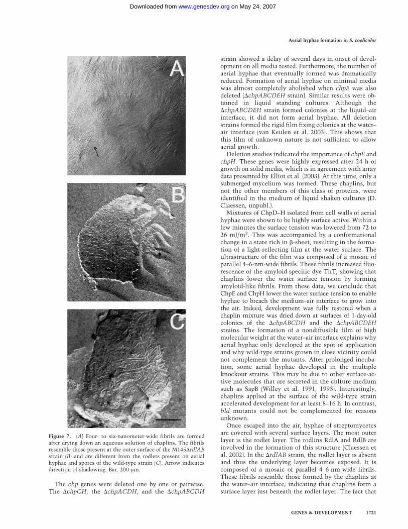
formation of aerial hyphae in S. coelicolor (Fig. 9). Thismodel probably also holds for other streptomycetes be-cause chpD hybridized at stringent conditions with ge-nomic DNA of various members of this group of bacte-ria. During formation of a feeding submerged mycelium,chpE and chpH are expressed. The proteins diffuse to thewater–air interface, where they form a highly insolublefilm that lowers the water surface tension. This enableshyphae to escape into the air. In the aerial hyphae, allchp genes are expressed. The proteins diffuse to the outersurface and assemble on contacting air. The resultinglayer, composed of a mosaic of parallel 4–6-nm-widefibrils, confers hydrophobicity to the aerial hypha. It istempting to speculate that the large chaplins ChpA–Ccoassemble with the small chaplins ChpD–H with theirhomologous sequences. The hydrophilic region of thelarge chaplins may traverse the cell wall, whereas theLAXTG motif is cross-linked to the peptidoglycan. As aresult, the small chaplins would be immobilized at thecell surface by the larger chaplins. The chaplin layer pro-vides a hydrophobic surface and this may induce the for-mation of the rodlet layer formed by RdlA and RdlB.Chaplins resemble the hydrophobins of filamentous
fungi. These proteins, too, enable hyphae to breach thewater–air interface by lowering the water surface tension(Wosten et al. 1999) on assembly in a highly insolublefilm composed of a mosaic of fibrils (Wosten et al. 1993).Moreover, the assembled hydrophobin is responsible forthe hydrophobicity of fungal aerial structures (Wosten etal. 1993, 1994; Wosten 2001). Similar to the chaplins,hydrophobins assemble in an amyloid-like structure(Wosten and de Vocht 2000; Butko et al. 2001; Mackay et
al. 2001). Until recently, amyloids were mainly associ-ated with debilitating human diseases like Huntington’sdisease and Alzheimer’s (Chiti et al. 1999; Dobson 1999).Chapman and colleagues (2002) reported that the curliproteins of Escherichia coli are functional amyloids in-volved in colonization of inert surfaces, biofilm forma-tion, and attachment to a variety of host proteins. Thus,like in fungi, amyloids also serve a wide spectrum offunctions in bacterial development.
Materials and methods
Strains and plasmids
Cloning was done in E. coli DH5�, SCS110 or BW25113 (Dat-senko and Wanner 2000). E. coli ET12567 containing pUZ8002was used for conjugation to S. coelicolor. S. coelicolor strainsM145 (Kieser et al. 2000), �rdlAB6 (Claessen et al. 2002), bldJ261,bldD53 (Willey et al. 1993), bldH109 (Champness 1988), andbldA39 (Piret and Chater 1985) were used in this study as wellas Streptomyces lividans TK23 (Kieser et al. 2000), Streptomy-ces tendae Tü901/8c (Richter et al. 1998), and Streptomycesgriseus (DSMZ 40236). Vectors and constructs are summarizedin Table 2.
Growth conditions and media
Streptomyces strains were grown at 30°C on solid MS or R5medium (Kieser et al. 2000) or on solid or liquid modifiedNMMP medium (van Keulen et al. 2003). The solid medium R5was used for regenerating protoplasts. Yeast extract–malt ex-tract (YEME) medium was used for liquid shaken cultures (Kie-ser et al. 2000).
Molecular techniques
Standard molecular techniques followed Sambrook et al. (1989).Protoplast preparation and transformation were performed asdescribed by Kieser et al. (2000). Chromosomal DNA of S. coe-licolor strains was isolated according to Verhasselt et al. (1989)and modified by Nagy et al. (1995). Total RNA of S. coelicolorwas isolated using the SV Total RNA Isolation System (Pro-mega) according to Veenendaal and Wosten (1998) and 5 µg was
Figure 8. Surface activity of a mixture ofchaplins (A), as indicated by the shape ofan 8-µL droplet, water serving as a control(B).
Claessen et al.
1722 GENES & DEVELOPMENT
on May 24, 2007 www.genesdev.orgDownloaded from
loaded on gel. DNA and RNA were blotted on Nylon filters(Boehringer) and hybridized at 63°C under conditions describedby Church and Gilbert (1984). Under these conditions, cross-hybridization between the different chp genes was weak, if pres-ent at all. Probes were radioactively labeled using the Prime-a-Gene Labelling kit (Promega).
Construction of the S. coelicolor M145 chp deletion strains
Gene deletions were made in S. coelicolor M145 using the Re-direct technology as described (Gust et al. 2002, 2003). Gene
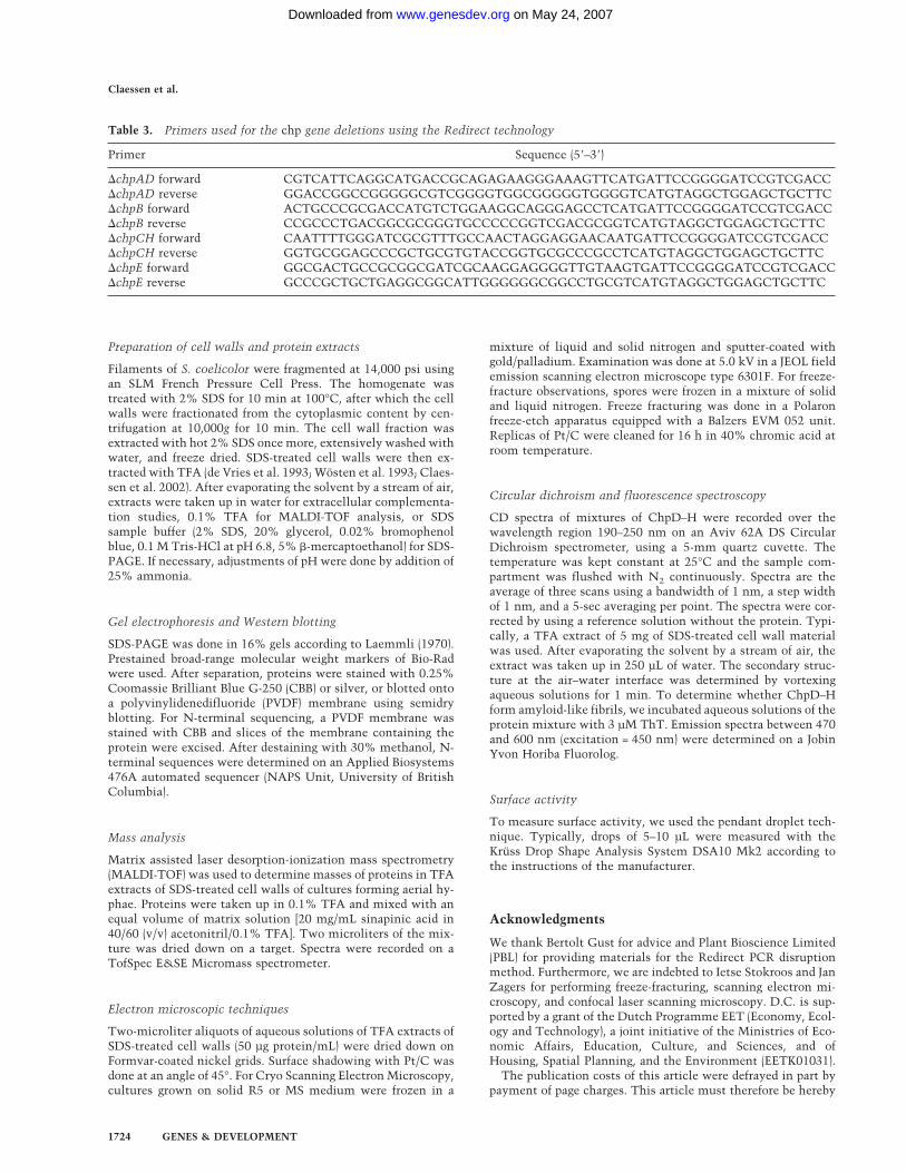
replacements of chpAD, chpB, chpCH, or chpE were made withthe apramycin resistance cassette. For the deletion of chpCH inthe �chpAD strain, the aadA spectinomycin–streptomycin re-sistance cassette was used, resulting in �chpACDH. For thedeletion of chpB in the �chpACDH strain, the viomycin resis-tance cassette was used (�chpABCDH). The FLP recombinasewas used to remove the apramycin cassette in the �chpABCDHstrain (Gust et al. 2002, 2003), enabling us to reuse this cassetteto delete chpE. This resulted in the �chpABCDEH strain. Prim-ers used for the Redirect technology are listed in Table 3. Genedeletions were confirmed by Southern analysis.
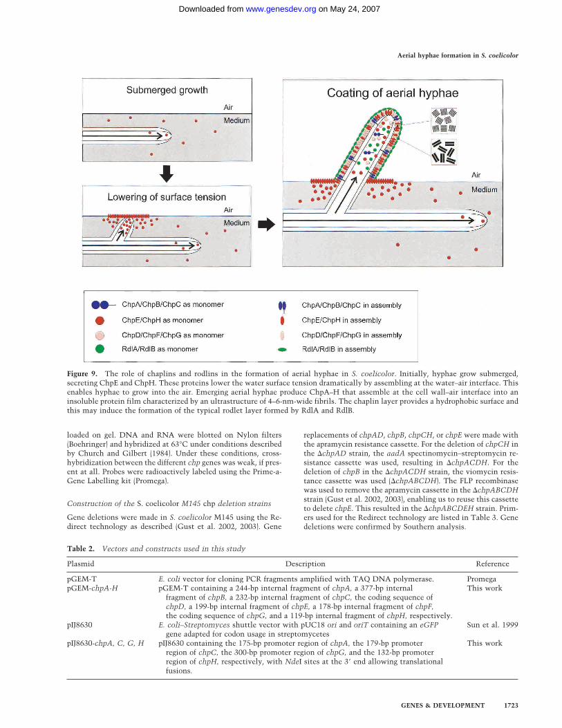
Table 2. Vectors and constructs used in this study
Plasmid Description Reference
pGEM-T E. coli vector for cloning PCR fragments amplified with TAQ DNA polymerase. PromegapGEM-chpA-H pGEM-T containing a 244-bp internal fragment of chpA, a 377-bp internal
fragment of chpB, a 232-bp internal fragment of chpC, the coding sequence ofchpD, a 199-bp internal fragment of chpE, a 178-bp internal fragment of chpF,the coding sequence of chpG, and a 119-bp internal fragment of chpH, respectively.
This work
pIJ8630 E. coli–Streptomyces shuttle vector with pUC18 ori and oriT containing an eGFPgene adapted for codon usage in streptomycetes
Sun et al. 1999
pIJ8630-chpA, C, G, H pIJ8630 containing the 175-bp promoter region of chpA, the 179-bp promoterregion of chpC, the 300-bp promoter region of chpG, and the 132-bp promoterregion of chpH, respectively, with NdeI sites at the 3� end allowing translationalfusions.
This work
Figure 9. The role of chaplins and rodlins in the formation of aerial hyphae in S. coelicolor. Initially, hyphae grow submerged,secreting ChpE and ChpH. These proteins lower the water surface tension dramatically by assembling at the water–air interface. Thisenables hyphae to grow into the air. Emerging aerial hyphae produce ChpA–H that assemble at the cell wall–air interface into aninsoluble protein film characterized by an ultrastructure of 4–6-nm-wide fibrils. The chaplin layer provides a hydrophobic surface andthis may induce the formation of the typical rodlet layer formed by RdlA and RdlB.
Aerial hyphae formation in S. coelicolor
GENES & DEVELOPMENT 1723
on May 24, 2007 www.genesdev.orgDownloaded from
Preparation of cell walls and protein extracts
Filaments of S. coelicolor were fragmented at 14,000 psi usingan SLM French Pressure Cell Press. The homogenate wastreated with 2% SDS for 10 min at 100°C, after which the cellwalls were fractionated from the cytoplasmic content by cen-trifugation at 10,000g for 10 min. The cell wall fraction wasextracted with hot 2% SDS once more, extensively washed withwater, and freeze dried. SDS-treated cell walls were then ex-tracted with TFA (de Vries et al. 1993; Wosten et al. 1993; Claes-sen et al. 2002). After evaporating the solvent by a stream of air,extracts were taken up in water for extracellular complementa-tion studies, 0.1% TFA for MALDI-TOF analysis, or SDSsample buffer (2% SDS, 20% glycerol, 0.02% bromophenolblue, 0.1 M Tris-HCl at pH 6.8, 5% �-mercaptoethanol) for SDS-PAGE. If necessary, adjustments of pH were done by addition of25% ammonia.
Gel electrophoresis and Western blotting
SDS-PAGE was done in 16% gels according to Laemmli (1970).Prestained broad-range molecular weight markers of Bio-Radwere used. After separation, proteins were stained with 0.25%Coomassie Brilliant Blue G-250 (CBB) or silver, or blotted ontoa polyvinylidenedifluoride (PVDF) membrane using semidryblotting. For N-terminal sequencing, a PVDF membrane wasstained with CBB and slices of the membrane containing theprotein were excised. After destaining with 30% methanol, N-terminal sequences were determined on an Applied Biosystems476A automated sequencer (NAPS Unit, University of BritishColumbia).
Mass analysis
Matrix assisted laser desorption-ionization mass spectrometry(MALDI-TOF) was used to determine masses of proteins in TFAextracts of SDS-treated cell walls of cultures forming aerial hy-phae. Proteins were taken up in 0.1% TFA and mixed with anequal volume of matrix solution [20 mg/mL sinapinic acid in40/60 (v/v) acetonitril/0.1% TFA]. Two microliters of the mix-ture was dried down on a target. Spectra were recorded on aTofSpec E&SE Micromass spectrometer.
Electron microscopic techniques
Two-microliter aliquots of aqueous solutions of TFA extracts ofSDS-treated cell walls (50 µg protein/mL) were dried down onFormvar-coated nickel grids. Surface shadowing with Pt/C wasdone at an angle of 45°. For Cryo Scanning Electron Microscopy,cultures grown on solid R5 or MS medium were frozen in a
mixture of liquid and solid nitrogen and sputter-coated withgold/palladium. Examination was done at 5.0 kV in a JEOL fieldemission scanning electron microscope type 6301F. For freeze-fracture observations, spores were frozen in a mixture of solidand liquid nitrogen. Freeze fracturing was done in a Polaronfreeze-etch apparatus equipped with a Balzers EVM 052 unit.Replicas of Pt/C were cleaned for 16 h in 40% chromic acid atroom temperature.
Circular dichroism and fluorescence spectroscopy
CD spectra of mixtures of ChpD–H were recorded over thewavelength region 190–250 nm on an Aviv 62A DS CircularDichroism spectrometer, using a 5-mm quartz cuvette. Thetemperature was kept constant at 25°C and the sample com-partment was flushed with N2 continuously. Spectra are theaverage of three scans using a bandwidth of 1 nm, a step widthof 1 nm, and a 5-sec averaging per point. The spectra were cor-rected by using a reference solution without the protein. Typi-cally, a TFA extract of 5 mg of SDS-treated cell wall materialwas used. After evaporating the solvent by a stream of air, theextract was taken up in 250 µL of water. The secondary struc-ture at the air–water interface was determined by vortexingaqueous solutions for 1 min. To determine whether ChpD–Hform amyloid-like fibrils, we incubated aqueous solutions of theprotein mixture with 3 µM ThT. Emission spectra between 470and 600 nm (excitation = 450 nm) were determined on a JobinYvon Horiba Fluorolog.
Surface activity
To measure surface activity, we used the pendant droplet tech-nique. Typically, drops of 5–10 µL were measured with theKrüss Drop Shape Analysis System DSA10 Mk2 according tothe instructions of the manufacturer.
Acknowledgments
We thank Bertolt Gust for advice and Plant Bioscience Limited(PBL) for providing materials for the Redirect PCR disruptionmethod. Furthermore, we are indebted to Ietse Stokroos and JanZagers for performing freeze-fracturing, scanning electron mi-croscopy, and confocal laser scanning microscopy. D.C. is sup-ported by a grant of the Dutch Programme EET (Economy, Ecol-ogy and Technology), a joint initiative of the Ministries of Eco-nomic Affairs, Education, Culture, and Sciences, and ofHousing, Spatial Planning, and the Environment (EETK01031).The publication costs of this article were defrayed in part by
payment of page charges. This article must therefore be hereby
Table 3. Primers used for the chp gene deletions using the Redirect technology
Primer Sequence (5�–3�)
�chpAD forward CGTCATTCAGGCATGACCGCAGAGAAGGGAAAGTTCATGATTCCGGGGATCCGTCGACC�chpAD reverse GGACCGGCCGGGGGCGTCGGGGTGGCGGGGGTGGGGTCATGTAGGCTGGAGCTGCTTC�chpB forward ACTGCCCGCGACCATGTCTGGAAGGCAGGGAGCCTCATGATTCCGGGGATCCGTCGACC�chpB reverse CCGCCCTGACGGCGCGGGTGCCCCCGGTCGACGCGGTCATGTAGGCTGGAGCTGCTTC�chpCH forward CAATTTTGGGATCGCGTTTGCCAACTAGGAGGAACAATGATTCCGGGGATCCGTCGACC�chpCH reverse GGTGCGGAGCCCGCTGCGTGTACCGGTGCGCCCGCCTCATGTAGGCTGGAGCTGCTTC�chpE forward GGCGACTGCCGCGGCGATCGCAAGGAGGGGTTGTAAGTGATTCCGGGGATCCGTCGACC�chpE reverse GCCCGCTGCTGAGGCGGCATTGGGGGGCGGCCTGCGTCATGTAGGCTGGAGCTGCTTC
Claessen et al.
1724 GENES & DEVELOPMENT
on May 24, 2007 www.genesdev.orgDownloaded from
marked “advertisement” in accordance with 18 USC section1734 solely to indicate this fact.
References
Bentley, S.D., Chater, K.F., Cerdeño-Tarraga, A.-M., Challis,G.L., Thomson, N.R., James, K.D., Harris, D.E., Quail, M.A.,Kieser, H., Harper, D., et al. 2002. Complete genome se-quence of the model actinomycete Streptomyces coelicolorA3(2). Nature 417: 141–147.
Butko, P., Buford, J.P., Goodwin, J.S., Stroud, P.A., McCormick,C.L., and Cannon, G.C. 2001. Spectroscopic evidence foramyloid-like interfacial self-assembly of hydrophobin Sc3.Biochem. Biophys. Res. Commun. 280: 212–215.
Champness, W.C. 1988. New loci required for Streptomycescoelicolor morphological and physiological differentiation.J. Bacteriol. 170: 1168–1174.
Chapman, M.R., Robinson, L.S., Pinkner, J.S., Roth, R., Heuser,J., Hammar, M., Normark, S., and Hultgren, S.J. 2002. Role ofEscherichia coli curli operons in directing amyloid fiber for-mation. Science 295: 851–855.
Chater, K.F. 1998. Taking a genetic scalpel to the Streptomycescolony. Microbiology 144: 1465–1478.
———. 2001. Regulation of sporulation in Streptomyces coeli-color A3(2): A checkpoint multiplex? Curr. Opin. Microbiol.4: 667–673.
Chiti, F., Webster, P., Taddei, N., Clark, A., Stefani, M., Ram-poni, G., and Dobson, C.M. 1999. Designing conditions forin vitro formation of amyloid protofilaments and fibrils.Proc. Natl. Acad. Sci. 96: 3590–3594.
Church, G.M. and Gilbert, W. 1984. Genomic sequencing. Proc.Natl. Acad. Sci. 81: 1991–1995.
Claessen, D., Wosten, H.A.B., van Keulen, G., Faber, O.G.,Alves, A.M.C.R., Meijer, W.G., and Dijkhuizen, L. 2002.Two novel homologous proteins of Streptomyces coelicolorand Streptomyces lividans are involved in the formation ofthe rodlet layer and mediate attachment to a hydrophobicsurface. Mol. Microbiol. 44: 1483–1492.
Datsenko, K.A. and Wanner, B.L. 2000. One-step inactivation ofchromosomal genes in Escherichia coli K-12 using PCRproducts. Proc. Natl. Acad. Sci. 97: 6640–6645.
de Vries, O.M.H., Fekkes, M.P., Wosten, H.A.B., and Wessels,J.G.H. 1993. Insoluble hydrophobin complexes in the wallsof Schizophyllum commune and other filamentous fungi.Arch. Microbiol. 159: 330–335.
Dobson, C.M. 1999. Protein misfolding, evolution and disease.Trends Biochem. Sci. 24: 329–332.
Elliot, M.A., Karoonuthaisiri, N., Huang, J., Bibb, M.J., Cohen,S.N., Kao, C.M., and Buttner, M.J. 2003. The chaplins: Afamily of hydrophobic cell-surface proteins involved in aerialmycelium formation in Streptomyces coelicolor. Genes &Dev. (this issue).
Gust, B., Kieser, T., and Chater, K.F. 2002. REDIRECT technol-ogy: PCR-targeting system in Streptomyces coelicolor. TheJohn Innes Centre, Norwich, United Kingdom.
Gust, B., Challis, G.L., Fowler, K., Kieser, T., and Chater, K.F.2003. PCR-targeted Streptomyces gene replacement identi-fies a protein domain needed for biosynthesis of the sesqui-terpene soil odor geosmin. Proc. Natl. Acad. Sci. 100: 1541–1546. (Published online ahead of print on January 31, 2003,as 10.1073/pnas.0337542100.)
Kelemen, G.H. and Buttner, M.J. 1998. Initiation of aerial my-celium formation in Streptomyces. Curr. Opin. Microbiol.1: 656–662.
Kieser, T., Bibb, M.J., Buttner, M.J., Chater, K.F., and Hopwood,
D.A. 2000. Practical Streptomyces genetics. The John InnesFoundation, Norwich, United Kingdom.
Laemmli, U.K. 1970. Cleavage of structural proteins during theassembly of the head of bacteriophage T4. Nature 227: 680–685.
Mackay, J.P., Matthews, J.M., Winefield, R.D., Mackay, L.G.,Haverkamp, R.G., and Templeton, M.D. 2001. Structure9: 83–91.
Mazmanian, S.K., Liu, G., Ton-That, H., and Schneewind, O.1999. Staphylococcus aureus sortase, an enzyme that an-chors surface proteins to the cell wall. Science 285: 760–763.
Nagy, I., Schoofs, G., Compernolle, F., Proost, P., Vanderleyden,J., and de Mot, R. 1995. Degradation of the thiocarbonateherbicide EPTC (S-ethyl dipropylcarbamotioate) and bio-safening by Rhodococcus sp. strain NI86/21 involve an in-ducible cytochrome P-450 system and aldehyde dehydroge-nase. J. Bacteriol. 177: 676–687.
Pallen, M.J., Lam, A.C., Antonio, M., and Dunbar, K. 2001. Anembarrassment of sortases—A richness of substrates?Trends Microbiol. 9: 97–102.
Piret, J.M. and Chater, K.F. 1985. Phage-mediated cloning ofBldA, a region involved in Streptomyces coelicolor morpho-logical development, and its analysis by genetic complemen-tation. J. Bacteriol. 163: 965–972.
Richter, M., Willey, J.M., Süßmuth, R., Jung, G., and Fiedler,H.-P. 1998. Streptofactin, a novel biosurfactant with aerialmycelium inducing activity from Streptomyces tendae Tü901/8c. FEMS Microbiol. Lett. 163: 165–171.
Sambrook, J., Fritsch, E.F., and Maniatis, T. 1989. Molecularcloning. A laboratory manual. Cold Spring Harbor Labora-tory Press, Cold Spring Harbor, NY.
Smucker, R.A. and Pfister, R.M. 1978. Characteristics of Strep-tomyces coelicolor A3(2) aerial spore rodlet mosaic. Can.J. Microbiol. 24: 397–408.
Sun, J., Kelemen, G.H., Fernandez-Abalos, J.M., and Bibb, M.J.1999. Green fluorescent protein as a reporter for spatial andtemporal gene expression in Streptomyces coelicolor A3(2).Microbiology 145: 2221–2227.
Tillotson, R.D., Wosten, H.A.B., Richter, M., and Willey, J.M.1998. A surface active protein involved in aerial hyphae for-mation in the filamentous fungus Schizophyllum communerestores the capacity of a bald mutant of the filamentousbacterium Streptomyces coelicolor to erect aerial structures.Mol. Microbiol. 30: 595–602.
Ton-That, H., Faull, K.F., and Schneewind, O. 1997. Anchorstructure of staphylococcal surface proteins. A branchedpeptide that links the carboxyl terminus of proteins to thecell wall. J. Biol. Chem. 272: 22285–22292.
van Keulen, G., Jonkers, H.M., Claessen, D., Dijkhuizen, L., andWosten, H.A.B. 2003. Differentiation and anaerobiosis instanding liquid cultures of Streptomyces coelicolor. J. Bac-teriol. 185: 1455–1458.
Veenendaal, A.K.J. and Wosten, H.A.B. 1998. Total RNA fromthe Gram-positive bacterium Streptomyces lividans. Pro-mega Benelux News 17: 7.
Verhasselt, P., Poncelet, F., Vits, K., and Vanderleyden, J. 1989.Cloning and expression of a Clostridium acetobutylicum al-pha-amylase gene in Escherichia coli. FEMS Microbiol. Lett.59: 135–140.
Wildermuth, H., Wehrli, E., and Horne, R.W. 1971. The surfacestructure of spores and aerial mycelium in Streptomycescoelicolor. J. Ultrastruct. Res. 35: 168–180.
Willey, J.M., Santamaría, R.I., Guijarro, J., Geistlich, M., andLosick, R. 1991. Extracellular complementation of a devel-opmental mutation implicates a small sporulation protein inaerial mycelium formation by S. coelicolor. Cell 65: 641–
Aerial hyphae formation in S. coelicolor
GENES & DEVELOPMENT 1725
on May 24, 2007 www.genesdev.orgDownloaded from

650.Willey, J.M., Schwedock, J., and Losick, R. 1993. Multiple ex-
tracellular signals govern the production of a morphogeneticprotein involved in aerial mycelium formation by Strepto-myces coelicolor. Genes & Dev. 7: 895–903.
Wosten, H.A.B. 2001. Hydrophobins: Multipurpose proteins.Annu. Rev. Microbiol. 55: 625–646.
Wosten, H.A.B. and de Vocht, M.L. 2000. Hydrophobins, thefungal coat unravelled. Biochim. Biophys. Acta 1469: 79–86.
Wosten, H.A.B. and Willey, J.M. 2000. Surface-active proteinsenable microbial aerial hyphae to grow into the air. Micro-biology 146: 767–773.
Wosten, H.A.B., de Vries, O.M.H., and Wessels, J.G.H. 1993.Interfacial self-assembly of a fungal hydrophobin into a hy-drophobin rodlet layer. Plant Cell 5: 1567–1574.
Wosten, H.A.B., Ásgeirsdóttir, S.A., Krook, J.H., Drenth, J.H.H.,and Wessels, J.G.H. 1994. The fungal hydrophobin Sc3p self-assembles at the surface of aerial hyphae as a protein mem-brane constituting the hydrophobic rodlet layer. Eur. J. CellBiol. 63: 122–129.
Wosten, H.A.B., van Wetter, M.-A., Lugones, L.G., van der Mei,H.C., Busscher, H.J., and Wessels, J.G.H. 1999. How a fungusescapes the water to grow into the air. Curr. Biol. 9: 85–88.
Claessen et al.
1726 GENES & DEVELOPMENT
on May 24, 2007 www.genesdev.orgDownloaded from
Copyright © 2022 FDOKUMEN