Spitzenkorper Localization and Intracellular Traffic of Green Fluorescent Protein-Labeled CHS-3 and...
12
EUKARYOTIC CELL, Oct. 2007, p. 1853–1864 Vol. 6, No. 10 1535-9778/07/$08.000 doi:10.1128/EC.00088-07 Copyright © 2007, American Society for Microbiology. All Rights Reserved. Spitzenko ¨rper Localization and Intracellular Traffic of Green Fluorescent Protein-Labeled CHS-3 and CHS-6 Chitin Synthases in Living Hyphae of Neurospora crassa † Meritxell Riquelme, 1 * Salomon Bartnicki-Garcı ´a, 1 Juan Manuel Gonza ´lez-Prieto, 2 Eddy Sa ´nchez-Leo ´n, 1 Jorge A. Verdı ´n-Ramos, 1 Alejandro Beltra ´n-Aguilar, 1 and Michael Freitag 3 Department of Microbiology, Center for Scientific Research and Higher Education of Ensenada (CICESE), Km 107 Ctra. Tijuana-Ensenada, 22860 Ensenada, Baja California, Me ´xico 1 ; Center for Genomic Biotechnology, Blvd. del Maestro s/n. 88710 Cd. Reynosa, Tamaulipas, Me ´xico 2 ; and Department of Biochemistry and Biophysics ALS 2011, Oregon State University, Corvallis, Oregon 97331-7305 3 Received 19 March 2007/Accepted 12 July 2007 The subcellular location and traffic of two selected chitin synthases (CHS) from Neurospora crassa, CHS-3 and CHS-6, labeled with green fluorescent protein (GFP), were studied by high-resolution confocal laser scanning microscopy. While we found some differences in the overall distribution patterns and appearances of CHS-3–GFP and CHS-6–GFP, most features were similar and were observed consistently. At the hyphal apex, fluorescence congregated into a conspicuous single body corresponding to the location of the Spitzenko ¨rper (Spk). In distal regions (beyond 40 m from the apex), CHS-GFP revealed a network of large endomembra- nous compartments that was predominantly comprised of irregular tubular shapes, while some compartments were distinctly spherical. In the distal subapex (20 to 40 m from the apex), fluorescence was observed in globular bodies that appeared to disintegrate into vesicles as they advanced forward until reaching the proximal subapex (5 to 20 m from the apex). CHS-GFP was also conspicuously found delineating developing septa. Analysis of fluorescence recovery after photobleaching suggested that the fluorescence of the Spk originated from the advancing population of microvesicles (chitosomes) in the subapex. The inability of brefeldin A to interfere with the traffic of CHS-containing microvesicles and the lack of colocalization of CHS-GFP with the endoplasmic reticulum (ER)-Golgi body fluorescent dyes lend support to the idea that CHS proteins are delivered to the cell surface via an alternative route distinct from the classical ER-Golgi body secretory pathway. Fungal hyphae elongate and branch by a complex process based on polarized secretion. Many studies have investigated the cellular and molecular components involved in shaping fungal cells, but no detailed understanding of the mechanisms that govern and regulate polarized fungal growth has been achieved (4, 25). In the yeast Saccharomyces cerevisiae, many of the main components of the secretory pathway, including some of the enzymes involved in cell wall formation, have been extensively characterized (32). Filamentous fungi encode ho- mologues of some key components known from the yeast se- cretory pathway, but despite their apparent orthology, rela- tively little is known about how this pathway is organized to accomplish the highly polarized growth typical of hyphae. There are some differences in cell wall synthesis between fila- mentous fungi and S. cerevisiae. In hyphae of septate fungi, vesicles and other components accumulate at the apex, as part of the Spitzenko ¨rper (Spk) (14, 22–24, 28). The composition and mode of action of this pleomorphic and dynamic structure have intrigued fungal biologists for many decades. Fungal cells have at least two types of well-defined secretory vesicles (5). It has been suggested that macrovesicles, or con- ventional secretory vesicles, carry the components of the amor- phous phase of the cell wall, in addition to the load of extra- cellular enzymes (5, 27). There is a large body of evidence characterizing the chitin synthase (CHS)-carrying mi- crovesicles as chitosomes (3, 8, 13, 30). CHS are -glycosyl- transferases that catalyze the polymerization of N-acetylglu- cosamine from UDP N-acetylglucosamine into chitin (47), a major structural polymer of the fungal cell wall (2). Chitin synthesis occurs in highly localized fashion both at the hyphal apices (7) and at nascent septa (29). Chitosomes are the small- est vesicles with the ability to form chitin microfibrils in vitro and have been suggested to carry and transport CHS to the cell surface at the apex of hyphae for cell wall synthesis (13, 37, 48, 55, 56). In recent years, studies on fungal CHS have concen- trated mainly on gene identification. Given this wealth of in- formation, we chose CHS as candidate markers to investigate vesicle traffic in fungal hyphae. Fungi have multiple chs genes grouped into two divisions, with seven classes, primarily on the basis of similarities in the primary sequence of the predicted proteins (12, 16, 37, 50). Division I includes classes I, II, and III, which share a catalytic domain surrounded by a hydrophilic N-terminal region and a hydrophobic C-terminal region (12). Division II includes classes IV, V, and VII, all with a catalytic domain preceded by a cytochrome b 5 -like domain. In addition, classes V and VII * Corresponding author. Mailing address: Department of Microbi- ology, Center for Scientific Research and Higher Education of Ensenada (CICESE), P.O. Box 430222, San Ysidro, CA 92143-0222. Phone: (646) 175-0500, ext. 27051. Fax: (646) 175-0595, ext. 27052. E-mail: [email protected]. † Supplemental material for this article may be found at http://ec .asm.org/. Published ahead of print on 20 July 2007. 1853
Transcript of Spitzenkorper Localization and Intracellular Traffic of Green Fluorescent Protein-Labeled CHS-3 and...

EUKARYOTIC CELL, Oct. 2007, p. 1853–1864 Vol. 6, No. 101535-9778/07/$08.00�0 doi:10.1128/EC.00088-07Copyright © 2007, American Society for Microbiology. All Rights Reserved.
Spitzenkorper Localization and Intracellular Traffic of Green FluorescentProtein-Labeled CHS-3 and CHS-6 Chitin Synthases in
Living Hyphae of Neurospora crassa�†Meritxell Riquelme,1* Salomon Bartnicki-Garcıa,1 Juan Manuel Gonzalez-Prieto,2 Eddy Sanchez-Leon,1
Jorge A. Verdın-Ramos,1 Alejandro Beltran-Aguilar,1 and Michael Freitag3
Department of Microbiology, Center for Scientific Research and Higher Education of Ensenada (CICESE), Km 107 Ctra. Tijuana-Ensenada,22860 Ensenada, Baja California, Mexico1; Center for Genomic Biotechnology, Blvd. del Maestro s/n. 88710 Cd. Reynosa, Tamaulipas,
Mexico2; and Department of Biochemistry and Biophysics ALS 2011, Oregon State University, Corvallis, Oregon 97331-73053
Received 19 March 2007/Accepted 12 July 2007
The subcellular location and traffic of two selected chitin synthases (CHS) from Neurospora crassa, CHS-3and CHS-6, labeled with green fluorescent protein (GFP), were studied by high-resolution confocal laserscanning microscopy. While we found some differences in the overall distribution patterns and appearances ofCHS-3–GFP and CHS-6–GFP, most features were similar and were observed consistently. At the hyphal apex,fluorescence congregated into a conspicuous single body corresponding to the location of the Spitzenkorper(Spk). In distal regions (beyond 40 �m from the apex), CHS-GFP revealed a network of large endomembra-nous compartments that was predominantly comprised of irregular tubular shapes, while some compartmentswere distinctly spherical. In the distal subapex (20 to 40 �m from the apex), fluorescence was observed inglobular bodies that appeared to disintegrate into vesicles as they advanced forward until reaching theproximal subapex (5 to 20 �m from the apex). CHS-GFP was also conspicuously found delineating developingsepta. Analysis of fluorescence recovery after photobleaching suggested that the fluorescence of the Spkoriginated from the advancing population of microvesicles (chitosomes) in the subapex. The inability ofbrefeldin A to interfere with the traffic of CHS-containing microvesicles and the lack of colocalization ofCHS-GFP with the endoplasmic reticulum (ER)-Golgi body fluorescent dyes lend support to the idea that CHSproteins are delivered to the cell surface via an alternative route distinct from the classical ER-Golgi bodysecretory pathway.
Fungal hyphae elongate and branch by a complex processbased on polarized secretion. Many studies have investigatedthe cellular and molecular components involved in shapingfungal cells, but no detailed understanding of the mechanismsthat govern and regulate polarized fungal growth has beenachieved (4, 25). In the yeast Saccharomyces cerevisiae, many ofthe main components of the secretory pathway, including someof the enzymes involved in cell wall formation, have beenextensively characterized (32). Filamentous fungi encode ho-mologues of some key components known from the yeast se-cretory pathway, but despite their apparent orthology, rela-tively little is known about how this pathway is organized toaccomplish the highly polarized growth typical of hyphae.There are some differences in cell wall synthesis between fila-mentous fungi and S. cerevisiae. In hyphae of septate fungi,vesicles and other components accumulate at the apex, as partof the Spitzenkorper (Spk) (14, 22–24, 28). The compositionand mode of action of this pleomorphic and dynamic structurehave intrigued fungal biologists for many decades.
Fungal cells have at least two types of well-defined secretoryvesicles (5). It has been suggested that macrovesicles, or con-ventional secretory vesicles, carry the components of the amor-phous phase of the cell wall, in addition to the load of extra-cellular enzymes (5, 27). There is a large body of evidencecharacterizing the chitin synthase (CHS)-carrying mi-crovesicles as chitosomes (3, 8, 13, 30). CHS are �-glycosyl-transferases that catalyze the polymerization of N-acetylglu-cosamine from UDP N-acetylglucosamine into chitin (47), amajor structural polymer of the fungal cell wall (2). Chitinsynthesis occurs in highly localized fashion both at the hyphalapices (7) and at nascent septa (29). Chitosomes are the small-est vesicles with the ability to form chitin microfibrils in vitroand have been suggested to carry and transport CHS to the cellsurface at the apex of hyphae for cell wall synthesis (13, 37, 48,55, 56). In recent years, studies on fungal CHS have concen-trated mainly on gene identification. Given this wealth of in-formation, we chose CHS as candidate markers to investigatevesicle traffic in fungal hyphae.
Fungi have multiple chs genes grouped into two divisions,with seven classes, primarily on the basis of similarities in theprimary sequence of the predicted proteins (12, 16, 37, 50).Division I includes classes I, II, and III, which share a catalyticdomain surrounded by a hydrophilic N-terminal region and ahydrophobic C-terminal region (12). Division II includesclasses IV, V, and VII, all with a catalytic domain preceded bya cytochrome b5-like domain. In addition, classes V and VII
* Corresponding author. Mailing address: Department of Microbi-ology, Center for Scientific Research and Higher Education ofEnsenada (CICESE), P.O. Box 430222, San Ysidro, CA 92143-0222.Phone: (646) 175-0500, ext. 27051. Fax: (646) 175-0595, ext. 27052.E-mail: [email protected].
† Supplemental material for this article may be found at http://ec.asm.org/.
� Published ahead of print on 20 July 2007.
1853

contain an N-terminal myosin motor-like domain, suggesting adirect interaction with the actin cytoskeleton (15, 20, 58). ClassVI has not been assigned to either division and includes re-cently identified CHS of unknown function (16). Earlier stud-ies suggest that the various CHS have specific roles in chitincell wall synthesis that are time or space dependent (60). Incontrast to most filamentous fungi, S. cerevisiae (46) and Can-dida albicans (40) have only three or four CHS isozymes, re-spectively. S. cerevisiae Chs1p, C. albicans Chs2p, and C. albi-cans Chs8p belong to class I; S. cerevisiae Chs2p and C. albicansChs1p belong to class II; and S. cerevisiae Chs3p and C. albi-cans Chs3p belong to class IV (46). While potential roles inhyphal growth have been suggested for some of the seven CHSclasses described in filamentous fungi (9, 64, 65), we lack spe-cific information on the cellular localization and trafficking totheir sites of action in regions of active cell wall growth formost of these proteins.
The goal of this study was to elucidate the traffic of CHS-containing vesicles en route from their site of genesis to theirsite of exocytosis in living hyphae of Neurospora crassa. Theavailability of an almost-complete genome sequence for thisfungus allowed the identification of seven open reading frameswith high homology to previously described chs genes (10). Wechose to trace the intracellular location and secretory paths ofCHS-3 and CHS-6. Neurospora CHS-3 belongs to the previ-ously reported class I CHS with known homologues in all fungitested, including S. cerevisiae Chs1p. In contrast, CHS-6 is anewly identified CHS assigned to class VI, homologous toAspergillus fumigatus ChsD (39) and Coccidioides posadasiiCHS-6 (34) but with no apparent homologues in S. cerevisiae orC. albicans. To trace both proteins, we fused green fluorescent
protein (GFP) to the carboxyl terminus of the CHS codingregions and analyzed the fate of the resulting CHS-3–GFP andCHS-6–GFP fusion proteins by high-resolution confocal laserscanning microscopy (CLSM) in living hyphae of N. crassa.
MATERIALS AND METHODS
Strains and culture conditions. For transformations, N. crassa strain N623(mat A his-3; FGSC 6103) was grown at 32°C for 5 to 10 days on Vogel’s minimalmedium (VMM) agar (59) containing 1.5% sucrose and supplemented withhistidine (0.5 mg/ml). Transformed conidia were spread on VMM-FGS (0.5%fructose, 0.5% glucose, 20% sorbose) medium. Five to 10 His� prototrophictransformants per plate were transferred to VMM tubes and incubated at 32°Cfor 3 days. For epifluorescence and confocal microscopy, transformants wereroutinely grown on VMM and observed using the “inverted agar block method”(26).
Recombinant DNA techniques and plasmid constructions. Standard PCR andcloning procedures were used to fuse the sgfp gene to the carboxyl terminus ofchs-3 and chs-6 (53). The 2.7-kb chs-3 gene (class I CHS; GenBank accession no.XM_323590) and the 2.5-kb chs-6 gene (class VI CHS; GenBank accession no.XM_324624) were amplified by PCR from N. crassa N1 (mat a; FGSC 988)genomic DNA with custom-designed primers that included XbaI and PacI re-striction endonuclease sites at their respective 5� termini (Table 1). PCR wasperformed in a Bio-Rad Thermal Cycler with platinum Pfu polymerase (Invitro-gen) according to the manufacturer’s instructions, under the following condi-tions: denaturation at 94°C for 1 min, followed by 30 cycles of 94°C (30 s), 61°C(30 s), and 72°C (3 min), and a final extension step at 72°C for 5 min. Theamplified and gel-purified PCR products were digested with XbaI and PacI andcloned into XbaI- and PacI-digested plasmid pMF272 (19; GenBank accessionno. AY598428). This yielded pJMGT5 (chs-3) and pJMG58 (chs-6). Inserts weresequenced at the Core Instrumentation Facility of the Institute for IntegrativeGenome Biology at the University of California, Riverside, with primerspMF272F and pMF272R2 (Table 1) designed for the upstream and downstreamflanks of the multiple cloning site of pMF272.
Neurospora genetics. Transformation of N. crassa strain N623 his-3 conidiawith plasmids pJMGT5 (linearized by digestion with SspI) and pJMG58 (linear-ized by digestion with NdeI) was carried out by electroporation on a Bio-Rad
TABLE 1. Strains, plasmids, and oligonucleotides used or generated for this study
Plasmid, N. crassa strain,a
or oligonucleotide Description, genotype, or sequenceb Reference or accession no.
PlasmidspMF272 Pccg-1::sgfp� 19pGP003 Pccg-1::chs-3�::sgfp� This studypGP002 Pccg-1::chs-6�::sgfp� This study
StrainsN39 mat A; fl FGSC 4317N40 mat a; fl FGSC 4347N150 mat A FGSC 9013N623 mat A his-3 FGSC 6103N625 mat a his-3 FGSC 6525NMR3 mat A his-3�::Pccg-1::chs-3�::sgfp� This studyNMR6 mat A his-3�::Pccg-1::chs-6�::sgfp� This studyN1 mat a FGSC 988
OligonucleotidesMRp10 5�-AGAGACAAGAAAATTACCCCCTTCTT-3� This studyMRp11 5�-AACTACAACAGCCACAACGTCTATATC-3� This studyMRp12 5�-ATAATGAACGGAAGGTAGTTGTAGAAAG-3� This studyMRp13 5�-ATGGATATAATGTGGCTGTTGAAAG-3� This studypMF272F 5�-CAAATCAACACAACACTCAAACCA-3� 19pMF272R2 5�-AGATGAACTTCAGGGTCAGCTTG-3� This studyChs3F 5�-GAAAGTTCTAGAATGGATCCTCGAATGCATACG-3� This studyChs3R 5�-AGCATTTAATTAAAACACCCCTAAACATCCTCAC-3� This studyChs6F 5�-AAATAATCTAGAATGACTATCAATTACCTTGGG-3� This studyChs6R 5�-TCACCTTAATTAATCTCTGATTTGACCCCCTCGA-3� This study
a Transformants NMR3 and NMR6 appear homokaryotic because Southern blotting analyses revealed the presence of his-3�::chs::sgfp loci exclusively.b Restriction endonuclease sites for cloning are shown in bold type, and predicted CHS start codons are underlined.
1854 RIQUELME ET AL. EUKARYOT. CELL

Gene Pulser (capacitance, 25 �F; 1.5 kV; resistance, 600 �) as previously de-scribed (35). Prototrophic His� transformants were screened for the expressionof CHS-GFP by epifluorescence microscopy as described previously (19). Noneof the primary transformants showed sufficient GFP expression under a fluores-cence stereomicroscope, but almost all transformants showed fluorescence atvaried levels when screened under an inverted epifluorescence microscope.Transformants showing robust fluorescence were selected (strains TMR3-1 toTMR3-4 and strains TMR6-1 to TMR6-4). Potential heterokaryotic prototrophictransformants were crossed to N. crassa strain N625 (mat a his-3; FGSC 6525) onsynthetic crossing medium (63). Ascospores were heat-shocked on VMM at 60°Cfor 1 h, germinated, picked to VMM slants, and analyzed for fluorescence. Twostrains, NMR3 and NMR6, were selected for further analysis.
For genomic DNA extraction, we used the DNeasy plant extraction kit(QIAGEN Inc.). Mycelium for DNA extraction was grown at 28°C for 7 days onliquid medium with no shaking and no light, filtered, submerged in liquid nitro-gen and lyophilized. Integration of chs-sgfp fusion genes was verified by PCR withtwo sets of primers (MRp10 and MRp11 or MRp12 and MRp13; Table 1). Forconfirmatory Southern blotting analyses, Neurospora genomic DNA from strainsNMR3 and NMR6 was digested with NdeI, separated on a 0.8% agarose gel,blotted to charged nylon membranes, and probed with a 2.67-kb HindIII frag-ment from pMF272 that contains part of the his-3 gene. Probes were generatedwith a nonradioactive labeling kit (Roche Applied Science).
Confocal microscopy and image processing. Growing hyphae were imaged at20 to 22°C using an inverted Zeiss LSM-510 META CLSM fitted with an argon/2ion laser with a GFP filter set (excitation, 488 nm; emission, 515 to 530 nm). A�100 Ph3 Plan Neofluar oil immersion objective (N.A. 1.3) or a �63 DIC PlanApochromat oil immersion objective (N.A. 1.4) were used for image acquisition.A photomultiplier module allowed us to merge the confocal with the phase-contrast images, thus providing simultaneous precise imaging of fluorescentlylabeled proteins in the entire cell context.
Confocal images were captured using LSM-510 software (version 3.2; CarlZeiss), evaluated with an LSM-510 image examiner (version 3.2), and furtherprocessed with Adobe Photoshop CS2 (version 9.0). Selected image series wereconverted into movies with the same software. For fluorescence recovery afterphotobleaching (FRAP) analysis, the “bleach control” command of the LSM-510software was used. Areas to be bleached were selected with the ROI (region ofinterest) tool. Bleaching was applied during a time series sequence acquisitionwith 35 iterations and 85% laser intensity. For each strain, we analyzed at leastfive sequences for both subapical and apical photobleaching.
To track the movement of fluorescent particles, we used the “track object”option of Image Pro Plus v. 6.1 (Media Cybernetics). We manually traced thetrajectory of the Spk and putative vesicles that remained in view for at least 3 to4 frames. The paths and speeds of 9 and 37 vesicles were analyzed in differentsequences of NMR3 and NMR6, respectively.
Fluorescent dyes and inhibitors. Concentrated stock solutions of organelle-specific dyes and inhibitors were made in either dimethyl sulfoxide or deionizedsterile water as suggested by the manufacturer, kept at �20°C, diluted in liquidVMM to the appropriate working concentration, and allowed to warm up toroom temperature before being applied to growing hyphae. For each dye treat-ment, the agar blocks containing Neurospora that were growing mycelia wereinverted onto a coverslip containing a 10-�l drop of the corresponding solution.Stained cells were imaged after a 3- to 5-min recovery period. As a marker forvacuolar content, we used a 10 �M diacetate derivative of carboxydifluorofluo-rescein (CDFFDA, catalog number O6151; Molecular Probes) imaged with theargon/2 ion laser (excitation maximum, 498 nm, and emission maximum, 520nm). As a marker of endocytic organelles, we used 25 �M of the styril dyeN-(3-triethylammoniumpropyl)-4-(6-[4-{diethylamino} phenyl] hexatrienyl) pyr-idinium dibromide (FM4-64, catalog number T3166; Molecular Probes), imagedwith the argon/2 ion laser (excitation, 514 nm, and emission, 670 nm) (26). As amarker of Golgi body equivalents and the endoplasmic reticulum (ER), we used5 �M brefeldin A (BFA) conjugated to bodipy 558/568 (catalog number B7449;Molecular Probes). Observations were made with a He/Ne-2 laser (excitation,558 nm, and emission, 568 nm).
RESULTS
Selection of positive transformants. After transformation ofa Neurospora his-3 host strain with either pJMGT5 or pJMG58,we obtained prototrophic putative transformants that ex-pressed CHS-3–GFP (strain TMR3) or CHS-6–GFP (strainTMR6), respectively, when screened by epifluorescence mi-
croscopy. After TMR3 and TMR6 were crossed with N. crassaN625, two homokaryotic strains, NMR3 and NMR6, showedstable fluorescence and were selected for further analysis byCSLM. Strains NMR3 and NMR6 were spot tested on plateswith fluffy strains; as expected, both were mat A, reflecting theclose linkage of mat and his-3.
The integration of the chs-sgfp fusion genes at his-3 wasconfirmed by PCR and Southern blotting analyses. For chs-3-sgfp, an expected 3.2-kb PCR product was obtained with prim-ers MRp10 and MRp11, and for chs-6-sgfp, an expected 2.1-kbPCR product was obtained with primers MRp12 and MRp13.Integration by gene replacement at his-3 was also confirmed bySouthern blotting analysis for both strains. While the hoststrain revealed the single expected 3.9-kb band, we observed anadditional 6.2-kb or 18.9-kb band for strains NMR3 andNMR6, respectively, indicative of the integration (data notshown).
The primary transformants showed variable levels of GFP,presumably reflecting the number of GFP� nuclei in GFP�/GFP� heterokaryons. The distribution patterns of CHS-GFPwere equivalent in the low- or high-expression heterokaryonsand homokaryons, suggesting that the expression of eitherchs-3-sgfp or chs-6-sgfp from the inducible ccg-1 promoter didnot significantly alter the characteristic CHS-GFP distributiondescribed below. Preliminary results with CHS-1-GFP fusiongenes that were integrated into the endogenous locus by a“knock-in” procedure to be described elsewhere (E. Sanchez-Leon, M. Riquelme, S. Bartnicki-Garcia, and M. Freitag, un-published data) also suggest that expression from the ccg-1promoter does not significantly alter CHS-GFP expression inthis system. Moreover, both strain NMR3 and strain NMR6grown at 28°C had colony extension rates (2.8 � 0.4 [mean �standard deviation] mm/h and 2.2 � 0.4 mm/h, respectively)comparable to those measured for the wild-type strain (2.3 �0.4 mm/h) and presented colony and hyphal morphology andconidiation patterns similar to those of the wild-type strain,suggesting that the presence of the GFP-labeled CHS proteinsdid not interfere with normal hyphal growth.
Localization of CHS-3 and CHS-6 fusion proteins in livinghyphae. The hyphae of both strain NMR3 and strain NMR6were extensively labeled by CHS-3–GFP and CHS-6–GFP fu-sion proteins, respectively. Despite the overall similarity, therewere some differences in the morphology and distribution pat-terns of structures labeled with the two different CHS fusions(Fig. 1 and 2). The most striking common feature in strainswith CHS-3–GFP and CHS-6–GFP was the presence of a con-spicuous green fluorescent body at the hyphal apex. CHS-GFPfluorescence congregated into a round body (Fig. 1A, D, H,and K and Fig. 2A, C, and D), corresponding to the location ofthe Spk as revealed by either phase-contrast microscopy (Fig.1B and E) or fluorescence staining with FM4-64 (Fig. 1G andJ). The round apical structure labeled by CHS-GFP (Fig. 1Hand K) was concentric with, but smaller than, the area stainedby FM4-64 (Fig. 1G and J), suggesting that the CHS-GFPlabeling was localized at the core of the Spk (Fig. 1I and L).For CHS-3–GFP, the area of fluorescence had a diameter of0.8 � 0.3 �m, whereas the FM4-64-stained area had a diameterof 1.3 � 0.3 �m (n 11 hyphae). For CHS-6–GFP, thesevalues were 0.7 � 0.1 �m and 1.0 � 0.1 �m, respectively (n 4 hyphae). Upon prolonged incubation, FM4-64 also stained
VOL. 6, 2007 CHITIN SYNTHASES IN LIVING HYPHAE OF NEUROSPORA CRASSA 1855

the mitochondria (Fig. 1J). Except for the Spk core region, therest of the subapical structures stained by FM4-64 did notcolocalize with structures labeled by either one of the CHS-GFP fusion proteins (Fig. 1I and L).
To better describe the distribution of CHS-GFP, we dividedeach hypha into four different regions (Fig. 2A) as previouslydescribed (45): one apical region (2 to 5 �m from the apicalpole); two subapical regions, a proximal and a distal one (5 to
FIG. 1. Apical distribution of CHS-3–GFP and CHS-6–GFP in N. crassa (A to F) and comparison of CHS-GFP and FM4-64 (endocytic marker)localization (G to L). At the hyphal apex, fluorescence CSLM of GFP-labeled CHS-3 (A) and CHS-6 (D) shows a prominent apical body thatcoincides with the Spk, as revealed by phase-contrast microscopy (arrows in B, E) and confirmed in the overlaid images (C, F). Arrowheads in Apoint at fluorescent punctate structures at or near the plasma membrane; note the lack of correspondence with phase-dark structures in the mergedimage (C). Dual fluorescent labeling with FM4-64 (G, J) and CHS-3–GFP (H) or CHS-6–GFP (K) indicates no correspondence of labeledstructures except for a concentric but only partial colocalization in the Spk. Overlaid images (I, L) show that the CHS-GFP occupies the centerof a larger body stained by FM4-64. Scale bars, 5 �m.
1856 RIQUELME ET AL. EUKARYOT. CELL

20 �m and 20 to 40 �m, respectively, from the apical pole); anda distal region (40 �m from the apex). High-magnificationconfocal images of each region were assembled in Adobe Pho-toshop CS2 to obtain an overall view of CHS-GFP localizationalong a hypha (Fig. 2A and D). GFP-labeled structures weredistributed along the hyphal tube in gradient-like fashion, fromlarger, more intensely fluorescent structures in the distal re-gions to discrete punctate structures about 0.1 �m in diameterin the distal subapex and to finely dispersed fluorescence in theproximal subapex.
At the proximal subapex, there was very little punctatevesiculoid fluorescence in the cytoplasm of either NMR3 orNMR6 (Fig. 2A and D). Some of the punctate CHS-3–GFPfluorescence was either attached or adjacent to the plasmamembrane in this region (Fig. 2B and C). While punctatefluorescence at or near the plasma membrane was observed, itwas more difficult to differentiate in CHS-6–GFP strains. TheCHS-3–GFP fluorescent punctate structures did not corre-spond to phase-dark structures (Fig. 1A to F). Except for theSpk, no obvious phase-dark match was found for other CHS-3–GFP fluorescent structures.
In the distal subapex, there was a higher concentration ofpunctate fluorescence, especially in strain NMR6 (Fig. 2D). Inthis region, the fluorescence of both CHS-3–GFP and CHS-6–GFP was frequently observed in globular bodies (possiblyvacuoles) 1.5 to 3 �m in diameter that appeared to disintegrateinto smaller bodies (putative vesicles or groups of vesicles)while advancing. These vesicles moved predominantly forward
until they reached the proximal subapex, where they were nolonger distinctly visible (Fig. 3A to F; see movies S3 and S4 inthe supplemental material).
In distal regions (40 �m), fluorescence, especially of CHS-6–GFP, was mostly found in the lumen of a network of tubularand globular (3 �m) vacuolar compartments (Fig. 3G to J).
Spk and vesicle traffic. Overall, the entire array of fluores-cent vesicles labeled with CHS-3–GFP and CHS-6–GFP, i.e.,the discrete punctate structures about 0.1 �m in diameter,moved predominantly forward as the hyphae elongated (Fig. 4;see movies S1 and S2 in the supplemental material). The speedof vesicles varied significantly, ranging from 0.09 to 0.4 �m/s.Most of the average speed values for the traced vesicles werewithin the range of the Spk advancement rates obtained for N.crassa during CSLM observation (0.1 to 0.4 �m/s). Sporadi-cally, some vesicles moved backwards at a much lower speed(0.04 �m/s).
FRAP analysis. FRAP analysis was conducted to monitorthe flow of CHS-GFP towards the Spk in both strain NMR3and strain NMR6. A small subapical area, 4 �m behind theSpk, or an apical area including the Spk was selected in hyphaeof each strain and exposed to a high-intensity laser irradiation.We did FRAP analysis of eight subapical and four apical re-gions of CHS-3–GFP and CHS-6–GFP strains, respectively.The growth rate of hyphae (0.14 to 0.16 �m/s) was similar tothat of wild-type or unbleached strains and appeared not to beaffected by the photobleaching. Photobleached hyphae withCHS-3–GFP and CHS-6–GFP showed similar recovery pat-
FIG. 2. Distribution of GFP-labeled CHS-3 and CHS-6 along hyphae. Reconstruction of a Neurospora hypha, showing overall distribution ofCHS-3–GFP (A). Hyphae were divided into four regions measured from the tip: 1, apex (2 to 5 �m); 2, proximal subapex (5 to 20 �m); 3, distalsubapex (20 to 40 �m); and 4, distal region (40 �m). In the distal (B) and proximal subapex (C) CHS-3–GFP shows a punctate distribution; somefluorescent dots appear to be at the plasma membrane (arrowheads in panel B, C). Reconstruction of a hypha showing overall distribution ofCHS-6–GFP (D). In distal regions, both CHS-3–GFP and CHS-6–GFP are found in a highly stained network of spherical and tubular compart-ments (arrowheads in panel A, D). Arrows in panels A, C, and D point to the Spk. Scale bars: 10 �m (A, D); 5 �m (B, C).
VOL. 6, 2007 CHITIN SYNTHASES IN LIVING HYPHAE OF NEUROSPORA CRASSA 1857

terns. The intensity of apical fluorescence after photobleachingwas analyzed with LSM510 software. In NMR3, Spk fluores-cence decreased to 56% 18 s after the pulse and recovered to98% 20 s later, even though no change in Spk fluorescence wasvisually apparent (e.g., see Fig. 5A to E). Bleaching the apicalarea caused a drastic reduction in the apical fluorescence andthe disappearance of the Spk signal (e.g., see Fig. 5G). Fluo-rescence intensity measurements in NMR6 showed that apicalfluorescence had not disappeared completely but decreased to16% 14 s after photobleaching and recovered to 73% of itsoriginal intensity 120 s later (e.g., see Fig. 5H and I).
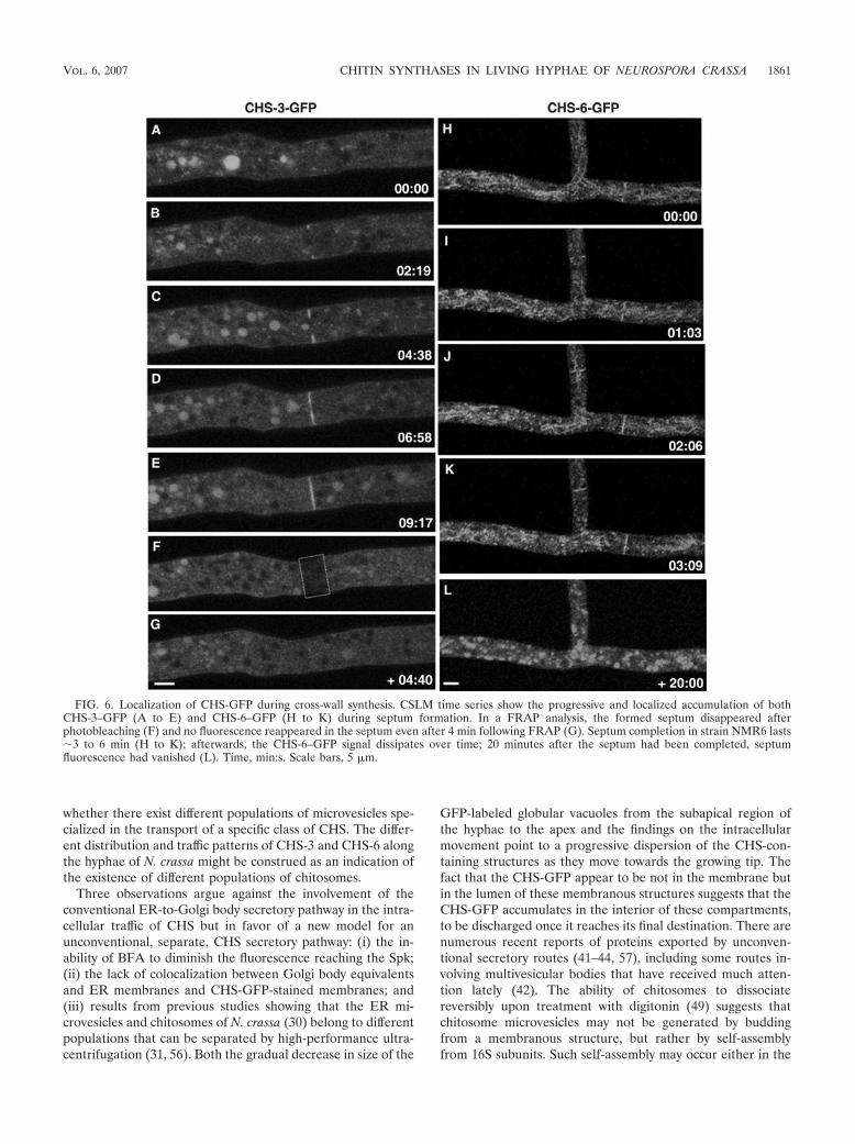
Localization of CHS-3 and CHS-6 in septa. As in the grow-ing apex, septa are zones of intense cell wall synthesis. BothCHS-3–GFP (Fig. 6A to E; see movie S5 in the supplementalmaterial) and CHS-6–GFP (Fig. 6H to K) were found at thedeveloping septa. Septum development lasted about 3 to 6 minas observed in different septa of strains NMR3 and NMR6.After the completion of septum formation, FRAP experimentsshowed that there was no additional accumulation of CHS-3–GFP in the photobleached septum (Fig. 6F and G; see movieS5 in the supplemental material). In addition, we observed thatCHS-6–GFP fluorescence did not persist in the septa forlonger than 10 to 20 min after septum formation had beencompleted (Fig. 6). Furthermore, existing septa identified byphase-contrast optics in far distal—and therefore older—re-gions of hyphae showed very low or no fluorescence (data notshown).
Lack of colocalization of CHS-GFP with compartments ofthe conventional secretory pathway. Strains NMR3, NMR6,and N150 (wild type; mat A; FGSC 9013) were stained withspecific vital fluorescent dyes to determine whether CHS-GFPcolocalized with various endomembranous compartments (i.e.,the ER, Golgi body equivalents, vacuoles, and endocytic com-
partments) involved in the conventional secretory routes. Alldyes tested were incorporated into actively growing hyphae �5to 10 min after application.
The putative Golgi body equivalents and ER stained by theBFA bodipy 558/568 conjugate did not colocalize with theGFP-labeled endomembranous system (Fig. 7A to G). BFAinterferes with Golgi body-dependent secretion and also withendosomal/post-Golgi body trafficking (11, 18, 21, 52), al-though the effects of this inhibitor can be tissue and speciesspecific (21, 54). In Neurospora, BFA caused a decrease inhyphal elongation and an increase in branching, but the treat-ment did not interfere with the traffic of CHS-containing mi-crovesicles to the Spk. For a short, 5-min initial period, the dyestained putative Golgi equivalents present at the hyphal apexand proximal subapex (Fig. 7B). During the following 5 to 20min, as a result of the effect of the BFA, the dye was redis-tributed and stained the ER surrounding the nuclei (Fig. 7D).During that period, GFP fluorescence was still very prominentat the Spk of both strain NMR3 and strain NMR6 (Fig. 7A andC). In some hyphae, growth ceased upon prolonged exposureto BFA-bodipy. This caused the disappearance of the Spkconcomitantly with a movement of the dye away from theapical region, while staining the ER more strongly (Fig. 7E toG). The lack of colocalization of CHS-GFP-labeled andbodipy-stained membranes showed clearly that CHS-GFP isnot transported by a conventional ER-Golgi body pathway.
Evidence of a possible relationship between CHS and thevacuole system is shown in Fig. 7H to L. CHS-GFP fluores-cence was found in the lumen of vacuoles but, significantly, notin their membranes (Fig. 7H to J). The membrane, but not thelumen, was stained by the endocytic marker FM4-64. Also thedistribution of CHS-GFP in distal regions is reminiscent of
FIG. 3. Progressive disintegration of GFP-labeled vacuoles and dispersal of fluorescence in strain NMR3 (A to F) and strain NMR6 (G to J).Arrowheads point at spherical bodies whose size gradually diminishes until they are no longer visible. Similarly, the CHS-6–GFP-labeled tubularcompartments in NMR6 undergo progressive disintegration (G to J). Time, min:s. Scale bars, 5 �m.
1858 RIQUELME ET AL. EUKARYOT. CELL

the globular and tubular vacuolar system as revealed byCDFFDA staining (Fig. 7K and L).
DISCUSSION
By tagging chs genes with sgfp, we have succeeded in deter-mining the intracellular location of two CHS in Neurospora andobtained a first glimpse of CHS trafficking inside the cell.Despite some differences in the patterns of distribution be-tween CHS-3 and CHS-6, four major conclusions can be drawnabout the intracellular location of these two CHS: (i) most ofthe GFP-tagged CHS in distal regions is found in rather largecompartments, some tubular, some spherical; (ii) the subapi-cal, round, GFP-labeled structures diminish dramatically insize as they approach the apex; (iii) in the proximal subapex,the GFP label is finely dispersed into putative microvesiclesthat are likely chitosomes; and (iv) most strikingly, at the apex,there is a dense accumulation of CHS-GFP into a sphericalbody that coincides with the position of the Spk.
A salient finding in the present study was the localization ofCHS-3 and CHS-6 in the Spk, which has previously not beenobserved or reported. Thus far, A. fumigatus ChsD, a class VICHS, has been shown to be expressed during hyphal growth(39), but no localization data were reported. In Ustilago may-dis, the class I CHS-3 and CHS-4—both labeled with fluores-cent proteins—have been localized at septa and at corticalregions of the cytoplasm (61) but were not found at the Spk.
Given the fragile nature of the Spk (33), it is not surprisingthat it may not have survived procedures employed previouslyfor autoradiography or electron microscopy. Another crucialfactor may be the fungal growth rate: the high elongation rateand large diameter of the N. crassa hyphae produce a well-defined Spk. In slow-growing fungi, the Spk is much smallerand harder to see, and this may help explain why CHS-GFPfusions, although present at tips, were not detected in the Spkin studies with Colletotrichum graminicola (1). On the otherhand, our finding that both CHS-3–GFP and CHS-6–GFP ac-cumulate at growing septa but are turned over fairly rapidlycoincides with previous reports on the localization of CHS atthe septa of C. graminicola (1), A. nidulans (58), and U. maydis(51, 61, 62).
Our results show that CHS-GFP accumulates at the core ofthe Spk, presumably the same region in which microvesicleshave been detected in earlier studies by transmission electronmicroscopy (23, 38). To better discern whether the CHS-GFPdistribution indeed coincides with the core identified by trans-mission electron microscopy, we plan to further characterizethe CHS-GFP distribution at the ultrastructural level by im-munoelectron microscopy. The dimensions of the smallestCHS-GFP fluorescent vesicles cannot be determined from ourconfocal images with high confidence, but they are probablysmaller than 100 nm in diameter, a size in the size range ofchitosomes, which measure 40 to 70 nm and are known to
FIG. 4. Kinetic analysis of fluorescent structures carrying CHS-3–GFP and CHS-6–GFP. The trajectories (t) of individual particles and the Spkwere followed. Fluorescent vesicles were traced moving predominantly forward until reaching the proximal subapex (15 to 20 �m from the apex)where they were no longer visible. The average speed (in �m/s) of the Spk and the different vesicles was, for strain NMR3 Spk, 0.11 � 0.02; t2,0.11 � 0.06; t3, 0.13 � 0.04; t4, 0.12 � 0.09; t5, 0.11 � 0.04; t6, 0.18 � 0.05; t7, 0.12 � 0.04; t8, 0.13 � 0.04; and t9, 0.09 � 0.03; and for strainNMR6 Spk, 0.24 � 0.02; t2, 0.18 � 0.05; t3, 0.19 � 0.06; t4, 0.21 � 0.02; t5, 0.18 � 0.07; t6, 0.26 � 0.02; t7, 0.19 � 0.06; and t8, 0.22 � 0.05. Time,min:s. Scale bars, 5 �m. Particle movement can be better appreciated in movies S1 and S2 in the supplemental material.
VOL. 6, 2007 CHITIN SYNTHASES IN LIVING HYPHAE OF NEUROSPORA CRASSA 1859

contain most of the CHS in fungal cells (6). In a separate,ongoing study (J. Verdın-Ramos, M. Riquelme, E. Castro-Longoria, and S. Bartnicki-Garcıa, unpublished data), cell ex-tracts of strains NMR3 and NMR6 were separated by isopycnicsedimentation and analyzed by Western blotting with a GFP-specific monoclonal antibody. Several bands of a higher mo-lecular weight than that of GFP alone were detected in cellfractions, with a density around 1.13 g/ml, the characteristicbuoyant density of chitosomes of N. crassa (36).
FRAP analysis indicated that the fluorescence of the Spkoriginates from the finely dispersed fluorescence in the proxi-mal subapex, i.e., it originated in the population of CHS-3–GFP- and CHS-6–GFP-labeled microvesicles. These findingslend credence to the long-standing prediction that the Spk isdirectly involved in guiding cell wall formation (4, 8).
Our observations that CHS-6–GFP and CHS-3–GFP do notaccumulate at the plasma membrane in N. crassa hyphal tipssupport the idea that, upon reaching the cell surface, CHSturns over rapidly (56). Our dynamic images of CHS-GFPtraffic, plus the identification of the GFP-labeled particles asmicrovesicles (likely chitosomes, because of their low specificgravity), support previous findings obtained by immunolocal-ization in N. crassa (56) and by autoradiography in Mucorrouxii (55).
Our findings on the localization of CHS-3 and CHS-6 in N.crassa, plus previous studies on the three CHS from S. cerevi-siae, Chs1p, Chs2p (31), and Chs3p (17), show that they all canbe detected in chitosomes. This raises the question as towhether these different CHS belong to one multienzyme com-plex transported within the same population of chitosomes or
FIG. 5. FRAP analysis of the proximal subapex of a CHS-3–GFP hypha (A to E) and the apex of a CHS-6–GFP hypha (F to J). Prebleachedhyphae. Arrows point at the Spk (A, F). Photobleaching was applied to the selected areas indicated by the dotted rectangles (B, G). Recovery ofSpk fluorescence occurs after either apical (H to J) or subapical (C to E) bleaching. Note a temporary departure of the Spk from its centric positionin the apical dome (D). Time, min:s. Scale bars, 5 �m.
1860 RIQUELME ET AL. EUKARYOT. CELL
whether there exist different populations of microvesicles spe-cialized in the transport of a specific class of CHS. The differ-ent distribution and traffic patterns of CHS-3 and CHS-6 alongthe hyphae of N. crassa might be construed as an indication ofthe existence of different populations of chitosomes.
Three observations argue against the involvement of theconventional ER-to-Golgi body secretory pathway in the intra-cellular traffic of CHS but in favor of a new model for anunconventional, separate, CHS secretory pathway: (i) the in-ability of BFA to diminish the fluorescence reaching the Spk;(ii) the lack of colocalization between Golgi body equivalentsand ER membranes and CHS-GFP-stained membranes; and(iii) results from previous studies showing that the ER mi-crovesicles and chitosomes of N. crassa (30) belong to differentpopulations that can be separated by high-performance ultra-centrifugation (31, 56). Both the gradual decrease in size of the
GFP-labeled globular vacuoles from the subapical region ofthe hyphae to the apex and the findings on the intracellularmovement point to a progressive dispersion of the CHS-con-taining structures as they move towards the growing tip. Thefact that the CHS-GFP appear to be not in the membrane butin the lumen of these membranous structures suggests that theCHS-GFP accumulates in the interior of these compartments,to be discharged once it reaches its final destination. There arenumerous recent reports of proteins exported by unconven-tional secretory routes (41–44, 57), including some routes in-volving multivesicular bodies that have received much atten-tion lately (42). The ability of chitosomes to dissociatereversibly upon treatment with digitonin (49) suggests thatchitosome microvesicles may not be generated by buddingfrom a membranous structure, but rather by self-assemblyfrom 16S subunits. Such self-assembly may occur either in the
FIG. 6. Localization of CHS-GFP during cross-wall synthesis. CSLM time series show the progressive and localized accumulation of bothCHS-3–GFP (A to E) and CHS-6–GFP (H to K) during septum formation. In a FRAP analysis, the formed septum disappeared afterphotobleaching (F) and no fluorescence reappeared in the septum even after 4 min following FRAP (G). Septum completion in strain NMR6 lasts�3 to 6 min (H to K); afterwards, the CHS-6–GFP signal dissipates over time; 20 minutes after the septum had been completed, septumfluorescence had vanished (L). Time, min:s. Scale bars, 5 �m.
VOL. 6, 2007 CHITIN SYNTHASES IN LIVING HYPHAE OF NEUROSPORA CRASSA 1861

FIG. 7. Distinct localization of CHS-GFP and conventional secretory compartments. (A to G) Comparative distribution of fluorescence fromCHS-3–GFP (A) and CHS-6–GFP (C, E) with Golgi body structures revealed after staining with 5 �M BFA-bodipy 558/568 conjugate. After ashort period (�5 min), the dye stained apical and subapical Golgi body equivalents (arrowheads in B). Note the preservation of CHS-GFP in theSpk (arrow in panel A). Over 5 to 20 min, the dye stained the ER (arrowheads in D). During that period, GFP fluorescence was still very prominentat the Spk of strain NMR6 (arrow in panel C). After a longer exposure (20 to 25 min), the hyphae stopped growing (E to G), the Spk disappeared,and both fluorescence and dye moved away from the apical region and strongly stained the ER (arrowheads in panel F), but the separation betweenfluorescent CHS-6–GFP and the bodipy-stained structures, particularly the ER around the nuclei, remained clearly evident (G). Scale bars, 5 �m.(H to L) Relationship between CHS-3–GFP and the vacuolar system. Localization of CHS-3–GFP fluorescence in the vacuoles of NMR3 in hyphaestained with FM4-64 (H to J); note that CHS-3–GFP is found in the lumen of the vacuole (I), whereas FM4-64 stains the vacuolar membrane (H,J). Scale bars, 5 �m. For comparison, the vacuolar system in wild-type hyphae of N. crassa is shown after staining with 10 �M CDFFDA (K, L).Scale bars, 10 �m.
1862

cytosol or in vacuoles, the latter giving rise to multivesicularbodies (13).
In conclusion, our in vivo studies on the localization andmovement of Neurospora CHS-GFP have already yielded valu-able insights into the pathway that delivers proteins for cellwall synthesis to the Spk in fungal hyphae. Future studies onthe structure and localization of Neurospora CHS at the reso-lution level of the electron microscope will characterize thestructures revealed here by fluorescence microscopy. This ini-tial study sets the stage for further work that will test theunconventional transport model proposed here and that couldeventually result in a more complete understanding of how thedifferent components of the CHS family are transported andintegrated into microfibril-assembling complexes.
ACKNOWLEDGMENTS
This work was supported by CONACYT (Consejo Nacional deCiencia y Tecnologıa, Mexico) grants DG/2003-1158 and U-45818Q.The Freitag lab is supported by start-up funds from the Computationaland Genome Biology Initiative at Oregon State University.
We thank Eric Selker (University of Oregon, Eugene) for support ofthis work.
REFERENCES
1. Amnuaykanjanasin, A., and L. Epstein. 2006. A class Vb chitin synthase inColletotrichum graminicola is localized in the growing tips of multiple celltypes, in nascent septa, and during septum conversion to an end wall afterhyphal breakage. Protoplasma 227:155–164.
2. Bartnicki-Garcia, S. 1968. Cell wall chemistry, morphogenesis and taxonomyof fungi. Annu. Rev. Microbiol. 22:87–107.
3. Bartnicki-Garcia, S. 2006. Chitosomes: past, present and future. FEMSYeast Res. 6:957–965.
4. Bartnicki-Garcia, S. 2002. Hyphal tip growth: outstanding questions, p.29–58. In H. D. Osiewacz (ed.), Molecular biology of fungal development.Marcel Dekker, Inc., New York, NY.
5. Bartnicki-Garcia, S. 1990. Role of vesicles in apical growth and a newmathematical model of hyphal morphogenesis, p. 211–232. In I. B. Heath(ed.), Tip growth in plant and fungal cells. Academic Press, San Diego, CA.
6. Bartnicki-Garcia, S., C. E. Bracker, E. Reyes, and J. Ruiz Herrera. 1978.Isolation of chitosomes from taxonomically diverse fungi and synthesis ofchitin microfibrils in vitro. Exp. Mycol. 2:173–192.
7. Bartnicki-Garcia, S., and E. Lippman. 1969. Fungal morphogenesis: cell wallconstruction in Mucor rouxii. Science 165:302–304.
8. Bartnicki-Garcia, S., J. Ruiz-Herrera, and C. E. Bracker. 1979. Chitosomesand chitin synthesis, p. 149–168. In J. H. Burnett and A. P. J. Trinci (ed.),Fungal walls and hyphal growth. Cambridge University Press, Cambridge,United Kingdom.
9. Borgia, P. T., N. Iartchouk, P. J. Riggle, K. R. Winter, Y. Koltin, and C. E.Bulawa. 1996. The chsB gene of Aspergillus nidulans is necessary for normalhyphal growth and development. Fungal Genet. Biol. 20:193–203.
10. Borkovich, K. A., L. A. Alex, O. Yarden, M. Freitag, G. E. Turner, N. D.Read, S. Seiler, D. Bell-Pedersen, J. Paietta, N. Plesofsky, M. Plamann, M.Goodrich-Tanrikulu, U. Schulte, G. Mannhaupt, F. E. Nargang, A. Radford,C. Selitrennikoff, J. E. Galagan, J. C. Dunlap, J. J. Loros, D. Catcheside, H.Inoue, R. Aramayo, M. Polymenis, E. U. Selker, M. S. Sachs, G. A. Marzluf,I. Paulsen, R. Davis, D. J. Ebbole, A. Zelter, E. R. Kalkman, R. O’Rourke, F.Bowring, J. Yeadon, C. Ishii, K. Suzuki, W. Sakai, and R. Pratt. 2004.Lessons from the genome sequence of Neurospora crassa: tracing the pathfrom genomic blueprint to multicellular organism. Microbiol. Mol. Biol. Rev.68:1–108.
11. Bourett, T. M., and R. J. Howard. 1996. Brefeldin A-induced structuralchanges in the endomembrane system of a filamentous fungus, Magnaporthegrisea. Protoplasma 190:151–163.
12. Bowen, A. R., J. L. Chen-Wu, M. Momany, R. Young, P. J. Szaniszlo, andP. W. Robbins. 1992. Classification of fungal chitin synthases. Proc. Natl.Acad. Sci. USA 89:519–523.
13. Bracker, C. E., J. Ruiz-Herrera, and S. Bartnicki-Garcia. 1976. Structureand transformation of chitin synthetase particles (chitosomes) during micro-fibril synthesis in vitro. Proc. Natl. Acad. Sci. USA 73:4570–4574.
14. Brunswik, H. 1924. Untersuchungen uber die Geschlechts- und Kernverhalt-nisse bei der Hymenomyzetengattung Coprinus. Bot. Abh. 5:1–152.
15. Chigira, Y., K. Abe, K. Gomi, and T. Nakajima. 2002. chsZ, a gene for anovel class of chitin synthase from Aspergillus oryzae. Curr. Genet. 41:261–267.
16. Choquer, M., M. Boccara, I. R. Goncalves, M. Soulie, and A. Vidal-Cros.2004. Survey of the Botrytis cinerea chitin synthase multigenic family throughthe analysis of six euascomycetes genomes. Eur. J. Biochem. 271:2153–2164.
17. Chuang, J. S., and R. W. Schekman. 1996. Differential trafficking and timedlocalization of two chitin synthase proteins, Chs2p and Chs3p. J. Cell Biol.135:597–610.
18. Cole, L., D. Davies, G. J. Hyde, and A. E. Ashford. 2000. Brefeldin A affectsgrowth, endoplasmic reticulum, Golgi bodies, tubular vacuole system, andsecretory pathway in Pisolithus tinctorius. Fungal Genet. Biol. 29:95–106.
19. Freitag, M., P. C. Hickey, N. B. Raju, E. U. Selker, and N. D. Read. 2004.GFP as a tool to analyze the organization, dynamics and function of nucleiand microtubules in Neurospora crassa. Fungal Genet. Biol. 41:897–910.
20. Fujiwara, M., H. Horiuchi, A. Ohta, and M. Takagi. 1997. A novel fungalgene encoding chitin synthase with a myosin motor-like domain. Biochem.Biophys. Res. Commun. 236:75–78.
21. Geldner, N. 2004. The plant endosomal system—its structure and role insignal transduction and plant development. Planta 219:547–560.
22. Girbardt, M. 1957. Der Spitzenkorper von Polystictus versicolor. Planta 50:47–59.
23. Girbardt, M. 1969. Die Ultrastruktur der Apikalregion von Pilzhyphen.Protoplasma 67:413–441.
24. Grove, S. N., and C. E. Bracker. 1970. Protoplasmic organization of hyphaltips among fungi: vesicles and Spitzenkorper. J. Bacteriol. 104:989–1009.
25. Harris, S. D., N. D. Read, R. W. Roberson, B. Shaw, S. Seiler, M. Plamann,and M. Momany. 2005. Polarisome meets Spitzenkorper: microscopy, genet-ics, and genomics converge. Eukaryot. Cell 4:225–229.
26. Hickey, P. C., S. M. Swift, M. G. Roca, and N. D. Read. 2005. Live-cellimaging of filamentous fungi using vital fluorescent dyes. Methods Micro-biol. 34:63–87.
27. Holcomb, C. L., W. J. Hansen, T. Etcheverry, and R. Schekman. 1988.Secretory vesicles externalize the major plasma membrane ATPase in yeast.J. Cell Biol. 106:641–648.
28. Howard, R. J. 1981. Ultrastructural analysis of hyphal tip cell growth in fungi:Spitzenkorper, cytoskeleton and endomembranes after freeze-substitution.J. Cell Sci. 48:89–103.
29. Hunsley, D., and G. W. Gooday. 1974. The structure and development ofsepta in Neurospora crassa. Protoplasma 82:125–146.
30. Leal-Morales, C., C. E. Bracker, and S. Bartnicki-Garcia. 1994. Distributionof chitin synthetase and various membrane marker enzymes in chitosomesand other organelles of the slime mutant of Neurospora crassa. Exp. Mycol.18:168–179.
31. Leal-Morales, C. A., C. E. Bracker, and S. Bartnicki-Garcia. 1994. Subcel-lular localization, abundance and stability of chitin synthetases 1 and 2 fromSaccharomyces cerevisiae. Microbiology 140:2207–2216.
32. Lesage, G., and H. Bussey. 2006. Cell wall assembly in Saccharomyces cer-evisiae. Microbiol. Mol. Biol. Rev. 70:317–343.
33. Lopez-Franco, R., and C. E. Bracker. 1996. Diversity and dynamics of theSpitzenkorper in growing hyphal tips of higher fungi. Protoplasma 195:90–111.
34. Mandel, M. A., J. N. Galgiani, S. Kroken, and M. J. Orbach. 2006. Coccid-ioides posadasii contains single chitin synthase genes corresponding to classesI to VII. Fungal Genet. Biol. 43:775–788.
35. Margolin, B. S., M. Freitag, and E. U. Selker. 1997. Improved plasmids forgene targeting at the his-3 locus of Neurospora crassa by electroporation.Fungal Genet. Newslett. 44:34–36.
36. Martinez, J. P., G. Gimenez, and S. Bartnicki Garcia. 1987. Intracellularlocalization of UDP-N-acetylglucosamine in Neurospora crassa wild-type andslime mutant strains. Exp. Mycol. 11:278–286.
37. Martın-Udıroz, M., M. P. Madrid, and M. I. G. Roncero. 2004. Role of chitinsynthase genes in Fusarium oxysporum. Microbiol. Mol. Biol. Rev. 150:3175–3187.
38. McClure, W. K., D. Park, and P. M. Robinson. 1968. Apical organization inthe somatic hyphae of fungi. J. Gen. Microbiol. 50:177–182.
39. Mellado, E., C. A. Specht, P. W. Robbins, and D. W. Holden. 1996. Cloningand characterization of chsD, a chitin synthase-like gene of Aspergillus fu-migatus. FEMS Microbiol. Lett. 143:69–76.
40. Munro, C. A., D. A. Schofield, G. W. Gooday, and N. A. Gow. 1998. Regu-lation of chitin synthesis during dimorphic growth of Candida albicans.Microbiology 144:391–401.
41. Nickel, W. 2003. The mystery of nonclassical protein secretion: a currentview on cargo proteins and potential export routes. Eur. J. Biochem. 270:2109.
42. Nickel, W. 2005. Unconventional secretory routes: direct protein exportacross the plasma membrane of mammalian cells. Traffic 6:607–614.
43. Rayner, J. C., and H. R. Pelham. 1997. Transmembrane domain-dependentsorting of proteins to the ER and plasma membrane in yeast. EMBO J.16:1832–1841.
44. Reggiori, F., and H. R. Pelham. 2001. Sorting of proteins into multivesicularbodies: ubiquitin-dependent and -independent targeting. EMBO J. 20:5176–5186.
45. Riquelme, M., R. Roberson, D. McDaniel, and S. Bartnicki-Garcia. 2002.
VOL. 6, 2007 CHITIN SYNTHASES IN LIVING HYPHAE OF NEUROSPORA CRASSA 1863

The effect of ropy-1 mutation on cytoplasmic organization and intracellularmotility in mature Neurospora crassa. Fungal Genet. Biol. 37:171–179.
46. Roncero, C. 2002. The genetic complexity of chitin synthesis in fungi. Curr.Genet. 41:367–378.
47. Ruiz-Herrera, J. 1992. Fungal cell wall: structure, synthesis and assembly.CRC Press, Boca Raton, FL.
48. Ruiz-Herrera, J., and S. Bartnicki-Garcia. 1974. Synthesis of cell wall mi-crofibrils in vitro by a “soluble” chitin synthetase from Mucor rouxii. Science186:357–359.
49. Ruiz-Herrera, J., S. Bartnicki-Garcia, and C. E. Bracker. 1980. Dissociationof chitosomes by digitonin into 16 S subunits with chitin synthetase activity.Biochim. Biophys. Acta 629:201–206.
50. Ruiz-Herrera, J., J. M. Gonzalez-Prieto, and R. Ruiz-Medrano. 2002. Evo-lution and phylogenetic relationships of chitin synthases from yeasts andfungi. FEMS Yeast Res. 1:247–256.
51. Ruiz-Herrera, J., B. Xoconostle-Cazare, C. G. Reynaga-Pena, C. Leon-Ramirez, and A. Carabez-Trejo. 2006. Immunolocalization of chitin syn-thases in the phytopathogenic dimorphic fungus Ustilago maydis. FEMSYeast Res. 6:999–1009.
52. Rupes, I., W. Z. Mao, H. Åstrom, and M. Raudaskoski. 1995. Effects ofnocodazole and brefeldin A on microtubule cytoskeleton and membraneorganization in the homobasidiomycete Schizophyllum commune. Proto-plasma 185:212–221.
53. Sambrook, J., E. F. Fritsch, and T. Maniatis. 1989. Molecular cloning: alaboratory manual. Cold Spring Harbor Laboratory Press, Cold Spring Har-bor, NY.
54. Satiat-Jeunemaitre, B., L. Cole, T. Bourett, R. Howard, and C. Hawes. 1996.Brefeldin A effects in plant and fungal cells: something new about vesicletrafficking? J. Microsc. 181:162–177.
55. Sentandreu, R., A. Martinez-Ramon, and J. Ruiz-Herrera. 1984. Localiza-
tion of chitin synthase in Mucor rouxii by an autoradiographic method.J. Gen. Microbiol. 130:1193–1199.
56. Sietsma, J. H., A. Beth Din, V. Ziv, K. A. Sjollema, and O. Yarden. 1996. Thelocalization of chitin synthase in membranous vesicles (chitosomes) in Neu-rospora crassa. Microbiology 142:1591–1596.
57. Stoorvogel, W., M. J. Kleijmeer, H. J. Geuze, and G. Raposo. 2002. Thebiogenesis and functions of exosomes. Traffic 3:321–330.
58. Takeshita, N., A. Ohta, and H. Horiuchi. 2005. CsmA, a class V chitinsynthase with a myosin motor-like domain, is localized through direct inter-action with the actin cytokeleton in Aspergillus nidulans. Mol. Biol. Cell16:1961–1970.
59. Vogel, H. J. 1956. A convenient growth medium for Neurospora (medium N).Microbiol. Gen. Bull. 13:42–43.
60. Wang, Q., H. Liu, and P. J. Szaniszlo. 2002. Compensatory expression of fivechitin synthase genes, a response to stress stimuli, in Wangiella (Exophiala)dermatitidis, a melanized fungal pathogen of humans. Microbiology 148:2811–2817.
61. Weber, I., D. Assmann, E. Thines, and G. Steinberg. 2006. Polar localizingclass V myosin chitin synthases are essential during early plant infection inthe plant pathogenic fungus Ustilago maydis. Plant Cell 18:225–242.
62. Weber, I., C. Gruber, and G. Steinberg. 2003. A class-V myosin required formating, hyphal growth, and pathogenicity in the dimorphic plant pathogenUstilago maydis. Plant Cell 15:2826–2842.
63. Westergaard, M., and H. K. Mitchell. 1947. Neurospora V. A syntheticmedium favoring sexual reproduction. Am. J. Bot. 34:573–577.
64. Yanai, K., N. Kojima, N. Takaya, H. Horiuchi, A. Ohta, and M. Takagi. 1994.Isolation and characterization of two chitin synthase genes from Aspergillusnidulans. Biosci. Biotechnol. Biochem. 58:1828–1835.
65. Yarden, O., and C. Yanofsky. 1991. Chitin synthase 1 plays a major role incell wall biogenesis in Neurospora crassa. Genes Dev. 5:2420–2430.
1864 RIQUELME ET AL. EUKARYOT. CELL