MreB of Streptomyces coelicolor is not essential for vegetative growth but is required for the...
15
Molecular Microbiology (2006) 60(4), 838–852 doi:10.1111/j.1365-2958.2006.05134.x First published online 21 April 2006 © 2006 The Authors Journal compilation © 2006 Blackwell Publishing Ltd Blackwell Publishing LtdOxford, UKMMIMolecular Microbiology0950-382X© 2006 The Authors; Journal compilation © 2006 Blackwell Publishing Ltd ? 2006604838852Original ArticleMreB in Streptomyces coelicolorP. Mazza et al. Accepted 15 February, 2006. For correspondence. *E-mail g.wezel@ chem.leidenuniv.nl; Tel. (+31) 71 527 4310; Fax (+31) 71 527 4340; **E-mail [email protected]; Tel. (+49) 7071 297 6944; Fax (+49) 7071 295 979. † Present address: Depart- ment of Cell and Organism Biology, Lund University, Sölvegatan 35, SE-223 62, Lund, Sweden. MreB of Streptomyces coelicolor is not essential for vegetative growth but is required for the integrity of aerial hyphae and spores Paola Mazza, 1 Elke E. Noens, 2,3 Kathrin Schirner, 1 Nina Grantcharova, 4 A. Mieke Mommaas, 3 Henk K. Koerten, 3 Günther Muth, 1 Klas Flärdh, 4† Gilles P. van Wezel 2 * and Wolfgang Wohlleben 1 ** 1 Microbiology/Biotechnology, Institute of Microbiology, Faculty of Biology, Eberhard-Karls-Universität Tübingen, Auf der Morgenstelle 28, 72076 Tübingen, Germany. 2 Department of Biochemistry, Leiden Institute of Chemistry, Leiden University, PO Box 9502, 2300RA, Leiden, the Netherlands. 3 Department of Electron Microscopy, Leiden University Medical Centre, PO Box 9503, 2300RA Leiden, the Netherlands. 4 Department of Cell and Molecular Biology, Biomedical Center, Uppsala University, PO Box 596, 75124 Uppsala, Sweden. Summary MreB forms a cytoskeleton in many rod-shaped bac- teria which is involved in cell shape determination and chromosome segregation. PCR-based and Southern analysis of various actinomycetes, supported by anal- ysis of genome sequences, revealed mreB homo- logues only in genera that form an aerial mycelium and sporulate. We analysed MreB in one such organ- ism, Streptomyces coelicolor . Ectopic overexpres- sion of mreB impaired growth, and caused swellings and lysis of hyphae. A null mutant with apparently normal vegetative growth was generated. However, aerial hyphae of this mutant were swelling and lysing; spores doubled their volume and lost their character- istic resistance to stress conditions. Loss of cell wall consistency was observed in MreB-depleted spores by transmission electron microscopy. An MreB–EGFP fusion was constructed to localize MreB in the myce- lium. No clearly localized signal was seen in vegeta- tive mycelium. However, strong fluorescence was observed at the septa of sporulating aerial hyphae, then as bipolar foci in young spores, and finally in a ring- or shell-like pattern inside the spores. Immu- nogold electron microscopy using MreB-specific anti- bodies revealed that MreB is located immediately underneath the internal spore wall. Thus, MreB is not essential for vegetative growth of S. coelicolor , but exerts its function in the formation of environmentally stable spores, and appears to primarily influence the assembly of the spore cell wall. Introduction Bacterial morphologies range from spherical and rod- shaped to curved, helical and filamentous. A major deter- minant of cell shape is the bacterial cell wall, which con- sists of glycan strands cross-linked by short peptides. Isolated peptidoglycan sacculi can retain the shape of the bacterial cell, and mutants defective in peptidoglycan syn- thesis typically show an altered morphology (Young, 2003; Cabeen and Jacobs-Wagner, 2005). However, not only the enzymes that are directly involved in the synthesis and assembly of peptidoglycan can affect the morphology. It is becoming clear that cytoskeletal elements exist in the bacterial cytoplasm (Löwe et al ., 2004), and that they are determining the architecture of the cell wall and have strong impacts on cell shape. These include homologues of all three major types of eukaryotic cytoskeletons. Thus, MreB proteins are actin homologues that produce micro- filament-like fibres and determine rod shape in many bacteria (van den Ent et al ., 2001; Jones et al ., 2001); crescentin (CreS) produces intermediate filament-like ele- ments in Caulobacter crescentus and give rise to the curved shape of those cells (Ausmees et al ., 2003); and FtsZ is a tubulin homologue that assembles into a cyto- kinetic ring at the site of cell division and directs cell division and formation of the septal peptidoglycan (Mar- golin, 2005). MreB is a member of the HSP70-actin-sugar kinase (ASHKA) superfamily of proteins (Bork et al ., 1992), and its crystal structure is strikingly similar to the structure of actin (van den Ent et al ., 2001). Purified MreB of Thermo- toga maritima polymerizes in vitro to form filaments with a spacing between the MreB monomers of 51 Å, which is
-
Upload
independent -
Category
Documents
-
view
2 -
download
0
Transcript of MreB of Streptomyces coelicolor is not essential for vegetative growth but is required for the...

Molecular Microbiology (2006)
60
(4), 838–852 doi:10.1111/j.1365-2958.2006.05134.xFirst published online 21 April 2006
© 2006 The AuthorsJournal compilation © 2006 Blackwell Publishing Ltd
Blackwell Publishing LtdOxford, UKMMIMolecular Microbiology0950-382X© 2006 The Authors; Journal compilation © 2006 Blackwell Publishing Ltd
? 2006
60
4838852
Original Article
MreB in Streptomyces coelicolorP. Mazza
et al.
Accepted 15 February, 2006. For correspondence. *E-mail [email protected]; Tel. (
+
31) 71 527 4310; Fax (
+
31) 71 527 4340;**E-mail [email protected]; Tel. (
+
49)7071 297 6944; Fax (
+
49) 7071 295 979.
†
Present address: Depart-ment of Cell and Organism Biology, Lund University, Sölvegatan 35,SE-223 62, Lund, Sweden.
MreB of
Streptomyces coelicolor
is not essential for vegetative growth but is required for the integrity of aerial hyphae and spores
Paola Mazza,
1
Elke E. Noens,
2,3
Kathrin Schirner,
1
Nina Grantcharova,
4
A. Mieke Mommaas,
3
Henk K. Koerten,
3
Günther Muth,
1
Klas Flärdh,
4†
Gilles P. van Wezel
2
* and Wolfgang Wohlleben
1
**
1
Microbiology/Biotechnology, Institute of Microbiology, Faculty of Biology, Eberhard-Karls-Universität Tübingen, Auf der Morgenstelle 28, 72076 Tübingen, Germany.
2
Department of Biochemistry, Leiden Institute of Chemistry, Leiden University, PO Box 9502, 2300RA, Leiden, the Netherlands.
3
Department of Electron Microscopy, Leiden University Medical Centre, PO Box 9503, 2300RA Leiden, the Netherlands.
4
Department of Cell and Molecular Biology, Biomedical Center, Uppsala University, PO Box 596, 75124 Uppsala, Sweden.
Summary
MreB forms a cytoskeleton in many rod-shaped bac-teria which is involved in cell shape determination andchromosome segregation. PCR-based and Southernanalysis of various actinomycetes, supported by anal-ysis of genome sequences, revealed
mreB
homo-logues only in genera that form an aerial myceliumand sporulate. We analysed MreB in one such organ-ism,
Streptomyces coelicolor
. Ectopic overexpres-sion of
mreB
impaired growth, and caused swellingsand lysis of hyphae. A null mutant with apparentlynormal vegetative growth was generated. However,aerial hyphae of this mutant were swelling and lysing;spores doubled their volume and lost their character-istic resistance to stress conditions. Loss of cell wallconsistency was observed in MreB-depleted sporesby transmission electron microscopy. An MreB–EGFPfusion was constructed to localize MreB in the myce-lium. No clearly localized signal was seen in vegeta-tive mycelium. However, strong fluorescence was
observed at the septa of sporulating aerial hyphae,then as bipolar foci in young spores, and finally in aring- or shell-like pattern inside the spores. Immu-nogold electron microscopy using MreB-specific anti-bodies revealed that MreB is located immediatelyunderneath the internal spore wall. Thus, MreB is notessential for vegetative growth of
S. coelicolor
, butexerts its function in the formation of environmentallystable spores, and appears to primarily influence theassembly of the spore cell wall.
Introduction
Bacterial morphologies range from spherical and rod-shaped to curved, helical and filamentous. A major deter-minant of cell shape is the bacterial cell wall, which con-sists of glycan strands cross-linked by short peptides.Isolated peptidoglycan sacculi can retain the shape of thebacterial cell, and mutants defective in peptidoglycan syn-thesis typically show an altered morphology (Young, 2003;Cabeen and Jacobs-Wagner, 2005). However, not onlythe enzymes that are directly involved in the synthesis andassembly of peptidoglycan can affect the morphology. Itis becoming clear that cytoskeletal elements exist in thebacterial cytoplasm (Löwe
et al
., 2004), and that they aredetermining the architecture of the cell wall and havestrong impacts on cell shape. These include homologuesof all three major types of eukaryotic cytoskeletons. Thus,MreB proteins are actin homologues that produce micro-filament-like fibres and determine rod shape in manybacteria (van den Ent
et al
., 2001; Jones
et al
., 2001);crescentin (CreS) produces intermediate filament-like ele-ments in
Caulobacter crescentus
and give rise to thecurved shape of those cells (Ausmees
et al
., 2003); andFtsZ is a tubulin homologue that assembles into a cyto-kinetic ring at the site of cell division and directs celldivision and formation of the septal peptidoglycan (Mar-golin, 2005).
MreB is a member of the HSP70-actin-sugar kinase(ASHKA) superfamily of proteins (Bork
et al
., 1992), andits crystal structure is strikingly similar to the structure ofactin (van den Ent
et al
., 2001). Purified MreB of
Thermo-toga maritima
polymerizes
in vitro
to form filaments witha spacing between the MreB monomers of 51 Å, which is

MreB in
Streptomyces coelicolor 839
© 2006 The AuthorsJournal compilation © 2006 Blackwell Publishing Ltd,
Molecular Microbiology
,
60
, 838–852
reminiscent of the spacing between the subunits (55 Å)in actin filaments (van den Ent
et al
., 2001). MreBhomologues have been studied primarily in
Escherichiacoli
(
mreB
),
Bacillus subtilis
(
mreB
,
mbl
,
mreBH
) and
C. crescentus
(
mreB
), and been found with immunofluo-rescence microscopy and GFP tagging to form helical-likestructures underneath the cell envelope (Jones
et al
.,2001; Shih
et al
., 2003; Figge
et al
., 2004; Soufo andGraumann, 2003). Deletion of
mreB
in
E. coli
resulted inloss of the rod shape and in the production of sphericalcells (Doi
et al
., 1988). In
B. subtilis
,
mreB
is an essentialgene, and depletion of MreB resulted in increased cellwidth, loss of cell shape and eventually lysis (Jones
et al
.,2001; Lee and Stewart, 2003; Formstone and Errington,2005). Furthermore, the MreB-like Mbl protein is requiredfor determination of the rod shape of
B. subtilis
, and formshelical cytoskeletal structures that are needed for incor-poration of new peptidoglycan along the lateral wall duringcell elongation (Daniel and Errington, 2003). Despitethese observations that suggest that MreB proteins formactin-like fibres that define the bacterial cell shape, thespecific function of MreB proteins remain still unclear.Recent studies link MreB to peptidoglycan synthesis(Daniel and Errington, 2003), correct chromosome segre-gation (Kruse
et al
., 2003; Soufo and Graumann, 2003;Gitai
et al
., 2005) and establishment of cell polarity (Gitai
et al
., 2004). Interestingly, in the rod- and coccobacillus-shaped
Rhodobacter sphaeroides
MreB failed to producethe typical helical structures observed in
Bacillus
butrather formed a ring at the mid-cell position of elongatingcells, suggesting a role for MreB both in septation and inpeptidoglycan synthesis in this organism (Slovak
et al
.,2005). Thus, it appears that MreB may have differentfunctions in different organisms.
All three genes in the highly conserved
mre
gene clus-ter, encompassing
mreB
,
mreC
and
mreD
, are requiredfor cell shape determination in the rod-shaped
E. coli
and
B. subtilis
(Wachi
et al
., 1987; Jones
et al
., 2001; Kruse
et al
., 2005; Leaver and Errington, 2005). In both organ-isms,
mreC
and
mreD
are essential genes, and depletionof the gene products resulted in lost control of cell shape,spheroid morphology and eventually lysis (Lee and Stew-art, 2003; Kruse
et al
., 2005; Leaver and Errington, 2005).It has been suggested that the membrane proteins MreCand MreD may connect the cytoplasmic MreB and Mblfilaments to the cell wall synthetic machinery that islocated on the outer face of the membrane (Kruse
et al
.,2005; Leaver and Errington, 2005).
The observation that the
mre
gene cluster is absentfrom coccoid species such as
Streptococcus
,
Staphylo-coccus
and
Lactococcus
suggested that a sphericalshape may be correlated with the absence of an
mre
-dependent system, and that MreB proteins are particularlyimportant in rod-shaped cells (Jones
et al
., 2001). How-
ever, in contrast to most other rod-shaped bacteria, theactinomycete
Corynebacterium glutamicum
lacks
mreB
-like genes in its genome. In accordance with this, it appar-ently establishes its rod shape in a different and MreB-independent way, through growth of the cell wall only atthe cell poles (Daniel and Errington, 2003). Most otheractinomycetes also lack MreBs, but the
Streptomyces
genus is a notable exception as they encode well con-served MreB homologues (see below). This raises thequestion of what the roles may be for the
Streptomyces
MreBs. Streptomycetes are Gram-positive filamentousactinobacteria known for their complex morphology andtheir ability to produce a wide range of secondary metab-olites. Their morphological development resembles inmany ways that of filamentous fungi. They produce avegetative mycelium consisting of a network of branching,multinucleoidal, hyphal filaments, which grow in a highlypolarized way by tip extension. This polarized growth dif-fers from that of
C. glutamicum
by being independent ofcell division and occurring at hyphal tips generated bybranching or germ tube emergence, rather than at thepoles created by cell division (Flärdh, 2003). Upon star-vation colonies enter the reproductive phase by formingaerial hyphae that eventually produce chains of spores(Chater and Hopwood, 1993). This involves a specializedform of cell division that divides long multigenomic aerialhyphae into a large number of spore compartments, whichthen mature into thick-walled dormant spores (Chater andLosick, 1997; Flärdh, 2003). The genome of
Streptomycescoelicolor
contains two homologues of
mreB
, but the func-tions of them during growth and development are poorlyunderstood. One of these genes,
mreB
, is located in acluster with
mreC
and
mreD
, while the other one is locatedelsewhere. We have previously identified and studied the
mreBCD
homologues in
S. coelicolor
, and gene disruptionexperiments resulted in a viable
mreC
knockout mutantthat showed growth retardation and reduced sporulation,while an
mreB
mutant could not be created by conven-tional gene replacement (Burger
et al
., 2000). Transcrip-tional analysis of the
mreBCD
gene cluster by promoterprobing and S1 nuclease mapping suggested that thethree genes are regulated as an operon from three pro-moters upstream of
mreB
(Burger
et al
., 2000). In thisarticle, we provide evidence that MreB is not essential forvegetative growth in
S. coelicolor
but plays a vital role information of spores resistant to various forms of environ-mental stress.
Results
MreB
is found only in actinomycetes that convert aerial hyphae into spore chains
The
mre
genes are highly conserved among strepto-mycetes, and analysis of the sequenced genomes of

840
P. Mazza
et al.
© 2006 The AuthorsJournal compilation © 2006 Blackwell Publishing Ltd,
Molecular Microbiology
,
60
, 838–852
S. coelicolor
(Bentley
et al
., 2002) and
S. avermitilis
(Ikeda
et al
., 2003) and the partial sequence of
S. scabies
(http://www.sanger.ac.uk/Projects/S_scabies) revealedalmost complete conservation of MreB, and between85% and 90% amino acid identity for MreC and forMreD. In addition, a second
mreB
homologue wasdetected in all these organisms. So far no
mreB
homo-logue was identified in non-sporulating actinomycetes,including
Corynebacterium
,
Kineococcus
,
Mycobacte-rium
,
Nocardia and Rhodococcus (Jones et al., 2001;http://nocardia.nih.go.j/; http://Rhodococcus.ca; http://genome.jgi-psf.org/mic_home.html).
In S. coelicolor, S. avermitilis and S. scabies, the mre-BCD operon lies between ndk, encoding a nucleosidediphosphate kinase, and pbp83 (= pbp2), encoding a pen-icillin-binding protein (PBP) (Burger et al., 1998). In orderto test how widespread the mreB genes are among rareactinomycetes, genomic DNA was isolated from variousactinomycetes and PCR was performed using primersPM13 and PM14 (see Table 4) that were designed at twohighly conserved regions of mreB. The primers did notdistinguish between the two homologues of mreB foundin the streptomycete genomes, and should detect bothgenes, although the products did not differ in size. Follow-ing the example from B. subtilis, we designate the secondhomologue that is located at a position distant from themre cluster, mbl. We obtained a band of the expected sizein all tested Streptomyces strains, in Streptoverticillummobaraense and in Streptosporangium roseum (Table 1).In contrast, we failed to detect such a band with actino-mycetes that do not produce aerial hyphae (Miyadoh,
1997), such as Actinoplanes (A. friuliensis and Actino-planes sp.), Micromonospora sp., C. glutamicum, Micro-bacterium testaceum, Nocardioides simplex, Gordonia sp.and Rhodococcus rhodochrous, suggesting the lack of anmreB homologue in these organisms. These findings wereconfirmed by Southern blot analyses using mreB ofS. coelicolor as a probe (Table 1). MreB proteins thereforeseem to be present only in strains that produce both aerialmycelium and spores (see Discussion).
Expression of MreB in S. coelicolor leads to growth impairment and lysis
Overexpression of E. coli MreB in E. coli leads to inhibitionof cell division (filamentous phenotype), probably dueto a reduction of FtsI activity, which is enhanced inmreB mutants (Wachi and Matsuhashi, 1989). To analysethe effects of overexpressed S. coelicolor MreB inS. coelicolor, the gene was cloned under the control of thethiostrepton-inducible tipA promoter in the E. coli-Strepto-myces shuttle vector pGM190; the construct was desig-nated pPM1. When S. coelicolor M145 transformed withpPM1 was plated on MS plates containing 10 µg ml−1
thiostrepton, the strain failed to grow (Fig. 1B), while thesame strain harbouring the control plasmid pGM190 (with-out insert) showed normal growth (Fig. 1D); this indicatedthat overexpression of MreB on solid medium is lethal.
To study the effect of S. coelicolor MreB overexpressionon the growth of substrate mycelium, spores of S. coeli-color carrying either pPM1 or the control plasmid wereinoculated in S-medium and grown at 30°C. After 8–12 h
Table 1. Presence of mreB in different actinomycetes and the relationship of the ability to form aerial mycelium and spores.
Strain ReferenceFormation of aerial mycelium1
Productionof spores2
Amplifiedproduct3
Detection by Southern blot4
Microbacterium testaceum DSM 20166 – – – –Nocardioides simplex DSM 20130 – – – –Corynebacterium glutamicum* DSM 20300 – – – –Rhodococcus rhodochrous DSM 43241 – – – –Tsukamurella paurometabola DSM 20262 – – – –Gordonia sp. ACTA 2262 Lab collection – – – –Actinoplanes friuliensis Aretz et al. (2000) – + – –Actinoplanes sp. ATCC 31042 – + – n.t.Micromonospora sp. Tü53 Lab collection – + – –Streptomyces olivaceus Tü8 Lab collection + + + +Streptomyces reticuli Tü45 Lab collection + + + +Streptomyces rimosus Tü58 Lab collection + + + +Streptosporangium roseum Tü74 Lab collection + + + +Streptoverticillum mobaraense Tü1063 Lab collection + + + +
* mreB not found after BLAST search in the sequenced genome (http://www.expasy.org/tools/blast/?CORGL).–, absence of an amplified product of the expected size (3), absence of aerial mycelium formation (1), absence of spores (2), absence of a bandhybridizing to the S. coelicolor mreB probe (4).+, presence of an amplified product of the expected size (3), a band hybridising to a S. coelicolor mreB probe (4), formation of aerial mycelium(1), formation of spores (2).n.t., not tested.3. The presence of an mreB homologue in the tested strains deduced by the amplified PCR product obtained using primers designed in highlyconserved regions of mreB.

MreB in Streptomyces coelicolor 841
© 2006 The AuthorsJournal compilation © 2006 Blackwell Publishing Ltd, Molecular Microbiology, 60, 838–852
of growth, MreB expression was induced by the additionof 10 µg ml−1 thiostrepton. Interestingly, it was possible tooverexpress MreB in liquid-grown cultures, although thesecultures grew significantly slower than those of the paren-tal strain harbouring the empty vector pGM190 (Fig. 2).Swelling of the extremities of the hyphae and extensivelysis of the mycelium of pPM1 transformants wereobserved already 2 h after induction (Fig. 2C), while thepGM190 transformants remained unaffected. Overexpres-sion of MreB in the pPM1 strain was confirmed by West-ern blot analysis with anti-MreB antibodies (data notshown).
To analyse whether MreB overexpression affects sporegermination, spores of S. coelicolor M145 transformedwith pPM1 were inoculated in liquid culture containingthiostrepton (Fig. 2D–F). In this case the spores were ableto germinate, but the elongation of germ tubes was inhib-ited (Fig. 2F) and the hyphae failed to elongate properly.
A mutant deficient in mreB was constructed by PCR targeting
Previous attempts to inactivate mreB by gene disruptionusing the temperature-sensitive Streptomyces vectorpGM9 were unsuccessful (Burger et al., 2000). However,the use of REDIRECT technology (see Experimentalprocedures) allowed the isolation of an insertion mutant
Fig. 1. Lethal effect of MreB overexpression on solid growth. S. coelicolor M145 strains were plated in the absence (A and C) and presence (B and D) of 10 µg ml−1 of the inducer thiostrepton. S. coelicolor M145 carrying pPM1 [tipAp-mreB] (A and B) and the vector pGM190 (C and D) were streaked onto the plates indicated. Each patch is an independent isolate of the strains.
Fig. 2. Effect of the MreB overexpression on vegetative growth (A and C) in liquid culture and on spore germination (D and E).A–C. Spores of S. coelicolor M145 carrying pPM1 [tipAp-mreB] were inoculated in S-medium. After 12 h of growth (A) the cultures were supplemented (C) or not (B) with 10 µg ml−1 of the inducer thiostrepton. Images were taken 8 h after induction. Scale bar: 10 µm. (C) Arrow heads show hyphal lysis and arrows swelling of hyphae induced by MreB overexpression.D–F. To analyse the effects on spore germina-tion, spores of the strain carrying pPM1 [tipAp-mreB] were inoculated in S-medium (D) and grown in the absence (E) and presence (F) of 25 µg ml−1 of the inducer thiostrepton. Under inducing conditions spores were able to germi-nate (arrow), but elongation of germ tubes was inhibited.Scale bar: 10 µm.

842 P. Mazza et al.
© 2006 The AuthorsJournal compilation © 2006 Blackwell Publishing Ltd, Molecular Microbiology, 60, 838–852
(mreB-IM) in which mreB was replaced with the apramycinresistance cassette aacC4 (1384 bp). The replacement ofmreB with the apramycin cassette was verified with PCRand Southern blot analysis (Fig. 3A and B), and the lossof expression of MreB was confirmed with Western blotanalysis using anti-MreB antiserum (Fig. 3C). The mreB-IM mutation is expected to have polar effects to mreC andmreD, as these appear to be transcribed as an operon(Burger et al., 2000). In order to avoid such polar effects,an mreB in-frame deletion mutant (mreB-IFD) was alsoconstructed removing the apramycin cassette from thechromosome of the mreB-IM mutant (see Experimentalprocedures). The loss of the resistance marker and theexact location of the deletion were confirmed by PCR(data not shown) and Southern blot analysis (Fig. 3B).
The mreB deletion mutant produces swollen aerial hyphae and swollen spores
Unexpectedly, in the case of the mreB-IM mutant, colonymorphology and vegetative growth of the substrate myce-lium was normal on plates and in liquid-grown cultures(data not shown). However, differentiation was signifi-cantly affected: many aerial hyphae had a swollen appear-
ance, were irregular in shape (Fig. 4B) and showed alarge degree of lysis. Spore dimensions were also abnor-mal. Measurement of spore sizes (Table 2) using theANALYSIS® program (SIS, Soft Imaging System GmbH)showed that the mreB-IM mutant spores had an averagewidth of 1.31 µm while the spores of the parental strainS. coelicolor M145 had an average width of only 0.75 µm.Mutant spores were also longer (Table 2), as the averagelength of mreB-IM mutant spores was 1.65 µm while theaverage length of parental spores measured 1.09 µm. Inaddition, both light (Fig. 4C) and scanning electronmicroscopy (data not shown) frequently showed prema-ture germination of spores already in the spore chains.The phenotype of the in-frame mreB deletion mutant(mreB-IFD) strongly resembled that of the mreB-IMreplacement mutant, again with swollen, kinky and lysingaerial hyphae (data not shown). Spores were also signif-icantly larger and wider than the parental strain showingaverage values of width (1.16 µm) and length (1.64 µm)comparable to the insertional mutant (Table 2).
To confirm that the absence of an intact mreB wasresponsible for the observed phenotype, a single copy ofthe mreB gene under its own promoter (construct pPM6;see Experimental procedures) was inserted at the φC31
Fig. 3. Confirmation of the mreB mutants by PCR (A), Southern (B) and immunoblot (C) analysis.A. PCR was conducted on DNA of M145 (lane 2) and mreB-IM (lane 1) using a pair of primers that anneals 200 bases upstream the start codon (P1 in D) and 20 bases downstream the stop codon (P2 in D). If the gene has been replaced by the aacC4 cassette the PCR prod-uct obtained with these primers has to be 356 bases bigger than the wild type. M: 1 kb ladder, Fermentas.B. Southern blot analysis was performed with genomic DNA of M145 (1), mreB-IFD (2) and mreB-IM (3) digested with BamHI or MluI using the mreC gene as a dig-labelled probe (dotted line in D). M, DNA marker VII, DIG-labelled (Roche), with fragment sizes (bp) of 8576, 7427, 6106, 4899, 3639, 2799, 1953, 1882, 1515, 1482, 1164, 992, 710.C. For Immunoblot analysis protein extracts of M145 and mreB-IM were separated by SDS-PAGE and blotted on a nitrocellulose mem-brane. Immunoblotting performed with MreB-specific antiserum detected a band of the expected size (37 kDa) in the wild type but not in the mreB-IM mutant.D. The schematic drawing shows maps of the chromosomal mreBCD regions of S. coelicolor M145, and the mreB mutants mreB-IM and mreB-IFD. Restriction sites used in Southern blot analysis are indicated.

MreB in Streptomyces coelicolor 843
© 2006 The AuthorsJournal compilation © 2006 Blackwell Publishing Ltd, Molecular Microbiology, 60, 838–852
attachment site in the chromosome of the mreB-IFDmutant, generating the strain mreB-IFDc. Insertion ofmreB complemented the phenotype of the mreB mutant.Aerial hyphae of the strain mreB-IFDc did not swell andlyse (data not shown). Length (1.23 µm) and width(0.84 µm) of the spores of mreB-IFDc were almost com-pletely restored to parental values (Table 2), suggestingthat indeed the altered spore morphology was due todeletion of mreB.
Integrity of the envelope is severely compromised in mreB mutant spores
To analyse the defects of the mreB mutant ofS. coelicolor in more detail, thin sections of sporulatingaerial mycelia of the mutant and its parental strain M145were analysed by high-resolution transmission electronmicroscopy (TEM). Typical examples of dividing aerialhyphae, prespore chains and mature spores are pre-sented in Fig. 5. In line with the observations with lightmicroscopy, the mreB mutant showed extensive lysis ofthe aerial hyphae, producing bloated structures (data notshown). Especially remarkable was the altered appear-ance of the nucleoids, invariably surrounded by electron-lucent (white) material (Fig. 5B). Spores had highlyheteromorphous shapes, and collapsed, dented andbroken spores evidenced loss of spore wall consistency(Fig. 5B), when compared with the parental M145(Fig. 5A). Apparently, both nucleoid organization and
integrity of the spore envelope were severely compro-mised in the mreB mutant.
The spores of the mreB mutants are sensitive to heat and detergent
Streptomyces spores are dormant cells which are rela-tively resistant to desiccation, sonic vibration, enzymaticdigestion and exposure to moderately high temperature(McBride and Ensign, 1987). To test whether the mutantspores were as heat resistant as those of the parentalstrain, spore suspensions (105 spores ml−1) were incu-bated at 60°C for different intervals and then plated onSFM and further cultivated for 2 days at 30°C. After 5 minof heat treatment, 84% of the spores of the parental M145survived, while of the mreB mutant only 7.5% (mreB-IFD)had survived (Fig. 6). Extended heat treatment (60 min)was required to decrease the survival rate of M145 sporesto 30%, and during those conditions only 0.01% of themreB-IFD mutant could survive (Fig. 6).
It is known that Streptomyces spores are resistantto treatment with SDS and this detergent has alsobeen reported to activate spore germination (Grund andEnsign, 1982). The effect of SDS on mre-depletedS. coelicolor spores was therefore tested. SDS (final con-centration 5%) or water (as a control) was added to sporesuspensions of M145 and of the mreB-IFD mutant andincubated at room temperature for 1 h. Spores were thendiluted, plated on MS agar and incubated at 30°C for
Fig. 4. Effect of mreB deletion on morphologi-cal differentiation. S. coelicolor M145 (A) and mreB-IM (B and C) were grown on MS agar for 2–3 days before being prepared for light microscopy. White arrows indicate swollen aerial hyphae (B) and prematurely germinating spores (C) caused by the deletion of mreB. Bar = 10 µm.
Table 2. Average values of spore length and width of spores in chains of M145, MreB-depleted strains, complemented mutants and mre-IM com-plemented with the mreB–egfp fusion gene.
M145 mreB-IM mreB-IMgfp mreB-IFD mreB-IFDc
Spores length (µm)a 1.09 ± 0.29 1.65 ± 0.33 1.26 ± 0.31 1.64 ± 0.24 1.23 ± 0.27Spores width (µm)a 0.75 ± 0.12 1.31 ± 0.25 0.82 ± 0.11 1.16 ± 0.21 0.84 ± 0.14
a. Average values for 60 spores measured by phase contrast microscopy.

844 P. Mazza et al.
© 2006 The AuthorsJournal compilation © 2006 Blackwell Publishing Ltd, Molecular Microbiology, 60, 838–852
2 days. Viable count of wild-type spores did not decreaseafter incubation either in water or in SDS. However, theviable count of the mreB mutant was reduced to 0.1%after SDS treatment. As the spores of the mreB mutantstrains are, unlike the wild type, sensitive to some physi-ological stresses, the response of these spores tomechanical stress was also analysed. Spore suspensionswere treated with ultrasound as described in Experimentalprocedures. The wild type and the mutants were equallyresistant to this treatment, and even after five cycles ofultrasonication the survival rate was more than 50% inboth cases (data not shown).
MreB is a membrane-associated protein that localizes at the septa of aerial hyphae and at the spore wall
Recent results showed that MreB in B. subtilis and E. coliforms helical structures located on the inner surface of thecytoplasmic membrane (Jones et al., 2001; Kruse et al.,2003; Shih et al., 2003; Formstone and Errington, 2005)and that it is only weakly associated with the membrane(Slovak et al., 2005), while MreC and MreD are mem-
brane proteins (Kruse et al., 2005). In order to localizeMreB in S. coelicolor M145, the entire mreB gene and itsthree promoters were positioned upstream of the egfpgene, to generate a fusion between the two genes, encod-
Fig. 5. Transmission electron micrographs of the mreB mutant. Thin sections of sporogenic aerial hyphae of S. coelicolor M145 (A) and its mreB-IM gene replacement mutant (B) were analysed at high resolution by transmission electron microscopy. Left, middle and right pan-els show prespores, almost mature spores and mature spores respectively. While septation (arrowheads) is not affected by the deletion of mreB, integrity of cell walls is highly compro-mised, resulting in strongly deformed spores (arrows). Bar = 500 nm.
Fig. 6. Effect of the mreB mutation on resistance of the spores to heat treatment. Spore suspensions of S. coelicolor M145 and the mreB mutant mreB-IFD were incubated at 60°C for different intervals (indicated on the x-axis) and plated on MS agar. Survival rate was calculated as described in Experimental procedures. After 60 min heat treatment the survival rate of mreB-IFD was about 1000 times lower than that of the parental strain S. coelicolor M145. Each value is the mean of three to five replicates.
MreB in Streptomyces coelicolor 845
© 2006 The AuthorsJournal compilation © 2006 Blackwell Publishing Ltd, Molecular Microbiology, 60, 838–852
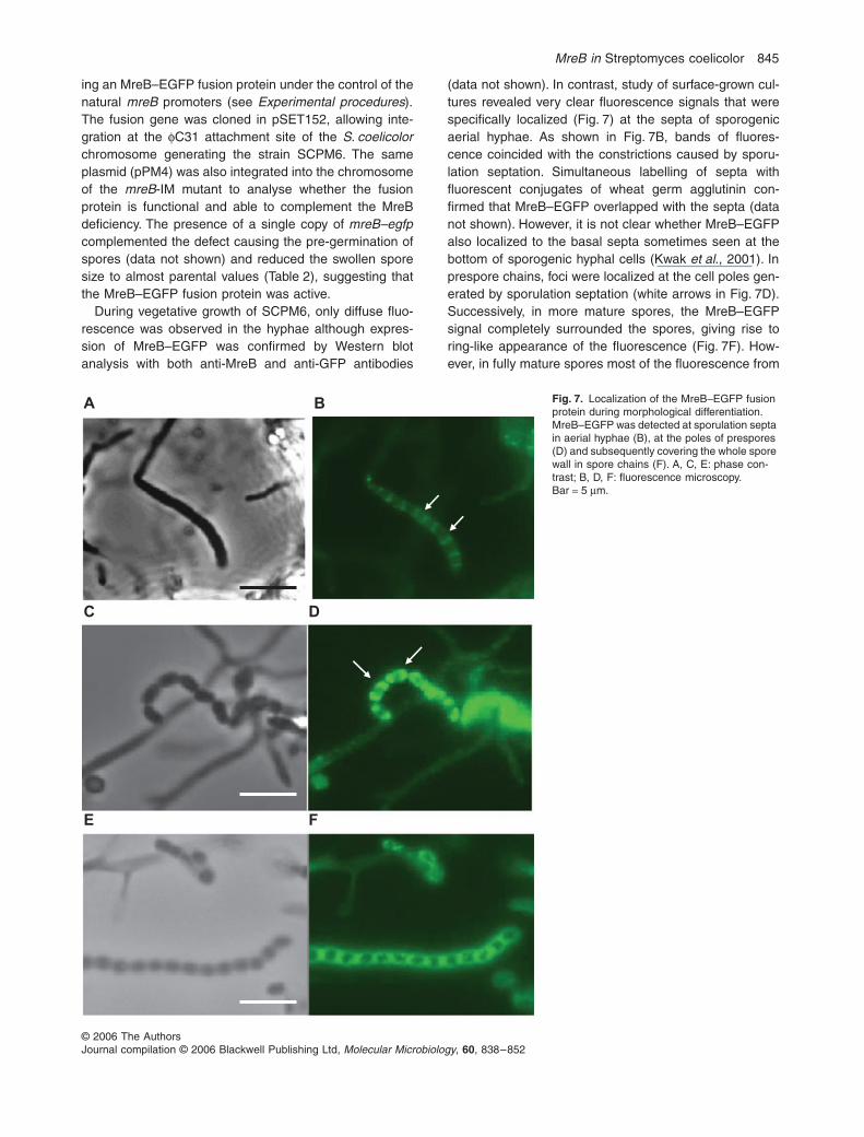
ing an MreB–EGFP fusion protein under the control of thenatural mreB promoters (see Experimental procedures).The fusion gene was cloned in pSET152, allowing inte-gration at the φC31 attachment site of the S. coelicolorchromosome generating the strain SCPM6. The sameplasmid (pPM4) was also integrated into the chromosomeof the mreB-IM mutant to analyse whether the fusionprotein is functional and able to complement the MreBdeficiency. The presence of a single copy of mreB–egfpcomplemented the defect causing the pre-germination ofspores (data not shown) and reduced the swollen sporesize to almost parental values (Table 2), suggesting thatthe MreB–EGFP fusion protein was active.
During vegetative growth of SCPM6, only diffuse fluo-rescence was observed in the hyphae although expres-sion of MreB–EGFP was confirmed by Western blotanalysis with both anti-MreB and anti-GFP antibodies
(data not shown). In contrast, study of surface-grown cul-tures revealed very clear fluorescence signals that werespecifically localized (Fig. 7) at the septa of sporogenicaerial hyphae. As shown in Fig. 7B, bands of fluores-cence coincided with the constrictions caused by sporu-lation septation. Simultaneous labelling of septa withfluorescent conjugates of wheat germ agglutinin con-firmed that MreB–EGFP overlapped with the septa (datanot shown). However, it is not clear whether MreB–EGFPalso localized to the basal septa sometimes seen at thebottom of sporogenic hyphal cells (Kwak et al., 2001). Inprespore chains, foci were localized at the cell poles gen-erated by sporulation septation (white arrows in Fig. 7D).Successively, in more mature spores, the MreB–EGFPsignal completely surrounded the spores, giving rise toring-like appearance of the fluorescence (Fig. 7F). How-ever, in fully mature spores most of the fluorescence from
Fig. 7. Localization of the MreB–EGFP fusion protein during morphological differentiation. MreB–EGFP was detected at sporulation septa in aerial hyphae (B), at the poles of prespores (D) and subsequently covering the whole spore wall in spore chains (F). A, C, E: phase con-trast; B, D, F: fluorescence microscopy. Bar = 5 µm.

846 P. Mazza et al.
© 2006 The AuthorsJournal compilation © 2006 Blackwell Publishing Ltd, Molecular Microbiology, 60, 838–852
the MreB–EGFP fusion had disappeared (data notshown).
Closer inspection of the localization of MreB was per-formed by immunogold electron microscopy of sporepreparations from S. coelicolor M145 (Fig. 8). As a nega-tive control we used the mreB insertion mutant (mreB-IM).While in the mutant only background gold labelling wasobtained, similar to when anti-MreB antibody was omitted(not shown), in the parental strain clear and specific label-ling, particularly close to the inside of the spore walls wasdetected.
The cellular localization of MreB was further examinedby cell fractionation and Western analysis using anti-MreBantibodies. For this, the mycelium and spores grown onMS agar were disrupted by French press treatment andthe cytoplasmic, the membrane-associated and themembrane fractions were individually isolated. MreB wasdetected both in the soluble fraction and mostly in themembrane-associated fraction, while we failed to detectMreB in the membrane fraction (data not shown). Cellular
localization of the MreB–EGFP fusion protein was alsoconducted. Indeed, the protein was localized in the samefractions as the native MreB, namely in the soluble fractionand in the membrane associated fractions (data notshown), indicating that the fusion protein was localizedequivalent to the parent protein.
Discussion
In this study, an mreB deletion mutant was obtained in thefilamentous Gram-positive bacterium S. coelicolor, and itwas demonstrated that MreB is dispensable for vegetativegrowth, but essential for spore formation and assembly ofthe spore cell wall. Previous attempts to inactivate mreBin S. coelicolor by gene disruption using the temperature-sensitive Streptomyces vector pGM9 (Muth et al., 1989)were unsuccessful (Burger et al., 2000). However, usinga PCR-targeting method (REDIRECT method; Gust et al.,2003) we were able to generate deletion mutants in mreB.The discrepancy may be explained by a higher frequencyof allelic exchange in the REDIRECT method, due to themuch larger regions flanking the mreB locus that wereavailable for recombination. As several mreB mutantswere isolated in independent experiments with frequen-cies that are typical of gene inactivation experiments usingthe REDIRECT procedure, it appears unlikely that mreBis essential in Streptomyces and that lethality of the mreBmutations was masked by (a) suppressor mutation(s), ashas been described for other bacteria (Kruse et al., 2005).This is further supported by the observation that mreB isabsent from many non-sporulating actinomycetes.
Deletion of mreB in S. coelicolor gave no obviousphenotype during vegetative growth in liquid culture oron solid plates. However, transcriptional analyses by S1nuclease mapping (Burger et al., 2000) and by microarrayand reverse transcription polymerase chain reaction (RT-PCR) studies (E.E. Noens and G.P. van Wezel, unpub-lished) revealed that S. coelicolor mreB is transcribed dur-ing vegetative growth, and expression was confirmed byWestern blot analysis of MreB protein in liquid-grown cul-tures (data not shown). Thus, the possible role of MreB inyoung mycelium is unclear and needs further analysis,and it can not be excluded that there could be somedegree of redundancy between MreB and the secondMreB homologue Mbl. However, based on the lack of aclear phenotype of the mreB mutant during vegetativegrowth, and the lack of MreB–EGFP signals at hyphal tips,it seems unlikely that MreB would have a direct role in theelongation of the hyphal cell wall, which occurs primarilyat the tips. Sequence analysis of genomic DNA of differentactinomycetes showed that species forming only sub-strate mycelium, such as Rhodococcus sp. and Nocardiasp. (http://Rhodococcus.ca; http://nocardia.nih.go.jp), do nothave an mreBCD cluster, supporting the idea that the Mre
Fig. 8. High-resolution localization of MreB in S. coelicolor spores. Specific localization of MreB in spores by in situ hybridization of thin sections (immunoelectron microscopy) of spore preparations of S. coelicolor M145 (A), using gold-labelled anti-MreB antibodies. The MreB-specific antibodies localized almost exclusively around the inner side of the spore envelope. Occasional particles found outside the spores were attached to cell debris. Expectedly, no specific label-ling was obtained with any of the independent mreB mutants (B). Notice the highly variable spore sizes and less dense spore wall of the mreB null mutant. Bar = 500 nm.

MreB in Streptomyces coelicolor 847
© 2006 The AuthorsJournal compilation © 2006 Blackwell Publishing Ltd, Molecular Microbiology, 60, 838–852
proteins may not be required for vegetative growth inactinomycetes.
While inactivation of mreB did not compromise vegeta-tive growth of S. coelicolor, ectopic expression of mreBwas highly toxic. After germination of spores the elonga-tion of germ tubes was inhibited and hyphae lysed. Obvi-ously, the presence of (too much) MreB at the wrongspatial compartment severely interferes with normalgrowth, perhaps by recruiting PBPs and preventing themfrom their function in cell wall synthesis.
Mutational analysis clearly demonstrated that MreB inS. coelicolor is required for correct sporulation, andappears to affect spore wall formation. The aerial hyphaeand spores of the mreB deletion mutant were swollen(Fig. 4; Table 2), spores were sensitive to heat and treat-ment with SDS (Fig. 6), and irregularities in the spore cellwalls were observed using TEM (Fig. 5B). This is consis-tent with the observation made in B. subtilis that deletionof mreB causes increased cell width and cell lysis (Form-stone and Errington, 2005). Formstone and Errington(2005) proposed that loss of MreB lead to a change inresistance of the cell wall to osmotic or mechanical stress,due to the absence or incorrect assembly of cell wallcomponents (Formstone and Errington, 2005). In analogy,the depletion of MreB in S. coelicolor appears to impairassembly of the cell wall, but only during sporulation. Thespore wall composition of streptomycetes is not well stud-ied. However, it is different in thickness from vegetativehyphal walls and has often two layers, although qualitativedifferences in peptidoglycan components have not beenreported (Glauert and Hopwood, 1961; Ensign, 1978).The importance of the spore wall may be indicated by thefact that spores acquire resistance to different physiolog-ical and mechanical stresses, which the substrate myce-lium does not have. Consistently, the mreB mutants failedto mount resistance against two types of stress.
The involvement of MreB in spore formation is consis-tent with the PCR and Southern blot analysis of differentactinomycetes. Using primers corresponding to a highlyconserved region of mreB, genomic DNA of sporulatingactinomycetes gave rise to an amplified fragment of theexpected size, while genomic DNA of non-sporulating act-inomycetes did not (Table 1). Surprisingly two sporulatinggenera, Actinoplanes and Micromonospora, did not givean amplified product (Table 1). These two genera sharean important characteristic: they sporulate, but they do notform aerial mycelium. The genus Actinoplanes producesmotile spores enveloped in structures called sporangia(Waksman, 1961). The genus Micromonospora grows asstraight or curved branching substrate hyphae withoutcross-walls (Waksman, 1961). Like in Actinoplanes, aerialmycelium is not formed and multiplication occurs bymeans of fragments of mycelium and special sporesformed singularly. The genus Actinoplanes is a member
of the family of the Actinoplanaceae together with thegenus Streptosporangium which in contrast produces anaerial mycelium which resembles that of most speciesof Streptomyces (Waksman, 1961). Unlike Actinoplanes,Streptosporangium DNA was shown to encode an mreBhomologue (Table 1). Apparently, the differentiation pro-cess in the two genera is highly different, suggesting thatthe mre genes are needed only in Streptosporangiumsporulation that indeed resembles that of Streptomyces.From these observations, MreB seems not to be requiredin sporulation processes of actinomycetes in general, butonly in a sporulation process which involves septation ofaerial mycelium.
In B. subtilis and E. coli, which both are rod-shapedbacteria, MreB forms helical filaments lying beneath thecell surface (Jones et al., 2001; Shih et al., 2003). InC. crescentus, MreB undergoes two distinct patterns oflocalizations: during the cell elongation phase it appearslike spirals that traverse along the longitudinal axis of thecell, while during cell division it forms an FtsZ-dependenttransverse band at the mid-cell (Figge et al., 2004).Therefore, it was speculated that MreB has a role in co-ordinating the switch between longitudinal and septal pep-tidoglycan synthesis. In S. coelicolor, we failed to detectthe typical helical-like structures described for E. coli,B. subtilis and C. crescentus. The signal generated fromthe MreB–EGFP fusion during vegetative growth resultedin diffused fluorescence in the hyphae, indicating thatMreB monomers are randomly distributed in the vegeta-tive mycelium without a specific localization. In contrast,during differentiation the MreB–EGFP localized at thesepta of aerial hyphae and successively, when sporeswere formed, in the spores (Fig. 7) probably underneaththe cytoplasmic membrane, as indicated by immunogoldelectron microscopy (Fig. 8). Considering that MreB isorganized in helical structures in rod-shaped bacteria, itis possible that in S. coelicolor MreB forms dense spiralsjust underneath the cytoplasmic membrane, but no suchpatterns could be discerned in our analyses. As it is pos-sible that fusions of GFP to the C-terminus of MreB caninterfere with its function, we can not exclude that our C-terminal MreB–EGFP fusion did not fully reproduce thelocalization patterns of MreB itself. However, we coulddemonstrate that MreB–EGFP localized to the same sub-cellular fractions of S. coelicolor as the MreB protein did.Furthermore, MreB–EGFP was able to complement themutant phenotype of the mreB-IM mutant by restoring thespore size to almost parental values (Table 2) as well asby preventing pre-germination of spore chains.
In S. coelicolor, septation occurs during both vegetativegrowth and sporulation, but MreB localization was specificfor sporulation septa. It is clear that MreB is not requiredfor the formation of sporulation septa per se, as mreBmutants were still able to septate (Fig. 5B) and produced

848 P. Mazza et al.
© 2006 The AuthorsJournal compilation © 2006 Blackwell Publishing Ltd, Molecular Microbiology, 60, 838–852
viable spores. On the other hand, it should be testedwhether MreB localization could be FtsZ dependent, asalready seen in C. crescentus (Figge et al., 2004). It willbe therefore interesting to test in future experiments MreBlocalization in different S. coelicolor ftsZ mutants; forexample, an ftsZ mutant impaired in Z-ring formation insporogenic aerial hyphae, but not in vegetative mycelium(Grantcharova et al., 2003). In R. sphaeroides, MreBlocalizes predominantly at the mid-cell position (Slovaket al., 2005). The authors speculate that this mid-celllocalization for MreB might reflect the fact that this regionis the main site of peptidoglycan synthesis rather than thatMreB plays a direct role in septation. In an analogousmanner, MreB in S. coelicolor which was shown to localizeat the sporulation septa, could be needed in subsequentsteps for spore formation, rather than playing a crucial rolein the sporulation-specific cell division. Thus, MreB inS. coelicolor has a novel localization pattern as well as asomewhat unique mode of action that has not beenobserved in the rod-shaped bacteria and may be typicalof actinomycetes that form an aerial mycelium and spores.
Based on the localization of MreB at the spore wall andits important role in maintaining the integrity of the sporewall, we anticipate that MreB is involved in thickening ofthe spore wall and may be recruiting PBPs and otherpeptidoglycan-related proteins during the sporulation pro-cess. The mre genes in S. coelicolor and S. avermitilis arephysically linked with pbp83 and sfr (Burger et al., 2000).Pbp83 (Pbp2; Bentley et al., 2002), encodes a PBP withhigh similarity to E. coli PBP2, which is involved in cellelongation in E. coli (Vinella et al., 1993), and with SpoVD,a PBP important for sporulation of B. subtilis (Daniel et al.,1994). The product of sfr shows similarity to RodA, aprotein involved in peptidoglycan synthesis during cellelongation in E. coli, and SpoVE, a protein required forthe synthesis of the spore cortex peptidoglycan inB. subtilis (Henriques et al., 1998). Inactivation of pbp83by gene disruption lead to an impairment in differentiation(A. Burger and W. Wohlleben, unpublished), a phenotypethat resembles that of the mreB mutant that is reported inthis article. A protein interaction model of the MreBCD,RodA and PBPs in B. subtilis has been proposed by Err-ington (2003). According to this model a multi-PBP com-plex interacts with RodA, MreC and MreD and thisassembly is spatially controlled by the MreB cables in thecytosol. These proposed interactions between the Mreproteins were partially confirmed in E. coli (Kruse et al.,2005). We observed that in S. coelicolor, deletion of mreB,mreC, mreD individually, or of the whole mreBCD cluster,leads to a similar phenotype (K. Schirner, P. Mazza, G.Muth, and W. Wohlleben, unpublished) suggesting thatalso in this organism the three proteins may participate inthe same process. A complex that includes PBPs, a RodAhomologue, and the Mre proteins, and which primarily is
acting during spore wall assembly, is therefore imaginablein S. coelicolor. This leads to the following hypothesis onthe mode of action of MreB in S. coelicolor: it is expressedat a basal level during vegetative growth and it is localizedrather dispersely in the cytosol. When sporulation septa-tion occurs, MreB condenses at the sporulation septa,possibly in an FtsZ-dependent manner. An increasedexpression of mreB before sporulation that was detectedby S1-mapping experiments (Burger et al., 2000) mayallow the cells to accumulate a sufficient number of MreBunits to form a ring- or a shell-like structure underneaththe spore wall. As MreB most likely is a membrane-associated protein, this structure may be anchored to thecell membrane through the two membrane proteins MreCand MreD and used to recruit and localize proteinsresponsible for spore wall formation, perhaps includingPbp83 and the product of sfr. Further studies to localizePbp83 and Sfr in mre mutants and interaction studiesbetween the Mre proteins and the products of pbp83 andsfr are underway to understand the unique role of MreBin S. coelicolor A3(2). It will also be important to clarify therole of Mbl in this organism, and to determine whetherthere could be some functional redundancy between thisprotein and MreB.
Experimental procedures
Bacterial strains and media
The E. coli and S. coelicolor A3(2) strains used are listed inTable 3. S. coelicolor strains were cultivated on MS agarplates or in S-medium (Kieser et al., 2000). Cultivation ofstrains and procedure for DNA manipulation were performedas previously described for E. coli (Sambrook et al., 1989)and S. coelicolor (Kieser et al., 2000).
Construction of plasmids
Plasmid pPM1 was obtained by amplifying the mreB genefrom the S. coelicolor M145 genome using the primers PM1and PM2 ( Table 4), digesting the PCR product with NdeIand HindIII and cloning the fragment under the control of thethiostrepton-inducible promoter PtipA into the Streptomycesmulticopy expression plasmid pGM190 (G. Muth, unpubl.data). To construct a C-terminal fusion with the EGFP pro-tein, the mreB gene was amplified using the primers PM5and PM6 (Table 4), cut with NdeI and BglII and cloned infront of the egfp gene in the plasmid pTST101 (J. Altenbuch-ner, pers. comm.) cut with NdeI and BamHI. To transformS. coelicolor M145 the resulting mreB–egfp fusion was cutout from pTST101 and cloned in the integrative Streptomy-ces vector pSET152 (Bierman et al., 1992) generatingpPM4. Plasmid pPM6 was constructed by amplifying themreB gene with its promoter region from S. coelicolor M145with the primers PM11 and PM12 (Table 4) and cloning thePCR product as BglII–EcoRI fragment in pSET152 (Biermanet al., 1992).

MreB in Streptomyces coelicolor 849
© 2006 The AuthorsJournal compilation © 2006 Blackwell Publishing Ltd, Molecular Microbiology, 60, 838–852
Protein expression, purification and antibody production
mreB from S. coelicolor M145 was amplified by PCR with theprimers PM7 and PM2 that include BamHI and HindIII site,respectively, and cloned in pRSETB (Invitrogen) so that itforms an in-frame fusion with the His-tag (pPM5). His-taggedMreB was expressed in E. coli BL21 cells containing pPM5and purified with Ni-Nta spin columns (Qiagen) under dena-turing condition as described in the Ni-Nta manual (Qiagen).Rabbit polyclonal antibodies were raised against the purifiedprotein (Eurogentec).
Overexpression of MreB
Protoplasts of S. coelicolor M145 were transformed with theplasmids pPM1 or pGM190 as described previously (Kieseret al., 2000). Spores of transformants were inoculated in S-medium containing 50 µg ml−1 kanamycin and incubated withshaking at 30°C. To overexpress MreB, thiostrepton wasincluded at a concentration of 10 µg ml−1.
Overexpression of MreB was confirmed by Western blotanalysis with anti-MreB serum. Mycelium was collected after18 h of growth with or without thiostrepton and broken withFrench press. Crude extracts were mixed with an equal vol-ume of 2× sample buffer (125 mM Tris-HCl pH 6.8; 4% SDS;20% glycerol; 2.0 mM EDTA; 0.02% bromophenol blue; 3%dithiothreitol), boiled for 5 min and loaded on SDS-polyacry-lamide gels (12.5%). Proteins were capillary transferred to apure nitrocellulose blotting membrane (Pall Corporation) andMreB was detected using a 1:2500 dilution of polyclonal anti-MreB serum.
Cell fractionation
Streptomyces coelicolor M145 and the strain SCPM6 carry-ing the mreB–egfp fusion were grown for 2–3 days in 150 mlof S-medium with or without antibiotics at 30°C and har-vested by centrifugation. Mycelium was resuspended in 6 mlof 25 mM Tris-HCl (pH 7.5), 100 mM NaCl and 1 mM pro-tease inhibitor (Complete EDTA-free tablets, Roche), and thecells were broken using the French press. The cell extractwas centrifuged at 90 000 g for 30 min at 4°C and the super-natant, containing the cytoplasmic fraction, was saved andstored at −20°C. Pellet was resuspended in 4 ml of 25 mMTris-HCl (pH 7.5), 1 M NaCl and 20% glycerol, stirred for 2 hat 4°C and centrifuged at 90 000 g for 30 min at 4°C. Thesupernatant containing membrane-associated proteins wassaved and stored at −20°C. To obtain the membrane fraction,pellet was resuspended in 1.5 ml of 25 mM Tris-HCl (pH 7.5),1 M NaCl, 20% glycerol and 2% Triton X-100, stirred over-night at 4°C and again centrifuged for 30 min at 90 000 g.The samples obtained from cell fractionation were loaded onSDS-gels and Western blot was performed as describedabove.
Creation of an mreB null allele
To create an mreB mutant, the PCR-targeting proceduredescribed by Gust et al. (2003) was used. The ΩaacC4 cas-sette (which confers apramycin resistance) from the plasmidpIJ773 was amplified using the oligonucleotides PM9 andPM10 (Table 4) designed so that the 3′-ends can anneal to
Primer Sequencea
PM1 cgcatatggggaactcaatgtcgttcPM2 gaaagcttacgtcatctacggggcgPM5 cgcatatgtgatccttcttcgggacPM6 ggagatctgatgaacgacattgaPM7 aaggatccggggaactcaatgtcgPM9 ccctcaaaagctcctgggaaggccagtcgaatcctgatggatatcattccggggatccgcgtaccPM10 ggagatcgtctcgtacggcggaaccgaagtgttacgtcagatatctgtaggctggagctgcttcPM11 gcagatctgacgccatgtcagtcgaPM12 gcgaattcggcggaaccgaagtgttaPM13 gccgtgcgcccgctgaaggacggPM14 agcagcgcgcgccgccgccggt
a. Restriction sites are written in italics.
Table 4. Primers used in this work.
Table 3. Strains of E. coli and S. coelicolor used in this work.
Strain Characteristics Reference
E. coli BL21(DE3)pLysS F–ompT hsdSB(rB–mB
–) gal dcm araB::T7RNAP-tetA InvitrogenE. coli ET 12567 GM2929 hsdM–hsdR–zjj-202 MacNeil et al. (1992)E. coli DH5α supE44 ∆lacU169 (80 lacZ∆M15) hsdR17 recA1 endA1 gyrA96 thi-1 relA1 Woodcock et al. (1989)E. coli BW 25113/pIJ790 Chromosome: ∆(araD-araB)567 ∆lacZ4787 (::rrnB-4) lacIp-4000 (lacIQ)
λ−1rpoS369(Am) rph-1 ∆(rhaD-rhaB)568 hsd514;Gust et al. (2003)
Plasmid: [oriR101] [repA101(ts)] araBp-gam-beta-exoS. coelicolor M145 Prototrophic, SCP1– SCP2– Kieser et al. (2000)mreB-IM M145 ∆mreB::ΩaacC4 This workmreB-IFD M145 ∆mreB This workmreB-IFDc M145 ∆mreB attBΦC31 ::mreB This workSCPM6 M145 attBΦC31 ::mreB–egfp This work

850 P. Mazza et al.
© 2006 The AuthorsJournal compilation © 2006 Blackwell Publishing Ltd, Molecular Microbiology, 60, 838–852
the resistance cassette gene, whereas the 5′-ends arehomologous to the sequences flanking S. coelicolor mreB.The PCR products was transferred into E. coli BW 25113/pIJ790 carrying the cosmid C88 and the λ RED systeminduced as described (Gust et al., 2003). The correct genereplacement on the cosmid C88 isolated from an apramy-cin-resistant clone was confirmed with PCR using a set ofoligonucleotides priming outside the region of recombina-tion. This cosmid was designated pPM11. pPM11 was intro-duced into E. coli ET12567 containing the plasmid pUZ8002and then transferred to S. coelicolor M145 by conjugationas previously described (Kieser et al., 2000). Exconjugantswere selected for kanamycin and apramycin resistance andthen replicated on plates with apramycin and with or withoutkanamycin. DNA of kanamycin-sensitive and apramycin-resistant clones were isolated and tested with PCR andSouthern blot to confirm the absence of the chromosomalmreB.
In order to remove the disruption cassette, the cosmidpPM11 was introduced in the E. coli strain DH5α/pCP20that expresses the FLP recombinase (Gust et al., 2003).The resulting cosmid carrying the in-frame deletion wasnamed pPM12 and used to transform the S. coelicolormutant strain containing the marked deletion (Kieser et al.,2000). Transformants were first selected for insertion of thecosmid by single cross-over (kanamycin resistant) and thenscreened for the double-cross-over event (kanamycin sensi-tive and apramycin sensitive). The loss of the disruptioncassette was confirmed with PCR and Southern blotanalysis.
Heat and detergent treatment
To test whether the mutant spores were as heat resistant asthe ones of the parental strain, spore suspensions of differentstrains (105 spores ml−1) were incubated at 60°C for differenttime. After incubation spore suspensions were diluted 1:10or 1:100 and 100 µl of the dilution was plated on MS agar.Spore suspension of the mreB mutant incubated longer than20 min at 60°C were plated without being diluted. Plates werefurther cultivated for 2 days at 30°C. Survival rate was calcu-lated as number of colonies grown on plates after treatmentat 60°C divided by the number of colonies grown on plateswithout treatment at 60°C in percentage.
To test the effect of the detergent SDS on mreB-depletedspores, 50 µl of spore suspensions (109 ml−1) of M145 and ofthe mreB mutant (mreB-IFD) was incubated in 5% SDS, 1%SDS or water for 1 h at room temperature. Appropriate sporedilutions were plated on MS agar and incubated at 30°C for2 days. Titre of colonies surviving SDS treatment were deter-mined in relation to those incubated in water.
Ultrasonic treatment
To test spore resistance to ultrasonication, spore suspension(105 spores ml−1) was treated with ultrasounds (SONOPULSGM2070 digital ultrasonic homogenizers, 20 kHz; BANDELINelectronic GmbH) five cycles of 10 s and after each cycles100 µl of spore suspension was collected, diluted, plated onMS agar and further incubated at 30°C for 2 days.
Microscopy
Phase contrast microscopy. For light microscopy, sterilecoverslips were inserted at a 45° angle into MS agar andspores were inoculated in the acute angle along the glasssurface. Coverslips were removed after 3- to 4-day incubationat 30°C and mounted in PBS containing 50% glycerol on polyL-lysine-coated slides. Alternatively liquid culture was spotteddirectly on microscope slides covered with 1% agarose andcovered with coverslips. Samples were observed with anOlympus System Microscope BX60 and pictures taken withan Olympus F-view II camera.
Electron microscopy. TEM for the analysis of ultrathincross-sections of hyphae and spores was performed asdescribed previously (van Wezel et al., 2000).
For immunoelectron microscopy spores were fixed in 2%paraformaldehyde with 0.2% glutaraldehyde in PHEM bufferfor 2 h at room temperature. After washing in PBS the sporeswere pelleted and embedded in 12% gelatine. The pellet wascut into 1 mm3 cubes, cryoprotected in 2.3 M sucrose andsnapfrozen in liquid nitrogen. Ultrathin cryosections werelabelled with rabbit anti-MreB (1:2000) followed by 15 nmprotein A-gold particles. The labelled sections were embed-ded and contrasted in methylcellulose with uranyl acetate. Allsamples were viewed with a Philips EM 410 electron micro-scope (Eindhoven, the Netherlands).
Acknowledgements
Thanks to Eriko Takano for fruitful discussion and many help-ful comments on the manuscript. This work was supportedby grants from the DFG (Forschergruppe 449 ‘BakterielleZellhülle’) to W.W. and from the Netherlands Royal Societyfor Arts and Sciences (KNAW) to G.P.v.W.
References
Aretz, W., Meiwes, G., Seibert, G., Vobis, G., and Wink, J.(2000) Friulimicins: novel lipopeptide antibiotics with pep-tidoglycan synthesis inhibiting activity from Actinoplanesfriuliensis sp. nov. I. Taxonomics studies of the producingmicroorganism and fermentation. J Antibiot (Tokyo) 53:807–815.
Ausmees, N., Kuhn, J.R., and Jacobs-Wagner, C. (2003) Thebacterial cytoskeleton: an intermediate filament-like func-tion in cell shape. Cell 115: 705–713.
Bentley, S.D., Chater, K.F., Cerdeno-Tarraga, A.M., Challis,G.L., Thomson, N.R., James, K.D., et al. (2002) Completegenome sequence of the model actinomycete Streptomy-ces coelicolor A3(2). Nature 417: 141–147.
Bierman, M., Logan, R., O’Brien, K., Seno, E.T., Nagaraja-Rao, R., and Schoner, B.E. (1992) Plasmid cloning vectorsfor the conjugal transfer of DNA from Escherichia coli toStreptomyces spp. Gene 116: 43–49.
Bork, P., Sander, C., and Valencia, A. (1992) An ATPasedomain common to prokaryotic cell cycle proteins, sugarkinases, actin, and Hsp70 heat shock proteins. Proc NatlAcad Sci USA 89: 7290–7294.
Burger, A., Brandt, B., Süsstrunk, U., Thompson, C.J., andWohlleben, W. (1998) Analysis of a Streptomyces coeli-

MreB in Streptomyces coelicolor 851
© 2006 The AuthorsJournal compilation © 2006 Blackwell Publishing Ltd, Molecular Microbiology, 60, 838–852
color A3(2) locus containing the nucleoside diphosphatekinase (ndk) and folylpolyglutamate synthetase (folC)genes. FEMS Microbiol Lett 159: 283–291.
Burger, A., Sichler, K., Kelemen, G., Buttner, M., and Wohlle-ben, W. (2000) Identification and characterization of themre gene region of Streptomyces coelicolor A3(2). MolGen Gene 263: 1053–1060.
Cabeen, M.T., and Jacobs-Wagner, C. (2005) Bacterial cellshape. Nat Rev Microbiol 3: 601–610.
Chater, K.F., and Hopwood, D.A. (1993) Streptomyces. InBacillus subtilis and Other Gram-positive Bacteria. Bio-chemistry, Physiology and Molecular Genetics. Sonen-shein, J.P. (ed.). Washington, DC; ASM, pp. 83–89.
Chater, K.F., and Losick, R. (1997) Mycelial life style ofStreptomyces coelicolor A3(2) and its relatives. In Shapiro,J.A., and Dworkin, M. (eds). Bacteria as MulticellularOrganisms. New York: Oxford University Press, pp. 149–182.
Daniel, R.A., and Errington, J. (2003) Control of cell morpho-genesis in bacteria: two distinct ways to make a rod-shaped cell. Cell 113: 767–776.
Daniel, R.A., Drake, S., Buchanan, C.E., Scholle, R., andErrington, J. (1994) The Bacillus subtilis spoVD geneencodes a mother-cell-specific penicillin-binding proteinrequired for spore morphogenesis. J Mol Biol 235: 209–220.
Doi, M., Wachi, M., Ishino, F., Tomioka, S., Ito, M., Sakagami,Y., et al. (1988) Determinations of the DNA sequence ofthe mreB gene and of the gene products of the mre regionthat function in formation of the rod shape of Escherichiacoli cells. J Bacteriol 170: 4619–4624.
Ensign, J.C. (1978) Formation, properties and germinationactinomycete spores. Annu Rev Microbiol 32: 185–219.
van den Ent, F., Amos, L.A., and Löwe, J. (2001) Prokaryoticorigin of the actin cytoskeleton. Nature 413: 39–44.
Errington, J. (2003) Dynamic proteins and a cytoskeleton inbacteria. Nat Cell Biol 5: 175–178.
Figge, R.M., Divakaruni, A.V., and Gober, J.W. (2004) MreB,the cell shape-determining bacterial actin homologue, co-ordinates cell wall morphogenesis in Caulobacter crescen-tus. Mol Microbiol 51: 1321–1332.
Flärdh, K. (2003) Growth polarity and cell division in Strep-tomyces. Curr Opin Microbiol 6: 564–571.
Formstone, A., and Errington, J. (2005) A magnesium-dependent mreB null mutant: implications for the role ofmreB in Bacillus subtilis. Mol Microbiol 55: 1646–1657.
Gitai, Z., Dye, N., and Shapiro, L. (2004) An actin-like genecan determine cell polarity in bacteria. Proc Natl Acad SciUSA 101: 8643–8648.
Gitai, Z., Dye, N.A., Reisenauer, A., Wachi, M., and Shapiro,L. (2005) MreB actin-mediated segregation of a specificregion of a bacterial chromosome. Cell 120: 329–341.
Glauert, A.M., and Hopwood, D.A. (1961) The fine structureof Streptomyces violaceoruber (S. coelicolor) III. The wallsof the mycelium and spores. J Biophys Biochem Cytol 10:505–516.
Grantcharova, N., Ubhayasekera, W., Mowbray, S.L., McCor-mick, J.R., and Flärdh, K. (2003) A missense mutation inftsZ differentially affects vegetative and developmentallycontrolled cell division in Streptomyces coelicolor A3(2).Mol Microbiol 47: 645–656.
Grund, A.D., and Ensign, J.C. (1982) Activation of Strepto-myces viridochromogenes spores by detergents. CurrMicrobiol 7: 223–228.
Gust, B., Challis, G.L., Fowler, K., Kieser, T., and Chater,K.F. (2003) PCR-targeted Streptomyces gene replacementidentifies a protein domain needed for biosynthesis of thesesquiterpene soil odor geosmin. Proc Natl Acad Sci USA100: 1541–1546.
Henriques, A.O., Glaser, P., Piggot, P.J., and Moran, C.P.,Jr (1998) Control of cell shape and elongation by the rodAgene in Bacillus subtilis. Mol Microbiol 28: 235–247.
Ikeda, H., Ishikawa, J., Hanamoto, A., Shinose, M., Kikuchi,H., Shiba, T., et al. (2003) Complete genome sequenceand comparative analysis of the industrial microorganismStreptomyces avermitilis. Nat Biotechnol 21: 526–531.
Jones, L.J., Carballido-Lopez, R., and Errington, J. (2001)Control of cell shape in bacteria: helical, actin-like filamentsin Bacillus subtilis. Cell 104: 913–922.
Kieser, T., Bibb, M.J., Buttner, M.J., Chater, K.F., and Hop-wood, D.A. (2000) Practical Streptomyces Genetics. Nor-wich: The John Innes Foundation.
Kruse, T., Moller-Jensen, J., Lobner-Olesen, A., and Gerdes,K. (2003) Dysfunctional MreB inhibits chromosome segre-gation in Escherichia coli. EMBO J 22: 5283–5292.
Kruse, T., Bork-Jensen, J., and Gerdes, K. (2005) The mor-phogenetic MreBCD proteins of Escherichia coli form anessential membrane-bound complex. Mol Microbiol 55:78–89.
Kwak, J., Dharmatilake, A.J., Jiang, H., and Kendrick, K.E.(2001) Differential regulation of ftsZ transcription duringseptation of Streptomyces griseus. J Bacteriol 2001:5092–5101.
Leaver, M., and Errington, J. (2005) Roles for MreC andMreD proteins in helical growth of the cylindrical cell wallsin Bacillus subtilis. Mol Microbiol 57: 1196–1209.
Lee, J.C., and Stewart, G.C. (2003) Essential nature of themreC determinant of Bacillus subtilis. J Bacteriol 185:4490–4498.
Löwe, J., van den Ent, E.F., and Amos, L.A. (2004) Moleculesof the bacterial cytoskeleton. Annu Rev Biophys BiomolStruct 33: 177–198.
McBride, M.J., and Ensign, J.C. (1987) Effects of intracellulartrehalose content on Streptomyces griseus spores. J Bac-teriol 169: 4995–5001.
MacNeil, D.J., Gewain, K.M., Ruby, C.L., Dezeny, G., Gib-bons, P.H., and MacNeil, T. (1992) Analysis of Streptomy-ces avermitilis gene required for avermectin biosynthesisutilizing a novel integration vector. Gene 111: 61–68.
Margolin, W. (2005) FtsZ and the division of prokaryotic cellsand organelles. Nat Rev Mol Cell Biol 6: 862–871.
Miyadoh, S. (1997) Atlas of Actinomycetes. Tokyo: TheSociety for Actinomycetes Japan.
Muth, G., Nussbaumer, B., Wohlleben, W., and Pühler, A.(1989) A vector-system with temperature sensitive replica-tion for gene disruption and mutational cloning in strepto-mycetes. Mol Gen Genet 219: 341–348.
Sambrook, J., Fritsch, E.F., and Manatis, T. (1989) MolecularCloning: A Laboratory Manual, 2nd edn. Cold Spring Har-bour, NY: Cold Spring Harbour Laboratory Press.
Shih, Y.L., Le, T., and Rothfield, L. (2003) Division site selec-tion in Escherichia coli involves dynamic redistribution of

852 P. Mazza et al.
© 2006 The AuthorsJournal compilation © 2006 Blackwell Publishing Ltd, Molecular Microbiology, 60, 838–852
Min proteins within coiled structures that extend betweenthe two cell poles. Proc Natl Acad Sci USA 100: 7865–7870.
Slovak, P.M., Wadhams, G.H., and Armitage, J.P. (2005)Localization of MreB in Rhodobacter sphaeroides underconditions causing changes in cell shape and membranestructure. J Bacteriol 187: 54–64.
Soufo, H.J., and Graumann, P.L. (2003) Actin-like proteinsMreB and Mbl from Bacillus subtilis are required for bipo-lar positioning of replication origins. Curr Biol 13: 1916–1920.
Vinella, D., Joselau-Petit, D., Thévenet, D., Bouloc, P., andD’Ari, R. (1993) Penicillin-binding protein 2 inactivation inEscherichia coli results in cell division inhibition, which isrelieved by FtsZ overexpression. J Bacteriol 175: 6704–6710.
Wachi, M., and Matsuhashi, M. (1989) Negative control ofcell division by mreB, a gene that functions in determiningthe rod shape of Escherichia coli cells. J Bacteriol 171:3123–3127.
Wachi, M., Doi, M., Tamaki, S., Park, W., Nakajima-Iijima, S.,and Matsuhashi, M. (1987) Mutant isolation and molecularcloning of mre genes, which determine cell shape, sensi-tivity to mecillinam, and amount of penicillin-binding pro-teins in Escherichia coli. J Bacteriol 169: 4935–4940.
Waksman, S.A. (1961) The Actinomycetes. Classification,Identification and Description of Genera and Species. Bal-timore: The Williams & Wilkins company.
van Wezel, G.P., van der Meulen, J., Kawamoto, S., Luiten,R.G., Koerten, H.K., and Kraal, B. (2000) ssgA is essentialfor sporulation of Streptomyces coelicolor A3(2) andaffects hyphal development by stimulating septum forma-tion. J Bacteriol 182: 5653–5662.
Woodcock, D.M., Crowther, P.J., Doherty, J., Jefferson, S.,DeCruz, E., Noyer-Weidner, M.S., et al. (1989) Quantita-tive evaluation of Escherichia coli host strains for toleranceto cytosine methylation in plasmid and phage recombi-nants. Nucleic Acids Res 17: 3469–3478.
Young, K.D. (2003) Bacterial shape. Mol Microbiol 49: 571–580.