The ARC2 Response in Streptomyces coelicolor - TSpace
205
The ARC2 Response in Streptomyces coelicolor: Genetic and Physiological Changes Induced by a Chemical Probe by Vanessa Yoon Calvelo A thesis submitted in conformity with the requirements for the degree of Doctor of Philosophy Department of Biochemistry University of Toronto © Copyright by Vanessa Yoon Calvelo 2021
-
Upload
khangminh22 -
Category
Documents
-
view
1 -
download
0
Transcript of The ARC2 Response in Streptomyces coelicolor - TSpace

The ARC2 Response in Streptomyces coelicolor: Genetic and Physiological Changes Induced by a Chemical Probe
by
Vanessa Yoon Calvelo
A thesis submitted in conformity with the requirements for the degree of Doctor of Philosophy
Department of Biochemistry University of Toronto
© Copyright by Vanessa Yoon Calvelo 2021

ii
The ARC2 Response in Streptomyces coelicolor: Genetic and
Physiological Changes Induced by a Chemical Probe
Vanessa Yoon Calvelo
Doctor of Philosophy
Department of Biochemistry University of Toronto
2021
Abstract Streptomyces are known for the production of biologically active secondary metabolites that are
encoded in discrete gene clusters. Expression of these clusters is often controlled by factors that
include growth rate, signaling molecules, metabolism, and physiological and environmental
stresses. These diverse signals along with signal transduction proteins and transcription factors
constitute a complex regulatory network that mediate various types of responses including the
induction of secondary metabolism. In this work, I have taken a chemical genetic approach, using
a chemical elicitor called ARC2, to investigate the regulatory network of Streptomyces coelicolor.
I show that ARC2 alters the expression of primary metabolic genes associated with amino acid
metabolism, carbohydrate metabolism, lipid metabolism and nucleic acid metabolism. I also show
that ARC2 alters the expression of at least 16 different secondary metabolic gene clusters,
including the gene cluster encoding the blue-pigmented secondary metabolite actinhorhodin. I go
on to show that AfsR and AfsS, two regulators from the AfsK/AfsR/AfsS (AfsK/R/S) pleiotropic
regulation system for secondary metabolism, are required for both the basal production of
actinorhodin and the stimulation of actinorhodin production by the ARC2 elicitor. In addition, I
show that the serine/threonine kinase AfsK is not required for actinorhodin production in S.
coelicolor. Lastly, with the use of ARC2, I identified a functionally relevant repeat in the AfsS
regulator that is required to activate secondary metabolism. This work demonstrates the use of a
chemical tool to uncover novel aspects about the biology of secondary metabolism in the model
organism S. coelicolor.

iii
Acknowledgments First, I would like to thank my supervisor Dr. Justin Nodwell for his patience and mentorship
throughout my graduate studies. Thank you for taking a chance on me and welcoming me into
your lab. With your guidance, I have grown tremendously as a scientist over the years and I hope
to continue to grow in my next endeavour. I would also like to thank my past advisory committee
members, Dr. Marie Elliot and Dr. Michael Surette, during my brief time at McMaster University
as well as my current advisory committee members, Dr. Shana Kelley and Dr. Andrew Wilde, at
the University of Toronto. Your feedback has always been appreciated.
Second, I want to thank the past and present members of the Nodwell lab as you were the ones that
provided a healthy and stable work environment. In particular, I want to thank Dr. Sheila Pimentel-
Elardo and Dr. Stefanie Mak for the constant emotional and technical support both inside and
outside of the lab. I also want to thank Saif Hossain for giving me the opportunity to gain
mentorship experience, and I want to thank Jan Falguera for being just as excited about
Streptomyces biology and committed to continuing this work.
Lastly, a huge thank you to my family and friends for their encouragement and support. To my
parents, thank you for all your hard work and sacrifices in supporting me throughout the course of
my educational development. To my in-laws, thank you for always being kind and caring during
this journey. To my husband Kevin Calvelo, thank you for your endless love and support in all
aspects of our lives. Without all of you, I would not be where I am today. Finally, to my dogs
Foster and Beauregard, thank you for being undeniably cute and the goodest of boys.
*I would also like to acknowledge some of the life events that have made my Ph.D. experience that much more memorable: the passing of both my grandmothers, the passing of my family dog Toby, two physical lab moves, two flooding occurrences in my apartment, planning one wedding, two dead laptops, being asked and serving as a bridesmaid in six weddings, and one pandemic.

iv
Table of Contents Acknowledgments ........................................................................................................................ iii Table of Contents ......................................................................................................................... iv List of Tables ............................................................................................................................... vii List of Figures ............................................................................................................................. viii List of Appendices ........................................................................................................................ ix Chapter 1 Introduction ................................................................................................................ 1 Introduction ........................................................................................................................... 1 1.1 Streptomyces – a fascinating genus of bacteria ............................................................... 1
1.1.1 Features of Streptomyces genomes ............................................................................. 1 1.1.2 Multicellular development in Streptomyces ................................................................ 3
1.2 Secondary metabolism in Streptomyces .......................................................................... 9 1.2.1 Secondary metabolism in S. coelicolor ..................................................................... 12 1.2.2 Regulation of secondary metabolism in S. coelicolor .............................................. 19
1.3 Strategies to activate secondary metabolism ................................................................ 29 1.3.1 Genetic strategies ...................................................................................................... 30 1.3.2 Synthetic strategies ................................................................................................... 32 1.3.3 Ecological and chemical strategies ........................................................................... 34
1.4 Significance and Thesis Objectives .............................................................................. 38 Chapter 2 ARC2 remodels gene expression in S. coelicolor .................................................... 40 ARC2 remodels gene expression in S. coelicolor .............................................................. 41 2.1 Abstract ......................................................................................................................... 41 2.2 Introduction ................................................................................................................... 42 2.3 Results ........................................................................................................................... 44
2.3.1 ARC2 perturbs the S. coelicolor transcriptome ........................................................ 44 2.3.2 The effect of ARC2 on secondary metabolic genes .................................................. 71 2.3.3 The effect of ARC2 on primary metabolic genes ..................................................... 75 2.3.4 The effect of ARC2 on developmental genes ........................................................... 76 2.3.5 The effect of ARC2 on regulatory genes .................................................................. 78
2.4 Discussion ..................................................................................................................... 81 Chapter 3 afsK, afsR and afsS in the ARC2 response ............................................................. 85 afsK, afsR and afsS in the ARC2 response ........................................................................ 86 3.1 Abstract ......................................................................................................................... 86 3.2 Introduction ................................................................................................................... 87 3.3 Results ........................................................................................................................... 88
3.3.1 afsS and actII-ORF4 expression increases in response to ARC2 ............................. 88 3.3.2 afsR and afsS deletion, but not afsK deletion, compromises actinorhodin production 89 3.3.3 afsR and afsS deletion, but not afsK deletion, compromises the ARC2 response .... 91
3.4 Discussion ..................................................................................................................... 94

v
Chapter 4 A novel, functionally relevant repeat sequence in AfsS ........................................ 96 A novel, functionally relevant repeat sequence in AfsS ................................................... 97 4.1 Abstract ......................................................................................................................... 97 4.2 Introduction ................................................................................................................... 98 4.3 Results ........................................................................................................................... 99
4.3.1 AfsS contains disordered regions and three conserved sequence repeats ................. 99 4.3.2 A D31 mutation in AfsS compromises the ARC2 response ................................... 101 4.3.3 Yeast two-hybrid screening reveals a potentially complex AfsS interactome ....... 104
4.4 Discussion ................................................................................................................... 104 Chapter 5 Concluding Remarks .............................................................................................. 107 Concluding Remarks ........................................................................................................ 107 5.1 Thesis Summary .......................................................................................................... 107 5.2 Future Directions ........................................................................................................ 108
5.2.1 Identification of biological partners and targets of AfsS ........................................ 108 5.2.2 Biophysical characterization of AfsS ...................................................................... 110 5.2.3 Identification of a signal transduction kinase involved in secondary metabolism . 111
5.3 Conclusion .................................................................................................................. 112 Chapter 6 Materials & Methods .............................................................................................. 113 Materials & Methods ........................................................................................................ 114 6.1 Strains, plasmids and compounds ............................................................................... 114 6.2 Transcriptome analysis ............................................................................................... 117 6.3 Lux reporter assays ..................................................................................................... 118 6.4 Construction of the DafsK, DafsR and DafsS mutants ................................................. 119 6.5 Reconstitution of DafsK, DafsR and DafsS mutants .................................................... 123 6.6 Sequence analysis of AfsS and AfsS-like proteins ..................................................... 124 6.7 Generation of AfsS point mutations ........................................................................... 124 6.8 Chemical elicitation and quantification of actinorhodin ............................................. 125 6.9 Yeast two-hybrid screen for AfsS binding partners .................................................... 125
References .................................................................................................................................. 127 Appendix 1 ................................................................................................................................. 151 A1 Investigation of serine/threonine kinases in S. coelicolor .......................................... 152
A1.1 Abstract ....................................................................................................................... 152 A1.2 Introduction ................................................................................................................. 153 A1.3 Results ......................................................................................................................... 158
A1.3.1 Identifying serine/threonine kinases for the ARC2 response ................................. 158 A1.3.2 Sequence alignment of STKs .................................................................................. 160 A1.3.3 Construction of S. coelicolor M145 kinase mutants ............................................... 168
A1.4 Discussion ................................................................................................................... 169 A1.5 Materials & Methods .................................................................................................. 170
A1.5.1 Strains, plasmids and compounds ........................................................................... 170 A1.5.2 Transcriptome analysis ........................................................................................... 172 A1.5.3 Chemical elicitation of actinorhodin ....................................................................... 172 A1.5.4 Sequence analysis of serine/threonine kinases ....................................................... 172

vi
A1.5.5 Generating deletions of the SCO3820, SCO4817 and SCO6219 genes .................. 172 A1.6 References ................................................................................................................... 175
Appendix 2 ................................................................................................................................. 179 A2 Investigation of the AfsS-like protein in S. venezuelae .............................................. 180
A2.1 Abstract ....................................................................................................................... 180 A2.2 Introduction ................................................................................................................. 181 A2.3 Results ......................................................................................................................... 183
A2.3.1 AfsSSv is a conserved protein with disordered regions ........................................... 183 A2.3.2 Bacterial two-hybrid screening with the AfsSSv bait protein .................................. 186
A2.4 Discussion ................................................................................................................... 189 A2.5 Materials & Methods .................................................................................................. 190
A2.5.1 Strains, plasmids and compounds ........................................................................... 190 A2.5.2 Sequence analysis of AfsSSv and AfsSSv-like proteins ........................................... 191 A2.5.3 Bacterial two-hybrid screen for AfsSSv binding partners ....................................... 191
A2.6 References ................................................................................................................... 193 Copyright Acknowledgements ................................................................................................. 196

vii
List of Tables Table 1.1. bld genes required for aerial hyphae formation. ............................................................ 6 Table 1.2. whi genes required for sporulation. ................................................................................ 8 Table 1.3. Streptomyces coelicolor secondary metabolites. ......................................................... 12 Table 2.1. Streptomyces coelicolor M145 genes up-regulated by ARC2 treatment. .................... 46 Table 2.2. Streptomyces coelicolor M145 genes down-regulated by ARC2 treatment. ............... 59 Table 2.3. Differentially expressed secondary metabolic genes in response to ARC2 treatment in Streptomyces coelicolor M145. .................................................................................................... 72 Table 2.4. Differentially expressed developmental genes in response to ARC2 treatment in Streptomyces coelicolor M145. .................................................................................................... 77 Table 2.5. Differentially expressed regulatory genes in response to ARC2 treatment in Streptomyces coelicolor M145. .................................................................................................... 78 Table 4.1. Interaction data from Y2H screens with the AfsS bait protein. ................................. 104 Table 6.1. Bacterial and yeast strains. ......................................................................................... 115 Table 6.2. Plasmids. .................................................................................................................... 116 Table 6.3. Oligonucleotides/primers used in the construction of the afsK, afsR and afsS deletion strains. ......................................................................................................................................... 121 Table 6.4. Primers used in the construction of the afsK, afsR and afsS reconstitution strains. .. 123 Table 6.5. Nucleotide substitution(s) in afsS and the resulting amino acid substitutions. .......... 125 Table 6.6. Primers used in the Y2H screen with the AfsS bait protein. ..................................... 126 Table A1.1. Numbers of putative serine/threonine kinases in selected bacterial genomes. ....... 154 Table A1.2. Putative serine/threonine kinases of Streptomyces coelicolor (adapted from (Petříčkova & Petříček, 2003)). .................................................................................................. 157 Table A1.3. Differentially expressed signal transduction genes in response to ARC2 treatment in Streptomyces coelicolor M145. .................................................................................................. 158 Table A1.4. Bacterial strains. ...................................................................................................... 171 Table A1.5. Plasmids. ................................................................................................................. 172 Table A1.6. Oligonucleotides/primers used to generate deletions of the SCO3820, SCO4817 and SCO6219 genes. .......................................................................................................................... 173 Table A2.1. Streptomyces venezuelae secondary metabolites. ................................................... 181 Table A2.2. Interaction data from BACTH screen with the AfsSSv bait protein. ....................... 187 Table A2.3. Bacterial strains. ...................................................................................................... 190 Table A2.4. Plasmids. ................................................................................................................. 190 Table A2.5. Primers used in the BACTH screen with the AfsSSv bait protein. .......................... 192

viii
List of Figures Figure 1.1. Representation of the linear Streptomyces coelicolor genome. .................................... 2 Figure 1.2. Developmental life cycle of Streptomyces coelicolor. ................................................. 4 Figure 1.3. Key findings and dates of antibiotics derived from streptomycetes. .......................... 11 Figure 1.4. Actinorhodin biosynthesis. ......................................................................................... 15 Figure 1.5. Prodiginine biosynthesis. ............................................................................................ 18 Figure 1.6. Two-component systems implicated in the regulation of secondary metabolism in Streptomyces coelicolor. ............................................................................................................... 22 Figure 1.7. One-component and multi-component systems implicated in the regulation of secondary metabolism in Streptomyces coelicolor. ...................................................................... 26 Figure 1.8. Genetic strategies for activating the production of secondary metabolites. ............... 31 Figure 1.9. Synthetic strategies for activating the production of secondary metabolites. ............ 33 Figure 1.10. Ecological strategies for activating the production of secondary metabolites. ........ 36 Figure 1.11. Chemical strategies for activating the production of secondary metabolites. .......... 38 Figure 2.1. The ARC2 series inhibits FabI in the fatty acid biosynthetic pathway (Craney et al., 2012). ............................................................................................................................................ 43 Figure 2.2. ARC2 globally changes gene expression in Streptomyces coelicolor M145. ............ 45 Figure 3.1. Current model of the signal transduction pathway involving AfsK/R/S in Streptomyces coelicolor. ............................................................................................................... 88 Figure 3.2. PafsS-lux and PactII-ORF4-lux activity is increased in response to ARC2. ...................... 89 Figure 3.3. Actinorhodin production is compromised in DafsR and DafsS. ................................. 90 Figure 3.4. Actinorhodin production is not compromised in DafsK. ............................................ 90 Figure 3.5. The ARC2 response is compromised in DafsR and DafsS. ......................................... 92 Figure 3.6. The ARC2 response is not compromised in DafsK. ................................................... 93 Figure 4.1. Organization of the afsK, afsR and afsS genes on the Streptomyces coelicolor genome and the AfsS protein sequence. ..................................................................................................... 99 Figure 4.2. AfsS is a conserved protein with three sequence repeats. ........................................ 100 Figure 4.3. AfsS is predicted to be a highly disordered protein. ................................................ 101 Figure 4.4. Point mutations in the AfsS sequence repeats compromise basal actinorhodin production. .................................................................................................................................. 102 Figure 4.5. The ARC2 response is compromised in DafsS[ermE*:afsSD31A]. ............................ 103 Figure A1.1. Primary sequence alignment of the catalytic kinase domains from PkA of Mus musculus and PknB of Mycobacterium tuberculosis. ................................................................. 156 Figure A1.2. The ARC2 response is compromised in Streptomyces coelicolor M600 DSCO3820::apr. ......................................................................................................................... 160 Figure A1.3. The SCO6219 catalytic kinase domain does not have high homology with that of the serine/threonine kinases of Streptomyces coelicolor. ........................................................... 162 Figure A1.4. Summary of protein domain predictions in AfsK, PkaG and SCO6219. .............. 167 Figure A1.5. Deletion of SCO3820 presents two different phenotypes of Streptomyces coelicolor M145. .......................................................................................................................................... 168 Figure A2.1. Organization of the afsR and afsS orthologs on the Streptomyces venezuelae genome and the AfsSSv protein sequence. ................................................................................... 183 Figure A2.2. AfsSSv-like proteins are conserved among the streptomycetes. ............................. 185 Figure A2.3. AfsSSv is predicted to be a disordered protein. ...................................................... 186

ix
List of Appendices Appendix 1. Investigation of serine/threonine kinases in S. coelicolor. ………………………151 Appendix 2. Investigation of the AfsS-like protein of S. venezuelae. …………………………179

1
Chapter 1 Introduction
Introduction
1.1 Streptomyces – a fascinating genus of bacteria
1.1.1 Features of Streptomyces genomes
Streptomyces, one of 120 genera from the order Actinomycetales, comprise a group of Gram-
positive, chemo-heterotrophic bacteria that primarily inhabit the soil (David A. Hopwood, 2006).
Streptomyces spp., collectively known as the streptomycetes, are uniquely characterized by single,
large, linear chromosomes of more than eight megabase pairs (Mbp) with a much higher G+C
content of over 70% of their DNA (David A. Hopwood, 2006). Within their linear chromosomes
exist a centrally located origin of replication (oriC) and terminal inverted repeats (TIRs) that carry
covalently bound terminal proteins at the free 5’ ends (Bentley et al., 2002). DNA sequences at
these ends are generally not conserved among the streptomycetes and often undergo extensive
deletions and amplifications without compromising viability in the organism (Volff &
Altenbuchner, 1998). TIRs can range in size from 21,653 base pairs (bp) in Streptomyces
coelicolor A3(2) to 550 kilobase pairs (kbp) in Streptomyces rimosus (Bentley et al., 2002; Volff
& Altenbuchner, 1998).
TIRs and the terminal proteins play a functional role in the replication of Streptomyces linear
chromosomes (C. C. Yang et al., 2002). Replication begins at the centrally located oriC and
proceeds bidirectionally, as seen with circular bacterial chromosomes; however, the completion of
linear replicons is unlike that of circular replicons (D. Jakimowicz et al., 2000). With linear
chromosomes, the leading strand is synthesized right to the chromosomal ends, but the lagging
strand contains a single-stranded 3’-gap at its terminal end after the last Okazaki fragment is
removed (David A. Hopwood, 2006). The resulting gap is thought to be filled in by a unique DNA
synthesis process called ‘end-patching’ that is primed from the terminal protein (Bao & Cohen,
2001; C. C. Yang et al., 2002).
Much of the genetic information on the streptomycetes came from the original work on S.
coelicolor A3(2), which has since become one of the major model organisms for this genus (David
A. Hopwood, 2006). It is the most genetically characterized representative of the streptomycetes

2
with its genome fully sequenced from the M145 strain, a prototrophic derivative of the wild-type
A3(2) (Bentley et al., 2002). The S. coelicolor genome is 8,667,507 bp in length and has 7825
identified genes (Bentley et al., 2002) (Figure 1.1). To compare this number, the model Gram-
negative bacterium Escherchia coli has 4288 genes; another model Gram-positive bacterium
Bacillus subtilis has 4099 genes; and the model lower eukaryote Saccharomyces cerevisiae has
6203 genes (Bentley et al., 2002). The distribution of all 7825 genes in S. coelicolor is largely
uniform across the chromosome; however, the essential genes tend to be situated in the central
core (from ~1.5 to 6.4 Mbp) and the non-essential genes largely lie in the arms (Bentley et al.,
2002) (Figure 1.1). The essential genes, comprising approximately half the chromosome, include
those for cell division, DNA replication, transcription, translation, and other primary cellular
functions (Bentley et al., 2002). The non-essential genes, such as those coding for conditionally
adaptive functions (ie. secondary metabolism), roughly make up the other half of the chromosome
that flank the central core (Bentley et al., 2002).
Figure 1.1. Representation of the linear Streptomyces coelicolor genome. The top scale is numbered left to right in megabase pairs. The origin of replication (oriC) and the terminal proteins (black circles) are also indicated. The central core region of the chromosome is indicated in dark blue. The arm regions of the chromosome are indicated in light blue. Coding sequences on the sense (positive) strand are indicated by green arrows. Coding sequences on the antisense (negative) strand are indicated by red arrows.
The larger coding capacity observed in the S. coelicolor genome reflects an expansion of many
protein families including those involved in regulation, transport and breakdown of extracellular
nutrients (Bentley et al., 2002). Its genome sequence reveals 965 proteins predicted to be involved
in regulation and signal transduction. This includes 65 putative sigma factors, 85 putative sensor
kinases, 79 putative response regulators, 34 putative serine/threonine kinases and 25 putative
DNA-binding proteins (Bentley et al., 2002). In addition, S. coelicolor has 614 proteins with
predicted transport function and 819 putative secreted proteins (Bentley et al., 2002). The
compendium of all these proteins allows S. coelicolor to sense and respond to a variety of stimuli
3
and exploit nutrients to adapt to life in a highly complex and competitive soil environment (Bentley
et al., 2002).
1.1.2 Multicellular development in Streptomyces
Most streptomycetes live as saprophytes in the soil, though there are some species that successfully
inhabit other terrestrial and aquatic niches (Flärdh & Buttner, 2009). Presumably as an adaptation
to the complex soil environment, streptomycetes have developed intricate life cycles that resemble
those of fungi, which share similar ecological niches (Flärdh & Buttner, 2009). Like fungi, the
developmental stages of streptomycetes consist of filamentous vegetative growth, aerial hyphae
formation and differentiation into spore chains (Elliot et al., 2008).
The S. coelicolor life cycle (Figure 1.2) begins with spore germination and the growth of one or
two germ tubes under a suitable source of nutrients (Elliot et al., 2008). These germ tubes grow by
tip extension and branching to form vegetative hyphae, which gradually build a network of
filamentous cells called the vegetative mycelium, or substrate mycelium given that they often
expand into the surrounding substrate (Elliot et al., 2008). As the vegetative cells age and nutrients
are depleted, morphological differentiation is initiated by producing a second filamentous cell type
called the aerial hyphae (Elliot et al., 2008). These aerial hyphae break the surface tension and
grow upwards and away from the substrate (Elliot et al., 2008). Each aerial hypha then undergoes
a round of controlled cell division called septation to form a long chain of equally sized
compartments (Elliot et al., 2008). These compartments make up the “prespores”, which go on to
form spores through a number of maturation steps including the development of thick walls and
the synthesis of a gray spore pigment that turns the aerial mycelium from white to gray (Elliot et
al., 2008).
Streptomyces morphogenesis is just one of the many fascinating aspects of this genus. Their life
cycles separate them from the simpler microorganisms (ie. Escherichia coli and B. subtilis) and
provide one example of some of the complexities observed among different types of bacteria.
Many years of research progress have added to the understanding of the biology and genetics
involved in Streptomyces growth and development; however, a detailed discussion of all the
factors involved is beyond the scope here. An overview of some of the key components will be
reviewed below.

4
Figure 1.2. Developmental life cycle of Streptomyces coelicolor. The developmental stages of germination, vegetative growth into the substrate, aerial growth away from the substrate and sporulation are depicted.
DivIVA in hyphal tip growth and branching
Streptomycetes are defined by their mycelial growth habit, which occurs through tip extension and
branch initiation, as new cell walls are synthesized only at the hyphal tips (Flärdh & Buttner, 2009).
Tip extension during hyphal branching requires selecting a site where growth will occur (Flärdh
& Buttner, 2009). At this site, the cell wall biosynthetic machinery must then be recruited to initiate
growth and create a new hyphal tip (Flärdh & Buttner, 2009). This mode of apical growth in the
streptomycetes seems to depend on the protein DivIVA (Flärdh, 2003). In S. coelicolor, DivIVA
is essential and influences tip extension and branching by localizing at each growing hyphal tip
(Flärdh, 2003; A. Hempel et al., 2008). Additionally, germ tubes do not emerge from the spores
without an associated DivIVA protein (Flärdh, 2003). DivIVA appears to be the landmark protein
for vegetative mycelial growth; however, other proteins have been revealed to localize at the
hyphal tips indicating their potential involvement (Flärdh & Buttner, 2009). Research has shown
that the serine/threonine kinase AfsK co-localizes with DivIVA at the hyphal tips (A. M. Hempel
et al., 2012). AfsK appears to phosphorylate the C-terminal domain of DivIVA to regulate apical
growth and branching (A. M. Hempel et al., 2012). For other proteins that share the same
Spore
Germ tube
Vegetativehypha
Aerial hypha
Presporecompartment Spore
Germination Vegetative growth Aerial growth Sporulation

5
subcellular location as DivIVA, such as the cellulose synthase-like CslA, the nature of their
interactions is not yet known (H. Xu et al., 2008).
Chaplins, rodlins and SapB in aerial hyphae growth
In S. coelicolor, cellular differentiation from the vegetative state begins upon the formation of
aerial hyphae. In order to break through the surface tension of the medium and grow upwards, the
aerial hyphae must be coated in a hydrophobic sheath, and on rich media, the surfactant peptide
SapB must be produced (Flärdh & Buttner, 2009). SapB is a peptide that is structurally and
biosynthetically related to the lantibiotics (Flärdh & Buttner, 2009). A lantibiotic is a ribosomally
synthesized antibiotic that is initially translated as an inactive prepeptide, which undergoes
extensive post-translational modifications and cleavage to yield the mature peptide (Flärdh &
Buttner, 2009). SapB has been characterized as the lantibiotic-like product of the ram genes (Elliot
et al., 2008). The ram gene cluster (ramC/S/A/B) encodes the prepeptide RamS, the lantibiotic
synthetase-like RamC and the putative exporters RamA and RamB (Kodani et al., 2004; O’Connor
et al., 2002; Willey et al., 2006). The 42-amino-acid prepeptide RamS is post-translationally
processed at the C-terminal end by RamC; four serine residues are dehydrated to dehydroalanine,
of which two react with cysteine thiols to form the lanthionine rings (Kodani et al., 2004). The
leader peptide at the N-terminal end is then removed by an unknown protease yielding the mature
21-amino-acid surfactant SapB (Kodani et al., 2004). Unlike lantibiotics, SapB apparently has no
antibiotic activity and instead must have evolved to function as a surfactant peptide involved in
the morphological differentiation in S. coelicolor (Elliot et al., 2008).
The ram gene cluster for SapB is only expressed on rich media, yet S. coelicolor can independently
develop aerial hyphae on minimal media (Willey et al., 2006). SapB-independent development of
aerial hyphae is likely mediated by the proteins that make up the hydrophobic sheath: the chaplins
and the rodlins (Flärdh & Buttner, 2009). In S. coelicolor, there are eight secreted chaplin proteins
encoded by the chp genes (chpA/B/C/D/E/F/G/H) (Claessen et al., 2003; Elliot et al., 2003).
ChpA-C are long chaplins and ChpD-H are short chaplins (Claessen et al., 2003; Elliot et al.,
2003). They all share a conserved hydrophobic “chaplin domain” of ~50 amino acids; the long
chaplins have two of these domains, while the short chaplins only have one (Claessen et al., 2003;
Elliot et al., 2003). The long chaplins covalently attach to the peptidoglycan, a principle constituent
of bacterial cell walls, by a sortase enzyme, while the short chaplins heteropolymerize with the

6
long chaplins to anchor to the cell surface of the aerial hyphae (Claessen et al., 2003; Elliot et al.,
2003). These chaplins self-assemble into hydrophobic filaments at the air-water interface of the
cell surface to aid in breaking the surface tension of the medium (Claessen et al., 2003).
Both the chaplins and SapB are morphogenetic surfactants that reduce the surface tension and aid
in the emergence of the aerial hyphae from the aqueous vegetative environment; however, it is not
known if these two sets of proteins interact in this process (Claessen et al., 2003; Elliot et al., 2003;
Willey et al., 2006). Instead, there is evidence indicating an interaction between the chaplins and
the rodlins, the other components of the hydrophobic sheath (Claessen et al., 2004). In S.
coelicolor, there are two rodlin proteins encoded by the rdlA and rdlB genes (Claessen et al., 2002).
The rodlins appear to be important for organizing the chaplin filaments into a larger “paired rodlet
ultrastructure” (Claessen et al., 2002). Unlike the chaplins and SapB, the rodlins are not universally
conserved among the streptomycetes (Claessen et al., 2004). Those that lack the rodlin genes (ie.
Streptomyces avermitilis) are able to undergo normal aerial hyphae development (Claessen et al.,
2004).
bld genes in aerial hyphae growth
The production of the chaplins and SapB appears to be a key step in initiating cellular
differentiation in S. coelicolor. The corresponding chp and ram genes are activated before aerial
hyphae formation and are expressed on rich media; however, the regulatory pathways governing
the expression of these genes are not fully understood (Capstick et al., 2007; Keith F. Chater,
2001). A group of genes, identified as the bld genes, were found to be required for aerial hyphae
formation, as well as SapB and chaplin production on rich media (Elliot et al., 2003; Kodani et al.,
2004). All bld mutants of S. coelicolor tend to lack the typical “fuzzy” morphology of the wild
type and appear “bald”, hence the bld designation for these genes (Elliot et al., 2008). All bld genes
and their gene products are listed in Table 1.1.
Table 1.1. bld genes required for aerial hyphae formation. Gene Name
Gene SCO No.
Description Reference
bldA tRNA for the leucine UUA codon (Lawlor et al., 1987; Leskiw et al., 1991)
bldB SCO5723 small DNA-binding protein (Eccleston et al., 2002; Pope et al., 1998)

7
Table 1.1 (continued) Gene Name
Gene SCO No.
Description Reference
bldC SCO4091 putative MerR-like DNA-binding protein
(Hunt et al., 2005)
bldD SCO1489 small DNA-binding protein (Elliot et al., 2001; Elliot & Leskiw, 1999)
bldG SCO3549 putative anti-anti-sigma factor (Bignell et al., 2000) bldH SCO2792 DNA-binding transcription factor
(adpA) (E. Takano et al., 2003)
bldI unknown unknown (Leskiw & Mah, 1995) bldJ unknown unknown (Nodwell et al., 1996) bldK SCO5112-16 oligopeptide permease (Nodwell & Losick, 1998;
Nodwell et al., 1996) bldL unknown unknown (Nodwell et al., 1999) bldM SCO4768 two-component response regulator (Molle & Buttner, 2000) bldN SCO3323 ECF sigma factor (Maureen J. Bibb et al., 2000)
Although many of the characterized bld genes encode regulatory proteins, very little is known
about how these proteins interact to bring about aerial hyphae formation. There were no clear roles
for any of the bld genes, until bldD was revealed to encode a small DNA-binding protein that
represses several developmental genes (Elliot et al., 2001; Elliot & Leskiw, 1999). One of these
was the bldN gene, which encodes a sigma factor (Elliot et al., 2001). At the onset of aerial hyphae
emergence, BldD seems to release its repression on bldN (Maureen J. Bibb et al., 2000). sBldN then
directs RNA polymerase to transcribe the bldM gene, which encodes a response regulator that is
in some way required for aerial growth (Maureen J. Bibb et al., 2000; Molle & Buttner, 2000).
Though their mechanisms and interactions are not completely characterized, a hierarchical
signaling pathway called the bld cascade has been proposed. This signaling cascade was largely
established on the extracellular complementation of bld mutants by certain other bld mutants
(Nodwell et al., 1999; Willey et al., 1993). Specifically, when certain pairs of bld mutants are
grown in close proximity, the wild-type phenotype is initially restored in the “receptor strain”
(Nodwell et al., 1999; Willey et al., 1993). Eventually, both strains regain the ability to form aerial
hyphae and undergo full development (Nodwell et al., 1999; Willey et al., 1993). It has been
suggested that each bld gene may be involved in synthesizing/sensing/responding to different
extracellular signaling molecules during the cascade (Claessen et al., 2006). These putative signals,
along with the action of the bld genes, are believed to culminate in SapB production and aerial
hyphae formation (Claessen et al., 2006).

8
whi genes in sporulation
Another group of developmental genes, which have been designated as the whi genes, are
associated with the differentiation of aerial hyphae into mature spores (Elliot et al., 2008). The whi
gene designation stems from the white morphology that is characteristic of all whi mutants in S.
coelicolor (Elliot et al., 2008). These mutants are able to form aerial hyphae but do not form the
gray-pigmented spores, giving them that white appearance (Elliot et al., 2008). All whi genes and
their gene products are listed in Table 1.2.
Table 1.2. whi genes required for sporulation. Gene Name
Gene SCO No. Description Reference
whiA SCO1950 unknown (Aínsa et al., 2000) whiB SCO3034 putative transcription factor (Davis & Chater, 1992;
Soliveri et al., 2000) whiD SCO4767 putative transcription factor (P. Jakimowicz et al., 2005;
Molle et al., 2000) whiE SCO5314-5321 gray spore pigment polyketide (Davis & Chater, 1990)
whiG SCO5621 sigma factor (Mendez & Chater, 1987) whiH SCO5819 GntR-like regulatory protein (Ryding et al., 1998) whiI SCO6029 two-component response regulator (Aínsa et al., 1999) whiJ SCO4543 putative transcription factor (Gehring et al., 2001)
The whiG gene, encoding the sigma factor sWhiG, is one of the crucial early genes to initiate the
developmental course of action from aerial hyphae to sporulation (Kelemen et al., 1996; Mendez
& Chater, 1987). It is not known what activates sWhiG; however, it appears that whiG is repressed
by BldD, the repressor discussed above in the regulation of aerial hyphae formation (Elliot et al.,
2001). This interaction is the first regulatory link between the bld and whi genes, though the
developmental importance of whiG repression by BldD is unclear (Elliot et al., 2008). The RNA
polymerase-sWhiG holoenzyme goes on to transcribe two other early sporulation genes whiH and
whiI both of which encode regulatory proteins (Aínsa et al., 1999; Ryding et al., 1998). Both genes
seem to be autoregulatory, which appears to cause their low-level expression initially; however,
there is a sudden increase in their expression upon initiation of sporulation septation (Aínsa et al.,
1999). As regulatory proteins, both WhiH and WhiI contain a DNA-binding domain, but they also
contain a signal-sensing domain (Aínsa et al., 1999; Ryding et al., 1998). It has been suggested
that both regulators may sense a signal change when aerial hyphae growth stops, thus releasing

9
their autorepression (Keith F. Chater, 2001). WhiH and WhiI then ultimately go on to activate
certain late sporulation genes including whiE, which encodes the enzymes required for the
biosynthesis of the gray spore pigment (Kelemen et al., 1998).
Secondary metabolism coincides with multicellular development
In addition to their fungi-like lifestyle, the streptomycetes are also recognized as prolific producers
of secondary metabolites with a range of biological activities (Mervyn J. Bibb, 2005). The
production of these secondary metabolites coincides with the onset of aerial hyphae formation and
in S. coelicolor the two processes share some regulatory elements (Elliot et al., 2008). Specifically,
certain bld mutants (mutations in bldA/B/C/D/G/H/J), which are impaired in aerial hyphae
formation, are also impaired in secondary metabolite production (Elliot et al., 2008). During aerial
growth, a proportion of mycelia is sacrificed to release nutrients to further drive the sporulation
process (van Wezel & McDowall, 2011). It is believed that a diverse arsenal of secondary
metabolites is produced at this stage because the streptomycetes are particularly vulnerable to the
competing organisms in the soil environment (van Wezel & McDowall, 2011). The capacity to
produce these secondary metabolites, which make up over two-thirds of the naturally derived
antibiotics and other pharmaceuticals, makes the streptomycetes a medically relevant group
(Bentley et al., 2002). A discussion of Streptomyces secondary metabolites and their complex
biosynthetic and regulatory pathways will be addressed next.
1.2 Secondary metabolism in Streptomyces Secondary metabolites, also referred to as natural products, are small organic molecules that are
differentiated from the anabolic and catabolic activities of primary metabolism (O’Brien & Wright,
2011). Whereas primary metabolites (ie., polysaccharides, amino acids, nucleic acids, fatty acids)
are essential for cell growth and are highly conserved across species, genera and kingdoms,
secondary metabolites are molecules of adaptation that are produced by individual species or
genera (O’Brien & Wright, 2011). In particular, many bacteria have been recognized as skilled
producers due to their capacities to synthesize secondary metabolites of diverse chemical nature
(X. Zhang et al., 2019). The biological roles of these compounds, however, are largely obscure
since their production is not essential for viability (Yoon & Nodwell, 2014). Instead, it is presumed

10
that they are produced for certain physiological, social and/or competitive reasons, conferring a
fitness advantage to the producer (O’Brien & Wright, 2011).
The microbial secondary metabolites of utmost interest are the ones that have wide-ranging and
potent biological activities. The range in activities are often separated into four broad classes: 1)
anatagonistic agents such as antibacterials, antifungals, antiprotozoans, antivirals; 2)
pharmacological agents such as antitumorals, immunomodulators, neurological agents, enzyme
inhibitors; 3) agrobiological agents such as insecticides, pesticides, herbicides; and 4) regulatory
compounds such as growth factors, siderophores, morphological agents (Tarkka & Hampp, 2008).
From this vast source of biologically active compounds come some of the most economically
important pharmaceutical products on the market today. Examples include the antibiotic
tetracycline, the anti-tumor agent daunorubicin, the immunosuppressant rapamycin, and the anti-
helminthic agent avermectin (de Lima Procópio et al., 2012). These examples represent just some
of the diverse natural products of secondary metabolism from the streptomycetes: tetracycline is
produced by Streptomyces aureofaciens, daunorubicin is produced by Streptomyces peucetius,
rapamycin is produced by Streptomyces hygroscopicus, and avermectin is produced by S.
avermitilis (Aparicio et al., 1996; Darken et al., 1960; Ikeda et al., 1999; Otten et al., 1990).
While many specific organisms (bacteria, fungi and plants) can produce biologically active
compounds, the streptomycetes are an especially rich source. An estimated 7600 biologically
active secondary metabolites are derived from the streptomycetes alone; compared with the ~3800
produced by non-Actinomycetales bacteria (Bérdy, 2005). Those that are synthesized by the
streptomycetes often have complex and intricate chemical structures. Some of these include
peptides, polyketides, lipids, and terpenoids, which themselves are prepared from primary
metabolites – linking the two branches of metabolism. The activities of these compounds can vary
greatly; however, it is the antibiotics that represent the largest group of biologically active
secondary metabolites (6550 out of 7600) from the streptomycetes (Bérdy, 2005). The first
antibiotic derived from the streptomycetes was streptothricin, which was discovered in 1942
(Waksman & Woodruff, 1942). Two years later, the discovery of streptomycin was just the
beginning for systematic screening of antibiotics from the streptomycetes (Schatz et al., 1944).
With continued efforts towards screening for biologically active compounds, about 80% of the
antibiotics discovered between 1955-1962 originated primarily from the streptomycetes (Watve et

11
al., 2001). A subset of antibiotics derived from the genus Streptomyces with key findings and dates
is depicted in Figure 1.3.
Figure 1.3. Key findings and dates of antibiotics derived from streptomycetes.
With ~7600 biologically active secondary metabolites produced by the streptomycetes, their
competencies for natural product biosynthesis is unsurpassed amongst all bacteria (Bérdy, 2005;
Nett et al., 2009). Consequently, research interests have primarily focused on the genetics and
biochemistry of secondary metabolism in these organisms. From the ongoing sequencing of
Streptomyces genomes, it has been revealed that the majority of these genomes possess the coding
capacity to produce at least 20 secondary metabolites (Nett et al., 2009). Two species of
Streptomyces, in particular, have been well studied: Streptomyces griseus, which was the first
streptomycete to be used for industrial production of streptomycin; and the model organism S.
coelicolor, which is the most widely used streptomycete in genetic studies. In this section, the
secondary metabolic features of S. coelicolor will be highlighted with a focus on the regulation of
secondary metabolite production.
2006: Platensimycin - S. platensis 2003: Daptomycin - S. roseosporus
1970: Ribostamycin - S. ribosidificus 1969: Fosfomycin - S. fradiae
1957: Kanamycin - S. kanamyceticus 1956: Novobiocin - S. niveus 1956: Vancomycin - S. orientalis 1955: Cycloserine - S. garyphalus 1952: Lincomycin - S. lincolnensis 1952: Virginiamycin - S. virginiae 1951: Viomycin - S. vinaceus 1950: Nystatin - S. noursei 1950: Tetracycline - S. aureofaciens1949: Neomycin - S. fradiae1949: Chloramphenicol - S. venezuelae1945: Cephalosporin - S. clavuligerus1944: Streptomycin - S. griseus1942: Streptothricin - S. lavendulae1940
1950
1960
1970
2000

12
1.2.1 Secondary metabolism in S. coelicolor
One of the many reasons that makes S. coelicolor a robust model for secondary metabolism is its
production of the blue-pigmented secondary metabolite, actinorhodin, and the red-pigmented
secondary metabolites, the prodiginines (Craney et al., 2013). These pigmented compounds
provide visual cues for the onset of secondary metabolism, facilitating the genetic analysis of the
biosynthetic pathways and regulatory elements (Craney et al., 2013). The S. coelicolor genome
sequence, which has been available for almost two decades, has revealed 29 predicted secondary
metabolites; however, only 17 secondary metabolites have characterized structures while five
secondary metabolites have predicted structures (Bentley et al., 2002; Craney et al., 2013). The
genes that encode the biosynthetic pathways for these secondary metabolites are organized into
clusters, ranging in size from 1 kb up to 83 kb, on the chromosome (Bentley et al., 2002). Most of
the biosynthetic gene clusters are found in the arms of the chromosome rather than the central core;
though one secondary metabolite (methylenomycin) is encoded in a plasmid found in S. coelicolor
(Bentley et al., 2002). Within these clusters, genes for biosynthesis, regulation and resistance to
the compound can typically be found (Nett et al., 2009). These genes encode for various enzymes
and proteins that are required for the production of diverse secondary metabolites, including
polyketides, nonribosomal peptides, bacteriocins, terpenoids and other natural products (Bentley
et al., 2002; Nett et al., 2009). For a complete list of the secondary metabolites from S. coelicolor,
refer to Table 1.3.
Table 1.3. Streptomyces coelicolor secondary metabolites.
Secondary Metabolite Gene Cluster
Gene Cluster
Size (kb) Type Reference
Eicosapentaenoic acidb
SCO0124-0129 16.6 Fatty acid (Nett et al., 2009)
Isorenieratenea SCO0185-0191 8.3 Terpenoid (H. Takano et al., 2005)
Lantibioticc SCO0267-0270 5.8 Lantipeptide (Nett et al., 2009) Deoxysugarc SCO0381-0401 27.7 N/A (Nett et al., 2009) Coelichelina SCO0484-0499 29.9 Non-ribosomal
peptide (Lautru et al., 2005)
Bacteriocinb SCO0753-0756 6.2 Bacteriocin (Nett et al., 2009) THN/flaviolina SCO1206-1208 2.9 Polyketide
(type III) (Austin et al., 2004)
Polyketidec SCO1265-1273 8.1 Polyketide (type II)
(Nett et al., 2009)

13
Table 1.3 (continued)
Secondary Metabolite Gene Cluster
Gene Cluster
Size (kb) Type Reference
5-Hydroxyectoinea SCO1864-1867 3.3 Cyclic amino acid (Bursy et al., 2008)
Melaninb SCO2700-2701 1.4 Melanin (Nett et al., 2009) Desferrioxaminea SCO2782-2785 5.0 Tris-hydroxymate (Barona-Gómez et
al., 2004) Calcium-Dependent Antibiotica
SCO3210-3249 82.9 Non-ribosomal peptide
(Hojati et al., 2002)
Actinorhodina SCO5071-5092 21.3 Polyketide (type II)
(L. F. Wright & Hopwood, 1976a)
Albaflavenonea SCO5222-5223 2.5 Terpenoid (Zhao et al., 2008) Grey Spore Pigmentc SCO5314-5321 7.8 Polyketide
(type II) (Davis & Chater, 1990)
Siderophorec SCO5799-5801 4.3
(Nett et al., 2009) Prodigininea SCO5877-5898 31.4 Oligopyrrole (J. S. Feitelson et
al., 1985) Geosmina SCO6073 2.2 Terpenoid (Cane & Watt,
2003) SCB1a SCO6266 0.9 g-butyrolactone (E. Takano et al.,
2000) Coelimycin P1 (CPK) a
SCO6273-6288 47.5 Polyketide (type I)
(Gomez-Escribano et al., 2012)
Dipeptidec SCO6429-6438 17.5 Unknown (Nett et al., 2009) SapBa SCO6681-6685 7.3 Lantipeptide (Kodani et al.,
2004) Hopenea SCO6759-6771 13.8 Polyketide
(type III) (Poralla et al., 2000)
Arsenopolyketideb SCO6812-6837 33.7 Polyketide (type II)
(Cruz-Morales et al., 2016)
Lantibioticc SCO6927-6932 8.0 Lantipeptide (Nett et al., 2009) Germicidina SCO7221 1.2 Polyketide
(type III) (Song et al., 2006)
Polyketidec SCO7669-7671 2.8 Polyketide (type III)
(Nett et al., 2009)
Coelibactinb SCO7676-7692 30.9 Non-ribosomal peptide
(Bentley et al., 2002)
2-Methylisoborneola SCO7700-7701 2.2 Terpenoid (Komatsu et al., 2008)
Methylenomycina SCP1.228c-246 19.4 Cyclopentanoid (L. F. Wright & Hopwood, 1976b)
aSecondary metabolites with identified structures. bSecondary metabolites with predicted structures. cSecondary metabolites with unknown structures.

14
The S. coelicolor genome sequence indicates many predicted secondary metabolites that range in
chemical and functional diversity. Some examples include: isorenieratene, which is a light-
harvesting terpenoid; geosmin, which is a volatile terpenoid that confers the “earthy” smell to soil;
and the developmentally associated WhiE, which is a grey-pigmented polyketide that decorates
the spore walls of S. coelicolor. There is a growing understanding of these different secondary
metabolites as more tools become available to facilitate genetic and biochemical analyses. The
most thoroughly investigated secondary metabolites, however, are the blue-pigmented polyketide
actinorhodin and the group of red-pigmented tripyrroles called the prodiginines. The biosynthesis
of these two sets of secondary metabolites will be reviewed here.
Biosynthesis of actinorhodin
Actinorhodin is the blue-pigmented antibiotic that belongs to a class of aromatic polyketides
(Okamoto et al., 2009). Its biosynthetic pathway encompasses 14 enzymes, 3 putative resistance
transporters, 2 regulators and 3 proteins of uncharacterized function that are all encoded in a 21.3-
kb act gene cluster (Figure 1.4). In fact, actinorhodin was the first secondary metabolite whose
complete pathway was cloned for systematic analysis (Okamoto et al., 2009). The synthesis of
actinorhodin occurs by a type II polyketide synthase, which is encoded by the actI-ORF1/2/3 genes
(Beltran-Alvarez et al., 2007). The polyketide synthase carries out the initial synthesis of the
carbon backbone via iterative condensation of the fatty acid precursors (malonyl-CoA) that are
shared from primary metabolism (Beltran-Alvarez et al., 2007). The four proteins used repeatedly
in this process include: 1) the acyl carrier protein (ACP) ActI-ORF3, which serves as the tether for
the growing carbon backbone; 2) the b-ketoacyl-ACP synthase ActI-ORF1, which catalyzes
decarboxylative condensation to incorporate each precursor group; 3) the chain length factor ActI-
ORF2, which stabilizes the growing carbon chain and control its length; and 4) the malonyl-
CoA:ACP transacylase (MCAT) that is borrowed from primary metabolism and is responsible for
transferring each precursor group to ActI-ORF3 (Beltran-Alvarez et al., 2007).
Biosynthesis begins with the transfer of the malonyl group from CoA to ActI-ORF3, which can be
catalyzed by the MCAT or be self-catalyzed (Figure 1.4) (Beltran-Alvarez et al., 2007). The
malonyl group is then decarboxylated to form the acetyl starter group, which is catalyzed by and
subsequently bound to ActI-ORF1 (Beltran-Alvarez et al., 2007).

15
Figure 1.4. Actinorhodin biosynthesis. A: Organization of the actinorhodin biosynthetic gene cluster. Coloured arrows indicate predicted gene annotations as follows: green, regulator; red, resistance transporter; orange, polyketide synthase; blue (all shades), tailoring enzyme; grey, uncharacterized. B: Actinorhodin biosynthetic pathway. The carbon backbone is formed via condensation of 8x malonyl-CoA by ActI-ORF1/2/3, followed by cyclization by ActIII/VII/IV to form a bicyclic intermediate. Modifications by ActVI-ORF3/1 and spontaneous dehydration leads to the formation of the first three-ring intermediate, (S)-DNPA. Further modifications by ActVI-ORF2/4 and ActVA-ORF6 leads to the second three-ring intermediate, DHK. Dimerization of 2 DHK by ActVA-ORF5 and ActVB results in the formation of actinorhodin.
A
B
act III VII IV VBVI
1A 2 3 4
VA
1 2 3 4 5 6
II
1 2 3 4
I
1 2 3
Actinorhodin
DHK (S)-DNPA Bicyclicintermediate
8x
Malonyl-CoA Carbon backbone
dimerization of 2 DHK
ActI-ORF1/2/3
ActIIIActVIIActIV
ActVI-ORF3/1ActVI-ORF2/4ActVA-ORF6
ActVA-ORF5ActVB
ActI-ORF3
O OOOOOO
O
CH3
CoA-S OH
O O
OH O O
O
OHO
O
OH CH3O
OHO
O
O
OOH CH3
OHO
OO
OH
O
CH3OOH
OH O
CH3 O
O OH
OH
HO
O

16
Next, a second malonyl group is loaded onto ActI-ORF3 by the MCAT, which then undergoes
decarboxylative condensation (catalyzed by ActI-ORF1) with the acetyl starter group (Beltran-
Alvarez et al., 2007). Through six more iterative condensation reactions by Act1-ORF1, the
subsequent six malonyl groups are individually transferred and incorporated to the growing carbon
chain (Beltran-Alvarez et al., 2007). The length of the polyketide backbone is determined by ActI-
ORF2, which controls the number of condensation reactions; this amounts to 8 condensation
reactions giving rise to a 16-carbon backbone for actinorhodin (Beltran-Alvarez et al., 2007).
A series of modifications to the carbon backbone are catalyzed by the tailoring enzymes encoded
by the actIII, actIV, actVA-ORF5/6, actVB, actVI-ORF1/2/3/4 and actVII genes (Figure 1.4)
(Craney et al., 2013). The first set of modifications result in the conversion of the octaketide to a
bicyclic intermediate, which is catalyzed by the ketoreductase ActIII and the cyclases ActVII and
ActIV. ActIII reduces the C-9 keto-group to a hydroxyl group, while ActVII and ActIV catalyzes
the formation of the first and second rings, respectively (Hadfield et al., 2004; McDaniel et al.,
1994). At this point, the bicyclic intermediate is released from ActI-ORF3. The next set of
modifications lead to (S)-DNPA, the first three-ring intermediate structure. These modifications
involve the hydroxylacyl-CoA dehydrogenase ActVI-ORF1, which reduces the C-3 keto group,
and the dehydratase ActVI-ORF3, which assists in the formation of a pyran ring (Ichinose et al.,
1999). Spontaneous dehydration then occurs to result in (S)-DNPA. From (S)-DNPA,
modifications are carried out by ActVI-ORF2, ActVI-ORF4 and ActVA-ORF6 to form
dihydrokalafungin (DHK), which is the second three-ring intermediate structure. ActVI-ORF2 and
ActVI-ORF4 are two oxidoreductases that reduce the C-14/15 double bond and assists in the
formation of a chemically favoured isomer; while the monooxygenase ActVA-ORF6 oxygenates
the C-6 and C-8 positions to give rise to DHK (Okamoto et al., 2009; Taguchi et al., 2000). Finally,
the last set of modifications are catalyzed by the hydrolase ActVA-5 and dimerase ActVB to result
in the dimerization of two DHK subunits to generate the final actinorhodin product (Okamoto et
al., 2009).
Biosynthesis of prodiginines
The prodiginines are a group of tripyrrole red pigments that are of interest for their
immunosuppressive and anti-tumoral activities (Han et al., 1998; Pérez-Tomás et al., 2003; Z.
Wang et al., 2016). The red pigment produced by S. coelicolor is a mixture of prodiginines, with

17
a 2:1 ratio of undecylprodigiosin and the cyclized derivative streptorubin B (Tsao et al., 1985).
Their biosynthetic pathway proceeds by a bifurcated process resulting in the production of
specialized precursors that undergo condensation to form undecylprodigiosin; these two precursors
are 4-methoxy-2,2’-bipyrrole-5-carbaldehyde (MBC) and 2-undecylpyrrole (Williamson et al.,
2006). In S. coelicolor, the enzymes required for prodiginine biosynthesis are encoded by 23 genes
in the 31.4-kb red gene cluster (Figure 1.5); however, the complete biosynthetic process involves
metabolites and enzymes drawn from primary metabolism as well (Cerdeño et al., 2001;
Malpartida et al., 1990). Specifically, proline, serine, glycine, acetyl-CoA and malonyl-CoA, along
with enzymes from fatty acid biosynthesis, are all required (Williamson et al., 2006).
Synthesis of MBC, the dipyrrole precursor from the bifurcated pathway, occurs by the enzymes
encoded by the redF/I/M/N/O/V/W/X genes to incorporate proline and serine (Figure 1.5) (Cerdeño
et al., 2001; Malpartida et al., 1990). First, proline is modified to pyrrolyl-2-carboxyl to form a
pyrrole ring; the prolyl-PCP synthetase RedM and the prolyl-PCP dehydrogenase RedW catalyze
the incorporation of two double bonds in the proline ring structure, which is tethered to the peptidyl
carrier protein RedO (Cerdeño et al., 2001; Thomas et al., 2002). Pyrrolyl-2-carboxyl is then
transferred to the b-ketomyristoyl-ACP synthase RedX and undergoes decarboxylative
condensation with a malonyl group attached to RedN (Cerdeño et al., 2001; Williamson et al.,
2006). RedN, a pyrrolinone synthase, is also predicted to catalyze the formation of the second
pyrrole ring by incorporating a serine to form 4-hydroxy-2,2’-bipyrrole-5-methanol (HBM)
(Cerdeño et al., 2001; Stanley et al., 2006). HBM is then converted to 4-hydroxy-2,2′-bipyrrole-5-
carbaldehyde, or HBC, which undergoes methylation to culminate in the final MBC product
(Williamson et al., 2006). It is suggested that the RedV protein carries out the oxidation of HBM
to HBC, while the oxidoreductase RedF and the O-methyltransferase RedI are involved in the final
methylation step (Cerdeño et al., 2001; Jerald S. Feitelson & Hopwood, 1983; Williamson et al.,
2006).
The other process in the bifurcated pathway leads to the formation of the monopyrrole precursor,
2-undecylepyrrole (Figure 1.5). Its synthesis is predicted to be catalyzed by the enzymes encoded
by the redK/L/P/Q/R genes and begins with the production of a 12-carbon lipid (Cerdeño et al.,
2001).

18
Figure 1.5. Prodiginine biosynthesis. A: Organization of the prodiginine biosynthetic gene cluster. Coloured arrows indicate predicted gene annotations as follows: green, regulator; red, enzymes for MBC synthesis; orange, enzymes for 2-undecylpyrrole synthesis; brown, enzyme for condensation of MBC and 2-undecylpyrrole; purple, enzyme for cyclization of undecylprodigiosin; gray, uncharacterized. B: Bifurcated prodiginine biosynthetic pathway. Prodiginine biosynthesis requires the production of the dipyrrole MBC and the monopyrrole 2-undecylpyrrole. Synthesis of MBC requires the incorporation of proline, a malonyl group and serine to form the HBC intermediate, which is catalyzed by RedM/O/W/X/N/V. HBC is then modified by RedF/I to form the MBC product. Synthesis of 2-undecylpyrrole begins with the formation of a 12-carbon lipid chain, which is catalyzed by RedP/Q/R with the help of enzymes from fatty acid biosynthesis (FAS). A malonyl group and glycine are then incorporated into the lipid chain, catalyzed by RedL/K, to form the 2-undecylpyrrole product. MBC and 2-undecylpyrrole are condensed by RedH to form undecylprodigiosin, some of which is further cyclized by RedG to form streptorubin B.
D X W Y Z V U T S R Q P O N M L K J I H G F Ered
A
B
RedM/O/W RedX RedN RedV
Malonyl-CoA
Serine
Proline HBM HBC MBC
5x
Malonyl-CoA
Acetyl-CoA
Glycine
2-undecylpyrrole Undecylprodigiosin
Streptorubin B
RedL RedK
Malonyl-CoA
RedG
RedF/I
condensation
RedHRedP/Q/R
FAS
NH
HOO
NH
RedO-SO
NH
SO
RedX
O O
OHCoA-S
H2N OH
O
NH2
NH
NH
HOOH
NH
NH
HO
O
NH
NH
O
O
H3C
O O
OHCoA-S
CH3
O
CoA-S
O
RedQ-S C11H23
O O
OHCoA-S
OH
O
NH2
HN
O
C11H23
HN
C11H23 NH
NH N
C11H23
OCH3
NH
NH N
OCH3 C4H9

19
RedP, a b-ketoacyl-ACP synthase III, is proposed to initiate the lipid chain by condensation of an
acetyl-CoA starter group with a malonyl group tethered to the ACP RedQ, which is subsequently
reduced by type II fatty acid biosynthesis enzymes (Cerdeño et al., 2001; Williamson et al., 2006).
RedR, a b-ketoacyl-ACP synthase II, then catalyzes four subsequent elongation steps to give rise
to the dodecyl group (Cerdeño et al., 2001; Williamson et al., 2006). This lipid chain is transferred
to the type I polyketide synthase RedL, which catalyzes a ketosynthase reaction with a malonyl
group followed by the incorporation of a glycine, to generate an intermediate that is released and
cyclized (Cerdeño et al., 2001; Williamson et al., 2006). The oxidoreductase RedK then catalyzes
the final reduction and dehydration of the intermediate to produce 2-undecylpyrrole (Cerdeño et
al., 2001; Williamson et al., 2006). Finally, one unit of 2-undecylpyrrole and one unit of MBC are
condensed by the phophotransferase RedH to complete undecylprodigiosin biosynthesis (Cerdeño
et al., 2001; Williamson et al., 2006). Following this condensation reaction, some units of
undecylprodigiosin undergo oxidative cyclization to form streptorubin B, which is predicted to be
catalyzed by the dioxygenase RedG (Cerdeño et al., 2001; Williamson et al., 2006).
1.2.2 Regulation of secondary metabolism in S. coelicolor
The production of secondary metabolites by streptomycetes is often the result of integrating
diverse physiological and environmental signals through a myriad of signal transduction and
regulation events (Daniel-Ivad et al., 2018). For most streptomycetes, the vast regulatory network
controlling secondary metabolism has yet to be fully elucidated. The most progress has been made
in understanding the mechanisms controlling the expression of secondary metabolism in S.
coelicolor, particularly those that affect the production of the blue and red pigments (Craney et al.,
2013). Analysis of the biosynthetic gene clusters revealed that they typically contain regulatory
genes, which impact the production of the cognate secondary metabolite (van Wezel & McDowall,
2011). These genes encode “cluster-situated” regulators (CSRs) that control the expression of the
corresponding biosynthetic genes. The best characterized family of CSRs are the Streptomyces
antibiotic regulatory proteins, or SARPs (Wietzorrek & Bibb, 1997). These proteins are described
by a N-terminal winged helix-turn-helix DNA-binding motif and a C-terminal transcriptional
activation domain (Wietzorrek & Bibb, 1997). The N-terminus recognizes and binds to specific
sequences that generally overlap the -35 regions in the target promoters and the C-terminus
switches on the expression of the target biosynthetic genes (Arias et al., 1999). In S. coelicolor, at

20
least four SARPs have been identified including ActII-ORF4 and RedD for the actinorhodin and
prodiginine biosynthetic gene clusters, respectively. ActII-ORF4 binds the intergenic region
between actVI-ORFA and actVI-ORF1, as well as the intergenic region between actIII and actI-
ORF1, facilitating their transcription (Arias et al., 1999; Wietzorrek & Bibb, 1997). RedD is
suggested to enhance the transcription of the redE/F/X genes and redD itself is transcriptionally
regulated by the other CSR RedZ (Narva & Feitelson, 1990; E. Takano et al., 1992; White & Bibb,
1997). Though most CSRs behave as transcriptional activators, some gene clusters are regulated
by a CSR repressor (ie. PtmR1 for the platensimycin/platencin biosynthetic gene cluster in
Streptomyces platensis) and some do not encode a CSR at all (ie. the biosynthetic gene cluster for
moenomycin, which is produced by at least four Streptomyces spp.) (Ostash & Walker, 2010;
Smanski et al., 2009). Those that do not encode a CSR are instead controlled by more globally
acting regulators, which will be addressed next.
Global regulation of secondary metabolism
The astoundingly large regulatory network for secondary metabolism in the streptomycetes can be
divided into two main levels: at the lower level are the above-mentioned CSRs and at the upper
level are the global regulators that control the production of multiple secondary metabolites
(Daniel-Ivad et al., 2018). In many cases, the global regulators extensively regulate the CSRs;
however, in some cases (ie. biosynthetic gene clusters with no associated CSRs), it is the
biosynthetic genes themselves that are subject to regulation (Daniel-Ivad et al., 2018). In S.
coelicolor, the production of the actinorhodin and prodiginine pigments have facilitated the
identification of a vast array of global regulators. These regulators sense and respond to different
metabolic and/or environmental cues that often determine whether the secondary metabolite will
be expressed or repressed (Craney et al., 2013; Daniel-Ivad et al., 2018). Some of what is known
about these regulatory mechanisms, with a focus on the regulation of actinorhodin production, will
be covered here.
Global regulators of secondary metabolism: Two-component systems
The most abundant global regulators found in S. coelicolor are the two-component systems, which
consist of a membrane-bound sensor histidine kinase and a cognate response regulator (Hutchings
et al., 2004). Within the S. coelicolor genome, approximately 85 histidine kinase genes and 79
response regulator genes have been identified (Bentley et al., 2002). Of these, 67 histidine kinase

21
genes are found adjacent to response regulator genes and are predicted as two-component systems
(Hutchings et al., 2004). Through its extracytoplasmic sensor domain, the histidine kinase
responds to the specific environmental signal, which is relayed to its conserved histidine residue
via autophosphorylation and then transferred to the aspartate residue in the response regulator
(Stock et al., 2000). The cellular response to the signal is ultimately mediated by the response
regulator through transcriptional regulation of the target genes (Stock et al., 2000). For most of the
two-component systems in S. coelicolor, their functions and targets have yet to be revealed;
however, a handful of them have been associated with the regulation of secondary metabolism
(Figure 1.6).
Several two-component systems in S. coelicolor are implicated in coupling nitrogen availability
with secondary metabolite production. One of these is the AfsQ1/Q2 system: AfsQ2 is the histidine
kinase and AfsQ1 is its cognate response regulator (Shu et al., 2009). It has been suggested that
the AfsQ1/Q2 system activates secondary metabolism in response to glutamate as a fixed nitrogen
source (Shu et al., 2009). Under minimal media supplemented with glutamate, a decrease in the
production of several secondary metabolites, including actinorhodin and undecylprodigiosin, was
observed in S. coelicolor mutants with the afsQ1/Q2 gene deletions (Shu et al., 2009).
Subsequently, it was revealed that AfsQ1 interacts directly with the CSR genes actII-ORF4 and
redZ from the actinorhodin and prodiginine biosynthetic gene clusters, respectively (R. Wang et
al., 2013). The binding sites for the AfsQ1 regulator have been located in the promoters of these
CSR genes (R. Wang et al., 2013).
The other two-component systems implicated in regulating secondary metabolism in response to
a nitrogen source are DraR/K and GluR/K: DraK/GluK are the histidine kinases and DraR/GluR
are the response regulators. Under minimal media supplemented with glutamate, deletions of the
draR/K genes in S. coelicolor resulted in the reduced production of actinorhodin and enhanced
production of undecylprodigiosin (Z. Yu et al., 2012). Deletions of the gluR/K genes had the
opposite effect (enhanced actinorhodin and reduced undecylprodigiosin) under similar minimal
conditions (L. Li et al., 2017). While DraR binds upstream of the actII-ORF4 gene to mediate its
effect on actinorhodin biosynthesis, its effect on undecylprodigiosin appears to be independent of
any interaction with redZ (Z. Yu et al., 2012). The effect of GluR on the production of these two
pigments are also not mediated through the CSR genes, as this response regulator cannot bind
upstream of actII-ORF4 or redZ (L. Li et al., 2017).

22
Figure 1.6. Two-component systems implicated in the regulation of secondary metabolism in Streptomyces coelicolor. The sensor histidine kinases are positioned along the cell membrane adjacent to their corresponding response regulators. Signals that activate the two-component systems are as follows: , nitrogen availability; , phosphate availability; , unknown signal. Regulatory responses are as follows: , positive regulation; , negative regulation; , temporal regulation. Secondary metabolites that were measured are as follows: ACT, actinorhodin; RED, prodiginines; CDA, calcium-dependent antibiotic; CPK, coelimycin P1.
N P ?

23
As with nitrogen availability, S. coelicolor senses and modulates phosphate availability primarily
through a two-component system. This two-component system, known as the PhoR/P system,
comprises the histidine kinase PhoR, which phosphorylates its cognate response regulator PhoP
under limited phosphate conditions (Apel et al., 2007). In its active state, PhoP transcriptionally
regulates numerous genes, particularly those associated with the phosphate uptake system (pst
genes); however, PhoP is also implicated in modulating the production of secondary metabolites
(Martín, 2004; Sola-Landa et al., 2005). Deletion of the phoP gene in S. coelicolor resulted in
lower production of actinorhodin and undecylprodigiosin in response to the limited inorganic
phosphate (Santos-Beneit et al., 2009). It was revealed that PhoP does not bind to the promoter
regions of actII-ORF4 and redZ, respectively (Martín et al., 2011; Sola-Landa et al., 2005).
Instead, PhoP binds to other global regulators that govern secondary metabolism. For example,
PhoP binds upstream of the afsS gene, which encodes an activator of the actinorhodin biosynthetic
genes (Santos-Beneit et al., 2009). The binding of PhoP to the afsS promoter, stimulates afsS
transcription and mediates increased production of actinorhodin under phosphate limitation
(Santos-Beneit et al., 2011).
Aside from the four systems (AfsQ1/Q2, DraR/K, GluR/K, PhoR/P) discussed above, the
activating signals of most two-component systems remain unknown. Some of these two-
component systems, however, have been alluded to regulating the production of either one or more
secondary metabolites, given that their deletions lead to altered production in S. coelicolor. Those
that regulate one secondary metabolite include: CutR/S, SCO0203/0204, EcrA1/A2 and EcrE1/E2.
The CutR/S and SCO0203/0204 systems have been identified to negatively regulate actinorhodin
production, while the EcrA1/A2 and EcrE1/E2 systems were identified to positively regulate
undecylprodigiosin production (Chang et al., 1996; Y. Li et al., 2004; C. Wang et al., 2007; W.
Wang et al., 2009). The two-component systems (with unknown signals) that regulate multiple
secondary metabolites include: AbsA1/A2, AbrA1/A2, RapA1/A2, AbrC1/C2/C3, SCO5784/5785
and MtrA/B. The AbsA1/A2 and AbrA1/A2 systems have been identified to repress the production
of three secondary metabolites: actinorhodin, undecylprodigiosin, and calcium-dependent
antibiotic (McKenzie & Nodwell, 2007; Sheeler et al., 2005; Yepes et al., 2011). It has been
confirmed only in the AbsA1/A2 system that this cellular response is mediated by the direct
interaction between the response regulator AbsA2 and the actII-ORF4, redZ and cdaR (the CSR
gene from the calcium-dependent antibiotic biosynthetic gene cluster) genes (McKenzie &

24
Nodwell, 2007). The RapA1/A2 system has been identified to positively regulate the production
of actinorhodin and another secondary metabolite called coelimycin P1 (Lu et al., 2007). The
AbrC1/C2/C3 system, which consists of two histidine kinases (AbrC1/C2) that can activate the
cognate response regulator (AbrC3), has been identified to activate the production of actinorhodin,
undecylprodigiosin and calcium-dependent antibiotic (Yepes et al., 2011). The SCO5784/5785
system is suggested to regulate the timing of secondary metabolite production, as S. coelicolor
mutants with the corresponding gene deletions were delayed in the production of actinorhodin and
undecylprodigiosin (Rozas et al., 2012). Lastly, the MtrA/B system, which has been previously
implicated in the regulation of cell division, also seems to control and enhance the production of
actinorhodin and the prodiginines by binding to the promoters of actII-ORF4 and redZ,
respectively (Som et al., 2017).
Global regulators of secondary metabolism: One-component and multi-component systems
In addition to the two-component systems, S. coelicolor is rich in other types of regulatory and
signal transduction proteins that exert their effects on secondary metabolism. For a comprehensive
review of all the regulatory factors involved, readers can refer to (Daniel-Ivad et al., 2018; Liu et
al., 2013; van Wezel & McDowall, 2011). For the scope here, a small subset of one-component
and multi-component systems that alter the production of the pigmented secondary metabolites
will be discussed (Figure 1.7).
One example of a one-component system is the DasR regulator. DasR is a well-characterized
GntR-family repressor that responds to the supply of N-acetyl glucosamine (GlcNAc) as a carbon
source (Rigali et al., 2008; Świątek et al., 2012). In S. coelicolor, DasR regulates both primary and
secondary metabolism: it represses the pts and nag genes for the phosphotransferase-based uptake
and assimilation of GlcNAc, the chi genes for the chitinolytic pathway, and the CSR genes for the
biosynthetic gene clusters of actinorhodin (actII-ORF4), undecylprodigiosin (redZ) and calcium-
dependent antibiotic (cdaR) (Rigali et al., 2008; Świątek-Połatyńska et al., 2015). In the absence
of glucose, the DasR system makes it possible for cells to utilize GlcNAc. GlcNAc is derived from
chitin, which is an environmental carbohydrate found as a major component of insect exoskeletons
and fungal cell walls (Merzendorfer & Zimoch, 2003). It is also a major constituent of the bacterial
cell wall peptidoglycan. Upon internalization by a permease or ABC transporters, GlcNAc is
converted to glucosamine-6-phosphate (GlcN-P), which then enters glycolysis and/or inhibits the

25
activity of DasR (Nothaft et al., 2010; Saito et al., 2007; Świątek et al., 2012). Subsequently,
repression is lifted from the DasR-targeted genes leading to a self-reinforced feedback loop for the
uptake of GlcNAc, as well as the stimulation of secondary metabolite production (Rigali et al.,
2006, 2008). Under minimal media supplemented with GlcNAc, the production of actinorhodin
and undecylprodigiosin is triggered in S. coelicolor (Rigali et al., 2008).
Other one-component systems that have been identified to modulate secondary metabolite
production include AbsC and DmdR1/Adm; these global regulators respond to the two key trace
elements zinc and iron, respectively (Hesketh et al., 2009; Tunca et al., 2009). Under specific
conditions of low zinc, AbsC is required for the production of actinorhodin and
undecylprodigiosin, since its gene deletion abolishes the production of these two pigments in S.
coelicolor (Hesketh et al., 2009). Additionally, under the same conditions, AbsC has been revealed
to repress the production of another secondary metabolite called coelibactin, a non-ribosomal
peptide that has putative metal-chelating activity (Hesketh et al., 2009). It has been suggested that
the repression of coelibactin biosynthesis leads to an increase in zinc availability, which in turn
enables the production of actinorhodin and undecylprodigiosin (Hesketh et al., 2009). The other
global regulator DmdR1 is one of the major components controlling iron homeostasis in S.
coelicolor (Tunca et al., 2009). The dmdR1 gene overlaps with the adm gene (on the opposite
strand) and its deletion increases the production of actinorhodin and undecylprodigiosin (Tunca et
al., 2009). Deletion of both genes, however, results in a defective strain that fails to produce both
pigments as well as the siderophore desferrioxamine (Tunca et al., 2009). The underlying
mechanisms for AbsC, DmdR1 and Adm remain to be determined; however, it is possible that the
changes in secondary metabolite production may be an indirect effect of physiological stress
caused by a metabolic imbalance (Hesketh et al., 2009; Tunca et al., 2009).
Apart from the one- and two-component systems already described, one global regulatory system
illustrates the complexity of regulation in S. coelicolor; this is the AfsK/R/S system (Horinouchi,
2003; Umeyama et al., 2002). This multi-component system is at the interface of different
regulatory pathways involved in nutrition, development and secondary metabolism. At the core is
the AfsR transcriptional activator, which is described by a N-terminal SARP-like domain, a central
ATPase domain and a C-terminal tetratricopeptide repeat domain that drive its DNA-binding and
transcriptional activity (Horinouchi et al., 1990).

26
Figure 1.7. One-component and multi-component systems implicated in the regulation of secondary metabolism in Streptomyces coelicolor. Signals that activate the one-component and multi-component systems are as follows: , iron availability; , zinc availability; , N-acetylglucosamine availability; , S-adenosyl-L-methionine; , cell-wall stress; , phosphate availability; , unknown signal. Regulatory responses are as follows: , positive regulation; , negative regulation. Secondary metabolites that were measured are as follows: ACT, actinorhodin; RED.
Fe
Zn GlcNAc SAM
CWS P ?

27
Overexpression of the afsR gene has been correlated with increases in the transcript levels of the
actII-ORF4 gene; however, deletion of afsR had no apparent effect on the transcription of the
actinorhodin SARP (Floriano & Bibb, 1996). Later, it was revealed that AfsR binds to the promoter
of a small gene called afsS, which is located downstream of afsR (P. C. Lee et al., 2002). AfsR
was shown to recruit RNA polymerase to the afsS promoter and activate its transcription (Tanaka
et al., 2007). Though the precise mechanism of AfsS is unclear, it has been described as an
activator of secondary metabolism with some homology to the sigma factors (Lian et al., 2008).
Like afsR, overexpression of the afsS gene enhances the production of actinorhodin, which is
correlated with an increase in actII-ORF4 transcripts (P. C. Lee et al., 2002).
One metabolic input found to control the expression of the afsS gene is phosphate limitation, which
is sensed by the PhoR/P two-component system as described above. This places the AfsK/R/S
system at the interface of phosphate availability and secondary metabolism. In addition to AfsR,
the response regulator PhoP also binds to the promoter of the afsS gene (Santos-Beneit et al., 2011,
2009). Specifically, the binding sites of PhoP and AfsR overlap with each other, indicating some
competition between the two regulators (Santos-Beneit et al., 2011). In response to limited
inorganic phosphate, both PhoP and AfsR can stimulate afsS transcription; however, AfsR was
found to be the more potent activator (Santos-Beneit et al., 2011). Moreover, reciprocal regulation
of the two-component system by AfsR has been suggested due to the identification of AfsR-
binding sites in the promoter region of the phoR/P operon (Santos-Beneit et al., 2011).
AfsR-binding activity is thought to occur upon phosphorylation by a serine/threonine kinase called
AfsK (Matsumoto et al., 1994). Correspondingly, phosphorylation of AfsR by AfsK has been
demonstrated in vitro (Matsumoto et al., 1994). To be able to phosphorylate AfsR, it was revealed
that AfsK undergoes autophosphorylation (Tomono et al., 2006). This autokinase activity in turn
is subject to regulation by a repressor called KbpA, which is encoded by a neighbouring gene
(Umeyama & Horinouchi, 2001). The close proximity of the afsR, afsK and kbpA genes in the S.
coelicolor genome could indicate meaningful interactions in vivo; however, it has been shown that
disruption of the afsK gene causes a reduction in actinorhodin production, but not in AfsR
phosphorylation (Matsumoto et al., 1994). This prompted a search for additional kinases given that
AfsK is only one of 34 putative serine/threonine kinases found in S. coelicolor. So far, two other
serine/threonine kinases, PkaG and AfsL, were identified and shown to phosphorylate AfsR in

28
vitro (Sawai et al., 2004). As a result, it has been suggested that multiple sensory inputs through
different signal transduction kinases are integrated at the level of AfsR phosphorylation.
The activating signals of the other two kinases, PkaG and AfsL, have yet to be identified. AfsK,
on the other hand, may potentially have at least two signals (Sawai et al., 2004). One of these is S-
adenosyl-L-methionine (SAM), which is suggested to bind the AfsK kinase. It has been reported
that exogenously added SAM stimulates the production of several secondary metabolites;
however, it remains to be confirmed whether if SAM is in fact a natural ligand of AfsK. More
recently, AfsK has been implicated in responding to cell wall stress caused by the antibiotic
bacitracin (A. M. Hempel et al., 2012). In response to the inhibition of cell wall biosynthesis, AfsK
has been shown to phosphorylate DivIVA, the landmark protein for vegetative mycelial growth
that was discussed in Section 1.1.2 (A. M. Hempel et al., 2012). It was found that AfsK localized
at the hyphal tips where it phosphorylates DivIVA to initiate new hyphal branches (A. M. Hempel
et al., 2012). Additionally, AfsK seemed to be involved in phosphorylating the replication initiator
protein DnaA to regulate the initiation of chromosome replication during apical cellular growth
(Łebkowski et al., 2020). This places the AfsK/R/S system at the interface of cell wall synthesis
and secondary metabolism; however, it is yet not known if the two different activities of AfsK are
linked.
The regulation of secondary metabolism is complex
Secondary metabolism is subject to diverse and complex regulatory inputs from the integration of
various environmental and metabolic cues (Daniel-Ivad et al., 2018; Liu et al., 2013; van Wezel &
McDowall, 2011). Consequently, it is often difficult to predict which biosynthetic gene cluster is
expressed in any given species. The compendium of molecules that are produced are different, and
there is no universal regulatory network that governs the production of these compounds; however,
there are shared aspects (ie. two-component systems, CSRs, SARPs) between the regulatory
systems in each streptomycete (Craney et al., 2013; Daniel-Ivad et al., 2018). The most well-
characterized secondary metabolome comes from S. coelicolor, though even in this case, not all of
it is entirely understood. Most of its regulatory pathways for secondary metabolism have been
discovered by analyzing mutations that alter yields of the two pigmented compounds, actinorhodin
and the prodiginines (Liu et al., 2013). Insights into the genetic and molecular regulation of these
metabolic pathways have allowed majority of the research to focus on the discovery of novel

29
secondary metabolites. Given the value of these compounds for medicine, streptomycete research
will continue to follow this path and lean towards developing strategies to activate and improve
expression of the various biosynthetic gene clusters. These strategies will be discussed in the
following section.
1.3 Strategies to activate secondary metabolism Streptomyces secondary metabolites garnering the most interest are those that damage other
organisms or inhibit their growth. It is these metabolites that make up most of the clinical
antibiotics and antifungals, as well as some antitumorals, antiparasitics and immunosuppresants
(de Lima Procópio et al., 2012). Majority of the compounds in use today were identified in screens
that involved testing the culture supernatants for the capacity to inhibit the growth of common
pathogenic microorganisms (Staphylococcus aureus and Mycobacterium tuberculosis) (Yoon &
Nodwell, 2014). This work has led to the discovery of most of the antibiotics in the current
armamentarium, such as the tetracyclines, chloramphenicol, vancomycin and many others (de
Lima Procópio et al., 2012; Yoon & Nodwell, 2014). These extensive screens, which was primarily
conducted between 1940 and 1980, ushered in the “golden era” of antibiotic discovery; however,
by the 1990s, new secondary metabolites of interest were becoming difficult to find (Brown &
Wright, 2016).
As complete genome sequences became increasingly accessible, it led to the realization that the
secondary metabolic potential of streptomycetes is far more extensive than what has been
previously observed and/or predicted (Yoon & Nodwell, 2014). Indeed, every streptomycete
genome was found to encode between 20-40 secondary metabolite biosynthetic gene clusters,
indicating a great deal of chemical diversity that may be worth investigating (Bentley et al., 2002;
Ikeda et al., 2003; Ohnishi et al., 2008). The difficulty, however, is that many of these gene clusters
are poorly expressed in laboratory environments, leaving their corresponding products
uncharacterized. The discovery of these untapped repositories has led to widespread efforts, by
both academia and industry, to activate the synthesis of these unknown, or “cryptic”, secondary
metabolites (Yoon & Nodwell, 2014; X. Zhang et al., 2019). Generally, three types of strategies
have been applied in these endeavours: 1) genetic strategies that leverage the regulatory networks
of secondary metabolism; 2) synthetic strategies that involve manipulating and/or expressing gene

30
clusters in native or heterologous hosts; and 3) ecological and chemical strategies that remodel
metabolic output (Yoon & Nodwell, 2014; X. Zhang et al., 2019). Specific examples of each will
be discussed below.
1.3.1 Genetic strategies
Secondary metabolism in the streptomycetes are subject to multi-level regulation by a large
number of regulatory mechanisms that respond to diverse inputs (Craney et al., 2013; Daniel-Ivad
et al., 2018; Liu et al., 2013). These complex networks that govern the secondary metabolic
processes do not seem to be universally conserved; however, they do share regulatory features,
such as CSRs and global regulators. For example, the moenomycin biosynthetic gene cluster
(identified in Streptomyces ghanaensis, Streptomyces bambergiensis, Streptomyces ederensis
and Streptomyces geysiriensis) does not contain a CSR gene and is instead under the control of
three global regulators, which have been found to be highly conserved in many streptomycetes
(Makitrynskyy et al., 2013; Ostash & Walker, 2010). The shared regulatory features suggest that
there is potential for widely applicable methods to stimulate the production of cryptic secondary
metabolites in different streptomycetes.
One of these methods is the engineering of global regulators to be overexpressed within the host
organism or expressed heterologously in other streptomycetes. This has already been demonstrated
with the global regulators AbsA1/A2, a two-component system in S. coelicolor (as described in
Section 1.2.2). In its native host, the overexpression of a kinase-defective allele of AbsA1, thereby
altering the phosphorylation state of the endogenous AbsA2 repressor, has resulted in the
overproduction of actinorhodin, the prodiginines and calcium-dependent antibiotic (McKenzie et
al., 2010). In heterologous hosts, the kinase-defective AbsA1 allele had the capacity to enhance or
stimulate the production of different secondary metabolites: in S. griseus, streptomycin production
was increased; in Streptomyces griseochromogenes, blasticidin S production was increased; and
in Streptomyces flavopersicus, pulvomycin production was activated, which was previously
undetected in this species (McKenzie et al., 2010). Another example of manipulating the genetic
control of secondary metabolite production was demonstrated with the response regulator AfsQ1
from the AfsQ1/Q2 two-component system, as described in Section 1.2.2 (Figure 1.8).
Specifically, the expression of an activated AfsQ1 allele, which bypasses the need for the AfsQ2
sensor histidine kinase, resulted in the activation of different secondary metabolites in several

31
streptomycetes (Daniel-Ivad et al., 2017). Among the metabolites that were expressed was the
otherwise cryptic antibiotic Siamycin-1 from the wild Streptomyces isolate WAC00263 (Daniel-
Ivad et al., 2017).
Figure 1.8. Genetic strategies for activating the production of secondary metabolites. Manipulation and overexpression of global regulators (ie. afsQ1*; top) or CSRs (ie. actII-ORF4; bottom) has been demonstrated to be an effective means of activating and/or improving the expression of biosynthetic gene clusters (ie. actinorhodin gene cluster).
In contrast to the global regulators, overexpression of CSRs from biosynthetic gene clusters is
another, more targeted approach to stimulating secondary metabolite production. For example,
overexpression of actII-ORF4 and redZ, the respective CSRs of the actinorhodin and prodiginine
biosynthetic gene clusters in S. coelicolor, leads to enhanced yields of the cognate secondary
metabolites (Figure 1.8) (Huang et al., 2005). Similarly, overexpression of the CSRs AveR and
StrR results in higher yields of avermectin in S. avermitilis and streptomycin in S. griseus,
respectively (Guo et al., 2010; Retzlaff & Distler, 1995). Finally, this approach has been used
successfully to activate the production of stambomycin A-D (a family of glycosylated macrolides)
in Streptomyces ambofaciens, which otherwise would not be produced by the organism (Laureti et
al., 2011).
Over the years, there has been steady progress in understanding the events that control the
expression of biosynthetic gene clusters (Craney et al., 2013; Daniel-Ivad et al., 2018; Liu et al.,
2013). As a result, regulatory engineering has been a broadly successful approach to stimulating
the production of secondary metabolites in the streptomycetes. The one caveat, however, is that
afsQ1*
AfsQ1*
actII-ORF4ActII-ORF4
+++ +++
+++
Actinorhodin cluster

32
these regulatory networks are quite complex and have yet to be fully fleshed out for any given
species. Therefore, alternatives to the classical genetic strategies are required to tease apart the
diverse regulatory mechanisms and the putative genetic and/or protein interactions.
1.3.2 Synthetic strategies
To effectively exploit the genetic strategies described in Section 1.3.1, some knowledge about the
regulatory network in the producing organism is required. Though new regulators are continually
being identified, there is a great deal that remains to be elucidated (Hou et al., 2018; Som et al.,
2017). As an alternative approach, the refactoring of biosynthetic gene clusters within a single
producing organism has been used to access and enhance secondary metabolite production. One
example has been demonstrated with five streptomycetes in which the native promoters of the
biosynthetic gene clusters that are otherwise inactive were replaced with constitutive promoters
that are highly active (M. M. Zhang et al., 2017). Upon promoter swapping, the indigoidine cluster
of Streptomyces albus and the actinorhodin and prodiginine clusters of Streptomyces lividans were
expressed (M. M. Zhang et al., 2017). In Streptomyces roseosporus, Streptomyces venezuelae and
Streptomyces viridochromogenes, the introduction of constitutive promoters into uncharacterized
biosynthetic gene clusters led to the production of unique compounds that were not observed in
the parent strains (M. M. Zhang et al., 2017).
The availability of new genome editing methods, such as the CRISPR (clustered regularly
interspaced palindromic repeat)/Cas (CRISPR-associated) homology-directed repair method, has
permitted the refactoring of biosynthetic gene clusters in different streptomycetes; however, not
all host organisms can be genetically manipulated with such ease (M. M. Zhang et al., 2017).
Consequently, entire biosynthetic gene clusters have been cloned and introduced into heterologous
hosts that are more amenable for manipulation and overexpression (Nah et al., 2017). For example,
S. lividans and S. albus are two species that have originally been used as heterologous hosts due
to their minimal output of secondary metabolites and limited genetic restriction barriers (K. F.
Chater & Wilde, 1980; D. A. Hopwood et al., 1983). More recently, a number of Streptomyces
“chassis strains” have been developed for heterologous expression (N. Lee et al., 2019). These
chassis strains lack some of their own biosynthetic gene clusters to minimize competition and
redirect the metabolic precursors towards the expression of the heterologous secondary metabolites
(N. Lee et al., 2019). Among some of the chassis strains that are available include the genome-

33
minimized versions of S. albus, S. avermitilis and S. coelicolor: the S. albus chassis strain lacks
the biosynthetic gene clusters for 15 different secondary metabolites; the S. avermitilis chassis
strain lacks the biosynthetic gene clusters for the avermectins, filipin, oligomycin and terpenes;
and the S. coelicolor chassis strain lacks the biosynthetic gene clusters for actinorhodin, the
prodiginines, calcium-dependent antibiotic and coelimycin P1 (Gomez-Escribano & Bibb, 2011;
Komatsu et al., 2013; Myronovskyi et al., 2018). These chassis strains have enabled the expression
of approximately 100 biosynthetic gene clusters including those that may otherwise be cryptic
(Nah et al., 2017). Examples include the heterologous expression of the lavendiol biosynthetic
gene cluster from Streptomyces lavendulae using the S. avermitilis chassis strain and the
heterologous expression of the venemycin biosynthetic gene cluster from S. venezuelae using the
S. coelicolor chassis strain (Figure 1.9) (Pait et al., 2018; Thanapipatsiri et al., 2016).
Figure 1.9. Synthetic strategies for activating the production of secondary metabolites. Expression of the entire biosynthetic gene cluster native to one streptomycete (ie. venemycin gene cluster from S. venezuelae) using a heterologous host (ie. S. coelicolor) has been effectively used to express secondary metabolites that otherwise would be silent (ie. venemycin).
S. venezuelae (native host)
Venemycin cluster with native promoter
Venemycin cluster
S. coelicolor (heterologous host)
Venemycin cluster with constitutive promoter

34
Chassis strains may have provided a general solution to expressing diverse secondary metabolites
of interest; however, there are some limitations to these synthetic strategies. Expressing the same
biosynthetic gene cluster in different heterologous hosts does not always yield equivalent levels of
the metabolic product (N. Lee et al., 2019). This may be due the different precursor pools or the
metabolic competitions that arise from the residual endogenous biosynthetic gene clusters in the
native hosts (N. Lee et al., 2019). Additionally, the large sizes of the biosynthetic gene clusters
often make genetic manipulation very challenging (N. Lee et al., 2019). In lieu of these difficulties,
there has been some advancement in strategies that do not require the manipulation of genes, or
entire clusters, to activate secondary metabolite production. These strategies will be discussed
next.
1.3.3 Ecological and chemical strategies
The most familiar application of secondary metabolites is in human health, in the form of
antibiotics and other therapeutics; however, the biological roles of many secondary metabolites
within the streptomycetes are largely unknown (Yoon & Nodwell, 2014). One simple explanation
for why the streptomycetes produce these compounds is that they serve as tools for communication
and survival in times of competition and scarcity of resources (O’Brien & Wright, 2011). It is
believed that the presence of other microorganisms and competitors provide cues and changes to
the ecological surroundings, which stimulate the production of secondary metabolites that may
otherwise be poorly expressed (X. Zhang et al., 2019). This is the basis for many strategies
involving the manipulation of culture conditions and application of chemical elicitors (Yoon &
Nodwell, 2014; X. Zhang et al., 2019). These strategies attempt to mimic the microbial interactions
that may occur in their natural environments. It is often hard to predict the outcomes of these
strategies since different streptomycetes have different responses to each manipulation. Some
examples of exploiting culture conditions and chemical elicitors to activate secondary metabolism
will be discussed here.
Ecological strategies: Manipulation of culture conditions
Some of the classical methods for activating secondary metabolite production involves
manipulating the culture conditions, which often provokes biological stress responses in the
microorganism. Two conditions that have been widely applied are heat and ethanol shock, both of

35
which act by damaging the cell envelope and accumulating misfolded or unfolded proteins (Puglia
et al., 1995; Yura et al., 1993). Jadomycin B, a benzoxazolophenanthridine antibiotic from S.
venezuelae ISP5230, is a classic example of a secondary metabolite that is produced upon changing
the culture conditions (Figure 1.10) (Jakeman et al., 2009). During normal laboratory growth at
27°C, negligible amounts of jadomycin B is produced (Doull et al., 1993). Upon a temperature
shift from 27°C to 42°C, thereby inducing heat shock, higher yields of the antibiotic were observed
with titers of jadomycin B as high as 25 μg/mL (Doull et al., 1993). Similarly, enhanced yields of
jadomycin B (as high as 30 μg/mL) were observed upon adding 6% ethanol to induce ethanol
shock in S. venezuelae cultures without increasing the temperature condition (Doull et al., 1994).
Another compound that is produced in response to heat/ethanol shock is validamycin A, an
aminocyclitol antifungal from S. hygroscopicus (Liao et al., 2009; Zhou et al., 2012). In this
instance, the application of high heat or ethanol conferred yields of validamycin A on the order of
1.3´104 μg/mL (Liao et al., 2009; Zhou et al., 2012). These stress responses are not entirely
understood in the streptomycetes. There is some indication that an accumulation of chaperones
and proteases occur due to the presence of mis-folded protein (Puglia et al., 1995; Servant &
Mazodier, 2001). It is unclear, however, how these biological stress responses influence the
regulators and the rest of the biosynthetic genes for secondary metabolite production. For example,
the biosynthetic gene cluster for jadomycin production is primarily regulated by two CSRs: the
transcriptional activator JadR1 and the repressor JadR2 (K. Yang et al., 2001). Jadomycin B
production occurs upon activation of its biosynthetic genes by JadR1; however, JadR2 negatively
regulates jadomycin B production by directly repressing the jadR1 gene (K. Yang et al., 2001).
Though the mechanism is unclear, it appears that the repression by JadR2 is negated under heat
and ethanol shock conditions.
Altering a single parameter in the growth conditions to explore the secondary metabolic potential
of different streptomycetes is the foundation of the one strain many compounds (OSMAC)
approach (Bode et al., 2002). In addition to heat/ethanol shock, several other culture conditions
have been manipulated to observe their effects on secondary metabolite production. For example,
increasing the hydrostatic pressure during fermentation of Streptomyces parvulus cultures yielded
new metabolites belonging to the manumycin family (Bode et al., 2002). Another example is the
production of ectoine and 5-hydroxyectoine by S. coelicolor in response to an increase in the
salinity of the culture conditions (Bursy et al., 2008). Additionally, limiting the supply of alanine

36
and/or lowering the pH in S. coelicolor cultures can trigger methylenomycin production (Hayes et
al., 1997). The mechanisms by which these ecological signals trigger secondary metabolism are
poorly understood; however, manipulating the culture conditions have provided a rather simple
strategy to stimulate the production of secondary metabolites, time and again.
Figure 1.10. Ecological strategies for activating the production of secondary metabolites. Alteration of ecological growth conditions (ie. temperature) of the streptomycete (ie. S. venezuelae) is a classical method for activating and/or improving the production of secondary metabolites (ie. jadomycin B).
Chemical strategies: Application of chemical elicitors
Exploiting the effects of chemical elicitors on the parvome (the entire complement of secondary
metabolites that can be produced) (Davies & Ryan, 2012) of streptomycetes is a relatively new
strategy compared to the classical approaches discussed above. Chemical elicitors can come in
many forms, whether it be naturally or synthetically derived. One example that has already been
discussed is the application of GlcNAc; however, there have also been other attempts to chemically
probe the production of secondary metabolites (Rigali et al., 2008). Some chemical elicitors are
themselves biologically active natural products. An early example is the modified oligopeptide
produced by Streptomyces sp. TP-A0584 (Onaka, 2009). This oligopeptide, called goadsporin, was
shown to stimulate the production of prodiginine antibiotics in S. lividans, as well as promote
-40-30-20-10
01020304050
27ºC
O O
OHHO
H3CN O
OO
CH3HO
CH3H3C
H
H
Jadomycin B
-40-30-20-10
01020304050
42ºC
Jadomycin B cluster
O O
OHHO
H3CN O
OO
CH3HO
CH3H3C
H
H
Jadomycin B
Jadomycin B cluster

37
pigment production and alter morphogenesis in 36 other streptomycetes (Onaka et al., 2001). A
more recent example is the anti-parasitic compound ivermectin, which is derived from the natural
product avermectin produced by S. avermitilis (Chabala et al., 1980). The application of ivermectin
to S. albus led to the production and identification of 14 distinct metabolites, some of which with
biologically active properties (F. Xu et al., 2017).
Another class of naturally produced chemical elicitors is the autoregulatory signaling molecules
that are synthesized and secreted by the producing streptomycetes themselves. Examples include
the γ-butyrolactone A-factor, which regulates the onset of streptomycin production in S. griseus,
and PI factor (2,3-diamino-2,3-bis(hydroxymethyl)-1,4-butanediol), which induces the production
of pimaricin, a glycosylated polyene antifungal, in Streptomyces natalensis (Ohnishi et al., 1999;
Recio et al., 2004). Finally, a third class of hydroxymethylfuran signaling molecule was identified
in S. coelicolor, which activates the production of methylenomycin (Corre et al., 2008).
The first synthetic molecule reported to influence secondary metabolism was a compound named
7ae, which was synthesized based on the pharmacophore of an ACP synthase inhibitor
(anthranilate 4H-oxazol-5-one) (Foley et al., 2009). This synthetic compound was initially
designed as an antibiotic that targets phosphopantetheinyl transferase enzymes involved in fatty
acid biosynthesis; however, in cultures of S. coelicolor, the 7ae compound did not inhibit its
growth, but instead enhanced the production of actinorhodin (Foley et al., 2009). The mechanism
by which this compound stimulates actinorhodin production in S. coelicolor is not yet known.
Another synthetic compound found to target fatty acid biosynthesis and elicit a secondary
metabolic response is ARC2 (Craney et al., 2012). This chemical elicitor was identified from a
screen of >30,000 compounds (natural and synthetic) against S. coelicolor, which was conducted
by our group (Craney et al., 2012). ARC2 was found to enhance the yields of actinorhodin as well
as other secondary metabolites produced by S. coelicolor (Figure 1.11) (Craney et al., 2012).
Addtionally, ARC2 appeared to alter the secondary metabolic output in >60 streptomycetes, giving
it potential as a widely useful chemical elicitor. In all the strains, the yield of at least one secondary
metabolite was elevated in response to ARC2 (Ahmed et al., 2013; Craney et al., 2012; Pimentel-
Elardo et al., 2015). This included enhanced yields of known antibiotics and antifungals, as well
as a number of unknown compounds of unknown function (Pimentel-Elardo et al., 2015).

38
Figure 1.11. Chemical strategies for activating the production of secondary metabolites. Application of chemical elicitors (ie. ARC2) to streptomycete cultures (ie. S. coelicolor) can be exploited to activate and/or improve the production of secondary metabolites (ie. actinorhodin).
The effect of chemical elicitors can be exploited by applying a single validated compound to a
library of bacterial isolates, as described in the ARC2 example, or by using different high-
throughput strategies, such as the application of a wide array of compounds to a single
microorganism (Craney et al., 2012; F. Xu et al., 2017). Both strategies, however, aim to drive the
unbiased production of poorly expressed secondary metabolites, illustrating one of the important
uses of chemical elicitors.
1.4 Significance and Thesis Objectives The availability of complete genome sequences has revealed the secondary metabolic potential of
each stretpomycete that otherwise may have been overlooked. Within in each genome lies a vast
number of biosynthetic gene clusters, some of which are characterized and some still remain
cryptic (Bentley et al., 2002). Those that are cryptic are progressively being identified and
characterized as various genetic, synthetic or ecological/chemical strategies are being leveraged
(Craney et al., 2013; X. Zhang et al., 2019). Much of the previous efforts to activate secondary
metabolite production have focused on genetic and synthetic strategies, as they tend to have more
specific outcomes. These strategies, however, are often cumbersome and limited by the availability
Actinorhodin cluster
O
NH
SO
OH
O
OF
FF
OO
OH
O
CH3OOH
OH O
CH3 O
O OH
OH
HO
O
Actinorhodin
OO
OH
O
CH3OOH
OH O
CH3 O
O OH
OH
HO
O
Actinorhodin
Actinorhodin cluster
ARC2

39
of tools for genetic manipulation. Therefore, some of the efforts have prioritized the
ecological/chemical strategies that can be applied topically. These strategies include the classical
methods of culture manipulation as well as the newer methods of chemical elicitor application
(Craney et al., 2013; X. Zhang et al., 2019). Though these methods have been successfully
exploited in the past, it is still clear that their underlying mechanisms are not well understood.
Even the best characterized chemical elicitors or stress signals and their links to the secondary
metabolic genes remain unclear.
The growing number of chemical elicitors of secondary metabolism provides an opportunity to
develop a suite of technologies that could be used to induce sufficient yields of all of the secondary
metabolites encoded in a given Streptomyces genome. To fully capitalize on this strategy, there
needs to be a thorough understanding of the molecular genetic mechanisms controlling the action
of the existing and new chemical elicitors. Integrating this information into the growing number
of biological pathways leading to secondary metabolism will make it possible to identify many of
the cryptic secondary metabolites and investigate their metabolic effects in greater detail.
Thesis Goals
Therefore, the objective of my Ph.D. research is to conduct an investigation into the chemical
elicitor ARC2 and contribute to the understanding of the regulatory logic behind secondary
metabolism. This work has been divided into three separate chapters. First, I conducted a genome-
scale investigation of ARC2 in S. coelicolor to understand its mechanism as a chemical elicitor of
secondary metabolism. I observed a significant change in its transcriptome that includes the up-
regulation of numerous secondary metabolic genes as well as regulatory genes, including the genes
that encode the global regulators AfsR and AfsS. Second, I investigated the role of the AfsK/R/S
regulatory system in the ARC2 response using chemical genetic approaches. I found that the ARC2
response of eliciting secondary metabolism requires the AfsR and AfsS transcriptional regulators,
but not the AfsK sensor kinase. Third, I explored the uncharacterized AfsS regulator to advance
any current knowledge on this regulatory protein. With bioinformatic and genetic tools, I revealed
a novel, functionally relevant repeat in the AfsS protein, which may be involved in a complex
mechanism of action.
40
Chapter 2 ARC2 remodels gene expression in S. coelicolor
Contributions by others to this work: RNA-seq library preparation and sequencing were performed by Christine King at the Farncombe Metagenomics Facility (McMaster University, Hamilton, Ontario, Canada).

41
ARC2 remodels gene expression in S. coelicolor
2.1 Abstract ARC2 is a synthetic chemical elicitor that activates the production of many secondary metabolites
in the Streptomyces genus of bacteria. It was initially identified in a compound screen against S.
coelicolor to induce the blue-pigmented secondary metabolite actinorhodin (Craney et al., 2012).
It was previously thought that ARC2 elevates actinorhodin production by targeting fatty acid
biosynthesis and redirecting the fatty acid precursors from primary metabolism to secondary
metabolism (Craney et al., 2012). In this work, I demonstrate that the addition of ARC2 to S.
coelicolor cultures altered gene expression, most notably, that of the secondary metabolic genes.
In particular, ARC2 had a profound effect on all 22 genes of the actinorhodin biosynthetic gene
cluster, which coincided with the original compound screen. I also reveal that ARC2 perturbed the
overall expression of many primary metabolic genes as well as regulatory genes, demonstrating
the multifaceted nature of this response that goes beyond the simple liberation of precursors.

42
2.2 Introduction Most streptomycete genomes encode polyketides, non-ribosomal peptides and other secondary
metabolites of unknown function, many of which appear to be novel (Bentley et al., 2002; Craney
et al., 2013). Collectively, these molecules have been harnessed extensively for drug development;
the majority of the antibiotics in clinical use are derived from secondary metabolites, as are other
drugs used for cancer therapy and other indications (de Lima Procópio et al., 2012). To this day,
there is continued interest in mining the stretpomycetes for new drug leads. The challenge,
however, is that these cryptic secondary metabolic genes are often not expressed in the laboratory
making it difficult to purify and characterize them (Craney et al., 2013; Daniel-Ivad et al., 2018;
Yoon & Nodwell, 2014; X. Zhang et al., 2019).
One approach that our group has taken to address this problem is to identify chemical elicitors that
remodel secondary metabolism. We conducted a screen for chemical elicitors by measuring the
output of the blue-pigmented compound actinorhodin in S. coelicolor (Craney et al., 2012). This
screen resulted in the identification of 19 compounds that enhanced the yields of actinorhodin as
well as other secondary metabolites (Craney et al., 2012). Among these elicitors was ARC2, which
along with three other chemical elicitors collectively identified as the ARC2 series (ARC3, ARC4
and ARC5) were found to be structurally related to the antibiotic triclosan (Figure 2.1) We were
able to show that the ARC2 series acted on the same target as triclosan by inhibiting the enoyl
reductase FabI of fatty acid biosynthesis (Craney et al., 2012). FabI functions iteratively, removing
double bonds from the growing fatty acid chain following every Claissen condensation (Figure
2.1) (Heath & Rock, 1995). The link to fatty acid metabolism was further confirmed by expressing
the triclosan resistant enoyl reductase FabV of Pseudomonas aeruginosa, which interfered with
the effect of ARC2 to induce secondary metabolism in S. coelicolor (Craney et al., 2012).
From this work, we initially hypothesized that the down-regulation of fatty acid production by
ARC2 permitted the shunting of precursor molecules (ie. acetyl-CoA and malonyl-CoA) from
primary metabolism to secondary metabolism (Craney et al., 2012); however, subsequent
experiments suggested a more complex ARC2 response. It was found that ARC2 did not reduce
the overall levels of fatty acids in the cells and instead, caused an increase in the proportion of
unsaturated fatty acids (Ahmed et al., 2013). This coincided with the inhibition of the enzyme
(FabI) that removes the double bonds in the growing fatty acid chain (Ahmed et al., 2013).

43
Figure 2.1. The ARC2 series inhibits FabI in the fatty acid biosynthetic pathway (Craney et al., 2012). Chemical structures of the ARC2 series and the structurally related antibiotic triclosan (top). Type II fatty acid biosynthetic pathway of bacteria (bottom).
In prokaryotes, fatty acids are primarily found as phospholipid components that often define the
fluidity of the cell membrane (Fujita et al., 2007). Consequently, most bacterial phospholipids are
comprised of saturated fatty acids, while unsaturated fatty acids scarcely occur due to their rigidity
inflicted by the double bonds in the carbon chain (Cronan & Thomas, 2009). It appears that only
under certain strenuous circumstances that the proportion of unsaturated fatty acids increase in the
O
NH
SO
OH
O
OF
FFARC2
O
NH
OOH
OF
FF
O
ARC3
O
OHOF
FF
OS
HNO
ARC4
O
OF
FF
N
CH3
OCH3
NH2
ARC5
OOH Cl
ClCl
Triclosan
CoA-S OH
O O
CH3
O
CoA-S
ACP-S
O O
( )1+2n
ACP-S
O OH
( )1+2n ACP-S
O
( )1+2n
ACP-S
O
( )1+2n
FabI
FabA
FabZ
FabG
FabH
Malonyl-CoA Acetyl-CoA

44
cell membranes of some bacterial species. For example, it has been reported that the amount of
unsaturated fatty acids in Vibrio cholerae cells increase in response to starvation (Keweloh &
Heipieper, 1996). Another example is the increase in these fatty acids during the exposure of
Pseudomonas putida cells to high concentrations of toxic/inhibitory compounds (Keweloh &
Heipieper, 1996). Finally, there are some examples in which temperature influences fatty acid
content in E. coli and P. aeruginosa; in both species, the proportion of unsaturated fatty acids
increased as growth temperature decreased (Gill & Suisted, 1978). A common element among
these cases is the extreme conditions that the bacterial cells are exposed to and so it appears that
the accumulation of unsaturated fatty acids may be more of a response to the imposed stress.
Currently, there is no known link between unsaturated fatty acids and secondary metabolism in
the streptomycetes, and with our existing knowledge of ARC2, it seems less likely that a simple
freeing up of precursors is the basis of its effect. Since elevated levels of unsaturated fatty acids in
bacterial cells seem to be indicative of some sort of stress response, perhaps ARC2 is perturbing
the regulatory or biosynthetic machinery of secondary metabolism in response to this particular
stress. In this chapter, I have taken a systems-wide approach using RNA sequencing to explore the
global transcriptional effects induced by ARC2 in S. coelicolor. I analyze the effects of ARC2 on
not only secondary metabolic gene expression but also primary metabolic gene expression to
further the understanding of its subsequent metabolic and regulatory responses.
2.3 Results
2.3.1 ARC2 perturbs the S. coelicolor transcriptome
To obtain a global view of how ARC2 influences gene expression, I used RNA sequencing (RNA-
seq) to compare the gene expression profiles of S. coelicolor M145 in the presence and absence of
ARC2 (Figure 2.2). After treatment for 10 hours with either 50 µM ARC2 or the solvent control
DMSO, total RNA was harvested and subjected to the RNA-seq protocol (refer to Chapter 6) for
differential gene expression analysis. Upon analysis of two biological replicates, 980 genes were
identified to be differentially expressed in the ARC2-treated cells when compared to the DMSO-
treated cells. Complete lists of the 490 up-regulated genes and the 490 down-regulated genes in
response to ARC2 are shown in Tables 2.1 and 2.2, respectively. Observed changes in expression
ranged between modest changes at 1.5 fold to more striking changes at 19.8 fold (adjusted P-values
of £0.05) (Tables 2.1 and 2.2).

45
Figure 2.2. ARC2 globally changes gene expression in Streptomyces coelicolor M145. Transcriptional profiling of S. coelicolor cells in response to 50 µM ARC2 or DMSO exposure for 10 h, grown at 30°C, 200 rpm in R5MX liquid medium. Genes with a fold change of ³1.5 and adjusted P-values of £0.05 were identified as being differentially expressed, as determined using DESeq2. Fold change was determined as the ratio of the signal values between ARC2- and DMSO-treated cells for two biological replicates. All genes were categorized based on their predicted functional pathways obtained from the EggNOG database of orthologous groups and functional annotation (Huerta-Cepas et al., 2016).
Secondary MetabolismSignal TransductionTranscription
Unknown Function
-20
-15
-10
-5
0
5
10
15
20Fo
ld C
hang
e
Amino Acid MetabolismCarbohydrate MetabolismCell Cycle ControlCell Wall/Membrane/Envelope BiogenesisCoenzyme MetabolismDefense MechanismsDevelopment
Energy ProductionInorganic Ion Metabolism Intracellular TraffickingLipid MetabolismNucleotide MetabolismPost-translational Modification
Replication, Recombination & Repair
Translation

46
Table 2.1. Streptomyces coelicolor M145 genes up-regulated by ARC2 treatment. Functional Pathwaya
Gene Fold Changeb
Description
Amino acid metabolism
SCO0315 2.9 decarboxylase SCO0820 2.8 hypothetical protein SCO0821 2.9 threonine dehydratase SCO0985 2.0 methionine synthase SCO1028 3.3 lyase SCO1086 1.5 transglutaminase SCO1294 1.7 cystathionine g-synthase SCO1570 1.5 argininosuccinate lyase (argH) SCO1577 1.6 acetylornithine aminotransferase (argD) SCO1620 1.6 glycine/betaine ABC transporter permease SCO1621 1.5 glycine/betaine transport ATP-binding protein SCO1657 1.8 methionine synthase SCO1868 1.8 cysteine desulfurase SCO1901 1.7 zinc-binding dehydrogenase SCO1932 1.5 aminohydralase SCO2117 1.9 anthranilate synthase (trpE1) SCO2999 1.5 dehydrogenase SCO3064 2.8 peptide transporter SCO3362 2.7 membrane protein SCO3363 1.6 acetyltransferase SCO4089 1.9 valine dehydrogenase (vdh) SCO4683 1.7 glutamate dehydrogenase SCO4828 2.0 betaine aldehyde dehydrogenase SCO4829 1.8 oxidoreductase SCO4830 2.2 glycine/betaine ABC transporter ATP-binding
protein SCO4831 2.0 glycine/betaine ABC transporter membrane
protein SCO4832 1.9 glycine/betaine-binding lipoprotein SCO5746 2.0 aminotransferase SCO5776 1.5 glutamate binding protein SCO6222 1.7 aminotransferase SCO6289 1.8 oxidoreductase SCO6522 1.8 aminotransferase SCO6523 1.6 Zn-dependent hydrolase SCO6734 2.4 L-asparagine permease SCO7036 1.5 argininosuccinate synthase (argG) SCO7467 2.3 prenyltransferase SCO7468 1.6 flavin-binding monooxygenase SCO7470 1.6 acyl-CoA thioesterase (paaI)

47
Table 2.1 (continued) Functional Pathwaya
Gene Fold Changeb
Description
Carbohydrate metabolism
SCO0136 1.7 PTS system ascorbate-specific transporter subunit IIC
SCO0137 2.0 sugar-transport protein SCO0812 1.9 sugar isomerase SCO1010 2.6 integral membrane transport protein SCO1077 1.6 sugar kinase SCO1314 1.8 sugar acetyltransferase SCO1641 1.8 transporter SCO2746 3.1 ribose ABC transporter ATP-binding protein
(rbsA) SCO2747 2.8 bifunctional carbohydrate binding and
transport protein (rbsBC) SCO2748 1.8 carbohydrate kinase (rbsK) SCO2795 1.5 sugar binding secreted protein SCO2907 1.6 PTS transmembrane protein (nagE2) SCO3097 1.5 secreted protein SCO3186 1.7 ABC transporter membrane protein SCO3197 1.7 1-phosphofructokinase SCO4286 1.9 sugar ABC transporter SCO4886 1.6 sugar ABC transporter ATP-binding protein SCO4887 1.6 sugar ABC transporter integral membrane
protein SCO4888 1.6 sugar ABC transporter integral membrane
protein SCO5672 1.8 hypothetical protein SCO5673 1.5 chitinase (chiB) SCO6009 1.5 xylose-binding protein ABC transporter SCO6010 1.5 ABC transporter ATP-binding protein SCO6011 1.5 xylose ABC transporter SCO6267 8.9 epimerase dehydratase SCO6665 2.0 glucosidase
Cell cycle control
SCO4923 1.5 nucleotide binding protein SCO4924 1.9 membrane protein SCO4925 1.7 hypothetical protein SCO4926 1.8 propionyl-CoA carboxylase complex B
subunit (pccB) Cell
wall/membrane/ envelope
biogenesis
SCO2070 1.5 mechanosensitive ion channel SCO2318 1.6 glycosyl transferase SCO2998 1.8 glycosyl transferase SCO3949 1.6 peptidase SCO4816 1.9 peptidoglycan-binding domain 1 protein SCO4877 2.1 hypothetical protein

48
Table 2.1 (continued) Functional Pathwaya
Gene Fold Changeb
Description
Cell wall/membrane/
envelope biogenesis
SCO4878 1.9 glycosyltransferase SCO4879 1.8 hypothetical protein SCO4880 2.2 transferase SCO4881 1.9 polysaccharide biosynthesis-family protein SCO5039 2.0 penicillin-binding protein SCO6220 1.7 RHS-family protein SCO6221 1.8 lipoprotein SCO6773 1.8 peptidase
Coenzyme metabolism
SCO7021 1.5 secreted protein SCO7023 1.7 hypothetical protein SCO1243 1.6 8-amino-7-oxononanoate synthase (bioF) SCO1244 1.8 biotin synthase (bioB) SCO1245 2.0 adenosylmethionine-8-amino-7-oxononanoate
aminotransferase (bioA) SCO1246 1.9 dithiobiotin synthetase (bioD) SCO1441 1.6 bifunctional 3,4-dihydroxy-2-butanone 4-
phosphate synthase/GTP cyclohydrolase II protein (ribAB)
SCO1443 1.6 riboflavin synthase subunit a SCO1523 2.0 pyridoxal biosynthesis lyase PdxS SCO1847 2.3 cobalamin biosynthesis protein (cobD) SCO1848 2.4 cobyric acid synthase (cobQ) SCO1849 2.3 cobaltochelatase subunit (cobN) SCO1850 2.0 magnesium chelatase SCO1851 2.0 cob(I)yrinic acid a,c-diamide
adenosyltransferase (cobO) SCO1852 2.1 cobyrinic acid a,c-diamide synthase (cobB) SCO1853 2.1 precorrin-2 C20-methyltransferase (cobI) SCO1854 2.0 permease SCO1855 2.0 precorrin-4 C11-methyltransferase SCO1856 2.1 precorrin-6Y C5,15-methyltransferase SCO1857 2.0 bifunctional CbiGH SCO1858 1.9 hypothetical protein SCO2630 1.8 biotin synthase SCO3023 1.8 S-adenosyl-L-homocysteine hydrolase (sahH) SCO4471 1.5 deacetylase SCO5252 1.9 dihydrofolate reductase SCO5253 1.6 hypothetical protein SCO5312 2.1 hypothetical protein SCO5313 2.8 membrane protein SCO5381 1.5 cob(I)alamin adenosyltransferase SCO6799 1.6 L-threonine 3-dehydrogenase (tdh)

49
Table 2.1 (continued) Functional Pathwaya
Gene Fold Changeb
Description
Defense mechanisms
SCO0314 2.5 b-lactamase SCO1144 2.7 ABC transporter ATP-binding protein SCO2164 1.9 efflux protein SCO2373 1.5 efflux protein (tcmA) SCO2396 5.1 hydroperoxide reductase SCO3110 1.5 ABC transporter membrane protein SCO4031 2.1 drug resistance transporter SCO4224 2.0 lanthionine synthetase SCO4265 1.6 drug resistance transporter SCO5032 1.5 alkyl hydroperoxide reductase (ahpC) SCO5502 2.4 drug resistance transporter SCO6091 1.5 integral membrane efflux protein SCO6204 18.3 catalase (katA) SCO6250 2.3 transport protein SCO7008 1.8 ABC-transport protein SCO7536 1.5 integral membrane efflux protein
Energy production
SCO0735 1.9 oxidoreductase SCO1012 1.7 oxidoreductase SCO1081 1.8 electron transfer flavoprotein subunit a SCO1082 1.8 electron transfer flavoprotein subunit b SCO1338 2.0 monooxygenase SCO1659 2.1 glycerol uptake facilitator protein (glpF) SCO1660 2.4 glycerol kinase (glpK) SCO1661 2.0 glycerol-3-phosphate dehydrogenase SCO2478 2.0 NADPH-dependent FMN reductase SCO2951 1.6 malate oxidoreductase SCO3815 2.3 branched-chain a-keto acid dehydrogenase E2
(bkdC1) SCO3816 2.3 branched-chain a-keto acid dehydrogenase E1
subunit b (bkdB1) SCO3817 2.6 branched-chain a-keto acid dehydrogenase E1
subunit a (bkdA1) SCO4808 1.6 succinyl-CoA synthetase subunit b SCO4809 1.7 succinyl-CoA synthetase subunit a SCO4856 1.5 succinate dehydrogenase flavoprotein subunit
(dhsA) SCO4857 1.5 succinate dehydrogenase membrane subunit
(dhsD) SCO4858 1.5 succinate dehydrogenase membrane subunit
(dhsC) SCO4870 2.1 monooxygenase SCO4979 1.5 phosphoenolpyruvate carboxykinase

50
Table 2.1 (continued) Functional Pathwaya
Gene Fold Changeb
Description
Energy production
SCO5398 2.3 glyoxylase SCO6269 3.4 oxidoreductase subunit b SCO6956 2.1 monovalent cation/H+ antiporter subunit D SCO6957 2.1 monovalent cation/H+ antiporter subunit E SCO6958 1.8 membrane protein SCO6959 1.7 monovalent cation/H+ antiporter subunit G SCO7714 1.7 acetyltransferase
Inorganic ion metabolism
SCO0973 1.6 integral membrane protein SCO2275 2.0 lipoprotein SCO2277 1.9 iron permease SCO2505 4.9 zinc ABC transporter binding lipoprotein
(znuA) SCO2506 2.7 zinc ABC transporter (znuC) SCO2507 2.3 zinc ABC transporter (znuB) SCO3426 2.2 zinc chaperone (yciC) SCO4139 1.7 phosphate transporter ATP-binding protein SCO4140 1.6 phosphate ABC transporter permease SCO4141 1.8 phosphate ABC transporter permease SCO4142 2.0 phosphate-binding protein SCO4225 2.0 integral membrane protein SCO5957 1.8 transporter SCO5958 2.0 cobalt transport system ATP binding protein
(cbiO) SCO5959 1.8 cobalt transport integral membrane protein
(cbiQ) SCO5960 1.8 cobalt transport protein (cbiN) SCO5961 2.0 cobalt transport protein (cbiM) SCO6320 2.2 chloride integral membrane transport protein
Lipid metabolism
SCO0320 2.7 enoyl reductase SCO0321 1.8 carboxylesterase SCO1085 1.5 acyltransferase SCO1345 2.3 3-oxoacyl-ACP reductase (fabG2) SCO1346 2.0 3-oxoacyl-ACP reductase (fabG3) SCO1393 1.8 acetoacetyl-CoA synthetase SCO1428 2.0 acyl-CoA dehydrogenase SCO1698 1.6 dehydratase SCO2131 1.6 long chain fatty acid CoA ligase SCO2727 1.8 epi-inositol hydrolase (iolD) SCO2773 2.3 acyl CoA thioesterase II (tesB2) SCO2774 1.9 acyl-CoA dehydrogenase (acdH2) SCO2776 2.5 acetyl/propionyl CoA carboxylase subunit b
(accD1)

51
Table 2.1 (continued) Functional Pathwaya
Gene Fold Changeb
Description
Lipid metabolism
SCO2777 2.7 acetyl/propionyl CoA carboxylase subunit a (accC)
SCO2778 3.1 hydroxymethylglutaryl-CoA lyase (hmgL) SCO2779 2.7 acyl-CoA dehydrogenase (acdH) SCO2780 2.4 periplasmic-binding protein SCO3051 2.3 acyl-CoA dehydrogenase (fadE) SCO3079 1.8 acetyl-CoA acetyltransferase SCO3800 1.5 acyl-CoA dehydrogenase SCO3897 1.5 hypothetical protein SCO3898 1.6 membrane protein SCO3899 2.0 synthase SCO4266 3.1 enoyl reductase SCO4800 1.9 isobutyryl-CoA mutase, small subunit SCO4869 2.1 methylmalonyl CoA mutase (mutA2) SCO4930 1.6 enoyl-CoA hydratase SCO5144 1.6 acyl CoA isomerase SCO5385 2.1 3-hydroxybutyryl-CoA dehydrogenase SCO5399 2.3 acetyl-CoA acetyltransferase SCO5400 2.8 transport system kinase SCO5401 1.7 integral membrane transport protein SCO5899 2.8 acyltransferase SCO6026 2.3 fatty acid oxidation complex a-subunit SCO6027 1.9 acetyl-CoA acetyltransferase SCO6249 8.9 3-oxoacyl-ACP reductase SCO6698 1.6 3-carboxy-cis,cis-muconate cycloisomerase
(pcaB) SCO6975 2.0 epi-inositol hydrolase SCO6976 2.3 myo-inositol catabolism protein (iolB) SCO6977 2.1 deoxyribose-phosphate aldolase (iolJ) SCO6978 2.6 carbohydrate kinase (iolC) SCO6979 1.7 solute-binding lipoprotein (iolTA2) SCO6980 2.1 ABC transporter membrane protein (iolTB2) SCO6981 2.0 ABC transporter ATP-binding protein
(iolTC2) SCO6982 1.8 inosose dehydratase (iolE2) SCO6985 2.0 phytanoyl-CoA dioxygenase SCO6987 1.7 membrane protein SCO6988 1.6 oxidoreductase SCO7697 1.5 phytase
Nucleotide metabolism
SCO1461 1.5 inosine 5-monophosphate dehydrogenase SCO1486 1.5 dihydroorotase (pyrC) SCO2015 1.5 nucleotidase (cpdB)

52
Table 2.1 (continued) Functional Pathwaya
Gene Fold Changeb
Description
Nucleotide metabolism
SCO4889 1.8 cytidine deaminase SCO4890 1.7 thymidine phosphorylase SCO4912 1.6 aldehyde dehydrogenase (aldH2) SCO4913 1.5 aldehyde dehydrogenase SCO4914 1.5 deoxyribose-phosphate aldolase (deoC) SCO4916 1.7 phosphomannomutase SCO4917 1.7 purine nucleoside phosphorylase SCO4971 4.9 xanthine dehydrogenase SCO4972 5.9 xanthine dehydrogenase SCO4973 3.7 xanthine dehydrogenase accessory protein SCO4974 2.1 deaminase SCO5579 1.6 purines/cytosine/uracil/thiamine permease SCO6171 1.8 oxidoreductase SCO6172 1.6 oxidoreductase SCO6173 2.1 permease SCO6174 1.8 xanthine dehydrogenase SCO6201 6.8 glyoxylate carboligase (gcl) SCO6202 2.0 hypothetical protein SCO6203 4.4 hypothetical protein SCO6205 14.2 tartronate semialdehyde reductase (glxR) SCO6206 16.9 hydroxypyruvate isomerase (hyi) SCO6209 5.7 decarboxylase SCO6210 4.2 hydroxyisourate hydrolase SCO6211 4.7 uricase SCO6212 4.9 permease SCO6213 3.2 hydrolase SCO6214 3.8 permease SCO6243 5.0 malate synthase (aceB1) SCO6247 9.5 allantoinase (allB) SCO6248 7.9 allantoicase (alC)
Post-translational modification
SCO2898 1.6 molecular chaperone (sugE) SCO3669 1.6 chaperone protein (dnaJ) SCO3670 1.5 heat shock protein (grpE) SCO4259 1.5 ATPase AAA SCO4260 1.5 hypothetical protein SCO7188 1.8 peptidase SCO7189 2.3 hypothetical protein SCO7461 3.0 serine protease
Replication, recombination
& repair
SCO4000 2.3 hypothetical protein SCO4001 1.9 bifunctional DNA primase/polymerase

53
Table 2.1 (continued) Functional Pathwaya
Gene Fold Changeb
Description
Secondary metabolism
SCO0801 1.7 cytochrome P450-family protein SCO1864 1.6 acetyltransferase SCO1866 1.9 L-ectoine synthase SCO1867 1.7 hydroxylase SCO3210 2.5 2-dehydro-3-deoxyheptonate aldolase SCO3211 2.6 indoleglycerol phosphate synthase (trpC2) SCO3212 2.3 anthranilate phosphoribotransferase (trpD2) SCO3213 2.7 anthranilate synthase component II (trpG) SCO3214 2.5 anthranilate synthase component I (trpE2) SCO3215 2.4 methyltransferase (glmT) SCO3217 1.9 transcriptional regulator (cdaR) SCO3218 1.6 hypothetical protein SCO3220 1.6 secreted protein SCO3221 2.3 prephenate dehydrogenase SCO3222 1.7 phospholipase A2 SCO3228 2.1 glycolate oxidase (hmo) SCO3229 2.0 4-hydroxyphenylpyruvic acid dioxygenase
(hmaS) SCO3230 2.7 CDA peptide synthetase I (cdaPS1) SCO3231 2.3 CDA peptide synthetase II (cdaPS2) SCO3232 2.1 CDA peptide synthetase III (cdaPS3) SCO3233 1.7 hydrolase SCO3234 1.6 phosphotransferase (hasP) SCO3235 2.4 ABC transporter membrane protein SCO3236 2.4 oxygenase (asnO) SCO3237 2.0 type I phosphodiesterase/nucleotide
pyrophosphatase SCO3238 2.0 zinc-binding protein SCO3239 2.0 hydrolase SCO3240 2.1 hypothetical protein SCO3241 2.4 isomerase SCO3243 2.2 myo-inositol phosphate synthase SCO3244 1.8 hypothetical protein SCO3245 2.3 salicylate hydroxylase (hcmO) SCO3246 2.3 3-oxoacyl-ACP synthase (fabH4) SCO3247 2.4 acyl CoA oxidase (hxcO) SCO3248 2.7 3-oxoacyl-ACP synthase (fabF3) SCO3249 2.7 acyl carrier protein SCO5070 1.7 hydroxylacyl-CoA dehydrogenase (actVI-
ORFB) SCO5071 18.1 hydroxylacyl-CoA dehydrogenase (actVI-
ORFA)

54
Table 2.1 (continued) Functional Pathwaya
Gene Fold Changeb
Description
Secondary metabolism
SCO5072 18.5 hydroxylacyl-CoA dehydrogenase (actVI-ORF1)
SCO5073 13.0 oxidoreductase (actVI-ORF2) SCO5074 18.3 dehydratase (actVI-ORF3) SCO5075 11.2 oxidoreductase (actVI-ORF4) SCO5076 3.8 integral membrane protein (actVA-ORF1) SCO5077 1.9 oxidoreductase (actVA-ORF2) SCO5078 5.2 hypothetical protein (actVA-ORF3) SCO5079 9.4 hypothetical protein (actVA-ORF4) SCO5080 9.0 hydrolase (actVA-ORF5) SCO5081 4.9 biosynthesis monooxygenase (actVA-ORF6) SCO5082 1.9 transcriptional regulator (actII-ORF1) SCO5083 4.8 transporter (actII-ORF2) SCO5084 4.7 membrane protein (actII-ORF3) SCO5085 4.5 regulator (actII-ORF4) SCO5086 17.9 ketoacyl reductase (actIII) SCO5087 19.8 polyketide b-ketoacyl synthase subunit a
(actI-ORF1) SCO5088 18.5 polyketide b-ketoacyl synthase subunit b
(actI-ORF2) SCO5089 14.1 polyketide synthase ACP (actI-ORF3) SCO5090 17.7 polyketide synthase bifunctional
cyclase/dehydratase (actVII) SCO5091 10.8 cyclase (actIV) SCO5092 9.4 polyketide dimerase (actVB) SCO5093 5.4 mini-circle protein (ORF7) SCO5094 2.3 methyltransferase (ORF8) SCO5877 2.3 transcriptional regulator (redD) SCO5880 2.0 RedY protein (redY) SCO5881 1.5 response regulator (redZ) SCO5882 3.2 RedV protein (redV) SCO5883 3.1 holo-ACP synthase (redU) SCO5884 3.3 hypothetical protein SCO5885 2.9 membrane protein SCO5886 2.7 3-oxoacyl-ACP synthase (redR) SCO5887 4.7 acyl carrier protein (redQ) SCO5888 3.2 3-oxoacyl-ACP synthase (redP) SCO5889 4.3 hypothetical protein (redO) SCO5890 3.1 8-amino-7-oxononanoate synthase (redN) SCO5891 3.0 peptide synthase (redM) SCO5892 2.8 prodigiosin polyketide synthase (redL) SCO5893 2.5 prodigiosin oxidoreductase (redK)

55
Table 2.1 (continued) Functional Pathwaya
Gene Fold Changeb
Description
Secondary metabolism
SCO5894 2.6 thioesterase (redJ) SCO5895 2.7 methyltransferase (redI) SCO5896 2.6 phosphoenolpyruvate-utilizing enzyme (redH) SCO5897 2.5 prodigiosin oxidase (redG) SCO5898 2.7 membrane protein (redF) SCO6266 7.5 ScbA protein (scbA) SCO6281 1.8 FAD-binding protein (cpkH) SCO6770 1.6 hopene DNA-binding protein SCO6816 1.9 ABC transporter binding lipoprotein SCO6818 1.7 phosphoglyceromutase SCO6819 1.7 3-phosphoshikimate 1-carboxyvinyltransferase
(aroA) SCO6820 2.0 molybdopterin oxidoreductase SCO6821 1.9 CTP synthase SCO6824 1.8 phosphonopyruvate decarboxylase (fragment) SCO6826 2.1 3-oxoacyl-ACP synthase SCO6827 2.4 polyketide synthase SCO6927 1.7 hypothetical protein SCO7221 2.0 germicidin polyketide synthase (gcs) SCO7417 1.5 cytochrome P450-family protein SCO7677 2.0 solute-binding protein
Signal transduction
SCO3589 1.6 two-component histidine kinase (vanS) SCO3590 1.6 two-component response regulator (vanR) SCO4229 1.6 two-component histidine kinase (phoR) SCO4261 1.5 two-component response regulator SCO4817 2.9 serine/threonine protein kinase SCO4931 2.5 diguanylate cyclase SCO5544 1.6 histidine kinase (cvnA1) SCO6218 1.9 phosphatase SCO6219 1.7 serine/threonine protein kinase SCO6268 3.4 histidine kinase SCO7422 1.5 histidine kinase (cvnA10)
Transcription SCO0608 1.6 ScbR-family regulatory protein (slbR) SCO0610 1.9 GntR-family transcriptional regulator SCO0822 3.1 DNA-binding protein SCO1177 2.1 GntR-family transcriptional regulator SCO1658 1.6 glycerol operon regulatory protein (gylR) SCO1699 1.6 TetR-family transcriptional regulator SCO1748 1.5 expression regulator SCO1979 1.9 DNA-binding protein SCO2100 1.8 TetR-family transcriptional regulator SCO2745 3.3 LacI-family transcriptional regulator

56
Table 2.1 (continued) Functional Pathwaya
Gene Fold Changeb
Description
Transcription SCO3198 1.9 DeoR-family transcriptional regulator SCO3209 1.7 IcIR-family transcriptional regulator SCO3335 1.6 AraC-family transcriptional regulator SCO3606 1.5 regulator SCO3900 1.9 transcriptional regulator SCO4118 1.7 TetR-family transcriptional regulator (atrA) SCO4198 1.6 DNA-binding protein SCO4425 1.6 sigma-family protein (afsS) SCO4426 1.6 regulatory protein (afsR) SCO5386 1.7 anti-sigma factor antagonist SCO5416 1.5 AraC-family transcriptional regulator SCO5968 1.7 bldA-regulated nucleotide binding protein SCO6246 1.5 transcriptional regulator for glyoxylate bypass
(allR) SCO6715 2.4 transcriptional regulator (wblH) SCO6801 1.6 LysR-family transcriptional regulator SCO6974 2.0 GntR-family transcriptional regulator (iolR) SCO6986 1.6 DNA-binding protein
Translation SCO2971 1.6 secreted protein SCO3334 1.7 tryptophanyl-tRNA synthetase (trpS) SCO6344 1.6 amidase
Unknown SCO0319 2.3 hypothetical protein SCO0324 1.6 secreted protein SCO0437 1.6 hypothetical protein SCO0607 1.8 lipoprotein SCO0609 1.9 integral membrane protein SCO0736 1.7 secreted protein SCO0909 1.5 hypothetical protein SCO1079 2.1 hypothetical protein SCO1080 5.4 hypothetical protein SCO1118 1.7 integral membrane protein SCO1143 2.9 hypothetical protein SCO1178 2.3 hypothetical protein SCO1293 1.9 hypothetical protein SCO1313 1.7 integral membrane protein SCO1316 1.5 hypothetical protein SCO1364 1.8 hypothetical protein SCO1427 1.9 hypothetical protein SCO1550 1.5 small membrane protein SCO1862 1.9 integral membrane protein SCO1909 1.5 hypothetical protein SCO1933 1.7 hypothetical protein

57
Table 2.1 (continued) Functional Pathwaya
Gene Fold Changeb
Description
Unknown SCO1940 1.5 secreted protein SCO1980 2.1 hypothetical protein SCO2200 1.5 hypothetical protein SCO2317 1.5 methyltransferase SCO2334 1.6 integral membrane protein SCO2395 1.5 integral membrane protein SCO2557 1.5 hypothetical protein SCO3011 1.5 lipoprotein SCO3146 1.6 secreted protein SCO3301 1.5 secreted protein SCO3343 1.7 hypothetical protein SCO3546 1.6 secreted protein SCO3607 1.6 secreted protein SCO3608 1.9 hypothetical protein SCO3790 1.9 hypothetical protein SCO3862 1.5 membrane protein SCO3999 2.2 lipoprotein SCO4011 1.6 integral membrane protein SCO4012 1.6 integral membrane protein SCO4056 2.0 secreted protein SCO4199 1.5 hypothetical protein SCO4257 1.8 hydrolytic protein SCO4401 1.8 lipoprotein SCO4424 2.0 secreted protein SCO4765 1.5 hypothetical protein SCO4801 2.3 hypothetical protein SCO4818 2.9 hypothetical protein SCO4862 1.6 hypothetical protein SCO4863 1.9 hypothetical protein SCO4885 1.8 lipoprotein SCO4980 1.5 hypothetical protein SCO5022 1.5 lipoprotein SCO5023 1.6 secreted protein SCO5193 1.8 secreted protein SCO5402 2.2 membrane protein SCO5541 1.5 ATP-GTP binding protein SCO5542 1.6 hypothetical protein SCO5543 1.5 hypothetical protein SCO5797 1.7 secreted protein SCO6021 1.5 hypothetical protein SCO6025 1.6 hypothetical protein SCO6075 1.5 metallophosphoesterase

58
Table 2.1 (continued) Functional Pathwaya
Gene Fold Changeb
Description
Unknown SCO6133 1.5 membrane protein SCO6136 1.5 transmembrane protein SCO6197 1.5 secreted protein SCO6198 1.8 secreted protein SCO6199 1.9 secreted esterase SCO6217 1.5 hypothetical protein SCO6242 1.7 hypothetical protein SCO6264 1.5 reductase SCO6382 1.8 secreted protein SCO6590 2.2 secreted esterase SCO6591 2.3 secreted protein SCO6592 2.7 secreted protein SCO6593 2.6 hypothetical protein SCO6716 1.8 hypothetical protein SCO6721 1.7 integral membrane protein SCO6728 2.0 membrane protein SCO6729 2.6 membrane protein SCO7204 2.2 hypothetical protein SCO7205 1.8 hydrolase SCO7368 1.6 hypothetical protein SCO7460 2.5 lipoprotein SCO7613 1.6 integral membrane protein SCO7715 2.4 S-adenosyl methyltransferase SCO7716 2.8 hypothetical protein SCO7717 2.2 secreted protein
aFunctional pathways were predicted from the EggNOG database of orthologous groups and functional annotation (Huerta-Cepas et al., 2016). bFold change was determined as the ratio of the signal values between ARC2-treated and DMSO-treated cells.

59
Table 2.2. Streptomyces coelicolor M145 genes down-regulated by ARC2 treatment. Functional Pathwaya
Gene Fold Changeb
Description
Amino acid metabolism
SCO0938 -1.5 transporter protein SCO1222 -2.0 amidinotransferase SCO1223 -1.7 ornithine aminotransferase (rocD) SCO1454 -1.5 amino oxidase SCO2008 -1.7 branched chain amino acid binding protein SCO2009 -1.5 branched chain amino acid transport permease SCO2586 -1.6 hypothetical protein SCO2910 -1.8 cysteine synthase (cysM) SCO2911 -2.2 hypothetical protein SCO2927 -14.3 4-hydroxyphenylpyruvate dioxygenase SCO3070 -1.6 imidazolonepropionase SCO3072 -1.5 allantoate aminohydrolase SCO3073 -2.1 urocanase (hutU) SCO3195 -1.8 hypothetical protein SCO3622 -1.6 aspartate aminotransferase SCO3645 -2.1 kynureninase SCO3646 -2.1 oxidoreductase SCO3652 -1.5 membrane protein SCO3655 -1.7 spermidine synthase SCO3656 -2.2 hypothetical protein SCO4366 -1.6 phosphoserine aminotransferase SCO4589 -1.6 aminopeptidase SCO4883 -1.5 aminohydrolase SCO4932 -1.8 histidine ammonia-lyase SCO4962 -1.5 threonine dehydratase SCO5110 -2.4 lipoprotein SCO5447 -3.1 neutral zinc metalloprotease SCO5511 -1.6 membrane associated phosphodiesterase SCO5512 -2.0 acetolactate synthase (ilvB) SCO5513 -2.5 acetolactate synthase small subunit (ilvN) SCO5514 -1.5 ketol-acid reductoisomerase (ilvC) SCO5515 -1.5 D-3-phosphoglycerate dehydrogenase (serA) SCO5519 -1.6 proline dehydrogenase SCO5521 -11.9 hypothetical protein SCO5522 -8.9 3-isopropylmalate dehydrogenase (leuB) SCO5529 -2.0 (R)-citramalate synthase SCO5553 -1.8 3-isopropylmalate dehydratase large subunit
(leuC) SCO5554 -1.8 3-isopropylmalate dehydratase small subunit
(leuD) SCO6038 -1.7 serine protease SCO6643 -2.2 acetyltransferase SCO6644 -1.7 solute-binding lipoprotein

60
Table 2.2 (continued)
Functional Pathwaya
Gene Fold Changeb
Description
Amino acid metabolism
SCO6645 -1.5 transport system permease SCO6646 -1.9 permease SCO6647 -2.7 CDP-alcohol phosphatidyltransferase SCO6654 -1.8 creatinine amidohydrolase SCO7002 -1.7 permease SCO7605 -1.5 metallopeptidase
Carbohydrate metabolism
SCO1411 -3.0 transmembrane transport protein SCO1412 -2.7 hypothetical protein SCO1590 -1.5 secreted protein SCO1734 -5.9 secreted cellulose-binding protein SCO1777 -1.5 glycosyl hydrolase SCO2408 -2.1 histidinol-phosphate/aromatic
aminotransferase SCO2529 -2.6 metalloprotease SCO2530 -2.2 hypothetical protein SCO2531 -1.7 b-glucosidase SCO2758 -1.7 b-N-acetylglucosaminidase SCO2786 -1.6 b-N-acetylhexosaminidase (hex3) SCO2799 -3.2 chitinase SCO2977 -1.8 hypothetical protein SCO3809 -1.5 transmembrane transport protein SCO5430 -1.9 extracellular sugar-binding lipoprotein SCO5442 -1.6 trehalose synthase SCO6110 -1.7 sugar kinase
Cell cycle control
SCO3406 -2.6 tRNA(Ile)-lysidine synthase SCO5004 -1.7 hypothetical protein SCO5005 -1.8 integral membrane protein SCO5006 -1.7 septum site-determining protein SCO5007 -1.5 septum site-determining protein SCO5008 -1.5 septum site-determining protein SCO5009 -1.5 secretory protein SCO5010 -1.7 integral membrane protein SCO5011 -1.6 integral membrane protein SCO5396 -1.6 cellulose-binding protein (filP)
Cell wall/membrane/
envelope biogenesis
SCO0954 -1.7 acetyltransferase SCO0955 -1.9 hypothetical protein SCO1172 -1.5 N-acetylmuramoyl-L-alanine amidase SCO1240 -2.0 secreted protein SCO2480 -2.0 sortase family protein SCO3091 -1.5 cyclopropane-fatty-acyl-phospholipid synthase SCO3190 -1.5 mechanosensitive channel SCO4108 -1.5 peptidase

61
Table 2.2 (continued)
Functional Pathwaya
Gene Fold Changeb
Description
Cell wall/membrane/
envelope biogenesis
SCO4109 -1.6 aldo-keto reductase SCO4204 -1.6 D-inositol 3-phosphate glycosyltransferase SCO4238 -2.6 guanyltransferase SCO4239 -3.5 small membrane protein SCO4796 -1.8 secreted protein SCO5012 -1.8 integral membrane protein SCO5013 -1.7 secreted protein SCO5014 -1.5 secreted protein SCO5015 -1.6 outer membrane protein SCO5174 -2.2 glycosyl transferase SCO5175 -2.1 integral membrane protein SCO5176 -2.2 NAD-dependent epimerase/dehydratase SCO5177 -2.4 spherulation-specific family 4 SCO5248 -1.6 peptidoglycan-binding domain 1 protein SCO5466 -1.9 muramidase SCO6650 -2.6 6-pyruvoyl tetrahydropterin synthase SCO6651 -2.7 glycosyl transferase SCO6652 -2.4 hypothetical protein SCO6653 -2.0 hypothetical protein
Coenzyme metabolism
SCO1573 -2.6 oxidoreductase SCO1574 -2.6 hypothetical protein SCO1575 -2.0 FAD:protein FMN transferase SCO1996 -1.6 dephospho-CoA kinase (coaE) SCO1997 -2.0 PAC2-family protein SCO2208 -1.9 carboxylesterase SCO2849 -1.5 oxidoreductase SCO5178 -2.2 molybdopterin biosynthesis-like protein
(moeB) SCO5250 -1.6 polyprenyl synthetase (gtr) SCO6423 -1.8 lipoate-protein ligase SCO6655 -2.2 GTP cyclohydrolase-2
Defense mechanisms
SCO0560 -1.8 catalase/peroxidase (cpeB) SCO0885 -1.7 thioredoxin 2 SCO0999 -2.7 superoxide dismutase (sodF2) SCO1194 -2.0 drug resistance transporter SCO1903 -1.5 resistance protein SCO2250 -1.7 b-lactamase domain protein SCO2633 -6.0 superoxide dismutase (sodF) SCO2763 -1.6 ABC transporter ATP-binding protein SCO2937 -1.5 drug resistance transporter SCO2986 -2.8 hydroperoxide reductase (ohrA) SCO3162 -1.5 b-lactamase

62
Table 2.2 (continued)
Functional Pathwaya
Gene Fold Changeb
Description
Defense mechanisms
SCO3206 -2.8 drug resistance transporter SCO3763 -2.5 resistance protein SCO3767 -1.5 resistance protein SCO3889 -1.9 thioredoxin 1 SCO6108 -1.9 esterase (fusH) SCO6483 -1.6 drug resistance transporter SCO6531 -1.5 hydroperoxide reductase SCO6950 -1.8 hypothetical protein
Development SCO1674 -3.0 secreted protein (chpC) SCO1675 -4.0 small membrane protein (chpH) SCO1800 -2.5 small secreted protein (chpE) SCO2699 -2.2 small membrane protein (chpG) SCO2717 -3.6 small membrane protein (chpD) SCO2718 -3.8 secreted rodlin protein (rdlA) SCO2719 -2.1 secreted rodlin protein (rdlB) SCO3540 -2.2 proteinase (slpD) SCO4069 -1.6 integral membrane protein (sarA) SCO4070 -1.8 hypothetical protein SCO6166 -4.8 MreB-family protein SCO6167 -3.8 membrane protein SCO7257 -2.3 secreted protein (chpB)
Energy production
SCO0219 -2.0 nitrate reductase d chain (narl2) SCO0888 -1.7 NADPH-dependent FMN reductase SCO0917 -1.7 monooxygenase SCO1174 -3.9 aldehyde dehydrogenase (thcA) SCO1968 -1.8 glycerophosphoryl diester phosphodiesterase
(glpQ2) SCO2371 -1.8 pyruvate dehydrogenase E1 component SCO3829 -1.6 dihydrolipoamide acetyltransferase (bkdC) SCO3830 -1.6 branched-chain a keto acid dehydrogenase E1
b subunit (bkdB) SCO3831 -1.8 E1-a branched-chain a keto acid
dehydrogenase (bkdA) SCO3967 -2.0 membrane protein SCO3968 -1.6 integral membrane protein SCO4498 -3.6 proton transport protein SCO4562 -5.3 NADH-quinone oxidoreductase subunit A
(nuoA) SCO4563 -5.6 NADH-quinone oxidoreductase subunit B1
(nuoB) SCO4564 -4.7 NADH-quinone oxidoreductase subunit C
(nuoC)

63
Table 2.2 (continued)
Functional Pathwaya
Gene Fold Changeb
Description
Energy production
SCO4565 -3.5 NADH-quinone oxidoreductase subunit D2 (nuoD)
SCO4566 -1.7 NADH dehydrogenase subunit (nuoE) SCO4567 -2.0 NADH-quinone oxidoreductase subunit F
(nuoF) SCO4568 -6.7 NADH-quinone oxidoreductase subunit G
(nuoG) SCO4569 -6.7 NADH-quinone oxidoreductase subunit H
(nuoH) SCO4570 -5.4 NADH-quinone oxidoreductase subunit I1
(nuoI) SCO4571 -5.3 NADH dehydrogenase subunit (nuoJ) SCO4572 -6.1 NADH-quinone oxidoreductase subunit K2
(nuoK) SCO4573 -5.6 NADH-quinone oxidoreductase subunit L
(nuoL) SCO4574 -5.6 NADH dehydrogenase subunit (nuoM) SCO4575 -5.7 NADH-quinone oxidoreductase subunit N
(nuoN) SCO4853 -2.0 glyoxylase SCO4854 -2.0 integral membrane protein SCO4859 -1.6 decarboxylase SCO4947 -1.6 nitrate reductase a chain (narG3) SCO5042 -1.7 fumarate hydratase class II SCO5107 -2.6 succinate dehydrogenase flavoprotein subunit SCO5108 -2.8 integral membrane protein SCO5109 -2.9 hypothetical protein SCO5390 -1.9 alkanal monooxygenase SCO5465 -1.7 oxidoreductase SCO5679 -1.5 aldehyde dehydrogenase SCO7435 -2.1 transmembrane transport protein SCO7436 -1.7 aldehyde dehydrogenase
Inorganic ion metabolism
SCO0559 -1.6 hypothetical protein SCO0860 -2.0 cation-transporting ATPase SCO0861 -1.9 secreted protein SCO0862 -1.9 integral membrane protein SCO0863 -2.3 integral membrane protein SCO1356 -2.7 iron sulphur protein SCO1357 -2.2 pyridoxamine 5'-phosphate oxidase SCO3159 -1.7 cobalt transport protein (nikMN) SCO3160 -1.8 cobalt transport protein (nikQ) SCO3161 -1.5 ABC transporter ATP-binding protein SCO3562 -4.3 sulfate transporter

64
Table 2.2 (continued)
Functional Pathwaya
Gene Fold Changeb
Description
Inorganic ion metabolism
SCO3578 -1.7 ion-transporting ATPase SCO3603 -1.7 Na(+)/H(+) antiporter SCO3604 -2.1 membrane protein SCO4164 -2.2 sulfurtransferase SCO4165 -2.4 hypothetical protein SCO6055 -1.7 carbonic anhydrase SCO6056 -1.5 secreted protein SCO6094 -1.9 integral membrane transport protein SCO6095 -1.8 ABC transporter ATP-binding protein SCO6096 -2.0 aliphatic sulfonates ABC transporter SCO6097 -2.5 sulfate adenylyltransferase subunit 1 (cysN) SCO6098 -3.0 sulfate adenylyltransferase (cysD) SCO6099 -2.9 adenylyl-sulfate kinase SCO6100 -2.9 phosphoadenosine phosphosulfate reductase SCO6101 -2.3 hypothetical protein SCO6102 -2.6 nitrite/sulphite reductase SCO6451 -3.9 nickel binding protein (nikA) SCO6452 -3.4 nickel transport permease protein (nikB) SCO6453 -3.5 nickel transport permease protein (nikC) SCO6454 -1.9 nickel ABC transporter ATP-binding protein
(nikD) SCO6751 -2.1 Co/Zn/Cd efflux protein
Intracellular trafficking
SCO6160 -3.6 protein translocase subunit SecD SCO6161 -2.9 secreted protein SCO6505 -1.8 gas vesicle protein (gvpJ)
Lipid metabolism
SCO1175 -3.5 hydrolase SCO1611 -2.0 3-ketoacyl-ACP reductase SCO1612 -1.9 aldehyde dehydrogenase SCO1613 -2.0 glutamine synthetase SCO1735 -2.0 triacylglycerol lipase SCO2493 -1.5 acyltransferase SCO3563 -1.5 acetyl-CoA synthetase (acsA) SCO6195 -3.2 acetyl-coenzyme A synthetase SCO7066 -1.5 2,4-dienoyl-CoA reductase [NADPH] (fadH) SCO7329 -1.5 long-chain-fatty-acid-CoA ligase
Nucleotide metabolism
SCO2469 -1.5 pyridine nucleotide-disulfide oxidoreductase SCO4334 -1.6 guanine-hypoxanthine permease (guaT) SCO4634 -2.1 deaminase SCO5431 -1.5 nucleosidase SCO6414 -2.7 N-carbamoylputrescine amidase SCO6415 -2.3 D-hydantoinase

65
Table 2.2 (continued)
Functional Pathwaya
Gene Fold Changeb
Description
Nucleotide metabolism
SCO6416 -2.1 oxidoreductase SCO6417 -2.2 permease
Post-translational modification
SCO0752 -3.1 protease (salO) SCO2618 -1.5 ATP-dependent Clp protease proteolytic
subunit (clpP2) SCO3890 -1.5 thioredoxin reductase (trxB) SCO4956 -1.8 peptide methionine sulfoxide reductase SCO5187 -2.0 glutaredoxin-family protein SCO6059 -1.7 hypothetical protein SCO6061 -1.7 peptide methionine sulfoxide reductase SCO6109 -1.8 protease SCO7432 -2.8 extracellular small neutral protease
Replication, recombination
& repair
SCO1969 -1.6 methylated-DNA-protein-cysteine methyltransferase
SCO3329 -1.5 hypothetical protein SCO3714 -1.5 transposase SCO4347 -2.6 hypothetical protein SCO4348 -2.6 hypothetical protein SCO4349 -2.4 hypothetical protein SCO4350 -2.4 integrase SCO4398 -2.3 bifunctional DNA primase/polymerase SCO4576 -1.8 endonuclease SCO4772 -1.5 transposase SCO6393 -2.3 transposase SCO6394 -2.4 IS element ATP binding protein
Secondary metabolism
SCO0185 -4.8 geranylgeranyl pyrophosphate synthase (crtE) SCO0186 -4.7 phytoene dehydrogenase (crtI) SCO0187 -4.4 phytoene synthase (crtB) SCO0188 -3.8 methylesterase (crtV) SCO0189 -3.0 dehydrogenase (crtU) SCO0190 -3.2 methyltransferase (crtT) SCO0191 -3.0 lycopene cyclase (crtY) SCO0192 -2.2 phytoene dehydrogenase SCO0193 -1.8 transcriptional regulator (litR) SCO0194 -1.7 ECF-family sigma factor (litS) SCO0195 -2.4 lipoprotein SCO0196 -2.5 hypothetical protein SCO0398 -1.6 glycosyl transferase SCO0399 -1.5 polysaccharide pyruvyl transferase SCO0400 -1.6 epimerase SCO0401 -1.5 glutamate-1-semialdehyde aminotransferase SCO0583 -1.5 cytochrome P450-family protein

66
Table 2.2 (continued)
Functional Pathwaya
Gene Fold Changeb
Description
Secondary metabolism
SCO0584 -2.7 cytochrome P450-family protein SCO1626 -1.7 cytochrome P450-family protein (rarE) SCO1627 -1.6 ATP/GTP binding protein (rarD) SCO1628 -1.7 hypothetical protein (rarC) SCO1629 -1.8 hypothetical protein (rarB) SCO1630 -1.6 integral membrane protein (rarA) SCO1715 -1.7 homogentisate 1,2-dioxygenase (hgd) SCO2016 -1.5 lysine/ornithine N-monooxygenase SCO2700 -1.8 tyrosinase (melC2) SCO2701 -2.1 tyrosinase co-factor (melC1) SCO2823 -1.6 decarboxylase SCO2883 -1.7 cytochrome P450-family protein SCO2884 -1.5 cytochrome P450-family protein SCO3216 -1.5 integral membrane ATPase SCO3219 -2.1 lipase SCO3223 -1.5 ABC transporter SCO5799 -1.6 aminotransferase SCO5800 -1.6 siderophore biosynthesis protein SCO5801 -1.9 acetyltransferase SCO6681 -4.0 serine/threonine protein kinase (ramC) SCO6682 -4.5 lanthionine-containing peptide SapB (ramS) SCO6683 -4.1 ABC transporter (ramA) SCO6684 -2.9 ABC transporter (ramB) SCO6828 -1.7 oxidoreductase
Signal transduction
SCO0588 -2.6 histidine kinase (cvnA11) SCO3369 -1.7 large ATP-binding protein SCO3370 -1.7 large ATP-binding protein SCO4411 -1.5 calcium binding protein SCO4768 -1.8 two-component response regulator (bldM) SCO5028 -1.5 PhoH-family ATP-binding protein SCO5289 -1.5 histidine kinase (cvnA5) SCO5828 -1.6 two-component response regulator SCO6029 -1.8 two-component response regulator (whiI) SCO6162 -2.3 two-component response regulator SCO6163 -2.0 two-component histidine kinase SCO6194 -1.7 two-component response regulator (acsR) SCO6424 -1.9 two-component histidine kinase SCO7647 -2.1 calcium binding protein SCO7648 -1.7 two-component response regulator SCO7711 -1.7 two-component histidine kinase SCO7712 -1.8 two-component response regulator

67
Table 2.2 (continued)
Functional Pathwaya
Gene Fold Changeb
Description
Transcription SCO0561 -1.5 regulatory protein (furS) SCO0672 -1.5 anti-sigma factor antagonist SCO0866 -1.9 ECF-family sigma factor SCO1173 -1.6 transcriptional regulator SCO1193 -2.4 TetR-family transcriptional regulator SCO1504 -1.5 regulator SCO1716 -1.6 GntR-family transcriptional regulator SCO2426 -1.7 regulatory protein SCO2481 -2.2 transcriptional regulator SCO2928 -2.1 AsnC-family transcriptional regulator SCO2935 -1.5 IclR-family transcriptional regulator (scrX) SCO2954 -2.1 ECF-family sigma factor (sigU) SCO3075 -1.8 RpiR-family transcriptional regulator SCO3207 -3.3 TetR-family transcriptional regulator SCO3323 -1.8 ECF-family sigma factor (bldN) SCO3549 -1.5 anti-sigma factor antagonist (bldG) SCO3571 -1.7 Crp/Fnr-family transcriptional regulator (crp) SCO3579 -1.9 transcriptional regulator (wblA) SCO3810 -1.5 GntR-family transcriptional regulator SCO3814 -2.1 DNA-binding protein SCO3926 -2.3 regulatory protein (ssgA) SCO3943 -1.7 PurR-family transcriptional regulator (rstP) SCO4034 -1.5 RNA polymerase sigma factor SCO4214 -1.5 AbaA-family regulatory protein SCO4301 -2.4 DNA-binding protein SCO4499 -1.8 TetR-family transcriptional regulator SCO4677 -1.6 SigF-family anti-sigma factor (rsfA) SCO5244 -1.5 SigH-family anti-sigma factor (prs) SCO5249 -1.7 nucleotide-binding protein SCO5405 -1.6 MarR-family transcriptional regulator SCO5621 -1.5 RNA polymerase sigma factor (whiG) SCO5803 -1.7 SOS regulatory protein (lexA) SCO5811 -1.6 transcriptional regulator SCO5819 -3.7 RNA polymerase sigma factor (whiH) SCO6129 -2.2 DNA-binding protein SCO6164 -5.3 transcriptional regulator SCO6165 -4.9 transcriptional regulator SCO6323 -1.7 TetR-family transcriptional regulator SCO7279 -1.5 DNA-binding protein (popR)
Translation SCO0569 -1.7 50S ribosomal protein L36 2 SCO2526 -2.0 acetyltransferase SCO2728 -2.0 hypothetical protein

68
Table 2.2 (continued)
Functional Pathwaya
Gene Fold Changeb
Description
Translation SCO3009 -1.5 sigma 54 modulation protein (hpf) SCO4038 -1.5 tRNA-specific adenosine deaminase SCO4039 -1.7 hypothetical protein SCO4558 -1.7 acetyltransferase
Unknown SCO0585 -2.9 ATP/GTP binding protein (cvnD11) SCO0586 -2.9 hypothetical protein (cvnC11) SCO0587 -2.9 hypothetical protein (cvnB11) SCO0644 -3.2 membrane protein SCO0682 -1.5 hypothetical protein SCO0867 -2.0 integral membrane protein SCO0881 -1.6 prin-related protein SCO0930 -3.3 lipoprotein SCO0931 -2.0 secreted proline-rich protein SCO0932 -2.9 integral membrane protein SCO0943 -1.8 hypothetical protein SCO1030 -1.7 hypothetical protein SCO1121 -1.5 secreted protein SCO1165 -1.6 integral membrane protein SCO1166 -1.9 integral membrane protein SCO1192 -2.5 hypothetical protein SCO1241 -1.7 hypothetical protein SCO1304 -1.5 CoA-binding protein SCO1305 -1.5 hypothetical protein SCO1432 -1.5 membrane protein SCO1474 -2.1 hypothetical protein SCO1673 -1.8 hypothetical protein SCO1695 -1.5 hypothetical protein SCO1756 -2.9 hypothetical protein SCO1757 -2.5 hypothetical protein SCO1860 -2.0 secreted protein SCO1992 -1.6 integral membrane protein SCO1993 -2.6 hypothetical protein SCO2035 -2.3 membrane protein SCO2207 -1.9 secreted protein SCO2217 -3.7 secreted protein SCO2218 -2.2 lipoprotein SCO2220 -1.9 hypothetical protein SCO2245 -1.8 hypothetical protein SCO2246 -1.7 hypothetical protein SCO2251 -1.5 integral membrane protein SCO2253 -1.7 hypothetical protein SCO2263 -1.8 hypothetical protein

69
Table 2.2 (continued)
Functional Pathwaya
Gene Fold Changeb
Description
Unknown SCO2302 -1.8 hypothetical protein SCO2311 -1.6 hypothetical protein SCO2381 -3.4 hypothetical protein SCO2382 -1.6 hypothetical protein SCO2383 -1.6 secreted protein SCO2492 -3.3 membrane protein SCO2495 -2.5 membrane protein SCO2527 -2.9 hypothetical protein SCO2588 -2.5 integral membrane protein SCO2591 -1.5 secreted protein SCO2634 -2.1 hypothetical protein SCO2702 -2.5 secreted protein SCO2703 -2.4 membrane protein SCO2705 -3.4 membrane protein SCO2804 -2.4 hypothetical protein SCO2805 -1.9 hypothetical protein SCO2879 -1.5 membrane protein SCO2880 -1.5 hypothetical protein (cvnB12) SCO2881 -1.5 hypothetical protein (cvnC12) SCO2882 -1.5 ATP/GTP-binding protein (cvnD12) SCO2921 -1.5 membrane protein SCO2922 -1.6 membrane protein SCO2926 -3.6 secreted protein SCO2953 -1.8 membrane protein SCO2976 -1.5 hypothetical protein SCO3101 -1.7 lipoprotein SCO3105 -1.9 hypothetical protein SCO3106 -1.7 lipoprotein SCO3288 -1.9 integral membrane protein SCO3289 -2.0 membrane protein SCO3290 -2.0 hypothetical protein SCO3294 -1.9 transferase SCO3324 -1.5 hypothetical protein SCO3350 -1.6 hypothetical protein SCO3365 -2.3 hypothetical protein SCO3509 -1.8 hypothetical protein SCO3617 -1.5 hypothetical protein SCO3620 -1.5 hypothetical protein SCO3762 -2.1 hypothetical protein SCO3808 -1.5 hypothetical protein SCO3924 -1.9 membrane protein SCO4080 -2.3 hypothetical protein

70
Table 2.2 (continued)
Functional Pathwaya
Gene Fold Changeb
Description
Unknown SCO4237 -2.2 integral membrane protein SCO4335 -1.6 hypothetical protein SCO4354 -1.6 hypothetical protein SCO4390 -1.7 hypothetical protein SCO4440 -2.5 hypothetical protein SCO4544 -1.6 hypothetical protein SCO4545 -1.8 hypothetical protein SCO4546 -1.6 lipoprotein SCO4590 -1.6 hypothetical protein SCO4674 -1.7 hypothetical protein SCO4675 -1.8 hypothetical protein SCO4676 -1.5 hypothetical protein SCO4678 -1.6 hypothetical protein SCO4763 -1.5 hypothetical protein SCO4910 -2.3 hypothetical protein SCO5137 -2.8 ATP/GTP binding protein SCO5219 -1.5 lipoprotein SCO5290 -1.7 hypothetical protein (cvnB5) SCO5291 -1.9 hypothetical protein (cvnC5) SCO5292 -1.8 ATP/GTP-binding protein (cvnD5) SCO5293 -1.5 oxygenase subunit SCO5457 -2.1 lipoprotein SCO5490 -2.5 hypothetical protein SCO5589 -1.8 hypothetical protein SCO5650 -1.8 membrane protein SCO5790 -1.9 hypothetical protein SCO5826 -1.9 membrane protein SCO5827 -2.1 transmembrane transport protein SCO5970 -1.7 hypothetical protein SCO5986 -1.5 hydrolase SCO5987 -1.9 hypothetical protein SCO5995 -1.8 secreted protein SCO5996 -1.8 membrane protein SCO6128 -1.7 hypothetical protein SCO6157 -2.0 transmembrane protein SCO6168 -1.9 hypothetical protein SCO6476 -1.6 hypothetical protein SCO6503 -1.6 hypothetical protein SCO6544 -1.6 membrane protein SCO6609 -1.9 esterase SCO6629 -1.9 hypothetical protein SCO6631 -1.5 membrane protein

71
Table 2.2 (continued)
Functional Pathwaya
Gene Fold Changeb
Description
Unknown SCO6708 -2.2 membrane protein SCO6795 -1.6 hypothetical protein (cvnB7) SCO6797 -1.5 ATP/GTP binding protein (cvnD7) SCO6906 -1.5 hypothetical protein SCO7187 -1.7 lipoprotein SCO7540 -1.6 hypothetical protein SCO7713 -1.7 hypothetical protein
aFunctional pathways were predicted from the EggNOG database of orthologous groups and functional annotation (Huerta-Cepas et al., 2016). bFold change was determined as the ratio of the signal values between ARC2-treated and DMSO-treated cells.
2.3.2 The effect of ARC2 on secondary metabolic genes
One of the more striking effects of ARC2 observed in the RNA-seq data was the altered expression
of 116 genes from 16 different secondary metabolic gene clusters (Table 2.3). Up-regulated gene
clusters included those for ectoine, calcium-dependent antibiotic, actinorhodin, prodiginine, the g-
butyrolactone SCB1, cryptic polyketide, hopene, arsenopolyketide, an uncharacterized lantibiotic,
germicidin, and coelibactin (Table 2.3). Those for isorenieratene, an uncharacterized deoxysugar,
melanin, an uncharacterized siderophore, and the lantibiotic-like peptide SapB were all down-
regulated (Table 2.3). Consistent with previous work showing that ARC2 enhances production of
the blue pigment actinorhodin (Craney et al., 2012), all 22 biosynthetic genes in the actinorhodin
cluster were up-regulated (Table 2.3). This included the SARP gene actII-ORF4 (SCO5085),
which was up-regulated by 4.5 fold (adjusted P-value £0.05), as well as the polyketide synthase
genes actI-ORF1 (SCO5087), actI-ORF2 (SCO5088), and actI-ORF3 (SCO5089), which were up-
regulated by 19.8, 18.5 and 14.1 fold (adjusted P-values £0.05), respectively (Table 2.3). The
observed enrichment for secondary metabolic genes in the RNA-seq data confirm that ARC2’s
first and foremost effect is the remodelling of secondary metabolism in S. coelicolor.

72
Table 2.3. Differentially expressed secondary metabolic genes in response to ARC2 treatment in Streptomyces coelicolor M145.
Secondary Metabolite Product
Gene Fold
Changea Description
Isorenieratene SCO0185 -4.8 geranylgeranyl pyrophosphate synthase (crtE)
SCO0186 -4.7 phytoene dehydrogenase (crtI) SCO0187 -4.4 phytoene synthase (crtB) SCO0188 -3.8 methylesterase (crtV) SCO0189 -3.0 dehydrogenase (crtU) SCO0190 -3.2 methyltransferase (crtT) SCO0191 -3.0 lycopene cyclase (crtY)
Deoxysugar SCO0398 -1.6 glycosyl transferase SCO0399 -1.5 polysaccharide pyruvyl transferase SCO0400 -1.6 epimerase SCO0401 -1.5 glutamate-1-semialdehyde
aminotransferase Ectoine SCO1864 1.6 acetyltransferase
SCO1866 1.9 L-ectoine synthase SCO1867 1.7 hydroxylase
Melanin SCO2700 -1.8 tyrosinase (melC2) SCO2701 -2.1 tyrosinase co-factor (melC1)
Calcium-dependent antibiotic
SCO3210 2.5 2-dehydro-3-deoxyheptonate aldolase SCO3211 2.6 indoleglycerol phosphate synthase
(trpC) SCO3212 2.3 anthranilate phosphoribotransferase
(trpD2) SCO3213 2.7 anthranilate synthase component II
(trpG) SCO3214 2.5 anthranilate synthase component I
(trpE2) SCO3215 2.4 methyltransferase (glmT) SCO3216 -1.5 integral membrane ATPase SCO3217 1.9 transcriptional regulator (cdaR) SCO3218 1.6 hypothetical protein SCO3219 -2.1 lipase SCO3220 1.6 secreted protein SCO3221 2.3 prephenate dehydrogenase SCO3222 1.7 phospholipase A2 SCO3223 -1.5 ABC transporter SCO3228 2.1 glycolate oxidase (hmo) SCO3229 2.0 4-hydroxyphenylpyruvic acid
dioxygenase (hmaS) SCO3230 2.7 CDA peptide synthetase I (cdaPS1) SCO3231 2.3 CDA peptide synthetase II (cdaPS2)

73
Table 2.3 (continued) Secondary
Metabolite Product Gene Fold
Changea Description
Calcium-dependent antibiotic
SCO3232 2.1 CDA peptide synthetase III (cdaPS3) SCO3233 1.7 hydrolase SCO3234 1.6 phosphotransferase (hasP) SCO3235 2.4 ABC transporter membrane protein SCO3236 2.4 oxygenase (asnO) SCO3237 2.0 type I phosphodiesterase/nucleotide
pyrophosphatase SCO3238 2.0 zinc-binding protein SCO3239 2.0 hydrolase SCO3240 2.1 hypothetical protein SCO3241 2.4 isomerase SCO3243 2.2 myo-inositol phosphate synthase SCO3244 1.8 hypothetical protein SCO3245 2.3 salicylate hydroxylase (hcmO) SCO3246 2.3 3-oxoacyl-ACP synthase (fabH4) SCO3247 2.4 acyl CoA oxidase (hxcO) SCO3248 2.7 3-oxoacyl-ACP synthase (fabF3) SCO3249 2.7 acyl carrier protein
Actinorhodin SCO5071 18.1 hydroxylacyl-CoA dehydrogenase (actVI-ORFA)
SCO5072 18.5 hydroxylacyl-CoA dehydrogenase (actVI-ORF1)
SCO5073 13.0 oxidoreductase (actVI-ORF2) SCO5074 18.3 dehydratase (actVI-ORF3) SCO5075 11.2 oxidoreductase (actVI-ORF4) SCO5076 3.8 integral membrane protein (actVA-
ORF1) SCO5077 1.9 oxidoreductase (actVA-ORF2) SCO5078 5.2 hypothetical protein (actVA-ORF3) SCO5079 9.4 hypothetical protein (actVA-ORF4) SCO5080 9.0 hydrolase (actVA-ORF5) SCO5081 4.9 biosynthesis monooxygenase (actVA-
ORF6) SCO5082 1.9 transcriptional regulator (actII-ORF1) SCO5083 4.8 transporter (actII-ORF2) SCO5084 4.7 membrane protein (actII-ORF3) SCO5085 4.5 regulator (actII-ORF4) SCO5086 17.9 ketoacyl reductase (actIII) SCO5087 19.8 polyketide b-ketoacyl synthase
subunit a (actI-ORF1) SCO5088 18.5 polyketide b-ketoacyl synthase
subunit b (actI-ORF2) SCO5089 14.1 polyketide synthase ACP (actI-ORF3)

74
Table 2.3 (continued) Secondary
Metabolite Product Gene Fold
Changea Description
Actinorhodin SCO5090 17.7 polyketide synthase bifunctional cyclase/dehydratase (actVII)
SCO5091 10.8 cyclase (actIV) SCO5092 9.4 polyketide dimerase (actVB)
Siderophore SCO5799 -1.6 aminotransferase SCO5800 -1.6 siderophore biosynthesis protein SCO5801 -1.9 acetyltransferase
Prodiginine SCO5877 2.3 transcriptional regulator (redD) SCO5880 2.0 RedY protein (redY) SCO5881 1.5 response regulator (redZ) SCO5882 3.2 RedV protein (redV) SCO5883 3.1 holo-ACP synthase (redU) SCO5884 3.3 hypothetical protein SCO5885 2.9 membrane protein SCO5886 2.7 3-oxoacyl-ACP synthase (redR) SCO5887 4.7 acyl carrier protein (redQ) SCO5888 3.2 3-oxoacyl-ACP synthase (redP) SCO5889 4.3 hypothetical protein (redO) SCO5890 3.1 8-amino-7-oxononanoate synthase
(redN) SCO5891 3.0 peptide synthase (redM) SCO5892 2.8 prodigiosin polyketide synthase
(redL) SCO5893 2.5 prodigiosin oxidoreductase (redK) SCO5894 2.6 thioesterase (redJ) SCO5895 2.7 methyltransferase (redI) SCO5896 2.6 phosphoenolpyruvate-utilizing
enzyme (redH) SCO5897 2.5 prodigiosin oxidase (redG) SCO5898 2.7 membrane protein (redF)
g-Butyrolactone SCB1 SCO6266 7.5 ScbA protein (scbA) Cryptic polyketide SCO6281 1.8 FAD-binding protein (cpkH)
Lanthionine-containing peptide
SapB
SCO6681 -4.0 serine/threonine protein kinase (ramC)
SCO6682 -4.5 lanthionine-containing peptide SapB (ramS)
SCO6683 -4.1 ABC transporter (ramA) SCO6684 -2.9 ABC transporter (ramB)
Hopene SCO6770 1.6 hopene DNA-binding protein Arsenopolyketide SCO6816 1.9 ABC transporter binding lipoprotein
SCO6818 1.7 Phosphoglyceromutase SCO6819 1.7 3-phosphoshikimate 1-
carboxyvinyltransferase (aroA)

75
Table 2.3 (continued) Secondary
Metabolite Product Gene Fold
Changea Description
Arsenopolyketide SCO6820 2.0 molybdopterin oxidoreductase SCO6821 1.9 CTP synthase SCO6824 1.8 phosphonopyruvate decarboxylase
(fragment) SCO6826 2.1 3-oxoacyl-ACP synthase SCO6827 2.4 polyketide synthase SCO6828 -1.7 oxidoreductase
Lantibiotic SCO6927 1.7 hypothetical protein Germicidin SCO7221 2.0 germicidin polyketide synthase (gcs) Coelibactin SCO7677 2.0 solute-binding protein
aFold change was determined as the ratio of the signal values between ARC2-treated and DMSO-treated cells.
2.3.3 The effect of ARC2 on primary metabolic genes
ARC2 also influenced the expression of numerous primary metabolic genes involved in amino
acid metabolism, carbohydrate metabolism, lipid metabolism, and nucleic acid metabolism (Figure
2.1, Table 2.1 and 2.2). One notable effect was the up-regulation of the genes associated with
allantoin metabolism. This included the six genes encoding the functional enzymes of the allantoin
pathway: gcl (SCO6201), glxR (SCO6205), hyi (SCO6206), aceB1 (SCO6243), allB (SCO6247)
and alC (SCO6248), which were up-regulated by 6.8, 14.2, 16.9, 5.0, 9.5 and 7.9 fold (adjusted P-
values of £0.05), respectively (Table 2.1). It also included the pathway regulator, encoded by the
gene allR (SCO6246), which was up-regulated by 1.5 fold (adjusted P-values of £0.05) (Table
2.1). Allantoin is a purine derivative produced by most organisms, including bacteria, and is often
used as a secondary nitrogen source under nutrient-limiting conditions (Ma et al., 2016). Its
degradation results in the formation of ammonia, which is used as a primary nitrogen source (Ma
et al., 2016).
For the rest of the primary metabolic genes, I observed modest perturbations at ~2.0 fold change
(adjusted P-values of £0.05); however, there was an enrichment for genes associated with amino
acid metabolism (85 genes) and lipid metabolism (59 genes). Among those associated with lipid
metabolism included the genes that have been identified for the myo-inositol catabolic pathway:
SCO6975, iolB (SCO6976), iolC (SCO6978), iolD (SCO2727), iolJ (SCO6977), iolTA2
(SCO6979), iolTB2 (SCO6980), iolTC2 (SCO6981), iolE2 (SCO6982) and SCO6985. All 10 genes
of the myo-inositol catabolic pathway, including its regulator iolR (SCO6974), were up-regulated

76
in response to ARC2 (Table 2.1). Myo-inositol is a precursor of phosphatidylinositol, which is a
common phospholipid found in eukaryotic cells. In bacteria, however, its presence is mostly
limited to the cellular membranes of the Actinobacteria class, such as the streptomycetes
(Sandoval-Calderón et al., 2017).
Of the 85 genes associated with amino acid metabolism, 38 were up-regulated and 47 were down-
regulated. These included genes associated with the biosynthesis and breakdown of various amino
acids. Those that were up-regulated consisted of genes involved in the biosynthesis of arginine,
argD (SCO1577), argG (SCO7036) and argH (SCO1570) (Table 2.1). Additionally, the vdh
(SCO4089) gene, which is involved in valine catabolism, was up-regulated (Table 2.1). Those that
were down-regulated consisted of genes involved in leucine biosynthesis, leuB (SCO5522), leuC
(SCO5553) and leuD (SCO5554), as well as isoleucine biosynthesis, ilvB (SCO5512), ilvC
(SCO5514) and ilvN (SCO5513) (Table 2.2).
These data demonstrate some of the broad effects on primary metabolism within the S. coelicolor
cell and indicate the complexity of the ARC2 response that goes beyond remodelling secondary
metabolism.
2.3.4 The effect of ARC2 on developmental genes
Another interesting aspect of the ARC2 response was its effect on a number of genes associated
with the morphological development of S. coelicolor. Indeed, all the developmental genes that
were differentially expressed were exclusively down-regulated. The aerial hyphae formation genes
bldG (SCO3549), bldN (SCO3323), bldM (SCO4768) and sigU (SCO2954) were down-regulated
by 1.5, 1.8, 1.8 and 2.1 fold change (adjusted P-values of £0.05), respectively (Table 2.4).
Likewise, the sporulation genes whiG (SCO5621), whiH (SCO5819) and whiI (SCO6029) were
down-regulated by 1.5, 3.7 and 1.8 fold change (adjusted P-values of £0.05), respectively (Table
2.4). Additionally, I observed changes in several other developmental genes. Specifically, six out
of the eight chaplin genes (chpB/C/D/E/G/H) and both rodlin genes (rdlA/B) were down-regulated:
the expression of chpB (SCO7257), chpC (SCO1674), chpD (SCO2717), chpE (SCO1800), chpG
(SCO2699) and chpH (SCO1675) were down by 2.3, 3.0, 3.6, 2.5, 2.2 and 4.0 fold (adjusted P-
values of £0.05), respectively; the expression of rdlA (SCO2718) was down by 3.8 fold (adjusted
P-values of £0.05), while the expression of rdlB (SCO2719) was down by 2.1 fold (adjusted P-

77
values of £0.05) (Table 2.4). Both the chaplins and rodlins are key components of the hydrophobic
sheath that coats the aerial hyphae (Claessen et al., 2004; Elliot et al., 2003). This, along with the
production of the surfactant peptide SapB, allows the aerial hyphae to break the surface tension of
the medium and grow upwards (Claessen et al., 2004; Elliot et al., 2003; Kodani et al., 2004).
Therefore, the reduced expression of the SapB biosynthetic genes (SCO6681-SCO6684) (Table
2.3), in addition to the genes encoding the chaplins, rodlins and the developmental regulators, all
coincide with our previous observation that sporulation is repressed in the presence of ARC2
(Ahmed et al., 2013).
Table 2.4. Differentially expressed developmental genes in response to ARC2 treatment in Streptomyces coelicolor M145.
Functional Pathway Gene Fold
Changea Description
Regulation - aerial hyphae formation
SCO2954 -2.1 ECF-family sigma factor (sigU) SCO3323 -1.8 ECF-family sigma factor (bldN)
SCO3549 -1.5 anti-sigma factor antagonist (bldG) SCO4768 -1.8 two-component response regulator
(bldM) Regulation - sporulation SCO5621 -1.5 RNA polymerase sigma factor (whiG)
SCO5819 -3.7 RNA polymerase sigma factor (whiH) SCO6029 -1.8 two-component response regulator
(whiI) Chaplin production SCO1674 -3.0 secreted protein (chpC)
SCO1675 -4.0 small membrane protein (chpH) SCO1800 -2.5 small secreted protein (chpE) SCO2699 -2.2 small membrane protein (chpG) SCO2717 -3.6 small membrane protein (chpD) SCO7257 -2.3 secreted protein (chpB)
Rodlin production SCO2718 -3.8 secreted rodlin protein (rdlA) SCO2719 -2.1 secreted rodlin protein (rdlB)
Development - other SCO3540 -2.2 proteinase (slpD) SCO4069 -1.6 integral membrane protein (sarA) SCO4070 -1.8 hypothetical protein SCO6166 -4.8 MreB-family protein SCO6167 -3.8 membrane protein
aFold change was determined as the ratio of the signal values between ARC2-treated and DMSO-treated cells.

78
2.3.5 The effect of ARC2 on regulatory genes
With the multitude of transcriptional effects elicited by ARC2, an enrichment of regulatory genes
was expected. A total of 66 regulatory genes were differentially expressed in the ARC2-treated
cells: 27 were up-regulated while 39 were down-regulated (Table 2.5). Those that increased in
expression were largely associated with regulating primary metabolic processes (ie. allR and iolR)
and those that displayed reduced expression were largely associated with regulating developmental
processes (ie. bldG, bldN, sigU, whiG, whiH). Most of these genes, however, have yet to be
characterized in their regulatory roles.
Table 2.5. Differentially expressed regulatory genes in response to ARC2 treatment in Streptomyces coelicolor M145.
Gene Fold
Changea Description
Predicted Functional Pathwayb
Up-regulated
SCO0608 1.6 ScbR-family regulatory protein (slbR)
Secondary metabolism
SCO0610 1.9 GntR-family transcriptional regulator
SCO0822 3.1 DNA-binding protein
SCO1177 2.1 GntR-family transcriptional regulator
SCO1658 1.6 glycerol operon regulatory
protein (gylR) Glycerol metabolism
SCO1699 1.6 TetR-family transcriptional
regulator
SCO1748 1.5 expression regulator
SCO1979 1.9 DNA-binding protein
SCO2100 1.8 TetR-family transcriptional
regulator
SCO2745 3.3 LacI-family transcriptional
regulator Carbon metabolism
SCO3198 1.9 DeoR-family transcriptional
regulator Sugar metabolism
SCO3209 1.7 IcIR-family transcriptional
regulator Carbon metabolism
SCO3335 1.6 AraC-family transcriptional
regulator Carbon metabolism, stress response
SCO3606 1.5 regulator
SCO3900 1.9 transcriptional regulator Development SCO4118 1.7 TetR-family transcriptional
regulator (atrA) Secondary metabolism
SCO4198 1.6 DNA-binding protein
SCO4425 1.6 sigma-family protein (afsS) Secondary metabolism

79
Table 2.5 (continued) Gene Fold
Changea Description Predicted Functional
Pathwayb Up-regulated
SCO4426 1.6 regulatory protein (afsR) Secondary metabolism SCO5386 1.7 anti-sigma factor antagonist
SCO5416 1.5 AraC-family transcriptional regulator
Carbon metabolism, stress response
SCO5968 1.7 bldA-regulated nucleotide binding protein
SCO6246 1.5 transcriptional regulator
(allR) Allantoin metabolism
SCO6715 2.4 transcriptional regulator
(wblH)
SCO6801 1.6 LysR-family transcriptional
regulator Carbon/nitrogen metabolism
SCO6974 2.0 GntR-family transcriptional regulator (iolR)
Phospholipid metabolism
SCO6986 1.6 DNA-binding protein
Down-regulated
SCO0561 -1.5 regulatory protein (furS) Iron-uptake
SCO0672 -1.5 anti-sigma factor antagonist
SCO0866 -1.9 ECF-family sigma factor
SCO1173 -1.6 transcriptional regulator Carbon metabolism SCO1193 -2.4 TetR-family transcriptional
regulator
SCO1504 -1.5 regulator
SCO1716 -1.6 GntR-family transcriptional regulator
SCO2426 -1.7 regulatory protein
SCO2481 -2.2 transcriptional regulator
SCO2928 -2.1 AsnC-family transcriptional
regulator Amino acid metabolism
SCO2935 -1.5 IclR-family transcriptional regulator (scrX)
Carbon metabolism
SCO2954 -2.1 ECF-family sigma factor (sigU)
Development
SCO3075 -1.8 RpiR-family transcriptional regulator
Sugar metabolism
SCO3207 -3.3 TetR-family transcriptional regulator
SCO3323 -1.8 ECF-family sigma factor
(bldN) Development
SCO3549 -1.5 anti-sigma factor antagonist
(bldG) Development
SCO3571 -1.7 Crp/Fnr-family
transcriptional regulator

80
Table 2.5 (continued) Gene Fold
Changea Description Predicted Functional
Pathwayb Down-regulated
SCO3579 -1.9 transcriptional regulator (wblA)
Development
SCO3810 -1.5 GntR-family transcriptional regulator
SCO3814 -2.1 DNA-binding protein
SCO3926 -2.3 regulatory protein (ssgA) Development SCO3943 -1.7 PurR-family transcriptional
regulator (rstP) Nucleotide metabolism
SCO4034 -1.5 RNA polymerase sigma factor
SCO4214 -1.5 AbaA-family regulatory
protein Secondary metabolism
SCO4301 -2.4 DNA-binding protein
SCO4499 -1.8 TetR-family transcriptional regulator
SCO4677 -1.6 SigF-family anti-sigma factor
(rsfA) Development
SCO5244 -1.5 SigH-family anti-sigma factor
(prs) Stress response
SCO5249 -1.7 nucleotide-binding protein
SCO5405 -1.6 MarR-family transcriptional regulator
Multiple antibiotic resistance
SCO5621 -1.5 RNA polymerase sigma factor (whiG)
Development
SCO5803 -1.7 SOS regulatory protein (lexA) Stress response
SCO5811 -1.6 transcriptional regulator
SCO5819 -3.7 GntR-like regulatory protein
(whiH) Development
SCO6129 -2.2 DNA-binding protein
SCO6164 -5.3 transcriptional regulator
SCO6165 -4.9 transcriptional regulator
SCO6323 -1.7 TetR-family transcriptional regulator
SCO7279 -1.5 DNA-binding protein (popR)
aFold change was determined as the ratio of the signal values between ARC2-treated and DMSO-treated cells. bFunctional pathways were predicted from the EggNOG database of orthologous groups and functional annotation (Huerta-Cepas et al., 2016). Genes with unspecified functional pathway currently have no known predicted function
Given that the dominant effect of ARC2 is on the secondary metabolic genes, I then surveyed the
regulatory genes for those that may be associated with regulating secondary metabolism.
Specifically, five differentially-expressed genes were identified to encode putative regulators of

81
secondary metabolism: four were up-regulated (SCO0608, SCO4118, SCO4425 and SCO4426)
and one was down-regulated (SCO4214) (Table 2.5). SCO0608 (up-regulated by 1.6 fold change,
adjusted P-values of £0.05) has been characterized as SlbR, which has been shown to negatively
regulate the production of actinorhodin and undecylprodigiosin (Y. H. Yang et al., 2012).
SCO4118 (up-regulated by 1.7 fold change, adjusted P-values of £0.05) has been identified as
AtrA, an activator of actinorhodin production, since its gene deletion results in the reduced
production of the blue pigment (Uguru et al., 2005). SCO4214 (down-regulated by 1.5, adjusted
P-values of £0.05) has yet to be characterized; however, it is predicted to encode a putative AbaA-
like regulator. AbaA is another regulator of secondary metabolism that has been shown to activate
the production of actinorhodin, undecylprodigiosin and calcium-dependent antibiotic (Fernandez-
Moreno et al., 1992). The two regulatory genes that particularly stood out were SCO4425 and
SCO4426, both of which were up-regulated by 1.6 fold change (adjusted P-values of £0.05) (Table
2.5). These genes encode AfsS and AfsR, respectively. AfsR and AfsS are two known global
regulators part of a multi-component system, along with the serine/threonine kinase AfsK, that
activates the production of multiple secondary metabolites including actinorhodin (Floriano &
Bibb, 1996; P. C. Lee et al., 2002; Matsumoto et al., 1994).
Overall, the changes in expression of the regulatory genes were relatively modest and there did
not seem to be a specific regulatory mechanism that dominated the ARC2 response. afsR and afsS,
however, have garnered my interest as they were the only up-regulated genes that encoded global
regulators of secondary metabolism. Therefore, it seemed like these genes were sensible
candidates to follow up with and explore the regulatory logic behind the ARC2 response.
2.4 Discussion In this chapter, I have shown that the chemical elicitor ARC2 causes an extensive remodeling of
gene expression in S. coelicolor. Though the changes observed were quite expansive, the dominant
effect of ARC2 was the activation of many secondary metabolic genes from various biosynthetic
gene clusters. This included the activation of all 22 genes encompassing the entire biosynthetic
gene cluster for actinorhodin. This data agrees with our original compound screen, in which the
stimulation of the actinorhodin pigment led us to this particular elicitor (Craney et al., 2012). Given
that ARC2 activated the actinorhodin SARP gene actII-ORF4, it is likely that the enhancement of

82
actinorhodin yields in response to ARC2 is due to transcriptional up-regulation rather than the
liberation of precursors from competing pathways (Ahmed et al., 2013).
Among other transcriptional effects produced by ARC2 was the overall perturbation of the primary
metabolic genes. This included the apparent activation of the allantoin catabolic pathway genes.
Previously, allantoin catabolism has been shown to influence secondary metabolism by reducing
actinorhodin levels in S. coelicolor (Navone et al., 2014). Subsequent research, however, had
indicated that the impairment of actinorhodin production was an indirect effect of the imbalance
in nitrogen metabolism (Navone et al., 2015). The provision of nitrogen sources is often a common
factor that influences not only growth, but also the secondary metabolic output of the
streptomycetes. Many secondary metabolites are subject to nitrogen repression whereby their
production is either impaired, reduced or delayed by the presence of a readily catabolized nitrogen
source (Hodgson, 2000). There are numerous examples of ammonium repression on the production
of different secondary metabolites (Brana et al., 1985; Hobbs et al., 1990; Lebrihi et al., 1992).
Therefore, under normal growth conditions such as those in the previous work (Navone et al.,
2015), it is possible that actinorhodin may be subject to nitrogen repression as a result of allantoin
degradation into ammonium. On the other hand, nitrogen repression in primary metabolism defines
the hierarchy of nitrogen sources that are exploited under varying ecological conditions:
ammonium, glutamate and glutamine are typically taken up first since they are often easiest to
assimilate (Hodgson, 2000). Under nutrient limiting conditions, allantoin is often used by bacteria
as a secondary nitrogen source (Ma et al., 2016); so, it is possible that ARC2 may be activating
this primary metabolic pathway to offset the nitrogen needs of its robust secondary metabolic
response.
A more modest primary metabolic effect of the ARC2 response included the activation of the myo-
inositol catabolic pathway genes. Previous work has indicated the importance of myo-inositol for
morphological development and secondary metabolism in S. coelicolor (L. Yu et al., 2015).
Specifically, up-regulation of the myo-inositol catabolic pathway led to reduced sporulation and
increased actinorhodin production, as observed in the ARC2 response (L. Yu et al., 2015). It has
been suggested that promoting the catabolism of myo-inositol leads to the reduction of its
intracellular pools, limiting its use as a precursor for phosphatidylinositol biosynthesis (L. Yu et
al., 2015). Phosphatidylinositol is a crucial phospholipid in streptomycete cell membranes and is
required in large amounts during the process of sporulation septation (G. Zhang et al., 2012). A

83
reduction in myo-inositol and subsequent reduction in sporulation could coincide with the action
of ARC2 and the down-regulation of various developmental genes. Though the availability of
precursors for sporulation may have diminished, it is believed that the catabolic products of myo-
inositol catabolism may be influencing the secondary metabolic output of S. coelicolor (L. Yu et
al., 2015). Specifically, one of its catabolites is acetyl-CoA, which is a precursor for actinorhodin
biosynthesis; however, it has not been confirmed whether if the precursors are being redirected
from one pathway to another (L. Yu et al., 2015). Likewise, in the ARC2 response, it is hard to
say that the activation of the myo-inositol catabolic pathway genes causes a shift in the precursor
pools. Given that these genes were modestly expressed, it is likely that the activation of this
primary metabolic pathway may be more of a response by-product rather than a direct mechanism
of ARC2.
Another potential by-product of the ARC2 response could be the transcriptional effects on the
metabolic genes for amino acid metabolism, as these genes were also modestly expressed. There
are numerous examples of amino acids that feed into different secondary metabolic pathways.
Arginine is a precursor for a number of secondary metabolites including streptomycin, which is
produced by S. griseus, and clavulanic acid, which is produced by Streptomyces clavuligerus
(Hodgson, 2000). Valine, leucine and isoleucine (branched-chain amino acids) are also important
since their catabolic pathways proceed through a number of intermediates that serve as fatty acid
precursors to a wide range of secondary metabolites (Hodgson, 2000). Specifically, valine
catabolism is a major source of precursors for tylosin production in S. fradiae and for spiramycin
production in S. ambofaciens (Tang et al., 1994). Leucine catabolism can be a source of precursors
for the volatile compound geosmin, which is synthesized in many streptomycetes (Díaz-Pérez et
al., 2016). Finally, the catabolism of all three branched-chain amino acids (valine, leucine and
isoleucine) can contribute to about 50% of the acetyl-CoA precursor for actinorhodin synthesis in
S. coelicolor (Stirrett et al., 2009).
These broad transcriptional effects seem to indicate system-wide changes in the cell, potentially
in response to the robust activation of secondary metabolism by the ARC2 elicitor. Surveying my
genome-scale data revealed the multifaceted nature of the ARC2 response. To understand this
response requires understanding the regulatory events that occur within the system. The up-
regulation of the afsR and afsS genes may be a potential lead in the regulatory pathway governing
the ARC2 response. These global regulators have been previously shown to activate the expression

84
of the actinorhodin SARP gene actII-ORF4; however, both genes were modestly expressed in
response to ARC2 so it is also possible that their transcriptional changes may be an indirect cellular
effect. Therefore, in the following chapter, I have conducted a more thorough investigation on afsR
and afsS to define their regulatory roles in the ARC2 response.

85
Chapter 3 afsK, afsR and afsS in the ARC2 response
Contributions by others to this work: The afsR and afsS null mutants were constructed by David Crisante in the Department of Biology at McMaster University (Hamilton, Ontario, Canada).

86
afsK, afsR and afsS in the ARC2 response
3.1 Abstract ARC2 activates the blue-pigmented antibiotic actinorhodin, and many others, in S. coelicolor.
Here, the ARC2 response is further explored as part of an ongoing effort to understand the
chemical elicitor and secondary metabolic expression in general. ARC2 was confirmed to activate
the previously described pleiotropic regulatory gene afsS (P. C. Lee et al., 2002), and it was
revealed that actinorhodin production depends on this gene as well as a protein encoded by the
neighbouring pleiotropic regulatory gene afsR. The ARC2 response, however, did not require the
afsK gene, which encodes a previously reported serine/threonine kinase of AfsR (Matsumoto et
al., 1994). Interestingly, the constitutive expression of afsS was shown to bypass the need for afsR
but not vice versa. These data are consistent with a model in which AfsR activates the afsS gene
and AfsS then triggers the production of actinorhodin both at basal and ARC2-enchanced levels.

87
3.2 Introduction Streptomyces coelicolor, the best-known representative of the genus, has been adopted as a model
streptomycete because of its amenability to genetic analysis and its capacity to produce diverse
secondary metabolites, including the blue-pigmented antibiotic actinorhodin (Liu et al., 2013).
Actinorhodin, like many other secondary metabolites, is produced by a biosynthetic pathway
encoded in a contiguous gene cluster that is activated by the CSR ActII-ORF4 (Liu et al., 2013).
CSRs, like ActII-ORF4, are then subject to extensive regulation by pleiotropic regulators, and it is
these global signaling events that often determine whether a secondary metabolite will be produced
by a given strain (Daniel-Ivad et al., 2018). Currently, there are numerous signal transduction
systems in S. coelicolor that integrate a variety of environmental and/or metabolic cues to control
the production of secondary metabolites, such as actinorhodin (refer to Chapter 1).
The putative AfsK/R/S pathway is one of many pleiotropic signal transduction systems governing
secondary metabolism in S. coelicolor (Figure 3.1; refer to Chapter 1) (Mervyn J. Bibb, 2005;
Daniel-Ivad et al., 2018; Horinouchi, 2003). The existing model involves AfsK acting as a signal
transduction kinase that self-activates via auto-phosphorylation at threonine-168 and then
phosphorylates AfsR at serine and threonine residues of unknown position (Matsumoto et al.,
1994, 1995; Tomono et al., 2006). AfsR, a transcriptional activator, binds to the afsS promoter,
recruiting RNA polymerase to initiate transcription of the afsS gene that encodes a small “sigma
factor-like” protein (Tanaka et al., 2007). AfsS then activates the production of several secondary
metabolites, including actinorhodin, through an unknown mechanism (P. C. Lee et al., 2002).
In this chapter, I aim to evaluate the AfsK/R/S pathway and its role in the ARC2 response. I have
put specific focus on afsR and afsS, which are the only known regulators of secondary metabolism
up-regulated in our transcriptome data. Using a transcriptional reporter, I analyze the effect of
ARC2 on the expression of the afsK, afsR and afsS genes. I also examine each gene on the basal
production and ARC2-stimulated production of actinorhodin in deletion mutant strains.

88
Figure 3.1. Current model of the signal transduction pathway involving AfsK/R/S in Streptomyces coelicolor. AfsK undergoes autophosphorylation and subsequently phosphorylates AfsR. AfsR binds and recruits RNA polymerase to the promoter of afsS to initiate transcription. AfsS activates secondary metabolite production, though its mechanism is unknown.
3.3 Results
3.3.1 afsS and actII-ORF4 expression increases in response to ARC2
To test the putative response of afsR and afsS to ARC2, I measured the activities of each gene’s
promoter fused to the luminescence transcriptional reporter plux (PafsR-lux and PafsS-lux) (Craney
et al., 2007). I also included a fusion of the afsK promoter (PafsK-lux) as this gene has been
implicated in AfsR activity and afsS expression (Matsumoto et al., 1994, 1995). As controls, I used
promoter fusions for the SARP (Streptomyces Antibiotic Regulatory Protein) gene actII-ORF4
(PactII-ORF4-lux), which was enhanced 4.5-fold (adjusted P-values of £0.05) in the RNA-seq data,
and the housekeeping sigma factor gene hrdB (PhrdB-lux), which was not differentially expressed
in the RNA-seq data. These reporter fusions were introduced into the wild-type S. coelicolor strain
M145 and each strain was treated with either 50 µM ARC2 or the control solvent DMSO. From
each of the PafsK-, PafsR-, PafsS-, PactII-ORF4- and PhrdB-lux reporter strains, I detected a basal
luminescence output for all DMSO-exposed control cultures (Figure 3.2). In the presence of
ARC2, however, I observed an increase in PafsS-lux and PactII-ORF4-lux activity (4.0 and 3.5 fold
change, respectively) (Figure 3.2). In contrast, there was no change in activity from the hrdB
housekeeping promoter (Figure 3.2). Furthermore, there was little or no change in luminescence
P
P
AfsK
AfsR
AfsS
Secondary metabolismafsS

89
from the PafsK-lux and PafsR-lux reporter strains in response to ARC2 (Figure 3.2). These data
support a role for AfsS in the ARC2 response and are consistent with a model in which AfsS
activates actII-ORF4.
Figure 3.2. PafsS-lux and PactII-ORF4-lux activity is increased in response to ARC2. Luminescence output from the PafsK-lux, PafsR-lux, PafsS-lux, PactII-ORF4-lux and PhrdB-lux transcriptional reporter strains grown in the presence of 50 µM ARC2 or DMSO for 20 h at 30°C, 200 rpm in R5MX liquid medium. Fold change ±SD was determined as the ratio of luminescence values between ARC2- and DMSO-treated cells for three biological replicates.
3.3.2 afsR and afsS deletion, but not afsK deletion, compromises actinorhodin production
In parallel to the gene expression data above, I generated deletion mutants of afsK, afsR and afsS
using a CRISPR-Cas9 system engineered for streptomycetes (Cobb et al., 2015; Tong et al., 2015).
Consistent with previous reports, I found that deleting either afsR (DafsR) or afsS (DafsS) impaired
actinorhodin production in S. coelicolor M145 (Figure 3.3). Deleting afsK (DafsK), however, had
no effect on actinorhodin yields, which remained consistent with wild-type levels (Figure 3.4). I
then reintroduced afsK, afsR and afsS back into their cognate mutant strains, under the control of
the constitutively active promoter ermE* using the integrative vector pSET152 (Bierman et al.,
1992). Introduction of the empty vector (ermE*:EV) into the DafsR and DafsS strains did not alter
their mutant phenotypes (Figure 3.3). Actinorhodin production was only restored to the respective
PafsK-lux
PafsR-lux
PafsS-lux
PactII-ORF4-lux
PhrdB-lux
0
1
2
3
4
5Fo
ld C
hang
eDMSOARC2

90
mutants upon reconstitution of ermE*:afsR and ermE*:afsS, essentially restoring the mutant
phenotypes to wild-type (Figure 3.3). As noted above, actinorhodin production remained normal
in the DafsK strain, and introducing ermE*:EV and ermE*:afsK into the mutant did not change
this phenotype (Figure 3.4).
Figure 3.3. Actinorhodin production is compromised in DafsR and DafsS. Phenotypes of the wild-type (WT[ermE*:EV]), DafsR (DafsR[ermE*:EV]) and DafsS (DafsS[ermE*:EV]) mutant strains grown on R5MX agar medium at 30°C for 3 days. The phenotypes of DafsR[ermE*:EV] and DafsS[ermE*:EV] were compared with the DafsR[ermE*:afsR] and DafsS[ermE*:afsS] reconstitution strains, as well as with the DafsR[ermE*:afsS] and DafsS[ermE*:afsR] overexpression strains, grown on the same media for the same length of time.
Figure 3.4. Actinorhodin production is not compromised in DafsK. The phenotype of the DafsK (DafsK[ermE*:EV]) mutant strain was compared with the DafsK[ermE*:afsK] reconstitution strain, both grown on R5MX agar medium at 30°C for 3 days.
ǻafsS
ǻafsR
WT
ermE*:afsR
ermE*:afsS
ermE*:EV
ǻafsK
ermE*:afsK
ermE*:EV

91
Additionally, I introduced ermE*:afsS into the DafsR mutant strain, and ermE*:afsR into the △afsS
mutant strain to determine whether the expression of afsR or afsS could bypass the need for the
other gene. Constitutive expression of afsS restored actinorhodin production to the DafsR mutant,
whereas the constitutive expression of afsR in the DafsS mutant had no effect on actinorhodin
yields (Figure 3.3). These data confirm previous reports that afsR and afsS are important for
actinorhodin production. Furthermore, the bypass effect of expressing afsS in the afsR mutant is
consistent with previous work indicating that AfsR activates the afsS promoter and that it is AfsS
that has the most direct influence on the secondary metabolic genes (Floriano & Bibb, 1996; P. C.
Lee et al., 2002; Tanaka et al., 2007).
3.3.3 afsR and afsS deletion, but not afsK deletion, compromises the ARC2 response
To define afsR and afsS function in ARC2-stimulated actinorhodin production, I assessed the effect
of each gene deletion on its response to the chemical elicitor. I treated the DafsK, DafsR and DafsS
mutants, as well as the wild-type parent strain, with either 50 µM ARC2 or the control solvent
DMSO, focussing on the earlier time points in the culture when actinorhodin production normally
does not occur. In the absence of ARC2, there was little to no production of actinorhodin in all
strains over the two days of the experiment (Figure 3.5 and Figure 3.6). In the presence of ARC2,
precocious actinorhodin production was observed in the wild-type parent strains (WT); however,
it was undetectable in both the DafsR and DafsS mutants (Figure 3.5). Wild-type responses to
ARC2 were only restored in the DafsR and DafsS mutants upon reconstitution with the ermE*:afsR
and ermE*:afsS constructs, respectively (Figure 3.5). The DafsK mutant, on the other hand,
responded to ARC2 in a manner that was indistinguishable from the wild-type parent (Figure 3.6).
Furthermore, adding the ermE*:afsK construct to the DafsK mutant had no effect on basal
actinorhodin production or induction by ARC2 (Figure 3.6).

92
Figure 3.5. The ARC2 response is compromised in DafsR and DafsS. A: Actinorhodin production from the wild-type (WT[ermE*:EV]), WT[ermE*:afsR], WT[ermE*:afsS], DafsR[ermE*:EV], DafsR[ermE*:afsR], DafsR[ermE*:afsS], DafsS[ermE*:EV], DafsS[ermE*:afsR], and DafsS[ermE*:afsS] strains grown in the presence of 50 µM ARC2 or DMSO for 2 days at 30°C, 200 rpm in R5MX liquid medium. B: Spectrophotometric analysis of actinorhodin yields from all strains listed in A.
B
A
WT
ǻafsR
ǻafsS
ermE*:afsR
ermE*:afsS
DMSO ARC2 DMSO ARC2 DMSO ARC2
ermE*:EV
WT[ermE*:EV]
WT[ermE*:afsR]
WT[ermE*:afsS]
afsR[ermE*:EV]
afsR[ermE*:afsR]
afsR[ermE*:afsS]
afsS[ermE*:EV]
afsS[ermE*:afsR]
afsS[ermE*:afsS]
0.0
0.1
0.2
0.3
0.4
0.5
0.6
0.7
A640/g
DMSOARC2

93
Figure 3.6. The ARC2 response is not compromised in DafsK. A: Actinorhodin production from the DafsK mutant and the DafsK[ermE*:afsK] reconstitution strains grown in the presence of 50 µM ARC2 or DMSO for 2 days at 30°C, 200 rpm in R5MX liquid medium. B: Spectrophotometric analysis of actinorhodin yields from the DafsK mutant and DafsK[ermE*:afsK] reconstitution strains. A640/g ±SD was determined for three biological replicates of each strain.
Finally, I measured the ARC2 response in the “bypass” strain DafsR[ermE*:afsS]. Consistent with
a model in which AfsS is the direct activator of secondary metabolism, the presence of the
constitutively expressed afsS bypassed the need for afsR (Figure 3.5). The converse scenario,
where ermE*:afsR was introduced into the DafsS mutant, did not support either basal actinorhodin
production or induction by ARC2 (Figure 3.5).
From this work, I conclude that both afsR and afsS are important for the ARC2 effect. Furthermore,
this data does not support a role for AfsK in secondary metabolism.
A
B
DMSO ARC2 DMSO ARC2
ǻafsK
ermE*:afsK
ermE*:EV
afsK[ermE*:EV]
afsK[ermE*:afsK]
0.0
0.1
0.2
0.3
0.4
A640/g
DMSOARC2

94
3.4 Discussion In this chapter, I have demonstrated that the pleiotropic regulators of secondary metabolism AfsR
and AfsS are important for both basal actinorhodin production and for stimulation of actinorhodin
production by the chemical elicitor ARC2. My observations support the current model in which
AfsR activates afsS and that AfsS in turn promotes the activation of actinorhodin biosynthesis
under all conditions (Floriano & Bibb, 1996; P. C. Lee et al., 2002; Tanaka et al., 2007).
In agreement with previous studies, actinorhodin production is impaired in the afsR null mutant;
however, there is a discrepancy in the afsS null phenotype (Floriano & Bibb, 1996; P. C. Lee et
al., 2002). I found that deleting afsS noticeably impairs actinorhodin production in S. coelicolor
M145, which is not the case in S. coelicolor M130 (P. C. Lee et al., 2002). The inconsistent
observations may be due to the differences in the genetic backgrounds of the strains used. A
distinct difference between the M130 and M145 strains is the sizes of their TIRs. M130 boasts a
long TIR of 1 Mb, while M145 has a shorter TIR of 22 kb (Bentley et al., 2002; Weaver et al.,
2004). Although TIRs mainly have a functional role in chromosomal replication, the number and
types of genes located in the TIRs are not fully understood. It is possible that M130 contains
additional genetic elements in its long TIR that may influence the regulation of secondary
metabolism differently.
I have also shown that AfsR is not strictly essential and that AfsS has the most direct influence on
actinorhodin biosynthesis. This is in contrast with a previous report stating that AfsS cannot
independently activate actinorhodin production without AfsR (Floriano & Bibb, 1996). This
discrepancy is likely due to the choice in the expression system used. The researchers introduced
multicopies of afsS with its native promoter, which would require AfsR to transcribe afsS (Floriano
& Bibb, 1996). Alternatively, I chose to express afsS under the constitutive ermE* promoter, which
does not require AfsR for transcription, explaining the observed AfsS activity in the afsR null
background.
Lastly, my findings do not support the observation of AfsK promoting secondary metabolism
through AfsR phosphorylation and subsequent activation of afsS expression (Matsumoto et al.,
1994). I found that neither the basal nor ARC2-stimulated production of actinorhodin was
dependent on or influenced by the afsK gene. Likewise, other reports have also noted little or no
effect of an afsK mutation on actinorhodin production. Instead, these reports have found that AfsK

95
phosphorylates the growth-associated protein DivIVA, which modulates polar cell wall synthesis
(A. M. Hempel et al., 2012), and the replication initiator protein DnaA, which regulates the
initiation of chromosome replication (Łebkowski et al., 2020).
These data indicate that the AfsK/R/S pathway is not completely characterized, and additional
studies are required to obtain a stronger understanding of the complex regulation events that occur
in S. coelicolor. Previous reports have primarily focused on the AfsR regulator, while AfsS
remains elusive. The following chapter focuses on my initial investigation into the AfsS regulator
in S. coelicolor.

96
Chapter 4 A novel, functionally relevant repeat sequence in AfsS
Contributions by others to this work: The mating of yeast strains and selection of interactions for binding partners of AfsS was performed by Xiafei Zhang in the Department of Biology at McMaster University (Hamilton, Ontario, Canada).

97
A novel, functionally relevant repeat sequence in AfsS
4.1 Abstract AfsS, one part of the putative AfsK/R/S signalling pathway, appears to have the most direct
influence on secondary metabolism; however, its mechanism remains elusive. Here, AfsS is further
explored to gain some insight into its sequence composition and protein function. A BLAST search
has revealed that AfsS-like proteins were encoded in many other streptomycete genomes, and their
coding sequences were consistently next to an afsR-like gene. Within the AfsS sequence and the
sequences of each AfsS ortholog, three conserved, novel repeats were identified. One such repeat
was found to be functionally relevant in AfsS for the ARC2 response. Interestingly, AfsS was also
predicted to be a highly disordered protein, with 81% of its residues encompassing the disordered
regions. Finally, a screen for binding partners of AfsS suggested a potentially complex interactome
for the “sigma factor-like” regulator. These data indicate a complicated mechanism of AfsS action
in activating secondary metabolism.

98
4.2 Introduction With the availability of a complete genome sequence, genetic research has identified key
regulators that control secondary metabolism in S. coelicolor. One such set of regulators includes
AfsK/R/S, which make up a putative signalling cascade to activate the production of secondary
metabolites, including actinorhodin (Mervyn J. Bibb, 2005; Daniel-Ivad et al., 2018; Horinouchi,
2003). Previous work has suggested that AfsR, phosphorylated by AfsK, binds to the afsS promoter
to positively regulate afsS transcription (Horinouchi, 2003). Data from my work, however,
indicates the lack of AfsK involvement in secondary metabolic regulation (refer to Chapter 3).
Preceding efforts have mostly focused on the phosphorylation of AfsR and its DNA-binding
activity (Hong et al., 1991; P. C. Lee et al., 2002; Matsumoto et al., 1994). AfsS, on the other hand,
remains elusive and its mechanism leading to the onset of antibiotic production is still unknown.
AfsS is a 63-amino-acid, 6.7 kDa polypeptide that is encoded in a small open reading frame located
between the afsK and afsR genes on the S. coelicolor genome (Figure 4.1). This small protein,
which has been described as “sigma factor-like”, contains three repeats of Thr-X2-Asp-Asn-His-
Met-Pro-X2-Pro-Ala (X2 represents non-conserved amino acid) over its amino acid sequence
(Figure 4.1). To date, reports have shown that overexpression of afsS results in the overproduction
of actinorhodin in S. coelicolor (Floriano & Bibb, 1996; Matsumoto et al., 1995). This stimulatory
effect on actinorhodin is also observed with afsR2, the afsS counterpart in S. lividans (Vögtil et al.,
1994). Though the mechanism is unknown, multicopies of both afsS and afsR2 were shown to
increase transcripts of the actinorhodin SARP actII-ORF4 (Umeyama et al., 2002; Vögtil et al.,
1994). In my work, I have confirmed that deleting the afsS gene impairs actinorhodin production
in S. coelicolor (Chapter 3). All these observations strongly suggest the involvement of afsS in the
regulation of secondary metabolism, especially in the production of actinorhodin; however, very
little is understood about this putative “sigma factor-like” regulator.
In this chapter, I aim to explore the AfsS regulator to obtain a further understanding of its sequence
structure and mechanism. Using BLAST and multiple sequence alignments, I first analyze the
protein sequence of AfsS from S. coelicolor with AfsS-like proteins from other streptomycetes. I
then examine the impact of the redundant repeat nature of AfsS on its function to activate
secondary metabolism. Lastly, I conduct a preliminary screen for potential binding partners of
AfsS using a yeast two-hybrid (Y2H) system.

99
Figure 4.1. Organization of the afsK, afsR and afsS genes on the Streptomyces coelicolor genome and the AfsS protein sequence. Coloured arrows indicate predicted gene annotations as follows: purple, kinase; green, regulator; white, unknown. The three repeats in the AfsS protein sequence are highlighted in yellow.
4.3 Results
4.3.1 AfsS contains disordered regions and three conserved sequence repeats
To begin exploring AfsS, I ran a BLAST search with the AfsS protein sequence and found that
AfsS-like proteins were encoded in many other streptomycete genomes. These AfsS-like proteins
ranged in size between 59-64 amino acids and had 63-90% sequence identity with AfsS. In all
cases, their coding sequences were found adjacent to an afsR-like gene. Using MAFFT (multiple
alignment using fast Fourier transform v7.221.3) (K. Katoh, 2002), I aligned the AfsS sequence
with the sequences of AfsS-like proteins from 14 Streptomyces species. All of the aligned protein
sequences appeared to be enriched for proline, aliphatic/hydrophobic (alanine and methionine),
hydrophilic (threonine) and negatively-charged (aspartate) residues (Figure 4.2). I also noted that
the protein sequences similarly contain three repeats indicating their conservation. These
conserved repeats consist of 12 amino acids sharing the consensus sequence of Thr-X2-Asp-Asn-
X-Met-Pro-X2-Pro-Ala (X: non-conserved amino acids) (Figure 4.2).
In tandem, I analyzed the AfsS protein sequence using Phyre2 (Protein homology/analogy
recognition engine v2.0) (Kelley et al., 2015). The secondary structure of AfsS was predicted to
consist of two a helices and two b strands; however, it was also predicted to be highly disordered
with 81% of its residues in the disordered regions (Figure 4.3). Because AfsS appears to be a
disordered protein, its structure prediction is unreliable, which explains the low confidence in
predicting the a helices and b strands.
afsK afsRafsS
MSDKMKDADATPQDNHMPTPPATEEPVTTLDNHMPSGPANEAITTMDNHMPAPPALDLDGDGK

100
Figure 4.2. AfsS is a conserved protein with three sequence repeats. MAFFT alignment of Streptomyces coelicolor AfsS and 14 AfsS-like primary sequences from other streptomycetes. Amino acid residues are coloured according to their physicochemical properties as follows: pink, aliphatic/hydrophobic; blue, positive; red, negative; green, hydrophilic; purple, conformationally special. The consensus sequence for the AfsS-like proteins is shown below the alignment followed by conservation scores for each amino acid. Residues are given a numerical index from 0 to 9 or a * indicating maximum conservation (numerical index of 11). The 12 amino acids in each sequence repeat are demarcated by square brackets and numbered sequentially from N to C terminus.

101
Figure 4.3. AfsS is predicted to be a highly disordered protein. Phyre2 prediction and analysis of the AfsS protein sequence. a helices are marked as green spirals, b strands are marked as blue arrows and disorder predictions are marked as question marks. Prediction confidences are indicated from low with a value of 0 (dark blue) to high with a value of 9 (red).
4.3.2 A D31 mutation in AfsS compromises the ARC2 response
The presence of the repeat sequences in AfsS and other AfsS-like proteins potentially suggests
their importance in the protein’s conformation and/or regulatory function. To further explore this,
I mutagenized three of the universally conserved residues from each of the repeats in the AfsS
sequence. The codons for the aspartate (D), asparagine (N) and methionine (M) residues in each
repeat were changed to result in alanine (A) residues. Single point mutant variants of AfsS
included: 1) D14A, N15A and M17A from the first repeat; 2) D31A, N32A and M34A from the
second repeat; and 3) D47A, N48A and M50A from the third repeat. Triple point mutations at all
three aspartates (3xDA), all three asparagines (3xNA) and all three methionines (3xMA) in AfsS
were also generated. Each mutagenized afsS gene was then placed under the control of the ermE*
promoter in pSET152 (Bierman et al., 1992) and introduced as constructs into the DafsS mutant.
Under some conditions, the effect of each single and triple point mutations on basal actinorhodin
production were indistinguishable from that of the DafsS null mutant, indicating a possible role for
all three repeats (Figure 4.4). This was in stark contrast to the DafsS mutant reconstituted with the
ermE*:afsS construct in which actinorhodin production was restored (Figure 4.4). I then proceeded
to investigate each mutagenized afsS allele on the ability to support an ARC2 response in the DafsS
mutant, by treating each strain with either 50 µM ARC2 or the control solvent DMSO. In the
absence of ARC2, there was little to no production of actinorhodin in any of the strains (Figure
1 . . . . . . . . 10 . . . . . . . . . 20 . . . . . . . . . 30 . . . . . . . . . 40 . . . . . . . . . 50 . . . . . . . . . 60
MSDK M KDADATPQDNHMPTPPATEEPVT T L D N HMPSGPANEA I T T MDNHMPAPPALDLDG. . .
D GKSequence
Secondarystructure
SSconfidence
? ? ? ? ? ? ? ? ? ? ? ? ? ? ? ? ? ? ? ? ? ? ? ? ? ? ? ? ? ? ? ? ? ? ? ? ? ? ? ? ? ? ? ?? ?? ? ? ? ?Disorder
Disorderconfidence
? 81%8%10%
High (9) Low (0)Confidence Key

102
4.5). In the presence of ARC2, the wild-type allele restored robust actinorhodin production to the
DafsS mutant, as observed in the previous experiment (Figure 4.5). The three triple-mutant strains
(DafsS[ermE*:afsS3xDA], DafsS[ermE*:afsS3xNA] and DafsS[ermE*:afsS3xMA]) behaved as DafsS
null mutants and supported no detectable response to ARC2 at all (Figure 4.5). Of the single-
mutant strains, all behaved like the wild-type, with the exception of the strain expressing the D31A
mutation in AfsS (DafsS[ermE*:afsSD31A]) (Figure 4.5). DafsS[ermE*:afsSD31A] exhibited a defect
in ARC2 induction of actinorhodin that was almost as severe as that of the null mutant and the
triple mutant substitutions (Figure 4.5). These data indicate some functional redundancy in the
AfsS repeats but show that they may be important for the stability, conformation and/or function
of the protein.
Figure 4.4. Point mutations in the AfsS sequence repeats compromise basal actinorhodin production. Phenotypes of the DafsS[ermE*:EV], DafsS[ermE*:afsS], DafsS[ermE*:afsSD14A], DafsS[ermE*:afsSN15A], DafsS[ermE*:afsSM17A], DafsS[ermE*:afsSD31A], DafsS[ermE*:afsSN32A], DafsS[ermE*:afsSM34A], DafsS[ermE*:afsSD47A], DafsS[ermE*:afsSN48A], DafsS[ermE*:afsSM50A], DafsS[ermE*:afsS3xDA], DafsS[ermE*:afsS3xNA] and DafsS[ermE*:afsS3xMA] strains grown on MS agar medium at 30°C for 5 days.
Repeat 1
D14A N15A M17A
¨afsS[ermE*:afsS]
ermE*:EV
ermE*:afsS
¨afsS
Repeat 2
D31A N32A M34A
Repeat 3
D47A N48A M50A
All 3 repeats
3xDA 3xNA 3xMA

103
Figure 4.5. The ARC2 response is compromised in DafsS[ermE*:afsSD31A]. A: Actinorhodin production from DafsS[ermE*:EV], DafsS[ermE*:afsS], DafsS[ermE*:afsSD14A], DafsS[ermE*:afsSN15A], DafsS[ermE*:afsSM17A], DafsS[ermE*:afsSD31A], DafsS[ermE*:afsSN32A], DafsS[ermE*:afsSM34A], DafsS[ermE*:afsSD47A], DafsS[ermE*:afsSN48A], DafsS[ermE*:afsSM50A], DafsS[ermE*:afsS3xDA], DafsS[ermE*:afsS3xNA] and DafsS[ermE*:afsS3xMA] strains grown in the presence of 50 µM ARC2 or DMSO for 2 days at 30°C, 200 rpm in R5MX liquid medium. B: Spectrophotometric analysis of actinorhodin yields from all strains listed in A. A640/g ±SD was determined for three biological replicates of each strain.
A ǻafsS[ermE*:afsS]
DMSO ARC2
Repeat 1
M17ADMSO ARC2
N15ADMSO ARC2
D14ADMSO ARC2
Repeat 2
M34ADMSO ARC2
N32ADMSO ARC2
D31ADMSO ARC2
Repeat 3
M50ADMSO ARC2
N48ADMSO ARC2
D47ADMSO ARC2
All 3 repeats
3xMADMSO ARC2
3xNADMSO ARC2
3xDADMSO ARC2
ǻafsS[ermE*:afsS]
DMSO ARC2
ǻafsS[ermE*:EV]
B
afsS[ermE*
:EV]
afsS[ermE*
:afsS
]
afsS[ermE*
:afsS D14
A]
afsS[ermE*
:afsS N15
A]
afsS[ermE*
:afsS M17
A]
afsS[ermE*
:afsS D31
A]
afsS[ermE*
:afsS N32
A]
afsS[ermE*
:afsS M34
A]
afsS[ermE*
:afsS D47
A]
afsS[ermE*
:afsS N48
A]
afsS[ermE*
:afsS M50
A]
afsS[ermE*
:afsS 3x
DA]
afsS[ermE*
:afsS 3x
NA]
afsS[ermE*
:afsS 3x
MA]
0.0
0.1
0.2
0.3
0.4
0.5
A64
0/g
DMSO
ARC2

104
4.3.3 Yeast two-hybrid screening reveals a potentially complex AfsS interactome
As a “sigma factor-like” regulator, AfsS is presumed to activate the transcription of actII-ORF4;
however, it is not known whether the interaction is direct or indirect. One way or another, there is
some conjecture that the AfsS repeats may be involved in its regulatory function. To gain some
insight into the protein and its mechanism, I conducted a preliminary screen for protein binding
partners of AfsS using the Y2H system. AfsS was used as the bait protein and screened against a
Y2H library with 99% coverage of the S. coelicolor genome. Initially, three putative prey
interactors were obtained. These included a b-xylosidase (SCO0293), a putative membrane protein
(SCO3410), and a phosphoribosyl formylglycinamidine synthase II (SCO4079) (Table 4.1). Since
all three prey interactors were quite different, another independent round of Y2H screening was
conducted to see if any of the putative interactions were reproducible. In this round, two putative
prey interactors were obtained; one being the b-xylosidase (SCO0293) that was observed in the
initial round of screening and the other being a different putative membrane protein (SCO4789)
(Table 4.1). The observed variability in putative binding partners could potentially indicate a
complex interactome for AfsS.
Table 4.1. Interaction data from Y2H screens with the AfsS bait protein. Screening Rounda
Prey Interactor Prey Interactor Description Prey Entrez
Protein ID 1 SCO0293 b-xylosidase NP_624621.1 SCO3410 putative membrane protein NP_627616.1 SCO4079 phosphoribosyl formylglycinamidine synthase II
(purL) NP_628260.1
2 SCO0293 b-xylosidase NP_624621.1 SCO4789 putative integral membrane protein NP_628947.1
aTwo independent rounds of Y2H screening were conducted.
4.4 Discussion
In this chapter, I have identified a novel sequence repeat in the AfsS protein that appears to be
functionally relevant. Very little is understood about how AfsS works, aside from the fact that it
can activate secondary metabolic genes (Umeyama et al., 2002; Vögtil et al., 1994). Currently, it
is suggested that AfsS could be related in sequence to the sigma subunit of RNA polymerase.

105
While a sigma-like mechanism cannot be ruled out, to my knowledge, the unique 12-amino-acid
sequence motif observed in AfsS is not found in any previously described sigma protein.
My work shows that the sequence repeats in AfsS may be important for the stability, conformation
and/or function of the protein. The application of the ARC2 elicitor shows that AfsS action,
perhaps involving the aspartate in the second repeat, is in some way involved in enhancing the
expression of the actII-ORF4 gene. While AfsS could be acting directly on the actinorhodin SARP,
I cannot exclude the possibility of AfsS working in concert with, or in parallel with, some other
mechanism that controls the expression of the actII-ORF4 gene. The redundant repeat nature of
the protein might be consistent with the formation of a multiprotein complex, by analogy to other
repeat sequences such as the tetratricopeptide repeat (Perez-Riba & Itzhaki, 2019).
Further analysis of the AfsS sequence predicted the protein to contain disordered regions. This
could indicate that AfsS is an intrinsically disordered protein, which is not common in prokaryotes
(Basile et al., 2019). Intrinsically disordered proteins are functional proteins that are unable to fold
into stable, well-defined, three-dimensional structures (P. E. Wright & Dyson, 2015). They are,
instead, dynamic and fluctuate in conformation (P. E. Wright & Dyson, 2015). In eukaryotes, these
disordered proteins commonly function as regulatory hubs due to their extensive abilities to control
cellular signaling processes (P. E. Wright & Dyson, 2015). Some of the physical attributes that
allow this level of control include: 1) the presence of multiple small recognition elements for
partner-binding; 2) a degree of flexibility that permits promiscuous interactions with different
targets; 3) efficient utilization of conserved sequence motifs to mediate binding; and 4) the ability
to bind partners with high specificity, but modest affinity (P. E. Wright & Dyson, 2015). In
addition to their regulatory function, intrinsically disordered proteins can have roles in the ordered
assembly of macromolecular machines as well as in the separation of functional protein domains
as flexible linkers (P. E. Wright & Dyson, 2015).
The presence of motifs containing consecutive prolines (XPPX) in AfsS indicates that it could
interact with some cellular components, as XPPX motifs are commonly found at sites of protein-
protein or protein-nucleic acid interaction (Pinheiro et al., 2020). Therefore, a search for protein
binding partner(s) seemed like a logical step forward. My findings, however, appear to be pointing
towards a more complex interactome for AfsS. The putative interactors observed in the Y2H
screens could indicate that AfsS may have some flexibility in binding different targets, which

106
would be consistent with the disordered nature of the protein. Previous analysis of the
transcriptome in an afsS disruption mutant has shown major changes in genes related to nutritional
starvation and stress responses, indicating the complexity of the afsS regulon (Lian et al., 2008). It
is possible, however, that the two-hybrid assays may be generating inaccurate interactions, since
false-positive (non-reproducible interactions detected in the screen) and false-negative (potential
interactions not detected in the screen) results can occur in these types of screens (Brückner et al.,
2009). The data presented here are preliminary and must be approached with additional
experiments, which will be addressed in the following chapter.

107
Chapter 5 Concluding Remarks
Concluding Remarks
5.1 Thesis Summary
Advances in genome sequencing have led to the identification of many gene clusters within
streptomycete genomes that encode various secondary metabolites. These diverse compounds
have been steadily harnessed for modern clinical use as antibiotics, antifungals, antitumorals and
other therapeutic agents (de Lima Procópio et al., 2012). As a result, mining the streptomycetes
for potential and innovative drug leads remains relevant, particularly as a response to the rise in
occurrences of drug resistance. Numerous ecological, genetic and synthetic strategies have been
exploited to this endeavor; however, the secondary metabolic potential for many streptomycetes,
as indicated by their sequenced genomes, remain largely untapped (Craney et al., 2013; Daniel-
Ivad et al., 2018; Yoon & Nodwell, 2014; X. Zhang et al., 2019). One of the major hurdles is that
these cryptic or silent gene clusters are often not expressed by the streptomycete under laboratory
conditions, making the purification and identification of the compounds very difficult (Craney et
al., 2013; Yoon & Nodwell, 2014; X. Zhang et al., 2019).
One of the ways our group has adapted is through the use of chemical elicitors to activate the
expression of the biosynthetic gene clusters for various secondary metabolites from different
streptomycetes (Craney et al., 2012; Pimentel-Elardo et al., 2015). One such elicitor was ARC2,
which was identified by screening a compound library against S. coelicolor for its capacity to
elevate the production of actinorhodin (Craney et al., 2012). We found that ARC2 targeted the
FabI enoyl reductase of the fatty acid biosynthetic pathway and thus initially hypothesized that the
fatty acid precursors (ie. acetyl-CoA and malonyl-CoA) were being shunted from primary to
secondary metabolism (Craney et al., 2012). Subsequent experiments, however, revealed that
ARC2 did not reduce the overall levels of fatty acids in the cells but instead caused an
accumulation of unsaturated fatty acids (Ahmed et al., 2013). This indicated a more complex
response induced by the chemical elicitor that did not exclusively reflect the liberation of
precursors. My work has revealed the multifaceted nature of the ARC2 response. The data is
consistent with a view in which ARC2 induces cell-wide transcriptional changes that ultimately

108
lead to a robust secondary metabolic response. The activation of actinorhodin production by ARC2
can be attributed to the up-regulation of the global regulators AfsR and AfsS, which is supported
by both the transcriptome data as well as the genetic studies. Moreover, it appears that the
serine/threonine kinase AfsK, formerly associated with the AfsR/S system, is not required to elicit
the production of actinorhodin under any condition. Lastly, the bioinformatics and mutagenesis
analyses of the AfsS regulator has revealed the significance of its three conserved repeats, with
one particular repeat as functionally relevant for the ARC2 response.
5.2 Future Directions
The activation of the AfsR/S regulatory pathway using the ARC2 elicitor has opened up a number
of important questions that remain to be answered. Below, I have proposed three potential
directions that may further advance this project.
5.2.1 Identification of biological partners and targets of AfsS
The predicted disordered regions in AfsS, as well as the diverse putative interactors revealed in
the Y2H screens, indicate some level of flexibility of the protein to bind different targets (refer to
Chapter 4). Ongoing work will be required to either confirm or identify any putative binding
partners of AfsS. A common alternative approach to the genetic methods (ie. Y2H) for the
identification of protein-protein interactions is co-immunoprecipitation (co-IP). Co-IP involves the
use of antibodies that are directed towards a target protein of a multiprotein complex (Lin & Lai,
2017). The antibody specifically binds to its antigen on the target component, and the entire
antibody-protein complex assembly is captured by coupling the antibodies to affinity beads (Lin
& Lai, 2017). The antibody-protein complex can then be eluted and identified by mass
spectrometry or western blot analysis (Lin & Lai, 2017).
One caveat of co-IP, however, is the requirement of specific antibodies to the protein of interest.
Since there are no known antibodies that are specific to AfsS, and antibody production is often a
cumbersome process, a FLAG polypeptide protein tag can be fused to AfsS in order to proceed
with co-IP. The FLAG-tag system involves a short, hydrophilic 8-amino-acid peptide with the
sequence DYKDDDDK that can be fused to the target protein (Einhauer & Jungbauer, 2001). The
polypeptide tag itself is an artificial antigen that can bind to the specific monoclonal antibodies

109
M1, M2 and M5 (Einhauer & Jungbauer, 2001). Therefore, FLAG-tagged AfsS, along with any of
its putative binding partners that are expressed in S. coelicolor, can be extracted and captured by
the M1, M2 or M5 antibodies that are coupled to the affinity matrix. Some of the advantages of
the FLAG-tag system include: 1) its adaptability in a variety of cell types including bacterial, yeast
and mammalian cells; 2) the option to fuse the polypeptide tag to either the N- or C-terminus of
the given protein; and 3) the non-denaturing conditions for purification, through the use of
chelating agents such as EDTA, allows for the purification of active fusion proteins (Einhauer &
Jungbauer, 2001).
Due to the predicted disordered regions and the XPPX motifs in its protein sequence (refer to
Chapter 4), it seems less likely that AfsS acts alone to exert its regulatory effects. Co-IP of AfsS
and its putative partner(s) may provide the much-needed information on how this protein
functions. It can also lead to further questions that still remain unanswered. For example, seeing
as AfsS is described as a “sigma factor-like transcriptional activator”, it would be interesting to
explore the possibility of AfsS and its putative partner(s) in the binding of DNA, such as the actII-
ORF4 promoter. A couple of common techniques that can be used to inform whether if the putative
AfsS complex does in fact interact with DNA are: electrophoretic mobility shift assays, which
detect interactions based on the electrophoretic migration of the protein complex bound to the
DNA; or DNaseI footprinting, which detect interactions based on the protection of the DNA
fragment by the bound protein from enzymatic cleavage (Moss & Leblanc, 2015). If a protein-
DNA interaction can be established, this could lead to further analysis in determining other genetic
targets of the putative AfsS complex. AfsS has been classified as a pleiotropic regulator of
secondary metabolism so another exciting observation to make would be the binding of the
putative AfsS complex to other CSR and/or biosynthetic genes. This could be done with chromatin
immunoprecipitation coupled with high-throughput sequencing (ChIP-seq). ChIP-seq is similar to
co-IP in that its method relies on the use of specific antibodies (Myers et al., 2015). The differences
between the two techniques, however, lie in the target of interest, which is DNA rather than other
proteins, and that the protein-DNA interaction is stabilized by a crosslinking agent, such as
formaldehyde (Myers et al., 2015).
Though AfsS makes up one part of a “well-characterized” regulatory system of secondary
metabolism, there is still a plethora of information that remains elusive regarding this pleiotropic
regulator. Very little is understood about the depths of the regulatory mechanisms that direct

110
secondary metabolism in the streptomycetes, and this area is ripe with information that is worth
exploring.
5.2.2 Biophysical characterization of AfsS
As part of the continued effort to understand AfsS function, characterization of its structural
properties is another direction that may complement the co-immunoprecipitation studies discussed
above. One obstacle, however, is the indication that AfsS may be an intrinsically disordered protein
(refer to Chapter 4), which do not have well-defined three-dimensional structures. This could
complicate the approaches that are typically taken in structural studies, making it difficult to obtain
a high-resolution protein structure for AfsS. Intrinsically disordered proteins are not common
among prokaryotes so much of the structural studies that are available have focused on disordered
proteins in eukaryotes (Basile et al., 2019). A common approach in these studies, which could be
applied to AfsS, is to obtain experimentally constrained models or ensembles that describe the
structural properties of intrinsically disordered proteins (Eliezer, 2009). Two techniques that are
often used to construct and define structural ensembles are: nuclear magnetic resonance (NMR)
spectroscopy, which is a technique that observes the atomic resonances of the purified protein
sample placed under a strong magnetic field and probed with radio waves; and small-angle X-ray
scattering (SAXS), which is a technique that observes the scattered intensity of an X-ray beam on
the purified protein sample to detect and quantify nanoscale density differences (Mittag & Forman-
Kay, 2007). SAXS is typically used to complement NMR spectroscopy for the analyses of
macromolecules of higher molecular mass (>30-40 kDa) (Mittag & Forman-Kay, 2007; Tompa,
2012). Since AfsS is only a 6.7 kDa protein, NMR spectroscopy may be enough to provide the
atomic-resolution data on any secondary structure, tertiary contacts, solvent exposure, overall
molecular dimensions and dynamic properties (Mittag & Forman-Kay, 2007).
Alternatively, it may be possible to obtain a high-resolution structure of AfsS if it is bound to a
protein partner. Many disordered proteins can adopt more highly ordered conformations upon
interacting with other cellular components (Dyson & Wright, 2002). In these cases, the same
methods that are applied for well-defined protein complexes can be used for the structure
elucidation of disordered proteins in their bound states (Eliezer, 2009). These can include, but are
not limited to, X-ray crystallography, which is a technique that observes the diffraction of an X-
ray beam on a crystalized protein sample to determine the distribution of electrons in the protein,

111
or cryogenic electron microscopy, which is a technique that observes the emission of particles
from an electron beam on a protein sample that is preserved under cryogenic conditions and
imaged using a system of electron lenses (Frank, 2002; Yee et al., 2005).
In eukaryotes, intrinsically disordered proteins have been associated with a variety of biological
functions including those that pertain to human disease (Dyson & Wright, 2005). Consequently,
the interest in these proteins have increased over time. Though the occurrences of disordered
proteins in prokaryotic genomes are lower, it may be worthwhile to investigate these proteins in
prokaryotes since there is a possibility that some may not simply occur as ‘fillers’ or linkers for
larger protein complexes.
5.2.3 Identification of a signal transduction kinase involved in secondary metabolism
While much about AfsS remains unknown, there seems to be some grasp of its upstream
signalling/activating components in the AfsK/R/S cascade (refer to Chapter 1). My work, however,
has demonstrated just how little we truly understand about the regulation of secondary metabolism,
even in model organisms such as S. coelicolor. The previous notion of AfsK’s direct involvement
in the control of secondary metabolism seems highly suspect, since its gene deletion did not
influence the output of at least one secondary metabolite (refer to Chapter 3). Analysis of the S.
coelicolor genome has revealed 34 genes (including afsK) that encode putative serine/threonine
kinases, many of which have not been linked to specific signalling functions (Petříčkova &
Petříček, 2003). Therefore, another area to address, and one that also remains largely untapped,
are the serine/threonine kinases in S. coelicolor. It would be interesting to see if any of these other
putative kinases have a regulatory effect on the output of secondary metabolites and if any of them
are required for the ARC2 response.
For further information on the serine/threonine kinases of S. coelicolor, as well as a preliminary
investigation of these kinases, refer to Appendix 1.

112
5.3 Conclusion
My work has demonstrated at least two important applications for the ARC2 elicitor: 1) ARC2 can
be used to activate the regulatory mechanisms that drive the output of secondary metabolites from
streptomycetes; and 2) ARC2 can be used to further our understanding of the biology of secondary
metabolism. Investigating the molecular genetic mechanisms that control the action of ARC2 only
broadens the opportunity to develop this chemical elicitor for its effective use. The growing
number of chemical elicitors, much like ARC2, is an indication of the burgeoning use of these
tools in the fields of streptomycete biology, secondary metabolism and natural product discovery.

113
Chapter 6 Materials & Methods
Contributions by others to this work: The afsR and afsS null mutants were constructed by David Crisante in the Department of Biology at McMaster University (Hamilton, Ontario, Canada). The mating of yeast and selection of interactions for binding partners of AfsS was performed by Xiafei Zhang in the Department of Biology at McMaster University (Hamilton, Ontario, Canada).

114
Materials & Methods
6.1 Strains, plasmids and compounds
All strains and plasmids used in this study are listed in Table 6.1 and 6.2, respectively. Plasmid
propagation and cloning were carried out in E. coli DH5a. Plasmids pCRISPomyces (Cobb et
al., 2015) and pCRISPR-Cas9 (Tong et al., 2015) were used to generate mutant strains. Plasmid
pSET152-ermE* was used for reconstitution of mutant strains. Conjugal transfer of plasmids into
S. coelicolor M145 wild-type was carried out using E. coli ET12567/pUZ8002, as previously
described (Kieser et al., 2000). Plasmids pGBKT7 and pGADT7 were used in the yeast two-
hybrid (Y2H) screen.
All S. coelicolor M145 strains were grown in either agar or liquid modified R5M (R5MX)
medium (per L: 100 g maltose, 10.12 g MgCl2•6H2O, 0.5 g K2SO4, 0.2 g BD BactoTM Casamino
acids, 10 g BD BactoTM yeast extract, 11.46 g TES, 10 g agar for plates; after autoclaving: 4 mL
trace elements, 10 mL [0.5%] KH2PO4, 4 mL [5 M] CaCl2•2H2O, 15 mL [20%] L-proline, 7 mL
[1 M] NaOH). On agar R5MX, S. coelicolor M145 strains were grown at 30°C for 3 days, unless
indicated otherwise. In liquid R5MX, S. coelicolor M145 strains were grown at 30°C and 200
rpm for 2 days, unless indicated otherwise. All E. coli strains were grown in either agar or liquid
LB medium (per L: 10 g tryptone, 10 g NaCl, 5 g yeast extract, 10 g agar for plates) at 37°C for
1 day (and 200 rpm for liquid), unless indicated otherwise. All Saccharomyces cerevisiae strains
were grown in either agar or liquid SD medium (per L: 6.7 g yeast nitrogen base, 20 g glucose, 1
L H2O, amino acid dropout mix (0.7 g for -Trp or -Leu; 0.6 g for -Trp/-Leu/-His/-Ade), adjusted
to pH 5.6 with 5 M NaOH, 20 g agar for plates). All S. cerevisiae strains were grown at 30°C for
7 days (and 200 rpm for liquid), unless indicated otherwise.
Antibiotics and their concentrations applied in this study include apramycin (50 µg/mL),
kanamycin (50 µg/mL), chloramphenicol (25 µg/mL), ampicillin (100 µg/mL) (Millipore Sigma
Canada Co.) and/or aureobasidian A (200 ng/mL) (Takara Bio USA Inc.). EZ-gal (40 ug/mL)
(BioShop Canada Inc.) was used in the selection of interactions in the yeast two-hybrid screen.
DMSO was used as the solvent control compound (Millipore Sigma Canada Co.) and the ARC2
compound (dissolved in DMSO) was purchased from Maybridge, Ryan Scientific, Inc.
(Carlsbad, California, USA).

115
Table 6.1. Bacterial and yeast strains. Strain Description Source
Streptomyces coelicolor M145 (WT: wild-type) Phototrophic, SCP1-, SCP2- (Kieser et al., 2000) PafsK-lux M145[plux:PafsK] Present study PafsR-lux M145[plux:PafsR] Present study PafsS-lux M145[plux:PafsS] Present study PactII-ORF4-lux M145[plux:PactII-ORF4] Present study PhrdB-lux M145[plux:PhrdB] (Craney et al., 2007) WT[ermE*:EV] M145[pSET152-ermE*] Present study WT[ermE*:afsR] M145[pSET152-ermE*:afsR] Present study WT[ermE*:afsS] M145[pSET152-ermE*:afsS] Present study ΔafsK[ermE*:EV] M145:ΔafsK[pSET152-ermE*] Present study ΔafsK[ermE*:afsK] M145:ΔafsK[pSET152-ermE*:afsK] Present study ΔafsR[ermE*:EV] M145:ΔafsR[pSET152-ermE*] Present study ΔafsR[ermE*:afsR] M145:ΔafsR[pSET152-ermE*:afsR] Present study ΔafsR[ermE*:afsS] M145:ΔafsR[pSET152-ermE*:afsS] Present study ΔafsS[ermE*:EV] M145:ΔafsS[pSET152-ermE*] Present study ΔafsS[ermE*:afsS] M145:ΔafsS[pSET152-ermE*:afsS] Present study ΔafsS[ermE*:afsR] M145:ΔafsS[pSET152-ermE*:afsR] Present study ΔafsS[ermE*:afsSD14A] M145:ΔafsS[pSET152-ermE*:afsSD14A] Present study ΔafsS[ermE*:afsSD31A] M145:ΔafsS[pSET152-ermE*:afsSD31A] Present study ΔafsS[ermE*:afsSD47A] M145:ΔafsS[pSET152-ermE*:afsSD47A] Present study ΔafsS[ermE*:afsSN15A] M145:ΔafsS[pSET152-ermE*:afsSN15A] Present study ΔafsS[ermE*:afsSN32A] M145:ΔafsS[pSET152-ermE*:afsSN32A] Present study ΔafsS[ermE*:afsSN48A] M145:ΔafsS[pSET152-ermE*:afsSN48A] Present study ΔafsS[ermE*:afsSM17A] M145:ΔafsS[pSET152-ermE*:afsSM17A] Present study ΔafsS[ermE*:afsSM34A] M145:ΔafsS[pSET152-ermE*:afsSM34A] Present study ΔafsS[ermE*:afsSM50A] M145:ΔafsS[pSET152-ermE*:afsSM50A] Present study Escherchia coli DH5a Cloning strain Laboratory stock ET12567/pUZ8002 dam-13::Tn9 dcm-6 hsdM, carries RK2
derivative with defective oriT for plasmid mobilization, Kanr
(Kieser et al., 2000)
Saccharomyces cerevisiae Y2HGold Matchmaker host strain for pGBKT7,
auxotrophic for Trp, Leu, His, Ade Takara Bio USA Inc.
Y2HGold[afsS] Y2HGold[pGBKT7-afsS] Present study Y187 Matchmaker host strain for pGADT7,
auxotrophic for Trp, Leu, His Ade Takara Bio USA Inc.
Y187[S. coelicolor] Y187[pGADT7-S. coelicolor] Elliot Lab

116
Table 6.2. Plasmids. Plasmid Description Source
plux Expression vector of the transcriptional reporter Lux, Aprar
(Craney et al., 2007)
plux:PafsK Expression vector of the transcriptional reporter Lux driven by the afsK promoter, Aprar
Present study
plux:PafsR Expression vector of the transcriptional reporter Lux driven by the afsR promoter, Aprar
Present study
plux:PafsS Expression vector of the transcriptional reporter Lux driven by the afsS promoter, Aprar
Present study
plux:PactII-ORF4 Expression vector of the transcriptional reporter Lux driven by the actII-ORF4 promoter, Aprar
Present study
plux:PhrdB Expression vector of the transcriptional reporter Lux driven by the hrdB promoter, Aprar
(Craney et al., 2007)
pCRISPomyces-2 Expression vector of the codon-optimized Cas9 and custom gRNA, Aprar
(Cobb et al., 2015)
pCRISPomyces-2:afsR Expression vector of the codon-optimized Cas9 and afsR-targeting gRNA, Aprar
Present study
pCRISPomyces-2:afsS Expression vector of the codon-optimized Cas9 and afsS-targeting gRNA, Aprar
Present study
pCRISPR-Cas9 Expression vector of the codon-optimized Cas9 and custom gRNA, Aprar
(Tong et al., 2015)
pCRISPR-Cas9:afsK Expression vector of the codon-optimized Cas9 and afsK-targeting gRNA, Aprar
Present study
pSET152-ermE* Mobilizable expression vector that integrates at ΨC31 attB containing the constituitively-active promoter ermE*, Aprar
(Bierman et al., 1992)
pSET152-ermE*:afsK Expression vector with afsK cloned downstream of the ermE* promoter, Aprar
Present study
pSET152-ermE*:afsR Expression vector with afsR cloned downstream of the ermE* promoter, Aprar
Present study
pSET152-ermE*:afsS Expression vector with afsS cloned downstream of the ermE* promoter, Aprar
Present study
pSET152-ermE*:afsSD14A Expression vector with afsSD14A cloned downstream of the ermE* promoter, Aprar
Present study
pSET152-ermE*:afsSD31A Expression vector with afsSD31A cloned downstream of the ermE* promoter, Aprar
Present study
pSET152-ermE*:afsSD47A Expression vector with afsSD47A cloned downstream of the ermE* promoter, Aprar
Present study
pSET152-ermE*:afsSN15A Expression vector with afsSN15A cloned downstream of the ermE* promoter, Aprar
Present study

117
Table 6.2 (continued) Plasmid Description Source
pSET152-ermE*:afsSN32A Expression vector with afsSN32A cloned downstream of the ermE* promoter, Aprar
Present study
pSET152-ermE*:afsSN48A Expression vector with afsSN48A cloned downstream of the ermE* promoter, Aprar
Present study
pSET152-ermE*:afsSM17A Expression vector with afsSM17A cloned downstream of the ermE* promoter, Aprar
Present study
pSET152-ermE*:afsSM34A Expression vector with afsSM34A cloned downstream of the ermE* promoter, Aprar
Present study
pSET152-ermE*:afsSM50A Expression vector with afsSM50A cloned downstream of the ermE* promoter, Aprar
Present study
pGBKT7 Matchmaker yeast expression vector of protein-of-interest fused to the GAL4 DNA-binding domain, Kanr, TRP1
TakaraBio USA Inc.
pGBKT7-afsS Matchmaker yeast expression vector of AfsS fused to the GAL4 DNA-binding domain, Kanr, TRP1
Present study
pGADT7 Matchmaker yeast expression vector of protein-of-interest fused to the GAL4 activation domain, Ampr, LEU2
TakaraBio USA Inc.
pGADT7-S. coelicolor Matchmaker yeast expression vector of the S. coelicolor library fused to the GAL4 activation domain, Ampr, LEU2
Elliot Lab
6.2 Transcriptome analysis
Starter cultures (5 mL) of S. coelicolor M145 wild-type were grown for 8 h in R5MX liquid
medium at 30°C with shaking at 200 rpm. After 8 h of initial growth, the cultures were induced
with either 50 µM ARC2 or the control solvent DMSO. At 10 h post induction, total RNA was
extracted from 1 mL of each culture using a modified version of the Kirby procedure (Kirby et al.,
1967). The cells were harvested by centrifugation at 3700 ´g for five minutes at 4°C. Cold lysis
solution (4 M guanidine thiocyanate, 25 mM trisodium citrate dihydrate, 0.5%w/v sodium N-
lauroylsarcosinate, 0.8%w/v b-mercaptoethanol, DNase-free and RNase-free dH2O) along with 3-
mm2 glass beads were added to the cells and vortexed at maximum speed for 2 min. The total RNA
was extracted using a 50:50:1 phenol:chloroform:isoamyl alcohol solution. The RNA was then
separated into the aqueous phase by centrifugation at 3700 ´g for 5 min at 4°C. Two more rounds
of phenol-chloroform extractions were performed on the aqueous phase to completely remove the
unwanted cell debris. To precipitate the RNA, 3 M CH3COONa (pH 6) and 100% isopropanol

118
were added to the aqueous phase and incubated overnight at -20°C. The RNA pellet was then
collected by centrifugation at 3700 ´g for 30 min at 4°C and washed with 70% ethanol. To remove
any contaminating DNA, RNA preparations were treated once with DNaseI for 15 minutes at
37°C, which were then purified using the RNeasyÒ Mini Kit (QIAGEN) as per the manufacturer’s
instructions.
Two biological replicate samples of RNA for each ARC2- and DMSO-treated conditions
(concentration of 10 µg) were submitted to the Farncombe Metagenomics Facility (McMaster
University, Hamilton, Ontario, Canada) for RNA library construction and sequencing. Sequencing
was performed using the V2 rapid run mode with paired-end 2´75 bp reads on the Illumina HiSeq
platform. From the 3¢ end of each sequencing read, we trimmed off the standard Illumina TruSeq
adaptor sequence AGATCGGAAGAGC using Trimmomatic v0.32.3 (Bolger et al., 2014).
Additionally, for quality purposes, the first six bases were trimmed off the 5¢ end of each
sequencing read using Trimmomatic v0.32.3 (Bolger et al., 2014). Sequencing reads were aligned
to the S. coelicolor A3(2) reference genome (GenBank accession: NC_003888.3) using Bowtie2
v2.2.6.2 (Langmead & Salzberg, 2012). The total number of mapped reads were counted using
HTSeq v0.6 (Anders et al., 2015) and differential gene expression analysis of the count data was
performed using DESeq2 v2.11.38 (Love et al., 2014). Genes with adjusted P-values of £0.05 and
a fold change of ³1.5 were identified as being differentially expressed for the ARC2-treated cells.
Each gene was annotated and categorized into a predicted functional pathway based off of
annotations from the eggNOG v4.5.1 (Huerta-Cepas et al., 2016) and strepDB databases.
6.3 Lux reporter assays
To measure the promoter activity for afsK, afsR, afsS, actII-ORF4, and hrdB, plux fusions for each
promoter were created and introduced into S. coelicolor M145 wild-type as previously described
(Craney et al., 2007). A 5-mL starter culture of each plux reporter strain was grown for 8 h in
R5MX liquid medium at 30°C with shaking at 200 rpm. After 8 h of initial growth, each culture
was aliquoted into a 96-well plate and induced by adding either 50 µM ARC2 or the control solvent
DMSO. The induced cultures were incubated at 30°C with shaking at 200 rpm in the BioTek
Synergy H1 microplate reader. At 20 h post induction, luminescence was measured for three

119
biological replicates of each plux reporter strain. Fold change was determined as the ratio of
luminescence values between ARC2- and DMSO-treated cells.
6.4 Construction of the DafsK, DafsR and DafsS mutants
All oligonucleotides/primers used in the construction of the afsK, afsR and afsS deletion strains
are listed in Table 6.3. The DafsR and DafsS mutants were generated as described in Cobb et al.
(2015) using the temperature-sensitive pCRISPomyces-2 system gifted by Huimin Zhao (Addgene
plasmid #61737; http://n2t.net/addgene:61737; RRID:Addgene_61737) (Cobb et al., 2015). To
create the DafsS mutant, a 20-bp oligonucleotide (afsS-gRNA-F) was designed to incorporate the
protospacer sequence (Cobb et al., 2015). The gRNA, for targeting the afsS gene, was constructed
by annealing the protospacer oligonucleotide with its complementary oligonucleotide (afsS-
gRNA-R) and introduced into pCRISPomyces-2 at the BbsI site via Golden Gate assembly (New
England Biolabs® Inc.). To create the DafsR mutant, two 23-bp protospacer sequences were
designed to be used in a two-gRNA construct that targets the afsR gene at two locations (afsR-2-
gRNA). The afsR-2-gRNA construct was synthesized as a gBlock by Integrated DNA
Technologies (Coralville, Iowa, USA), and introduced into pCRISPomyces-2 at the BbsI site via
Golden Gate assembly (New England Biolabs® Inc.).
To construct the editing template, two homologous 1-kb sequences that flank the upstream and
downstream regions of afsR or afsS were PCR amplified from M145 wild-type genomic DNA
using the afsR-LE/-RE, afsS-LE/-RE forward and reverse primers. Both the upstream and
downstream sequences were designed to share 20 nt of overlapping sequence at the 5¢ and 3¢ ends
of each sequence. The overhangs were used to insert the 1-kb sequences into pCRISPomyces-2 at
the XbaI site via Gibson assembly (New England Biolabs® Inc.). pCRISPomyces-2[afsR] and
pCRISPomyces-2[afsS] were individually introduced into M145 wild-type by conjugal transfer to
generate deletions of each gene. Upon obtaining ex-conjugants, the temperature-sensitive
pCRISPomyces-2 plasmid was cured by incubating cultures at 37°C for 3 days. Candidate mutants
were apramycin sensitive, indicating the successful clearance of the temperature-sensitive plasmid.
Genomic DNA was extracted from the DafsR and DafsS mutants using the DNeasyÒ Blood &
Tissue Kit (QIAGEN) as per the manufacturer’s instructions. Deletions were confirmed by
amplifying across the deleted regions using the afsR-del and afsS-del forward and reverse primers,

120
as well as using one primer specific to the deleted region to ensure no product was obtained (afsR-
mid-F & afsR-mid-R).
The DafsK mutant was generated as described in Tong et al. (2015) using the temperature-sensitive
pCRISPR-Cas9 system gifted by Tilmann Weber and Sang Yup Lee (Tong et al., 2015). A 50-bp
primer was designed to incorporate the 20-bp protospacer sequence and the gRNA cassette
sequence (afsK-gRNA-F). A functional gRNA was amplified from pCRISPR-Cas9 using the 50-
bp primer and a designated gRNA reverse primer (gRNA-R) as specified from Tong et al. (2015)
(Tong et al., 2015). I then cloned the functional gRNA as a NcoI-SnaBI fragment into pCRISPR-
Cas9. Two homologous 1.1-kb sequences that flank the upstream and downstream regions of afsK
were amplified from M145 wild-type genomic DNA using the CloneAmp HiFi PCR Premix
(Takara Bio USA, Inc.) and the afsK-RE/-LE forward and reverse primers. Both the upstream and
downstream sequences were designed to share 20 nt of overlapping sequence at the 5¢ and 3¢ ends
of each sequence. The overhangs were used to insert the 1.1-kb sequences into pCRISPR-Cas9 at
the StuI site via In-Fusion® HD Cloning Plus (Takara Bio USA, Inc.). pCRISPR-Cas9[afsK] was
introduced into M145 wild-type by conjugal transfer to generate the gene deletion. Upon obtaining
ex-conjugants that displayed apramycin resistance, the temperature-sensitive pCRISPR-Cas9
plasmid was cured by incubating cultures at 40°C for 7 days. Candidate colonies that were
apramycin sensitive indicated successful clearance of the temperature-sensitive plasmid. Genomic
DNA from the DafsK mutant was extracted using the DNeasyÒ Blood & Tissue Kit (QIAGEN) as
per the manufacturer’s instructions. I then confirmed the deletion by amplifying across the deleted
region using the afsK-del-F and afsK-del-R primers, as well as using one primer specific to the
deleted region to ensure no product was obtained (afsK-mid-R).

121
Table 6.3. Oligonucleotides/primers used in the construction of the afsK, afsR and afsS deletion strains. Oligonucleotide
/Primer Sequencea,b Source
afsK-gRNA-F CATGCCATGGACTCGTTCACTATCTCCTCGGTTTTAGAGCTAGAAATAGC
Present study
gRNA-R ACGCCTACGTAAAAAAAGCACCGACTCGGTGCC
(Tong et al., 2015)
afsR-2-gRNA GAGACATCTTTGAAGACAAACGCGGATTCCCGGCGGAAGAGGAGTTTTAGAGCTAGAAATAGCAAGTTAAAATAAGGCTAGTCCGTTATCAACTCTAGTTATTGCTCAGCGGGCTGCTCCTTCGGTCGGACGTGCGTCTACGGGCACCTTACCGCAGCCGTCGGCTGTGCGACACGGACGGATCGGGCGAACTGGCCGATGCTGGGAGAAGCGCGCTGCTGTACGGCGCGCACCGGGTGCGGAGCCCCTCGGCGAGCGGTGTGAAACTTCTGTGAATGGCCTGTTCGGTTGCTTTTTTTATACGGCTGCCAGATAAGGCTTGCAGCATCTGGGCGGCTACCGCTATGATCGGGGCGTTCCTGCAATTCTTAGTGCGAGTATCTGAAAGGGGATACGCGGCTCTGCTGGGACGCGCCCGTTTAAGTCTTCTTTCACGTGGC
Present study
afsS-gRNA-F ACGCTGGTTGTCGAGCGTCGTGAC Present study afsS-gRNA-R AAACGTCACGACGCTCGACAACCA Present study afsK-LE-F TCGTCGAAGGCACTAGAAGGTGCCGTCAG
CTTACCGACGG Present study
afsK-LE-R CACCGGCCGGGCACGGGGCCTTGCCTGCCACCTCCCCG
Present study
afsK-RE-F TACGGGGAGGTGGCAGGCAAGGCCCCGTGCCCGGCCGG
Present study
afsK-RE-R GGTCGATCCCCGCATATAGGGCAGTGTGGCGCGGTGAACCCGG
Present study
afsR-LE-F CATCATTCTAGAAACTGGAAGTTCGGGTTCGG
Present study
afsR-LE-R GCCGTCCCCCTAGACATCCT Present study afsR-RE-F AGGATGTCTAGGGGGACGGCACCCGGCCG
TCGACCGGCGG Present study
afsR-RE-R CATCATTCTAGATGGGCAGCGGCAAGGGCCTGT
Present study
afsS-LE-F CATCATTCTAGAGCGAACTCACACCAGTACGA
Present study
afsS-LE-R GGACTTCGCTCCTCATGGGT Present study afsS-RE-F ACCCATGAGGAGCGAAGTCCACCTTCCTC
CACACGGGGAT Present study
afsS-RE-R CATCATTCTAGAGTCTACCTGGCCGACTACCT
Present study

122
Table 6.3 (continued) Oligonucleotide
/Primer Sequencea,b Source
afsK-del-F CTCGGACAGGAAGAATCGGG Present study afsK-del-R ACGCAGGTGATCAGGTTCAG Present study afsK-mid-R GAGGACGTTGGAGGGCTTC Present study afsR-del-F GATGCCGAAAGCGCCGAACC Present study afsR-del-R GGCGTCCGCGTCCTTCATCT Present study afsR-mid-F AACTCACACCAGTACGAGGC Present study afsR-mid-R CACGGGAGAGGTTCGACAAC Present study afsS-del-F TCGGCGAGGTAAGGGCGTTG Present study afsS-del-R ACGCCCACAAGGACGACGAC Present study aRestriction sites are indicated by bolded bases. bProtospacer sequences are indicated by underlined bases.
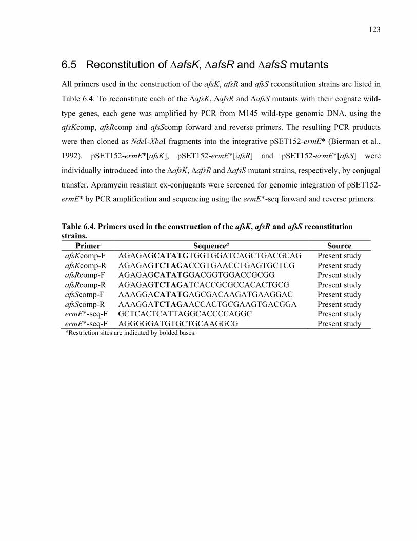
123
6.5 Reconstitution of DafsK, DafsR and DafsS mutants
All primers used in the construction of the afsK, afsR and afsS reconstitution strains are listed in
Table 6.4. To reconstitute each of the DafsK, DafsR and DafsS mutants with their cognate wild-
type genes, each gene was amplified by PCR from M145 wild-type genomic DNA, using the
afsKcomp, afsRcomp and afsScomp forward and reverse primers. The resulting PCR products
were then cloned as NdeI-XbaI fragments into the integrative pSET152-ermE* (Bierman et al.,
1992). pSET152-ermE*[afsK], pSET152-ermE*[afsR] and pSET152-ermE*[afsS] were
individually introduced into the DafsK, DafsR and DafsS mutant strains, respectively, by conjugal
transfer. Apramycin resistant ex-conjugants were screened for genomic integration of pSET152-
ermE* by PCR amplification and sequencing using the ermE*-seq forward and reverse primers.
Table 6.4. Primers used in the construction of the afsK, afsR and afsS reconstitution strains.
Primer Sequencea Source afsKcomp-F AGAGAGCATATGTGGTGGATCAGCTGACGCAG Present study afsKcomp-R AGAGAGTCTAGACCGTGAACCTGAGTGCTCG Present study afsRcomp-F AGAGAGCATATGGACGGTGGACCGCGG Present study afsRcomp-R AGAGAGTCTAGATCACCGCGCCACACTGCG Present study afsScomp-F AAAGGACATATGAGCGACAAGATGAAGGAC Present study afsScomp-R AAAGGATCTAGAACCACTGCGAAGTGACGGA Present study ermE*-seq-F GCTCACTCATTAGGCACCCCAGGC Present study ermE*-seq-F AGGGGGATGTGCTGCAAGGCG Present study aRestriction sites are indicated by bolded bases.

124
6.6 Sequence analysis of AfsS and AfsS-like proteins
Amino acid sequences of AfsS and 14 other AfsS-like proteins were retrieved from a NCBI
BLAST search. All 15 sequences were aligned and analyzed using MAFFT v7.221.3 (Kazutaka
Katoh & Standley, 2013) and visualized using Jalview v2.10.5 (Waterhouse et al., 2009). The
amino acid sequence of AfsS was analyzed using Phyre2 v2.0, a web-based server encompassing
a suite of bioinformatics tools to predict and analyze the structure, function and mutations of
proteins (Kelley et al., 2015).
6.7 Generation of AfsS point mutations
Single point mutations in each of the highly conserved D, N and M residues in each internal
sequence repeat of AfsS were synthesized by GenScript (Piscataway, New Jersey, USA). Triple
point mutations in the conserved D residues of all three repeats, the conserved N residues of all
three repeats, and the conserved M residues of all three repeats were also synthesized by GenScript
(Piscataway, New Jersey, USA). The nucleotide substitution(s) and the resulting codon and amino
acid changes are listed in Table 6.5. Each mutated AfsS construct was cloned as a NdeI-XbaI
fragment into pSET152-ermE* (Bierman et al., 1992). pSET152-ermE*[afsSD14A], pSET152-
ermE*[afsSN15A], pSET152-ermE*[afsSM17A], pSET152-ermE*[afsSD31A], pSET152-
ermE*[afsSN32A], pSET152-ermE*[afsSM34A], pSET152-ermE*[afsSD47A], pSET152-
ermE*[afsSN48A], pSET152-ermE*[afsSM50A], pSET152-ermE*[afsS3xDA], pSET152-
ermE*[afsS3xD=NA], and pSET152-ermE*[afsS3xMA] were individually introduced into the DafsS
mutant strain by conjugal transfer. Apramycin resistant ex-conjugants were screened for genomic
integration of pSET152-ermE* by PCR amplification and sequencing using the ermE*SC forward
and reverse primers (refer to Table 6.4).

125
Table 6.5. Nucleotide substitution(s) in afsS and the resulting amino acid substitutions.
Nucleotide Substitution(s) Codon Change Amino Acid Substitution
41A>C GAC à GCC D14A 43A>G, 44A>C AAC à GCC N15A 49A>G, 51T>C ATG à GCG M17A 92A>C GAC à GCC D31A 94A>G, 95A>C AAC à GCC N32A 100A>G, 101T>C ATG à GCG M34A 140A>C GAC à GCC D47A 142A>G, 143A>C AAC à GCC N48A 148A>G, 149T>C ATG à GCG M50A 41A>C, 92A>C, 140A>C GAC à GCC 3xDA 43A>G, 44A>C, 100A>G, 101T>C, 142A>G, 143A>C AAC à GCC 3xNA 49A>G, 51T>C, 100A>G, 101T>C, 148A>G, 149T>C ATG à GCG 3xMA
6.8 Chemical elicitation and quantification of actinorhodin
Three biological replicates of each of S. coelicolor M145 wild-type, mutant, and reconstitution
strains (5-mL cultures) were grown for 8 h in R5MX liquid medium at 30°C and 200 rpm. After 8
h of initial growth, the cultures were induced with either 50 µM ARC2 or the control solvent
DMSO. After 2 days of induction, actinorhodin production was visually assessed and quantified
spectrophotometrically. One milliliter of each culture was assayed by adding 5 M KOH to a final
concentration of 1 M. Each sample was vortexed, and then centrifuged at 15800 ´g for 1 min to
separate the cell pellet from the soluble compounds. The supernatant was sampled for total
actinorhodin by measuring the absorbance at 640 nm using the BioTek Epoch microplate
spectrophotometer.
6.9 Yeast two-hybrid screen for AfsS binding partners
All primers used in the Y2H screen with the AfsS bait protein are listed in Table 6.6. The system
used for the Y2H screen was the Matchmaker Gold Yeast Two-Hybrid System by TakaraBio USA
Inc. (Mountain View, California, USA). To construct the AfsS bait plasmid, afsS was amplified
by PCR from M145 wild-type genomic DNA using the afsS-Y2H forward and reverse primers.
The resulting PCR product was then cloned as a NdeI-BamHI fragment into pGBKT7, which
contains the GAL4 DNA-binding domain, a kanamycin resistance marker for selection in E. coli
and a tryptophan prototrophy marker for selection in S. cerevisiae. pGBKT7-afsS was then

126
transformed into the S. cerevisiae strain Y2HGold and selected for colonies on SD agar
medium/-Trp. As per the manufacturer’s instructions (TakaraBio USA Inc.), the two-hybrid
screen was performed via mating of Y2HGold[afsS] with the S. cerevisiae strain Y187 hosting the
S. coelicolor library. Provided by the Elliot Lab at McMaster University (Hamilton, Ontario,
Canada), the S. coelicolor library was fused to the GAL4 activation domain on pGADT7, which
contains an ampicillin resistance marker for selection in E. coli and a leucine prototrophy marker
for selection in S. cerevisiae. The mated culture was plated and grown for 7 days at 30°C on agar
SD medium/-Trp-Leu-His-Ade+AbA+kan+EZ-gal, which is selective for interactions based on
Gal4-driven transcription of the reporter genes for histidine protorophy (HIS3), adenine protrophy
(ADE2), aureobasidian A resistance (AUR1-C) and a-galactosidase activity (MEL1). Colonies
with putative interactions were then grown in liquid SD medium/-Leu for 7 days at 30°C and 200
rpm to select for pGADT7 containing the putative prey interactor. The prey plasmid was purified
using the QIAprepÒ Spin Miniprep Kit (QIAGEN) as per the manufacturer’s instructions for the
yeast plasmid isolation protocol. The putative prey interactor was then identified by sequencing
using the T7-F and pGAD-seq-R primers.
Table 6.6. Primers used in the Y2H screen with the AfsS bait protein.
Primer Sequencea Source afsS-Y2H-F AGAGAGCATATGAGCGACAAGATGAAGGA Present study afsS-Y2H-R AGAGAGGGATCCGGTCTACTTGCCGTCG Present study T7-F TAATACGACTCACTATAGGG Universal primer T7-R CAAGGGGTTATGCTAGTTAT Universal primer pGAD-seq-R AGATGGTGCACGATGCACAG Present study aRestriction sites are indicated by bolded bases.

127
References
Ahmed, S., Craney, A., Pimentel-Elardo, S. M., & Nodwell, J. R. (2013). A synthetic, species-specific activator of secondary metabolism and sporulation in Streptomyces coelicolor. ChemBioChem. https://doi.org/10.1002/cbic.201200619
Aínsa, J. A., Parry, H. D., & Chater, K. F. (1999). A response regulator-like protein that functions at an intermediate stage of sporulation in Streptomyces coelicolor A3(2). Molecular Microbiology, 34(3), 607–619. https://doi.org/10.1046/j.1365-2958.1999.01630.x
Aínsa, J. A., Ryding, N. J., Hartley, N., Findlay, K. C., Bruton, C. J., & Chater, K. F. (2000). WhiA, a protein of unknown function conserved among gram-positive bacteria, is essential for sporulation in Streptomyces coelicolor A3(2). Journal of Bacteriology, 182(19), 5470–5478. https://doi.org/10.1128/JB.182.19.5470-5478.2000
Anders, S., Pyl, P. T., & Huber, W. (2015). HTSeq-A Python framework to work with high-throughput sequencing data. Bioinformatics. https://doi.org/10.1093/bioinformatics/btu638
Aparicio, J. F., Molnár, I., Schwecke, T., König, A., Haydock, S. F., Khaw, L. E., Staunton, J., & Leadlay, P. F. (1996). Organization of the biosynthetic gene cluster for rapamycin in Streptomyces hygroscopicus: Analysis of the enzymatic domains in the modular polyketide synthase. Gene, 169, 9–16. https://doi.org/10.1016/0378-1119(95)00800-4
Apel, A. K., Sola-Landa, A., Rodríguez-García, A., & Martín, J. F. (2007). Phosphate control of phoA, phoC and phoD gene expression in Streptomyces coelicolor reveals significant differences in binding of PhoP to their promoter regions. Microbiology, 153(10), 3527–3537. https://doi.org/10.1099/mic.0.2007/007070-0
Arias, P., Fernández-Moreno, M. A., & Malpartida, F. (1999). Characterization of the pathway-specific positive transcriptional regulator for actinorhodin biosynthesis in Streptomyces coelicolor A3(2) as a DNA-binding protein. Journal of Bacteriology, 181(22), 6958–6968. https://doi.org/10.1128/jb.181.22.6958-6968.1999
Austin, M. B., Izumikawa, M., Bowman, M. E., Udwary, D. W., Ferrer, J. L., Moore, B. S., & Noel, J. P. (2004). Crystal structure of a bacterial type III polyketide synthase and enzymatic control of reactive polyketide intermediates. Journal of Biological Chemistry, 279(43), 45162–45174. https://doi.org/10.1074/jbc.M406567200
Bao, K., & Cohen, S. N. (2001). Terminal proteins essential for the replication of linear plasmids and chromosomes in Streptomyces. Genes and Development. https://doi.org/10.1101/gad.896201
Barona-Gómez, F., Wong, U., Giannakopulos, A. E., Derrick, P. J., & Challis, G. L. (2004). Identification of a cluster of genes that directs desferrioxamine biosynthesis in Streptomyces coelicolor M145. Journal of the American Chemical Society, 126(50), 16282–16283. https://doi.org/10.1021/ja045774k
128
Basile, W., Salvatore, M., Bassot, C., & Elofsson, A. (2019). Why do eukaryotic proteins contain more intrinsically disordered regions? PLoS Computational Biology, 15(7), 1–20. https://doi.org/10.1371/journal.pcbi.1007186
Beltran-Alvarez, P., Cox, R. J., Crosby, J., & Simpson, T. J. (2007). Dissecting the component reactions catalyzed by the actinorhodin minimal polyketide synthase. Biochemistry, 46(50), 14672–14681. https://doi.org/10.1021/bi701784c
Bentley, S. D., Chater, K. F., Cerdeño-Tárraga, A. M., Challis, G. L., Thomson, N. R., James, K. D., Harris, D. E., Quail, M. A., Kieser, H., Harper, D., Bateman, A., Brown, S., Chandra, G., Chen, C. W., Collins, M., Cronin, A., Fraser, A., Goble, A., Hidalgo, J., … Hopwood, D. A. (2002). Complete genome sequence of the model actinomycete Streptomyces coelicolor A3(2). Nature. https://doi.org/10.1038/417141a
Bérdy, J. (2005). Bioactive Microbial Metabolites. The Journal of Antibiotics. https://doi.org/10.1038/ja.2005.1
Bibb, Maureen J., Molle, V., & Buttner, M. J. (2000). σ(BldN), an extracytoplasmic function RNA polymerase sigma factor required for aerial mycelium formation in Streptomyces coelicolor A3(2). Journal of Bacteriology, 182(16), 4606–4616. https://doi.org/10.1128/JB.182.16.4606-4616.2000
Bibb, Mervyn J. (2005). Regulation of secondary metabolism in streptomycetes. In Current Opinion in Microbiology. https://doi.org/10.1016/j.mib.2005.02.016
Bierman, M., Logan, R., O’Brien, K., Seno, E. T., Nagaraja Rao, R., & Schoner, B. E. (1992). Plasmid cloning vectors for the conjugal transfer of DNA from Escherichia coli to Streptomyces spp. Gene. https://doi.org/10.1016/0378-1119(92)90627-2
Bignell, D. R. D., Warawa, J. L., Strap, J. L., Chater, K. F., & Leskiw, B. K. (2000). Study of the bldG locus suggests that an anti-anti-sigma factor and an anti-sigma factor may be involved in Streptomyces coelicolor antibiotic production and sporulation. Microbiology, 146(9), 2161–2173. https://doi.org/10.1099/00221287-146-9-2161
Bode, H. B., Bethe, B., Höfs, R., & Zeeck, A. (2002). Big effects from small changes: Possible ways to explore nature’s chemical diversity. ChemBioChem, 3(7), 619–627. https://doi.org/10.1002/1439-7633(20020703)3:7<619::AID-CBIC619>3.0.CO;2-9
Bolger, A. M., Lohse, M., & Usadel, B. (2014). Trimmomatic: A flexible trimmer for Illumina sequence data. Bioinformatics. https://doi.org/10.1093/bioinformatics/btu170
Brana, A. F., Wolfe, S., & Demain, A. L. (1985). Ammonium repression of cephalosporin production by Streptomyces clavuligerus. Canadian Journal of Microbiology, 31(8), 736–743. https://doi.org/10.1139/m85-138
Brown, E. D., & Wright, G. D. (2016). Antibacterial drug discovery in the resistance era. In Nature. https://doi.org/10.1038/nature17042

129
Brückner, A., Polge, C., Lentze, N., Auerbach, D., & Schlattner, U. (2009). Yeast two-hybrid, a powerful tool for systems biology. International Journal of Molecular Sciences, 10(6), 2763–2788. https://doi.org/10.3390/ijms10062763
Bursy, J., Kuhlmann, A. U., Pittelkow, M., Hartmann, H., Jebbar, M., Pierik, A. J., & Bremer, E. (2008). Synthesis and uptake of the compatible solutes ectoine and 5-hydroxyectoine by Streptomyces coelicolor A3(2) in response to salt and heat stresses. Applied and Environmental Microbiology, 74(23), 7286–7296. https://doi.org/10.1128/AEM.00768-08
Cane, D. E., & Watt, R. M. (2003). Expression and mechanistic analysis of a germacradienol synthase from Streptomyces coelicolor implicated in geosmin biosynthesis. Proceedings of the National Academy of Sciences of the United States of America, 100(4), 1547–1551. https://doi.org/10.1073/pnas.0337625100
Capstick, D. S., Willey, J. M., Buttner, M. J., & Elliot, M. A. (2007). SapB and the chaplins: Connections between morphogenetic proteins in Streptomyces coelicolor. Molecular Microbiology, 64(3), 602–613. https://doi.org/10.1111/j.1365-2958.2007.05674.x
Cerdeño, A. M., Bibb, M. J., & Challis, G. L. (2001). Analysis of the prodiginine biosynthesis gene cluster of Streptomyces coelicolor A3(2): New mechanisms for chain initiation and termination in modular multienzymes. Chemistry and Biology, 8(8), 817–829. https://doi.org/10.1016/S1074-5521(01)00054-0
Chabala, J. C., Mrozik, H., Tolman, R. L., Eskola, P., Lusi, A., Peterson, L. H., Woods, M. F., Fisher, M. H., Campbell, W. C., Egerton, J. R., & Ostlind, D. A. (1980). Ivermectin, a New Broad-Spectrum Antiparasitic Agent. Journal of Medicinal Chemistry, 23(10), 1134–1136. https://doi.org/10.1021/jm00184a014
Chang, H. M., Chen, M. Y., Chen, C. W., Shieh, Y. T., & Bibb, M. J. (1996). The cutRS signal transduction system of Streptomyces lividans represses the biosynthesis of the polyketide antibiotic actinorhodin. Molecular Microbiology, 21(5), 1075–1085.
Chater, K. F., & Wilde, L. C. (1980). Streptomyces albus G mutants defective in the SalGI restriction-modification system. Journal of General Microbiology, 116(2), 323–334. https://doi.org/10.1099/00221287-116-2-323
Chater, Keith F. (2001). Regulation of sporulation in Streptomyces coelicolor A3(2): A checkpoint multiplex? Current Opinion in Microbiology, 4, 667–673. https://doi.org/10.1016/S1369-5274(01)00267-3
Claessen, D., de Jong, W., Dijkhuizen, L., & Wösten, H. A. B. (2006). Regulation of Streptomyces development: reach for the sky! Trends in Microbiology, 14(7), 313–319. https://doi.org/10.1016/j.tim.2006.05.008
Claessen, D., Rink, R., De Jong, W., Siebring, J., De Vreugd, P., Hidde Boersma, F. G., Dijkhuizen, L., & Wösten, H. A. B. (2003). A novel class of secreted hydrophobic proteins is involved in aerial hyphae formation in Streptomyces coelicolor by forming amyloid-like fibrils. Genes and Development, 17(14), 1714–1726. https://doi.org/10.1101/gad.264303
130
Claessen, D., Stokroos, L., Deelstra, H. J., Penninga, N. A., Bormann, C., Salas, J. A., Dijkhuizen, L., & Wösten, H. A. B. (2004). The formation of the rodlet layer of streptomycetes is the result of the interplay between rodlins and chaplins. Molecular Microbiology, 53(2), 433–443. https://doi.org/10.1111/j.1365-2958.2004.04143.x
Claessen, D., Wösten, H. A. B., van Keulen, G., Faber, O. G., Alves, A. M. C. R., Meijer, W. G., & Dijkhuizen, L. (2002). Two novel homologous proteins of Streptomyces coelicolor and Streptomyces lividans are involved in the formation of the rodlet layer and mediate attachment to a hydrophobic surface. Molecular Microbiology, 44(6), 1483–1492. https://doi.org/10.1046/j.1365-2958.2002.02980.x
Cobb, R. E., Wang, Y., & Zhao, H. (2015). High-Efficiency Multiplex Genome Editing of Streptomyces Species Using an Engineered CRISPR/Cas System. ACS Synthetic Biology. https://doi.org/10.1021/sb500351f
Corre, C., Song, L., O’Rourke, S., Chater, K. F., & Challis, G. L. (2008). 2-Alkyl-4-hydroxymethylfuran-3-carboxylic acids, antibiotic production inducers discovered by Streptomyces coelicolor genome mining. Proceedings of the National Academy of Sciences of the United States of America, 105(45), 17510–17515. https://doi.org/10.1073/pnas.0805530105
Craney, A., Ahmed, S., & Nodwell, J. R. (2013). Towards a new science of secondary metabolism. In Journal of Antibiotics. https://doi.org/10.1038/ja.2013.25
Craney, A., Hohenauer, T., Xu, Y., Navani, N. K., Li, Y., & Nodwell, J. R. (2007). A synthetic luxCDABE gene cluster optimized for expression in high-GC bacteria. Nucleic Acids Research. https://doi.org/10.1093/nar/gkm086
Craney, A., Ozimok, C., Pimentel-Elardo, S. M., Capretta, A., & Nodwell, J. R. (2012). Chemical perturbation of secondary metabolism demonstrates important links to primary metabolism. Chemistry and Biology, 19(8), 1020–1027. https://doi.org/10.1016/j.chembiol.2012.06.013
Cronan, J. E., & Thomas, J. (2009). Bacterial fatty acid synthesis and its relationships with polyketide synthetic pathways. Methods in Enzymology, 459, 395–433. https://doi.org/10.1016/S0076-6879(09)04617-5.Bacterial
Cruz-Morales, P., Kopp, J. F., Martínez-Guerrero, C., Yáñez-Guerra, L. A., Selem-Mojica, N., Ramos-Aboites, H., Feldmann, J., & Barona-Gómez, F. (2016). Phylogenomic analysis of natural products biosynthetic gene clusters allows discovery of arseno-organic metabolites in model streptomycetes. Genome Biology and Evolution, 8(6), 1906–1916. https://doi.org/10.1093/gbe/evw125
Daniel-Ivad, M., Hameed, N., Tan, S., Dhanjal, R., Socko, D., Pak, P., Gverzdys, T., Elliot, M. A., & Nodwell, J. R. (2017). An engineered allele of afsQ1 facilitates the discovery and investigation of cryptic natural products. ACS Chemical Biology, 12(3), 628–634. https://doi.org/10.1021/acschembio.6b01002

131
Daniel-Ivad, M., Pimentel-Elardo, S. M., & Nodwell, J. R. (2018). Control of specialized metabolism by signaling and transcriptional regulation: Opportunities for new platforms for drug discovery? Annual Review of Microbiology. https://doi.org/10.1146/annurev-micro-022618-042458
Darken, M. A., Berenson, H., Shirk, R. J., & Sjolander, N. O. (1960). Production of tetracycline by Streptomyces aureofaciens in synthetic media. Applied Microbiology, 8(1), 46–51. https://doi.org/10.1128/aem.8.1.46-51.1960
Davies, J., & Ryan, K. S. (2012). Introducing the parvome: Bioactive compounds in the microbial world. ACS Chemical Biology, 7(2), 252–259. https://doi.org/10.1021/cb200337h
Davis, N. K., & Chater, K. F. (1990). Spore colour in Streptomyces coelicolor A3(2) involves the developmentally regulated synthesis of a compound biosynthetically related to polyketide antibiotics. Molecular Microbiology, 4(10), 1679–1691. https://doi.org/10.1111/j.1365-2958.1990.tb00545.x
Davis, N. K., & Chater, K. F. (1992). The Streptomyces coelicolor whiB gene encodes a small transcription factor-like protein dispensable for growth but essential for sporulation. MGG Molecular & General Genetics, 232(3), 351–358. https://doi.org/10.1007/BF00266237
de Lima Procópio, R. E., da Silva, I. R., Martins, M. K., de Azevedo, J. L., & de Araújo, J. M. (2012). Antibiotics produced by Streptomyces. Brazilian Journal of Infectious Diseases. https://doi.org/10.1016/j.bjid.2012.08.014
Díaz-Pérez, A. L., Díaz-Pérez, C., & Campos-García, J. (2016). Bacterial l-leucine catabolism as a source of secondary metabolites. Reviews in Environmental Science and Biotechnology, 15(1), 1–29. https://doi.org/10.1007/s11157-015-9385-3
Doull, J. L., Ayer, S. W., Thibault, P., & Singh, A. K. (1993). Production of a novel polyketide antibiotic, jadomycin b, by Streptomyces venezuelae following heat shock. Journal of Antibiotics, 46(5), 869–871. https://doi.org/10.7164/antibiotics.46.869
Doull, J. L., Singh, A. K., Hoare, M., & Ayer, S. W. (1994). Conditions for the production of jadomycin B by Streptomyces venezuelae ISP5230: Effects of heat shock, ethanol treatment and phage infection. Journal of Industrial Microbiology, 13(2), 120–125. https://doi.org/10.1007/BF01584109
Dyson, H. J., & Wright, P. E. (2002). Coupling of folding and binding for unstructured proteins. Current Opinion in Structural Biology, 12(1), 54–60. https://doi.org/10.1016/S0959-440X(02)00289-0
Dyson, H. J., & Wright, P. E. (2005). Intrinsically unstructured proteins and their functions. Nature Reviews Molecular Cell Biology, 6(3), 197–208. https://doi.org/10.1038/nrm1589
Eccleston, M., Ali, R. A., Seyler, R., Westpheling, J., & Nodwell, J. R. (2002). Structural and genetic analysis of the BldB protein of Streptomyces coelicolor. Journal of Bacteriology, 184(15), 4270–4276. https://doi.org/10.1128/JB.184.15.4270-4276.2002

132
Einhauer, A., & Jungbauer, A. (2001). The FLAGTM peptide, a versatile fusion tag for the purification of recombinant proteins. Journal of Biochemical and Biophysical Methods, 49(1–3), 455–465. https://doi.org/10.1016/S0165-022X(01)00213-5
Eliezer, D. (2009). Biophysical characterization of intrinsically disordered proteins. Current Opinion in Structural Biology, 19(1), 23–30. https://doi.org/10.1016/j.sbi.2008.12.004
Elliot, M. A., Bibb, M. J., Buttner, M. J., & Leskiw, B. K. (2001). BldD is a direct regulator of key developmental genes in Streptomyces coelicolor A3(2). Molecular Microbiology, 40(1), 257–269. https://doi.org/10.1046/j.1365-2958.2001.02387.x
Elliot, M. A., Buttner, M. J., & Nodwell, J. R. (2008). Multicellular Development in Streptomyces. In D. E. Whitworth (Ed.), Myxobacteria: Multicellularity and Differentiation (pp. 419–439). ASM Press. https://doi.org/10.1128/9781555815677.ch24
Elliot, M. A., Karoonuthaisiri, N., Huang, J., Bibb, M. J., Cohen, S. N., Kao, C. M., & Buttner, M. J. (2003). The chaplins: A family of hydrophobic cell-surface proteins involved in aerial mycelium formation in Streptomyces coelicolor. Genes and Development, 17(14), 1727–1740. https://doi.org/10.1101/gad.264403
Elliot, M. A., & Leskiw, B. K. (1999). The BldD protein from Streptomyces coelicolor is a DNA-binding protein. Journal of Bacteriology, 181(21), 6832–6835. https://doi.org/10.1128/jb.181.21.6832-6835.1999
Feitelson, J. S., Malpartida, F., & Hopwood, D. A. (1985). Genetic and biochemical characterization of the red gene cluster of Streptomyces coelicolor A3(2). Journal of General Microbiology, 131(9), 2431–2441. https://doi.org/10.1099/00221287-131-9-2431
Feitelson, Jerald S., & Hopwood, D. A. (1983). Cloning of a Streptomyces gene for an O-methyltransferase involved in antibiotic biosynthesis. MGG Molecular & General Genetics. https://doi.org/10.1007/BF00331065
Fernandez-Moreno, M. A., Martin-Triana, A. J., Martinez, E., Niemi, J., Kieser, H. M., Hopwood, D. A., & Malpartida, F. (1992). abaA, a new pleiotropic regulatory locus for antibiotic production in Streptomyces coelicolor. Journal of Bacteriology, 174(9), 2958–2967. https://doi.org/10.1128/jb.174.9.2958-2967.1992
Flärdh, K. (2003). Essential role of DivlVA in polar growth and morphogenesis in Streptomyces coelicolor A3(2). Molecular Microbiology, 49(6), 1523–1536. https://doi.org/10.1046/j.1365-2958.2003.03660.x
Flärdh, K., & Buttner, M. J. (2009). Streptomyces morphogenetics: Dissecting differentiation in a filamentous bacterium. Nature Reviews Microbiology, 7(1), 36–49. https://doi.org/10.1038/nrmicro1968
Floriano, B., & Bibb, M. J. (1996). afsR is a pleiotropic but conditionally required regulatory gene for antibiotic production in Streptomyces coelicolor A3(2). Molecular Microbiology. https://doi.org/10.1046/j.1365-2958.1996.6491364.x

133
Foley, T. L., Young, B. S., & Burkart, M. D. (2009). Phosphopantetheinyl transferase inhibition and secondary metabolism. FEBS Journal, 276(23), 7134–7145. https://doi.org/10.1111/j.1742-4658.2009.07425.x
Frank, J. (2002). Single-particle imaging of macromolecules by cryo-electron microscopy. Annual Review of Biophysics and Biomolecular Structure, 31, 303–319. https://doi.org/10.1146/annurev.biophys.31.082901.134202
Fujita, Y., Matsuoka, H., & Hirooka, K. (2007). Regulation of fatty acid metabolism in bacteria. In Molecular Microbiology. https://doi.org/10.1111/j.1365-2958.2007.05947.x
Gehring, A. M., Yoo, N. J., & Losick, R. (2001). RNA polymerase sigma factor that blocks morphological differentiation by Streptomyces coelicolor. Journal of Bacteriology. https://doi.org/10.1128/JB.183.20.5991-5996.2001
Gill, C. O., & Suisted, J. R. (1978). The effects of temperature and growth rate on the proportion of unsaturated fatty acids in bacterial lipids. Journal of General Microbiology, 104(1), 31–36. https://doi.org/10.1099/00221287-104-1-31
Gomez-Escribano, J. P., & Bibb, M. J. (2011). Engineering Streptomyces coelicolor for heterologous expression of secondary metabolite gene clusters. Microbial Biotechnology, 4(2), 207–215. https://doi.org/10.1111/j.1751-7915.2010.00219.x
Gomez-Escribano, J. P., Song, L., Fox, D. J., Yeo, V., Bibb, M. J., & Challis, G. L. (2012). Structure and biosynthesis of the unusual polyketide alkaloid coelimycin P1, a metabolic product of the cpk gene cluster of Streptomyces coelicolor M145. Chemical Science, 3(9), 2716–2720. https://doi.org/10.1039/c2sc20410j
Guo, J., Zhao, J., Li, L., Chen, Z., Wen, Y., & Li, J. (2010). The pathway-specific regulator AveR from Streptomyces avermitilis positively regulates avermectin production while it negatively affects oligomycin biosynthesis. Molecular Genetics and Genomics, 283(2), 123–133. https://doi.org/10.1007/s00438-009-0502-2
Hadfield, A. T., Limpkin, C., Teartasin, W., Simpson, T. J., Crosby, J., & Crump, M. P. (2004). The crystal structure of the actIII actinorhodin polyketide reductase: Proposed mechanism for ACP and polyketide binding. Structure, 12(10), 1865–1875. https://doi.org/10.1016/j.str.2004.08.002
Han, S. B., Kim, H. M., Kim, Y. H., Lee, C. W., Jang, E. S., Son, K. H., Kim, S. U., & Kim, Y. K. (1998). T-cell specific immunosuppression by prodigiosin isolated from Serratia marcescens. International Journal of Immunopharmacology, 20(1–3), 1–13. https://doi.org/10.1016/S0192-0561(97)00062-3
Hayes, A., Hobbs, G., Smith, C. P., Oliver, S. G., & Butler, P. R. (1997). Environmental signals triggering methylenomycin production by Streptomyces coelicolor A3(2). Journal of Bacteriology, 179(17), 5511–5515.

134
Heath, R. J., & Rock, C. O. (1995). Enoyl-acyl carrier protein reductase (fabI) plays a determinant role in completing cycles of fatty acid elongation in Escherichia coli. Journal of Biological Chemistry, 270(44), 26538–26542. https://doi.org/10.1074/jbc.270.44.26538
Hempel, A. M., Cantlay, S., Molle, V., Wang, S. B., Naldrett, M. J., Parker, J. L., Richards, D. M., Jung, Y. G., Buttner, M. J., & Flärdh, K. (2012). The ser/thr protein kinase AfsK regulates polar growth and hyphal branching in the filamentous bacteria Streptomyces. Proceedings of the National Academy of Sciences of the United States of America. https://doi.org/10.1073/pnas.1207409109
Hempel, A., Wang, S., Letek, M., Gil, J., & Flärdh, K. (2008). Assemblies of DivIVA mark sites for hyphal branching and can establish new zones of cell wall growth in Streptomyces coelicolor. Journal of Bacteriology, 190(22), 7579–7583. https://doi.org/10.1128/JB.00839-08
Hesketh, A., Kock, H., Mootien, S., & Bibb, M. (2009). The role of absC, a novel regulatory gene for secondary metabolism, in zinc-dependent antibiotic production in Streptomyces coelicolor A3(2). Molecular Microbiology, 74(6), 1427–1444. https://doi.org/10.1111/j.1365-2958.2009.06941.x
Hobbs, G., Frazer, C. M., Gardner, D. C. J., Flett, F., & Oliver, S. G. (1990). Pigmented antibiotic production by Streptomyces coelicolor A3(2): Kinetics and the influence of nutrients. Journal of General Microbiology. https://doi.org/10.1099/00221287-136-11-2291
Hodgson, D. A. (2000). Primary metabolism and its control in streptomycetes: A most unusual group of bacteria. Advances in Microbial Physiology, 42, 47–238. https://doi.org/10.1016/s0065-2911(00)42003-5
Hojati, Z., Milne, C., Harvey, B., Gordon, L., Borg, M., Flett, F., Wilkinson, B., Sidebottom, P. J., Rudd, B. A. M., Hayes, M. A., Smith, C. P., & Micklefield, J. (2002). Structure, biosynthetic origin, and engineered biosynthesis of calcium-dependent antibiotics from Streptomyces coelicolor. Chemistry and Biology, 9(11), 1175–1187. https://doi.org/10.1016/S1074-5521(02)00252-1
Hong, S. K., Kito, M., Beppu, T., & Horinouchi, S. (1991). Phosphorylation of the AfsR product, a global regulatory protein for secondary-metabolite formation in Streptomyces coelicolor A3(2). Journal of Bacteriology. https://doi.org/10.1128/jb.173.7.2311-2318.1991
Hopwood, D. A., Kieser, T., Wright, H. M., & Bibb, M. J. (1983). Plasmids, recombination and chromosome mapping in Streptomyces lividans 66. Journal of General Microbiology, 129, 2257–2269. https://doi.org/10.1099/00221287-129-7-2257
Hopwood, David A. (2006). Soil To Genomics: The Streptomyces Chromosome. Annual Review of Genetics. https://doi.org/10.1146/annurev.genet.40.110405.090639
Horinouchi, S. (2003). AfsR as an integrator of signals that are sensed by multiple serine/threonine kinases in Streptomyces coelicolor A3(2). Journal of Industrial Microbiology and Biotechnology. https://doi.org/10.1007/s10295-003-0063-z

135
Horinouchi, S., Kito, M., Nishiyama, M., Furuya, K., Hong, S. K., Miyake, K., & Beppu, T. (1990). Primary structure of AfsR, a global regulatory protein for secondary metabolite formation in Streptomyces coelicolor A3(2). Gene, 95, 49–56. https://doi.org/10.1016/0378-1119(90)90412-K
Hou, B., Lin, Y., Wu, H., Guo, M., Petkovic, H., Tao, L., Zhu, X., Ye, J., & Zhang, H. (2018). The novel transcriptional regulator LmbU promotes lincomycin biosynthesis through regulating expression of its target genes in Streptomyces lincolnensis. Journal of Bacteriology, 200(2), 1–17. https://doi.org/10.1128/JB.00447-17
Huang, J., Shi, J., Molle, V., Sohlberg, B., Weaver, D., Bibb, M. J., Karoonuthaisiri, N., Lih, C. J., Kao, C. M., Buttner, M. J., & Cohen, S. N. (2005). Cross-regulation among disparate antibiotic biosynthetic pathways of Streptomyces coelicolor. Molecular Microbiology, 58(5), 1276–1287. https://doi.org/10.1111/j.1365-2958.2005.04879.x
Huerta-Cepas, J., Szklarczyk, D., Forslund, K., Cook, H., Heller, D., Walter, M. C., Rattei, T., Mende, D. R., Sunagawa, S., Kuhn, M., Jensen, L. J., Von Mering, C., & Bork, P. (2016). EGGNOG 4.5: A hierarchical orthology framework with improved functional annotations for eukaryotic, prokaryotic and viral sequences. Nucleic Acids Research. https://doi.org/10.1093/nar/gkv1248
Hunt, A. C., Servín-González, L., Kelemen, G. H., & Buttner, M. J. (2005). The bldC developmental locus of Streptomyces coelicolor encodes a member of a family of small DNA-binding proteins related to the DNA-binding domains of the MerR family. Journal of Bacteriology, 187(2), 716–728. https://doi.org/10.1128/JB.187.2.716-728.2005
Hutchings, M. I., Hoskisson, P. A., Chandra, G., & Buttner, M. J. (2004). Sensing and responding to diverse extracellular signals? Analysis of the sensor kinases and response regulators of Streptomyces coelicolor A3(2). Microbiology, 150(9), 2795–2806. https://doi.org/10.1099/mic.0.27181-0
Ichinose, K., Surti, C., Taguchi, T., Malpartida, F., Booker-Milburn, K. I., Stephenson, G. R., Ebizuka, Y., & Hopwood, D. A. (1999). Proof that the actVI genetic region of Streptomyces coelicolor A3(2) is involved in stereospecific pyran ring formation in the biosynthesis of actinorhodin. Bioorganic and Medicinal Chemistry Letters, 9(3), 395–400. https://doi.org/10.1016/S0960-894X(99)00011-6
Ikeda, H., Ishikawa, J., Hanamoto, A., Shinose, M., Kikuchi, H., Shiba, T., Sakaki, Y., Hattori, M., & Omura, S. (2003). Complete genome sequence and comparative analysis of the industrial microorganism Streptomyces avermitilis. Nature Biotechnology, 21(5), 526–531. https://doi.org/10.1038/nbt820
Ikeda, H., Nonomiya, T., Usami, M., Ohta, T., & Omura, S. (1999). Organization of the biosynthetic gene cluster for the polyketide anthelmintic macrolide avermectin in Streptomyces avermitilis. Proceedings of the National Academy of Sciences of the United States of America, 96(17), 9509–9514. https://doi.org/10.1073/pnas.96.17.9509

136
Jakeman, D. L., Bandi, S., Graham, C. L., Reid, T. R., Wentzell, J. R., & Douglas, S. E. (2009). Antimicrobial activities of jadomycin B and structurally related analogues. Antimicrobial Agents and Chemotherapy, 53(3), 1245–1247. https://doi.org/10.1128/AAC.00801-08
Jakimowicz, D., Majka, J., Konopa, G., Wegrzyn, G., Messer, W., Schrempf, H., & Zakrzewska-Czerwińska, J. (2000). Architecture of the Streptomyces lividans DnaA protein-replication origin complexes. Journal of Molecular Biology, 298(3), 351–364. https://doi.org/10.1006/jmbi.2000.3686
Jakimowicz, P., Cheesman, M. R., Bishai, W. R., Chater, K. F., Thomson, A. J., & Buttner, M. J. (2005). Evidence that the Streptomyces developmental protein WhiD, a member of the WhiB family, binds a [4Fe-4S] cluster. Journal of Biological Chemistry, 280(9), 8309–8315. https://doi.org/10.1074/jbc.M412622200
Katoh, K. (2002). MAFFT: a novel method for rapid multiple sequence alignment based on fast Fourier transform. Nucleic Acids Research, 30(14), 3059–3066. https://doi.org/10.1093/nar/gkf436
Katoh, Kazutaka, & Standley, D. M. (2013). MAFFT multiple sequence alignment software version 7: Improvements in performance and usability. Molecular Biology and Evolution. https://doi.org/10.1093/molbev/mst010
Kelemen, G. H., Brian, P., Flärdh, K., Chamberlin, L., Chater, K. F., & Buttner, M. J. (1998). Developmental regulation of transcription of whiE, a locus specifying the polyketide spore pigment in Streptomyces coelicolor A3(2). Journal of Bacteriology, 180(9), 2515–2521. https://doi.org/10.1128/jb.180.9.2515-2521.1998
Kelemen, G. H., Brown, G. L., Kormanec, J., Potúčková, L., Chater, K. F., & Buttner, M. J. (1996). The positions of the sigma-factor genes, whiG and sigF, in the hierarchy controlling the development of spore chains in the aerial hyphae of Streptomyces coelicolor A3(2). Molecular Microbiology, 21(3), 593–603. https://doi.org/10.1111/j.1365-2958.1996.tb02567.x
Kelley, L. A., Mezulis, S., Yates, C. M., Wass, M. N., & Sternberg, M. J. E. (2015). The Phyre2 web portal for protein modeling, prediction and analysis. Nature Protocols. https://doi.org/10.1038/nprot.2015.053
Keweloh, H., & Heipieper, H. J. (1996). Trans unsaturated fatty acids in bacteria. Lipids, 31(2), 129–137. https://doi.org/10.1007/bf02522611
Kieser, T., Bibb, M. J., Buttner, M. J., & Chater, K. F. (2000). Practical Streptomyces Genetics. 2nd ed. In John Innes Foundation.
Kirby, K. S., Fox-Carter, E., & Guest, M. (1967). Isolation of deoxyribonucleic acid and ribosomal ribonucleic acid from bacteria. The Biochemical Journal, 104(1), 258–262. https://doi.org/10.1042/bj1040258

137
Kodani, S., Hudson, M. E., Durrant, M. C., Buttner, M. J., Nodwell, J. R., & Willey, J. M. (2004). The SapB morphogen is a lantibiotic-like peptide derived from the product of the developmental gene ramS in Streptomyces coelicolor. Proceedings of the National Academy of Sciences of the United States of America, 101(31), 11448–11453. https://doi.org/10.1073/pnas.0404220101
Komatsu, M., Komatsu, K., Koiwai, H., Yamada, Y., Kozone, I., Izumikawa, M., Hashimoto, J., Takagi, M., Omura, S., Shin-Ya, K., Cane, D. E., & Ikeda, H. (2013). Engineered Streptomyces avermitilis host for heterologous expression of biosynthetic gene cluster for secondary metabolites. ACS Synthetic Biology, 2(7), 384–396. https://doi.org/10.1021/sb3001003
Komatsu, M., Tsuda, M., Omura, S., Oikawa, H., & Ikeda, H. (2008). Identification and functional analysis of genes controlling biosynthesis of 2-methylisoborneol. Proceedings of the National Academy of Sciences of the United States of America, 105(21), 7422–7427. https://doi.org/10.1073/pnas.0802312105
Langmead, B., & Salzberg, S. L. (2012). Fast gapped-read alignment with Bowtie 2. Nature Methods. https://doi.org/10.1038/nmeth.1923
Laureti, L., Song, L., Huang, S., Corre, C., Leblond, P., Challis, G. L., & Aigle, B. (2011). Identification of a bioactive 51-membered macrolide complex by activation of a silent polyketide synthase in Streptomyces ambofaciens. Proceedings of the National Academy of Sciences of the United States of America, 108(15), 6258–6263. https://doi.org/10.1073/pnas.1019077108
Lautru, S., Deeth, R. J., Bailey, L. M., & Challis, G. L. (2005). Discovery of a new peptide natural product by Streptomyces coelicolor genome mining. Nature Chemical Biology, 1(5), 265–269. https://doi.org/10.1038/nchembio731
Lawlor, E. J., Baylis, H. A., & Chater, K. F. (1987). Pleiotropic morphological and antibiotic deficiencies result from mutations in a gene encoding a tRNA-like product in Streptomyces coelicolor A3(2). Genes & Development, 1(10), 1305–1310. https://doi.org/10.1101/gad.1.10.1305
Łebkowski, T., Wolański, M., Ołdziej, S., Flärdh, K., & Zakrzewska-Czerwińska, J. (2020). AfsK-mediated site-specific phosphorylation regulates DnaA initiator protein activity in Streptomyces coelicolor. Journal of Bacteriology, 202(3), e00597-19. https://doi.org/10.1128/JB.00597-19
Lebrihi, A., Lamsaif, D., Lefebvre, G., & Germain, P. (1992). Effect of ammonium ions on spiramycin biosynthesis in Streptomyces ambofaciens. Applied Microbiology, 37, 382–387.
Lee, N., Hwang, S., Lee, Y., Cho, S., Palsson, B., & Cho, B. K. (2019). Synthetic biology tools for novel secondary metabolite discovery in Streptomyces. Journal of Microbiology and Biotechnology, 29(5), 667–686. https://doi.org/10.4014/jmb.1904.04015

138
Lee, P. C., Umeyama, T., & Horinouchi, S. (2002). afsS is a target of AfsR, a transcriptional factor with ATPase activity that globally controls secondary metabolism in Streptomyces coelicolor A3(2). Molecular Microbiology. https://doi.org/10.1046/j.1365-2958.2002.02840.x
Leskiw, B. K., Lawlor, E. J., Fernandez-Abalos, J. M., & Chater, K. F. (1991). TTA codons in some genes prevent their expression in a class of developmental, antibiotic-negative, Streptomyces mutants. Proceedings of the National Academy of Sciences of the United States of America, 88(6), 2461–2465. https://doi.org/10.1073/pnas.88.6.2461
Leskiw, B. K., & Mah, R. (1995). The bldA-encoded tRNA is poorly expressed in the bldl mutant of Streptomyces coelicolor A3(2). Microbiology, 141(8), 1921–1926. https://doi.org/10.1099/13500872-141-8-1921
Li, L., Jiang, W., & Lu, Y. (2017). A novel twocomponent system, GluR-GluK, involved in glutamate sensing and uptake in Streptomyces coelicolor. Journal of Bacteriology. https://doi.org/10.1128/JB.00097-17
Li, Y., Chen, P., Chen, S., Wu, D., & Zheng, J. (2004). A pair of two-component regulatory genes ecrA1/A2 in S. coelicolor. Journal of Zhejiang University. Science, 5(2), 173–179. https://doi.org/10.1007/bf02840919
Lian, W., Jayapal, K. P., Charaniya, S., Mehra, S., Glod, F., Kyung, Y. S., Sherman, D. H., & Hu, W. S. (2008). Genome-wide transcriptome analysis reveals that a pleiotropic antibiotic regulator, AfsS, modulates nutritional stress response in Streptomyces coelicolorA3(2). BMC Genomics. https://doi.org/10.1186/1471-2164-9-56
Liao, Y., Wei, Z. H., Bai, L., Deng, Z., & Zhong, J. J. (2009). Effect of fermentation temperature on validamycin A production by Streptomyces hygroscopicus 5008. Journal of Biotechnology, 142(3–4), 271–274. https://doi.org/10.1016/j.jbiotec.2009.04.015
Lin, J., & Lai, E. (2017). Protein–protein interactions: co-immunoprecipitation. In: Journet L., Cascales E. (eds) Bacterial Protein Secretion Systems. In Methods in Molecular Biology (Vol. 1615, pp. 211–219). https://doi.org/10.1007/978-1-4939-7033-9
Liu, G., Chater, K. F., Chandra, G., Niu, G., & Tan, H. (2013). Molecular Regulation of Antibiotic Biosynthesis in Streptomyces. Microbiology and Molecular Biology Reviews. https://doi.org/10.1128/mmbr.00054-12
Love, M. I., Huber, W., & Anders, S. (2014). Moderated estimation of fold change and dispersion for RNA-seq data with DESeq2. Genome Biology. https://doi.org/10.1186/s13059-014-0550-8
Lu, Y., Wang, W., Shu, D., Zhang, W., Chen, L., Qin, Z., Yang, S., & Jiang, W. (2007). Characterization of a novel two-component regulatory system involved in the regulation of both actinorhodin and a type I polyketide in Streptomyces coelicolor. Applied Microbiology and Biotechnology, 77(3), 625–635. https://doi.org/10.1007/s00253-007-1184-5

139
Ma, P., Patching, S. G., Ivanova, E., Baldwin, J. M., Sharples, D., Baldwin, S. A., & Henderson, P. J. F. (2016). Allantoin transport protein, Pucl, from Bacillus subtilis: Evolutionary relationships, amplified expression, activity and specificity. Microbiology (United Kingdom). https://doi.org/10.1099/mic.0.000266
Makitrynskyy, R., Ostash, B., Tsypik, O., Rebets, Y., Doud, E., Meredith, T., Luzhetskyy, A., Bechthold, A., Walker, S., & Fedorenko, V. (2013). Pleiotropic regulatory genes bldA, adpA and absB are implicated in production of phosphoglycolipid antibiotic moenomycin. Open Biology, 3(10), 130121. https://doi.org/10.1098/rsob.130121
Malpartida, F., Niemi, J., Navarrete, R., & Hopwood, D. A. (1990). Cloning and expression in a heterologous host of the complete set of genes for biosynthesis of the Streptomyces coelicolor antibiotic undecylprodigiosin. Gene, 93(1), 91–99. https://doi.org/10.1016/0378-1119(90)90141-D
Martín, J. F. (2004). Phosphate control of the biosynthesis of antibiotics and other secondary metabolites is mediated by the PhoR-PhoP system: An unfinished story. Journal of Bacteriology, 186(16), 5197–5201. https://doi.org/10.1128/JB.186.16.5197-5201.2004
Martín, J. F., Sola-Landa, A., Santos-Beneit, F., Fernández-Martínez, L. T., Prieto, C., & Rodríguez-García, A. (2011). Cross-talk of global nutritional regulators in the control of primary and secondary metabolism in Streptomyces. In Microbial Biotechnology. https://doi.org/10.1111/j.1751-7915.2010.00235.x
Matsumoto, A., Hong, S. K., Ishizuka, H., Horinouchi, S., & Beppu, T. (1994). Phosphorylation of the AfsR protein involved in secondary metabolism in Streptomyces species by a eukaryotic-type protein kinase. Gene, 146, 47–56.
Matsumoto, A., Ishizuka, H., Beppu, T., & Horinouchi, S. (1995). Involvement of a small ORF downstream of the afsR gene in the regulation of secondary metabolism in Streptomyces coelicolor A3(2). Actinomycetologica. https://doi.org/10.3209/saj.9_37
McDaniel, R., Ebert-Khosla, S., Khosla, C., & Hopwood, D. A. (1994). Engineered biosynthesis of novel polyketides: actVII and actIV genes encode aromatase and cyclase enzymes, respectively. Journal of the American Chemical Society, 116(24), 10855–10859. https://doi.org/10.1021/ja00103a001
McKenzie, N. L., & Nodwell, J. R. (2007). Phosphorylated AbsA2 negatively regulates antibiotic production in Streptomyces coelicolor through interactions with pathway-specific regulatory gene promoters. Journal of Bacteriology, 189(14), 5284–5292. https://doi.org/10.1128/JB.00305-07
McKenzie, N. L., Thaker, M., Koteva, K., Hughes, D. W., Wright, G. D., & Nodwell, J. R. (2010). Induction of antimicrobial activities in heterologous streptomycetes using alleles of the Streptomyces coelicolor gene absA1. Journal of Antibiotics, 63(4), 177–182. https://doi.org/10.1038/ja.2010.13

140
Mendez, C., & Chater, K. F. (1987). Cloning of whiG, a gene critical for sporulation of Streptomyces coelicolor A3(2). Journal of Bacteriology. https://doi.org/10.1128/jb.169.12.5715-5720.1987
Merzendorfer, H., & Zimoch, L. (2003). Chitin metabolism in insects: Structure, function and regulation of chitin synthases and chitinases. Journal of Experimental Biology, 206(24), 4393–4412. https://doi.org/10.1242/jeb.00709
Mittag, T., & Forman-Kay, J. D. (2007). Atomic-level characterization of disordered protein ensembles. Current Opinion in Structural Biology, 17(1), 3–14. https://doi.org/10.1016/j.sbi.2007.01.009
Molle, V., & Buttner, M. J. (2000). Different alleles of the response regulator gene bldM arrest Streptomyces coelicolor development at distinct stages. Molecular Microbiology, 36(6), 1265–1278. https://doi.org/10.1046/j.1365-2958.2000.01977.x
Molle, V., Palframan, W. J., Findlay, K. C., & Buttner, M. J. (2000). WhiD and WhiB, homologous proteins required for different stages of sporulation in Streptomyces coelicolor A3(2). Journal of Bacteriology, 182(5), 1286–1295. https://doi.org/10.1128/JB.182.5.1286-1295.2000
Moss, T., & Leblanc, B. (2015). DNA-Protein Interactions: Principles and Protocols. In Methods in Molecular Biology. https://doi.org/10.1007/978-1-4939-2877-4_8
Myers, K. S., Park, D. M., Beauchene, N. A., & Kiley, P. J. (2015). Defining bacterial regulons using ChIP-seq. Methods, 86, 80–88. https://doi.org/10.1016/j.ymeth.2015.05.022
Myronovskyi, M., Rosenkränzer, B., Nadmid, S., Pujic, P., Normand, P., & Luzhetskyy, A. (2018). Generation of a cluster-free Streptomyces albus chassis strains for improved heterologous expression of secondary metabolite clusters. Metabolic Engineering, 49(August), 316–324. https://doi.org/10.1016/j.ymben.2018.09.004
Nah, H. J., Pyeon, H. R., Kang, S. H., Choi, S. S., & Kim, E. S. (2017). Cloning and heterologous expression of a large-sized natural product biosynthetic gene cluster in Streptomyces species. Frontiers in Microbiology, 8(MAR), 394. https://doi.org/10.3389/fmicb.2017.00394
Narva, K. E., & Feitelson, J. S. (1990). Nucleotide sequence and transcriptional analysis of the redD locus of Streptomyces coelicolor A3(2). Journal of Bacteriology, 172(1), 326–333. https://doi.org/10.1128/jb.172.1.326-333.1990
Navone, L., Casati, P., Licona-Cassani, C., Marcellin, E., Nielsen, L. K., Rodriguez, E., & Gramajo, H. (2014). Allantoin catabolism influences the production of antibiotics in Streptomyces coelicolor. Applied Microbiology and Biotechnology. https://doi.org/10.1007/s00253-013-5372-1

141
Navone, L., Macagno, J. P., Licona-Cassani, C., Marcellin, E., Nielsen, L. K., Gramajo, H., & Rodriguez, E. (2015). AllR controls the expression of Streptomyces coelicolor allantoin pathway genes. Applied and Environmental Microbiology. https://doi.org/10.1128/AEM.02098-15
Nett, M., Ikeda, H., & Moore, B. S. (2009). Genomic basis for natural product biosynthetic diversity in the actinomycetes. Natural Product Reports, 26(11), 1362–1384. https://doi.org/10.1039/b817069j
Nodwell, J. R., & Losick, R. (1998). Purification of an extracellular signaling molecule involved in production of aerial mycelium by Streptomyces coelicolor. Journal of Bacteriology, 180(5), 1334–1337. https://doi.org/10.1128/jb.180.5.1334-1337.1998
Nodwell, J. R., McGovern, K., & Losick, R. (1996). An oligopeptide permease responsible for the import of an extracellular signal governing aerial mycelium formation in Streptomyces coelicolor. Molecular Microbiology, 22(5), 881–893. https://doi.org/10.1046/j.1365-2958.1996.01540.x
Nodwell, J. R., Yang, M., Kuo, D., & Losick, R. (1999). Extracellular complementation and the identification of additional genes involved in aerial mycelium formation in Streptomyces coelicolor. Genetics, 151(2), 569–584.
Nothaft, H., Rigali, S., Boomsma, B., Świątek, M. A., van Wezel, G. P., & Titgemeyer, F. (2010). The permease gene nagE2 is the key to N-acetylglucosamine sensing and utilization in Streptomyces coelicolor and is subject to multi-level control. Molecular Microbiology, 75(5), 1133–1144. https://doi.org/10.1111/j.1365-2958.2009.07020.x
O’Brien, J., & Wright, G. D. (2011). An ecological perspective of microbial secondary metabolism. Current Opinion in Biotechnology, 22(4), 552–558. https://doi.org/10.1016/j.copbio.2011.03.010
O’Connor, T. J., Kanellis, P., & Nodwell, J. R. (2002). The ramC gene is required for morphogenesis in Streptomyces coelicolor and expressed in a cell type-specific manner under the direct control of RamR. Molecular Microbiology, 45(1), 45–57. https://doi.org/10.1046/j.1365-2958.2002.03004.x
Ohnishi, Y., Ishikawa, J., Hara, H., Suzuki, H., Ikenoya, M., Ikeda, H., Yamashita, A., Hattori, M., & Horinouchi, S. (2008). Genome sequence of the streptomycin-producing microorganism Streptomyces griseus IFO 13350. Journal of Bacteriology, 190(11), 4050–4060. https://doi.org/10.1128/JB.00204-08
Ohnishi, Y., Kameyama, S., Onaka, H., & Horinouchi, S. (1999). The A-factor regulatory cascade leading to streptomycin biosynthesis in Streptomyces griseus: Identification of a target gene of the A-factor receptor. Molecular Microbiology, 34(1), 102–111. https://doi.org/10.1046/j.1365-2958.1999.01579.x

142
Okamoto, S., Taguchi, T., Ochi, K., & Ichinose, K. (2009). Biosynthesis of actinorhodin and related antibiotics: Discovery of alternative routes for quinone formation encoded in the act gene cluster. Chemistry and Biology, 16(2), 226–236. https://doi.org/10.1016/j.chembiol.2009.01.015
Onaka, H. (2009). Biosynthesis of indolocarbazole and goadsporin, two different heterocyclic antibiotics produced by actinomycetes. Bioscience, Biotechnology and Biochemistry, 73(10), 2149–2155. https://doi.org/10.1271/bbb.90263
Onaka, H., Tabata, H., Igarashi, Y., Sato, Y., & Furumai, T. (2001). Goadsporin, a chemical substance which promotes secondary metabolism and morphogenesis in streptomycetes. I. Purification and characterization. Journal of Antibiotics, 54(12), 1036–1044. https://doi.org/10.7164/antibiotics.54.1036
Ostash, B., & Walker, S. (2010). Moenomycin family antibiotics: Chemical synthesis, biosynthesis, and biological activity. Natural Product Reports, 27(11), 1594–1617. https://doi.org/10.1039/c001461n
Otten, S. L., Stutzman-Engwall, K. J., & Hutchinson, C. R. (1990). Cloning and expression of daunorubicin biosynthesis genes from Streptomyces peucetius and S. peucetius subsp. caesius. Journal of Bacteriology, 172(6), 3427–3434. https://doi.org/10.1128/jb.172.6.3427-3434.1990
Pait, I. G. U., Kitani, S., Roslan, F. W., Ulanova, D., Arai, M., Ikeda, H., & Nihira, T. (2018). Discovery of a new diol-containing polyketide by heterologous expression of a silent biosynthetic gene cluster from Streptomyces lavendulae FRI-5. Journal of Industrial Microbiology and Biotechnology, 45(2), 77–87. https://doi.org/10.1007/s10295-017-1997-x
Perez-Riba, A., & Itzhaki, L. S. (2019). The tetratricopeptide-repeat motif is a versatile platform that enables diverse modes of molecular recognition. In Current Opinion in Structural Biology. https://doi.org/10.1016/j.sbi.2018.12.004
Pérez-Tomás, R., Montaner, B., Llagostera, E., & Soto-Cerrato, V. (2003). The prodigiosins, proapoptotic drugs with anticancer properties. Biochemical Pharmacology, 66(8), 1447–1452. https://doi.org/10.1016/S0006-2952(03)00496-9
Petříčkova, K., & Petříček, M. (2003). Eukaryotic-type protein kinases in Streptomyces coelicolor: Variations on a common theme. In Microbiology. https://doi.org/10.1099/mic.0.26275-0
Pimentel-Elardo, S. M., Sørensen, D., Ho, L. K., Ziko, M., Bueler, S. A., Lu, S., Tao, J., Moser, A., Lee, R., Agard, D., Fairn, G., Rubinstein, J. L., Shoichet, B. K., & Nodwell, J. R. (2015). Activity-independent discovery of secondary metabolites using chemical elicitation and cheminformatic inference. ACS Chemical Biology. https://doi.org/10.1021/acschembio.5b00612

143
Pinheiro, B., Scheidler, C. M., Kielkowski, P., Schmid, M., Forné, I., Ye, S., Reiling, N., Takano, E., Imhof, A., Sieber, S. A., Schneider, S., & Jung, K. (2020). Structure and function of an elongation factor P subfamily in actinobacteria. Cell Reports, 30(13), 4332-4342.e5. https://doi.org/10.1016/j.celrep.2020.03.009
Pope, M. K., Green, B., & Westpheling, J. (1998). The bldB gene encodes a small protein required for morphogenesis, antibiotic production, and catabolite control in Streptomyces coelicolor. Journal of Bacteriology, 180(6), 1556–1562. https://doi.org/10.1128/jb.180.6.1556-1562.1998
Poralla, K., Muth, G., & Härtner, T. (2000). Hopanoids are formed during transition from substrate to aerial hyphae in Streptomyces coelicolor A3(2). FEMS Microbiology Letters, 189(1), 93–95. https://doi.org/10.1016/S0378-1097(00)00259-7
Puglia, A. M., Vohradsky, J., & Thompson, C. J. (1995). Developmental control of the heat‐shock stress regulon in Streptomyces coelicolor. Molecular Microbiology, 17(4), 737–746. https://doi.org/10.1111/j.1365-2958.1995.mmi_17040737.x
Recio, E., Colinas, Á., Rumbero, Á., Aparicio, J. F., & Martín, J. F. (2004). PI factor, a novel type quorum-sensing inducer elicits pimaricin production in Streptomyces natalensis. Journal of Biological Chemistry, 279(40), 41586–41593. https://doi.org/10.1074/jbc.M402340200
Retzlaff, L., & Distler, J. (1995). The regulator of streptomycin gene expression, StrR, of Streptomyces griseus is a DNA binding activator protein with multiple recognition sites. Molecular Microbiology, 18(1), 151–162. https://doi.org/10.1111/j.1365-2958.1995.mmi_18010151.x
Rigali, S., Nothaft, H., Noens, E. E. E., Schlicht, M., Colson, S., Müller, M., Joris, B., Koerten, H. K., Hopwood, D. A., Titgemeyer, F., & van Wezel, G. P. (2006). The sugar phosphotransferase system of Streptomyces coelicolor is regulated by the GntR-family regulator DasR and links N-acetylglucosamine metabolism to the control of development. Molecular Microbiology, 61(5), 1237–1251. https://doi.org/10.1111/j.1365-2958.2006.05319.x
Rigali, S., Titgemeyer, F., Barends, S., Mulder, S., Thomae, A. W., Hopwood, D. A., & van Wezel, G. P. (2008). Feast or famine: The global regulator DasR links nutrient stress to antibiotic production by Streptomyces. EMBO Reports. https://doi.org/10.1038/embor.2008.83
Rozas, D., Gullón, S., & Mellado, R. P. (2012). A novel two-component system involved in the transition to secondary metabolism in Streptomyces coelicolor. PLoS ONE, 7(2). https://doi.org/10.1371/journal.pone.0031760
Ryding, N. J., Kelemen, G. H., Whatling, C. A., Flärdh, K., Buttner, M. J., & Chater, K. F. (1998). A developmentally regulated gene encoding a repressor-like protein is essential for sporulation in Streptomyces coelicolor A3(2). Molecular Microbiology, 29(1), 343–357. https://doi.org/10.1046/j.1365-2958.1998.00939.x

144
Saito, A., Shinya, T., Miyamoto, K., Yokoyama, T., Kaku, H., Minami, E., Shibuya, N., Tsujibo, H., Nagata, Y., Ando, A., Fujii, T., & Miyashita, K. (2007). The dasABC gene cluster, adjacent to dasR, encodes a novel ABC transporter for the uptake of N,N′-diacetylchitobiose in Streptomyces coelicolor A3(2). Applied and Environmental Microbiology, 73(9), 3000–3008. https://doi.org/10.1128/AEM.02612-06
Sandoval-Calderón, M., Guan, Z., & Sohlenkamp, C. (2017). Knowns and unknowns of membrane lipid synthesis in streptomycetes. Biochimie, 141, 21–29. https://doi.org/10.1016/j.biochi.2017.05.008
Santos-Beneit, F., Rodríguez-García, A., & Martín, J. F. (2011). Complex transcriptional control of the antibiotic regulator afsS in Streptomyces: PhoP and AfsR are overlapping, competitive activators. Journal of Bacteriology, 193(9), 2242–2251. https://doi.org/10.1128/JB.01462-10
Santos-Beneit, F., Rodríguez-García, A., Sola-Landa, A., & Martín, J. F. (2009). Cross-talk between two global regulators in Streptomyces: PhoP and AfsR interact in the control of afsS, pstS and phoRP transcription. Molecular Microbiology. https://doi.org/10.1111/j.1365-2958.2009.06624.x
Sawai, R., Suzuki, A., Takano, Y., Lee, P. C., & Horinouchi, S. (2004). Phosphorylation of AfsR by multiple serine/threonine kinases in Streptomyces coelicolor A3(2). Gene. https://doi.org/10.1016/j.gene.2004.02.046
Schatz, A., Bugle, E., & Waksman, S. A. (1944). Streptomycin, a substance exhibiting antibiotic activity against Gram-positive and Gram-negative bacteria. Proceedings of the Society for Experimental Biology and Medicine, 55(1), 66–69. https://doi.org/10.3181/00379727-55-14461
Servant, P., & Mazodier, P. (2001). Negative regulation of the heat shock response in Streptomyces. Archives of Microbiology, 176(4), 237–242. https://doi.org/10.1007/s002030100321
Sheeler, N. L., MacMillan, S. V., & Nodwell, J. R. (2005). Biochemical activities of the absA two-component system of Streptomyces coelicolor. Journal of Bacteriology, 187(2), 687 LP – 696. https://doi.org/10.1128/JB.187.2.687
Shu, D., Chen, L., Wang, W., Yu, Z., Ren, C., Zhang, W., Yang, S., Lu, Y., & Jiang, W. (2009). afsQ1-Q2-sigQ is a pleiotropic but conditionally required signal transduction system for both secondary metabolism and morphological development in Streptomyces coelicolor. Applied Microbiology and Biotechnology, 81(6), 1149–1160. https://doi.org/10.1007/s00253-008-1738-1
Smanski, M. J., Peterson, R. M., Rajski, S. R., & Shen, B. (2009). Engineered Streptomyces platensis strains that overproduce antibiotics platensimycin and platencin. Antimicrobial Agents and Chemotherapy, 53(4), 1299–1304. https://doi.org/10.1128/AAC.01358-08

145
Sola-Landa, A., Rodríguez-García, A., Franco-Domínguez, E., & Martín, J. F. (2005). Binding of PhoP to promoters of phosphate-regulated genes in Streptomyces coelicolor: Identification of PHO boxes. Molecular Microbiology. https://doi.org/10.1111/j.1365-2958.2005.04631.x
Soliveri, J. A., Gomez, J., Bishai, W. R., & Chater, K. F. (2000). Multiple paralogous genes related to the Streptomyces coelicolor developmental regulatory gene whiB are present in Streptomyces and other actinomycetes. Microbiology, 146(2), 333–343. https://doi.org/10.1099/00221287-146-2-333
Som, N. F., Heine, D., Holmes, N., Knowles, F., Chandra, G., Seipke, R. F., Hoskisson, P. A., Wilkinson, B., & Hutchings, M. I. (2017). The MtrAB two-component system controls antibiotic production in Streptomyces coelicolor A3(2). Microbiology (United Kingdom), 163(10), 1415–1419. https://doi.org/10.1099/mic.0.000524
Song, L., Barona-Gómez, F., Corre, C., Xiang, L., Udwary, D. W., Austin, M. B., Noel, J. P., Moore, B. S., & Challis, G. L. (2006). Type III polyketide synthase β-ketoacyl-ACP starter unit and ethylmalonyl-CoA extender unit selectivity discovered by Streptomyces coelicolor genome mining. Journal of the American Chemical Society, 128(46), 14754–14755. https://doi.org/10.1021/ja065247w
Stanley, A. E., Walton, L. J., Kourdi Zerikly, M., Corre, C., & Challis, G. L. (2006). Elucidation of the Streptomyces coelicolor pathway to 4-methoxy-2,2′- bipyrrole-5-carboxaldehyde, an intermediate in prodiginine biosynthesis. Chemical Communications, 38, 3981–3983. https://doi.org/10.1039/b609556a
Stirrett, K., Denoya, C., & Westpheling, J. (2009). Branched-chain amino acid catabolism provides precursors for the Type II polyketide antibiotic, actinorhodin, via pathways that are nutrient dependent. Journal of Industrial Microbiology and Biotechnology, 36(1), 129–137. https://doi.org/10.1007/s10295-008-0480-0
Stock, A. M., Robinson, V. L., & Goudreau, P. N. (2000). Two-component signal transduction. Annual Review of Biochemistry, 69, 183–215. https://doi.org/10.1146/annurev.biochem.69.1.183
Świątek-Połatyńska, M. A., Bucca, G., Laing, E., Gubbens, J., Titgemeyer, F., Smith, C. P., Rigali, S., & van Wezel, G. P. (2015). Genome-wide analysis of in vivo binding of the master regulator DasR in Streptomyces coelicolor identifies novel non-canonical targets. PLoS ONE, 10(4), 1–24. https://doi.org/10.1371/journal.pone.0122479
Świątek, M. A., Tenconi, E., Rigali, S., & van Wezel, G. P. (2012). Functional analysis of the N-acetylglucosamine metabolic genes of Streptomyces coelicolor and role in control of development and antibiotic production. Journal of Bacteriology, 194(5), 1136–1144. https://doi.org/10.1128/JB.06370-11
Taguchi, T., Itou, K., Ebizuka, Y., Malpartida, F., Hopwood, D. A., Surti, C. M., Booker-Milburn, K. I., Stephenson, G. R., & Ichinose, K. (2000). Chemical characterisation of disruptants of the Streptomyces coelicolor A3(2) actVI genes involved in actinorhodin biosynthesis. Journal of Antibiotics. https://doi.org/10.7164/antibiotics.53.144

146
Takano, E., Gramajo, H. C., Strauch, E., Andres, N., White, J., & Bibb, M. J. (1992). Transcriptional regulation of the redD transcriptional activator gene accounts for growth‐phase‐dependent production of the antibiotic undecylprodigiosin in Streptomyces coelicolor A3(2). Molecular Microbiology, 6(19), 2797–2804. https://doi.org/10.1111/j.1365-2958.1992.tb01459.x
Takano, E., Nihira, T., Hara, Y., Jones, J. J., Gershater, C. J. L., Yamada, Y., & Bibb, M. J. (2000). Purification and structural determination of SCB1, a γ-butyrolactone that elicits antibiotic production in Streptomyces coelicolor A3(2). Journal of Biological Chemistry, 275(15), 11010–11016. https://doi.org/10.1074/jbc.275.15.11010
Takano, E., Tao, M., Long, F., Bibb, M. J., Wang, L., Li, W., Buttner, M. J., Bibb, M. J., Deng, Z., & Chater, K. F. (2003). A rare leucine codon in adpA is implicated in the morphological defect of bldA mutants of Streptomyces coelicolor. Molecular Microbiology, 50(2), 475–486. https://doi.org/10.1046/j.1365-2958.2003.03728.x
Takano, H., Obitsu, S., Beppu, T., & Ueda, K. (2005). Light-induced carotenogenesis in Streptomyces coelicolor A3(2): Identification of an extracytoplasmic function sigma factor that directs photodependent transcription of the carotenoid biosynthesis gene cluster. Journal of Bacteriology. https://doi.org/10.1128/JB.187.5.1825-1832.2005
Tanaka, A., Takano, Y., Ohnishi, Y., & Horinouchi, S. (2007). AfsR recruits RNA polymerase to the afsS promoter: A model for transcriptional activation by SARPs. Journal of Molecular Biology. https://doi.org/10.1016/j.jmb.2007.02.096
Tang, L., Zhang, Y.-X., & Hutchinson, C. R. (1994). Amino acid catabolism and antibiotic synthesis: Valine is a source of precursors for macrolide biosynthesis in Streptomyces ambofaciens and Streptomyces fradiae. Journal of Bacteriology, 176(19), 6107–6119. https://doi.org/10.1128/jb.176.19.6107-6119.1994
Tarkka, M., & Hampp, R. (2008). Secondary metabolites of soil streptomycetes in biotic interactions. In P. Karlovsky (Ed.), Secondary Metabolites in Soil Ecology (pp. 107–126). Springer-Verlag Berlin Heidelberg. https://doi.org/10.1007/978-3-540-74543-3_6
Thanapipatsiri, A., Gomez-Escribano, J. P., Song, L., Bibb, M. J., Al-Bassam, M., Chandra, G., Thamchaipenet, A., Challis, G. L., & Bibb, M. J. (2016). Discovery of unusual biaryl polyketides by activation of a silent Streptomyces venezuelae biosynthetic gene cluster. ChemBioChem, 17(22), 2189–2198. https://doi.org/10.1002/cbic.201600396
Thomas, M. G., Burkart, M. D., & Walsh, C. T. (2002). Conversion of L-proline to pyrrolyl-2-carboxyl-S-PCP during undecylprodigiosin and pyoluteorin biosynthesis. Chemistry and Biology, 9(2), 171–184. https://doi.org/10.1016/S1074-5521(02)00100-X
Tomono, A., Mashiko, M., Shimazu, T., Inoue, H., Nagasawa, H., Yoshida, M., Ohnishi, Y., & Horinouchi, S. (2006). Self-activation of serine/threonine kinase AfsK on autophosphorylation at threonine-168. Journal of Antibiotics, 59(2), 117–123. https://doi.org/10.1038/ja.2006.18

147
Tompa, P. (2012). Intrinsically disordered proteins: a 10-year recap. Trends in Biochemical Sciences, 37(12), 509–516.
Tong, Y., Charusanti, P., Zhang, L., Weber, T., & Lee, S. Y. (2015). CRISPR-Cas9 based engineering of actinomycetal genomes. ACS Synthetic Biology. https://doi.org/10.1021/acssynbio.5b00038
Tsao, S. W., Rudd, B. A. M., He, X. G., Chang, C. J., & Floss, H. G. (1985). Identification of a red pigment from Streptomyces coelicolor A3(2) as a mixture of prodigiosin derivatives. Journal of Antibiotics, 38(1), 128–131. https://doi.org/10.7164/antibiotics.38.128
Tunca, S., Barreiro, C., Coque, J. J. R., & Martín, J. F. (2009). Two overlapping antiparallel genes encoding the iron regulator DmdR1 and the Adm proteins control sidephore and antibiotic biosynthesis in Streptomyces coelicolor A3(2). FEBS Journal, 276(17), 4814–4827. https://doi.org/10.1111/j.1742-4658.2009.07182.x
Uguru, G. C., Stephens, K. E., Stead, J. A., Towle, J. E., Baumberg, S., & McDowall, K. J. (2005). Transcriptional activation of the pathway-specific regulator of the actinorhodin biosynthetic genes in Streptomyces coelicolor. Molecular Microbiology, 58(1), 131–150. https://doi.org/10.1111/j.1365-2958.2005.04817.x
Umeyama, T., & Horinouchi, S. (2001). Autophosphorylation of a bacterial serine/threonine kinase, AfsK, is inhibited by KbpA, an AfsK-binding protein. Journal of Bacteriology, 183(19), 5506–5512. https://doi.org/10.1128/JB.183.19.5506-5512.2001
Umeyama, T., Lee, P. C., & Horinouchi, S. (2002). Protein serine/threonine kinases in signal transduction for secondary metabolism and morphogenesis in Streptomyces. In Applied Microbiology and Biotechnology. https://doi.org/10.1007/s00253-002-1045-1
van Wezel, G. P., & McDowall, K. J. (2011). The regulation of the secondary metabolism of Streptomyces: New links and experimental advances. Natural Product Reports, 28(7), 1311–1333. https://doi.org/10.1039/c1np00003a
Vögtil, M., Chang, P., & Cohen, S. N. (1994). afsR2: a previously undetected gene encoding a 63‐amino‐acid protein that stimulates antibiotic production in Streptomyces lividans. Molecular Microbiology. https://doi.org/10.1111/j.1365-2958.1994.tb01303.x
Volff, J. N., & Altenbuchner, J. (1998). Genetic instability of the Streptomyces chromosome. In Molecular Microbiology. https://doi.org/10.1046/j.1365-2958.1998.00652.x
Waksman, S. A., & Woodruff, H. B. (1942). Streptothricin, a new selective bacteriostatic and bactericidal agent, particularly active against Gram-negative bacteria. Proceedings of the Society for Experimental Biology and Medicine. https://doi.org/10.3181/00379727-49-13515

148
Wang, C., Ge, H., Dong, H., Zhu, C., Li, Y., Zheng, J., & Cen, P. (2007). A novel pair of two-component signal transduction system ecrE1/ecrE2 regulating antibiotic biosynthesis in Streptomyces coelicolor. Biologia, 62(5), 511–516. https://doi.org/10.2478/s11756-007-0101-9
Wang, R., Mast, Y., Wang, J., Zhang, W., Zhao, G., Wohlleben, W., Lu, Y., & Jiang, W. (2013). Identification of two-component system AfsQ1/Q2 regulon and its cross-regulation with GlnR in Streptomyces coelicolor. Molecular Microbiology, 87(1), 30–48. https://doi.org/10.1111/mmi.12080
Wang, W., Shu, D., Chen, L., Jiang, W., & Lu, Y. (2009). Cross-talk between an orphan response regulator and a noncognate histidine kinase in Streptomyces coelicolor. FEMS Microbiology Letters, 294(2), 150–156. https://doi.org/10.1111/j.1574-6968.2009.01563.x
Wang, Z., Li, B., Zhou, L., Yu, S., Su, Z., Song, J., Sun, Q., Sha, O., Wang, X., Jiang, W., Willert, K., Wei, L., Carson, D. A., & Lu, D. (2016). Prodigiosin inhibits Wnt/β-catenin signaling and exerts anticancer activity in breast cancer cells. Proceedings of the National Academy of Sciences of the United States of America, 113(46), 13150–13155. https://doi.org/10.1073/pnas.1616336113
Waterhouse, A. M., Procter, J. B., Martin, D. M. A., Clamp, M., & Barton, G. J. (2009). Jalview Version 2-A multiple sequence alignment editor and analysis workbench. Bioinformatics. https://doi.org/10.1093/bioinformatics/btp033
Watve, M. G., Tickoo, R., Jog, M. M., & Bhole, B. D. (2001). How many antibiotics are produced by the genus Streptomyces? Archives of Microbiology, 176(5), 386–390. https://doi.org/10.1007/s002030100345
Weaver, D., Karoonuthaisiri, N., Tsai, H. H., Huang, C. H., Ho, M. L., Gai, S., Patel, K. G., Huang, J., Cohen, S. N., Hopwood, D. A., Chen, C. W., & Kao, C. M. (2004). Genome plasticity in Streptomyces: Identification of 1 Mb TIRs in the Streptomyces coelicolor A3(2) chromosome. Molecular Microbiology, 51(6), 1535–1550. https://doi.org/10.1111/j.1365-2958.2003.03920.x
White, J., & Bibb, M. (1997). bldA dependence of undecylprodigiosin production in Streptomyces coelicolor A3(2) involves a pathway-specific regulatory cascade. Journal of Bacteriology, 179(3), 627–633. https://doi.org/10.1128/jb.179.3.627-633.1997
Wietzorrek, A., & Bibb, M. J. (1997). A novel family of proteins that regulates antibiotic production in streptomycetes appears to contain an OmpR-like DNA-binding fold. Molecular Microbiology, 25(6), 1177–1184. https://doi.org/10.1046/j.1365-2958.1997.5421903.x
Willey, J. M., Schwedock, J., & Losick, R. (1993). Multiple extracellular signals govern the production of a morphogenetic protein involved in aerial mycelium formation by Streptomyces coelicolor. Genes and Development, 7(5), 895–903. https://doi.org/10.1101/gad.7.5.895

149
Willey, J. M., Willems, A., Kodani, S., & Nodwell, J. R. (2006). Morphogenetic surfactants and their role in the formation of aerial hyphae in Streptomyces coelicolor. Molecular Microbiology, 59(3), 731–742. https://doi.org/10.1111/j.1365-2958.2005.05018.x
Williamson, N. R., Fineran, P. C., Leeper, F. J., & Salmond, G. P. C. (2006). The biosynthesis and regulation of bacterial prodiginines. Nature Reviews Microbiology, 4(12), 887–899. https://doi.org/10.1038/nrmicro1531
Wright, L. F., & Hopwood, D. A. (1976a). Actinorhodin is a chromosomally-determined antibiotic in Streptomyces coelicolor A3(2). Journal of General Microbiology, 96, 289–297.
Wright, L. F., & Hopwood, D. A. (1976b). Identification of the antibiotic determined by the SCPl plasmid of Streptomyces coelicolor. Journal of General Microbiology, 95(2), 96–106.
Wright, P. E., & Dyson, H. J. (2015). Intrinsically disordered proteins in cellular signalling and regulation. Nature Reviews Molecular Cell Biology, 16(1), 18–29. https://doi.org/10.1038/nrm3920
Xu, F., Nazari, B., Moon, K., Bushin, L. B., & Seyedsayamdost, M. R. (2017). Discovery of a cryptic antifungal compound from Streptomyces albus J1074 using high-throughput elicitor screens. Journal of the American Chemical Society, 139(27), 9203–9212. https://doi.org/10.1021/jacs.7b02716
Xu, H., Chater, K. F., Deng, Z., & Tao, M. (2008). A cellulose synthase-like protein involved in hyphal tip growth and morphological differentiation in Streptomyces. Journal of Bacteriology, 190(14), 4971–4978. https://doi.org/10.1128/JB.01849-07
Yang, C. C., Huang, C. H., Li, C. Y., Tsay, Y. G., Lee, S. C., & Chen, C. W. (2002). The terminal proteins of linear Streptomyces chromosomes and plasmids: A novel class of replication priming proteins. Molecular Microbiology, 43(2), 297–305. https://doi.org/10.1046/j.1365-2958.2002.02760.x
Yang, K., Han, L., He, J., Wang, L., & Vining, L. C. (2001). A repressor-response regulator gene pair controlling jadomycin B production in Streptomyces venezuelae ISP5230. Gene, 279(2), 165–173. https://doi.org/10.1016/S0378-1119(01)00723-5
Yang, Y. H., Song, E., Kim, J. N., Lee, B. R., Kim, E. J., Park, S. H., Kim, W. S., Park, H. Y., Jeon, J. M., Rajesh, T., Kim, Y. G., & Kim, B. G. (2012). Characterization of a new ScbR-like γ-butyrolactone binding regulator (SlbR) in Streptomyces coelicolor. Applied Microbiology and Biotechnology, 96(1), 113–121. https://doi.org/10.1007/s00253-011-3803-4
Yee, A. A., Savchenko, A., Ignachenko, A., Lukin, J., Xu, X., Skarina, T., Evdokimova, E., Liu, C. S., Semesi, A., Guido, V., Edwards, A. M., & Arrowsmith, C. H. (2005). NMR and X-ray crystallography, complementary tools in structural proteomics of small proteins. Journal of the American Chemical Society, 127(47), 16512–16517. https://doi.org/10.1021/ja053565+

150
Yepes, A., Rico, S., Rodríguez-García, A., Santamaría, R. I., & Díaz, M. (2011). Novel two-component systems Implied in antibiotic production in Streptomyces coelicolor. PLoS ONE, 6(5). https://doi.org/10.1371/journal.pone.0019980
Yoon, V., & Nodwell, J. R. (2014). Activating secondary metabolism with stress and chemicals. In Journal of Industrial Microbiology and Biotechnology. https://doi.org/10.1007/s10295-013-1387-y
Yu, L., Li, S., Gao, W., Pan, Y., Tan, H., & Liu, G. (2015). Regulation of myo-inositol catabolism by a GntR-type repressor SCO6974 in Streptomyces coelicolor. Applied Microbiology and Biotechnology, 99(7), 3141–3153. https://doi.org/10.1007/s00253-014-6368-1
Yu, Z., Zhu, H., Dang, F., Zhang, W., Qin, Z., Yang, S., Tan, H., Lu, Y., & Jiang, W. (2012). Differential regulation of antibiotic biosynthesis by DraR-K, a novel two-component system in Streptomyces coelicolor. Molecular Microbiology, 85(3), 535–556. https://doi.org/10.1111/j.1365-2958.2012.08126.x
Yura, T., Nagai, H., & Mori, H. (1993). Regulation of the heat-shock response in bacteria. Annual Review of Microbiology, 47, 321–350. www.annualreviews.org
Zhang, G., Tian, Y., Hu, K., Zhu, Y., Chater, K. F., Feng, C., Liu, G., & Tan, H. (2012). Importance and regulation of inositol biosynthesis during growth and differentiation of Streptomyces. Molecular Microbiology, 83(6), 1178–1194. https://doi.org/10.1111/j.1365-2958.2012.08000.x
Zhang, M. M., Wong, F. T., Wang, Y., Luo, S., Lim, Y. H., Heng, E., Yeo, W. L., Cobb, R. E., Enghiad, B., Ang, E. L., & Zhao, H. (2017). CRISPR-Cas9 strategy for activation of silent Streptomyces biosynthetic gene clusters. Nature Chemical Biology, 13(6), 607–609. https://doi.org/10.1038/nchembio.2341
Zhang, X., Hindra, & Elliot, M. A. (2019). Unlocking the trove of metabolic treasures: activating silent biosynthetic gene clusters in bacteria and fungi. Current Opinion in Microbiology, 51, 9–15. https://doi.org/10.1016/j.mib.2019.03.003
Zhao, B., Lin, X., Lei, L., Lamb, D. C., Kelly, S. L., Waterman, M. R., & Cane, D. E. (2008). Biosynthesis of the sesquiterpene antibiotic albaflavenone in Streptomyces coelicolor A3(2). Journal of Biological Chemistry, 283(13), 8183–8189. https://doi.org/10.1074/jbc.M710421200
Zhou, W. W., Ma, B., Tang, Y. J., Zhong, J. J., & Zheng, X. (2012). Enhancement of validamycin A production by addition of ethanol in fermentation of Streptomyces hygroscopicus 5008. Bioresource Technology, 114, 616–621. https://doi.org/10.1016/j.biortech.2012.03.124

151
Appendix 1
Investigation of serine/threonine kinases in S. coelicolor
Contributions by others to this work: M600 Serine/threonine kinase mutant strains of S. coelicolor were kindly gifted by Mark Buttner in the John Innes Centre (Norwich Research Park, Norwich, United Kingdom) and Klas Flärdh in the Department of Biology at Lund University (Lund, Sweden).

152
A1 Investigation of serine/threonine kinases in S. coelicolor
A1.1 Abstract
Annotation of the S. coelicolor genome has revealed 34 putative serine/threonine kinases. Their
abundance in the genome suggests their significance as potential hubs of signal transduction;
however, the roles of most of these putative kinases remain unclear (Petříčkova & Petříček, 2003).
Thus far, there are only two examples that have been reported with specific functional roles, one
of which is the AfsK kinase that has been previously associated with the AfsR/S pleiotropic
regulators of secondary metabolism. Recent work has indicated that AfsK does not seem to
influence secondary metabolic output and instead is involved in determining hyphal growth and
chromosome replication in S. coelicolor. My work has also indicated that the AfsK kinase is not
required for the basal production and chemically-induced production of actinorhodin, suggesting
the possibilities that there is no kinase for AfsR, or as suggested by other investigators, that there
is an alternative serine/threonine kinase that may be involved in the activation of the AfsR/S
regulatory cascade. Here, three putative serine/threonine kinases of S. coelicolor were identified
and explored with respect to their possible effects on secondary metabolism. This work is
preliminary in nature but would form a useful seed for future research on this family of proteins.

153
A1.2 Introduction
One of the fundamental and best understood forms of protein phosphorylation, in all prokaryotes,
occurs during the regulatory cascade between the sensor kinase and response regulator of the two-
component systems (Stock et al., 2000). Phosphorylation occurs on the histidine residue of the
sensor kinase upon which the phosphate group is subsequently transferred to the aspartic acid
residue of the response regulator (Stock et al., 2000). For some time, it was believed that this was
the only form of protein phosphorylation that occurred in prokaryotes (Cousin et al., 2013). Some
groups of bacteria, however, have been shown to employ another form of protein phosphorylation
specifically on the serine or threonine residues (Bakal & Davies, 2000). It was initially thought
that the kinases specific for serine/threonine phosphorylation only occurred in eukaryotes;
however, that was later debunked with the discovery of similar kinases in several different
prokaryotes (Bakal & Davies, 2000). The first serine/threonine kinase, Pkn1, was isolated and
characterized from Myxococcus xanthus and though not essential, it was found to be required for
normal development of the bacterium (Muñoz-Dorado et al., 1991). With the ongoing availability
of sequenced genomes, there has since been a rapid identification of over 100 serine/threonine
kinases from numerous prokaryotes (Pereira et al., 2011). These enzymes, however, are not
universally conserved and instead seem to be limited to specific bacterial groups with complex
lifestyles (Pereira et al., 2011). For example, the genome of E. coli, a bacterium with a relatively
simple lifestyle, does not encode any serine/threonine kinases, while the genome of Nostoc
punctiforme, a nitrogen-fixing cyanobacterium with a filamentous lifestyle, encodes 50 putative
serine/threonine kinases (Stancik et al., 2018; X. Zhang et al., 2007). It appears that the prevalence
of these enzymes increases with the complexity of lifestyle and genome size (Table A1.1).
Serine/threonine kinases in prokaryotes were classified as such based on the homology of their N-
terminal catalytic kinase domains with those found in eukaryotes (Hanks & Hunter, 1995; Pereira
et al., 2011). These domains consist of two lobes: a N-terminal lobe that is involved in binding and
orienting the phosphor-donor ATP molecule and a C-terminal lobe that is involved in binding the
protein substrate and initiating the transfer of the phosphate group (Hanks & Hunter, 1995; Pereira
et al., 2011) (Figure A1.1). The two lobes are typically organized into 12 subdomains that fold to
form the core structure with the catalytic active site situated in a cleft between the two lobes (Hanks
& Hunter, 1995; Pereira et al., 2011) (Figure A1.1).

154
Table A1.1. Numbers of putative serine/threonine kinases in selected bacterial genomes.
Bacterial Species Genome Size (Mbp)
Eukaryotic-type serine/threonine
kinasesa Source
Escherchia coli 4.6 0 (Blattner et al., 1997; Nolen et al., 2004)
Bacillus subtilis 4.2 3 (Kunst et al., 1997; Pereira et al., 2011)
Pseudomonas aeruginosa 6.2 3 (Mukhopadhyay et al., 1999; Stover et al., 2000)
Mycobacterium tuberculosis 4.4 11 (Av-Gay & Everett, 2000; Cole et al., 1998)
Myxococcus xanthus 9.5 13 (He et al., 1994; Inouye et al., 2000)
Streptomyces coelicolor 8.7 34 (Bentley et al., 2002; Petříčkova & Petříček, 2003)
Nostoc punctiforme 9.2 50 (Meeks et al., 2001; X. Zhang et al., 2007)
aNumbers of putative serine/threonine kinase genes based on genome annotations.
Each subdomain varies in size and there is little sequence homology between members of this
kinase family; however, the catalytic kinase domain is typically defined by the presence of specific
conserved motifs and 12 invariant residues, which are directly or indirectly required for kinase
activity (Figure A1.1) (Hanks & Hunter, 1995; Pereira et al., 2011). The conserved motifs include:
1) the activation loop, which encompasses at least one serine/threonine residue that is subject to
autophosphorylation (or transphosphorylation by another kinase) leading to the self-activation of
the kinase; 2) the catalytic loop, which encompasses both the catalytic aspartate residue for kinase
activity as well as an arginine residue that critically interacts with the phosphorylated
serine/threonine residue in the activation loop; 3) the P+1 loop, which is the point of contact
between the kinase and its substrate and determines substrate specificity between the
serine/threonine kinases; 4) the P-loop, a glycine-rich motif covering both the b- and g-phosphates
of ATP to play an important role in the phosphoryl transfer and ATP/ADP exchange during the
catalytic events; and 5) the magnesium-binding loop, which encompasses the conserved DFG
motif that is responsible for chelating a magnesium that positions the phosphates for transfer
(Figure A1.1) (Hanks & Hunter, 1995; Nolen et al., 2004; Pereira et al., 2011). The two lobes of
the catalytic kinase domain undergo extensive conformational changes that are essential for its
transition to the active state (Nolen et al., 2004; Pereira et al., 2011). Ultimately, the ATP g-

155
phosphate, the catalytic aspartate residue and the substrate serine/threonine residue are arranged
in a manner that permits the phosphotransfer between the ATP molecule and the substrate protein
(Nolen et al., 2004; Pereira et al., 2011).
The streptomycetes, known for their complex lifestyles (Chapter 1), are one bacterial genus that
harbours numerous representatives of serine/threonine kinases. Specifically, 34 genes encoding
these putative kinases have been identified in a previous survey of the S. coelicolor genome (Table
A1.2) (Petříčkova & Petříček, 2003). Their abundance in the genome suggests their significance
and involvement in the regulation of cellular processes in S. coelicolor; however, the roles of most
of these putative kinases remain unclear (Petříčkova & Petříček, 2003). As of yet, only one
serine/threonine kinase has been associated with a specific regulatory cascade: AfsK, which has
been previously implicated in different cellular processes including the regulation of secondary
metabolism and morphological development (Hempel et al., 2012; Łebkowski et al., 2020). A few
others, specifically PkaG, AfsL and PknA, and their roles in differentiation and/or secondary
metabolism still remain speculative (Petříčková et al., 2000; Sawai et al., 2004). The vast majority
of the serine/threonine kinases in S. coelicolor have been limited to computational analyses of their
protein sequences (Table A1.2). In addition to their N-terminal catalytic kinase domains, several
other functional domains at the C-terminal end have been predicted for the majority of these
kinases (Table A1.2). Without the tangible, experimental analyses, however, it is impossible to
assign any exact function to these putative kinases (Petříčkova & Petříček, 2003).
Until recently, it was believed that the primary physiological role of AfsK was regulating
secondary metabolism through the AfsR and AfsS cascade (Matsumoto et al., 1994; Umeyama et
al., 2002). It was later revealed, however, that the AfsK kinase had a role in determining hyphal
growth and chromosome replication (Hempel et al., 2012; Łebkowski et al., 2020). My own work
with the relevant DafsK mutant also suggested the lack of AfsK involvement in regulating the
secondary metabolic output, both at basal levels and induced levels with the chemical elicitor
ARC2 (Chapter 3). This could mean that there may be another serine/threonine kinase that
phosphorylates and thus activates the AfsR/S regulatory pathway. It has already been demonstrated
in vitro that AfsR can be phosphorylated by at least two other serine/threonine kinases, PkaG and
AfsL; however, their downstream phosphoproteomic effects have yet to be determined (Sawai et
al., 2004).

156
Figure A1.1. Primary sequence alignment of the catalytic kinase domains from PkA of Mus musculus and PknB of Mycobacterium tuberculosis. Amino acid residues are coloured according to their physicochemical properties as follows: pink, aliphatic/hydrophobic; orange, aromatic; blue, positive; red, negative; green, hydrophilic; purple, conformationally special; yellow, cysteine. The consensus sequence for the serine/threonine kinases is shown below the alignment. Features of the catalytic kinase domain are indicated as follows: , N-terminal lobe; , C-terminal lobe; subdomains are indicated by roman numerals; n, invariant residue; , catalytic aspartate residue; ¸, phosphorylated threonine residue; conserved motifs are demarcated by square brackets.
P-loop
I II III IV V
Catalytic loopMg-binding
loop Activation loop P+1 loop
VIA VIB VII VIII
IX X XI

157
Table A1.2. Putative serine/threonine kinases of Streptomyces coelicolor (adapted from (Petříčkova & Petříček, 2003)).
No. Gene Protein Name Size (aa) C-terminal variable
domaina Transmembrane
domainb 1 SCO1468 774 - 2 SCO1551 493 + 3 SCO1724 550 + 4 SCO2110 PkaF 667 PASTA b-lactam binding + 5 SCO2244 686 WD-40 b-transducin
repeat +
6 SCO2666 903 - 7 SCO2973 PkaB 417 + 8 SCO2974 PkaA 543 + 9 SCO3102 PkaE 510 10 SCO3344 720 PQQ repeat + 11 SCO3621 783 + 12 SCO3820 522 + 13 SCO3821 PksC 556 PASTA b-lactam binding + 14 SCO3848 673 PASTA b-lactam binding + 15 SCO3860 576 + 16 SCO4377 AfsL 580 + 17 SCO4423 AfsK 799 PQQ repeat - 18 SCO4481 632 sugar-binding + 19 SCO4487 PkaG 592 LamG-like jellyroll fold + 20 SCO4488 626 sugar-binding + 21 SCO4507 586 + 22 SCO4775 PkaH 717 + 23 SCO4776 979 + 24 SCO4777 PkaD 599 + 25 SCO4778 PkaI 380 + 26 SCO4779 PkaJ 548 + 27 SCO4817 452 peptidoglycan-binding
domain +
28 SCO4820 712 SLT transglycosylase + 29 SCO4911 670 solute-binding + 30 SCO6626 1557 helix-loop-helix type + 31 SCO6681 RamC 930 + 32 SCO6861 PknA 565 - 33 SCO7240 745 kinesin light-chain repeat - 34 SCO7291 538 + aDomains predictions as performed by (Petříčkova & Petříček, 2003). bTransmembrane helices predictions as performed by (Petříčkova & Petříček, 2003). +, transmembrane helices predicted; -, no transmembrane helices predicted.

158
In this appendix chapter, I aimed to explore the serine/threonine kinases of S. coelicolor that may
have a role in secondary metabolism as well as their potential responses to the ARC2 elicitor. From
the list of the 34 serine/threonine kinases encoded in the S. coelicolor genome, I shortlisted three
potential candidates for my preliminary investigation. I then attempted to generate deletion
mutations of each of the kinase genes to assess their effects on the production of the blue-
pigmented secondary metabolite actinorhodin.
A1.3 Results
A1.3.1 Identifying serine/threonine kinases for the ARC2 response
To generate a short list of serine/threonine kinases, I explored the list of signal transduction genes
that were differentially expressed in the transcriptome analysis of the ARC2 response in S.
coelicolor M145 (refer to Chapter 2). ARC2 influenced a total of 28 signal transduction genes, of
which 11 were up-regulated and 17 were down-regulated (Table A1.3). From those that were
differentially expressed, only two genes were identified to encode putative serine/threonine
kinases: SCO4817 and SCO6219, which were up-regulated by 2.9 and 1.7 fold, respectively (Table
A1.3).
Table A1.3. Differentially expressed signal transduction genes in response to ARC2 treatment in Streptomyces coelicolor M145.
Gene Fold
Changea Description
Up-regulated SCO3589 1.6 two-component histidine kinase (vanS) SCO3590 1.6 two-component response regulator (vanR) SCO4229 1.6 two-component histidine kinase (phoR) SCO4261 1.5 two-component response regulator SCO4817 2.9 serine/threonine protein kinase SCO4931 2.5 diguanylate cyclase SCO5544 1.6 histidine kinase (cvnA1) SCO6218 1.9 phosphatase SCO6219 1.7 serine/threonine protein kinase SCO6268 3.4 histidine kinase SCO7422 1.5 histidine kinase (cvnA10)
Down-regulated SCO0588 -2.6 histidine kinase (cvnA11) SCO3369 -1.7 large ATP-binding protein SCO3370 -1.7 large ATP-binding protein SCO4411 -1.5 calcium binding protein SCO4768 -1.8 two-component response regulator (bldM)

159
Table A1.3 (continued)
Gene Fold Changea Description
Down-regulated SCO5028 -1.5 PhoH-family ATP-binding protein SCO5289 -1.5 histidine kinase (cvnA5) SCO5828 -1.6 two-component response regulator SCO6029 -1.8 two-component response regulator (whiI) SCO6162 -2.3 two-component response regulator SCO6163 -2.0 two-component histidine kinase SCO6194 -1.7 two-component response regulator (acsR) SCO6424 -1.9 two-component histidine kinase SCO7647 -2.1 calcium binding protein SCO7648 -1.7 two-component response regulator SCO7711 -1.7 two-component histidine kinase SCO7712 -1.8 two-component response regulator
aFold change was determined as the ratio of the signal values between ARC2-treated and DMSO-treated cells.
In parallel, I assessed the effect of ARC2 on 14 different serine/threonine kinase mutants, which
were generated in the S. coelicolor strain M600 by replacing the entire coding sequence of each
kinase gene with an apramycin-resistance cassette. Each kinase mutant strain, as well as the wild-
type M600 strain, was treated with either 50 µM ARC2 or the control solvent DMSO and assessed
for the ARC2 response. In the absence of ARC2, there was little to no production of actinorhodin
in any of the strains over the course of the two-day experiment (Figure A1.2). As observed in the
respective M145 strains (refer to Chapter 3), the M600 wild-type strain and the M600 DafsK::apr
mutant strain precociously produced actinorhodin in the presence of ARC2 (Figure A1.2).
Likewise, the ARC2 response in all the other kinase mutant strains, with the exception of one, was
indistinguishable from the wild-type strain (Figure A1.2). M600 DSCO3820::apr was the only
kinase mutant strain that did not produce actinorhodin in the absence or presence of ARC2 (Figure
A1.2).
From these data, the serine/threonine kinase genes SCO3820, SCO4817 and SCO6219 were
shortlisted for the ARC2 response and further follow-up experiments.

160
Figure A1.2. The ARC2 response is compromised in Streptomyces coelicolor M600 DSCO3820::apr. Actinorhodin production from 14 different M600 serine/threonine kinase mutants grown in the presence of 50 µM ARC2 or DMSO for 2 days at 30°C, 200 rpm in R5MX liquid medium.
A1.3.2 Sequence alignment of STKs
To begin my investigation of the shortlisted kinases, I first went on to confirm that SCO3820,
SCO4817 and SCO6219 were indeed classified as serine/threonine kinases. Referring to the
original study conducted by Petříčkova & Petříček (2003), I noticed that while SCO3820 and
SCO4817 were assessed, SCO6219 appeared to be absent from their analysis of the 34 putative
serine/threonine kinases (Petříčkova & Petříček, 2003). To then confirm its annotation as a
serine/threonine kinase, I performed an alignment of the SCO6219 primary sequence with those
of the 34 putative kinases of S. coelicolor using MAFFT (multiple alignment using fast Fourier
transform v7.221.3). The sequence alignment of the catalytic kinase domains is shown in Figure
A1.3. Since each subdomain can vary in size and share little homology between the kinases, I
scanned the catalytic kinase domain of SCO6219 for the five conserved motifs and the 12 invariant
residues. Based on this alignment, the catalytic kinase domain of SCO6219 seems to consist some
variations in each of the motifs and the 12 invariant residues. Though the P-loop of SCO6219
contains two glycine residues, they do not seem to align with those of the other kinases (Figure
A1.3). Additionally, the SCO6219 sequence contains a postiviely-charged arginine residue in
place of the invariant lysine residue of subdomain II, as well as a negatively-charged aspartate
M600 WT
DMSO ARC2
ǻSCO1468::apr
ǻSCO2244::apr
ǻSCO3102::apr
ǻafsK::apr
ǻSCO2110::apr
ǻSCO3821::apr
ǻSCO3820::apr
DMSO ARC2
ǻSCO6681::apr
ǻSCO4487/4488::apr
ǻSCO4507::apr
ǻSCO4775/4776/4777/4778/4479::apr
ǻSCO6861::apr
ǻSCO7240::apr
ǻSCO3848::apr
DMSO ARC2

161
residue in place of the invariant glutamate residue of subdomain III (Figure A1.3). The greatest
variation was observed in the catalytic loop of the SCO6219 kinase domain: the critical arginine
residue that interacts with the phosphorylated serine/threonine residue in the activation loop as
well as the invariant asparagine residue are both absent (Figure A1.3). Moreover, the aspartate
residue in the catalytic loop does not seem to align with that of the other kinases (Figure A1.3).
The motifs and invariant residues of the Mg-binding loop, activation loop and P+1 loop, however,
appears to remain conserved (Figure A1.3).
SCO6219 is on the larger end in size consisting of 1261 amino acids. For reference, the largest
serine/threonine kinase in S. coelicolor is SCO6626 with 1557 amino acids and the smallest is PkaI
(SCO4778) with 380 amino acids (Table A1.2). The larger size of SCO6219 suggests that there
may be additional domains potentially contributing to the variation observed in its protein
sequence. Running the SCO6219 sequence through SMART and InterProScan (two web-based
protein domain prediction programs) predicted a transmembrane region at the N-terminus and a
LamG-like jellyroll fold at the C-terminus (Figure A1.4) . While a transmembrane region appears
to be characteristic of most serine/threonine kinases in S. coelicolor, the only other putative kinase
that possesses a LamG-like jellyroll fold is PkaG (SCO4487) (Table A1.2). Though these protein
domain predictions could provide some insight into what the phosphoproteomic targets and/or
physiological roles may be, the function(s) of these LamG-like jellyroll folds remain unknown.
The C-terminus of each of the putative serine/threonine kinases in S. coelicolor is quite variable
(Table A1.2). Therefore, the classification of these kinases as eukaryotic-like is largely based on
the N-terminal catalytic kinase domain. The sequence alignment in Figure A1.3 indicates
conservation of some of the subdomains in the SCO6219 catalytic kinase domain; however, it is
difficult to conclude that SCO6219 is indeed a serine/threonine kinase, particularly due to the
differences observed in the catalytic loop motif. Furthermore, a protein kinase domain, which is
predicted in other serine/threonine kinases including AfsK and PkaG, was not predicted in
SCO6219 (Figure A1.4). That being said, I opted to continue to follow up on SCO6219 as it was
annotated as a signal transduction gene that appears to be up-regulated in the ARC2 response.

162
Figure A1.3. The SCO6219 catalytic kinase domain does not have high homology with that of the serine/threonine kinases of Streptomyces coelicolor. Amino acid residues are coloured according to their physicochemical properties as follows: pink, aliphatic/hydrophobic; orange, aromatic; blue, positive; red, negative; green, hydrophilic; purple, conformationally special; yellow, cysteine. The consensus sequence for the kinase catalytic domains is shown below the alignment followed by conservation scores for each amino acid. Residues are given a numerical index from 0 to 9, or a + indicating conservation where all properties conserved even with mutations (numerical index of 10), or a * indicating maximum conservation (numerical index of 11). Features of the catalytic kinase domain are indicated as follows: subdomains are indicated by roman numerals; , catalytic aspartate residue; ¸, phosphorylated threonine residue; conserved motifs are demarcated by square brackets.
I
X33
X176
SCOConsensus
X33 X176
Conservation
II
Hardie & HanksConsensus G G g v a K
P-loop

163
Figure A1.3 (continued)
X10X16
Conservation
SCOConsensus X10 X16
III IV V
Hardie & HanksConsensus
E

164
Figure A1.3 (continued)
X5X18
X12
X6
Conservation
SCOConsensus
X5X18 X6-12
VIAVIB
Hardie & HanksConsensus
kh h r D N
Catalytic loop

165
Figure A1.3 (continued)
X13X13
X9
Conservation
SCOConsensus X13X9-13
VIB
VII
Hardie & HanksConsensus fD G g
Mg-bindingloop Activation loop P+1 loop

166
Figure A1.3 (continued)
X28 X13
X28 X13
SCOConsensus
Conservation
VIIIIX
Hardie & HanksConsensus E Dp G
P+1 loop

167
Figure A1.3 (continued)
Figure A1.4. Summary of protein domain predictions in AfsK, PkaG and SCO6219. Protein domains were predicted using SMART (Letunic & Bork, 2018) and InterProScan (Jones et al., 2014).
X21
X21
SCOConsensus
Conservation
X XI
Hardie & HanksConsensus p R
1 100 200 300 400 500 600 700 799
Protein kinase domainPyrrolo-quinolone quinone ȕ-propeller repeatPyrrolo-quinolone quinone repeat
AfsK
SCO62191 200 400 600 800 1000 1261
LamG-like jellyroll foldTransmembrane region
PkaG
1 100 200 300 400 500 592
Transmembrane regionLamG-like jellyroll fold
Protein kinase domain

168
A1.3.3 Construction of S. coelicolor M145 kinase mutants
The next rational step was to generate deletion mutations of SCO3820, SCO4817 and SCO6219 in
S. coelicolor M145 using a CRISPR-Cas9 system engineered for streptomycetes (Tong et al.,
2015). I found that deleting SCO3820 (DSCO3820) produced two different phenotypes of S.
coelicolor M145 (Figure A1.5). A red phenotype and blue phenotype were observed and each were
confirmed for the gene deletion via PCR amplification using genomic DNA from each mutant as
template (Figure A1.5). I was unable to achieve deletion mutations of SCO4817 or SCO6219 in S.
coelicolor M145. The effect of a SCO3820 gene deletion was, therefore, inconclusive as it was
difficult to identify a clear mutant phenotype.
Figure A1.5. Deletion of SCO3820 presents two different phenotypes of Streptomyces coelicolor M145. A: Blue and red phenotypes of the DSCO3820 mutant strain grown on R5MX agar medium at 30°C for 3 days. B: The gene deletion was screened by PCR using the genomic DNA from the DSCO3820 mutant strain as template. Lanes are labelled as follows: M, DNA marker (bp); R, genomic DNA from red phenotype colony; B, genomic DNA from blue phenotype colony; WT, genomic DNA from wild-type M145.
500 -
1000 -
2000 -3000 -
WTR R RB BMǻSCO3820
Topside of plate Backside of plate
ǻSCO3820 (Blue)
ǻSCO3820 (Red)
Topside of plate Backside of plate
A
B

169
A1.4 Discussion Given that AfsK is not required for the production of at least one secondary metabolite (refer to
Chapter 3), I have attempted to identify serine/threonine kinases that may have an effect on
secondary metabolism, possibly in response to the ARC2 elicitor. Here, I have identified three
putative serine/threonine kinases that were influenced by ARC2; however, the construction of
these kinase mutants in the M145 strain was problematic and yielded inconclusive results.
Therefore, I cannot confirm whether any of these kinases are implicated in secondary metabolism.
The lack of an ARC2 response observed in the DSCO3820::apr kinase mutant, constructed in the
M600 background, is a promising lead; however, it is difficult to say that the same gene deletion
in the M145 background would result in a similar phenotype.
Generally, genetic manipulation of the streptomycetes is a cumbersome and challenging feat
largely due to the limited existing genetic tools. The CRISPR/Cas technology (ie. CRISPR/Cas9-
based genome editing) has only been recently developed and adapted for the editing of
streptomycete genomes (Cobb et al., 2015; Tong et al., 2015; M. M. Zhang et al., 2017). Though
I have had some success with generating the afsK gene deletion (Chapter 3), I encountered several
difficulties in generating deletions of the SCO3820, SCO4817 and SCO6219 genes. The inability
to generate deletions of SCO4817 and SCO6219 could indicate that these particular kinases are
essential to S. coelicolor M145 since both of these genes are found within the core region of the
genome. Though it is possible that serine/threonine kinases can regulate many essential processes
in prokaryotes, most of these kinases do not seem to be required for viable growth (Pereira et al.,
2011). One exception to this, however, is observed in M. tuberculosis: PknA and PknB are two
serine/threonine kinases that are essential for mycobacterial growth (Fernandez et al., 2006;
Nagarajan et al., 2015).
As for SCO3820, it is not known why the deletion of this gene would generate two different
phenotypes of S. coelicolor M145. It is possible that this kinase gene may be genetically redundant
and therefore a deletion or mutation may be complemented by alternative genes. This has been
previously demonstrated in M. xanthus, whereby a large number of serine/threonine kinases were
observed to interact in intricate signaling pathways that control its complex life cycle (Nariya &
Inouye, 2005). Genetic redundancy, however, typically applies to a single biochemical function
(Ghosh & O’Connor, 2017), so it is peculiar that a single gene deletion would bring about two

170
different effects. The variability in the outcomes observed with the CRISPR/Cas technology may
indicate the need for optimization of this tool for use in the streptomycetes.
Over the years, genetic manipulation of the streptomycetes has advanced our understanding of
their biology and has allowed us to access, manipulate and improve different biosynthetic
pathways. The existing methods of genetic manipulation have been successfully used thus far;
however, these methods still have to be tested and optimized for each streptomycete. With further
optimization of the CRISPR/Cas technology, or the development of a novel tool for genetic
manipulation, it may become possible to target many more genes and expand many novel areas of
research. One such area could address the serine/threonine kinases in S. coelicolor. Generating
deletions of all 34 putative serine/threonine kinase genes could provide a vast amount of
information including the identification of those that are essential to the organism, as well as those
that are not essential. Very little is understood about any of these putative kinases and their genes
are ripe for further investigation in S. coelicolor. Computational analyses have provided a first step
in identifying these otherwise unknown signal transduction enzymes. The next logical step forward
would be to genetically manipulate their cognate genes. This can then perhaps lead to their
biochemical characterization to address areas such as the identification of their
autophosphorylation sites and the characterization of their phosphoproteomes.
A1.5 Materials & Methods
A1.5.1 Strains, plasmids and compounds
All strains and plasmids used in this study are listed in Table A1.4 and A1.5, respectively. All
M600 strains were kindly gifted by Mark Buttner and Klas Flärdh (Hempel et al., 2012). Plasmid
propagation and cloning were carried out in E. coli DH5a. The pCRISPR-Cas9 (Tong et al., 2015)
plasmid was used to generate gene deletions. Conjugal transfer of plasmids into S. coelicolor M145
wild-type was carried out using E. coli ET12567/pUZ8002, as previously described (Kieser et al.,
2000). All S. coelicolor M145 and M600 strains were grown in either agar or liquid modified R5M
(R5MX) medium (per L: 100 g maltose, 10.12 g MgCl2•6H2O, 0.5 g K2SO4, 0.2 g BD BactoTM
Casamino acids, 10 g BD BactoTM yeast extract, 11.46 g TES, 4 mL trace elements, 10 mL [0.5%]
KH2PO4, 4 mL [5 M] CaCl2•2H2O, 15 mL [20%] L-proline, 7 mL [1 M] NaOH, 10 g agar for
plates). On agar R5MX, S. coelicolor M145 or M600 strains were grown at 30°C for 3 days, unless

171
indicated otherwise. In liquid R5MX, S. coelicolor M145 or M600 strains were grown at 30°C and
200 rpm for 2 days, unless indicated otherwise. All E. coli strains were grown in either agar or
liquid LB medium (per L: 10 g tryptone, 10 g NaCl, 5 g yeast extract, 10 g agar for plates) at 37°C
for 1 day (and 200 rpm for liquid), unless indicated otherwise.
Antibiotics and their concentrations applied in this study include apramycin (50 µg/mL),
kanamycin (50 µg/mL), chloramphenicol (25 µg/mL) and/or ampicillin (100 µg/mL) (Millipore
Sigma Canada Co.). DMSO was used as the solvent control compound (Millipore Sigma Canada
Co.) and the ARC2 compound (dissolved in DMSO) was purchased from Maybridge, Ryan
Scientific, Inc. (Carlsbad, California, USA).
Table A1.4. Bacterial strains. Strain Description Source
Streptomyces coelicolor M145 (WT: wild-type) Phototrophic, SCP1-, SCP2- (Kieser et al., 2000) M600 (WT: wild-type) Phototrophic, SCP1-, SCP2- (Kieser et al., 2000) ΔafsK::apr M600:ΔafsK::apr (Hempel et al., 2012) ΔSCO1468::apr M600:ΔSCO1468::apr (Hempel et al., 2012) ΔSCO2110::apr M600:ΔSCO2110::apr (Hempel et al., 2012) ΔSCO2244::apr M600:ΔSCO2244::apr (Craney et al., 2007) ΔSCO3102::apr M600:ΔSCO3102::apr (Hempel et al., 2012) ΔSCO3820::apr M600:ΔSCO3820::apr (Hempel et al., 2012) ΔSCO3821::apr M600:ΔSCO3821::apr (Hempel et al., 2012) ΔSCO3848::apr M600:ΔSCO3848::apr (Hempel et al., 2012) ΔSCO4487/4488:apr M600:ΔSCO4487/4488:apr (Hempel et al., 2012) ΔSCO4507::apr M600:ΔSCO4507::apr (Hempel et al., 2012) ΔSCO4775/4776/4777/4778 /4779::apr
M600: ΔSCO4775/4776/4777/4778 /4779::apr
(Hempel et al., 2012)
ΔSCO6681::apr M600:ΔSCO6681::apr (Hempel et al., 2012) ΔSCO6861::apr M600:ΔSCO6861::apr (Hempel et al., 2012) ΔSCO7240::apr M600:ΔSCO7240::apr (Hempel et al., 2012) ΔSCO3820 M145:ΔSCO3820 Present study Escherchia coli DH5a Cloning strain Laboratory stock ET12567/pUZ8002 dam-13::Tn9 dcm-6 hsdM, carries RK2
derivative with defective oriT for plasmid mobilization, Kanr
(Kieser et al., 2000)

172
Table A1.5. Plasmids. Plasmid Description Source
pCRISPR-Cas9 Expression vector of the codon-optimized Cas9 and custom gRNA, Aprar
(Tong et al., 2015)
pCRISPR-Cas9:SCO3820 Expression vector of the codon-optimized Cas9 and SCO3820-targeting gRNA, Aprar
Present study
pCRISPR-Cas9:SCO4817 Expression vector of the codon-optimized Cas9 and SCO4817-targeting gRNA, Aprar
Present study
pCRISPR-Cas9:SCO6219 Expression vector of the codon-optimized Cas9 and SCO6219-targeting gRNA, Aprar
Present study
A1.5.2 Transcriptome analysis
Methods for the analysis of the transcriptome in response to ARC2 are detailed in Chapter 6,
Section 6.2.
A1.5.3 Chemical elicitation of actinorhodin
Three biological replicates of each of S. coelicolor M600 wild-type and the kinase mutant strains
(5-mL cultures) were grown for 8 h in R5MX liquid medium at 30°C and 200 rpm. After 8 h of
initial growth, the cultures were induced with either 50 µM ARC2 or the control solvent DMSO.
After 2 days of induction, actinorhodin production was visually assessed.
A1.5.4 Sequence analysis of serine/threonine kinases
Amino acid sequences of SCO6219 and the 34 serine/threonine kinases of S. coelicolor were
retrieved from a NCBI BLAST search. All 35 sequences were aligned and analyzed using
MAFFT v7.221.3 (Katoh & Standley, 2013) and visualized using Jalview v2.10.5 (Waterhouse
et al., 2009). Protein domains in AfsK, PkaG and SCO6219 were predicted using the two web-
based prediction programs SMART (Letunic & Bork, 2018) and InterProScan (Jones et al.,
2014).
A1.5.5 Generating deletions of the SCO3820, SCO4817 and SCO6219 genes
All oligonucleotides/primers used to generate deletions of the SCO3820, SCO4817 and SCO6219
genes are listed in Table A1.5 Generating deletions of the SCO3820, SCO4817 and SCO6219

173
genes were carried out using the temperature-sensitive pCRISPR-Cas9 system gifted by Tilmann
Weber and Sang Yup Lee, as described in Tong et al. (2015) (Tong et al., 2015). For each target
gene, a 50-bp primer was designed to incorporate the 20-bp protospacer sequence and the gRNA
cassette sequence (3820-gRNA-F, 4817-gRNA-F, 6219-gRNA-F). A functional gRNA was
amplified from pCRISPR-Cas9 using the 50-bp primer and a designated gRNA reverse primer
(gRNA-R) as specified from Tong et al. (2015) (Tong et al., 2015). I then cloned the functional
gRNA as a NcoI-SnaBI fragment into pCRISPR-Cas9.
To construct the editing template, two homologous 1-kb sequences that flank the upstream and
downstream regions of SCO3820, SCO4817, or SCO6219 were PCR amplified from M145 wild-
type genomic DNA using the 3820-LE/-RE, 4817-LE/-RE, 6219-LE/-RE forward and reverse
primers. Both the upstream and downstream sequences were designed to share 20 nt of overlapping
sequence at the 5¢ and 3¢ ends of each sequence. The overhangs were used to insert the 1-kb
sequences into pCRISPR-Cas9 at the StuI site via In-Fusion® HD Cloning Plus (Takara Bio USA,
Inc.). pCRISPR-Cas9[SCO3820], pCRISPR-Cas9[SCO4817] and pCRISPR-Cas9[SCO6219]
were individually introduced into M145 wild-type by conjugal transfer to generate the gene
deletion. Upon obtaining ex-conjugants that displayed apramycin resistance, the temperature-
sensitive pCRISPR-Cas9 plasmid was cured by incubating cultures at 40°C for 7 days. Candidate
colonies that were apramycin sensitive indicated successful clearance of the temperature-sensitive
plasmid. Genomic DNA from the DSCO3820 mutant was extracted using the DNeasyÒ Blood &
Tissue Kit (QIAGEN) as per the manufacturer’s instructions. I then confirmed the deletion by
amplifying across the deleted region using the 3820-del-F and 3820-del-R primers.
Table A1.6. Oligonucleotides/primers used to generate deletions of the SCO3820, SCO4817 and SCO6219 genes. Oligonucleotide
/Primer Sequencea,b Source
3820-gRNA-F CATGCCATGGGATGCCGTGCTGGTGCGAGTGTTTTAGAGCTAGAAATAGC
Present study
4817-gRNA-F CATGCCATGGGGTGGCGAAGGCGGGTGTGTGTTTTAGAGCTAGAAATAGC
Present study
6219-gRNA-F CATGCCATGGGCTGAACGACCACGTCTCGTGTTTTAGAGCTAGAAATAGC
Present study
gRNA-R ACGCCTACGTAAAAAAAGCACCGACTCGGTGCC
(Tong et al., 2015)
3820-LE-F TCGTCGAAGGCACTAGAAGGCTCCGGCGAGAGGTACTGCG
Present study

174
Table A1.6 (continued) Oligonucleotide
/Primer Sequencea,b Source
3820-LE-R CGTAGGGATCGTGGTACGGGACCGCCGCCAGATGCCTC
Present study
3820-RE-F CCGAGGCATCTGGCGGCGGTCCCGTACCACGATCCCTACG
Present study
3820-RE-R GGTCGATCCCCGCATATAGGCGAGGCCCTCTTCGACGC
Present study
4817-LE-F TCGTCGAAGGCACTAGAAGGCCAGGCGGGCACCGCGTC
Present study
4817-LE-R TCGGCCGTCGGCGCCGGGCGCGCTCCGCCCTGCTCGCC
Present study
4817-RE-F GTGGCGAGCAGGGCGGAGCGCGCCCGGCGCCGACG
Present study
4817-RE-R GGTCGATCCCCGCATATAGGACGAACTCCTCGGTGTCGTCGGTGT
Present study
6219-LE-F TCGTCGAAGGCACTAGAAGGCAGGAAGTGGTCGTGCGG
Present study
6219-LE-R AGGGGGGGAGGGCGTCGTGGATTTCTCATGCAATCTTCACAAT
Present study
6219-RE-F GAAGATTGCATGAGAAATCCACGACGCCCTCCCCCCCTC
Present study
6219-RE-R GGTCGATCCCCGCATATAGGCGCCTTCAGCGGGTAGGACCAGG
Present study
3820-del-F TTGCACGAGAGTACGGAACC Present study 3820-del-R TACGGAGCGGACAACCTGA Present study aRestriction sites are indicated by bolded bases bProtospacer sequences are indicated by underlined bases

175
A1.6 References Av-Gay, Y., & Everett, M. (2000). The eukaryotic-like ser/thr protein kinases of Mycobacterium
tuberculosis. Trends in Microbiology, 8(5), 238–244. https://doi.org/10.1016/S0966-842X(00)01734-0
Bakal, C. J., & Davies, J. E. (2000). No longer an exclusive club: Eukaryotic signalling domains in bacteria. Trends in Cell Biology, 10(1), 32–38. https://doi.org/10.1016/S0962-8924(99)01681-5
Bentley, S. D., Chater, K. F., Cerdeño-Tárraga, A. M., Challis, G. L., Thomson, N. R., James, K. D., Harris, D. E., Quail, M. A., Kieser, H., Harper, D., Bateman, A., Brown, S., Chandra, G., Chen, C. W., Collins, M., Cronin, A., Fraser, A., Goble, A., Hidalgo, J., … Hopwood, D. A. (2002). Complete genome sequence of the model actinomycete Streptomyces coelicolor A3(2). Nature. https://doi.org/10.1038/417141a
Blattner, F. R., Plunkett, G., Bloch, C. A., Perna, N. T., Burland, V., Riley, M., Collado-Vides, J., Glasner, J. D., Rode, C. K., Mayhew, G. F., Gregor, J., Davis, N. W., Kirkpatrick, H. A., Goeden, M. A., Rose, D. J., Mau, B., & Shao, Y. (1997). The complete genome sequence of Escherichia coli K-12. Science, 277(5331), 1453–1462. https://doi.org/10.1126/science.277.5331.1453
Cobb, R. E., Wang, Y., & Zhao, H. (2015). High-Efficiency Multiplex Genome Editing of Streptomyces Species Using an Engineered CRISPR/Cas System. ACS Synthetic Biology. https://doi.org/10.1021/sb500351f
Cole, S. T., Brosch, R., Parkhill, J., Garnier, T., Churcher, C., Harris, D., Gordon, S. V., Eiglmeier, K., Gas, S., Barry, C. E., Tekaia, F., Badcock, K., Basham, D., Brown, D., Chillingworth, T., Connor, R., Davies, R., Devlin, K., Feltwell, T., … Barrell, B. G. (1998). Deciphering the biology of Mycobacterium tuberculosis from the complete genome sequence. Nature, 393(6685), 537–544. https://doi.org/10.1038/31159
Cousin, C., Derouiche, A., Shi, L., Pagot, Y., Poncet, S., & Mijakovic, I. (2013). Protein-serine/threonine/tyrosine kinases in bacterial signaling and regulation. FEMS Microbiology Letters, 346(1), 11–19. https://doi.org/10.1111/1574-6968.12189
Craney, A., Hohenauer, T., Xu, Y., Navani, N. K., Li, Y., & Nodwell, J. R. (2007). A synthetic luxCDABE gene cluster optimized for expression in high-GC bacteria. Nucleic Acids Research. https://doi.org/10.1093/nar/gkm086
Fernandez, P., Saint-Joanis, B., Barilone, N., Jackson, M., Gicquel, B., Cole, S. T., & Alzari, P. M. (2006). The ser/thr protein kinase PknB is essential for sustaining mycobacterial growth. Journal of Bacteriology, 188(22), 7778–7784. https://doi.org/10.1128/JB.00963-06
Ghosh, S., & O’Connor, T. J. (2017). Beyond paralogs: The multiple layers of redundancy in bacterial pathogenesis. Frontiers in Cellular and Infection Microbiology, 7(NOV), 1–14. https://doi.org/10.3389/fcimb.2017.00467

176
Hanks, S. K., & Hunter, T. (1995). The eukaryotic protein kinase superfamily: Kinase (catalytic) domain structure and classification. FASEB Journal, 9(8), 576–596. https://doi.org/10.1096/fasebj.9.8.7768349
He, Q., Chen, H., Kuspa, A., Cheng, Y., Kaiser, D., & Shimkets, L. J. (1994). A physical map of the Myxococcus xanthus chromosome. Proceedings of the National Academy of Sciences of the United States of America, 91(20), 9584–9587. https://doi.org/10.1073/pnas.91.20.9584
Hempel, A. M., Cantlay, S., Molle, V., Wang, S. B., Naldrett, M. J., Parker, J. L., Richards, D. M., Jung, Y. G., Buttner, M. J., & Flärdh, K. (2012). The ser/thr protein kinase AfsK regulates polar growth and hyphal branching in the filamentous bacteria Streptomyces. Proceedings of the National Academy of Sciences of the United States of America. https://doi.org/10.1073/pnas.1207409109
Inouye, S., Jain, R., Ueki, T., Nariya, H., Xu, C., Hsu, M., Fernandez-Luque, B. A., Munoz-dorado, J., Farez-vidal, E., & Inouye, M. (2000). A large family of eukaryotic-like protein ser/thr kinases of Myxococcus xanthus, a developmental bacterium. Microbial & Comparative Genomics, 5(2), 103–120.
Jones, P., Binns, D., Chang, H. Y., Fraser, M., Li, W., McAnulla, C., McWilliam, H., Maslen, J., Mitchell, A., Nuka, G., Pesseat, S., Quinn, A. F., Sangrador-Vegas, A., Scheremetjew, M., Yong, S. Y., Lopez, R., & Hunter, S. (2014). InterProScan 5: Genome-scale protein function classification. Bioinformatics, 30(9), 1236–1240. https://doi.org/10.1093/bioinformatics/btu031
Katoh, K., & Standley, D. M. (2013). MAFFT multiple sequence alignment software version 7: Improvements in performance and usability. Molecular Biology and Evolution. https://doi.org/10.1093/molbev/mst010
Kieser, T., Bibb, M. J., Buttner, M. J., & Chater, K. F. (2000). Practical Streptomyces Genetics. 2nd ed. In John Innes Foundation.
Kunst, F., Ogasawara, N., Moszer, I., Albertini, A. M., Alloni, G., Azevedo, V., Bertero, M. G., Bessières, P., Bolotin, A., Borchert, S., Borriss, R., Boursier, L., Brans, A., Braun, M., Brignell, S. C., Bron, S., Brouillet, S., Bruschi, C. V., Caldwell, B., … Danchin, A. (1997). The complete genome sequence of the Gram-positive bacterium Bacillus subtilis. Nature, 390(6657), 249–256. https://doi.org/10.1038/36786
Łebkowski, T., Wolański, M., Ołdziej, S., Flärdh, K., & Zakrzewska-Czerwińska, J. (2020). AfsK-mediated site-specific phosphorylation regulates DnaA initiator protein activity in Streptomyces coelicolor. Journal of Bacteriology, 202(3), e00597-19. https://doi.org/10.1128/JB.00597-19
Letunic, I., & Bork, P. (2018). 20 years of the SMART protein domain annotation resource. Nucleic Acids Research, 46(D1), D493–D496. https://doi.org/10.1093/nar/gkx922
Matsumoto, A., Hong, S. K., Ishizuka, H., Horinouchi, S., & Beppu, T. (1994). Phosphorylation of the AfsR protein involved in secondary metabolism in Streptomyces species by a eukaryotic-type protein kinase. Gene, 146, 47–56.

177
Meeks, J. C., Elhai, J., Thiel, T., Potts, M., Larimer, F., Lamerdin, J., Predki, P., & Atlas, R. (2001). An overview of the genome of Nostoc punctiforme, a multicellular, symbiotic cyanobacterium. Photosynthesis Research, 70(1), 85–106. https://doi.org/10.1023/A:1013840025518
Mukhopadhyay, S., Kapatral, V., Xu, W., & Chakrabarty, A. M. (1999). Characterization of a Hank’s type serine/threonine kinase and serine/threonine phosphoprotein phosphatase in Pseudomonas aeruginosa. Journal of Bacteriology, 181(21), 6615–6622. https://doi.org/10.1128/jb.181.21.6615-6622.1999
Muñoz-Dorado, J., Inouye, S., & Inouye, M. (1991). A gene encoding a protein serine/threonine kinase is required for normal development of M. xanthus, a Gram-negative bacterium. Cell, 67(5), 995–1006. https://doi.org/10.1016/0092-8674(91)90372-6
Nagarajan, S. N., Upadhyay, S., Chawla, Y., Khan, S., Naz, S., Subramanian, J., Gandotra, S., & Nandicoori, V. K. (2015). Protein Kinase A (PknA) of Mycobacterium tuberculosis is independently activated and is critical for growth in vitro and survival of the pathogen in the host. Journal of Biological Chemistry, 290(15), 9626–9645. https://doi.org/10.1074/jbc.M114.611822
Nariya, H., & Inouye, S. (2005). Identification of a protein ser/thr kinase cascade that regulates essential transcriptional activators in Myxococcus xanthus development. Molecular Microbiology, 58(2), 367–379. https://doi.org/10.1111/j.1365-2958.2005.04826.x
Nolen, B., Taylor, S., & Ghosh, G. (2004). Regulation of protein kinases: Controlling activity through activation segment conformation. Molecular Cell, 15(5), 661–675. https://doi.org/10.1016/j.molcel.2004.08.024
Pereira, S. F. F., Goss, L., & Dworkin, J. (2011). Eukaryote-like serine/threonine kinases and phosphatases in bacteria. Microbiology and Molecular Biology Reviews. https://doi.org/10.1128/mmbr.00042-10
Petříčkova, K., & Petříček, M. (2003). Eukaryotic-type protein kinases in Streptomyces coelicolor: Variations on a common theme. In Microbiology. https://doi.org/10.1099/mic.0.26275-0
Petříčková, K., Tichý, P., & Petříček, M. (2000). Cloning and characterization of the pknA gene from Streptomyces coelicolor A3(2), coding for the Mn2+-dependent protein ser/thr kinase. Biochemical and Biophysical Research Communications, 279(3), 942–948. https://doi.org/10.1006/bbrc.2000.4054
Sawai, R., Suzuki, A., Takano, Y., Lee, P. C., & Horinouchi, S. (2004). Phosphorylation of AfsR by multiple serine/threonine kinases in Streptomyces coelicolor A3(2). Gene. https://doi.org/10.1016/j.gene.2004.02.046
Stancik, I. A., Šestak, M. S., Ji, B., Axelson-Fisk, M., Franjevic, D., Jers, C., Domazet-Lošo, T., & Mijakovic, I. (2018). Serine/threonine protein kinases from bacteria, archaea and eukarya share a common evolutionary origin deeply rooted in the tree of life. Journal of Molecular Biology, 430(1), 27–32. https://doi.org/10.1016/j.jmb.2017.11.004

178
Stock, A. M., Robinson, V. L., & Goudreau, P. N. (2000). Two-component signal transduction. Annual Review of Biochemistry, 69, 183–215. https://doi.org/10.1146/annurev.biochem.69.1.183
Stover, C. K., Pham, X. Q., Erwin, A. L., Mizoguchi, S. D., Warrener, P., Hickey, M. J., Brinkman, F. S. L., Hufnagle, W. O., Kowallk, D. J., Lagrou, M., Garber, R. L., Goltry, L., Tolentino, E., Westbrock-Wadman, S., Yuan, Y., Brody, L. L., Coulter, S. N., Folger, K. R., Kas, A., … Olson, M. V. (2000). Complete genome sequence of Pseudomonas aeruginosa PAO1, an opportunistic pathogen. Nature, 406(6799), 959–964. https://doi.org/10.1038/35023079
Tong, Y., Charusanti, P., Zhang, L., Weber, T., & Lee, S. Y. (2015). CRISPR-Cas9 based engineering of actinomycetal genomes. ACS Synthetic Biology. https://doi.org/10.1021/acssynbio.5b00038
Umeyama, T., Lee, P. C., & Horinouchi, S. (2002). Protein serine/threonine kinases in signal transduction for secondary metabolism and morphogenesis in Streptomyces. In Applied Microbiology and Biotechnology. https://doi.org/10.1007/s00253-002-1045-1
Waterhouse, A. M., Procter, J. B., Martin, D. M. A., Clamp, M., & Barton, G. J. (2009). Jalview Version 2-A multiple sequence alignment editor and analysis workbench. Bioinformatics. https://doi.org/10.1093/bioinformatics/btp033
Zhang, M. M., Wong, F. T., Wang, Y., Luo, S., Lim, Y. H., Heng, E., Yeo, W. L., Cobb, R. E., Enghiad, B., Ang, E. L., & Zhao, H. (2017). CRISPR-Cas9 strategy for activation of silent Streptomyces biosynthetic gene clusters. Nature Chemical Biology, 13(6), 607–609. https://doi.org/10.1038/nchembio.2341
Zhang, X., Zhao, F., Guan, X., Yang, Y., Liang, C., & Qin, S. (2007). Genome-wide survey of putative serine/threonine protein kinases in cyanobacteria. BMC Genomics, 8, 1–16. https://doi.org/10.1186/1471-2164-8-395

179
Appendix 2
Investigation of the AfsS-like protein of S. venezuelae
Contributions by others to this work: The co-transformation of bacterial competent cells and selection of interactions for binding partners of the AfsSSv-like protein was performed by Xiafei Zhang in the Department of Biology at McMaster University (Hamilton, Ontario, Canada).

180
A2 Investigation of the AfsS-like protein in S. venezuelae
A2.1 Abstract The regulatory networks governing secondary metabolism in streptomycetes are complex;
however, there are shared features between them, such as CSRs, SARPS and two-component
systems. AfsR, a conserved pleiotropic regulator of secondary metabolism, has been characterized
in many streptomycetes including the model systems S. coelicolor and S. venezuelae. Though its
mechanism in S. coelicolor involves the downstream AfsS regulator, it is unknown if the AfsR
ortholog in S. venezuelae acts through an additional regulatory component. Synteny of the afsR
and afsS genes is observed between both model systems, so an afsS-like gene is found adjacent to
the afsR-like gene on the S. venezuelae genome. Here, a preliminary analysis of the AfsS-like
protein of S. venezuelae provides some insight into its sequence composition as well as potential
groundwork for future research.

181
A2.2 Introduction Advancements in genome sequencing and analysis have provided complete genomic information
for diverse species of streptomycetes. As a result, several model systems for the Streptomyces
genus, beyond the classical S. coelicolor system, have been introduced. One such model system is
S. venezuelae. Due to its ability to sporulate in liquid culture (unlike S. coelicolor), S. venezuelae
has facilitated numerous in-depth studies of the regulatory networks controlling sporulation (Bush
et al., 2013). In addition to Streptomyces development, much progress has been achieved in
engineering specific S. venezuelae strains for the robust production of secondary metabolites (Kim
et al., 2015). The S. venezuelae genome is 8,222,198 bp in length and has 7625 identified genes
(NCBI reference: NZ_CP018074.1). As with any streptomycete, a portion of these genes encode
the biosynthetic pathways for diverse secondary metabolites. Every streptomycete genome has
between 20-40 secondary metabolic gene clusters, and the set differs for each species (Craney et
al., 2013; Daniel-Ivad et al., 2018). The S. venezuelae genome sequence has revealed 30 predicted
biosynthetic gene clusters, which are listed in Table A2.1.
Table A2.1. Streptomyces venezuelae secondary metabolites. Predicted Secondary
Metabolite Predicted Gene
Clustera,b Gene Cluster
Size (kb) Characterized
Product Ectoine vnz_01060-01105 8.6
Terpene – Geosmin vnz_01250-01320 21.0
Polyketide/non-ribosomal peptide
vnz_02290-02365 30.0 Venemycin (Thanapipatsiri et al., 2016)
Non-ribosomal peptide vnz_02390-02460 28.9 Thiazostatin/ Watasemycin (Inahashi et al., 2017)
Lantipeptide/terpene vnz_02580-02675 29.0
Lantipeptide vnz_02955-02965 5.1 Venezuelin (Goto et al., 2010)
Indole vnz_03620-03695 20.5 Non-ribosomal peptide vnz_04400-04480 22.0 Chloramphenicol (He
et al., 2001) Other vnz_09090-09185 20.5 Siderophore vnz_12575-12615 10.9 Desferrioxamine-like
(Gehrke et al., 2019) Lassopeptide vnz_15295-15400 21.7 Other vnz_20080-20290 42.2

182
Table A2.1 (continued) Predicted Secondary
Metabolite Predicted Gene
Clustera,b Gene Cluster
Size (kb) Characterized
Product Butyrolactone vnz_20635-20675 8.2 Gaburedin (Sidda et
al., 2014) Melanin vnz_22965-23015 8.2 Butyrolactone vnz_25145-25265 40.9
Thiopeptide vnz_25305-25435 32.7
Polyketide (type III) vnz_26470-26615 38.0
Siderophore vnz_26795-26850 12.5
Siderophore vnz_27080-27120 13.2
Bacteriocin vnz_28840-28880 10.8
Butyrolactone/
Polyketide (type II)
vnz_29390-29720 71.8
Other vnz_30270-30420 40.1 Non-ribosomal peptide/ladderane
vnz_30455-30990 133.2
Terpene vnz_31755-31865 25.1
Bacteriocin vnz_32195-32230 9.0 Polyketide (type III) – Spore pigment
vnz_33355-33670 71.4
Melanin vnz_33695-33730 9.4
Non-ribosomal peptide vnz_34695-34880 52.9
Terpene –
2-methylisoborneol
vnz_34995-35080 20.1
Polyketide (type III) vnz_35605-35770 40.0 Non-ribosomal peptide/terpene
vnz_36565-36730 35.7
aantiSMASH-predicted secondary metabolic gene clusters. bvnz_ identifiers are based off the S. venezuelae complete genome sequence under the NCBI reference NZ_CP018074.1.
The secondary metabolic potential of each stretpomycete is often associated with a complex
regulatory network that controls the production of these compounds. As discussed in Chapter 1,
these networks are comprised of multi-level regulatory components and/or systems, some of which
are conserved among the streptomycetes. The AfsR pleiotropic regulator is among the group of
regulatory components that is highly conserved. In S. coelicolor, AfsR has been linked to the
regulation of secondary metabolism via transcriptional activation of the AfsS pleiotropic regulator
(Floriano & Bibb, 1996; Tanaka et al., 2007). Orthologs of AfsR have been identified in numerous

183
streptomycetes including S. peucetius, S. lividans, S. clavuligerus, S. griseus and S. venezuelae
(Maharjan et al., 2009; Parajuli et al., 2005). Though these AfsR orthologs have been functionally
characterized as regulators of secondary metabolism, it is unknown if their regulatory mechanisms
involve AfsS orthologs (Maharjan et al., 2009; Parajuli et al., 2005). There appears to be synteny
of the afsR and afsS genes between S. coelicolor and the other streptomycetes: afsR-like genes are
often located adjacent to afsS-like genes. Thus, AfsS is a highly conserved protein among the
streptomycetes; however, current knowledge of AfsS is only limited to S. coelicolor.
Based on synteny, a small gene that could be indicative of an afsS ortholog is located adjacent to
the afsR ortholog on the S. venezuelae genome (Figure A2.1). Much like that of S. coelicolor
(hereon in identified as AfsSSc), the AfsS-like protein of S. venezuelae (hereon in identified as
AfsSSv) is a small protein of 76 amino acids with a mass of 7.7 kDa (Figure A2.1). The amino acid
composition of AfsSSv, however, is noticeably different from that of its S. coelicolor ortholog.
While AfsSSc is a recognized pleiotropic regulator of secondary metabolism, the function of AfsSSv
is unknown. In this appendix chapter, I begin to explore the AfsS-like protein of S. venezuelae.
Using BLAST and multiple sequence alignments, I analyze the protein sequences of numerous
AfsSSv-like proteins. I also conduct a preliminary screen for potential binding partners of AfsSSv
using a bacterial two-hybrid (BACTH) system.
Figure A2.1. Organization of the afsR and afsS orthologs on the Streptomyces venezuelae genome and the AfsSSv protein sequence.
A2.3 Results
A2.3.1 AfsSSv is a conserved protein with disordered regions
Since the AfsSSv sequence bears little similarity to that of S. coelicolor, I ran a BLAST search with
the AfsSSv protein sequence to determine its conservation. AfsSSv-like proteins were encoded in
MADLEPTPPVAKPTNMHATGEDIVSKNMHATDAPLADSVAAKLGINGNMHATTEPIDGSGGTASTDNMHATTEPAK
afsRSv(vnz_15230)
afsSSv(vnz_15235)

184
many other streptomycete genomes and ranged in size between 59-76 amino acids with 44-90%
sequence identity with AfsSSv. In all cases, their coding sequences were found adjacent to an afsR-
like gene. Using MAFFT (v7.221.3), I aligned the AfsSSv sequence with the sequences of the
AfsSSv-like proteins from 10 Streptomyces species (Figure A2.2). These sequences, much like the
AfsSSc protein sequence (Chapter 4), seemed to be enriched for proline, glycine,
aliphatic/hydrophobic (alanine and methionine), hydrophilic (threonine) and negatively-charged
(aspartate) residues (Figure A2.2). AfsSSv and all AfsSSv-like proteins, however, lack the repeats
that are found in the AfsSSc protein sequence. Additionally, analysis of the AfsSSv sequence with
Phyre2 (v.2.0) (Kelley et al., 2015) indicated that AfsSSv is also a disordered protein with relatively
little structure – two a helices were predicted and 58% of its residues make up the predicted
disordered regions (Figure A2.3).

185
Figure A2.2. AfsSSv-like proteins are conserved among the streptomycetes. MAFFT alignment of AfsSSv and 11 AfsSSv-like primary sequences from other streptomycetes. Amino acid residues are coloured according to their physicochemical properties as follows: pink, aliphatic/hydrophobic; blue, positive; red, negative; green, hydrophilic; purple, conformationally special. The consensus sequence for the AfsSSv-like proteins is shown below the alignment followed by conservation scores for each amino acid. Residues are given a numerical index from 0 to 9 or a * indicating maximum conservation (numerical index of 11).

186
Figure A2.3. AfsSSv is predicted to be a disordered protein. Phyre2 prediction and analysis of the AfsSSv protein sequence. a helices are marked as green spirals and disorder predictions are marked as question marks. Prediction confidences are indicated from low with a value of 0 (dark blue) to high with a value of 9 (red).
A2.3.2 Bacterial two-hybrid screening with the AfsSSv bait protein
To gain some insight into the protein and its putative mechanism, I conducted a preliminary screen
for protein binding partners of AfsSSv using the BACTH system. AfsSSv was used as the bait protein
and screened against a BACTH library with 99% coverage of the S. venezuelae genome. A total
of 36 putative prey interactors were obtained from the screen, of which three of them appeared
twice upon sequencing all candidates (Table A2.2). These three included an oxidoreductase
(vnz_07600), a serine/threonine protein kinase (vnz_17665), and an amino acid permease
(vnz_23335). The variability in putative binding partners could be consistent with the predicted
disordered state of AfsSSv as well as indicate a potentially complex interactome, as observed with
AfsSSc (Chapter 4).
1 . . . . . . . . 10 . . . . . . . . . 20 . . . . . . . . . 30 . . . . . . . . . 40 . . . . . . . . . 50 . . . . . . . . . 60 . . . . . . . . . 70 . . . . . .
MADLEPTP PVAKPTNMHATG E D I V S K N M HATDAP L A D S V A A K L GINGNMHATTEPIDGSGGTASTDNMHATTEPAK
? ? ? ? ? ? ? ? ? ? ? ? ? ? ? ? ? ? ? ? ? ? ? ? ? ? ? ? ? ? ? ? ? ? ? ? ? ? ? ? ? ? ? ?
Sequence
Secondarystructure
SSconfidence
Disorder
Disorderconfidence
? 58%25%High (9) Low (0)
Confidence Key

187
Table A2.2. Interaction data from BACTH screen with the AfsSSv bait protein.
PreySv Interactora PreySv Interactor Description
PreySv Entrez
Protein ID
SCO Homolog SCO Homolog Description
1 vnz_02030 NAD-dependent epimerase CCA53715.1 SCO5394 ABC transporter integral membrane
2 vnz_02335 MFS transporter CCA53778.1 N/A
3 vnz_04135 EamA family transporter CCA54147.1 SCO7394 integral membrane protein 4 vnz_04575 hypothetical protein CCA54235.1 SCO1361 hypothetical protein 5 vnz_04645 glycine dehydrogenase CCA54249.1 SCO1378 glycine dehydrogenase 6 vnz_06445 hypothetical protein CCA54604.1 N/A
7 vnz_07600* oxidoreductase CCA54836.1 N/A
8 vnz_07600* oxidoreductase CCA54836.1 N/A
9 vnz_09825 MFS transporter CCA55294.1 SCO2309 transmembrane transport protein 10 vnz_11600 sugar transferase CCA55656.1 SCO4876 sugar translocase 11 vnz_13250 hypothetical protein CCA55988.1 SCO5729 hypothetical protein 12 vnz_14390 transcription-repair coupling factor CCA56212.1 SCO3109 transcriptional-repair coupling
factor 13 vnz_15135 hypothetical protein CCA56363.1 SCO4440 hypothetical protein 14 vnz_15585 RNA-splicing ligase RtcB CCA56457.1 SCO3299 hypothetical protein 15 vnz_16340 sodium-translocating pyrophosphatase CCA56603.1 SCO3547 pyrophosphate synthase 16 vnz_16455 endonuclease III CCA56626.1 SCO3569 endonuclease 17 vnz_17665* serine/threonine protein kinase CCA56877.1 SCO3820 serine/threonine kinase 18 vnz_17665* serine/threonine protein kinase CCA56877.1 SCO3820 serine/threonine kinase 19 vnz_18850 two-component sensor histidine kinase CCA57115.1 SCO4073 sensor kinase (ragK) 20 vnz_19710 endonuclease CCA57276.1 N/A
21 vnz_21895 polysaccharide/chitin/xylan deacetylase CCA57717.1 N/A
22 vnz_22175 hypothetical protein CCA57774.1 SCO4811 integral membrane protein 23 vnz_22740 diguanylate cyclase CCA57888.1 SCO4931 diguanylate cyclase 24 vnz_22790 hypothetical protein CCA57898.1 SCO4957 membrane protein 25 vnz_23335* amino acid permease CCA58007.1 SCO5057 amino acid permease

188
Table A2.2 (continued)
PreySv Interactora PreySv Interactor Description
PreySv Entrez
Protein ID
SCO Homolog SCO Homolog Description
26 vnz_23335* amino acid permease CCA58007.1 SCO5057 amino acid permease 27 vnz_24540 a-ketoglutarate decarboxylase (kgd) CCA58252.1 SCO5281 2-oxoglutarate dehydrogenase 28 vnz_26075 chromosome segregation protein SMC CCA58558.1 SCO5577 chromosome associated protein
(smc) 29 vnz_27650 hypothetical protein CCA58878.1 SCO6842 hypothetical protein 30 vnz_27700 hypothetical protein CCA58888.1 SCO6849 membrane protein 31 vnz_29085 putative regulatory protein CCA59165.1 SCO6237 regulatory protein 32 vnz_29800 hypothetical protein CCA59317.1 N/A
33 vnz_33815 UDP-glucose:sterol glucosyltransferase CCA60146.1 N/A
34 vnz_35345 hypothetical protein CCA60458.1 SCO7454 membrane protein 35 vnz_35565 hypothetical protein CCA60502.1 N/A
36 vnz_36410 two-component sensor histidine kinase CCA60671.1 N/A
avnz_ identifiers are based off the S. venezuelae complete genome sequence under the NCBI reference NZ_CP018074.1. *PreySv interactors that have appeared more than once during screening.

189
A2.4 Discussion The BACTH data presented here is preliminary and must be approached with additional
experiments, such as those addressed in Chapter 5. One putative interactor that may be interesting
to pursue is the serine/threonine protein kinase vnz_17665. Its ortholog in S. coelicolor is
SCO3820, which is one of the putative serine/threonine kinases addressed in the previous appendix
chapter. vnz_17665 does appear to contain the conserved catalytic kinase domain and AfsSSv
appears to be enriched for threonine residues, most of which are located within the predicted
disordered regions. This is particularly intriguing because phosphorylation happens to be one of
the most common post-translational modifications that occur in intrinsically disordered eukaryotic
proteins (Bah & Forman-Kay, 2016; Iakoucheva et al., 2004; Koike et al., 2020). One example is
the multisite phosphorylation observed in the intrinsically disordered protein 4E-BP2 (Bah et al.,
2015). In its non-phosphorylated state, 4E-BP2 is a repressor that binds tightly to the 4E subunit
of the eukaryotic translation initiation factor (Bah et al., 2015). Phosphorylation of 4E-BP2 at
multiple threonine residues, however, induces a disorder-to-order transition resulting in a stable,
folded conformation that loosens its binding (Bah et al., 2015). Disordered proteins and
serine/threonine kinases are not common among prokaryotes and very little is known about either
of them (Basile et al., 2019; Pereira et al., 2011). Therefore, it is difficult to say that these
phosphorylation events can occur in disordered prokaryotic proteins. A confirmed interaction
between AfsSSv and vnz_17665 would be fascinating and may provide some insight into both of
these uncharacterized proteins.
AfsS-like proteins appear to be widespread among the streptomycetes and it seems that there may
be structurally and, perhaps, functionally different classes of these proteins. Here, I have identified
at least two different classes: one class consisting of those similar to AfsS of S. coelicolor (Chapter
4) and the other consisting of those similar to that of S. venezuelae. Though the sequence structures
are quite different between the two classes, all AfsS-like proteins share a relatively high level of
disordered prediction and lack of well-defined structures. Additionally, their coding sequences
appear to be consistently located near an afsR-like gene. The fact that these proteins are conserved
could be indicative of their significance and are certainly worth characterizing.

190
A2.5 Materials & Methods
A2.5.1 Strains, plasmids and compounds
All strains and plasmids used in this study are listed in Table A2.3 and A2.4, respectively. All E.
coli strains were grown in either agar or liquid LB medium (per L: 10 g tryptone, 10 g NaCl, 5 g
yeast extract, 10 g agar for plates) at 37°C for 1 day (and 200 rpm for liquid), unless indicated
otherwise. Bait-prey transformants in the BACTH screen were grown on agar M63 medium (per
L: 2 g (NH4)2SO4, 13.6 g KH2PO4, 0.5 mg FeSO4•7H2O, 15 g agar, adjusted to pH 7.0 with 1 M
KOH; after autoclaving: 1 mL [1 M] MgSO4•7H2O, 2 mL 0.05% thiamin).
Table A2.3. Bacterial strains. Strain Description Source
Escherchia coli
BTH101 F-, cya-99, araD139, galE15, galK16, rpsL1 (Strr), hsdR2, mcrA1, mcrB1
(Karimova et al., 1998)
T18-afsSSv BTH101[pUT18-afsSSv] Present study
T18C-afsSSv BTH101[pUT18C-afsSSv] Present study
T25-afsSSv BTH101[pKT25-afsSSv] Present study
NT25-afsSSv BTH101[pKNT25-afsSSv] Present study
Table A2.4. Plasmids. Plasmid Description Source
pUT18 Expression vector of protein-of-interest fused to N-terminal end of T18 adenylate cyclase fragment
Euromedex
pUT18C Expression vector of protein-of-interest fused to C-terminal end of T18 adenylate cyclase fragment
Euromedex
pUT18-afsSSv Expression vector of AfsSSv fused to N-terminal end of T18 adenylate cyclase fragment
Present study
pUT18C-afsSSv Expression vector of AfsSSv fused to C-terminal end of T18 adenylate cyclase fragment
Present study
pKNT25 Expression vector of protein-of-interest fused to N-terminal end of T25 adenylate cyclase fragment
Euromedex
pKT25 Expression vector of protein-of-interest fused to C-terminal end of T25 adenylate cyclase fragment
Euromedex
pKNT25-afsSSv Expression vector of AfsSSv fused to N-terminal end of T25 adenylate cyclase fragment
Present study
pKT25- afsSSv Expression vector of AfsSSv fused to C-terminal end of T25 adenylate cyclase fragment
Present study

191
A2.5.2 Sequence analysis of AfsSSv and AfsSSv-like proteins
Amino acid sequences of AfsSSv and 10 other AfsSSv-like proteins were retrieved from a NCBI
BLAST search. All 11 sequences were aligned and analyzed using MAFFT v7.221.3 (Katoh &
Standley, 2013) and visualized using Jalview v2.10.5 (Waterhouse et al., 2009). The amino acid
sequence of AfsSSv was analyzed using Phyre2 v2.0, a web-based server encompassing a suite of
bioinformatics tools to predict and analyze the structure, function and mutations of proteins
(Kelley et al., 2015).
A2.5.3 Bacterial two-hybrid screen for AfsSSv binding partners
All primers used in the BACTH screen with the AfsSSv bait protein are listed in Table A2.5. The
system used for the BACTH screen was the Bacterial Adenylate Cyclase Two-Hybrid System by
Euromedex (Souffelweyersheim, France). To construct the AfsSSv bait plasmids, afsSSv was
amplified by PCR from S. venezuelae NRRL B-65442 wild-type genomic DNA using the afsSSv-
BACTH forward and reverse primers. The resulting PCR product was then cloned as a XbaI-EcoRI
fragment into pUT18 and pUT18C, which contain the T18 adenylate cyclase fragment and an
ampicillin resistance marker, as well as into pKT25 and pKNT25, which contain the T25 adenylate
cyclase fragment and a kanamycin resistance marker. Both sets of plasmids also contain the genes
of the lac operon for selection of putative interactions. pUT18-afsSSv, pUT18C-afsSSv, pKNT25-
afsSSv and pKT25-afsSSv were each transformed into E. coli strain BTH101, from which
electrocompetent cells were prepared. Two versions of the S. venezuelae library were provided by
the Elliot Lab at McMaster University (Hamilton, Ontario Canada). In the first version (Library
1), the S. venezuelae library was fused to the T25 fragment on pKT25. In the second version
(Library 2), the S. venezuelae library was fused to the T18 fragment on pUT18C. While Library 1
was transformed into the T18-afsSSv and T18C-afsSSv electrocompetent cells, Library 2 was
transformed into the T25-afsSSv and NT25-afsSSv electrocompetent cells. The transformants were
plated and grown for 5 days at 30°C on agar M63 medium/+0.3% lactose+0.5mM IPTG+Amp(50
ug/ml)/Kan(25 ug/ml)+EZ gal. Blue colonies, indicative of putative interactions, were selected
and grown overnight at 37°C and 200 rpm in LB with the corresponding antibiotic that selects for
the prey plasmid. The prey plasmid was purified using the QIAprepÒ Spin Miniprep Kit
(QIAGEN) as per the manufacturer’s instructions and the putative prey interactor was identified

192
by sequencing using the T18C-seq forward and reverse primers or the T25-seq forward and reverse
primers.
Table A2.5. Primers used in the BACTH screen with the AfsSSv bait protein. Primer Sequencea Source
afsSSv-BACTH-F AGAGAGTCTAGAATGAGCGACAAGATGAAGGA
Present study
afsSSv-BACTH-R AGAGAGGAATTCGGTCTACTTGCCGTCG Present study
T18-seq-F CGTTCGAAGTTCTCGCCGGA Present study
T18-seq-R CGTCAGCGGGTGTTGGC Present study
T25-seq-F CGCATCTGTCCAACTTCC Present study
T25-seq-R CTCTTCGCTATTACGCCAG Present study aRestriction sites are indicated by bolded bases.

193
A2.6 References Bah, A., & Forman-Kay, J. D. (2016). Modulation of intrinsically disordered protein function by
post-translational modifications. Journal of Biological Chemistry, 291(13), 6696–6705. https://doi.org/10.1074/jbc.R115.695056
Bah, A., Vernon, R. M., Siddiqui, Z., Krzeminski, M., Muhandiram, R., Zhao, C., Sonenberg, N., Kay, L. E., & Forman-Kay, J. D. (2015). Folding of an intrinsically disordered protein by phosphorylation as a regulatory switch. Nature, 519(7541), 106–109. https://doi.org/10.1038/nature13999
Basile, W., Salvatore, M., Bassot, C., & Elofsson, A. (2019). Why do eukaryotic proteins contain more intrinsically disordered regions? PLoS Computational Biology, 15(7), 1–20. https://doi.org/10.1371/journal.pcbi.1007186
Bush, M. J., Bibb, M. J., Chandra, G., Findlay, K. C., & Buttner, M. J. (2013). Genes required for aerial growth, cell division, and chromosome segregation are targets of WhiA before sporulation in Streptomyces venezuelae. MBio, 4(5), 1–18. https://doi.org/10.1128/mBio.00684-13.Editor
Craney, A., Ahmed, S., & Nodwell, J. R. (2013). Towards a new science of secondary metabolism. In Journal of Antibiotics. https://doi.org/10.1038/ja.2013.25
Daniel-Ivad, M., Pimentel-Elardo, S. M., & Nodwell, J. R. (2018). Control of specialized metabolism by signaling and transcriptional regulation: Opportunities for new platforms for drug discovery? Annual Review of Microbiology. https://doi.org/10.1146/annurev-micro-022618-042458
Floriano, B., & Bibb, M. J. (1996). afsR is a pleiotropic but conditionally required regulatory gene for antibiotic production in Streptomyces coelicolor A3(2). Molecular Microbiology. https://doi.org/10.1046/j.1365-2958.1996.6491364.x
Gehrke, E. J., Zhang, X., Pimentel-Elardo, S. M., Johnson, A. R., Rees, C. A., Jones, S. E., Hindra, Gehrke, S. S., Turvey, S., Boursalie, S., Hill, J. E., Carlson, E. E., Nodwell, J. R., & Elliot, M. A. (2019). Silencing cryptic specialized metabolism in Streptomyces by the nucleoid-associated protein Lsr2. ELife, 8, 1–28. https://doi.org/10.7554/eLife.47691.001
Goto, Y., Li, B., Claesen, J., Shi, Y., Bibb, M. J., & van der Donk, W. A. (2010). Discovery of unique lanthionine synthetases reveals new mechanistic and evolutionary insights. PLoS Biology, 8(3), 4–13. https://doi.org/10.1371/journal.pbio.1000339
He, J., Magarvey, N., Piraee, M., & Vining, L. C. (2001). The gene cluster for chloramphenicol biosynthesis in Streptomyces venezuelae ISP5230 includes novel shikimate pathway homologues and a monomodular non-ribosomal peptide synthetase gene. Microbiology, 147(10), 2817–2829. https://doi.org/10.1099/00221287-147-10-2817

194
Iakoucheva, L. M., Radivojac, P., Brown, C. J., O’Connor, T. R., Sikes, J. G., Obradovic, Z., & Dunker, A. K. (2004). The importance of intrinsic disorder for protein phosphorylation. Nucleic Acids Research, 32(3), 1037–1049. https://doi.org/10.1093/nar/gkh253
Inahashi, Y., Zhou, S., Bibb, M. J., Song, L., Al-Bassam, M. M., Bibb, M. J., & Challis, G. L. (2017). Watasemycin biosynthesis in Streptomyces venezuelae: thiazoline C-methylation by a type B radical-SAM methylase homologue. Chemical Science, 8(4), 2823–2831. https://doi.org/10.1039/c6sc03533g
Karimova, G., Pidoux, J., Ullmann, A., & Ladant, D. (1998). A bacterial two-hybrid system based on a reconstituted signal transduction pathway. Proceedings of the National Academy of Sciences of the United States of America, 95(10), 5752–5756. https://doi.org/10.1073/pnas.95.10.5752
Katoh, K., & Standley, D. M. (2013). MAFFT multiple sequence alignment software version 7: Improvements in performance and usability. Molecular Biology and Evolution. https://doi.org/10.1093/molbev/mst010
Kelley, L. A., Mezulis, S., Yates, C. M., Wass, M. N., & Sternberg, M. J. E. (2015). The Phyre2 web portal for protein modeling, prediction and analysis. Nature Protocols. https://doi.org/10.1038/nprot.2015.053
Kim, E. J., Yang, I., & Yoon, Y. J. (2015). Developing Streptomyces venezuelae as a cell factory for the production of small molecules used in drug discovery. Archives of Pharmacal Research, 38(9), 1606–1616. https://doi.org/10.1007/s12272-015-0638-z
Koike, R., Amano, M., Kaibuchi, K., & Ota, M. (2020). Protein kinases phosphorylate long disordered regions in intrinsically disordered proteins. Protein Science, 29(2), 564–571. https://doi.org/10.1002/pro.3789
Maharjan, S., Oh, T. J., Lee, H. C., & Sohng, J. K. (2009). Identification and functional characterization of an afsR homolog regulatory gene from Streptomyces venezuelae ATCC 15439. Journal of Microbiology and Biotechnology, 19(2), 121–127. https://doi.org/10.4014/jmb.0803.223
Parajuli, N., Hung, T. V., Ishida, K., Hang, T. T., Hei, C. L., Liou, K., & Jae, K. S. (2005). Identification and characterization of the afsR homologue regulatory gene from Streptomyces peucetius ATCC 27952. Research in Microbiology, 156(5–6), 707–712. https://doi.org/10.1016/j.resmic.2005.03.005
Pereira, S. F. F., Goss, L., & Dworkin, J. (2011). Eukaryote-like serine/threonine kinases and phosphatases in bacteria. Microbiology and Molecular Biology Reviews. https://doi.org/10.1128/mmbr.00042-10
Sidda, J. D., Song, L., Poon, V., Al-Bassam, M., Lazos, O., Buttner, M. J., Challis, G. L., & Corre, C. (2014). Discovery of a family of γ-aminobutyrate ureas via rational derepression of a silent bacterial gene cluster. Chemical Science, 5(1), 86–89. https://doi.org/10.1039/c3sc52536h

195
Tanaka, A., Takano, Y., Ohnishi, Y., & Horinouchi, S. (2007). AfsR recruits RNA polymerase to the afsS promoter: A model for transcriptional activation by SARPs. Journal of Molecular Biology. https://doi.org/10.1016/j.jmb.2007.02.096
Thanapipatsiri, A., Gomez-Escribano, J. P., Song, L., Bibb, M. J., Al-Bassam, M., Chandra, G., Thamchaipenet, A., Challis, G. L., & Bibb, M. J. (2016). Discovery of unusual biaryl polyketides by activation of a silent Streptomyces venezuelae biosynthetic gene cluster. ChemBioChem, 17(22), 2189–2198. https://doi.org/10.1002/cbic.201600396
Waterhouse, A. M., Procter, J. B., Martin, D. M. A., Clamp, M., & Barton, G. J. (2009). Jalview Version 2-A multiple sequence alignment editor and analysis workbench. Bioinformatics. https://doi.org/10.1093/bioinformatics/btp033

196
Copyright Acknowledgements
Explicit copyright permission is not required for reproduction of the following articles in this
thesis:
Yoon, V., & Nodwell, J. R. (2014). Activating secondary metabolism with stress and chemicals. In Journal of Industrial Microbiology and Biotechnology. https://doi.org/10.1007/s10295-013-1387-y