The role of Mn-SOD and Fe-SOD genes in the response to low temperature in chs mutants of Arabidopsis
African Journal of Biotechnology Vol. 7 (15), pp. 2605-2617, 4 August, 2008 Available online at http://www.academicjournals.org/AJB DOI: 10.5897/AJB08.458 ISSN 1684–5315 © 2008 Academic Journals
Full Length Research Paper
Co-transfer of gfp, CHS and hptII genes into Oncidium Sharry Baby PLB using the biolistic gun
Ng Chea Yee1, Janna Ong Abdullah1*, Maziah Mahmood2, Nazir Basiron3
1Department of Microbiology, Faculty of Biotechnology and Biomolecular Sciences, Universiti Putra Malaysia, 43400
UPM, Serdang, Selangor Darul Ehsan, Malaysia. 2Department of Biochemistry, Faculty of Biotechnology and Biomolecular Sciences, Universiti Putra Malaysia, 43400
UPM, Serdang, Selangor Darul Ehsan, Malaysia. 3Malaysian Institute for Nuclear Technology Research (MINT), Bangi, 43000, Kajang, Selangor Darul Ehsan, Malaysia.
Accepted 25 June, 2008
This study was aimed at assessing the biolistic method of co-transforming non-linked genes on separate gene cassettes into Oncidium Sharry Baby’s protocorm-like-body (PLB). An expression vector containing a synthetic green fluorescence protein (sgfp) gene driven by the Cauliflower Mosaic Virus (CaMV) 35S promoter and another vector containing the antisense chalcone synthase (CHS) and hygromycin resistant (hpt II) genes, were successfully co-bombarded into Oncidium Sharry Baby PLB. Six critical parameters (PLB size, time course of gfp transient expression in PLB, DNA concentration, PLB age, presence of spermidine and CaCl2 in the DNA-microcarrier precipitation, and duration of PLB on fresh medium prior bombardment) were optimized based on transient gfp expression. One month
after bombardment, the PLB were subjected to 5 µµµµg/ml hygromycin selection for 8 months. A total of 137 regenerated putative transformants were randomly selected and verified by polymerase chain reaction (PCR) analysis. The results indicated the presence of the transgenes gfp, hptII and antisense CHS in 28, 61, and 11% of the selected putative transformants, respectively. Key words: Biolistic gun, co-transformation, orchidaceae, protocorm-like-body.
INTRODUCTION The family Orchidaceae includes a number of commer-cially important hybrids that are grown for cut flowers and potted plants. Cut-flower production and potted plant cultivation have been mainstays for agro-industries world-wide. Oncidium sp. is a popular ornamental plant for worldwide market, and is produced as a high value cash crop plant for cut-flower in Malaysia. The fragrant types provide additional commercial values. However, some of these fragrant orchids possess dull flower colours and are not suitable for use as cut flowers. On top of that, the *Corresponding author: E-mail: [email protected]. Tel: 603-89466697. Fax: 603-89430913.
Abbreviations: CaMV, Cauliflower Mosaic Virus; CHS, chalcone synthase; hpt II, hygromycin phosphotransferase type II; sgfp, synthetic green fluorescence protein; PLB, protocorm-like-body; PCR, polymerase chain reaction.
orchid industries in Malaysia are facing great competition from neighbouring exporting countries. Therefore, there is substantial interest in the improvement and production of these commercially valuable plants to give Malaysia a better competitive edge in worldwide market. Unfortu-nately orchids usually have long juvenile periods and reproductive cycle (several years), which restrict genetic improvement using the traditional sexual hybridization method. Therefore, the application of modern genetic engineering techniques provides an alternative for orchid improvement of flower colour and morphology in order to increase the commercial value of the orchid.
Until recently, transgenic orchid plants have been reported for only a few orchid genera. The first transgenic orchid plant was reported for Dendrobium which was generated using biolistic bombardment. The transforma-tion was verified using kanamycin selection and polymerase chain reaction analysis (Kuehnle and Sugii, 1992). Chia et al. (1994) also transformed Dendrobium
2606 Afr. J. Biotechnol. through biolistic bombardment and used a non-invasive selection system while Tee and Marziah (2005) used the GFP and GUS reporter systems for their Dendrobium callus transformation. Yu et al. (2001) transformed Dendrobium by inoculating thin sections of the PLB with Agrobacterium tumefaciens. Transformation of other orchid genera includes Cymbidium (Yang et al., 1999), Phalaenopsis (Anzai et al., 1996; Belarmino and Mii, 2000) and Vanilla (Malabadi and Nataraja, 2007). The various orchid genera each have its own optimal transfor-mation conditions. Therefore, an effective transformation system for gene transfer into Oncidium PLB needs to be established for producing new varieties of Oncidium orchid.
Most available useful genes are usually not cloned conveniently in the same vector with the preferred reporter or selectable marker gene. Hence, an alternative to reconstructing a new transforming vector harbouring all the desire genes would be to co-transform available constructs into the target tissues (Ramesh and Gupta, 2005; Yao et al., 2007). Besides that, to avoid unwanted DNA in the transgenic plant, it is also preferred to have the genes on separate gene cassettes (Zhao et al., 2007). In either case, co-transformation theoretically pro-vides equal chances for both genes on different vectors to enter the cell. The present work was carried out with the aim of assessing the biolistic system for the intro-duction of common markers genes (gfp and hpt) and a flower colour gene (CHS) on different vectors into Oncidium Sharry Baby PLB. PLB are the choice target tissues because they are the most easily obtained materials for most orchids and they have high capability of regenerating into new plants. Five critical parameters (size of PLB, time course of gfp transient expression in PLB, concentration of DNA, age of PLB, spermidine and CaCl2 in DNA-microcarrier precipitation and duration of PLB in fresh medium prior bombardment) were evaluated to provide useful information for future transformation work on biolistic introduction of specific beneficial genes into PLB of Oncidium Sharry Baby.
MATERIALS AND METHODS
Plasmids
The plasmid pSM-CHS (a generous gift from Dr. Abdullah Sipat, Department of Biochemistry, Faculty of Biotechnology and Biomole-cular Sciences, Universiti Putra Malaysia) contains an antisense chalcone synthase (CHS) gene and a hygromycin phosphotrans-ferase (hpt II) gene driven by the Cauliflower Mosaic Virus (CaMV) 35S promoter that confers hygromycin resistance. p35S is approxi-mately 4 kb, was obtained from Sheen et al. (1995). It carries a synthetic (s) gfp gene driven by the CaMV 35S promoter in pUC18, and an amp gene that confers ampicillin resistance. All bombard-ments were carried by co-transforming the two plasmids, p35S and pSMCHS, at a 1:1 ratio.
Plant materials
The protocorm-like-body (PLB) of Oncidium Sharry Baby were used as target tissues. These PLB were multiplied and maintained with
four-week sub-culturing interval in half-strength MS basal salt liquid medium (Murashige and Skoog, 1962) supplemented with 2% (w/v) sucrose, at pH 5.7 on a rotary shaker at 120 rpm with a 16 h
photoperiod at 25 ± 1°C. Light was provided by fluorescent tubes
with a photon flux density of 150 µmol/m2/s
1. After four weeks of
sub-culturing, secondary PLB were scalpel separated into individual PLB and sub-cultured onto ½ MS basal salt solid medium. A total of 20 individual PLB were plated at the centre of a 90-mm diameter Petri dish prior bombardment. Each Petri dish was bombarded once. Each parameter was performed in triplicates.
Biolistic bombardment
Bombardment was carried out using the PDS-1000/He System (BioRad). All the gold microcarriers, macrocarriers, stopping discs, and rupture discs were purchased from Bio-Rad (Hercules), California, USA. Precipitation of the DNA onto gold microcarriers was done according to the manufacturer’s instructions and all the bombardments were performed according to the standard proce-dure. Bombardments were carried out at the following conditions: 1100 psi rupture disc pressure, rupture disc to macrocarrier distance (11 mm) and stopping plate to target tissue distance (8 cm), 11 mm macrocarrier to stopping plate distance, and 27 mmHg vacuum pressure. Two controls were included, which consisted of unbombardment PLB and PLB bombarded with microcarriers only (not coated with DNA). The bombardment tissues were then
incubated at 25 ± 0.5°C under 16 hours illumination provided by
fluorescent tubes with a photon flux density of 150 µmol/m2/s
1 for
24 h prior to transient GFP expression observation. Five critical transformation parameters (size of PLB, time course of GFP transient expression of PLB, concentration of DNA, age of PLB, presence of spermidine and CaCl2 in DNA-microcarrier precipitation and duration of single PLB in fresh medium prior bombardment) were assessed in this study. GFP monitoring and transient assessment Assessment of the bombardment parameters was based on tran-sient gfp expression and subsequent hygromycin selection. The gfp expressing cells were detected using a fluorescence stereo-microscope (Leica MZFL 111) equipped with GFP2 (excitation filter: 480/40 nm; barrier filter: 510 nm) and GFP3 (excitation filter: 470/40 nm and barrier filter: 520/50 nm) filters. An imaging system (Leica DC 200) was attached to the fluorescence microscope to capture the image in real time using the Leica DC Viewer software. Selection of putative transformants
Hygromycin was used as a selective agent in the transformation. Four-week-old individual PLB, measuring 4 - 6 mm at the longest diameter, were subjected to a range of hygromycin treatment: 0, 1,
2, 3, 4, 5, 10, 15, 20, 25 and 30 µg/ml, on solid ½ MS medium to determine the minimal killing concentration. A total of 60 single PLB for each concentration was tested. The cultures were incubated at
25 ± 0.5°C under 16 h illumination provided by fluorescent tubes
with a photon flux density of 150 µmol/m2/s
1 for 1 month. Data was
recorded based on the percentage of surviving PLB. The minimal lethal concentration obtained was used to select the putative transformants in subsequent experiments.
The transformed PLB were maintained on ½ MS solid medium for one month with subsequent transfer to fresh ½ MS solid medium supplemented with the optimal hygromycin concentration for puta-tive transformants selection. Surviving and newly formed secondary PLB were further sub-cultured at four-week intervals on fresh ½ MS solid medium containing the same concentration of hygromycin as

previously for at least 8 months. After 8 months, the PLB were verified for the presence of the transgenes using PCR. Total genomic DNA were extracted from the leaves and analyzed for the presence of the antisense CHS, hpt II and gfp genes.
Genomic DNA isolation from leaves of regenerated bombarded plantlets
Genomic DNA was isolated from the leaves of 5 - 6 cm in vitro
plantlets using the FTA Classic Card (Whatman) according to manufacturer’s instructions. Briefly, the leaf (0.5 - 1 cm) was placed directly onto the FTA card and covered with a parafilm. Pressure was applied to the leaf with a blunt object such as a tack hammer. The leaf extract was drawn through to the back of the FTA card and the card was then allowed to dry for at least an hour at room temperature. A sample disc measuring 2.0 mm was punched out from the dried spot and placed in a PCR amplification tube. Two
hundred microliters (µl) of the FTA Purification Reagent
(Whatman) was added to the PCR tube and incubated for 5 min at room temperature with moderate manual mixing. All spent FTA Purification Reagent was removed and discarded using a pipette. This adding and discarding of the FTA Purification reagent was
repeated twice. Two hundred microliters (µl) of TE-1 Buffer (10 nM Tris-HCl, 0.1 mM EDTA, pH 8.0) was then added to the tube and incubated for 5 min at room temperature. All spent TE-1 Buffer was removed. This TE-1 Buffer wash was repeated once again before the disc was allowed to dry at room temperature for about an hour prior to the PCR analysis.
PCR analysis
The PCR reactions were carried out using the DNA Thermal Cycle 480 machine (Perkin-Elmer). Amplifications of the hptII, CHS anti-
sense and gfp genes were each carried out in a 25 µl reaction volume containing 10 x PCR buffer (10 mM Tris-Cl, pH 8.8, 50 mM KCl and 0.8% Nonidet P40), 1.5 mM MgCl2, 5 mM dNTP mix (2.5 mM of each nucleotide dATP,dCTP, dGTP, and dTTP), 5 U Taq polymerase, 15 pmole primer each and template DNA (disc). Amplification of the antisense CHS gene and CaMV35S promoter was done with the following primers: 5’ACGACACTCTCGTCTACTCCA3’ and 5’ ICCRAANCCRAANAGIACNCCCCA 3’ resulting in a 1.3 kb fragment. The PCR run condition was set to an initial denaturation
step of 1 min at 94°C and subsequent 30 cycles of denaturation
(94°C, 1 min), annealing (52 °C, 1 min) and extending (72°C, 1 min)
followed by a final extending step at 72°C for 5 min. A 750 bp fragment representing the coding region of sgfpS65T gene was amplified using the primers 5’ATGGTGAGCAAGGGCGAGGAG3’ and 5’ TTACTTGTACAGCTCGTCCAT3’ (Elliott et al., 1999) with the PCR run condition of an initial denaturation step of 1 min at
94°C and subsequent 30 cycles of denaturation (94°C, 1 min),
annealing (60°C, 1 min) and extending (72°C, 1 min) followed by a
final extension step at 72°C for 5 min. The hptII gene was amplified using the primers 5’ TCGTCCATCACAGTTTGCC-3’ and 5’AAAAGCCTGAACTCACCGC-3’ to give a 500bp fragment and the PCR conditions as described by Upadhyaya et al. (2000).
Statistical analysis
All data were analysed using one-way ANOVA and the differences contrasted using DUNCAN’s multiple range test performed at the level 5% using SPSS 10.0 (SPSS Inc. USA).
Yee et al. 2607 RESULTS AND DISCUSSION Minimal killing concentration of hygromycin on non-transformed Oncidium Sharry Baby PLB Determination of the minimal fatal concentration of selec-tive agent on the target tissue is an essential step in a transformation protocol (Chauvin et al., 1999), which entails killing almost all non-transformed cells and allow-ing the transformed cells to survive and regenerate. Evaluation of the survival of non-transformed PLB on various concentrations of hygromycin (0, 1, 2, 3, 4, 5, 10,
15, 20, 25 and 30 µg/ml) showed that 100% of the PLB
were killed at 4 µg/ml (Figure 1). All the PLB turned dark
brown, an indication of cells death. At 3 µg/ml, only 3% of the PLB remained green and growing. This indicates that
4 µg/ml of hygromycin is sufficient to select putative transformants. Contrary to Liau et al. (2003) who reported
that 5 µg/ml of hygromycin was sufficient for selecting their Agrobacterium- transformed Oncidium Sharry Baby PLB. However, as a precaution to avoid potential
escapees in the selection process, 5 µg/ml of hygromycin was used as the lethal dosage throughout the selection of putative transformants in all subsequent transformation experiments.
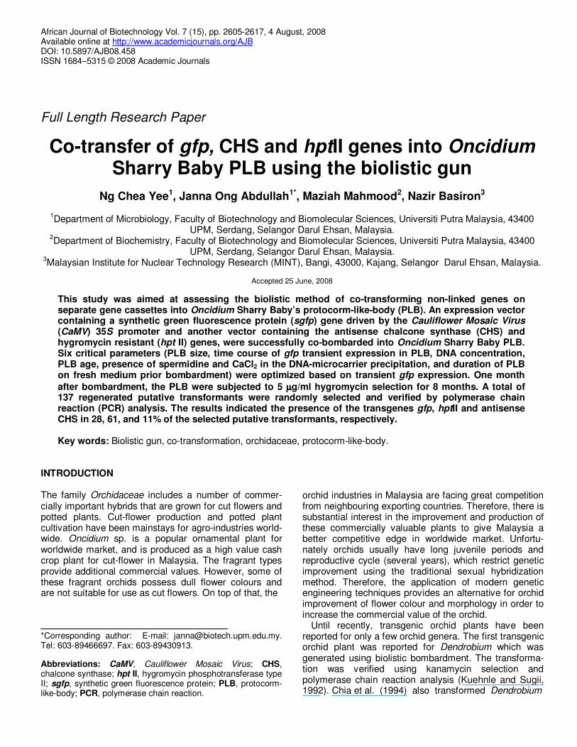
Assessment of transformation parameters Six critical transformation parameters were assessed based on transient gfp expression and/or hygromycin selection (Table 1, Figure 2) to increase the transfor-mation efficiency. Selection of the right size of target tissue is important because it is an indicator of the developmental stage of the tissue. According to Schenk el al (1998), gfp expression varied greatly with respect to shape and size of explant. In this study, 4-week-old single PLB, measuring 1 - 2 mm, 3 - 4 mm and 5 - 6 mm (diameter width) size ranges, were subjected to single bombardment. The results as summarized in Figure 3 show that PLB of 5 - 6 mm size range gave the highest transient gfp expression while the 1 - 2 mm size range PLB gave the lowest expression. Differences in gfp expression between the 5 - 6 mm size range and both 1 - 2 mm and 3 - 4 mm size ranges were significant (p = 0.5) but they were not significant between the 1 - 2 mm and 3 - 4 mm size ranges. However, all the bombarded PLB of size ranges 1 - 2 mm, 3 - 4 mm and 5 - 6 mm turned brown and died after 2 months of selection on 5 µg/ml hygromycin. This may be because this parameter was the first to be optimized, so the transformation condition was not optimal yet. Consequently optimization of other parameters was assessed to improve the transformation efficiency. To avoid low survival rate of smaller size target under the bombarding conditions, bigger PLB of 5 - 6 mm size range was chosen as the target size for subsequent experiments. Another reason is this size range is the

2608 Afr. J. Biotechnol.
(A)
0
20
40
60
80
100
120
0 1 2 3 4 5 10 15 20 25 30 35
Hygromycin concentration (ug/ml)
Perc
en
tag
e o
f su
rviv
ed
PL
B
Figure 1. Minimum inhibitory level of hyromycin as selective agent. (A) Non-transformed PLB were subjected to various concentrations of hygromycin (0, 1, 2,
3, 4, 5, 10, 15, 20, 25 and 30 µg/ml) for one month. For each selection, a total of 60 PLB were tested. Data plotted are the means ± SD of three replicates. (B) Lethal effect exhibited by PLB on media containing different concentrations of hygromycin.
A: control, 0 µg/ml ; B: 1 µg/ml; C: 2 µg/ml; D: 3 µg/ml; E: 4 µg/ml; F: 5 µg/ml. Green signifies surviving PLB (indicated with arrow).

Yee et al. 2609
Table 1. Bombardment parameters assessed in the co-bombardment of pSM-CHS and p35S into Oncidium Sharry Baby PLB.
Parameter Conditions assessed
Size of PLB 1-2 mm, 3-4 mm, 5-6 mm
Time course of gfp transient expression 1, 2, 3, 4, 5, 6, 7, 8, 9, 10, 11,12,13,14 days
Total amount of plasmid DNA (ratio of plamids 1:1) 0. 0.5, 1.0, 1.5, 2.0 µg/bombardment
Age of PLB 1-, 2-, 3-, 4-week old
Pre-culture period on fresh medium prior bombardment 0, 1, 2, 3, 4, 5, 6, 7 days
*Preparation of DNA coating with gold microcarriers CS, C, S, O
*CS means plasmid DNA was coated with microcarriers with the presence of spermidine and calcium chloride. C means plasmid DNA was coated with microcarriers with presence of calcium chloride only. S means plasmid DNA was coated with microcarriers with presence of spermidine only. O means plasmid DNA was coated without the presence of spermidine or calcium chloride.
Figure 2. gfp expression in bombarded Oncidium Sharry Baby PLB. Arrows indicate gfp expressing cells (green) visualized under Leica MZFLIII fluorescence microscope equipped with GFP2 filters (Excitation filter: 480/40 nm).
maximum size range of the Oncidium Sharry Baby PLB in culture.
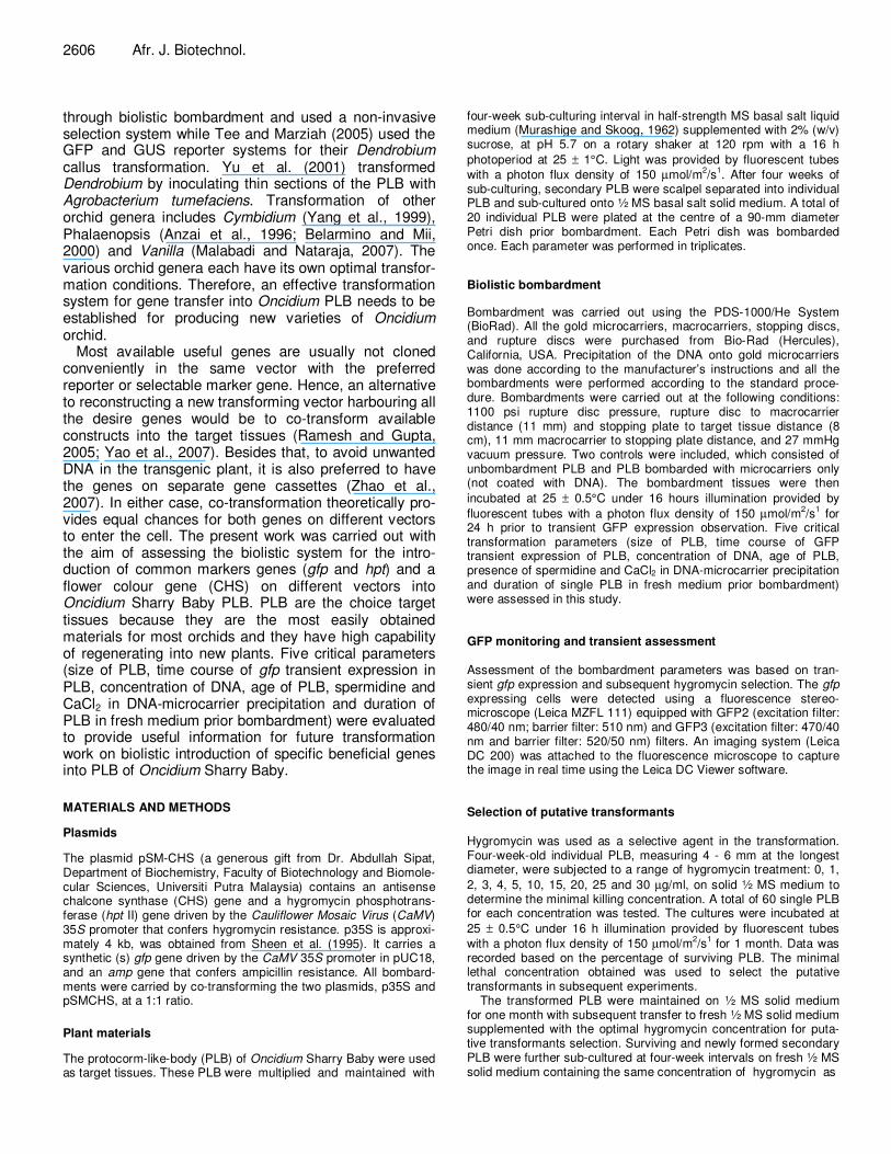
Determining the most suitable observation time for gfp expression after bombardment is also essential to opti-mize the transformation system. Tee et al. (2003) reported gfp expressing cells were detected 3 hours post-bombardment in their Dendrobium PLB. From the result obtained in this study, the earliest gfp expressing cell was detected on the same day of bombardment, and it showed that the number of gfp expressing cells increased during the first two days (Figure 4). The highest number
of gfp expressing cells and the most intense gfp expres-sion were observed 2 days post-bombardment. This shows that the best time to quantify cells transiently expressing gfp is 2 days post-bombardment.
The use of appropriate concentrations of DNA is important in order to produce efficient DNA-microcarrier binding. In assessing the amount of DNA used per
bombardment, it was found that 1.0 µg of DNA per bom-bardment gave the highest transient gfp expression (Figure 5A). Reducing or increasing the DNA concentra-tion beyond 1.0 µg significantly reduced the gfp expression.
2610 Afr. J. Biotechnol.
0
50
100
150
200
1-2mm 3-4mm 5-6mm
Size of PLB
Nu
mb
er o
f g
fpsp
ots
b
a
b
Figure 3. Effect of different PLB size ranges on transformation efficiency. Data was collected 2 days post-bombardment. Error bars correspond to standard deviation (n = 3). The average number of GFP spots is per Petri dish (each dish contained 20 pieces of 4-week-old, 5-6 mm size range PLB). Different letters indicate values are significantly different (p = 0.05).
Figure 4. Time course of gfp expression in PLB. Data was collected 2 days post-bombardment. Error bars correspond to standard deviation (n = 3). The average number of GFP spots is per Petri dish (each dish contained 20 pieces of 4-week-old, 5 - 6 mm size range PLB). Different letters indicate values are significantly different (p = 0.05): 1
ab, 2
a, 3
a, 4
a, 5
ab, 6
abc, 7
abc, 8
abc, 9
cd, 10
de, 11
de, 12
e, 13
e, 14
e.
These might probably due to insufficient amount of microcarriers to coat the available DNA molecules in the solution or maybe the large aggregates were less effi-cient in penetrating the target tissues thereby increasing cell injury and eventually cells death (Klein et al., 1988; Li et al., 1994). Parveez et al. (1998) also reported that
higher concentrations were not only ineffective but expensive. Only one – fifth of those PLB bombarded with
0.5 µg DNA survived on the hygromycin selection medium (Figure 5B). Even though this DNA concentration did not give the highest gfp expression level, it gave the
highest number of surviving PLB. In contrast, 1.0 µg DNA
0
20
40
60
80
100
120
140
160
1 2 3 4 5 6 7 8 9 10 11 12 13 14
Post-bombardment time (day)
Nu
mb
er
of
gfp
sp
ots

Yee et al. 2611
(A)
0
100
200
300
400
500
600
700
800
900
0.0 0.5 1.0 1.5 2.0
Concentration of DNA (µµµµg)
Nu
mb
er
of
gfp
sp
ots
(B)
0
5
10
15
20
25
30
0.5 1.0 1.5 2.0
DNA concentration (µµµµg)
Per
cen
tag
e of
surv
ived
PL
B
b
b b
b
a
a
a
a
a
Figure 5. Effects of different DNA concentrations on gfp expression and survival on hygromycin selection. Each point correspond to standard deviation (n = 3), with 20 pieces of 4-week-old, 5 - 6 mm size range PLB per replicate. Different letters indicate values are significantly different (p = 0.05). (A) gfp expression in PLB bombarded with different concentrations of total plasmid DNA (0, 0.5, 1.0, 1.5, 2.0 µg, with plasmids ratio of 1:1). Data was collected 2 days post-bombardment. (B) PLB surviving on hygromycin selection. The bombarded PLB were subjected to 5 µg/ml hygromycin selection for 8 months.
per bombardment gave the highest transient gfp expression but not the highest percentage of survivals. Overall, the results showed that high concentration of
DNA (beyond 1.0 µg) resulted in low survival rates.
Subsequent assessment of the suitable target tissue age for bombardment was carried out. According to Sanford et al. (1993), actively dividing cells were more receptive to receive DNA and gave higher gene expression.
2612 Afr. J. Biotechnol.
(A)
0
100
200
300
400
500
600
700
Week 1 Week 2 Week 3 Week 4
Age of PLB
Nu
mb
er
of
gfp
sp
ots
(B)
0
5
10
15
20
25
30
35
40
Week 1 Week 2 Week 3 Week 4
Age of PLBs
Per
cen
tag
e o
f su
rviv
ed P
LB
b b
a
ab
a
a
a
b
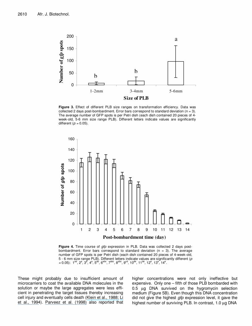
Figure 6. Effect of PLB age on gfp expression and survival on hygromycin selection. Data was collected 2 days post-bombardment. Error bars correspond to standard deviation (n = 3). Different letters indicate values are significantly different (p = 0.05). (A) The average number of GFP spots is per Petri dish (20 pieces of 4-week-old, 5-6 mm size range PLB). Each bombardment contains three replicates. (B) Bombarded PLB were subjected to 5 µg/ml hygromycin selection for 8 months.
In this work, PLB of 5-6 mm size range obtained from 1, 2, 3 and 4-week-old cultures, were subjected to bombardment, then transient gfp expression analysis and hygromycin selection. Neither 1 nor 2-week-old cultures exhibited good gfp expression (Figure 6A) compared to the 3 and 4-week-old PLB. After 8 months of hygromycin
selection, only 2% of 1-week-old bombarded PLB survived (Figure 6B). The numbers of surviving and proliferating PLB increased to 10 to 25% when 2, 3 and 4-week-old PLB were used as target tissues. Considering that majority of the PLB at these culture ages ranged from 5 – 6 mm in size, we therefore chose 4-week-old

Yee et al. 2613
(A)
0
100
200
300
400
500
600
700
0 1 2 3 4 5 6 7
Pre-culture period prior bombardment (day)
Nu
mb
er
of
gfp
sp
ots
(B)
0
10
20
30
40
50
60
70
80
90
7 6 5 4 3 2 1 0
Pre-culture period prior bombardment (day)
Perc
enta
ge
of
surv
ived
PL
B
a
b
c
cd cd
d d cd
bc
e
cd
e
ab
de
a
bc
Figure 7. Effect of duration of PLB on fresh medium prior bombardment on gfp expression and survival on hygromycin selection. Data was collected 2 days post-bombardment. Error bars correspond to standard deviation (n = 3). Different letters indicate values are significantly different (p = 0.05). (A) The average number of GFP spots is per Petri dish (20 pieces of 4-week-old, 5 - 6 mm size range PLB). Each bombardment contains three replicates. (B) Percentage of PLB survived after 8 months on 5 µg/ml hygromycin selection.
PLB to be the most suitable culture age for bombardment.
The effect of the culture period of the PLB on fresh medium prior bombardment was further examined. The PLB were sub-cultured onto ½ MS solid medium for various durations (0, 1, 2, 3, 4, 5, 6 and 7 days) before subjecting to bombardment. The result shows that tran-sient gfp expression is proportional to the pre-culturing period from 0 to 7 days (Figure 7A). Pre-culture period
beyond 7 days was not carried out in this study because it was previously observed that beyond 7 days, the PLB started to shoot. After 8 months on hygromycin selection, only 40% of the PLB that were not subjected to a pre-culturing period prior bombardment, survived (Figure 7B). The number of survivals increased to 67% with PLB that were pre-cultured for 1-day prior to bombardment. These bombarded PLB also gave the highest transient gfp expression. Hence, this time period was chosen for pre-
2614 Afr. J. Biotechnol.
(A)
0
50
100
150
200
250
300
350
400
0 Spermidine &
CaCl2
Spermidine CaCl2
Nu
mb
er
of
gfp
s
po
ts
(B)
0
10
20
30
40
50
60
70
80
90
CaCl2 Spermidine Spermidine &
CaCl2
0
Per
centa
ge
of su
rviv
ed P
LB
c c
a
b
a
b
ab
b
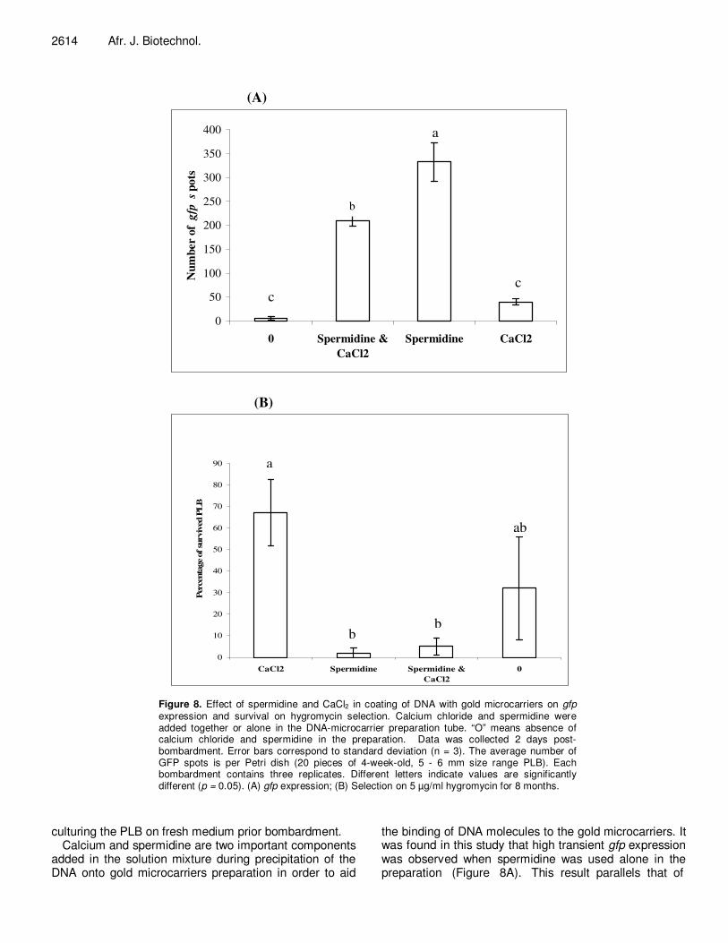
Figure 8. Effect of spermidine and CaCl2 in coating of DNA with gold microcarriers on gfp expression and survival on hygromycin selection. Calcium chloride and spermidine were added together or alone in the DNA-microcarrier preparation tube. “O” means absence of calcium chloride and spermidine in the preparation. Data was collected 2 days post-bombardment. Error bars correspond to standard deviation (n = 3). The average number of GFP spots is per Petri dish (20 pieces of 4-week-old, 5 - 6 mm size range PLB). Each bombardment contains three replicates. Different letters indicate values are significantly different (p = 0.05). (A) gfp expression; (B) Selection on 5 µg/ml hygromycin for 8 months.
culturing the PLB on fresh medium prior bombardment. Calcium and spermidine are two important components
added in the solution mixture during precipitation of the DNA onto gold microcarriers preparation in order to aid
the binding of DNA molecules to the gold microcarriers. It was found in this study that high transient gfp expression was observed when spermidine was used alone in the preparation (Figure 8A). This result parallels that of
Yee et al. 2615
(A)
(B)
(C)
bp
(D)
500
chs
sgfp
M 1 2 3 4
hpt II
M 1 2 3 4 5 6 7 8 9
2000
1 2 3 4 5 6 7 8 9
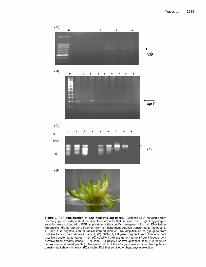
Figure 9. PCR amplification of chs, hptII and gfp genes. Genomic DNA harvested from randomly picked independent putative transformants that survived on 5 µg/ml hygromycin selection were subjected to PCR amplication of the specific transgene. M is 1Kb DNA ladder (A) specific 750 bp gfp gene fragment from 3 independent putative transformants (lanes 2, 3, 4), lane 1 is negative control (untransformed plantlet). No amplification of gfp gene from putative transformant shown in lane 2; (B) 500bp hpt II gene fragment from 9 independent putative transformants (lanes 1 - 9); (C) specific 1.3kb chs gene fragment from 7 independent putative transformants (lanes 1 - 7), lane 8 is positive control (plasmid), lane 9 is negative control (untransformed plantlet). No amplification of the chs gene was obtained from putative transformant shown in lane 4; (D) shooted PLB that survived on hygromycin selection.
2616 Afr. J. Biotechnol. Janna et al. (2006) observation for Dendrobium Sonia transformation. However, hygromycin selection recorded higher survivals (67%) for the PLB bombarded with DNA prepared with CaCl2 alone (Figure 8B), and spermidine is detrimental when included in the preparation process. Hence, only calcium is preferred for the DNA preparation in future experiments. PCR analysis Untransformed PLB turned brown and necrotic on the selection medium within 2 months, while the hygromycin-resistant PLB remained green and regenerated into plantlets during the 8 months of hygromycin selection (Figure 9D). A total of 137 regenerated putative transfor-mants were randomly picked to verify the presence of the transgenes (antisense CHS, gfp, and hpt II) by PCR analysis. Eleven of the putative transformants were positive for the antisense CHS gene, 61 positives for the hpt II gene and 28 for the gfp gene (Figure 9A, B, C). Conclusion Theoretically, gene transfer using biolistic gun (PDS 1000/He system) is applicable to all kinds of cells and tissues. However, the DNA delivery conditions and transient gfp expression in each species and tissues involved are different and hence, optimization of the delivery conditions is important in order to improve the transformation process (Nan and Kuehnle, 1995). Using the biolistic system, we have successfully introduced unlinked genes on different gene cassettes, p35S which carries a synthetic green fluorescence protein (sgfp) gene and pSM-CHS which carries the antisense chalcone synthase (CHS) and hygromycin resistance (hptII) genes, into Oncidium Sharry Baby PLB. We have shown that
hygromycin at 5.0 µg/ml is a good selective agent for Oncidium Sharry Baby PLB transformation. Four-week-old PLB of 5 – 6 mm size range and pre-incubated on fresh medium for 1 day prior to bombardment, are
optimal for transformation using 1.0 µg DNA prepared in the presence of calcium only. Transformation, selection, and regeneration of the transformants were also carried out successfully where 137 randomly selected regene-rated putative transformants were verified to be PCR positives for the antisense CHS, gfp, and hpt II genes at 11, 28 and 61%, respectively.
In conclusion, this study demonstrates that co-trans-formation of unlinked genes on different cassettes for Oncidium Sharry Baby PLB is possible using the established optimized conditions. This will facilitate the creation of new varieties through the introduction of commercially beneficial genes on different gene cas-settes into Oncidium Sharry Baby PLB. Because of the time and economic savings without having to re-construct
new gene cassette, the established protocol will also facilitate functional studies of orchid genes at the molecular and cellular levels. ACKNOWLEDGEMENTS The authors are grateful to the Ministry of Science, Technology and Innovation of Malaysia for supporting this work under IRPA grant. Special thanks to Dr. A. Elliot (CSIRO, Brisbane, AUS) and Dr. J. Sheen (Boston, USA) for kindly providing us with the GFP constructs. REFERENCES Anzai H, Ishii Y, Shichinohe M, Katsumata K, Nojiri C, Morikawa H,
Tanaka M (1996). Transformation of Phalaenopsis by particle
bombardment. Plant Tissue Cult. Lett. 13: 265-272. Belarmino MM, Mii M (2000). Agrobacterium –mediated genetic
transformation of a Phalaenopsis Orchid. Plant Cell Rep. 19: 435-
442. Chauvin J-E, Marhadour S, Cohat J, Le Nard M (1999). Effets of gelling
agents on in vitro regeneration and kanamycin efficiency as a
selective agent in plant transformation procedures. Plant Cell Tissue Organ Cult. 58: 213-217.
Chia TF, Chan YS, Chua NH (1994). The firefly luciferase gene as a non-invasive reporter for Dendrobium transformation. Plant J. 6: 441-446.
Elliott AR, Campbell JA, Dugdale B, Brettell RIS, Grof CPL (1999). Green-fluorescent protein facilitates rapid in vivo detection of genetically transformed plant cells. Plant Cell Rep. 18: 707-714.
Janna OA, Maziah M, Parveez GKA, Saleh K (2006). Factors affecting
delivery and transient expression of β-glucuronidase gene in Dendrobium Sonia protocorm-like-body. Afr. J. Biotechnol. 5: 88-94.
Klein TM, Gradziel T, Fromm ME, Sanford JC (1988). Factors influencing gene delivery into Zea mays cells by high velocity
microprojectiles. Bioresour. Technol. 6: 559-563. Kuehnle AR, Sugii N (1992). Transformation of dendrobium orchid using
particle bombardment of protocorms. Plant Cell Rep., 11: 484-488. Li YH, Trembley FM, Seguin A (1994). Transient transformation of
pollen and embryogenic tissues of white spruce (Picea glauca Moench) resulting from microprojectile bombardment. Plant Cell Rep.
13: 661-665. Liau CH, You SJ, Prasad V, Hsiao, HH, Lu JC, Yang NS, Chan MT
(2003). Agrobacterium tumefaciens-mediated transformation of an Oncidium orchid. Plant Cell Rep. 21: 993-998.
Malabadi RB, Nataraja K (2007). Genetic transformation of Vanilla planifolia by Agrobacterium-tumefaciens using shoot tip sections. Res. J. Bot. 2(2): 86-94.
Murashige T, Skoog T (1962). A revised method for rapid growth and bioassays with tobacco tissue cultures. Physiol. Plant 15: 473-497.
Nan GL, Kuehnle AR (1995). Factors affecting gene delivery by particle bombardment of Dendrobium orchids. In Vitro Cell Dev. Biol. Plant. 31: 131-136.
Parveez GKA, Chowdhury MKU, Saleh NM (1998). Physical parameters affecting transient GUS gene expression in oil palm (Elaeis guineensis Jacq.) embryogenic calli via microprojectile bombardment. Indian Crop. Prod. 8: 17-27.
Ramesh M, Gupta AK (2005). Transient expression of beta-glucuronidase gene in indica and japonica rice (Oryza sativa L.) callus after different stages of co-bombardment. Afr. J. Biotechnol. 4(7) :596-600.
Sanford J, Smith FD, Russel JA (1993). Optimizing the biolistic process for different biological applications. Method Enzymol. 217: 483-509.
Schenk PM, Elliot AR, Manner JM (1998). Assessment of transient gene expression in plant tissue using the green fluorescent protein as a reference. Plant Mol. Biol. Rep. 16: 313-322.

Sheen J, Hwang S, Niwa Y, Kobayashi H, Galbraith DW (1995). Green
fluorescent protein as a new vital marker in plant cells. Plant J. 8: 777-784.
Tee CS, Marziah M (2005). Optimization of biolistic bombardment parameters for Dendrobium Sonia 17 calluses using GFP and GUS as the reporter system. Plant Cell Tissue Organ Cult. 80: 77-89.
Tee CS, Marziah M, Tan CS, Abdullsh MP (2003). Evaluation of defferent promoters driving the GFP reporter gene and selected target tissues for particle bombardment of Dendrobium Sonia 17.
Plant Cell Rep. 21: 452-458. Upadhyaya NM, Surin B, Ramm K, Gaudron J, Schunmann PHD,
Taylor W, Waterhouse PM, Wang MB (2000). Agrobacterium-
mediated transformation of Australian rice cultivars Jarrah and Amaroo using modified promoters and selectable markers. Austr. J. Plant Physiol. 27: 201-210.
Yang J, Lee HJ, Shin DH, Oh SK, Seon JH, Paek KY, Han KH (1999). Genetic transformation of Cymbidium orchid by particle bombardment. Plant Cell Rep. 18: 978-984.
Yee et al. 2617 Yao Q, Cong L, He GY, Chang JL, Li KX, Yang GX (2007). Optimization
of wheat co-transformation procedure with gene cassettes resulted in an improvement in transformation efficiency. Mol. Biol. Rep. 34(1):
61-67. Yu H, Yang SH, Goh C J (2001). Agrobacterium-mediated
transformation of a Dendrobium orchid with the 1 knox gene DOH1.
Plant Cell Rep. 20: 301-305. Zhao Y, Qian Q, Wang HZ, Huang DN (2007). Co-transformation of
gene expression cassettes via particle bombardment to generate safe transgenic plant without any unwanted DNA. In Vitro Cell. Dev. Biol. 43(4): 325-334.
Copyright © 2022 FDOKUMEN