Dimethylfuran-Lactone Pheromone from Males of Galerucella calmariensis and Galerucella pusilla
Upload
universitasnegerimalangCategory
view
3download
0
Hormones and Behavior xxx (2009) xxx–xxx
YHBEH-02801; No. of pages: 10; 4C:
Contents lists available at ScienceDirect
Hormones and Behavior
j ourna l homepage: www.e lsev ie r.com/ locate /yhbeh
ARTICLE IN PRESS
Characterization of an alarm pheromone secreted by amphibian tadpoles thatinduces behavioral inhibition and suppression of the neuroendocrine stress axis
Michael E. Fraker a,b, Fang Hu a,b, Vindhya Cuddapah a,b, S. Andy McCollum c, Rick A. Relyea d,John Hempel d, Robert J. Denver a,b,⁎a Department of Ecology and Evolutionary Biology, The University of Michigan, Ann Arbor, MI 48109-1048, USAb Department of Molecular, Cellular and Developmental Biology, The University of Michigan, Ann Arbor, MI 48109-1048, USAc Department of Biology, Cornell College, Mount Vernon, IA 52314, USAd Department of Biological Science, University of Pittsburgh, PA 15260, USA
⁎ Corresponding author. 3065C Kraus Building, 830University of Michigan, Ann Arbor, MI 48109-1048, USA
E-mail address: [email protected] (R.J. Denver).
0018-506X/$ – see front matter © 2009 Elsevier Inc. Aldoi:10.1016/j.yhbeh.2009.01.007
Please cite this article as: Fraker, M.E., et al.,inhibition and suppression of the neuroend
a b s t r a c t
a r t i c l e i n f oArticle history:
Many species assess preda Received 17 November 2008Revised 19 January 2009Accepted 21 January 2009Available online xxxxKeywords:AmphibianTadpoleAnti-predator behaviorCorticosteroneStressPredation risk assessmentSkin secretion
tion risk through chemical cues, but the tissue source, chemical nature, andmechanisms of production or action of these cues are often unknown. Amphibian tadpoles show rapid andsustained behavioral inhibition when exposed to chemical cues of predation. Here we show that an alarmpheromone is produced by ranid tadpole skin cells, is released into the medium via an active secretoryprocess upon predator attack, and signals predator presence to conspecifics. The pheromone is composed oftwo components with distinct biophysical properties that must be combined to elicit the behavioralresponse. In addition to the behavioral response, exposure to the alarm pheromone caused rapid and strongsuppression of the hypothalamo–pituitary–adrenal (HPA) axis, as evidenced by a time and dose-dependentdecrease in whole body corticosterone content. Reversing the decline in endogenous corticosterone causedby exposure to the alarm pheromone through addition of corticosterone to the aquarium water (50 nM)partially blocked the anti-predator behavior, suggesting that the suppression of the HPA axis promotes theexpression and maintenance of a behaviorally quiescent state. To our knowledge this is the first evidence foraquatic vertebrate prey actively secreting an alarm pheromone in response to predator attack. We alsoprovide a neuroendocrine mechanism by which the behavioral inhibition caused by exposure to the alarmpheromone is maintained until the threat subsides.
© 2009 Elsevier Inc. All rights reserved.
Introduction
Many animals exhibit anti-predator, or alarm behavior whenexposed to chemical cues of predation (Mullerschwarze et al., 1984;Lima and Dill, 1990; Dodson et al., 1994; Kats and Dill, 1998; Lima,1998; Lin et al., 1998; Chivers and Mirza, 2000; Apfelbach et al., 2005;Wyatt, 2005; Thomas et al., 2006). These behaviors are species-specific, and include reducing activity (i.e., freezing, or behavioralinhibition), escape behaviors, shelter seeking, area avoidance, school-ing, and colony defense (Wisenden, 2000; Apfelbach et al., 2005;Lamprecht et al., 2008). Informative chemical cues can originate fromthe predator (e.g., kairomones) or the prey, and prey may usecombinations of chemicals in their assessment of predation risk (e.g.Schoeppner and Relyea, 2005; Richardson, 2006). Chemical cues ofpredation derived from the prey may be released incidentally as aresult of tissue damage (and perhaps after digestion of prey bypredators), or through active release as part of an anti-predator
North University Avenue, The. Fax: +1 734 647 0884.
l rights reserved.
Characterization of an alarmocrine stress axis, Horm. Be
mechanism (e.g., disturbance or alarm pheromones; Mullerschwarzeet al., 1984; Chivers and Smith, 1998; Madison et al., 2002; Wyatt,2005; Lamprecht et al., 2008). Here we use the term ‘chemical cue ofpredation’ as a general term to describe chemicals derived from preythat may be released actively or passively, and that signal toconspecifics the presence of a predator. We use the term ‘alarmpheromone’ to distinguish those chemical cues of predation that havebeen shown to be produced by specialized cells and/or to be releasedthrough a regulated secretory process, and that may induceantipredator behaviors in prey such as escape behavior or freezing.
A common process by which vertebrate prey releases chemicalcues of predation is through injury or consumption by a predator (i.e.damage-released cues, reviewed by Chivers and Smith, 1998). Thesechemical cues are derived from tissues and body fluids of prey, and areoften considered to be unintentional information sources forconspecifics; i.e., the emitter does not intentionally signal thepresence of a predator (e.g. hemolymph, Smith, 1992; Chivers et al.,1996; Acquistapace et al., 2005; but see Henderson et al., 1997;Cashner, 2004). Disturbed, but uninjured prey also releases chemicalswhile escaping from a predator or upon capture that act as predationcues for conspecifics; e.g., ammonium waste released by crayfish,
pheromone secreted by amphibian tadpoles that induces behavioralhav. (2009), doi:10.1016/j.yhbeh.2009.01.007
2 M.E. Fraker et al. / Hormones and Behavior xxx (2009) xxx–xxx
ARTICLE IN PRESS
amphibian tadpoles and fish (Hazlett, 1990; Kiesecker et al., 1999;Manteifel et al., 2005;Wisenden and Barbour, 2005) or skin secretionsreleased by adult salamanders (Graves and Quinn, 2000). Ostariophy-san fishes have specialized cells in their epidermis (club cells) thatrelease an alarm pheromone when attacked by a predator (Smith,1992). It is thought that release requires rupture of the club cells,although the possibility that these cells actively secrete alarmpheromone onto the skin surface cannot be ruled out. Limitedbiochemical evidence suggests that one component of the alarmpheromone may be the purine derivative hypoxanthine-3-N-oxide(Pfeiffer et al., 1985; Brown et al., 2000; Brown et al., 2001; Brownet al., 2003).
Chemical cues of predation cause rapid changes in behavior, butrelatively little is known about the neural and physiological processesinduced in vertebrate prey that underlie the behavioral responses. Infish, exposure to the putative alarm substance hypoxanthine-3-N-oxide leads to enhanced optical alertness, suggesting actions on thecentral nervous system (CNS) that influence visual acuity (Pfeifferet al., 1985). In mammals, exposure to predators or predator odorcauses behavioral inhibition (freezing behavior), activation of theneuroendocrine stress axis (the hypothalamus–pituitary–adrenal –
HPA – axis) and correlated changes in CNS limbic circuitry associatedwith fear and anxiety (Figueiredo et al., 2003; Apfelbach et al., 2005;Masini et al., 2005; Thomas et al., 2006; Roseboom et al., 2007). Theneuroendocrine stress response involves the release of corticotropin-releasing factor (CRF) from the hypothalamus, which stimulatessecretion of adrenocorticotropic hormone (ACTH) from the pituitarygland, and ACTH acts on adrenal cortical cells to increase biosynthesisand secretion of glucocorticoids (i.e., corticosterone or cortisol), theprimary vertebrate stress hormones (reviewed by Denver, in press).Glucocorticoids exert negative feedback at several points along theHPA axis. Glucocorticoids and CRF also have diverse actions onbehavior and physiology, including locomotion, food intake, andenergy utilization (Sapolsky et al., 2000; Crespi and Denver, 2004;Crespi and Denver, 2005).
Amphibian tadpoles reduce their activity level in response tochemical cues released by caged predators fed tadpoles (e.g., Relyea,2001; Fraker, 2008a). The level of swimming activity of tadpoles isrelated to their probability of capture by predators (i.e., the higher theactivity, the greater the probability of capture; Skelly, 1994), andreflects a trade-off between predation risk and foraging gain (Wernerand Anholt, 1993). However, the tissue source and chemical nature ofthe chemical cue of predation, and whether it is simply a damage-released, unintentional information source or is actively secreted (i.e.,an alarm or disturbance pheromone) is not known. In the currentstudy we used the anti-predator behavioral response of tadpoles as anassay for perceived predation risk following exposure to chemical cuesof predation. We investigated responses using tadpoles of two ranidanuran species, the wood frog (Rana sylvatica) and the green frog(Rana clamitans). These species represent two different life historystrategies. In southern Michigan, wood frogs typically breed duringlate March in ephemeral ponds and their tadpoles metamorphoseduring June of the same year (Wellborn et al., 1996). Green frogstypically breed from early June through early August in semi-permanent, fishless ponds and overwinter at least once (Wellbornet al., 1996). The differences between the species’ life history strategiesshould favor different anti-predator behavioral strategies (Anholt etal., 2000); this difference seems to be manifested in the duration ofthe behavioral response (i.e., behavioral inhibition) rather than thespeed that it is expressed (M. Fraker, unpublished data). Wedetermined the source of the chemical cue of predation, the meansby which it is released, and its biophysical properties. We alsoanalyzed activity of the tadpole HPA axis by changes in whole bodycorticosterone content following exposure to the chemical cue ofpredation, andwe investigated a role for suppression of corticosteronein the expression of the anti-predator behavioral response.
Please cite this article as: Fraker, M.E., et al., Characterization of an alarminhibition and suppression of the neuroendocrine stress axis, Horm. Be
Materials and methods
Animals
Four to five wood frog (R. sylvatica) and green frog (R. clamitans)egg masses were collected from several ephemeral and semi-permanent ponds at the University of Michigan's Edwin S. GeorgeReserve (ESGR) near Pinckney, Michigan. The top predators in all ofthe ponds were invertebrates (i.e., Anax dragonflies). The egg masseswere cultured in large plastic pools filled with well water that hadbeen inoculated with phytoplankton and zooplankton. The pools werecovered with shadecloth to keep predators from colonizing, andtadpoles were fed rabbit chow ad libitum (population densityapproximately 1–2 tadpoles/L). Once the tadpoles reached ∼50–100 mg body weight (Gosner stages 26–28; Gosner, 1960), they weretransported to the University of Michigan and housed in large holdingtanks (172 L, population density 1–2 tadpoles/L) filled with charcoal-purified (dechlorinated), pH-adjusted tap water in a controlledenvironmental chamber at 21–23 °C on a 12L:12D photoperiod.Tadpoles were then used in experiments two to three days aftertransporting. Predator-naive tadpoles were fed boiled spinach andpulverized rabbit chow ad libitum.
Predatory larval dragonflies (Anax junius) were also collected atthe ESGR and housed individually in plastic containers filled with500 ml dechlorinated tap water. A small piece of fiberglass screenwas added to the container as a perch for the dragonfly larvae andthey were fed 0.5–1.0 g R. sylvatica or R. clamitans tadpoles daily.All procedures involving animals were approved by the Universityof Michigan's University Committee on the Use and Care ofAnimals.
Behavioral assays
The proportion of time that tadpoles spent swimming vs. restingwhen exposed to different treatments was analyzed. In this assay,behavioral inhibition is indicated by reduced proportion of timespent swimming. For experiments 1–6, sets of 10 tadpoles werehaphazardly selected and distributed among four-to-six12H×16W×27L cm tanks (containing 3 L dechlorinated tap water)for each treatment (total number of replicates of each treatment isnoted in description of the particular experiment). Tadpoles wereplaced in tanks the day before the behavioral assay was conductedand the tanks were randomly positioned within the environmentalchamber to minimize microenvironmental effects. During eachbehavioral trial, the appropriate treatments were added to allreplicate tanks within 5 min. The investigator then exited thechamber, and behavioral data were recorded using two to three CCDcameras mounted on racks over the tanks (each camera recorded arectangular array of four tanks simultaneously). Recordings wereinitiated 30 min after the addition of treatments. In experiments withmore than three treatments, two or three replicate tanks from eachtreatment were assayed in multiple blocks due to having only threecameras. In these experiments, treatment additions were delayed sothat recording always began 30 min after addition of treatments. Thetime spent swimming by five haphazardly-selected tadpoles in eachtank over a one minute period was measured using the MS-DOScomputer program “Tadpole” (Van Buskirk and McCollum, 2000). Thefive tadpoles were chosen before viewing the recording to avoid bias.The mean proportion of time spent swimming for the tadpoles ineach tank served as one replicate. For each experiment, theproportion of time spent swimming by each individual tadpole waslog-transformed prior to statistical analysis. The data were analyzedby one-way ANOVA, followed by Scheffe's post hoc test, except forExpt. 10 (described below). The Pb0.05 criterion was used in theScheffe's tests. Statistical analyses were conducted using SAS 9.1software (SAS Institute, 2003).
pheromone secreted by amphibian tadpoles that induces behavioralhav. (2009), doi:10.1016/j.yhbeh.2009.01.007
3M.E. Fraker et al. / Hormones and Behavior xxx (2009) xxx–xxx
ARTICLE IN PRESS
Analysis of the nature and tissue source of a chemical cue of predation
Experiment 1 was designed to test 1) whether wood frog tadpolesrespond behaviorally to water conditioned by a predator fedconspecific tadpoles, and 2) whether the chemical cue of predationis derived from the attacked/consumed tadpole (i.e., homogenizing –
‘macerating’ – the tadpole will release the cue).The experiment had three treatments: control, predator-condi-
tioned water, and tadpoles homogenized in water. There were fourreplicate tanks per treatment. For the control treatment we added500 ml dechlorinated tap water to the tadpole tanks. For predator-conditionedwater, Anax larvaewere housed individually in containerswith 500 ml dechlorinated tap water and fed ∼0.25 g of live R.sylvatica tadpoles. Twenty to twenty four hours after feeding the Anaxthe screens were removed from the container, and the conditionedwater strained with a 1 mm mesh net to remove Anax waste ortadpole remains. The conditioned water from four Anax was pooled,and each tadpole tank (4 replicate tanks per treatment) received500 ml of predator conditioned water. For the tadpole homogenatetreatment, on the day of a behavioral trial tadpoles (∼1 g totalbiomass) were collected from the stock tank, deeply anesthetized byimmersion in 0.l% benzocaine for 10 min, and then rinsed in a beakerof fresh water. They were then transferred to a testtube with 4 mldechlorinated tap water and homogenized for 30 s using a PolytronPT-2000 homogenizer (Kinematica, Littau, Switzerland). The tadpolehomogenate was divided into four equal aliquots and each was addedto 500 ml dechlorinated tap water before being added to the tanks(∼0.25 g tadpole biomass equivalent per tank). This allowed a similarbiomass of tadpoles to be used in the predator-conditioned water andtadpole homogenate treatments.
Experiment 2 tested 1) whether the chemical cue of predation isreleased by living R. sylvatica tadpoles (i.e., an Anax simply eating anddigesting a dead tadpole will not release the cue), and 2) whether thechemical cue is released by living tadpoles in response to a genericpredatory event; i.e., its release does not require Anax per se or obvioustissue damage.
The experiment had four replicates of four treatments: control,predator-conditioned water (fed live tadpoles), predator-conditionedwater (fed dead tadpoles) andwater conditionedwith poked tadpoles.The predators were fed as described in experiment 1, except that onegroup was fed tadpoles that were first euthanized by immersion in0.01% benzocaine for 10 min. Tadpoles do not recover from this doseand duration of anesthesia and are thus euthanized by the treatment.In all cases tadpoles had been consumed by the Anax during the 20–24 h period before the experiment. Another treatment included in thisexperiment involved the addition of water conditioned by tadpolespokedwith a hypodermic needle. For this treatment, one large tadpole(∼1 g) was selected from the stock tank and submerged in 500 mldechlorinated tapwater within a net. A 26.5 gauge hypodermic needlewas used to “poke” the tadpoles' tail muscle five times for 10 s eachpoke (i.e. the needle remained in the tail muscle for 10 s). The tadpoleremained submerged throughout this process and was subsequentlyreleased into the water. This procedure was repeated until a total offive tadpoles per container were held and poked in each of fourcontainers. The tadpoles remained in the water for 20 min tocondition the water until they were removed and the water addedto the experimental tanks.
Experiment 3 tested whether the chemical cue is released via aclassical stimulus-secretion coupled pathway. The focal species usedfor this experiment was R. clamitans owing to its availability andknown behavioral response to predator chemical cue (Fraker, 2008a).We exposed tadpoles to 5 mM KCl for a brief period, then tested theconditioned water for a behavioral response by conspecifics. Exposureof cells to KCl is well known to cause plasma membrane depolariza-tion, and the release of secretory vesicles via a calcium-dependent,stimulus-secretion coupled pathway.
Please cite this article as: Fraker, M.E., et al., Characterization of an alarminhibition and suppression of the neuroendocrine stress axis, Horm. Be
Six sets of 10 small (30–50mg) green frog tadpoles were immersedin 20 ml 5 mM KCl or 20 ml dechlorinated tap water for 15 min, thenremoved. The tadpole-conditioned KCl solution or dechlorinated tapwater was then added to the tanks for behavioral assay. Two controltreatments included 20 ml of 5 mM KCl or dechlorinated tap water(without tadpoles having been added) added to the tanks. Behavioralobservations were made as described above.
Experiment 4 tested whether the chemical cue of predation isstored in membranous secretory vesicles that can be released uponhomogenization in a detergent (Triton X-100) and sonication. Thedetergent serves to solubilize lipid membranes of secretory vesicles,and sonication uses high-frequency sound waves to promote tissuedisruption.
The tadpole homogenate (R. sylvatica) was prepared as described inExperiment 1 (above) except that Triton X-100 was added to 4 mldechlorinated tap water to a final concentration of 1%. The homogenatewas then sonicated for 30 s using a Fisher Scientific Sonic Dismembrator,Model 100 (Fisher Scientific, Pittsburgh, PA, USA) and divided into fouraliquots, each added to 500ml dechlorinated tapwater (∼0.25 g tadpolemass equivalent per tank) whichwas then added to the assay tanks. Thebehavioral response resulting from these treatmentswas compared to apredator-conditionedwater treatment aswell as to awater only control.There were four replicates of each treatment.
Experiment 5 again tested whether the chemical cue of predationis stored in membranous secretory vesicles, but used both R. sylvaticaand R. clamitans tadpoles as focal species. The experiment wasconducted for each species separately.
The procedure used in Experiment 3 was repeated with anadditional control group in which Triton X-100 was added to theexperimental tanks to the same final concentration as with the TritonX-100 tadpole homogenate (0.0003% in the assay tank) to determine ifthe detergent alone influenced tadpole behavior. A predator-condi-tioned water treatment as described in Experiment 1 was alsoincluded. There were four replicates of each treatment.
Experiment 6 analyzed the tissue source of the chemical cue in R.clamitans tadpoles.
Ten tadpoles (∼100 mg BW each) were euthanized in 0.01% ben-zocaine, then dissected into their dorsal skin, ventral skin, tail, cloaca,nares, and internal organs. Some dorsal or ventral skin was likelyincluded with the cloacae and nares. The tissues were then homo-genized, sonicated in water with 1% Triton X-100, and divided intofour aliquots, as described above. The behavioral response of tadpolesexposed to each tissue homogenate was recorded as described above.Although there were differences in mass among the tissues, eachtissue samplewas derived from the same number of animals. Thus, if aspecific tissue was the major source of the chemical cue then theactivity would be detected in our assay.
Analysis of changes in HPA axis activity following exposure to thechemical cue of predation, and the role of suppression of the HPA axisin the expression of anti-predator behavior
The following experiments tested whether HPA axis activity wasaltered by exposure to the chemical cue of predation, and a possible rolefor corticosterone in behavioral inhibition. For experiments 7 and 8,tadpoles were haphazardly selected and distributed three to a tank(15H×9L×22W cm; containing 2 L dechlorinated tap water) with sixreplicate tanks for each treatment (6 treatments, 36 tanks total). Forexperiment 9, ten tadpoles were haphazardly selected and distributedamong six 12H×16W×27L cm tanks (containing 2 L dechlorinated tapwater) for each treatment. The tanks were shielded with opaque plexi-glass to minimize stress caused by the investigator entering the room.
Experiment 7 tested whether exposure to the predator chemicalcue caused a change in tadpole whole-body corticosterone content. R.clamitans tadpoles were exposed to the Triton X-100 tadpolehomogenate for 1, 2, and 4 h, then euthanized by immersion in
pheromone secreted by amphibian tadpoles that induces behavioralhav. (2009), doi:10.1016/j.yhbeh.2009.01.007
4 M.E. Fraker et al. / Hormones and Behavior xxx (2009) xxx–xxx
ARTICLE IN PRESS
0.01% benzocaine and snap-frozen in a dry ice/ethanol bath. Thecontrol treatment was an equal volume of dechlorinated tap wateradded to the tank (to control for disturbance effects), and animalswere sacrificed at the 4 h time point. Six replicate pools of threetadpoles per pool were collected for each treatment. Tadpoles wereextracted and whole body corticosterone content estimated byradioimmunoassay as described previously (Licht, 1983; Denver,1998). Briefly, tadpoles were homogenized and extracted in ethylacetate, and extracts were fractionated using thin-layer chromato-graphy (TLC) on silica plates. The region of the TLC lanes containingthe corticosteronewas collected, and the corticosteronewas extractedfrom the silica using diethyl ether. The extract was dried undernitrogen and resuspended in 0.02 M, pH 7.3 PBS-gelatin for RIA. Theextraction efficiency averaged 30%. All samples were analyzed in asingle radioimmunoassay, and the intra-assay coefficient of variationwas b10%. The corticosterone antiserum was purchased from MPBiomedicals (Solon, OH, USA).
Experiment 8 tested fordose-dependent effects of predator chemicalcue on tadpole whole-body corticosterone. R. clamitans tadpoles wereexposed to different concentrations of the Triton X-100 tadpolehomogenate for 4 h before sacrifice. The method used in Experiment 8was followed, except that tadpoleswere exposed to eitherdechlorinatedtap water (control), 0.1 ml, 0.5 ml, 1 ml, or 5 ml of the homogenate(0, 0.1×, 0.5×,1×or 5×, respectively). Three tadpoleswere pooled for eachreplicate, for a total of six replicates per treatment.
Experiment 9 tested whether the decline in whole-body corticos-terone content of tadpoles exposed to predator chemical cue ispermissive for the expression of behavioral inhibition. This hypothesis
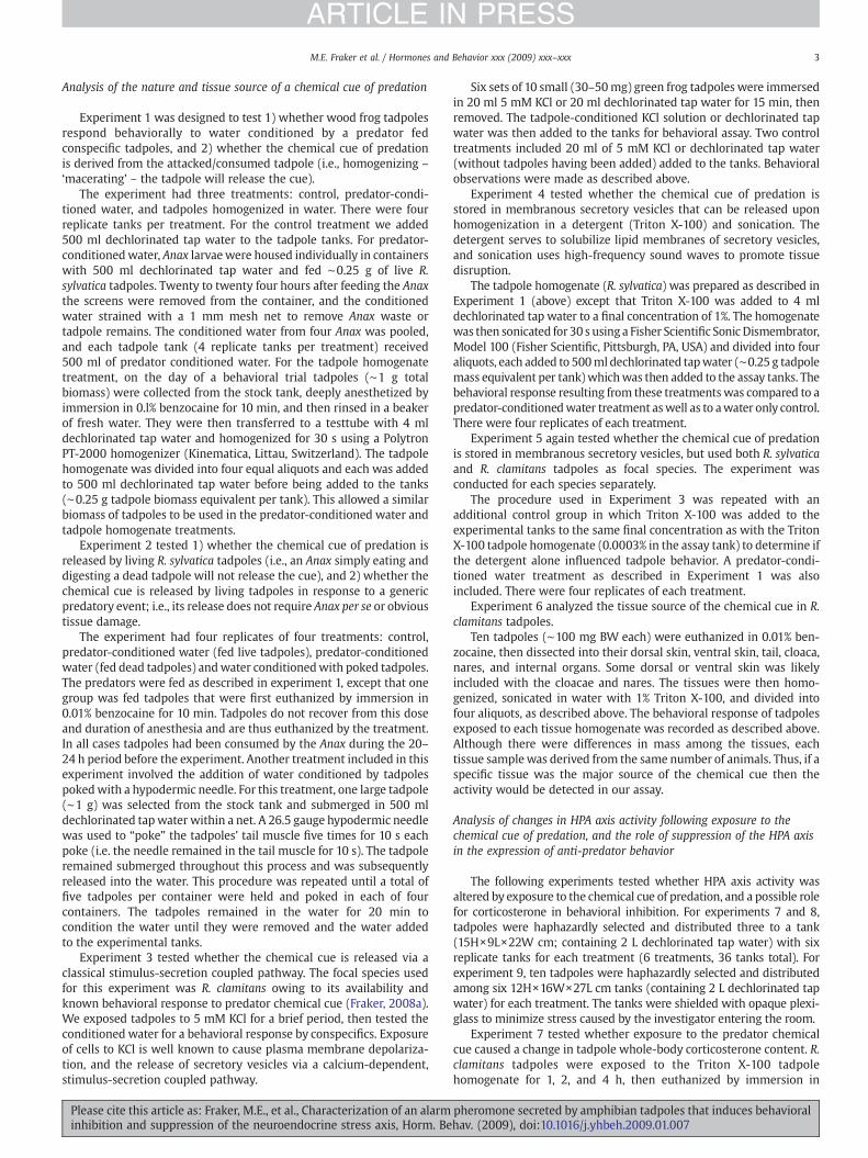
Fig. 1. A chemical cue of predation is produced by living ranid tadpoles and is released via a sttadpoles exposed to different water treatments. (A) Tadpoles (R. sylvatica) reduced activity wtadpoles homogenized inwater. (B) Tadpoles (R. sylvatica) reduced activity when exposed towdead tadpoles. (C) Tadpoles (R. clamitans) reduced activity when exposed to water conditionwater alone or the KCl alone. (D) Tadpoles (R. sylvatica) reduced activity when exposed tohomogenized in water alone. Similar results were obtained with R. clamitans (see Results). ExD, respectively. Pred— predator; Tad— tadpole; homog— homogenate; Triton— Triton X-10not significantly different (Pb0.05).
Please cite this article as: Fraker, M.E., et al., Characterization of an alarminhibition and suppression of the neuroendocrine stress axis, Horm. Be
was tested by reversing the decline in endogenous corticosteronethrough the addition of corticosterone to the aquarium water withpredator chemical cue and monitoring tadpole behavior.
R. clamitans tadpoles were used, and 6 replicates of 10 tadpoles pertank were held in 2 L with 50 nM corticosterone (ICN Biomedicals,Aurora, OH, USA) or dechlorinated tap water (control) for 1 h. Thecorticosterone was dissolved in 100% ethanol and added to the tanksto a final concentration of 50 nM (final concentration of ethanol0.001%). Controls were exposed to the ethanol vehicle to the samefinal concentration. This dose of corticosterone was chosen based onprevious studies in tadpoles and frogs that showed that it restorescorticosterone to baseline in animals in which corticosterone wasreduced by treatment with the corticosteroid synthesis inhibitormetyrapone (Glennemeier and Denver, 2002a,b; Yao et al., 2008).Triton X-100 tadpole homogenate was prepared as described inExperiment 3 and added to the control or corticosterone-treated tanksin aliquots of 20 μl every 15 min (a pilot study showed that thispreparation of tadpole homogenate had N10 times the potency of thehomogenates used in the previous experiments, thus a lower volumewas added to the tanks). Tenminutes after each aliquot of homogenatewas added, behavioral observations were made as described above.This process was repeated until 100 μl of homogenate had been added.
Tadpole behavior was analyzed using a repeated-measures generallinear mixed-effects model (Proc MIXED), with exogeneous corticos-terone treatment as a fixed effect, and cue concentration as a repeatedfactor (Littell et al., 1996). The AIC goodness of fit values were used todetermine which covariance structure to use. If a significantcorticosterone treatment x cue concentration interaction existed,
imulus-secretion coupled pathway. The graphs show themean time spent swimming ofhen exposed to predator-conditioned water (Anax fed tadpoles) but not by euthanizedater conditioned by tadpoles pokedwith a hypodermic needle, but not by predators feded by tadpoles that had been immersed in 5 mM KCl, but not by tadpoles immersed ina homogenate made with euthanized tadpoles in 1% Triton X-100, but not tadpolesperiments 1, 2, 3, 5 as described in the Materials and methods represented in panels A–0. The bars show the mean±SEM; means with the same letter within an experiment are
pheromone secreted by amphibian tadpoles that induces behavioralhav. (2009), doi:10.1016/j.yhbeh.2009.01.007
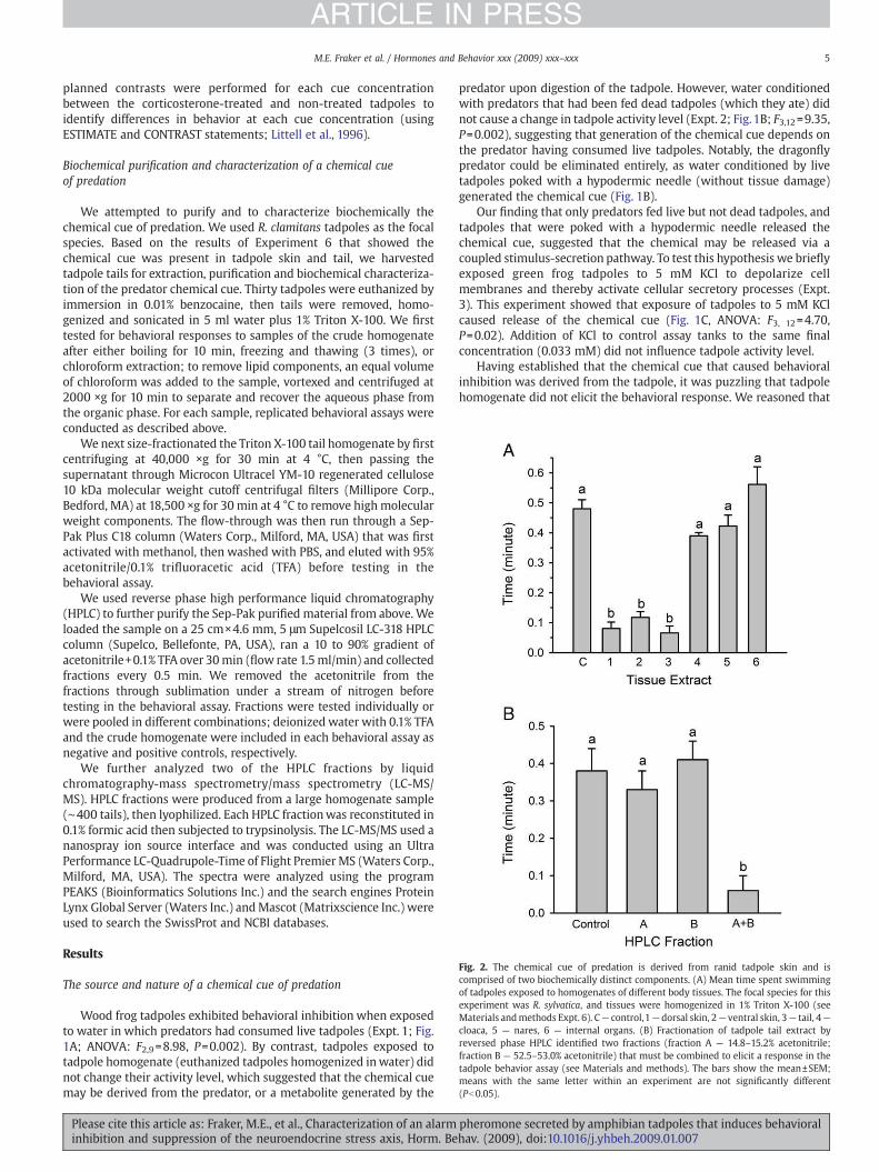
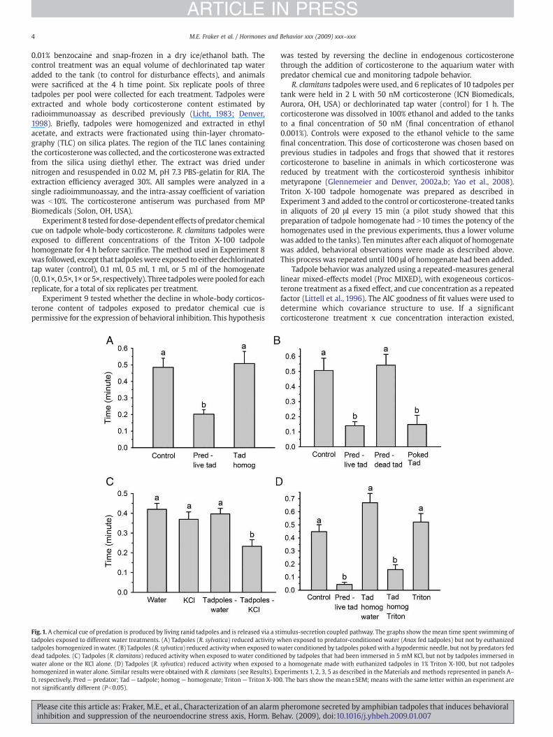
Fig. 2. The chemical cue of predation is derived from ranid tadpole skin and iscomprised of two biochemically distinct components. (A) Mean time spent swimmingof tadpoles exposed to homogenates of different body tissues. The focal species for thisexperiment was R. sylvatica, and tissues were homogenized in 1% Triton X-100 (seeMaterials andmethods Expt. 6). C— control,1— dorsal skin, 2— ventral skin, 3— tail, 4—
cloaca, 5 — nares, 6 — internal organs. (B) Fractionation of tadpole tail extract byreversed phase HPLC identified two fractions (fraction A — 14.8–15.2% acetonitrile;fraction B — 52.5–53.0% acetonitrile) that must be combined to elicit a response in thetadpole behavior assay (see Materials and methods). The bars show the mean±SEM;means with the same letter within an experiment are not significantly different(Pb0.05).
5M.E. Fraker et al. / Hormones and Behavior xxx (2009) xxx–xxx
ARTICLE IN PRESS
planned contrasts were performed for each cue concentrationbetween the corticosterone-treated and non-treated tadpoles toidentify differences in behavior at each cue concentration (usingESTIMATE and CONTRAST statements; Littell et al., 1996).
Biochemical purification and characterization of a chemical cueof predation
We attempted to purify and to characterize biochemically thechemical cue of predation. We used R. clamitans tadpoles as the focalspecies. Based on the results of Experiment 6 that showed thechemical cue was present in tadpole skin and tail, we harvestedtadpole tails for extraction, purification and biochemical characteriza-tion of the predator chemical cue. Thirty tadpoles were euthanized byimmersion in 0.01% benzocaine, then tails were removed, homo-genized and sonicated in 5 ml water plus 1% Triton X-100. We firsttested for behavioral responses to samples of the crude homogenateafter either boiling for 10 min, freezing and thawing (3 times), orchloroform extraction; to remove lipid components, an equal volumeof chloroform was added to the sample, vortexed and centrifuged at2000 ×g for 10 min to separate and recover the aqueous phase fromthe organic phase. For each sample, replicated behavioral assays wereconducted as described above.
We next size-fractionated the Triton X-100 tail homogenate by firstcentrifuging at 40,000 ×g for 30 min at 4 °C, then passing thesupernatant through Microcon Ultracel YM-10 regenerated cellulose10 kDa molecular weight cutoff centrifugal filters (Millipore Corp.,Bedford, MA) at 18,500 ×g for 30 min at 4 °C to remove high molecularweight components. The flow-through was then run through a Sep-Pak Plus C18 column (Waters Corp., Milford, MA, USA) that was firstactivated with methanol, then washed with PBS, and eluted with 95%acetonitrile/0.1% trifluoracetic acid (TFA) before testing in thebehavioral assay.
We used reverse phase high performance liquid chromatography(HPLC) to further purify the Sep-Pak purified material from above. Weloaded the sample on a 25 cm×4.6 mm, 5 μm Supelcosil LC-318 HPLCcolumn (Supelco, Bellefonte, PA, USA), ran a 10 to 90% gradient ofacetonitrile+0.1% TFA over 30min (flow rate 1.5ml/min) and collectedfractions every 0.5 min. We removed the acetonitrile from thefractions through sublimation under a stream of nitrogen beforetesting in the behavioral assay. Fractions were tested individually orwere pooled in different combinations; deionized water with 0.1% TFAand the crude homogenate were included in each behavioral assay asnegative and positive controls, respectively.
We further analyzed two of the HPLC fractions by liquidchromatography-mass spectrometry/mass spectrometry (LC-MS/MS). HPLC fractions were produced from a large homogenate sample(∼400 tails), then lyophilized. Each HPLC fractionwas reconstituted in0.1% formic acid then subjected to trypsinolysis. The LC-MS/MS used ananospray ion source interface and was conducted using an UltraPerformance LC-Quadrupole-Time of Flight PremierMS (Waters Corp.,Milford, MA, USA). The spectra were analyzed using the programPEAKS (Bioinformatics Solutions Inc.) and the search engines ProteinLynx Global Server (Waters Inc.) andMascot (Matrixscience Inc.) wereused to search the SwissProt and NCBI databases.
Results
The source and nature of a chemical cue of predation
Wood frog tadpoles exhibited behavioral inhibition when exposedto water in which predators had consumed live tadpoles (Expt. 1; Fig.1A; ANOVA: F2,9=8.98, P=0.002). By contrast, tadpoles exposed totadpole homogenate (euthanized tadpoles homogenized inwater) didnot change their activity level, which suggested that the chemical cuemay be derived from the predator, or a metabolite generated by the
Please cite this article as: Fraker, M.E., et al., Characterization of an alarminhibition and suppression of the neuroendocrine stress axis, Horm. Be
predator upon digestion of the tadpole. However, water conditionedwith predators that had been fed dead tadpoles (which they ate) didnot cause a change in tadpole activity level (Expt. 2; Fig.1B; F3,12=9.35,P=0.002), suggesting that generation of the chemical cue depends onthe predator having consumed live tadpoles. Notably, the dragonflypredator could be eliminated entirely, as water conditioned by livetadpoles poked with a hypodermic needle (without tissue damage)generated the chemical cue (Fig. 1B).
Our finding that only predators fed live but not dead tadpoles, andtadpoles that were poked with a hypodermic needle released thechemical cue, suggested that the chemical may be released via acoupled stimulus-secretion pathway. To test this hypothesis we brieflyexposed green frog tadpoles to 5 mM KCl to depolarize cellmembranes and thereby activate cellular secretory processes (Expt.3). This experiment showed that exposure of tadpoles to 5 mM KClcaused release of the chemical cue (Fig. 1C, ANOVA: F3, 12=4.70,P=0.02). Addition of KCl to control assay tanks to the same finalconcentration (0.033 mM) did not influence tadpole activity level.
Having established that the chemical cue that caused behavioralinhibition was derived from the tadpole, it was puzzling that tadpolehomogenate did not elicit the behavioral response. We reasoned that
pheromone secreted by amphibian tadpoles that induces behavioralhav. (2009), doi:10.1016/j.yhbeh.2009.01.007
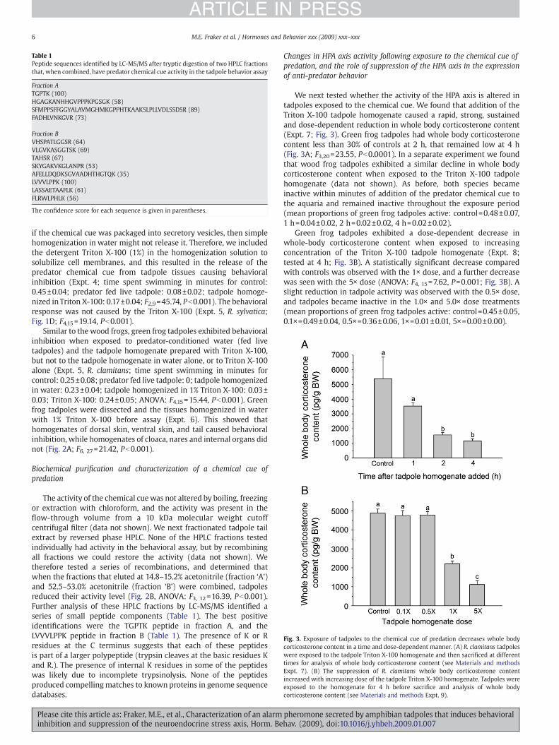
Fig. 3. Exposure of tadpoles to the chemical cue of predation decreases whole bodycorticosterone content in a time and dose-dependent manner. (A) R. clamitans tadpoleswere exposed to the tadpole Triton X-100 homogenate and then sacrificed at differenttimes for analysis of whole body corticosterone content (see Materials and methodsExpt. 7). (B) The suppression of R. clamitans whole body corticosterone contentincreased with increasing dose of the tadpole Triton X-100 homogenate. Tadpoles wereexposed to the homogenate for 4 h before sacrifice and analysis of whole bodycorticosterone content (see Materials and methods Expt. 9).
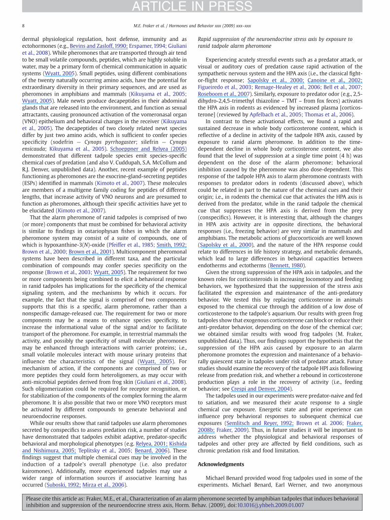
Table 1Peptide sequences identified by LC-MS/MS after tryptic digestion of two HPLC fractionsthat, when combined, have predator chemical cue activity in the tadpole behavior assay
Fraction ATGPTK (100)HGAGKANHHGVPPPKPGSGK (58)SFMPPSFFGGYALAVMGHMKGPPHTKAAKSLPLLVDLSSDSR (89)FADHLVNKGVR (73)
Fraction BVHSPATLGGSR (64)VLGVKASGGTSK (69)TAHSR (67)SKYGAKVKGLANPR (53)AFELLDQDKSGVAADHTHGTQK (35)LVVVLPPK (100)LASSAETAAFLK (61)FLRWLPHLK (56)
The confidence score for each sequence is given in parentheses.
6 M.E. Fraker et al. / Hormones and Behavior xxx (2009) xxx–xxx
ARTICLE IN PRESS
if the chemical cue was packaged into secretory vesicles, then simplehomogenization in water might not release it. Therefore, we includedthe detergent Triton X-100 (1%) in the homogenization solution tosolubilize cell membranes, and this resulted in the release of thepredator chemical cue from tadpole tissues causing behavioralinhibition (Expt. 4; time spent swimming in minutes for control:0.45±0.04; predator fed live tadpole: 0.08±0.02; tadpole homoge-nized in Triton X-100: 0.17±0.04; F2,9=45.74, Pb0.001). The behavioralresponse was not caused by the Triton X-100 (Expt. 5, R. sylvatica;Fig. 1D; F4,15=19.14, Pb0.001).
Similar to the wood frogs, green frog tadpoles exhibited behavioralinhibition when exposed to predator-conditioned water (fed livetadpoles) and the tadpole homogenate prepared with Triton X-100,but not to the tadpole homogenate in water alone, or to Triton X-100alone (Expt. 5, R. clamitans; time spent swimming in minutes forcontrol: 0.25±0.08; predator fed live tadpole: 0; tadpole homogenizedin water: 0.23±0.04; tadpole homogenized in 1% Triton X-100: 0.03±0.03; Triton X-100: 0.24±0.05; ANOVA: F4,15=15.44, Pb0.001). Greenfrog tadpoles were dissected and the tissues homogenized in waterwith 1% Triton X-100 before assay (Expt. 6). This showed thathomogenates of dorsal skin, ventral skin, and tail caused behavioralinhibition, while homogenates of cloaca, nares and internal organs didnot (Fig. 2A; F6, 27=21.42, Pb0.001).
Biochemical purification and characterization of a chemical cue ofpredation
The activity of the chemical cuewas not altered by boiling, freezingor extraction with chloroform, and the activity was present in theflow-through volume from a 10 kDa molecular weight cutoffcentrifugal filter (data not shown). We next fractionated tadpole tailextract by reversed phase HPLC. None of the HPLC fractions testedindividually had activity in the behavioral assay, but by recombiningall fractions we could restore the activity (data not shown). Wetherefore tested a series of recombinations, and determined thatwhen the fractions that eluted at 14.8–15.2% acetonitrile (fraction ‘A’)and 52.5–53.0% acetonitrile (fraction ‘B’) were combined, tadpolesreduced their activity level (Fig. 2B, ANOVA: F3, 12=16.39, Pb0.001).Further analysis of these HPLC fractions by LC-MS/MS identified aseries of small peptide components (Table 1). The best positiveidentifications were the TGPTK peptide in fraction A, and theLVVVLPPK peptide in fraction B (Table 1). The presence of K or Rresidues at the C terminus suggests that each of these peptidesis part of a larger polypeptide (trypsin cleaves at the basic residues Kand R.). The presence of internal K residues in some of the peptideswas likely due to incomplete trypsinolysis. None of the peptidesproduced compellingmatches to known proteins in genome sequencedatabases.
Please cite this article as: Fraker, M.E., et al., Characterization of an alarminhibition and suppression of the neuroendocrine stress axis, Horm. Be
Changes in HPA axis activity following exposure to the chemical cue ofpredation, and the role of suppression of the HPA axis in the expressionof anti-predator behavior
We next tested whether the activity of the HPA axis is altered intadpoles exposed to the chemical cue. We found that addition of theTriton X-100 tadpole homogenate caused a rapid, strong, sustainedand dose-dependent reduction in whole body corticosterone content(Expt. 7; Fig. 3). Green frog tadpoles had whole body corticosteronecontent less than 30% of controls at 2 h, that remained low at 4 h(Fig. 3A; F3,20=23.55, Pb0.0001). In a separate experiment we foundthat wood frog tadpoles exhibited a similar decline in whole bodycorticosterone content when exposed to the Triton X-100 tadpolehomogenate (data not shown). As before, both species becameinactive within minutes of addition of the predator chemical cue tothe aquaria and remained inactive throughout the exposure period(mean proportions of green frog tadpoles active: control=0.48±0.07,1 h=0.04±0.02, 2 h=0.02±0.02, 4 h=0.02±0.02).
Green frog tadpoles exhibited a dose-dependent decrease inwhole-body corticosterone content when exposed to increasingconcentration of the Triton X-100 tadpole homogenate (Expt. 8;tested at 4 h; Fig. 3B). A statistically significant decrease comparedwith controls was observed with the 1× dose, and a further decreasewas seen with the 5× dose (ANOVA: F4, 15=7.62, P=0.001; Fig. 3B). Aslight reduction in tadpole activity was observed with the 0.5× dose,and tadpoles became inactive in the 1.0× and 5.0× dose treatments(mean proportions of green frog tadpoles active: control=0.45±0.05,0.1×=0.49±0.04, 0.5×=0.36±0.06, 1×=0.01±0.01, 5×=0.00±0.00).
pheromone secreted by amphibian tadpoles that induces behavioralhav. (2009), doi:10.1016/j.yhbeh.2009.01.007
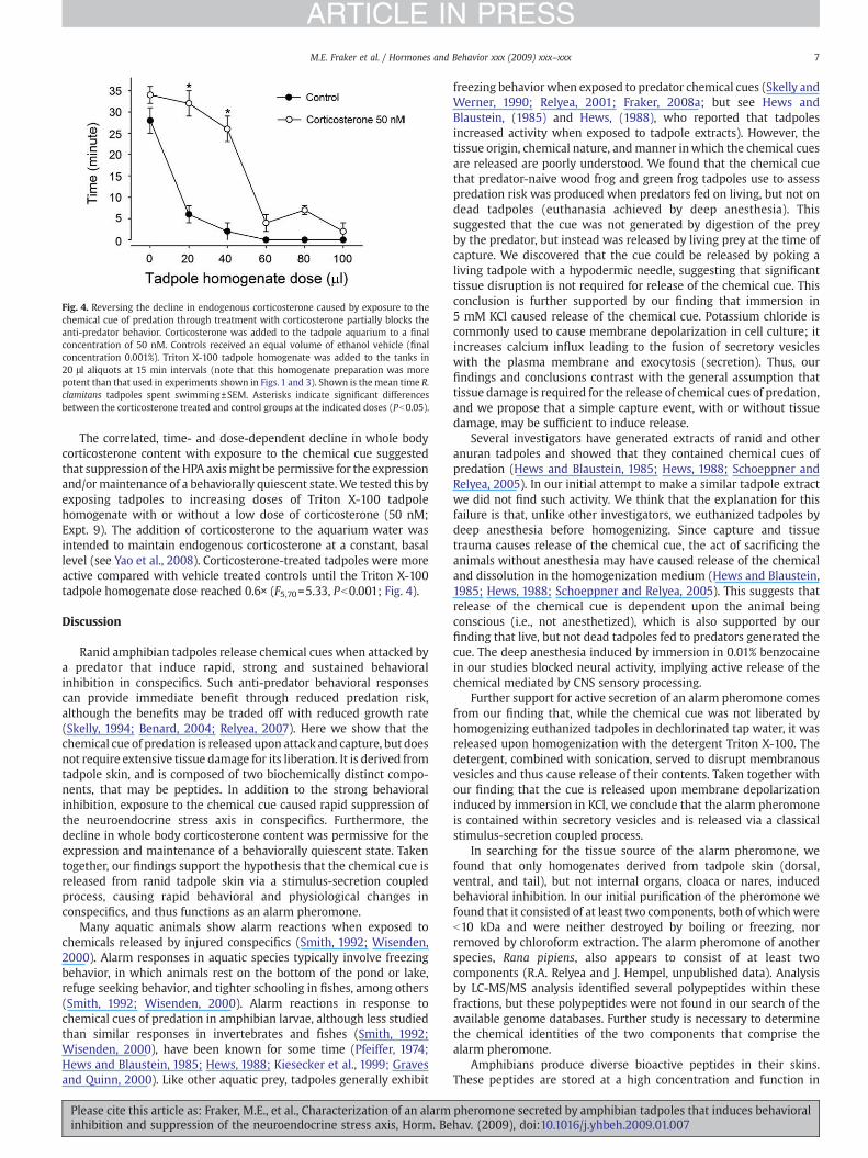
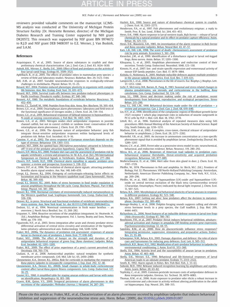
Fig. 4. Reversing the decline in endogenous corticosterone caused by exposure to thechemical cue of predation through treatment with corticosterone partially blocks theanti-predator behavior. Corticosterone was added to the tadpole aquarium to a finalconcentration of 50 nM. Controls received an equal volume of ethanol vehicle (finalconcentration 0.001%). Triton X-100 tadpole homogenate was added to the tanks in20 μl aliquots at 15 min intervals (note that this homogenate preparation was morepotent than that used in experiments shown in Figs. 1 and 3). Shown is the mean time R.clamitans tadpoles spent swimming±SEM. Asterisks indicate significant differencesbetween the corticosterone treated and control groups at the indicated doses (Pb0.05).
7M.E. Fraker et al. / Hormones and Behavior xxx (2009) xxx–xxx
ARTICLE IN PRESS
The correlated, time- and dose-dependent decline in whole bodycorticosterone content with exposure to the chemical cue suggestedthat suppression of the HPA axismight be permissive for the expressionand/ormaintenance of a behaviorally quiescent state.We tested this byexposing tadpoles to increasing doses of Triton X-100 tadpolehomogenate with or without a low dose of corticosterone (50 nM;Expt. 9). The addition of corticosterone to the aquarium water wasintended to maintain endogenous corticosterone at a constant, basallevel (see Yao et al., 2008). Corticosterone-treated tadpoles were moreactive compared with vehicle treated controls until the Triton X-100tadpole homogenate dose reached 0.6× (F5,70=5.33, Pb0.001; Fig. 4).
Discussion
Ranid amphibian tadpoles release chemical cues when attacked bya predator that induce rapid, strong and sustained behavioralinhibition in conspecifics. Such anti-predator behavioral responsescan provide immediate benefit through reduced predation risk,although the benefits may be traded off with reduced growth rate(Skelly, 1994; Benard, 2004; Relyea, 2007). Here we show that thechemical cue of predation is released upon attack and capture, but doesnot require extensive tissue damage for its liberation. It is derived fromtadpole skin, and is composed of two biochemically distinct compo-nents, that may be peptides. In addition to the strong behavioralinhibition, exposure to the chemical cue caused rapid suppression ofthe neuroendocrine stress axis in conspecifics. Furthermore, thedecline in whole body corticosterone content was permissive for theexpression and maintenance of a behaviorally quiescent state. Takentogether, our findings support the hypothesis that the chemical cue isreleased from ranid tadpole skin via a stimulus-secretion coupledprocess, causing rapid behavioral and physiological changes inconspecifics, and thus functions as an alarm pheromone.
Many aquatic animals show alarm reactions when exposed tochemicals released by injured conspecifics (Smith, 1992; Wisenden,2000). Alarm responses in aquatic species typically involve freezingbehavior, in which animals rest on the bottom of the pond or lake,refuge seeking behavior, and tighter schooling in fishes, among others(Smith, 1992; Wisenden, 2000). Alarm reactions in response tochemical cues of predation in amphibian larvae, although less studiedthan similar responses in invertebrates and fishes (Smith, 1992;Wisenden, 2000), have been known for some time (Pfeiffer, 1974;Hews and Blaustein, 1985; Hews, 1988; Kiesecker et al., 1999; Gravesand Quinn, 2000). Like other aquatic prey, tadpoles generally exhibit
Please cite this article as: Fraker, M.E., et al., Characterization of an alarminhibition and suppression of the neuroendocrine stress axis, Horm. Be
freezing behavior when exposed to predator chemical cues (Skelly andWerner, 1990; Relyea, 2001; Fraker, 2008a; but see Hews andBlaustein, (1985) and Hews, (1988), who reported that tadpolesincreased activity when exposed to tadpole extracts). However, thetissue origin, chemical nature, andmanner inwhich the chemical cuesare released are poorly understood. We found that the chemical cuethat predator-naive wood frog and green frog tadpoles use to assesspredation risk was produced when predators fed on living, but not ondead tadpoles (euthanasia achieved by deep anesthesia). Thissuggested that the cue was not generated by digestion of the preyby the predator, but instead was released by living prey at the time ofcapture. We discovered that the cue could be released by poking aliving tadpole with a hypodermic needle, suggesting that significanttissue disruption is not required for release of the chemical cue. Thisconclusion is further supported by our finding that immersion in5 mM KCl caused release of the chemical cue. Potassium chloride iscommonly used to cause membrane depolarization in cell culture; itincreases calcium influx leading to the fusion of secretory vesicleswith the plasma membrane and exocytosis (secretion). Thus, ourfindings and conclusions contrast with the general assumption thattissue damage is required for the release of chemical cues of predation,and we propose that a simple capture event, with or without tissuedamage, may be sufficient to induce release.
Several investigators have generated extracts of ranid and otheranuran tadpoles and showed that they contained chemical cues ofpredation (Hews and Blaustein, 1985; Hews, 1988; Schoeppner andRelyea, 2005). In our initial attempt to make a similar tadpole extractwe did not find such activity. We think that the explanation for thisfailure is that, unlike other investigators, we euthanized tadpoles bydeep anesthesia before homogenizing. Since capture and tissuetrauma causes release of the chemical cue, the act of sacrificing theanimals without anesthesia may have caused release of the chemicaland dissolution in the homogenization medium (Hews and Blaustein,1985; Hews, 1988; Schoeppner and Relyea, 2005). This suggests thatrelease of the chemical cue is dependent upon the animal beingconscious (i.e., not anesthetized), which is also supported by ourfinding that live, but not dead tadpoles fed to predators generated thecue. The deep anesthesia induced by immersion in 0.01% benzocainein our studies blocked neural activity, implying active release of thechemical mediated by CNS sensory processing.
Further support for active secretion of an alarm pheromone comesfrom our finding that, while the chemical cue was not liberated byhomogenizing euthanized tadpoles in dechlorinated tap water, it wasreleased upon homogenization with the detergent Triton X-100. Thedetergent, combined with sonication, served to disrupt membranousvesicles and thus cause release of their contents. Taken together withour finding that the cue is released upon membrane depolarizationinduced by immersion in KCl, we conclude that the alarm pheromoneis contained within secretory vesicles and is released via a classicalstimulus-secretion coupled process.
In searching for the tissue source of the alarm pheromone, wefound that only homogenates derived from tadpole skin (dorsal,ventral, and tail), but not internal organs, cloaca or nares, inducedbehavioral inhibition. In our initial purification of the pheromone wefound that it consisted of at least two components, both of whichwereb10 kDa and were neither destroyed by boiling or freezing, norremoved by chloroform extraction. The alarm pheromone of anotherspecies, Rana pipiens, also appears to consist of at least twocomponents (R.A. Relyea and J. Hempel, unpublished data). Analysisby LC-MS/MS analysis identified several polypeptides within thesefractions, but these polypeptides were not found in our search of theavailable genome databases. Further study is necessary to determinethe chemical identities of the two components that comprise thealarm pheromone.
Amphibians produce diverse bioactive peptides in their skins.These peptides are stored at a high concentration and function in
pheromone secreted by amphibian tadpoles that induces behavioralhav. (2009), doi:10.1016/j.yhbeh.2009.01.007
8 M.E. Fraker et al. / Hormones and Behavior xxx (2009) xxx–xxx
ARTICLE IN PRESS
dermal physiological regulation, host defense, immunity and asectohormones (e.g., Bevins and Zasloff, 1990; Erspamer, 1994; Giulianiet al., 2008). While pheromones that are transported through air tendto be small volatile compounds, peptides, which are highly soluble inwater, may be a primary form of chemical communication in aquaticsystems (Wyatt, 2005). Small peptides, using different combinationsof the twenty naturally occurring amino acids, have the potential forextraordinary diversity in their primary sequences, and are used aspheromones in amphibians and mammals (Kikuyama et al., 2005;Wyatt, 2005). Male newts produce decapeptides in their abdominalglands that are released into the environment, and function as sexualattractants, causing pronounced activation of the vomeronasal organ(VNO) epithelium and behavioral changes in the receiver (Kikuyamaet al., 2005). The decapeptides of two closely related newt speciesdiffer by just two amino acids, which is sufficient to confer speciesspecificity (sodefrin — Cynops pyrrhogaster; silefrin — Cynopsensicauda; Kikuyama et al., 2005). Schoeppner and Relyea (2005)demonstrated that different tadpole species emit species-specificchemical cues of predation (and also V. Cuddupah, S.A. McCollum andR.J. Denver, unpublished data). Another, recent example of peptidesfunctioning as pheromones are the exocrine-gland-secreting peptides(ESPs) identified in mammals (Kimoto et al., 2007). These moleculesare members of a multigene family coding for peptides of differentlengths, that increase activity of VNO neurons and are presumed tofunction as pheromones, although their specific activities have yet tobe elucidated (Kimoto et al., 2007).
That the alarm pheromone of ranid tadpoles is comprised of two(or more) components that must be combined for behavioral activityis similar to findings in ostariophysan fishes in which the alarmpheromone system may consist of a suite of compounds, one ofwhich is hypoxanthine-3(N)-oxide (Pfeiffer et al., 1985; Smith, 1992;Brown et al., 2000; Brown et al., 2001). Multicomponent pheromonalsystems have been described in different taxa, and the particularcombination of compounds may confer species specificity on theresponse (Brown et al., 2003; Wyatt, 2005). The requirement for twoor more components being combined to elicit a behavioral responsein ranid tadpoles has implications for the specificity of the chemicalsignaling system, and the mechanisms by which it occurs. Forexample, the fact that the signal is comprised of two componentssupports that this is a specific, alarm pheromone, rather than anonspecific damage-released cue. The requirement for two or morecomponents may be a means to enhance species specificity, toincrease the informational value of the signal and/or to facilitatetransport of the pheromone. For example, in terrestrial mammals theactivity, and possibly the specificity of small molecule pheromonesmay be enhanced through interactions with carrier proteins; i.e.,small volatile molecules interact with mouse urinary proteins thatinfluence the characteristics of the signal (Wyatt, 2005). Formechanism of action, if the components are comprised of two ormore peptides they could form heteroligomers, as may occur withanti-microbial peptides derived from frog skin (Giuliani et al., 2008).Such oligomerization could be required for receptor recognition, orfor stabilization of the components of the complex forming the alarmpheromone. It is also possible that two or more VNO receptors mustbe activated by different compounds to generate behavioral andneuroendocrine responses.
While our results show that ranid tadpoles use alarm pheromonessecreted by conspecifics to assess predation risk, a number of studieshave demonstrated that tadpoles exhibit adaptive, predator-specificbehavioral and morphological phenotypes (e.g. Relyea, 2001; Kishidaand Nishimura, 2005; Teplitsky et al., 2005; Benard, 2006). Thesefindings suggest that multiple chemical cues may be involved in theinduction of a tadpole's overall phenotype (i.e. also predatorkairomones). Additionally, more experienced tadpoles may use awider range of information sources if associative learning hasoccurred (Suboski, 1992; Mirza et al., 2006).
Please cite this article as: Fraker, M.E., et al., Characterization of an alarminhibition and suppression of the neuroendocrine stress axis, Horm. Be
Rapid suppression of the neuroendocrine stress axis by exposure toranid tadpole alarm pheromone
Experiencing acutely stressful events such as a predator attack, orvisual or auditory cues of predation cause rapid activation of thesympathetic nervous system and the HPA axis (i.e., the classical fight-or-flight response; Sapolsky et al., 2000; Canoine et al., 2002;Figueiredo et al., 2003; Remage-Healey et al., 2006; Bell et al., 2007;Roseboom et al., 2007). Similarly, exposure to predator odor (e.g., 2,5-dihydro-2,4,5-trimethyl thiazoline – TMT – from fox feces) activatesthe HPA axis in rodents as evidenced by increased plasma [corticos-terone] (reviewed by Apfelbach et al., 2005; Thomas et al., 2006).
In contrast to these activational effects, we found a rapid andsustained decrease in whole body corticosterone content, which isreflective of a decline in activity of the tadpole HPA axis, caused byexposure to ranid alarm pheromone. In addition to the time-dependent decline in whole body corticosterone content, we alsofound that the level of suppression at a single time point (4 h) wasdependent on the dose of the alarm pheromone; behavioralinhibition caused by the pheromone was also dose-dependent. Thisresponse of the tadpole HPA axis to alarm pheromone contrasts withresponses to predator odors in rodents (discussed above), whichcould be related in part to the nature of the chemical cues and theirorigin; i.e., in rodents the chemical cue that activates the HPA axis isderived from the predator, while in the ranid tadpole the chemicalcue that suppresses the HPA axis is derived from the prey(conspecifics). However, it is interesting that, although the changesin HPA axis activity are in opposite directions, the behavioralresponses (i.e., freezing behavior) are very similar in mammals andamphibians. The metabolic actions of glucocorticoids are well known(Sapolsky et al., 2000), and the nature of the HPA response couldrelate to differences in life history strategy, and metabolic demands,which lead to large differences in behavioral capacities betweenendotherms and ectotherms (Bennett, 1980).
Given the strong suppression of the HPA axis in tadpoles, and theknown roles for corticosteroids in increasing locomotory and feedingbehaviors, we hypothesized that the suppression of the stress axisfacilitated the expression and maintenance of the anti-predatorybehavior. We tested this by replacing corticosterone in animalsexposed to the chemical cue through the addition of a low dose ofcorticosterone to the tadpole's aquarium. Our results with green frogtadpoles show that exogenous corticosterone can block or reduce theiranti-predator behavior, depending on the dose of the chemical cue;we obtained similar results with wood frog tadpoles (M. Fraker,unpublished data). Thus, our findings support the hypothesis that thesuppression of the HPA axis caused by exposure to an alarmpheromone promotes the expression and maintenance of a behavio-rally quiescent state in tadpoles under risk of predator attack. Futurestudies should examine the recovery of the tadpole HPI axis followingrelease from predation risk, and whether a rebound in corticosteroneproduction plays a role in the recovery of activity (i.e., feedingbehavior; see Crespi and Denver, 2004).
The tadpoles used in our experiments were predator-naive and fedto satiation, and we measured their acute response to a singlechemical cue exposure. Energetic state and prior experience caninfluence prey behavioral responses to subsequent chemical cueexposures (Semlitsch and Reyer, 1992; Brown et al. 2006; Fraker,2008b; Fraker, 2009). Thus, in future studies it will be important toaddress whether the physiological and behavioral responses oftadpoles and other prey are affected by field conditions, such aschronic predation risk and food limitation.
Acknowledgments
Michael Benard provided wood frog tadpoles used in some of theexperiments. Michael Benard, Earl Werner, and two anonymous
pheromone secreted by amphibian tadpoles that induces behavioralhav. (2009), doi:10.1016/j.yhbeh.2009.01.007
9M.E. Fraker et al. / Hormones and Behavior xxx (2009) xxx–xxx
ARTICLE IN PRESS
reviewers provided valuable comments on the manuscript. LC/MS-MS analysis was conducted at The University of Michigan ProteinStructure Facility (Dr. Henriette Remmer, director) of the MichiganDiabetes Research and Training Center supported by NIH grantDK20572. This research was supported by NSF grant IBN 9974672to R.J.D and NSF grant DEB 9408397 to E.E. Werner, J. Van Buskirk,and S.A.M.
References
Acquistapace, P., et al., 2005. Source of alarm substances in crayfish and theirpreliminary chemical characterization. Can. J. Zool.-Can. J. Zool. 83, 1624–1630.
Anholt, B.R., Werner, E., Skelly, D.K., 2000. Effect of food and predators on the activity offour larval ranid frogs. Ecology 81, 3509–3521.
Apfelbach, R., et al., 2005. The effects of predator odors in mammalian prey species: areview of field and laboratory studies. Neurosci. Biobehav. Rev. 29, 1123–1144.
Bell, A.M., et al., 2007. Variable neuroendocrine responses to ecologically-relevantchallenges in sticklebacks. Physiol. Behav. 91, 15–25.
Benard, M.F., 2004. Predator-induced phenotypic plasticity in organisms with complexlife histories. Ann. Rev. Ecolog. Evol. Syst. 35, 651–673.
Benard, M.F., 2006. Survival trade-offs between two predator-induced phenotypes inPacific treefrogs (Pseudacris regilla). Ecology. 87, 340–346.
Bennett, A.F., 1980. The metabolic foundations of vertebrate behavior. Bioscience. 30,452–456.
Bevins, C.L., Zasloff, M., 1990. Peptides from frog skin. Annu. Rev. Biochem. 59, 395–414.Brown, G.E., et al., 2000. Ostariophysan alarm pheromones: laboratory and field tests of
the functional significance of nitrogen oxides. J. Chem. Ecol. 26, 139–154.Brown, G.E., et al., 2001. Behavioural responses of fathead minnows to hypoxanthine-3-
N-oxide at varying concentrations. J. Fish Biol. 58, 1465–1470.Brown, G.E., et al., 2003. Nitrogen oxides elicit antipredator responses in juvenile
channel catfish, but not in convict cichlids or rainbow trout: conservation of theostariophysan alarm pheromone. J. Chem. Ecol. 29, 1781–1796.
Brown, G.E., et al., 2006. The dynamic nature of antipredator behavior: prey fishintegrate threat-sensitive antipredator responses within background levels ofpredation risk. Behav. Ecol. Sociobiol. 61, 9–16.
Canoine, V., et al., 2002. The stress response of European stonechats depends on thetype of stressor. Behaviour 139, 1303–1311.
Cashner, M.F., 2004. Are spotted bass (Micropterus punctulatus) attracted to Schreckst-off? A test of the predator attraction hypothesis. Copeia 592–598.
Chivers, D.P., Mirza, R.S., 2000. Predator diet cues and the assessment of predation risk byaquatic vertebrates: a review and prospectus. In: M.-K.A., et al. (Ed.), 9th InternationalSymposium on Chemical Signals in Vertebrates. Krakow, Poland, pp. 277–284.
Chivers, D.P., Smith, R.J.F., 1998. Chemical alarm signalling in aquatic predator–preysystems: a review and prospectus. Ecoscience 5, 338–352.
Chivers, D.P., et al., 1996. The evolution of chemical alarm signals: attracting predatorsbenefits alarm signal senders. Am. Nat. 148, 649–659.
Crespi, E.J., Denver, R.J., 2004. Ontogeny of corticotropin-releasing factor effects onlocomotion and foraging in the Western spadefoot toad (Spea hammondii). Horm.Behav. 46, 399–410.
Crespi, E.J., Denver, R.J., 2005. Roles of stress hormones in food intake regulation inanuran amphibians throughout the life cycle. Comp. Biochem. Physiol., Part A Mol.Integr. Physiol. 141, 381–390.
Denver, R.J., 1998. Hormonal correlates of environmentally induced metamorphosis inthe Western spadefoot toad, Scaphiopus hammondii. Gen. Comp. Endocrinol. 110,326–336.
Denver, R.J., in press. Structural and functional evolution of vertebrate neuroendocrinestress systems. Ann. New York Acad. Sci. doi:10.1111/j.1749-6632.2009.04433.x.
Dodson, S.I., et al., 1994. Nonvisual communication in fresh water benthos — anoverview. J. North Am.n Benthol. Soc. 13, 268–282.
Erspamer, V., 1994. Bioactive secretions of the amphibian integument. In: Heatwole, H.(Ed.), Amphibian Biology: The Integument, Vol. 1. Surrey, Beatty and Sons, Norton,NSW, pp. 178–350.
Figueiredo, H.F., et al., 2003. Stress integration after acute and chronic predator stress:differential activation of central stress circuitry and sensitization of the hypotha-lamo–pituitary–adrenocortical axis. Endocrinology 144, 5249–5258.
Fraker, M.E., 2008a. The dynamics of predation risk assessment: responses of anuranlarvae to chemical cues of predators. J. Anim. Ecol. 77, 638–645.
Fraker, M.E., 2008b. The effect of hunger on the strength and duration of theantipredator behavioral response of green frog (Rana clamitans) tadpoles. Behav.Ecol. Sociobiol. 62, 1201–1205.
Fraker, M.E., 2009. The effect of prior experience of a prey's current perceived risk.Oecologia 158, 765–774.
Giuliani, A., et al., 2008. Antimicrobial peptides: natural templates for syntheticmembrane-active compounds. Cell. Mol. Life Sci. 65, 2450–2460.
Glennemeier, K.A., Denver, R.J., 2002a. Role for corticoids in mediating the response ofRana pipiens tadpoles to intraspecific competition. J. Exp. Zool. 292, 32–40.
Glennemeier, K.A., Denver, R.J., 2002b. Small changes in whole-body corticosteronecontent affect larval Rana pipiens fitness components. Gen. Comp. Endocrinol. 127,16–25.
Gosner, K.L., 1960. A simplified table for staging anuran embryos and larvae with noteson identification. Herpetlogica 16, 183–190.
Graves, B.M., Quinn, V.S., 2000. Temporal persistence of alarm pheromones in skinsecretions of the salamander, Plethodon cinereus. J. Herpetol. 34, 287–291.
Please cite this article as: Fraker, M.E., et al., Characterization of an alarminhibition and suppression of the neuroendocrine stress axis, Horm. Be
Hazlett, B.A., 1990. Source and nature of disturbance chemical system in crayfish.J. Chem. Ecol. 16, 2263–2275.
Henderson, P.A., et al., 1997. Fish pheromones and evolutionary enigmas: a reply toSmith. Proc. R. Soc. Lond., B Biol. Sci. 264, 451–453.
Hews, D.K., 1988. Alarm response in larval western toads, Bufo boreas— release of larvalchemicals by a natural predator and its effect on predator capture efficiency. Anim.Behav. 36, 125–133.
Hews, D.K., Blaustein, A.R., 1985. An investigation of the alarm response in Bufo boreasand Rana cascadae tadpoles. Behav. Neural Biol. 43, 47–57.
Kats, L.B., Dill, L.M., 1998. The scent of death: chemosensory assessment of predationrisk by prey animals. Ecoscience 5, 361–394.
Kiesecker, J.M., et al., 1999. Identification of a disturbance signal in larval red-leggedfrogs, Rana aurora. Anim. Behav. 57, 1295–1300.
Kikuyama, S., et al., 2005. Amphibian pheromones and endocrine control of theirsecretion. Trends Comp. Endocrinol. Neurobiol. 1040, 123–130.
Kimoto, H., et al., 2007. Sex- and strain-specific expression and vomeronasal activity ofmouse ESP family peptides. Curr. Biol. 17, 1879–1884.
Kishida, O., Nishimura, K., 2005. Multiple inducible defences against multiple predatorsin the anuran tadpole, Rana pirica. Evol. Ecol. Res. 7, 619–631.
Lamprecht, I., et al., 2008. Pheromones in the life of insects. Eur. Biophys. J. Biophys. Lett.37, 1253–1260.
Licht, P., McCreery, B.R., Barnes, R., Pang, R., 1983. Seasonal and stress related changes inplasma gonadotropins, sex steroids, and corticosterone in the bullfrog, Ranacatesbeiana. Gen. Comp. Endocrinol. 50, 124–145.
Lima, S.L., 1998. Stress and decision making under the risk of predation: recentdevelopments from behavioral, reproductive, and ecological perspectives. StressBehav. 215–290.
Lima, S.L., Dill, L.M., 1990. Behavioral decisions made under the risk of predation — areview and prospectus. Can. J. Zool.-Can. J. Zool. 68, 619–640.
Lin, H.Y., et al., 1998. Identification of the cytoplasmic regions of fibroblast growth factor(FGF) receptor 1 which play important roles in induction of neurite outgrowth inPC12 cells by FGF-1. Mol. Cell. Biol. 18, 3762–3770.
Littell, R.C., et al., 1996. Statistical analysis of repeated measures data using SASprocedures. 88th Annual Meeting of the American Society of Animal Science. RapidCity, South Dakota, pp. 1216–1231.
Madison, D.M., et al., 2002. A complex, cross-taxon, chemical releaser of antipredatorbehavior in amphibians. J. Chem. Ecol. 28, 2271–2282.
Manteifel, Y.B., et al., 2005. An increase in ammonium concentration as a non-specificpheromone signal that is avoided by amphibian larvae. Zoologichesky Zhurnal. 84,1289–1297.
Masini, C.V., et al., 2005. Ferret odor as a processive stress model in rats: neurochemical,behavioral, and endocrine evidence. Behav. Neurosci. 119, 280–292.
Mirza, R.S., et al., 2006. Responses of American toad tadpoles to predation cues:behavioural response thresholds, threat-sensitivity and acquired predationrecognition. Behaviour. 143, 877–889.
Mullerschwarze, D., et al., 1984. Alert odor from skin gland in deer. J. Chem. Ecol. 10,1707–1729.
Pfeiffer, W., 1974. Pheromones in fish and amphibia. In: Birch, M.C. (Ed.), Frontiers ofBiology, Vol. 32. Pheromones. VIII North-Holland Publishing Company: Amsterdam,Netherlands. American Elsevier Publishing Company, Inc., New York, N.Y., U.S.A.,pp. 269–296.
Pfeiffer, W., et al., 1985. Effect of hypoxanthine-3(N)-oxide and hypoxanthine-1(N)-oxide on central nervous excitation of the black tetra Gymnocorymbus ternetzi(Characidae, Ostariophysi, Pisces) indicated by dorsal light response. J. Chem. Ecol.11, 507–523.
Relyea, R.A., 2001. Morphological and behavioral plasticity of larval anurans in responseto different predators. Ecology 82, 523–540.
Relyea, R.A., 2007. Getting out alive: how predators affect the decision to metamor-phose. Oecologia 152, 389–400.
Remage-Healey, L., et al., 2006. Dolphin foraging sounds suppress calling and elevatestress hormone levels in a prey species, the Gulf toadfish. J. Exp. Biol. 209,4444–4451.
Richardson, J.L., 2006. Novel features of an inducible defense system in larval tree frogs(Hyla chrysoscelis). Ecology 87, 780–787.
Roseboom, P.H., et al., 2007. Predator threat induces behavioral inhibition, pituitary–adrenal activation and changes in amygdala CRF-binding protein gene expression.Psychoneuroendocrinology 32, 44–55.
Sapolsky, R.M., et al., 2000. How do glucocorticoids influence stress responses?Integrating permissive, suppressive, stimulatory, and preparative actions. Endocr.Rev. 21, 55–89.
Schoeppner, N.M., Relyea, R.A., 2005. Damage, digestion, and defence: the roles of alarmcues and kairomones for inducing prey defences. Ecol. Lett. 8, 505–512.
Semlitsch, R.D., Reyer, H.U.,1992. Modification of anti-predator behaviour in tadpoles byenvironmental conditioning. J. Anim. Ecol 61, 353–360.
Skelly, D.K., 1994. Activity level and the susceptibility of anuran larvae to predation.Anim. Behav. 47, 465–468.
Skelly, D.K., Werner, E.E., 1990. Behavioral and life-historical responses of larvalAmerican toads to an odonate predator. Ecology 71, 2313–2322.
Smith, R., 1992. Alarm signals in fishes. Rev. Fish Biol. Fish. 2, 33–63.Suboski, M.D., 1992. Releaser induced recognition learning by amphibians and reptiles.
Anim. Learn. Behav. 20, 63–82.Teplitsky, C., et al., 2005. Common pesticide increases costs of antipredator defenses in
Rana temporaria tadpoles. Environ. Sci. Technol. 39, 6079–6085.Thomas, R.M., et al., 2006. Acute exposure to predator odor elicits a robust increase in
corticosterone and a decrease in activity without altering proliferation in the adultrat hippocampus. Exp. Neurol. 201, 308–315.
pheromone secreted by amphibian tadpoles that induces behavioralhav. (2009), doi:10.1016/j.yhbeh.2009.01.007
10 M.E. Fraker et al. / Hormones and Behavior xxx (2009) xxx–xxx
ARTICLE IN PRESS
Van Buskirk, J., McCollum, S.A., 2000. Functional mechanisms of an inducible defence intadpoles: morphology and behaviour influence mortality risk from predation. J.Evol. Biol. 13, 336–347.
Wellborn, G.A., Skelly, D.K., Werner, E.E., 1996. Mechanisms creating communitystructure across a freshwater habitat gradient. Annu. Rev. Ecol. Syst. 27,337–363.
Werner, E.E., Anholt, B.R., 1993. Ecological consequences of the trade-off betweengrowth and mortality rates mediated by foraging activity. Am. Nat. 142,242–272.
Please cite this article as: Fraker, M.E., et al., Characterization of an alarminhibition and suppression of the neuroendocrine stress axis, Horm. Be
Wisenden, B.D., 2000. Olfactory assessment of predation risk in the aquaticenvironment. Philos. Trans. R. Soc. Lond., B Biol. Sci. 355, 1205–1208.
Wisenden, B.D., Barbour, K., 2005. Antipredator responses to skin extract of redbellydace, Phoxinus eos, by free-ranging populations of redbelly dace and fatheadminnows, Pimephales promelas. Environ. Biol. Fishes 72, 227–233.
Wyatt, T.D., 2005. Pheromones: convergence and contrasts in insects and vertebrates.Chem. Signals Vertebr. 10. 10, 7–20.
Yao, M., et al., 2008. Distribution and corticosteroid regulation of glucocorticoidreceptor in the brain of Xenopus laevis. J. Comp. Neurol. 508, 967–982.
pheromone secreted by amphibian tadpoles that induces behavioralhav. (2009), doi:10.1016/j.yhbeh.2009.01.007
Copyright © 2022 FDOKUMEN