Changing clinical presentation of angiosarcomas after breast cancer: from late tumors in edematous...
11
2014/06/04 1 Changing Clinical Presentation of Angiosarcomas after Breast Cancer: from late tumors in edematous arms to earlier tumors on the thoracic wall 1, 2 Emelie Styring, MD; 2 Josefin Fernebro, MD, PhD; 3, 4 Per-Ebbe Jönsson, MD, PhD; 5 Anna Ehinger, MD; 2 Jacob Engellau, MD, PhD; 6 Pehr Rissler, MD; 1 Anders Rydholm, MD, PhD; 2, 7 Mef Nilbert, MD, PhD; 1 Fredrik Vult von Steyern MD, PhD Departments of 1 Orthopedics, 2 Oncology, and 6 Pathology, Institute of Clinical Sciences, Lund University Hospital, Lund, SWEDEN Departments of 3 Surgery and 5 Pathology, Helsingborg Hospital, Helsingborg, SWEDEN 4 Department of Surgery, Institute of Clinical Sciences, Lund University, Malmö, SWEDEN 7 Clinical Research Centre, Hvidovre Hospital, Copenhagen University, DENMARK Correspondence: Emelie Styring, Department of Orthopedics, Lund University Hospital, 221 85 Lund, SWEDEN, fax: 46-46147327, e-mail: [email protected] Key words: soft tissue sarcoma, radiation-induced sarcoma, population-based, secondary sarcoma
-
Upload
independent -
Category
Documents
-
view
2 -
download
0
Transcript of Changing clinical presentation of angiosarcomas after breast cancer: from late tumors in edematous...
2014/06/04
1
Changing Clinical Presentation of Angiosarcomas after Breast Cancer:
from late tumors in edematous arms to earlier tumors on the thoracic wall
1, 2Emelie Styring, MD;
2Josefin Fernebro, MD, PhD;
3, 4Per-Ebbe Jönsson, MD, PhD;
5Anna Ehinger,
MD; 2Jacob Engellau, MD, PhD;
6Pehr Rissler, MD;
1Anders Rydholm, MD, PhD;
2, 7Mef
Nilbert, MD, PhD; 1Fredrik Vult von Steyern MD, PhD
Departments of 1Orthopedics,
2Oncology, and
6Pathology, Institute of Clinical Sciences, Lund University
Hospital, Lund, SWEDEN
Departments of 3Surgery and
5Pathology, Helsingborg Hospital, Helsingborg, SWEDEN
4Department of Surgery, Institute of Clinical Sciences, Lund University, Malmö, SWEDEN
7Clinical Research Centre, Hvidovre Hospital, Copenhagen University, DENMARK
Correspondence:
Emelie Styring, Department of Orthopedics, Lund University Hospital, 221 85 Lund, SWEDEN, fax:
46-46147327, e-mail: [email protected]
Key words: soft tissue sarcoma, radiation-induced sarcoma, population-based, secondary sarcoma
2014/06/04
2
Abstract
BACKGROUND: Angiosarcoma is a rare complication of breast cancer treatment. In order to define predictors,
clinical presentation, and outcome, we characterized a population-based 50-year cohort of angiosarcomas after
breast cancer.
METHODS: Clinical data were collected from all females with previous breast cancer who developed
angiosarcomas/lymphangiosarcomas on the thoracic wall/upper extremity between 1958 and 2008 in the
Southern Swedish health care region.
RESULTS: In total, 31 angiosarcomas developed at a median age of 71 years. The patients formed two distinct
groups; 14 females treated for breast cancer with radical mastectomy and radiotherapy 1949-1988 developed
angiosarcomas in edematous arms (Stewart-Treves syndrome) after median 11 years, and 17 females treated by
segmental resection, anti hormonal treatment and radiotherapy 1980-2005 developed angiosarcomas in the
irradiated field on the thoracic wall after median 7.3 years. The clinical presentations were heterogeneous and
included hematoma-like lesions, multiple bluish-reddish nodules, and asymptomatic lumps. The overall 5-year
survival was 16 %.
CONCLUSIONS: In this population-based cohort, the early angiosarcomas developed in edematous arms after
radical mastectomies, whereas more recent cases occurred after a shorter time period in the irradiated fields
following breast conserving surgery. We conclude that the clinical presentation of angiosarcomas has changed,
parallel with altered treatment principles for breast cancer.
2014/06/04
3
INTRODUCTION
Breast cancer affects 1 in 10 women in the western world. The prognosis is continuously improving with 4 out of
5 women alive 5 years after diagnosis. A major shift in the treatment of early-stage breast cancer occurred in the
1980-ies following studies that demonstrated that mastectomy could be replaced by segmental resection followed
by postoperative radiotherapy with similar results [1-3]. Furthermore, in the early 1980’ies, chemotherapy and
anti-hormonal therapies were introduced.
Angiosarcoma is a rare and severe complication to treatment of breast cancer. The first case was reported in
1906 by Lowenstein who identified an angiosarcoma in an arm with severe chronic lymphedema [4]. In 1948,
Stewart and Treves reported 6 cases of lymphangiosarcoma in edematous arms after mastectomy. Thereafter, the
condition has been denoted Stewart-Treves syndrome [5]. Already in their initial report, Stewart and Treves
suggested that these tumors could be mistaken for recurrent breast cancer. Following the initial observations,
multiple reports have described angiosarcomas associated with long-standing and pronounced edema [6-10]. In
1987, Body et al. reported secondary cutaneous angiosarcomas within irradiated fields, and this study has been
followed by a number of reports on radiation-induced angiosarcomas [9, 11-13].
Secondary sarcomas are rare long-term complications to radiotherapy, and angiosarcomas have been reported to
account for about one-third of these tumors [6-7, 14]. Angiosarcomas are composed of vascular spaces lined by
atypical tumor cells. The 5-year survival rates are typically 10-15% [12, 15]. We used a population-based
sarcoma cohort to characterize causative factors, clinical presentation, and outcome of angiosarcomas after
breast cancer treatment.
MATERIALS AND METHODS
Angiosarcomas were identified on a population basis in the southern Swedish Health Care Region, which
currently has an uptake area of 1.5 million inhabitants. The southern Swedish part of the national Swedish
Cancer Registry (established in 1958) and the pathology register (established in 1960) were used to identify all
females with angiosarcomas (ICD code 9120.3), lymphangiosarcomas (9170.3) and epithelioid
2014/06/04
4
hemangioendotheliomas (9133.3). Hemangiosarcoma, hemangioblastoma, lymphangioblastoma, malignant
hemangioendothelioma and malignant angioendothelioma were all considered synonymous with angiosarcoma
[16].
Women with breast cancer diagnosed prior to an angiosarcoma, were selected. Further requisites were that the
tumors should have been localized to the upper extremity or shoulder (ICD-9 code 171.2), the chest wall or the
mediastinum (171.4), the abdomen (171.5), the thoracic wall (171.7), or at an unspecified location (171.9). In
order not to disregard breast cancers diagnosed before the establishment of the registry, women with an
angiosarcoma diagnosed before 1979 were included, independent of previous cancer status. Hereby, 69 cases
were identified and clinical data could be retrieved from 67/69 cases (97%) with the missing cases being
diagnosed before 1980. We excluded 38 cases because they did not meet the inclusion criteria due to other tumor
sites or erroneous classifications. Hence, 31 women with confirmed angiosarcomas after treatment for breast
cancer were eligible for the study.
Ethical permission for the study was granted by the Lund University Ethics Committee. For statistical analysis
the logrank test was performed using Stata 11.0 (StataCorp LP, College Station, TX, USA).
RESULTS
Breast cancer therapy and time to angiosarcoma
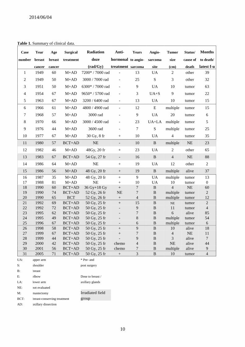
The median age at breast cancer diagnosis was 61 (range 35-83) years and at sarcoma diagnosis 71 (range 43-93)
years (table 1). The median time from radiation therapy to sarcoma development was 9 (range 3-25) years. Two
clinical patterns were observed; angiosarcomas that developed in edematous arms/shoulder regions, i.e. Stewart-
Treves syndrome, and lesions that developed in the irradiated field on the thoracic wall.
The Stewart-Treves group consisted of 14 females who had undergone mastectomy and axillary nodal dissection
followed by radiotherapy between 1949 and 1988. These women had been diagnosed with breast cancer at a
median age of 61 years (bilateral breast cancers had been diagnosed in 3 cases) and angiosarcoma at a median
age of 72 years, i.e. 11 years (range 3-25; 95% CI 9.0-19) later (table 2, figure 1). All 14 women suffered from
2014/06/04
5
edema in the affected arm. The doses and types of radiotherapy varied within this group. Anti hormonal therapy
was administered to 5/14 women, whereas none received chemotherapy.
Angiosarcomas in irradiated fields developed in 17 females, all of whom had undergone breast cancer treatment
between 1980 and 2005 (16 cases with segmental resection, 1 case of modified radical mastectomy, in all cases
followed by radiotherapy for breast cancer). In this subset, breast cancer had been diagnosed at a median age of
62 years and angiosarcoma at median 69 years of age, with a median interval of 7.3 years (range 3-15; 95% CI
5.8-8.8) (table 2, figure 1). Axillary nodal dissection had been performed in all but one case, but information on
arm edema was specified only in 6 cases, with edema reported in 2 cases. All of these angiosarcomas developed
within the irradiated fields on the thoracic wall. Most women in this group had received 50 Gy in 2 Gy daily
fractions over 5 weeks. Anti hormonal therapy had been administered to 7/17 women and 2 had been treated
with chemotherapy.
The time from breast cancer to sarcoma differed between the groups, from median 11 years in the Stewart-
Treves group to 7.3 years in the group with angiosarcomas in irradiated fields (p=0.01, log-rank test).
Clinical presentation and prognosis
Of the 14 cases with Stewart-Treves syndrome, 10 developed a solitary tumor, whereas 4 presented with
multiple tumors on the arm or the shoulder. Of the 17 cases with angiosarcomas on the thoracic wall, 8 had a
solitary tumor at diagnosis, 8 had multiple lesions, and data on tumor status was missing from 1 case. Among the
latter 17 tumors, 13 were restricted to the cutis/subcutis, whereas 4 had infiltrated into the breast parenchyma.
Symptom duration before diagnosis was longer (median 6, range 1-33 months) in the Stewart-Treves group than
in the group with angiosarcomas in irradiated fields (median 4, range 1-20 months).
Median follow-up for the patients in the Stewart-Treves group was 19 (0-65) months and for patients with
angiosarcomas in irradiated fields 12 (2-88) months. All 14 women in the former group were dead, 10 of them
from the angiosarcoma. In the latter cohort 6/17 patients are still alive and 7 had died from angiosarcoma (table
1). Median time to angiosarcoma-specific death was 15 months in the Stewart-Treves group and 4 months in the
irradiated-field group. The overall 5-year survival in the total cohort was 16%.
2014/06/04
6
DISCUSSION
Angiosarcoma is a rare complication of breast cancer treatment and in our population-based, 50-year cohort, we
identified 31 females with angiosarcomas of the thoracic wall, shoulder or arm following breast cancer
treatment. Two distinct groups were recognized; angiosarcomas in edematous upper arms, i.e. Stewart-Treves
syndrome, diagnosed before 1988, and tumors in irradiated fields on the thoracic wall, diagnosed after 1980
(figure 1). The distinct change in clinical presentation occurred parallel with altered treatment principles for
early-stage breast cancer. Modified radical mastectomy, axillary nodal dissection, and radiotherapy composed
the treatment until the 1980-ies, with a substantial risk of edema. Since then less aggressive surgery, i.e. breast
conservative treatment and selective dissection of axillary nodes, in combination with postoperative radiotherapy
and anti hormonal treatment or chemotherapy is considered standard treatment [1-2].
Before 1970-80, radiotherapy was poorly standardized. Treatment was generally administered postoperatively
and used orthovoltage techniques with doses of about 300 rad (approximately 3Gy) per fraction and split course
treatment. During the 1970’s linear accelerators were introduced and radiotherapy was given with combined
orthovoltage and megavoltage techniques, typically with 2.5 Gy daily doses in 2 to 3 series. In the 1980’s
radiotherapy was standardized with the commonest treatment being 48 Gy in 20 fractions in 2 series, with a
combination of electron (to the parasternal nodes and the thoracic wall) and megavoltage photon (to axillary and
supraclavicular nodes) treatment.
Angiosarcomas in irradiated fields following breast-conservative surgery developed median 4 years earlier than
in the Stewart-Treves group. Also symptom duration before diagnose was shorter in this group, median 4
months, than in the Stewart-Treves group, median 6 months. Intervals of 4-12 years between breast cancer and
angiosarcoma have been reported also by other investigators with lower time estimates in recent studies, which
likely reflect a larger contribution from radiation-induced sarcomas in recent cases [9, 12, 14, 17-19]. In our
cohort, extensive or breast conservative surgery constituted the major difference between the groups since all
women had been treated with radiotherapy (table 1). Our observation of a changing clinical presentation of
angiosarcoma is supported by other investigators. A study from Western Sweden reported 6 angiosarcomas, all
of which developed in edematous arms 7-14 years after treatment (including radiotherapy) for breast cancer
2014/06/04
7
during the time period 1960-1980 [6]. Billings et al, described 27 cutaneous angiosarcomas following
conservative surgery and radiotherapy for breast cancer with a median time to angiosarcoma that was shorter
than the time reported in the literature for patients with Stewart-Treves syndrome [9].
Atypical vascular lesions have been reported in irradiated field after treatment for breast cancer, but whether
these lesions represent precursors of angiosarcoma is controversial and many angiosarcomas also contain areas
with atypical vascular cellularity [12-13, 20]. Discoloration of the skin followed by hematoma-like lesions and
sometimes subsequent appearance of multiple bluish-reddish nodules was in our series reported by 13 women,
whereas the remaining 18 females presented with subcutaneous lumps without cutaneous precursors (although
they sometimes were ulcerated once the patient sought medical attention). Angiosarcoma carried a poor
prognosis with a 5-year survival of 16% in our study, compared to 10-15 % reported in the literature [12, 15].
The time to angiosarcoma-specific death seemed to be shorter for the cases presenting with angiosarcoma within
the irradiated field. If this is due to a more aggressive tumor form is unclear.
In summary, this population-based study demonstrates that angiosarcoma remains a rare complication of breast
cancer treatment, and reveals distinct differences in presentation, latency time, and tumor location linked to
altered treatment principles for breast cancer. With an increasing incidence of breast cancer (from 86/100 000 in
the southern Swedish Health Care Region in 1970 to currently 167/100 000), early diagnosis and improved
treatment, an increasing number of patients become long-term survivors. Thus a larger group is at risk of this late
treatment complication. Our series specifically addressed angiosarcomas, which has been estimated to represent
one third of radiation-induced sarcomas, suggesting that additional sarcomas of other histology appear in the
cohort studied [6-7, 14]. Several issues remain to be clarified, e.g. whether individual risk factors for
angiosarcoma development can be identified, whether the biological profiles differ depending on causative
factor, and how treatment should be optimized in this rare and severe complication to breast cancer treatment.
Acknowledgments
Pär-Ola Bendahl is acknowledged for statistical advice and review. The study was financially supported by the
Swedish Cancer Fund, the Swedish Children´s Cancer Fund, and the Swedish Research Council.
2014/06/04
8
References
1. Holmberg L (1989) Sector Resection With and Without Radiotherapy in Early Breast Cancer. Acta
Oncologica 28:927-929
2. The Uppsala-Örebro Breast Cancer Study Group (1990) Sector Resection With or Without Postoperative
Radiotherapy for Stage I Breast Cancer: A Randomized Trial. J Natl Cancer Inst 82:277-282
3. Fitzal F and Gnant M (2006) Breast Conservation: Evolution of Surgical Strategies. The Breast Journal
12:165-173
4. Lowenstein, S (1906) Der atiologische Zusammenhand zwischen akutem einmaligem Trauma and Sarkom.
Beitrage zur Klinsichen Chirurgie 48:780–824
5. Stewart FW, Treves N (1948) Lymphangiosarcoma in post-mastectomy lymphoedema. Report of six cases
in elephantiasis chirurgica. Cancer 1:64-81
6. Karlsson P, Holmberg E, Johansson K-A, Kindblom L-G, Carstensen J, Wallgren A (1996) Soft tissue
sarcoma after treatment for breast cancer. Radiotherapy and Oncology 38:25-31
7. Karlsson P, Holmberg E, Samuelsson A, Johansson K-A, Wallgren A. (1998) Soft Tissue Sarcoma after
Treatment for Breast Cancer – a Swedish Population-based Study. Eur J Cancer 34:2068-2075
8. Ruocco V, Schwartz RA, Ruocco E (2002) Lymphedema: An immunologically vulnerable site for
development of neoplasms. J Am Acad Dermatol 47:124-127
9. Billings SD, McKenny JK, Folpe AL, Hardacre MC, Weiss SW (2004) Cutaneous Angiosarcoma
Following Breast-conserving Surgery and Radiation. Am J Surg Pathology 28:781-788
10. Esler-Brauer L, Jaggernauth W, Zeitouni NC (2007) Angiosarcoma Developing after Conservative
Treatment for Breast Carcinoma: Case Report with Review of the Current Litterature. Dermatol Surg
33:749-755
11. Body G, Sauvanet E, Calais G, Fignon A, Fetissof F, Lansa CJ (1987) Angiosarcome cutané du sein aprés
adenocarcinome mammaire opéré et irradié. J Gynecol Obstet Biol Reprod 16:479-483
12. Brenn T and Fletcher CDM (2005) Radiation-Associated Cutaneous Atypical Vascular Lesions and
Angiosarcoma: Clinicopathological Analysis of 42 Cases. Am J Surg Pathology 29:983-996
13. Brodie C and Provenzano E (2008) Vascular proliferations of the breast. Histopathology 52:30-44
14. Neuhaus SJ, Pinnock N, Giblin V, Fisher C, Thway K, Thomas JM, Hayes AJ (2009) Treatment and
outcome of radiation-induced soft-tissue sarcomas at a specialist institution. Eur J Surg Oncol 1-6.
15. Monroe AT, Feigenberg SJ, Mendenhall NP (2002) Angiosarcoma after Breast-Conserving Therapy.
Cancer 97:1832-1840
2014/06/04
9
16. Roessner A, Boehlind T (2002) Angiosarcoma. In: Fletcher C, Unni K, Mertens F (eds) World Health
Organization Classification of Tumors, Pathology and Genetics of Tumors of Soft Tissue and Bone. IARC
press, Lyon, pp 322-323
17. Strobbe LJA, Peterse HL, van Tinteren H, Wijnmaalen A, Rutgers EJT (1998) Angiosarcoma of the breast
after conservation therapy for invasive cancer, the incidence and outcome. An unforeseen sequela. Breast
Cancer Research and Treatment 47:101-109
18. Vorburger SA, Xing Y, Hunt KK et al (2005) Angiosarcoma of the breast. Cancer 104:2682-2688.
19. Hodgson NC, Bowen-Wells C, Moffat F, Franceschi D, Avisar E (2007) Angiosarcoma of the Breast – A
review of 70 cases. Am J Clin Oncol 30:570-573
20. Patton K T, Deyrup A T, Weiss S W (2008) Atypical Vascular Lesions After Surgery and Radiation of the
Breast: A Clinicopathologic Study of 32 Cases Analyzing Histologic Heterogeneity and Association With
Angiosarcoma. Am J Surg Pathol 32:943-950
2014/06/04
10
Table 1. Summary of clinical data.
Case Year Age Surgical Radiation Anti- Years Angio- Tumor Status/ Months
number breast breast treatment doze hormonal to angio- sarcoma size cause of to death/
cancer cancer (rad/Gy) treatment sarcoma site (cm) death latest f-u
1 1949 60 M+AD 7200* / 7000 rad - 13 UA 2 other 39
2 1949 50 M+AD 3000 / 7000 rad - 25 S 3 other 32
3 1951 50 M+AD 6300* / 7000 rad - 9 UA 10 tumor 63
4 1954 67 M+AD 9650* / 5700 rad - 3 UA+S 9 tumor 22
5 1963 67 M+AD 3200 / 6400 rad - 13 UA 10 tumor 15
6 1966 61 M+AD 4800 / 4900 rad - 12 E multiple tumor 15
7 1968 57 M+AD 3000 rad - 9 UA 20 tumor 6
8 1970 66 M+AD 3000 / 4500 rad - 23 UA+LA multiple tumor 5
9 1976 44 M+AD 3600 rad - 7 S multiple tumor 25
10 1977 67 M+AD 30 Gy, 8 fr + 10 UA 4 tumor 35
11 1980 57 BCT+AD NE - 10 B multiple NE 23
12 1982 46 M+AD 48Gy, 20 fr + 23 UA 2 other 65
13 1983 67 BCT+AD 54 Gy, 27 fr - 16 B 4 NE 88
14 1986 64 M+AD NE + 19 UA 12 other 2
15 1986 56 M+AD 48 Gy, 20 fr + 19 B multiple alive 37
16 1987 35 M+AD 48 Gy, 20 fr + 9 UA multiple tumor 13
17 1988 81 M+AD NE + 10 UA 10 tumor 0
18 1990 60 BCT+AD 36 Gy+18 Gy + 7 B 4 NE 60
19 1990 74 BCT+AD 52 Gy, 26 fr NE 7 B multiple tumor 2
20 1990 65 BCT 52 Gy, 26 fr + 4 B multiple tumor 12
21 1992 69 BCT+AD 50 Gy, 25 fr + 15 B NE tumor 2
22 1992 72 BCT+AD 50 Gy, 25 fr - 9 B 11 tumor 4
23 1995 62 BCT+AD 50 Gy, 25 fr - 7 B 6 alive 85
24 1995 49 BCT+AD 50 Gy, 25 fr - 8 B multiple tumor 54
25 1996 67 BCT+AD 50 Gy, 25 fr - 6 B multiple tumor 6
26 1998 58 BCT+AD 50 Gy, 25 fr + 9 B 10 alive 18
27 1999 67 BCT+AD 50 Gy, 25 fr + 7 B 4 NE 11
28 1999 44 BCT+AD 50 Gy, 25 fr - 9 B 3 alive 7
29 2000 42 BCT+AD 50 Gy, 25 fr chemo 4 B NE alive 44
30 2001 56 BCT+AD 50 Gy, 25 fr chemo 7 B multiple alive 9
31 2005 71 BCT+AD 50 Gy, 25 fr + 3 B 10 tumor 4
UA: upper arm * Pre- and
S: shoulder post surgery
B: breast
E: elbow Dose to breast /
LA: lower arm axillary glands
NE: not evaluated
M: mastectomy Irradiated field
BCT: breast-conserving treatment group
AD: axillary dissection
2014/06/04
11
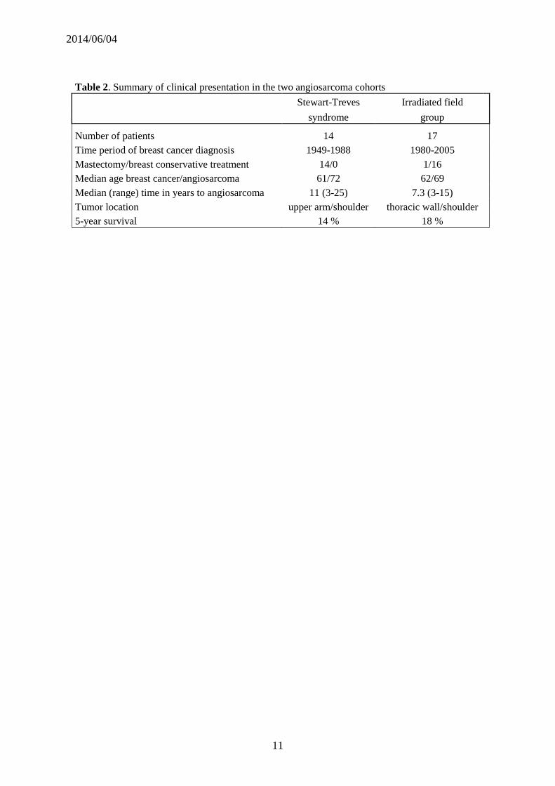
Table 2. Summary of clinical presentation in the two angiosarcoma cohorts
Stewart-Treves Irradiated field
syndrome group
Number of patients 14 17
Time period of breast cancer diagnosis 1949-1988 1980-2005
Mastectomy/breast conservative treatment 14/0 1/16
Median age breast cancer/angiosarcoma 61/72 62/69
Median (range) time in years to angiosarcoma 11 (3-25) 7.3 (3-15)
Tumor location upper arm/shoulder thoracic wall/shoulder
5-year survival 14 % 18 %