Changes in Growth, Biochemical Components, and Antioxidant Activity in Aquatic Plant Wolffia arrhiza...
11
Changes in Growth, Biochemical Components, and Antioxidant Activity in Aquatic Plant Wolffia arrhiza (Lemnaceae) Exposed to Cadmium and Lead Alicja Piotrowska • Andrzej Bajguz • Beata Godlewska- _ Zylkiewicz • El _ zbieta Zambrzycka Received: 21 May 2009 / Accepted: 28 September 2009 / Published online: 16 October 2009 Ó Springer Science+Business Media, LLC 2009 Abstract The present study investigated the biochemical response of aquatic plant Wolffia arrhiza (Lemnaceae) treated with lead (Pb) and cadmium (Cd) at a range of concentrations from 1 to 1000 lM. W. arrhiza has been identified as good scavenger of heavy metals from aqueous solution. Pb and Cd accumulation was found to be increased in a concentration- and duration-dependent manner. However, the highest bio- sorption of heavy metals was found in plants exposed to low levels (10 lM) of Cd and Pb in the nutrient medium. In observing the response to heavy-metal stress, we noted inhibited plant growth and decreased photosynthetic pig- ments, monosaccharides, and proteins. In addition, Cd was found to be more toxic to plants than Pb. Heavy metals also induced oxidative damage as evidenced by increased lipid peroxidation and hydrogen peroxide levels. In contrast, the deleterious effects resulting from the cellular oxidative state can be alleviated by enzymatic (catalase, ascorbate peroxi- dase, nicotinamide dinucleotide [NADH] peroxidase) and nonenzymatic (ascorbate, glutathione) antioxidant mecha- nisms activated in W. arrhiza plants exposed to Cd and Pb, especially at 10 lM. These results suggest that W. arrhiza is a promising bioindicator of heavy-metal toxicity. Duckweed (Lemnaceae) has been reported to be a potential scavenger of heavy metals from aquatic environments and is being used in wastewater renovation systems (Prasad et al. 2001; Artetxe et al. 2002; Hou et al. 2007). Wolffia arrhiza from the Lemnaceae family is one of the smallest vascular plants, which seldom exceed 1 mm in size. The plant has neither leaves nor a stem and even lacks roots. The whole plant, called a ‘‘frond,’’ may set flowers and seeds, although rapid multiplication is achieved by budding (Landolt 1986). W. arrhiza is also characterized by mixo- trophic feeding, high rate of absorption of nutrients from polluted water, quick growth, fast multiplication, and resistance to numerous toxins (Fujita et al. 1999). In par- ticular, W. arrhiza is reported to accumulate xenobiotics (Mical and Krotke 1999) and therefore is being used as experimental model system to investigate heavy-metal- induced responses. Cadmium (Cd) and lead (Pb) are strongly phytotoxic partly because of the generation of reactive oxygen species (ROS), which react with lipids, proteins, photosynthetic pigments, and nucleic acids, causing lipid peroxidation, membrane damage, metabolite degradation, inactivation of enzymes, and cell death (Heath and Packer 1968; Hegedu ¨s et al. 2001). In contrast, plants have evolved enzymatic (catalase, ascorbate peroxidase, nicotinamide dinucleotide [NADH] peroxidase) and nonenzymatic (ascorbate, gluta- thione) antioxidant mechanisms to prevent oxidative stress (Nakano and Asada 1981; Aeby 1984; Ishida et al. 1987; Xiang and Oliver 1998). Therefore, we studied the involvement of the antioxidant system in the biochemical detoxification of Cd and Pb in W. arrhiza culture in detail. The results of our study also demonstrate the effect of heavy-metal biosorption on the growth, lipid peroxidation, hydrogen peroxide level, and biochemical composition (proteins, monosaccharides, photosynthetic pigments) of this plant. The results of this study will be helpful in understanding the biochemical detoxification strategies that A. Piotrowska (&) A. Bajguz Department of Plant Biochemistry and Toxicology, Institute of Biology, University of Bialystok, Swierkowa 20 B, 15-950 Bialystok, Poland e-mail: [email protected] B. Godlewska- _ Zylkiewicz E. Zambrzycka Department of Analytic Chemistry, Institute of Chemistry, University of Bialystok, Hurtowa 1, 15-399 Bialystok, Poland 123 Arch Environ Contam Toxicol (2010) 58:594–604 DOI 10.1007/s00244-009-9408-6
-
Upload
independent -
Category
Documents
-
view
1 -
download
0
Transcript of Changes in Growth, Biochemical Components, and Antioxidant Activity in Aquatic Plant Wolffia arrhiza...

Changes in Growth, Biochemical Components, and AntioxidantActivity in Aquatic Plant Wolffia arrhiza (Lemnaceae) Exposedto Cadmium and Lead
Alicja Piotrowska • Andrzej Bajguz •
Beata Godlewska- _Zyłkiewicz • El _zbieta Zambrzycka
Received: 21 May 2009 / Accepted: 28 September 2009 / Published online: 16 October 2009
� Springer Science+Business Media, LLC 2009
Abstract The present study investigated the biochemical
response of aquatic plant Wolffia arrhiza (Lemnaceae) treated
with lead (Pb) and cadmium (Cd) at a range of concentrations
from 1 to 1000 lM. W. arrhiza has been identified as good
scavenger of heavy metals from aqueous solution. Pb and Cd
accumulation was found to be increased in a concentration-
and duration-dependent manner. However, the highest bio-
sorption of heavy metals was found in plants exposed to low
levels (10 lM) of Cd and Pb in the nutrient medium. In
observing the response to heavy-metal stress, we noted
inhibited plant growth and decreased photosynthetic pig-
ments, monosaccharides, and proteins. In addition, Cd was
found to be more toxic to plants than Pb. Heavy metals also
induced oxidative damage as evidenced by increased lipid
peroxidation and hydrogen peroxide levels. In contrast, the
deleterious effects resulting from the cellular oxidative state
can be alleviated by enzymatic (catalase, ascorbate peroxi-
dase, nicotinamide dinucleotide [NADH] peroxidase) and
nonenzymatic (ascorbate, glutathione) antioxidant mecha-
nisms activated in W. arrhiza plants exposed to Cd and Pb,
especially at 10 lM. These results suggest that W. arrhiza is a
promising bioindicator of heavy-metal toxicity.
Duckweed (Lemnaceae) has been reported to be a potential
scavenger of heavy metals from aquatic environments and
is being used in wastewater renovation systems (Prasad
et al. 2001; Artetxe et al. 2002; Hou et al. 2007). Wolffia
arrhiza from the Lemnaceae family is one of the smallest
vascular plants, which seldom exceed 1 mm in size. The
plant has neither leaves nor a stem and even lacks roots.
The whole plant, called a ‘‘frond,’’ may set flowers and
seeds, although rapid multiplication is achieved by budding
(Landolt 1986). W. arrhiza is also characterized by mixo-
trophic feeding, high rate of absorption of nutrients from
polluted water, quick growth, fast multiplication, and
resistance to numerous toxins (Fujita et al. 1999). In par-
ticular, W. arrhiza is reported to accumulate xenobiotics
(Mical and Krotke 1999) and therefore is being used as
experimental model system to investigate heavy-metal-
induced responses.
Cadmium (Cd) and lead (Pb) are strongly phytotoxic
partly because of the generation of reactive oxygen species
(ROS), which react with lipids, proteins, photosynthetic
pigments, and nucleic acids, causing lipid peroxidation,
membrane damage, metabolite degradation, inactivation of
enzymes, and cell death (Heath and Packer 1968; Hegedus
et al. 2001). In contrast, plants have evolved enzymatic
(catalase, ascorbate peroxidase, nicotinamide dinucleotide
[NADH] peroxidase) and nonenzymatic (ascorbate, gluta-
thione) antioxidant mechanisms to prevent oxidative stress
(Nakano and Asada 1981; Aeby 1984; Ishida et al. 1987;
Xiang and Oliver 1998). Therefore, we studied the
involvement of the antioxidant system in the biochemical
detoxification of Cd and Pb in W. arrhiza culture in detail.
The results of our study also demonstrate the effect of
heavy-metal biosorption on the growth, lipid peroxidation,
hydrogen peroxide level, and biochemical composition
(proteins, monosaccharides, photosynthetic pigments) of
this plant. The results of this study will be helpful in
understanding the biochemical detoxification strategies that
A. Piotrowska (&) � A. Bajguz
Department of Plant Biochemistry and Toxicology,
Institute of Biology, University of Bialystok, Swierkowa 20 B,
15-950 Bialystok, Poland
e-mail: [email protected]
B. Godlewska- _Zyłkiewicz � E. Zambrzycka
Department of Analytic Chemistry, Institute of Chemistry,
University of Bialystok, Hurtowa 1, 15-399 Bialystok, Poland
123
Arch Environ Contam Toxicol (2010) 58:594–604
DOI 10.1007/s00244-009-9408-6

aquatic plant W. arrhiza adopts against stress induced by
exposure to heavy metals.
Materials and Methods
Plant Material, Growth Conditions, and Treatment
Fronds of W. arrhiza (L.) Hork. ex. Wimm. (Lemnaceae) were
grown in small sterile, plastic vessels (Phytatray, Sigma-
Aldrich Co., USA) containing 100 mL culture solution under
controlled conditions at 25�C ± 0.5�C, with a day-to-night
cycle of 16:8 h (photon flux 50 lmol m-2 s-1). A 1/50
dilution of Hutner’s medium (Hutner 1953), pH 7.0, was used
in this study. The plants of W. arrhiza (0.5 ± 0.005 g fresh
weight) was treated with four different concentrations of Cd
and Pb (1, 10, 100, 1000 lM) on day 1 of culture. Heavy-
metal doses used in this work are environmentally relevant
and were chosen appropriately to expose the plants to low to
moderate levels of Cd and Pb. The different concentrations of
Cd and Pb were prepared by diluting Cd(NO3)2 and Pb(NO3)2
(Sigma-Aldrich Co., USA) in sterile 1/50 Hutner’s solution.
Plants were harvested at days 7 and 14, at the middle, and at the
end of the period of the plant developmental cycle. Biomass
was used for growth estimation and determination of the
biochemical parameters.
Heavy-Metal Determination
A Solaar M6 (Thermo Electron Corporation, UK) atomic
absorption spectrometer with deuterium background correc-
tion system was used for Pb and Cd determination in biomass
of W. arrhiza and in the medium. The absorbances of Pb and
Cd were measured in air-acetylene flame with 0.5 nm spectral
bandpass at k = 217.0 nm and k = 228.8 nm, respectively.
The Pb and Cd hollow cathode lamps (CPI International,
USA) were operated at 5 and 8 mA, respectively. A stock
solution (0.1 mol L-1) of Pb(II) and Cd were prepared by
dissolving Pb(NO3)2 and Cd(NO3)2 (Sigma-Aldrich Co.,
USA) in 2 mL 2 mol L-1 HNO3 and diluted with Milli-Q
water. Standard solutions were prepared from stock solutions
daily.
For metal determination in biomass, fronds were dried at
70�C for 12 h and ashed in a muffle furnace at 500�C ± 50�C
for 6 h. For measurement, the ash was dissolved in 2 mL
HNO3 (65%; Sigma-Aldrich Co., USA). Moreover, the con-
centration of heavy metals in the medium was measured
during 2 weeks of the culture. The calculated quantitation
limit (LOQ) for Cd dissolved in nitric acid was 0.0273
mg L-1, and the detection limit (LOD) was 0.00187 mg L-1.
LOQ = 0.0487 and LOD = 0.00609 mg L-1 were esti-
mated for Cd present in the medium. The calculated LOQ for
Pb dissolved in nitric acid was 0.228 mg L-1, and the LOD
was 0.0608 mg L-1. The calculated LOQ and LOD for Pb in
the medium were 0.176 and 0.039 mg L-1, respectively.
Determination of Growth, Proteins, Monosaccharides,
and Photosynthetic Pigments
For fresh-weight determination, the plants were filtered,
washed three times with distilled water, kept on filter
article for a few minutes to remove excess liquid, and then
weighed. For chlorophyll a and carotenoid determination,
the cultures were first collected by filtration, and then the
pellets (0.1 g) were homogenized in methanol. The
absorbance of the extract was measured at 652.4 and
665.2 nm for chlorophyll a and at 470.0 nm for carote-
noids. The amounts of photosynthetic pigments present in
the methanol extract were calculated according to the
equations of Wellburn (1994). For sugar determination, the
cultures were first collected by filtration, and then the
pellets (0.1 g) were assessed using the Somogyi (1954)
method. The measurement of the protein content was
performed by the homogenization of biomass. The
homogenate was centrifuged for 10 min at 12,000g, and an
aliquot of the extract was used to determine protein content
according to the Bradford (1976) method, using bovine
serum albumin as standard.
Stress-Marker Determination
The level of hydrogen peroxide in W. arrhiza fronds was
measured spectrophotometrically at 390 nm by reaction
with potassium iodide (KI). The results were calculated
using a standard curve prepared with fresh hydrogen per-
oxide solutions (Aliexieva et al. 2001).
Lipid peroxidation was estimated by measuring the
formation of malondialdehyde (MDA) with thiobarbituric
acid (TBA) reaction as described by Heath and Packer
(1968). Filtered biomass (0.5 g) was homogenized in 0.1%
trichloroacetic acid (TCA) and centrifuged at 10,000g for
5 min. Reaction mixture containing supernatant, 20%
TCA, and 0.5% TBA was heated at 95�C for 30 min and
then quickly cooled on ice. After centrifugation at 10,000g
for 10 min, the absorbance at 532 nm was read, and the
value for the nonspecific absorption at 600 nm was
subtracted.
Estimation of Antioxidants
Extraction and determination of total ascorbate was carried
out according to the method of Kampfenkel et al. (1995).
Plant material (1 g) was harvested by filtration and quickly
homogenized in liquid N2 and thereafter extracted with 5%
(w/v) TCA. The homogenate was centrifuged for 5 min at
15,600g (4�C). The supernatant was transferred to a new
Arch Environ Contam Toxicol (2010) 58:594–604 595
123
reaction vessel and immediately assayed for the ascorbate
content in a reaction mixture containing supernatant,
10 mM dithiothreitol, 0.2 M phosphate buffer (pH 7.4),
0.5% N-ethylmaleimide, 10% TCA, 42% H3PO4, 4% 2,20-dipyridyl, and 3% FeCl3. Glutathione extraction was per-
formed by the method of De Kok et al. (1986). The content
of glutathione was measured using a glutathione assay kit
(Sigma-Aldrich Co., USA).
Extraction of Antioxidant Enzymes and Estimation
of Their Activities
Enzymatic extracts were obtained from W. arrhiza (1 g
fresh weight). The biomass was filtered and then homog-
enized in liquid N2 and thereafter in 0.05 M phosphate
buffer (pH 7.0) containing 0.1 M ethylenediaminete-
traacetic acid and 1% polyvinylpyrrolidone at 4�C. The
homogenate was centrifuged for 10 min at 15,000g (4�C),
and the supernatant was dialyzed overnight in phosphate
buffer. Estimation of the activity of the selected enzymes
was performed as follows.
Catalase (EC 1.11.1.6) activity was measured spectro-
photometrically as the rate of H2O2 decomposition at
240 nm (Aeby 1984). The reaction mixture consisted of
0.05 M phosphate buffer, 0.1 mM H2O2, and supernatant.
One unit of catalase activity was assumed as the amount of
enzyme that decomposed 1 lmol of H2O2/mg soluble
protein/min at 30�C.
Total ascorbate peroxidase (EC 1.11.1.11) was deter-
mined according to the method of Nakano and Asada
(1981). The reaction mixture consisted of 0.05 M phos-
phate buffer, 5 mM sodium ascorbate, 0.1 mM H2O2, and
supernatant. Total ascorbate peroxidase activity was
determined as the decrease in absorbance of ascorbate at
290 nm. The enzyme activity was calculated as the amount
of the enzyme that oxidizes 1 lmol of ascorbate consumed/
mg soluble protein/min at 30�C.
NADH peroxidase (EC 1.11.1.1) activity was deter-
mined according to Ishida et al. (1987). The reaction
mixture consisted of 50 mM pH 6.0 sodium acetate buffer
and 0.2 mM NADH. The reaction was initiated by adding
the enzymatic extract and lasted for up to 5 min. The
peroxidase activators p-cumaric acid and 5 nM MnCl2were used. One unit of NADH peroxidase activity was
assumed to be the amount of the enzyme that oxidizes
1 lmol NADH/mg soluble protein/min at 30�C.
Replication and Statistical Analysis
Each treatment consisted of four replicates, and each
experiment was carried out at least twice at different times.
The data were analyzed by one-way analyses of variance,
and the means were separated using Duncan’s multiple-
range test (Statistica 6, StatSoft Co., USA). The level of
significance in all comparisons was p \ 0.05.
Results
Heavy-Metal Uptake
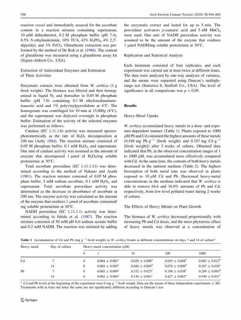
W. arrhiza accumulated heavy metals in a dose- and expo-
sure-dependent manner (Table 1). Plants exposed to 1000
lM Pb and Cd contained the highest amounts of these metals
(0.549 mg Pb g-1 [fresh weight] and 0.107 mg Cd g-1
[fresh weight]) after 2 weeks of culture. Obtained data
indicated that Pb, in the observed concentration ranges of 1
to 1000 lM, was accumulated more effectively compared
with Cd. At the same time, the contents of both heavy metals
decreased in the nutrient medium (Table 2). The highest
biosorption of both metal ions was observed in plants
exposed to 10 lM Cd and Pb. Decreased heavy-metal
concentrations in the medium indicated that W. arrhiza is
able to remove 64.6 and 36.0% amounts of Pb and Cd,
respectively, from low-level polluted water during 2 weeks
of culture.
The Effects of Heavy Metals on Plant Growth
The biomass of W. arrhiza decreased proportionally with
increasing Pb and Cd doses, and the most phytotoxic effect
of heavy metals was observed at a concentration of
Table 1 Accumulation of Cd and Pb (mg g-1 fresh weight) in W. arrhiza fronds at different concentrations on days 7 and 14 of culturea
Heavy metal Day of culture Heavy-metal concentration (lM)
0 1 10 100 1000
Cd 7 0 0.004 ± 0.001a 0.038 ± 0.008b 0.055 ± 0.004b 0.083 ± 0.012b
14 0 0.004 ± 0.005a 0.046 ± 0.004b 0.076 ± 0.009b 0.107 ± 0.010c
Pb 7 0 0.002 ± 0.009a 0.152 ± 0.025c 0.198 ± 0.038c 0.269 ± 0.093d
14 0 0.002 ± 0.004a 0.138 ± 0.081c 0.427 ± 0.062e 0.549 ± 0.031f
a Cd and Pb levels at the beginning of the experiment were 0 mg g-1 fresh weight. Data are the means of three independent experiments ± SD.
Treatments with at least one letter the same are not significantly different according to Duncan’s test
596 Arch Environ Contam Toxicol (2010) 58:594–604
123

1000 lM (Fig. 1a, b). However, the toxicities of Cd and Pb
in W. arrhiza differed remarkably. Fresh weight was more
intensively decreased in the presence of Cd in relation to
Pb-treated cultures because 1000 lM Pb caused a 47.2% to
67.2% decrease in biomass, whereas 1000 lM Cd induced
a 60.3% to 76.9% decrease in biomass.
The Effects of Heavy Metals on Photosynthetic
Pigments
W. arrhiza fronds treated with Cd and Pb displayed chlorosis
because a significant loss in chlorophyll a content was
observed during 2 weeks of culture (Fig. 2a, b). The results
showed also that Cd inhibited chlorophyll a content faster
and stronger than did Pb. Therefore, the maximum decrease
in chlorophyll a (75.4% and 72.7%) was obtained after
application of 1000 lM Cd and Pb, respectively, on day 14
of cultivation. Heavy metals at lower concentrations did
little harm to this chloroplast pigment in W. arrhiza plants.
In contrast, carotenoids were less sensitive than chlo-
rophyll a toward both Pb and Cd, probably protecting the
photosynthetic apparatus against heavy-metal stress
(Fig. 2c, d). The external supply of both 1000 lM Pb and
Cd (induced at 42.1 and 45.5%, respectively) decreased in
content by day 14 of the experiment. By contrast, heavy
metals at lower doses (1 lM) were characterized by a
stimulating effect of 7.4 to 9.7% on carotenoid content
compared with the control.
The Effects of Heavy Metals on Monosaccharides
Heavy metals were characterized by an inhibitory influence
on monosaccharide content in a concentration-dependent
manner (Fig. 2e, f). Therefore, the significant decrease (by
56.8% and 67.7%, respectively) in monosaccharide level
noted on days 7 and 14 of W. arrhiza cultivation was
obtained at the highest dose (1000 lM) of Cd. The appli-
cation of Pb at 1000 lM was characterized by a lower toxic
effect on this parameter because 49.5 and 61.4% inhibition
in monosaccharide accumulation was observed on days 7
and 14 of culture, respectively. Both Cd and Pb, at a range
of concentrations from 1 to 100 lM, possessed less
destructive influence on the sugar level in fronds.
The Effects of Heavy Metals on Proteins
The protein content in W. arrhiza fronds decreased pro-
portionally with increased Cd concentration at all of the
exposure periods, and the maximum decreases in this
biochemical parameter were recorded as 53.3% and 74.1%
on days 7 and 14 of cultivation, respectively, under the
Table 2 The initial and final contents of Cd and Pb (mg 100 mL-1) in the medium in which W. arrhiza fronds grewa
Heavy metal Day of culture Heavy-metal concentration (lM)
0 1 10 100 1000
Cd 0 0 0.014 ± 0.003a 0.139 ± 0.009b 1.399 ± 0.013b 13.995 ± 0.014c
7 0 0.013 ± 0.003a 0.111 ± 0.017b 1.339 ± 0.055b 13.762 ± 0.828c
14 0 0.009 ± 0.001a 0.050 ± 0.004a 1.196 ± 0.049b 13.844 ± 0.607c
Pb 0 0 0.021 ± 0.006a 0.212 ± 0.007b 2.072 ± 0.044b 20.720 ± 0.019d
7 0 0.020 ± 0.002a 0.088 ± 0.008a 1.692 ± 0.088b 20.501 ± 0.825d
14 0 0.019 ± 0.005a 0.075 ± 0.004a 1.145 ± 0.069b 19.942 ± 0.640d
a Data are the means of three independent experiments ± SD. Treatments with at least one letter the same are not significantly different
according to Duncan’s test
Fig. 1 Growth of W. arrhiza culture under the influence of Pb (a) and
Cd (b) at a range of 1 to 1000 lM. Data are the means of four
independent experiments ± SD. Treatments with at least one letter
the same are not significantly different according to Duncan’s test
Arch Environ Contam Toxicol (2010) 58:594–604 597
123

influence of 1000 lM Cd. In contrast, Pb provoked a
weaker response because a 41.3% to 59.7% decrease in
protein level was noted (Fig. 2g, h).
The Effects of Heavy Metals on H2O2 and Lipid
Peroxidation
Compared with unstressed plants, a significant increase in
hydrogen peroxide content was observed in the presence of
Cd and Pb in W. arrhiza culture (Fig. 3a, b). H2O2 pro-
duction was proportional with heavy-metal concentration.
For example, increases in hydrogen peroxide level of
130.1% and 125.6%, respectively, were observed in plants
treated with 1000 lM Cd on days 7 and 14. Pb at the same
dose provoked a weaker response because fronds treated
with 1000 lM Pb contained higher amounts (by 89% to
96%) of H2O2.
MDA content, a phytotoxic product of lipid peroxida-
tion, increased gradually in proportion to increased con-
centrations of heavy metals (Fig. 3c, d). The highest
stimulation by MDA level (105.9% and 76.1%, respec-
tively) was recorded under the influence of 1000 lM Cd on
days 7 and 14 of W. arrhiza culture. The application of Pb
ions at 1000 lM provoked a weaker response, leading to a
63.1% to 86.9% increase in MDA accumulation during
2 weeks of the experiment.
Fig. 2 The effects of Pb and Cd
at a range of 1 to 1000 lM on
chlorophyll a (a, b), carotenoid
(c, d), monosaccharide (e, f),and protein (g, h) content in
W. arrhiza. Data are the means
of four independent
experiments ± SD. Treatments
with at least one letter the same
are not significantly different
according to Duncan’s test
598 Arch Environ Contam Toxicol (2010) 58:594–604
123

The Effects of Heavy Metals on Nonenzymatic
Antioxidants
The highest enhancement in the ascorbate content (by
105.4%) was observed in fronds treated with 10 lM Pb on
day 7 of cultivation. In contrast, Cd at 10 lM possessed a
weaker effect on the ascorbate level because the content of
this antioxidant increased by 92.0% on the day 7 of the
experiment (Fig. 4a, b). In contrast, the inhibition of
ascorbate accumulation (by 21–68%, respectively) was
observed in fronds in the presence of Cd and Pb at 1000 lM.
Similarly, Pb at 10 lM was characterized by the highest
stimulating influence (by 86.5%) on the glutathione level on
day 7 of culture. Cd application at 10 lM resulted in a lower
increase in glutathione level (by 57.9% to 76.1%) compared
with the control (Fig. 4c, d). However, the 16–43% decrease
in glutathione content was found in fronds treated with
1000 lM Pb and Cd, respectively.
Fig. 3 The effects of Pb and Cd
at a range of 1 to 1000 lM on
hydrogen peroxide content (a,
b) and lipid peroxidation (c, d)
in W. arrhiza. Data are the
means of four independent
experiments ± SD. Treatment
with at least one letter the same
are not significantly different
according to Duncan’s test
Fig. 4 The effect of Pb and Cd
at range of 1 to 1000 lM on
ascorbate (a, b) and glutathione
(c, d) content in W. arrhiza.
Data are the means of four
independent experiments ± SD.
Treatment with at least one
letter the same are not
significantly different according
to Duncan’s test
Arch Environ Contam Toxicol (2010) 58:594–604 599
123

The Effects of Heavy Metals on Antioxidant Enzymes
Apart from the effect of Cd and Pb on nonenzymatic
antioxidants, heavy metals affected the activity of enzymes
involved in H2O2 metabolism (Fig. 5a–f). It was found that
the presence of 10 lM Cd and 10 lM Pb in the nutrient
solution resulted in the highest catalase and peroxidases
activities in W. arrhiza culture. In fronds treated with
10 lM Pb during 2 weeks of culture, ascorbate peroxidase
activity increased by 38.1% to 52.5%; catalase activity
increased by 42.7% to 65.5%; and NADH peroxidase
activity increased by 47.8% to 69.8%. Our data indicate
that Cd was characterized by a lower stimulating influence
on the antioxidant enzymes activities involved in ROS
scavenging in W. arrhiza fronds. Exogenously applied Cd
(at 10 lM) stimulated ascorbate peroxidase activity by
34.3 and 29.7% on days 7 and 14t of culture, respectively.
The significant 22.2 to 25.4% increase in NADH peroxi-
dase activity was also observed when plants were treated
with Cd at 10 lM during 2 weeks of the experiment. By
analogy, catalase activity increased by 36.7% to 59.1% in
response to exogenous 10 lM Cd. Both heavy metals at the
highest concentration (1000 lM) inhibited the activity of
all enzymes, which was probably caused by the harmful
effect of H2O2 overproduction or its poisonous ROS
derivatives.
Discussion
The results we obtained indicated that the biosorption of
heavy metals by plants is accompanied by an induction of a
variety of cellular changes, some of which directly con-
tribute to the metal-tolerance capacity of the plant. The
aquatic plant W. arrhiza accumulated heavy metals in a
dose- and exposure-dependent manner. This is in agreement
with earlier reports on aquatic plants, such as Chlorella
vulgaris, Nasturtium officinale, Mentha aquatica, and Ba-
copa monnieri (Aslan et al. 2003; Bajguz and Godlewska-_Zyłkiewicz 2004; Singh et al. 2006). In particular, members
of the duckweed family (Lemna minor, L. trisulca) have
been reported to bioaccumulate and bioconcentrate toxic
Fig. 5 The effects of Pb and Cd
at range of 1 to 1000 lM on the
activities of catalase (a, b),
ascorbate peroxidase (c, d), and
NADH peroxidase (e, f). Data
are the means of four
independent experiments ± SD.
Treatment with at least one
letter the same are not
significantly different according
to Duncan’s test
600 Arch Environ Contam Toxicol (2010) 58:594–604
123

metals from polluted water (Samardakiewicz and Wozny
2000). Results after 2 weeks of exposure to low doses
(10 lM) of Pb and Cd indicated that W. arrhiza achieved
high Pb (64.6%) and Cd (36.0%) removal efficiency. These
results correlate well with the observation that both heavy
metals present at a concentration of 10 lM induced defense
reactions in fronds, including antioxidant enzymes, ascor-
bate, and glutathione.
Literature data indicate that the most of Cd and Pb
accumulated by the aquatic plants was retained by the
root, leading to less translocation to the aerial parts.
Roots of plants are a barrier against heavy-metal trans-
location, and this may be a potential tolerance mecha-
nism operating in the roots (Singh et al. 2006). However,
W. arrhiza does not possess roots, so heavy metals are
absorbed by the fronds. Experiments performed by Sa-
mardakiewicz and Wozny (2000) showed that L. minor
treated with Pb showed maximum concentrations of
heavy metals in small vacuoles and the cell wall. The
localization of Pb between vacuoles and the cell wall
possibly results from redistribution of Pb, and it reflects
increased apoplastic transport. The presence of heavy
metals in small vesicles in Lemnaceae plants suggests
that endocytosis plays an important role in metal uptake
in these species. Moreover, the different accumulation of
Cd and Pb ions further suggest different cellular mech-
anisms of the biosorption of these nonessential trace
elements by W. arrhiza fronds.
Bioaccumulation of Cd and Pb has been reported to
induce negative effects on some key metabolic processes
coupled with plant development. The most dramatic
symptom of Cd and Pb toxicity in W. arrhiza culture was
the cessation of plant growth. Growth inhibition may be
connected with the decrease in mitotic index observed in
the case of Pb and Cd ion exposure (Vecchia et al. 2005).
Experiments performed on synchronized soybean (Glycine
max) cell suspension culture showed that Cd induced DNA
damage, decreased the rate of DNA synthesis, and blocked
cell division (Sobkowiak and Deckert 2004).
As a visible symptom, decreased chlorophyll a content
can be used to monitor heavy-metal-induced damage in
W. arrhiza fronds. Based on available data, it can be
assumed that Cd and Pb may inhibit chlorophyll synthesis
by causing impaired uptake of elements essential for photo-
synthetic pigments, such as magnesium, potassium, cal-
cium, and iron (Burzynski 1987). Moreover, enhancement
of chlorophyll damage occurs in plants growing in the
presence of Pb ions due to increased chlorophyllase
activity (Dra _zkiewicz 1994). The inhibition in photosyn-
thetic pigment accumulation in response to heavy-metal
stress may also be a consequence of peroxidation of
chloroplast membranes by way of increased rate of ROS
production. This observation is in good agreement with the
increased rate of H2O2 and lipid peroxide formation in W.
arrhiza exposed to Cd and Pb.
In contrast, carotenoids were less sensitive than chlo-
rophyll a toward both Pb and Cd, probably protecting the
photosynthetic apparatus against heavy-metal stress. The
increased carotenoid level in W. arrhiza plants exposed to
heavy metals is probably part of the strategy adopted by the
plant to counteract the toxic effect of free radicals gener-
ated under Cd and Pb stress, which is agreement with other
reports in aquatic plants, such as L. minor and L. trisulca
(Prasad et al. 2001; Singh et al. 2006; Hou et al. 2007).
Carotenoids, which play a part in guarding chlorophyll,
also serve as antioxidants to quench or scavenge the free
radicals and decrease damage to the cell, to the cell
membrane, and to the plant’s main genetic composition
induced by heavy metal (Artetxe et al. 2002).
Monosaccharides are building substances for plants as
well as a key source of energy necessary for inciting bio-
chemical processes. The decrease in monosaccharide con-
tent noted in W. arrhiza growing in the presence of Cd and
Pb may be caused by enhanced degradation of photosyn-
thetic pigments, thus contributing to decreased photosyn-
thesis and sugar accumulation. Significantly decreased
activities of enzymes involved in CO2 fixation in field-
grown Avena sativa exposed to heavy metals was reported
by Moustakas et al. (1994).
The protein content in W. arrhiza fronds decreased with
increased heavy-metal concentrations at all of the exposure
periods, and the maximum decrease in this biochemical
parameter was obtained in response to 1000 lM Cd. Sol-
uble-protein content in plants, which is an important indi-
cator of reversible and irreversible changes in metabolism,
is known to respond to a wide variety of stressors (Singh
and Tewari 2003). The inability of W. arrhiza fronds to
accumulate proteins after Cd and Pb application may be
caused by acute oxidative stress induced by heavy-metal
excess in plant cells. Protein degradation, as a consequence
of metal exposure, has been observed in many aquatic
plants, such as L. minor, L. trisulca and free-floating
freshwater macrophyte Ceratophyllum demersum (Mohan
and Hosetti 1997; Prasad et al. 2001; Aravind and Prasad
2003; Hou et al. 2007). This phytotoxic effect of Cd and Pb
in plant culture may be explained by their influence on
nucleic acid degradation. For example, Cd induces DNA
damage in plant cells, such as single- and double-strand
break, modified bases, abasic sites, DNA–protein cross-
links, oxidized bases, 8-hydroxyguanine, and even bulky
adducts (Liu et al. 2005).
Compared with unstressed plants, significantly increased
hydrogen peroxide content was observed in the presence of
Cd and Pb in W. arrhiza culture. The main sources of ROS
are enzymes localized at the external surface of plant cells,
plasma membrane NAD(P)H oxidases, and/or cell wall
Arch Environ Contam Toxicol (2010) 58:594–604 601
123

peroxidases (Ishida et al. 1987). In apoplast, H2O2 may be
produced by SOD for •O2-, resulting from the activity of
oxidase/peroxidase NADH complex, and accumulate in a
cell wall (Chaoui et al. 2004). Therefore, the present results
suggest a correlation between peroxidase activity, for
which NADH is a substrate (NADH peroxidase), and the
generation of H2O2 in W. arrhiza fronds in response to
exogenous Cd and Pb. MDA content also increased grad-
ually in proportion to increased concentrations of heavy
metals. MDA production in plants exposed to adverse
environmental conditions is an indicator of free radical
formation in biologic systems (Heath and Packer 1968). A
concentration-dependent increase in the level of lipid per-
oxides occurred in spruce (Picea abies) needles (Radotic
et al. 2000), barley (Hordeum vulgare) seedlings (Hegedus
et al. 2001), rice (Oriza sativa) shoots (Verma and Dubey
2003), water hyssop (Bacopa monnieri) plants (Singh et al.
2006), and duckweed (L. minor) fronds (Hou et al. 2007)
growing in the presence of increased concentrations of
heavy metals. However, the mechanism of oxidative stress
in W. arrhiza generated by exposure to heavy metals is not
clearly understood.
The stimulating influence of Cd and Pb on lipid perox-
idation and H2O2 levels indicated that fronds encountered
enhanced oxidative stress. Whenever ROS are produced,
plants activate nonenzymatic antioxidant defense mecha-
nisms comprising low molecular mass scavengers, such as
ascorbate and glutathione, to remove these highly reactive
molecules. Stimulation in the ascorbate level in response to
heavy metals at a concentration of 10 lM suggests its role
in ROS detoxification generated by stress. Ascorbate is
known to operate as an antioxidant either in direct chem-
ical interaction with free oxyradicals or during the reaction
catalyzed by ascorbate peroxidase (Nakano and Asada
1981). Moreover, ascorbate oxidation affects the redox
balance of other metabolites, such as glutathione, which are
themselves involved in the perception of cellular redox
unbalance (Kampfenkel et al. 1995). Similar results
obtained by Artetxe et al. (2002), Rucinska-Sobkowiak and
Pukacki (2006), and Singh et al. (2006) confirmed that
ascorbate accumulation leads to enhancement of plant
tolerance toward heavy-metal stress.
The adaptation of W. arrhiza to grow in the presence of
10 lM Cd and Pb was evidenced by the increment in
ascorbate and glutathione, together with increased activi-
ties of antioxidant enzymes involved in H2O2 detoxifica-
tion, such as ascorbate peroxidase, catalase, and NADH
peroxidase. H2O2 is destroyed in chloroplasts through the
action of a metabolic ascorbate–glutathione cycle involv-
ing successive oxidation and reduction of ascorbate, glu-
tathione, and NAD(P)H. The following pathway has been
proposed: ascorbate peroxidase reduces H2O2 into water
using ascorbate as the electron donor; the resulting
dehydroascorbate is cycled back to ascorbate using reduced
glutathione as electron donor; and the oxidized glutathione
formed is converted back to glutathione. The activation of
this cycle on metal-stress situations has been well docu-
mented (Chaoui et al. 1997; Hegedus et al. 2001). For each
metal treatment at a concentration of 10 lM, enhanced
antioxidant enzymes activities suggest an increased rate of
ascorbate and glutathione turnover in W. arrhiza exposed
to Cd and Pb. Despite the fact that neither reduced/oxidized
ratio of ascorbate and glutathione nor glutathione reductase
activity were assayed in this study, analysis of ascorbate
peroxidase as well as ascorbate and glutathione level lends
support to the hypothesis that the H2O2 scavenging ascor-
bate–glutathione cycle may be activated in W. arrhiza
treated with both heavy metals, especially at 10 lM.
Increased glutathione levels have been also shown to
correlate with plant adaptation to extreme heavy-metal
stress, and decreased glutathione pool shows marked
alterations in response to heavy-metal stress (Xiang and
Olivier 1998; Jin et al. 2008). Moreover, glutathione is also
a precursor of phytochelatins, which are low molecular
mass peptides produced by plants to immobilize toxic
heavy metals (Tsuji et al. 2005). Therefore, the increased
glutathione level noted in W. arrhiza treated with Cd and
Pb at a range of concentrations (1 to 100 lM) may precede
phytochelatin accumulation. However, ascorbate and glu-
tathione levels decreased in response to exposure to
1000 lM heavy metals. Reactive free oxygen radicals are
assumed to be involved in the oxidation of ascorbic acid to
dehydroascorbic acid, leading to decreased ascorbic acid
content in W. arrhiza.
Apart from the effects of Cd and Pb on nonenzymatic
antioxidants, heavy metals affected enzyme activities
involved in H2O2 metabolism. Both heavy metals at the
highest concentration (1000 lM) inhibited the activity of
all enzymes, probably because of the harmful effect of
H2O2 overproduction or its poisonous ROS derivatives.
Varying responses to heavy-metal-induced oxidative stress
might be related to the concentration of thiolic groups
already present or induced by Cd and Pb exogenous
application because they are consequently able to coun-
teract oxidative stress (Sanita di Toppi and Gabbrielli
1999). However, heavy metals at lower concentrations
(1 to 100 lM) were found to stimulate the activities of
antioxidant enzymes.
Peroxidases can function as effective quenchers of
reactive intermediary forms of oxygen and peroxy radicals
induced by increased metal levels in plant cells (Radotic
et al. 2000). They are considered to be heavy-metal stress-
related enzymes and can be used as stress markers in metal
(Cd, Pb) poisoning situations. Some acidic isoforms of cell
wall peroxidases have been implicated in lignification by
catalyzing oxidative polymerization of monolignols, e.g.,
602 Arch Environ Contam Toxicol (2010) 58:594–604
123

coumaryl, in the presence of H2O2, which is produced by
cell wall peroxidases having an NADH oxidase activity,
such as NADH-peroxidase (Ishida et al. 1987). Peroxidase-
catalyzed lignification represents a mechanical plant
adaptation to metal contamination (Gaspar et al. 1985).
Enhanced activity of ascorbate peroxidase in W. arrhiza is
generally associated with an acclimation to increased
amounts of active oxygen species generated by Cd and Pb.
Catalase participates in the main defence system against
accumulation and toxicity of active oxygen species, such as
hydrogen peroxide, and can play a key role in controlling
H2O2 levels in plant cells. Our findings correspond well
with other literature data showing the positive effect of
heavy-metal exposure on antioxidant enzymes activities
(Hegedus et al. 2001; Singh et al. 2006; Hou et al. 2007; Jin
et al. 2008).
The enhanced activities of the enzymatic antioxidant
system in W. arrhiza in response to exogenous metals, such
as Cd and Pb at 1 to 100 lM, may account for its better
growth and acclimation to metal pollution of the aquatic
environment. Interpretation of the biochemical detoxifica-
tion strategies of the plant against oxidative stress induced
by metal accumulation is a key to optimize the phyto-
remediation of heavy-metal-affected biomes. Summarizing
our results, it can be concluded that the antioxidant
enzymes activities, such as catalase, ascorbate peroxidise,
and NADH peroxidase, in W. arrhiza fronds were at their
peak when fronds were treated with 10 lM Cd and Pb,
whereas the highest dose (1000 lM) of heavy metals
decreased these activities and contributed dramatically to
inhibition of plant growth and development.
Conclusion
From the work presented here, the aquatic plant W. arrhiza
can be effective as a biosorbent for the removal of low
levels of Cd and Pb from nutrient medium. In the present
study, the accumulation of metals resulted in considerable
biochemical changes. The data confirm the inhibitory
effect of heavy metals on the growth and development of
W. arrhiza. Cd was found to be comparatively more toxic
than Pb. The rate of proteins, photosynthetic pigments, and
monosaccharide degradation was proportional to the
increased dose of heavy metals, and the most phytotoxic
influence of Cd and Pb was observed at the highest tested
concentration (1000 lM). Treatment with Cd and Pb also
caused oxidative damage as evidenced by increased lipid
peroxide and H2O2 formation, indicating the presence of
poisoning ROS. However, to cope with heavy-metal tox-
icity, W. arrhiza fronds are able to carry out a cellular
strategy involving activation of various enzymatic (cata-
lase, ascorbate peroxidase, NADH-peroxidase) and
nonenzymatic (ascorbate, glutathione) antioxidants, which
serve as important components of mechanisms that
detoxifying heavy metals. The highest enhancement in
antioxidant activity was observed in fronds exposed to
10 lM heavy metals, which may account for higher plant
tolerance and acclimation to metal contamination. The
results obtained with Cd and Pb, showing that Lemnaceae
are much more sensitive than other experimental plants
used so far, are in agreement with the use of W. arrhiza in
bioassays for phytotoxicity.
Acknowledgments The authors are grateful to Marta Kaczorowska
and Luba Siemianowicz for skilful technical assistance.
References
Aeby H (1984) Catalase in vitro. Meth Enzymol 105:125–212
Aliexieva V, Sergiev I, Mapelli S, Karanov E (2001) The effect of
drought and ultraviolet radiation on growth and stress markers in
pea and wheat. Plant Cell Environ 24:1337–1344
Aravind P, Prasad MNV (2003) Zinc alleviates cadmium-induced
oxidative stress in Ceratophyllum demersum L.: a free floating
freshwater macrophyte. Plant Physiol Biochem 41:391–397
Artetxe U, Garcıa-Plazaola JI, Hernandez A, Becerril JM (2002) Low
light grown duckweed plants are more protected against the
toxicity induced by Zn and Cd. Plant Physiol Biochem 40:859–
863
Aslan M, Unlu MY, Turkmen N, Yilmaz YZ (2003) Sorption of
cadmium and effects on growth, protein content, and photosyn-
thetic pigment composition of Nasturtium officinale R Br. and
Mentha aquatica L. Bull Environ Contam Toxicol 71:323–329
Bajguz A, Godlewska- _Zyłkiewicz B (2004) Protective role of 20-
hydroxyecdysone against lead stress in Chlorella vulgariscultures. Phytochemistry 65:711–720
Bradford MM (1976) A rapid and sensitive method for the
quantitation of microgram quantities of protein utilizing the
principle of protein–dye binding. Anal Biochem 72:248–254
Burzynski M (1987) The influence of lead and cadmium on the
absorption and distribution of potassium, calcium, magnesium
and iron in cucumber seedlings. Acta Physiol Plant 9:229–238
Chaoui A, Mazhoudi S, Ghorbal MH, Ferjani EE (1997) Cadmium
and zinc induction of lipid peroxidation and effects on antiox-
idant enzyme activities in bean (Phaseolus vulgaris L.) Plant Sci
127:139–147
Chaoui A, Jarrar B, El Ferjani E (2004) Effects of cadmium and
copper on peroxidase, NADH oxidase and IAA oxidase activities
in cell wall, soluble and microsomal membrane fractions of pea
roots. J Plant Physiol 161:1225–1234
De Kok LJ, Maas FM, Godeke J, Haaksma AB, Kuiper PJC (1986)
Glutathione, a tripeptide which may function as a temporary
storage compound of excessive reduced sulphur in H2S fumi-
gated spinach plants. Plant Soil 91:349–352
Dra _zkiewicz M (1994) Chorophyll-occurrence, functions, mechanism
of action, effects of internal and external factors. Photosynthetica
30:321–331
Fujita M, Mori K, Kodera T (1999) Nutrient removal and starch
production through cultivation of Wolffia arrhiza. J Biosci Bioeng
87:194–198
Gaspar T, Penel C, Castillo FJ, Greppin H (1985) A two step control
of basic and acidic peroxidases and its significance for growth
and development. Physiol Plant 64:418–423
Arch Environ Contam Toxicol (2010) 58:594–604 603
123
Heath RL, Packer L (1968) Photoperoxidation in isolated chloroplasts
I. Kinetics and stoichiometry of fatty acid peroxidation. Arch
Biochem Biophys 125:189–198
Hegedus A, Erdei S, Horvath G (2001) Comparative studies of H2O2
detoxifying enzymes in green and greening barley seedlings
under cadmium stress. Plant Sci 160:1085–1093
Hou W, Chen X, Song G, Wang Q, Chang CC (2007) Effect of copper
and cadmium on heavy metal polluter waterbody restoration by
duckweed (Lemna minor). Plant Physiol Biochem 45:62–69
Hutner SH (1953) Comparative physiology of heterotrophic growth in
plants. In: Loomis WE (ed) Growth and differentiation in plants.
Iowa State College Press, Ames, IA, pp 417–446
Ishida A, Ookubu K, Ono K (1987) Formation of hydrogen peroxide
by NAD(P)H oxidation with isolated cell wall-associated
peroxidase from cultured liverwort cells, Marchantia polymor-pha L. Plant Cell Physiol 28:723–726
Jin XF, Yang XE, Islam E, Liu D, Mahmood Q, Li H, Li J (2008)
Ultrastructural changes, zinc hyperaccumulation and its relation
with antioxidants in two ecotypes of Sedum alfredii Hance. Plant
Physiol Biochem 46:997–1006
Kampfenkel K, Van Montagu M, Inze D (1995) Extraction and
determination of ascorbate and dehydroascorbate from plant
tissue. Anal Biochem 225:165–167
Landolt E (1986) The family of Lemnaceae—a monographic study.
Veroff Geobot Inst ETH, Stiftung Rubel, Zurich 71:1–563
Liu W, Li PJ, Qi XM, Zhou QX, Zheng L, Sun TH et al (2005) DNA
changes in barley (Hordeum vulgare) seedlings induced by cadmium
pollution using RAPD analysis. Chemosphere 61:158–167
Mical AH, Krotke A (1999) Wolffia arrhiza (L.)—small but strong.
Acta Hydrobiol 41:165–170
Mohan BS, Hosetti BB (1997) Potential phytotoxicity of lead and
cadmium to Lemna minor grown in sewage stabilization ponds.
Environ Pollut 2:233–238
Moustakas M, Lanaras T, Symeonidis L, Kartaglis S (1994) Growth
and some photosynthetic characteristics of field grown Avenasativa under copper and lead stress. Photosynthetica 30:389–396
Nakano Y, Asada K (1981) Hydrogen peroxide is scavenged by
ascorbate-specific peroxidase in spinach chloroplasts. Plant Cell
Physiol 22:867–880
Prasad MNV, Malec P, Waloszek A, Bojko M, Strzałka K (2001)
Physiological responses of Lemna trisulca L (duckweed) to
cadmium and copper bioaccumulation. Plant Sci 161:881–889
Radotic K, Ducic T, Mutavdzic D (2000) Changes in peroxidase
activity and isoenzymes in spruce needles after exposure to
different concentrations of cadmium. Environ Exp Bot 44:
105–113
Rucinska-Sobkowiak R, Pukacki PM (2006) Antioxidative defense
system in lupin roots exposed to increasing concentrations of
lead. Acta Physiol Plant 28:357–364
Samardakiewicz S, Wozny A (2000) The distribution of lead in
duckweed (Lemna minor L.) root tip. Plant Soil 226:107–111
Sanita di Toppi L, Gabbrielli R (1999) Response to cadmium in
higher plants. Environ Exp Bot 41:105–130
Singh VP, Tewari RK (2003) Cadmium toxicity induced changes in
plant water relations and oxidative metabolism of Brassicajuncea L plants. J Environ Biol 24:107–112
Singh S, Eapen S, D’Souza SF (2006) Cadmium accumulation and its
influence on lipid peroxidation and antioxidative system in an
aquatic plant, Bacopa monnieri L. Chemosphere 62:233–246
Sobkowiak R, Deckert J (2004) The effect of cadmium on cell cycle
control in suspension culture cells of soybean. Acta Physiol Plant
26:335–344
Somogyi M (1954) Notes on sugar determination. J Biol Chem 195:
19–23
Tsuji N, Nishikori S, Ibawe O, Matsumoto S, Shiraki K, Miyasaka H
et al (2005) Comparative analysis of the two-step reaction
catalyzed by prokaryotic and eukaryotic phytochelatin synthase
by an ion-pair liquid chromatography assay. Planta 222:181–191
Vecchia FD, La Rocca N, Moro I, De Faveri S, Andreoli C, Rascio N
(2005) Morphogenetic, ultrastructural and physiological dam-
ages by submerged leaves of Elodea canadensis exposed to
cadmium. Plant Sci 168:329–338
Verma S, Dubey RS (2003) Lead toxicity induces lipid peroxidation
and alters the activities of antioxidant enzymes in growing rice
plants. Plant Sci 164:645–655
Wellburn AR (1994) The spectral determination of chlorophylls a and
b, as well as total carotenoids, using various solvents with
spectrophotometers of different resolution. J Plant Physiol 144:
307–313
Xiang C, Oliver DJ (1998) Glutathione metabolic genes coordinately
respond to heavy metals and jasmonic acid in Arabidopsis. Plant
Cell 10:1530–1550
604 Arch Environ Contam Toxicol (2010) 58:594–604
123