TrCP-Mediated Proteolysis of NF B1 p105 Requires Phosphorylation of p105 Serines 927 and 932

Chain initiation on type I modular polyketide synthasesrevealed by limited proteolysis and ion-trap massspectrometryHui Hong1, Antony N. Appleyard2, Alexandros P. Siskos2, Jose Garcia-Bernardo2, James Staunton1
and Peter F. Leadlay2
1 Department of Chemistry, University of Cambridge, UK
2 Department of Biochemistry, University of Cambridge, UK
Polyketides are a structurally diverse group of natural
products, which exhibit a broad range of biological
effects including antibiotic, antifungal, immunosup-
pressive, and anticancer activities [1]. They are synthes-
ized on polyketide synthases (PKSs), which convert
intracellular acyl-CoA precursors into complex poly-
ketide backbones via a stepwise chain building mech-
anism employing different combinations of a standard
set of biochemical reactions. There are three canonical
types of PKS, based on their structure and mecha-
nisms of operation: type I (iterative or modular),
type II and type III [2]. The best-studied modular
type I PKS is the 6-deoxyerythronolide B synthase
(EC 2.3.1.94) (DEBS) from Saccharopolyspora erythr-
aea, which produces the polyketide backbone of the
antibiotic erythromycin (Fig. 1A). DEBS consists of
three large bimodular polypeptides (DEBS1, DEBS2,
and DEBS3) (each >300 kDa) which together catalyze
the stepwise condensation of a propionyl-CoA-derived
primer unit with six methylmalonyl-CoA-derived exten-
der units to yield 6-deoxyerythronolide B (6dEB) [1].
The hallmark of a modular type I PKS is that there is a
separate domain for every step of the assembly of the
polyketide chain, and they are disposed along the PKS
Keywords
erythromycin; limited proteolysis; liquid
chromatography-mass spectrometry;
multienzyme; polyketide synthase
Correspondence
J. Staunton, Department of Chemistry,
University of Cambridge, Lensfield Road,
Cambridge CB2 1EW, UK
Fax: +44 1223 762018
Tel: +44 1223 766041
E-mail: [email protected]
(Received 10 November 2004, revised 28
January 2005, accepted 15 February 2005)
doi:10.1111/j.1742-4658.2005.04615.x
Limited proteolysis in combination with liquid chromatography-ion trap
mass spectrometry (LC-MS) was used to analyze engineered or natural
proteins derived from a type I modular polyketide synthase (PKS), the
6-deoxyerythronolide B synthase (DEBS), and comprising either the first
two extension modules linked to the chain-terminating thioesterase (TE)
(DEBS1-TE); or the last two extension modules (DEBS3) or the first exten-
sion module linked to TE (diketide synthase, DKS). Functional domains
were released by controlled proteolysis, and the exact boundaries of
released domains were obtained through mass spectrometry and N-terminal
sequencing analysis. The acyltransferase-acyl carrier protein required for
chain initiation (ATL-ACPL), was released as a didomain from both
DEBS1-TE and DKS, as well as the off-loading TE as a didomain with the
adjacent ACP. Mass spectrometry was used successfully to monitor in
detail both the release of individual domains, and the patterns of acylation
of both intact and digested DKS when either propionyl-CoA or n-butyryl-
CoA were used as initiation substrates. In particular, both loading domains
and the ketosynthase domain of the first extension module (KS1) were
directly observed to be simultaneously primed. The widely available and
simple MS methodology used here offers a convenient approach to the pro-
teolytic mapping of PKS multienzymes and to the direct monitoring of
enzyme-bound intermediates.
Abbreviations
ACP, acyl carrier protein; AT, acyl transferase; DEBS, 6-deoxyerythronolide B synthase; DKS, diketide synthase; KR, ketoreductase; KS,
ketosynthase; NPDS, 4-nitrophenyl disulfide; NRPS, nonribosomal peptide synthase; PKS, polyketide synthase; TE, thioesterase.
FEBS Journal 272 (2005) 2373–2387 ª 2005 FEBS 2373

multienzyme polypeptides essentially in the order that
they are used.
Modular PKSs are clearly amenable to rational gen-
etic manipulation of the biosynthetic enzymes, as a
promising way of creating new bioactive compounds
[3,4]. However to achieve this efficiently we need a
better understanding of the molecular basis underlying
the operation of these assembly line enzymes. To facili-
tate the detailed mechanistic study of the erythromycin
biosynthesis, model systems with shortened length have
been created. DEBS1-TE is a bimodular PKS, created
by moving the thioesterase (TE) domain from the ter-
minus of DEBS3 to the end of DEBS1 to cause prema-
ture release of the chain at the triketide stage (Fig. 1B)
[5]. The unimodular PKS, called diketide synthase
(DKS) was created by moving the TE domain from
the terminus of DEBS3 to the end of module 1 of
DEBS1, to cause premature release of the chain at the
diketide stage (Fig. 1C) [6]. It should be noted that the
engineering of these model proteins was designed to
preserve the native linker between the TE domain and
the adjacent acyl carrier protein (ACP). The ACP
domains are therefore hybrid structures comprising the
N-terminal of ACP2 (DEBS1-TE) and ACP1 (DKS),
respectively, fused to the C-terminal portion of ACP6.
(The domain number is the module number in which
the domain resides. This designation applies through
out the paper.) For simplicity in the following account,
these hybrid ACPs are designated ACP2 and ACP1,
respectively. The engineered proteins, DEBS1-TE and
DKS, have been purified to homogeneity and have
produced the expected products in vitro [6,7], and
therefore can serve as convenient models for the full
DEBS system.
A
B
C
Fig. 1. Organization of DEBS multienzyme proteins. (A) Organization of DEBS from S. erythraea, which catalyses the biosynthesis of 6-deoxy-
erythronolide B. DEBS consists of three large bimodular polypeptides DEBS1, DEBS2, and DEBS3. DEBS3 contains module 5, module 6 and
the TE. (B) Recombinant bimodular protein DEBS1-TE was created by moving the TE domain from the terminus of DEBS3 to the end of
DEBS1 to cause premature release of the chain at the triketide stage. (C) Recombinant unimodular protein DKS was created by moving the
TE domain from the terminus of DEBS3 to the end of module 1 of DEBS1 to cause premature release of the chain at the diketide stage. AT,
acyl transferase; ACP, acyl carrier protein; KS, ketosynthase; KR, ketoreductase; DH, dehydratase; ER, enoyl reductase; TE, thioesterase.
Limited proteolysis and MS of modular PKSs H. Hong et al.
2374 FEBS Journal 272 (2005) 2373–2387 ª 2005 FEBS
Multifunctional proteins are generally organized into
structural domains in which contiguous regions of the
polypeptide are folded into separate globular units,
each having specific functions. The domains are con-
nected by short, flexible, surface-exposed linker regions
which are especially susceptible to proteolysis [8]. Lim-
ited proteolysis has proved to be very useful in the
study of the structure, assembly and mechanism of
multifunctional proteins [9–12]. We have previously
made extensive use of limited proteolysis in the study
of DEBS proteins [13,14], including the use of radio-
labelled substrates to probe the effects of proteolysis
on enzymatic activity. Unfortunately, radiolabelling
methods can give misleading results [15], and in addition
this technology does not provide detailed information
on the exact chemical form of the labelled complex.
Over the last 10 years, mass spectrometry has played
an increasingly important role in the study of biologi-
cal systems, because of its high sensitivity, accuracy
and speed. Recently, Fourier transform mass spectro-
metry (FTMS) has been used successfully in the
observation of different acyl-ACP intermediates in
yersiniabactin [16] and also in epothilone biosynthesis
mixed PKS-nonribosomal peptide synthetases (NRPSs)
[17]. There are, however, significant limitations on the
size of protein fragments suitable for FTMS analysis
[16], and so to obtain specific information on domains
other than the ACP (� 11 kDa), they need to be diges-
ted extensively into smaller peptides.
Here, we show that entire functional domains from
modular type I PKSs can be released and detected
by controlled limited proteolysis in combination with
on-line liquid chromatography-mass spectrometry
(LC-MS) analysis. Domain identities as well as the exact
domain boundaries are obtained. The domains released
by proteolysis retain their intrinsic activity, and the
acylation details of the DEBS loading module as well as
KS1 domain have been observed directly using relatively
simple and affordable ion trap mass spectrometry. The
reduced resolving power is compensated for by the
increase of detectable size (over 79 kDa in this study) in
the proteins. We have used these protocols to make
direct observations of bound starter units on the DEBS
proteins. The methodology, which is sensitive, specific
and convenient, provides an additional and powerful
tool in the study of modular PKSs and NRPSs.
Results
Limited proteolysis of DEBS1-TE
DEBS1-TE was digested with trypsin at several different
weight ratios at 30 �C, as described under Experimental
procedures, and for various lengths of time. The pro-
gress of the reaction was monitored using LC-MS analy-
sis. Optimal digestion was achieved at a protein ⁄ trypsinratio in the range from 50 : 1 to 100 : 1 (w ⁄w) at 30 �Cfor 5 min. A typical LC trace of tryptic digestion at a
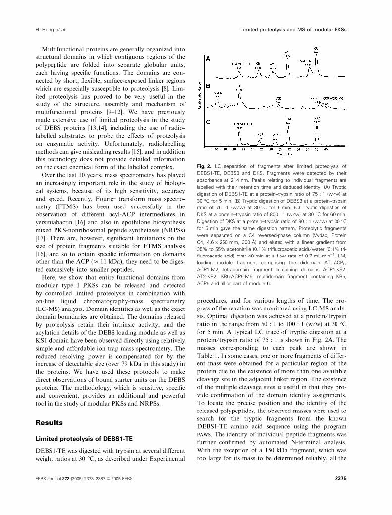
protein ⁄ trypsin ratio of 75 : 1 is shown in Fig. 2A. The
masses corresponding to each peak are shown in
Table 1. In some cases, one or more fragments of differ-
ent mass were obtained for a particular region of the
protein due to the existence of more than one available
cleavage site in the adjacent linker region. The existence
of the multiple cleavage sites is useful in that they pro-
vide confirmation of the domain identity assignments.
To locate the precise position and the identity of the
released polypeptides, the observed masses were used to
search for the tryptic fragments from the known
DEBS1-TE amino acid sequence using the program
paws. The identity of individual peptide fragments was
further confirmed by automated N-terminal analysis.
With the exception of a 150 kDa fragment, which was
too large for its mass to be determined reliably, all the
Fig. 2. LC separation of fragments after limited proteolysis of
DEBS1-TE, DEBS3 and DKS. Fragments were detected by their
absorbance at 214 nm. Peaks relating to individual fragments are
labelled with their retention time and deduced identity. (A) Tryptic
digestion of DEBS1-TE at a protein–trypsin ratio of 75 : 1 (w ⁄w) at
30 �C for 5 min. (B) Tryptic digestion of DEBS3 at a protein–trypsin
ratio of 75 : 1 (w ⁄w) at 30 �C for 5 min. (C) Tryptic digestion of
DKS at a protein–trypsin ratio of 800 : 1 (w ⁄w) at 30 �C for 60 min.
Digestion of DKS at a protein–trypsin ratio of 80 : 1 (w ⁄w) at 30 �Cfor 5 min gave the same digestion pattern. Proteolytic fragments
were separated on a C4 reversed-phase column (Vydac, Protein
C4, 4.6 · 250 mm, 300 A) and eluted with a linear gradient from
35% to 55% acetonitrile (0.1% trifluoroacetic acid) ⁄water (0.1% tri-
fluoroacetic acid) over 40 min at a flow rate of 0.7 mLÆmin)1. LM,
loading module fragment comprising the didomain ATL-ACPL;
ACP1-M2, tetradomain fragment containing domains ACP1-KS2-
AT2-KR2; KR5-ACP5-M6, multidomain fragment containing KR5,
ACP5 and all or part of module 6.
H. Hong et al. Limited proteolysis and MS of modular PKSs
FEBS Journal 272 (2005) 2373–2387 ª 2005 FEBS 2375

other fragments were detected with a mass accuracy of
0.01%, and therefore could be matched uniquely to the
amino acid sequence. Thus, both the N-terminal and the
exact C-terminal of the fragments as well as their identi-
ties were assigned (Table 1). Despite the uncertainty in
mass of the 150 kDa fragment, it was possible to con-
firm by N-terminal sequencing that this fragment starts
with ACP1, and based on the size of the observed mass
it probably comprises all of module 2 bar the C-terminal
ACP2 domain. However, ACP2 was observed separately
as part of the ACP2-TE didomain. Good LC separation
was achieved with the exception of the ACP2-TE and
TE fragments, which coelute. The digestion pattern of
DEBS1-TE generated by trypsin is in good agreement
with previous results from the tryptic digestion of
DEBS1 [13]. The loading module was released as a sta-
ble didomain ATL-ACPL. KR1 and TE were also both
released as stable single domains. Most of module 2
remained intact and did not release isolated domains
even when the protein was treated with up to 2 m urea
with the aim of partially unfolding the protein. To check
whether other proteinases could digest module 2, ela-
stase was also used to analyze DEBS1-TE. The resulting
digestion pattern from elastase was very similar to that
obtained following tryptic digestion (data not shown),
and again module 2 remained largely intact. Import-
antly, however, KS1 and AT1 were found to be released
as separate individual domains, which was in contrast to
the previous proteolysis results on DEBS1 and DKS,
where the KS1 and AT1 were always observed together,
either as a KS1-AT1 didomain or as part of larger pro-
teolytic fragments [6,13]. The ACP2 domain once
released seems to be susceptible to further proteolysis,
as it was never observed independently under the dig-
estion conditions employed, only as the ACP2-TE
didomain. Under harsher digestion conditions, even
ACP2-TE was degraded further leaving only the TE
domain intact. These observations suggest that the
ACP2 domain is stabilized by the presence of the TE
domain, as observed for PCP or ACP domains in other
NRPS and PKS proteins [12]. In contrast to the ACP2-
TE didomain, the loading didomain ATL-ACPL seemed
to be more resistant to proteolysis, and individual
domains were not observed, suggesting a strong inter-
action between the two domains. The correct post-trans-
lational modification with a 4¢-phosphopantetheinylprosthetic group of both the loading and extender ACPs
was confirmed by the fact that the observed mass of
ATL-ACPL and ACP2-TE could only be matched from
the DEBS1-TE amino acid sequence if the phospho-
pantetheinyl moiety is presumed to be present on both
ACPs (the calculated mass increase for addition of a
phosphopantetheinyl group is 339 Da).
Limited proteolysis of DEBS3
Purified DEBS3 was subjected to tryptic digestion as
described in Experimental procedures. Digestions were
carried out at two different protein ⁄ trypsin ratios,
250 : 1 (w ⁄w) and 75 : 1 (w ⁄w), but the resulting diges-
Table 1. Fragments identified after limited proteolysis of DEBS1-TE.
Fragment identity Corresponding sequence N-Terminal sequencea Observed mass (Da)b Expected mass (Da)
TE E3468-S3738 EASSALRDGY 28951 ± 1 28952
L3452-S3738 LAD**G 30610 ± 1 30613
R3451-S3738 RLA 30766 ± 1 30769
ACP2-TE A3363-S3738 AGEPETESLR 40592 ± 2 40255(apo)
40594 (holo)
KS1 T550-R1137 TNEAAPGEP 61196 ± 2 61200
A548-R1137 ARTNEAAPG 61424 ± 3 61428
AT1 E1138-R1418 EQDAALSTER 29770 ± 1 29770
E1138-R1429 31124 ± 1 31126
E1138-R1441 32588 ± 1 32587
ATL-ACPL T11-R544 TAQPGRIVRP 56003 ± 2 55667(apo)
56006 (holo)
T11-R547 56391 ± 2 56053(apo)
56392 (holo)
ACP1-KS2-AT2-KR2 V1925-R3362 VGALAS*PA 150114 ± 43 149829 (apo)
150168 (holo)
KR1 S1443-R1914 STEVDEVSAL 49642 ± 2 49647
R1442-R1914 RSTEVDEVS 49799 ± 3 49803
a All cleavages were at C-terminal of R residues (K is absent from the linker regions). b The error bars reported are based on at least three
independent experiments. *Signifies an unidentified residue.
Limited proteolysis and MS of modular PKSs H. Hong et al.
2376 FEBS Journal 272 (2005) 2373–2387 ª 2005 FEBS

tion pattern was the same in both cases. The LC trace
obtained for the digestion mixture at a protein ⁄ trypsinratio of 75 : 1 (w ⁄w) is shown in Fig. 2B. Only AT5
and ACP5 from module 5 and the TE domain were
observed as stable single domains. Their identities were
confirmed by both mass matching and N-terminal
sequencing analysis (Table 2). No single domain from
module 6 was observed. However, a large fragment
(greater than 100 kDa) was detected with a retention
time of 39.62 min. The identification of this fragment
was complicated by a neighbouring peak (retention time
of 38.82 min, observed mass of 57 195 Da), which
proved to arise from the E. coli chaperone protein
GroEL as judged by N-terminal sequencing and mass
spectrometric analysis. The 39.62-min polypeptide was
identified as beginning with KR5 by N-terminal sequen-
cing. Due to its large size and the relatively weak mass
spectrometric intensity, the exact C-terminus for this
fragment could not be identified. However, the approxi-
mate mass and the N-terminal sequencing results sug-
gested that this proteolytic fragment comprises KR5,
ACP5, and most or all of module 6. The didomain
ACP6-TE was not observed, but the TE domain itself
was obtained, with the same cleavage sites as observed
for DEBS1-TE. The release of ACP5 is significant in
that it is the only single ACP domain released in detect-
able quantities from the DEBS proteins. The observed
mass of ACP5 confirmed that it was in the apo form
without the phosphopantetheinyl prosthetic group
attached, as expected for the DEBS3 protein purified
from E. coli, which does not house a phosphopanthei-
nyltransferase active against DEBS [18,19]. In contrast,
DEBS1-TE and DKS, which were expressed in S. erythr-
aea, are expected to be in their holo forms.
Limited proteolysis of DKS
Purified DKS was subjected to limited tryptic pro-
teolysis under various conditions as described in
Experimental procedures. Domain and multidomain
fragments were reproducibly obtained when digestion
was carried out at a DKS ⁄ trypsin ratio of 800 : 1
(w ⁄w) at 30 �C for 1 h. In order to release the domains
rapidly for analysis following the acylation of DKS
(see later), a shorter digestion protocol was also inves-
tigated. We found that a 5-min digestion using a
DKS ⁄ trypsin ratio of 80 : 1 (w ⁄w) at 30 �C resulted in
the same digestion pattern as that from a 1-h digestion
at a DKS–trypsin ratio of 800 : 1 (w ⁄w). A typical LC
chromatogram of the proteolysed fragments from
DKS is shown in Fig. 2C. The masses corresponding
to each of the fractions are shown in Table 3. The pre-
cise location and identity of each digestion fragment
were assigned by mass mapping in combination with
N-terminal sequencing, and these data are also shown
in Table 3. The results were comparable to those of
DEBS1-TE in that all domains could be separated by
chromatography with the exception of the TE and
ACP1-TE fragments, which coeluted. Under the condi-
tions used, all the domain subunits from the DKS were
released either as individual domains or as a pair of
domains. The loading module was released as the sta-
ble didomain ATL-ACPL, and was resistant to further
digestion. KR1 and TE were released as stable indivi-
dual domains. Similarly, KS1 and AT1 were released
as individual domains (the deconvoluted mass spectra
for ATL-ACPL and KS1 are shown in Fig. 3A and
Fig. 4A, respectively). As for ACP2 in DEBS1-TE,
ACP1 was apparently too susceptible to further pro-
teolysis for it to be observed. The ACP1-TE didomain
could be observed under milder digestion conditions.
The complete post-translational modification of both
the loading and extender ACPs was also confirmed by
the observed masses.
Propionyl-CoA/n-butyryl-CoA incubation with
intact and digested DKS
The acyl-CoA substrates were incubated either with
intact protein or with the mixture of domain fragments
Table 2. Fragments identified after limited proteolysis of DEBS3. * Signifies an unidentified residue.
Fragment identity Corresponding sequence N-Terminal sequencea Observed mass (Da)b Expected mass (Da)
ACP5 Q1368-R1478 QSEEGPALAQ 12 006 ± 1 12 005(apo)
TE E2021-S2291 EASSALRDGY 28 950 ± 1 28 952
L2005-S2291 LADHIGQQ 30 611 ± 2 30 613
R2004-S2291 RL*DH 30 766 ± 1 30 769
AT5 T549-R894 TRRGVAMVF 36 676 ± 1 36 679
KR5-ACP5-M6 A907-? ARDEDDD*RY > 100 000
a All cleavages were at C-terminal of arginine residues (lysine is absent from the linker regions). b The error bars reported are based on at
least three independent experiments.
H. Hong et al. Limited proteolysis and MS of modular PKSs
FEBS Journal 272 (2005) 2373–2387 ª 2005 FEBS 2377

released by limited proteolysis, to detect any differ-
ences in acylation behaviour. (Overall polyketide syn-
thase activity was not measured.) For example, if
certain domains only become acylated via transfer of
starter units from tethered adjacent domains, they
might fail to be labelled in the mixture of fragments.
Intact DKS
The ability to release and obtain the precise mass of
individual domains and domain pairs from the DKS
enables the study of the acylation specificity for each
individual AT and ACP, as well as KS1 domains of
this multidomain enzyme.
Propionyl-CoA, the native substrate for the DEBS
loading module, was incubated with the intact DKS at
30 �C for 10 min, followed by a 5-min tryptic digestion
to release domains for analysis (Fig. 5B). Analysis of
the mass of each peak revealed that propionyl units
were specifically loaded onto fragments ATL-ACPL
and KS1 but not onto AT1, KR1, ACP1 or TE
domains. This clearly confirms that propionyl-CoA is
not a substrate for the extender AT1 and ACP1
domain. More significantly, after incubation with pro-
pionyl-CoA, the LC trace for the loading module frag-
ment showed two peaks, designated LM1 and LM2,
with a mass increase of 55 and 111 Da, respectively,
which within the experimental error corresponds to
loading of one and two propionyl units, respectively
(theoretical mass increase of 56 and 112 Da, respect-
ively) (Fig. 3B,C). No unacylated ATL-ACPL was
observed. The observation of a mass increase of
111 Da directly confirms that both active sites in the
loading didomain may be simultaneously acylated.
KS1 was also fully acylated by the incubation with
propionyl-CoA, with a mass increase of 55 Da, and no
residual free KS1 was observed (Fig. 4B). Similar
results were obtained when intact DEBS1-TE was trea-
ted with propionyl-CoA prior to digestion (data not
shown). So, for the first time, a stoichiometric binding
of the substrate on the DEBS loading module as well
as on the KS1 has been directly observed.
When the alternative non-natural substrate n-butyryl-
CoA, which also progressed to full-length polyketide
[20], was incubated with the intact DKS, similar results
were obtained (Fig. 5C). Like propionyl-CoA, the buty-
ryl group was specifically loaded onto fragment ATL-
ACPL and KS1 but not onto AT1, KR1, ACP1 or TE.
The loading module fragment also showed two peaks,
LM1 and LM2 with mass increase of 67 and 137 Da
(theoretical mass increase of 70 and 140 Da, respect-
ively), which corresponds to single and double acylation
by the butyryl group, respectively (Fig. 3D,E). KS1 was
also fully acylated by the butyryl group with a mass
increase of 68 Da (Fig. 4C). No residual free ATL-
ACPL and KS1 were observed. The results not only
provide direct evidence that the DEBS loading module
possesses flexible substrate specificity, which is in agree-
ment with previous radiolabelling studies [21], but also
demonstrate that the mass accuracy in our experiments
is sufficient to distinguish between propionyl and buty-
ryl groups even for a protein over 60 kDa.
Table 3. Fragments identified after limited proteolysis of DKS. *Signifies an unidentified residue.
Fragment identity Corresponding sequence N-Terminal sequencea Observed mass (Da)b Expected mass (Da)
TE E2021-S2291 EASSALRDGY 28 950 ± 1 28 952
L2005-S2291 LADH*GQQ 30 610 ± 2 30 613
R2004-S2291 RLADHI*QQ 30 766 ± 1 30 769
ACP1-TE V1925-S2291 VGALTGLPR 39 507 ± 1 39 171(apo)
39 510 (holo)
KS1 T550-R1137 TNEAAPG 61 195 ± 2 61 200
A548-R1137 ARTNEA 61 422 ± 2 61 428
AT1 E1138-R1418 EQDAALSTER 29 768 ± 1 29 770
E1138-R1429 31 124 ± 1 31 126
E1138-R1441 32 585 ± 1 32 587
E1138-R1442 32 742 ± 1 32 744
ATL-ACPL T11-R544 TAQPGRIVRP 56 003 ± 2 55 667(apo)
56 006 (holo)
T11-R547 56 389 ± 3 56 053(apo)
56 392 (holo)
KR1 S1443-R1914 STEVDEVS 49 642 ± 2 49 647
R1442-R1914 RSTEVDEVS 49 798 ± 2 49 803
a All cleavages were at C-terminal of R residues (K is absent from the linker regions). b The error bars reported are based on at least three
independent experiments.
Limited proteolysis and MS of modular PKSs H. Hong et al.
2378 FEBS Journal 272 (2005) 2373–2387 ª 2005 FEBS

Digested DKS
To check whether the domains released from the DKS
retain their catalytic activities after proteolysis, propio-
nyl-CoA and n-butyryl-CoA were also individually
incubated with predigested DKS at 30 �C for various
lengths of time. The maximum level of acylation was
found after 10-min incubation (data not shown). Care-
ful comparison of the LC traces as well as the acyla-
tion details of each domain revealed no discernible
difference between the acylation patterns when either
propionyl-CoA or n-butyryl-CoA were used, before or
after proteolysis. The loading module was either singly
or doubly acylated by the propionyl- or n-butyryl-
CoA, and no unacylated loading module was observed.
KS1 was also fully acylated by either substrate, while
no acylation was observed on other domains. The
results suggest that domains maintain the same intrin-
sic catalytic activity whether in isolation or within the
quaternary structure of an intact DEBS module.
Fig. 3. LC separation of fragments from trypsin-digested DKS and detection of acyl-enzymes. Fragments were detected through their absorb-
ance at 214 nm. Fragments are shown from tryptic digestion of (A) DKS (control); (B) DKS, followed by incubation with propionyl-CoA; (C)
DKS, followed by incubation with n-butyryl-CoA; (D) DKS, followed by incubation with thiol-directed reagent NPDS; (E) DKS, pretreated with
NPDS, and after digestion incubated with propionyl-CoA. The identity of domains present in each peak is indicated, together with their inferred
acylation status. Separation conditions are the same as in Fig. 2. In D and E, the first peak contains TE only, and the ACP1-TE is present as a
disulfide bond-linked dimer indicated by the arrow. *LM, loading module comprising ATL-ACPL; LM1 and LM2, signify singly and doubly acylat-
ed loading module, respectively; LM(S-S), loading module containing an internal disulfide bond between the ATL and the phosphopantetheine
of ACPL; LM1(S-S), singly acylated loading module containing an internal disulfide bond between the ATL and the phosphapantetheine of
ACPL. �It is not known whether the single acyl group is attached exclusively to the active site of ATL or of ACPL, or both.
H. Hong et al. Limited proteolysis and MS of modular PKSs
FEBS Journal 272 (2005) 2373–2387 ª 2005 FEBS 2379

Probing the sites of acylation of loading
didomain with 4-nitrophenyl disulfide
Previous experiments with apo DEBS loading module
using radiolabelling showed that the extent of labelling
was about half that when holo protein was used, as
expected, as the loading module has two active sites,
and the phosphopantetheinyl prosthetic group is
required for attachment of the substrate to the ACP
domain [21]. We wished to use mass spectrometry as an
analytical tool directly to probe the involvement of
phosphopantetheine by using a thiol-modifying reagent
4-nitrophenyl disulfide (NPDS) which reacts with sul-
fhydryl groups at neutral pH. The trypsin-digested DKS
was treated with an excess of NPDS at 30 �C for 5 min,
followed by LC-MS analysis (Fig. 5D). Comparison of
the digested DKS before and after the treatment of
NPDS showed that after NPDS treatment, the first elut-
ed peak no longer contained the ACP1-TE didomain,
only the TE domain. However, an extra peak was eluted
between the TE and the KS1, and had a molecular mass
of 79013 Da. N-terminal sequencing analysis showed
that it corresponded to the ACP1-TE. Therefore, it most
likely corresponds to a disulfide bond-linked dimer of
ACP1-TE, which has an expected mass of 79018 Da.
Unexpectedly, the loading module seemed unaffected by
NPDS, since no mass increase was observed. In addition,
careful analysis of each eluted peak showed no evidence
A
B
Fig. 4. Effect of 4-nitrophenyl disulfide
treatment on the electrospray mass spec-
trum of the loading didomain ATL-ACPL.
(A) Mass spectrum of the loading didomain
ATL-ACPL resulting from tryptic digestion of
DKS; (B) mass spectrum of the loading
didomain ATL-ACPL resulting from tryptic
digestion of DKS, after subsequent treat-
ment with NPDS. The formation of an inter-
nal disulfide bond between the ATL and
ACPL, induced by NPDS treatment, results
in alteration of the m ⁄ z distribution to a
higher mass range (see text for details).
Limited proteolysis and MS of modular PKSs H. Hong et al.
2380 FEBS Journal 272 (2005) 2373–2387 ª 2005 FEBS

for a disulfide bond-linked dimer of ATL-ACPL. How-
ever, incubation of propionyl-CoA with digested DKS,
which had been pretreated with NPDS, resulted in the
formation of only singly acylated loading module with
a mass increase of 54 Da [Fig. 5E, peak labelled as
LM1(S-S)], with no doubly acylated form being
observed. This indicated that the thiol of the phospho-
pantetheine of the ACPL was blocked by the treatment
Fig. 5. Deconvoluted mass spectra of loading didomain ATL-ACPL released from DKS by limited proteolysis. (A) unliganded loading module;
(B) and (C), loading didomain, respectively, singly and doubly acylated after incubation with propionyl-CoA either before or after proteolysis;
(D) and (E), loading didomain, respectively, singly and doubly acylated after incubation with n-butyryl-CoA either before or after proteolysis.
H. Hong et al. Limited proteolysis and MS of modular PKSs
FEBS Journal 272 (2005) 2373–2387 ª 2005 FEBS 2381

of NPDS, and only the active site serine residue of ATL
was left available for acylation. When the mass spectra
of the NPDS-treated and untreated loading module
were compared, the m ⁄ z distribution pattern showed
significant differences (Fig. 6A,B). The m ⁄ z envelope of
peaks shifted to higher values after the NPDS treatment,
indicating that an intramolecular disulfide bond might
have formed within the loading didomain (the mass
accuracy for the 56 kDa protein would not allow us to
detect the 2 Da mass decrease due to the formation of
such an internal disulfide bond). The formation of the
intramolecular disulfide bond would make the protein
more compact, therefore leaving fewer chargeable sites
available for electrospray ionization, which resulted in
higher m ⁄ z-values in the spectrum. To confirm that an
intramolecular disulfide bond had formed within the
loading didomain, the reducing reagent dithiothreitol
was added in excess to the NPDS pretreated digestion
mixture, before the mixture was analyzed using LC-MS.
As expected, the m ⁄ z distribution of the loading module
shifted back to its original position, suggesting that the
internal disulfide bond was reduced by dithiothreitol
(data not shown). Once the excess dithiothreitol in the
sample was removed, double acylation of the loading
module was observed again with a mass increase of
109 Da (a theoretical mass increase 112 Da, data not
shown), upon addition of propionyl-CoA. Taken
together, these experiments provide evidence that the
thiol of the phosphopantetheinyl arm of ACPL is
involved in the priming of the substrate. When propio-
nyl-CoA was incubated with digested DKS, which had
been pretreated with NPDS, KS1 was still fully acylated,
confirming that NPDS does not affect the active site cys-
teine of KS1. This activity can be attributed to KS1 self-
acylation. However, around 20% of the loading
didomain was found to be unacylated [Fig. 5E, peak
labelled as LM(S-S)], which was in contrast to the full
acylation without NPDS treatment. The 20% unacy-
lated product is probably due to the hydrolysis of an ini-
tially formed mono-acyl-intermediate. It was reported
previously that when the apo DEBS loading didomain
was incubated with [14C]propionyl-CoA, following an
initial burst of radioactivity, a gradual decrease was
observed. The decrease of radioactivity was attributed
by the authors to the progressive hydrolysis of the
labelled substrates from the ATL [21].
Discussion
DEBS1-TE, DEBS3 and DKS were subjected to limited
tryptic digestion, and the digestion conditions were
optimized for each protein so that domains rather than
unstructured peptides were released from modules. This
Fig. 6. Deconvoluted mass spectra of KS1 released from DKS by
limited proteolysis. (A) unliganded KS1; (B) singly acylated KS1
obtained after treatment of DKS with propionyl-CoA either before
or after proteolysis; (C) singly acylated KS1 obtained after treatment
of DKS with n-butyryl-CoA either before or after proteolysis.
Limited proteolysis and MS of modular PKSs H. Hong et al.
2382 FEBS Journal 272 (2005) 2373–2387 ª 2005 FEBS

greatly reduced the number of digestion fragments and
resulted in the released domains being readily separable
by a reverse phase C4 column and monitored by
on-line UV and mass spectrometry. Our choice of con-
ditions for proteolysis was guided by earlier studies
in which protein products were identified using only
N-terminal sequencing. The cleavage patterns therefore
followed earlier precedents, but with some notable
exceptions. For example, it is particularly interesting
that for the first time KS1 and AT1 were released as
separate individual domains following tryptic digestion
of both DEBS1-TE and DKS. This observation will
help further studies on these two key domains.
Chromatographic separation of fragments released by
proteolysis makes quantification much easier. For exam-
ple, singly and doubly acylated loading module can be
separated and the relative amounts of the two forms
determined. The peak area of either UV absorbance or
mass spectrometric intensity can be used for quantifica-
tion. Even though the different species may coelute, the
relative ratios can be obtained from the deconvoluted
spectrum. During the deconvolution process, every
charge state of a particular species in the spectrum is
considered to determine the relative abundance.
The identity of each fragment was obtained by mass
mapping from the known amino acid sequence. Given
the good mass accuracy (0.01%), for any particular
observed mass a match could normally be found with
a unique tryptic fragment from the amino acid
sequence. In combination with N-terminal analysis,
this allowed us unambiguously to assign the identity of
each released domain fragment, and to assign both its
N- and C-terminus. The precise knowledge of bound-
aries of domains defined by proteolysis has proved
vital in the design of experiments to swap domains
between PKS assembly lines, and it also greatly facili-
tates the cloning of individual domain components for
catalytic and structural studies.
In our work, ion trap mass spectrometry was used.
Compared with the Fourier transform-ion cyclotron
resonance (FT-ICR) mass spectrometer [16,17], the ion
trap is less expensive and more widely available. In
addition, its coupling with HPLC is less complicated
and more widely established. Even though the ion trap
is a low-resolution instrument, it is more than ade-
quate for most of the analytical problems likely to be
posed by proteolysis studies of modular polyketides
and polypeptides. Masses can be reliably and routinely
measured with a precision of 0.01% for strong, well-
resolved peaks. Even with weak or overlapping peaks
the precision of mass measurements is sufficiently good
to allow differentiation of ligands differing by 14 Da,
the mass of a methylene group. The protocols are
therefore suitable for reliable identification of sites of
C-terminal cleavage or the likely chemical composition
of an attached ligand.
Although the main purpose of the present work was
to assess the utility of ion trap mass spectrometry for
analysis of the protein fragments produced by limited
proteolysis of modular PKSs, a number of significant
conclusions can already be drawn concerning the struc-
ture and function of the DEBS proteins.
Comparison of the proteolysis patterns found for
DEBS1-TE and DKS shows that module 1 can be
readily digested using trypsin while module 2 is more
resistant to proteolysis. Careful investigation of the
amino acid sequence of DEBS1-TE reveals that there
is no available trypsin cleavage site in the linker region
between ACP1 and KS2, which might account for the
observation that ACP1 was left as part of the large
fragment ACP1-KS2-AT2-KR2. However, several
potential cleavage sites do exist in the linker regions
between KS2 and AT2 and between AT2 and KR2.
DEBS3 module 6 also seems to be more resistant to
proteolysis than module 5. Further studies would need
to be carried out employing a wider variety of proteas-
es and different type I PKSs, to decide whether C-ter-
minal modules are always more resistant. Nevertheless,
the proteolysis results do seem to suggest that,
although the primary domain organization between
modules is very similar, the compactness of their qua-
ternary organization may be significantly different.
No significant difference in the digestion patterns
was observed between DEBS1-TE obtained in this
study and DEBS1 reported previously [13], which does
not contain a C-terminal TE domain. The digestion
pattern for the DKS was also similar to that of mod-
ule 1 from DEBS1-TE as previously suggested [13].
The similarity in digestion pattern between these three
proteins suggests that re-positioning of the TE domain
at the end of module does not significantly alter the
structure of the adjacent module.
Post-translational modification of acyl carrier protein
domains (ACPs) of a PKS by the attachment of a
4¢-phosphopantetheinyl moiety to a unique serine resi-
due in each ACP is a prerequisite for PKS activity. We
have shown here that limited proteolysis linked to
LC-MS analysis provides a quick and unambiguous way
of checking whether and to what extent this modifica-
tion occurs. DEBS1-TE and DKS, which were expressed
in their native host S. erythraea, showed 100% holo
form, as observed for both the loading ACP (ACPL)
and the first extension ACP (ACP1); while DEBS3,
which was heterologously expressed in E. coli, was fully
in the apo form as observed for the ACP of module 5
(ACP5). These observations provide direct evidence that
H. Hong et al. Limited proteolysis and MS of modular PKSs
FEBS Journal 272 (2005) 2373–2387 ª 2005 FEBS 2383

E. coli is incapable of post-translationally modifying
ACP with phosphopantetheine [19]. Recently, we have
characterized a 4¢-phosphopantetheinyl transferase fromS. erythraea which acts on DEBS, using the MS-based
approach presented here [22].
Because the proteolytic map is reproducible, once
conditions have been optimized, enzyme-catalyzed acy-
lation on individual domains can be probed directly by
the resulting mass increase. As every domain from the
DKS has been released either as an individual domain
or as a pair of domains, DKS was chosen for acylation
studies. It has been previously shown that the DEBS
loading module can be specifically labelled after incuba-
tion with radiolabelled propionyl-CoA and some other
unnatural CoA thioesters [13,21]. DEBS1-TE catalyzed
synthesis of triketide lactones in vitro revealed that the
DEBS loading module has a relaxed specificity for the
starter unit [20]. It has been proposed that two active
sites exist in the DEBS loading module: one is the serine
of the ATL domain; the other is the thiol of phospho-
pantetheine prosthetic group of the ACPL. The priming
of the PKS with the propionate starter is accomplished
through the ATL first recruiting the propionyl from pro-
pionyl-CoA, then the propionyl group being transferred
to the phosphopantetheine ‘swinging arm’. To obtain
direct and detailed information on starter loading and
transfer along the enzymatic assembly line of DEBS, we
individually incubated either the natural substrate pro-
pionyl-CoA, or the alternative non-natural substrate
n-butyryl-CoA, with the intact DKS, followed by a
rapid tryptic digestion and analysis using LC-MS. In
both cases, the resulting digestion patterns were the
same as those obtained from the unacylated protein,
showing that substrate attachment does not alter the
accessibility of the linker regions in the multienzyme.
Acylation of the loading module by either propionyl- or
n-butyryl-CoA was clearly observed after 10-min incu-
bation, with a maximum of two acyl groups being
attached to the loading module. This agrees with the
prediction of two active sites present in the loading mod-
ule. However, not all of the loading didomain was fully
acylated even after prolonged incubation (up to 1 h,
data not shown). In all cases, two types of acylated load-
ing module, mono (LM1) and double (LM2) acylation
were observed. The two forms were chromatographi-
cally separated, and the relative amount could therefore
be quantified by the corresponding peak area. The ratio
of the two forms varied depending on the substrate used,
as demonstrated here by the propionyl-CoA and n-buty-
ryl-CoA experiments. When n-butyryl-CoA was used, a
higher ratio of LM2 to LM1 was observed (Fig. 5B,C).
The LM1 peak from both substrates had a very similar
retention time to that of the free loading module, but
the LM2 had almost one and a half minute longer
retention time compared to the free form, and it
increased as the carbon number increased from three to
four as from propionyl to butyryl (Fig. 5A–C). The
chromatographic results seem to suggest that for
the doubly acylated loading module, at least one of the
attached acyl groups is exposed, but for the singly acyl-
ated loading module, under the chromatographic condi-
tions used, the acyl group is buried.
Our results show that when priming of DEBS occurs
with either propionyl or n-butyryl units, the KS1
domain is efficiently acylated as well as the loading
didomain. In addition, the observation of acyl-inter-
mediates on ATL-ACPL and KS1 demonstrates that
the formed acyl-enzyme intermediates are stable under
the digestion and analytical conditions used here, and
the ion trap mass spectrometry used is capable of ana-
lyzing the formed acyl intermediates with the ability to
distinguish propionyl or butyryl modification on a pro-
tein of over 60 kDa. Our observations, for the first
time, provide direct proof of the proposed mechanism
for priming, and further clearly show that all the three
active sites, ATL, ACPL and KS1, can be simulta-
neously primed by the natural substrate, propionyl-
CoA, and unnatural substrate, n-butyryl-CoA, in a
single multidomain enzyme.
To test whether the proteolytically released domains
retain activity, propionyl-CoA and n-butyryl-CoA were
also incubated individually with the digested DKS.
The loading didomain followed the same acylation pat-
terns as those with intact protein, providing evidence
that domains of type I PKS retain their intrinsic activ-
ity after cleavage of their linkers. It is significant that
KS1 was still observed fully acylated after acylation of
digested protein. The explanation for this may be KS
self-acylation, which was previously proposed for the
DEBS [23,24]. The other possibility is that within the
digestion mixture, the KS acylation occurs by in trans
collaboration with the released loading didomain. Such
in trans collaboration between functional domains has
been observed in other multidomain systems [12].
Further experiments using the thiol-directed reagent
NPDS to treat the released loading module, provide
evidence that the phosphopantetheine prosthetic group
is involved in the loading of the starter unit. Interest-
ingly, the experiments revealed that an intramolecular
disulfide bond was formed within the loading dido-
main when digested DKS was treated with NPDS. The
internal disulfide bond was also formed when intact
DKS was treated with NPDS (data not shown). When
checking the amino acid sequence of the released
DEBS loading module, three cysteine residues (C26,
C139 and C206) were found, and they all reside in the
Limited proteolysis and MS of modular PKSs H. Hong et al.
2384 FEBS Journal 272 (2005) 2373–2387 ª 2005 FEBS

ATL domain. Although the identity of the cysteine
residue involved remains to be established, the forma-
tion of this internal disulfide bond indicates that the
4¢-phosphopantetheinyl ‘swinging arm’ on the ACPL
can readily approach the ATL domain.
In conclusion, our strategy for limited proteolysis in
combination with on-line liquid chromatography ion
trap mass spectrometry studies on the multifunctional
proteins, DEBS1-TE, DEBS3 and DKS as model sys-
tems, introduces an additional powerful experimental
tool for studies on the substrate specificity and organiza-
tion of type I PKSs. The methodology is based on
accessible and simple mass spectrometry equipment
rather than advanced FT-ICR-MS. Nevertheless, the
technology can achieve mass accuracy within 0.01% for
proteins up to 79 kDa and so can distinguish between
ligands which differ by as little as a methylene or an oxy-
gen. For chain initiation on DEBS, we have directly
demonstrated that multiple sites can be simultaneously
loaded with propionate or other starter acid units. This
raises the possibility that most sites on a longer assembly
line are operating simultaneously on different growing
chains. We have also discovered different degrees of sus-
ceptibility to proteolysis from one module to another,
which may reflect differential tightness of packing of
domains in the module. The technology appears appro-
priate for direct domain-by-domain investigation of
intermediates in the chain extension process on type I
modular PKS proteins. The information provided by
such studies should be particularly useful in optimizing
the efficiency of engineered PKS multienzymes.
Experimental procedures
General
Three proteins were used in this study: two bimodular pro-
teins, DEBS1-TE and DEBS3, and one unimodular protein
DKS. DEBS1-TE and DKS were expressed in S. erythraea
and purified as described previously [6,7]. DEBS3 was
heterologously expressed in E. coli, and purified as pre-
viously described [22]. Propionyl-CoA, n-butyryl-CoA and
l-1-tosylamido-2-phenylethyl chloromethyl ketone (TPCK)-
treated trypsin were purchased from Sigma (Poole, Dorset,
UK). 4-Nitrophenyl disulfide (NPDS) was purchased from
Aldrich (Poole, Dorset, UK).
Liquid chromatography-mass spectrometry
(LC-MS) analysis
Analysis was carried out on a HP 1100 (Hewlett-Packard,
Wilmington, DE, USA) liquid chromatography coupled
with an LCQ Classic (ThermoFinnigan, San Jose, USA)
mass spectrometer fitted with an electrospray-ionization
source. After proteolysis, the digestion mixture was applied
to a reverse phase column (Vydac, Protein C4,
4.6 · 250 mm, 300 A) and eluted with a linear gradient
from 35% to 55% acetonitrile (0.1% trifluoroacetic acid) ⁄water (0.1% trifluoroacetic acid) over 40 min at a flow rate
of 0.7 mLÆmin)1. The eluate was monitored by a diode array
detector selecting the UV absorbance at 214 and 280 nm.
The flow of eluate was split prior to the mass spectrometer.
Diverted eluate was collected manually and fractions were
N-terminally sequenced using an automated sequencer
(ABI, Foster City, CA, USA). The spray voltage of the
mass spectrometer was 4.5 kV, and the capillary tempera-
ture was 200 �C. All data were acquired in the positive-ion
mode at unit mass resolving power between m ⁄ z 600 to m ⁄ z2000. The LC-MS system was controlled by the xcalibur
software (version 1.1, ThermoFinnigan, San Jose, CA,
USA). The mass spectrometric data were processed and
transformed using the bioworks software (version 1.1,
ThermoFinnigan). The centroids of the ‘deconvoluted’
peaks were used to assign the observed masses. The software
paws (Proteometrics, NY, USA) was used to search the
tryptic fragments based on the known amino acid sequence.
Limited tryptic-digestion of DEBS1-TE, DEBS3
and DKS
Purified DEBS1-TE was incubated with TPCK-treated tryp-
sin at varying protein ⁄ trypsin ratios of 500 : 1, 200 : 1,
100 : 1 and 75 : 1 (w ⁄w) in digestion buffer (400 mm potas-
sium phosphate, pH 7.4, 1 mm EDTA, 1 mm dithiothreitol,
20% glycerol). Reactions were carried out at 30 �C for dif-
ferent times of incubation. Purified DEBS3 was incubated
with TPCK-treated trypsin at a protein ⁄ trypsin ratio of
either 250 : 1 or 75 : 1 (w ⁄w) in digestion buffer. Reactions
were carried out at 30 �C for 5 min. Purified DKS was incu-
bated with TPCK-treated trypsin at a protein ⁄ trypsin ratio
of 800 : 1 (w ⁄w) in digestion buffer. Reactions were carried
out at 30 �C for 5, 15, 30, and 60 min to determine the opti-
mal digestion conditions under which stable proteolytic
fragments were released. Digestion of DKS was also carried
out at a protein ⁄ trypsin ratio of 80 : 1 (w ⁄w) at 30 �C for
5 min. All the digestions were terminated by loading the
digestion mixture directly onto the pre-equilibrated (35%
acetonitrile ⁄ 0.1% trifluoroacetic acid) C4 column.
Propionyl-CoA/n-butyryl-CoA incubation with
intact and digested DKS
Intact DKS
Guided by previous measurements of substrate concentra-
tion required for saturation of the loading didomain [21],
purified DKS (6 lm) was incubated individually with each
acyl-CoA (6 mm) in a total volume of 30 lL, containing
H. Hong et al. Limited proteolysis and MS of modular PKSs
FEBS Journal 272 (2005) 2373–2387 ª 2005 FEBS 2385

400 mm potassium phosphate (pH 7.4), 1 mm EDTA, 1 mm
dithiothreitol, and 20% glycerol. The reaction was carried
out at 30 �C for 10 min. To minimize subsequent chemical
hydrolysis of the acyl-enzyme intermediates formed, trypsin
was added immediately to the reaction mixture at a pro-
tein ⁄ trypsin ratio of 80 : 1 (w ⁄w), and digestion was per-
formed at 30 �C for 5 min, before analysis by LC-MS.
Proteolysed DKS
Six micromolar DKS, proteolysed with trypsin as above,
was incubated with 6 mm propionyl-CoA in a total volume
of 30 lL, containing 400 mm potassium phosphate
(pH 7.4), 1 mm EDTA, 1 mm dithiothreitol, and 20% gly-
cerol. Incubations were at 30 �C for 5, 10 or 20 min,
respectively. After incubation, the reaction mixture was ana-
lyzed directly by LC-MS. Similarly, n-butyryl-CoA was also
incubated with proteolysed DKS, and analyzed by LC-MS.
Probing the sites of acylation of loading
didomain with 4-nitrophenyl disulfide
Purified DKS was subjected to tryptic proteolysis as des-
cribed above. After limited proteolysis, 4-nitrophenyl disul-
fide (NPDS) was added to the trypsin-digested DKS (6 lm)to a final concentration of 52 lm. The reaction mixture was
incubated at 30 �C for 5 min, and a portion was then ana-
lyzed by LC-MS. To the rest of the reaction mixture pro-
pionyl-CoA was added to a final concentration of 6 mm,
and after incubation at 30 �C for a further 10 min the mix-
ture was analyzed by LC-MS. To test whether NPDS had
induced the formation of an intramolecular disulfide bond
within the loading didomain (ATL-ACPL), after incubating
NPDS with the digested DKS, a large excess of dithiothrei-
tol was added to the reaction mixture, and left at room
temperature for 10 min. Excess dithiothreitol was then
removed from the peptide mixture by buffer exchange using
a CentriprepTM concentrator (10 kDa cut-off) (Amicon,
Bedford, MA, USA). Propionyl-CoA was added to the
mixture, and the reaction was performed at 30 �C for
10 min, and the product mixture then analyzed by LC-MS.
Acknowledgements
We gratefully acknowledge the financial support of the
Biotechnology and Biological Sciences Research Coun-
cil (UK) for this work through a project grant to PFL
and JS. JGB thanks the EU for a Marie Curie Post-
doctoral Research Fellowship.
References
1 Staunton J & Wilkinson B (1997) Biosynthesis of ery-
thromycin and rapamycin. Chem Rev 97, 2611–2629.
2 Staunton J & Weissman KJ (2001) Polyketide biosynth-
esis: a millennium review. Nat Prod Report 18, 380–416.
3 Leadlay PF (1997) Combinatorial approaches to poly-
ketide biosynthesis. Curr Opin Chem Biol 1, 162–168.
4 Tsoi CJ & Khosla C (1995) Combinatorial biosynthesis
of ‘unnatural’ natural products: the polyketide example.
Chem Biol 2, 355–362.
5 Cortes J, Wiesmann KEH, Roberts GA, Brown MJB,
Staunton J & Leadlay PF (1995) Repositioning of a
domain in a modular polyketide synthase to promote
specific chain cleavage. Science 268, 1487–1489.
6 Østergaard LH, Kellenberger L, Cortes J, Roddis MP,
Deacon M, Staunton J & Leadlay PF (2002) Stereo-
chemistry of catalysis by the ketoreductase activity in
the first extension module of the erythromycin poly-
ketide synthase. Biochemistry 41, 2719–2726.
7 Bycroft M, Weissman KJ, Staunton J & Leadlay PF
(2000) Efficient purification and kinetic characterization
of a bimodular derivative of the erythromycin poly-
ketide synthase. Eur J Biochem 267, 520–526.
8 Mally MI, Grayson DR & Evans DR (1981) Controlled
proteolysis of the multifunctional protein that initiates
pyrimidine biosynthesis in mammalian-cells – evidence
for discrete structural domains. Proc Natl Acad Sci
USA 78, 6647–6651.
9 Mattick JS, Tsukamoto Y, Nickless J & Wakil SJ
(1983) The Architecture of the animal fatty-acid synthe-
tase. 1. Proteolytic dissection and peptide-mapping.
J Biol Chem 258, 5291–5299.
10 Chauhan HJ, Domingo GJ, Jung HI & Perham RN
(2000) Sites of limited proteolysis in the pyruvate
decarboxylase component of the pyruvate dehydrogen-
ase multienzyme complex of Bacillus stearothermophilus
and their role in catalysis. Eur J Biochem 267, 7158–
7169.
11 Bantscheff M, Weiss V & Glocker MO (1999) Identifi-
cation of linker regions and domain borders of the tran-
scription activator protein NtrC from Escherichia coli
by limited proteolysis, in-gel digestion, and mass spec-
trometry. Biochemistry 38, 11012–11020.
12 Hijarrubia MJ, Aparicio JF & Martin JF (2003)
Domain structure characterization of the multifunc-
tional alpha- aminoadipate reductase from Penicillium
chrysogenum by limited proteolysis – activation of
alpha-aminoadipate does not require the peptidyl carrier
protein box or the reduction domain. J Biol Chem 278,
8250–8256.
13 Aparicio JF, Caffrey P, Marsden AFA, Staunton J &
Leadlay PF (1994) Limited proteolysis and active-site
studies of the first multienzyme component of the ery-
thromycin-producing polyketide synthase. J Biol Chem
269, 8524–8528.
14 Staunton J, Caffrey P, Aparicio JF, Roberts GA, Bethell
SS & Leadlay PF (1996) Evidence for a double-helical
Limited proteolysis and MS of modular PKSs H. Hong et al.
2386 FEBS Journal 272 (2005) 2373–2387 ª 2005 FEBS

structure for modular polyketide synthases. Nat Struct
Biol 3, 188–192.
15 Weissman KJ, Bycroft M, Staunton J & Leadlay PF
(1998) Origin of starter units for erythromycin biosynth-
esis. Biochemistry 37, 11012–11017.
16 Mazur MT, Walsh CT & Kelleher NL (2003) Site-speci-
fic observation of acyl intermediate processing in thio-
template biosynthesis by Fourier transform mass
spectrometry: The polyketide module of yersiniabactin
synthetase. Biochemistry 42, 13393–13400.
17 Hicks LM, O’Connor SE, Mazur MT, Walsh CT &
Kelleher NL (2004) Mass spectrometric interrogation of
thioester-bound intermediates in the initial stages of
epothilone biosynthesis. Chem Biol 11, 327–335.
18 Roberts GA, Staunton J & Leadlay PF (1993) Hetero-
logous expression in Escherichia coli of an intact multi-
enzyme component of the erythromycin-producing
polyketide synthase. Eur J Biochem 214, 305–311.
19 Pfeifer BA, Admiraal SJ, Gramajo H, Cane DE & Kho-
sla C (2001) Biosynthesis of complex polyketides in a
metabolically engineered strain of E. coli. Science 291,
1790–1792.
20 Wiesmann KEH, Cortes J, Brown MJB, Cutter AL,
Staunton J & Leadlay PF (1995) Polyketide synthesis
in vitro on a modular polyketide synthase. Chem Biol 2,
583–589.
21 Lau J, Cane DE & Khosla C (2000) Substrate specificity
of the loading didomain of the erythromycin polyketide
synthase. Biochemistry 39, 10514–10520.
22 Weissman KJ, Hong H, Oliynyk M, Siskos AP &
Leadlay PF (2004) Identification of a phosphopan-
tetheinyl transferase for erythromycin biosynthesis in
Saccharopolyspora erythraea. Chembiochem 5, 116–
125.
23 Pereda A, Summers RG, Stassi DL, Ruan XA & Katz
L (1998) The loading domain of the erythromycin poly-
ketide synthase is not essential for erythromycin bio-
synthesis in Saccharopolyspora erythraea. Microbiology
144, 543–553.
24 Wilkinson CJ, Frost EJ, Staunton J & Leadlay PF
(2001) Chain initiation on the soraphen-producing mod-
ular polyketide synthase from Sorangium cellulosum.
Chem Biol 8, 1197–1208.
H. Hong et al. Limited proteolysis and MS of modular PKSs
FEBS Journal 272 (2005) 2373–2387 ª 2005 FEBS 2387
Copyright © 2022 FDOKUMEN