ChemInform Abstract: Computational Design of Highly Selective Antimicrobial Peptides
Upload
independentCategory
view
0download
0
The biosynthesis of polyketide-derived polycyclic ethers†
Andrew R. Gallimore*
Received 10th September 2008
First published as an Advance Article on the web 21st November 2008
DOI: 10.1039/b807902c
Covering: up to 2008
The biosynthetic pathways to polyketide-derived polycyclic ethers, in bacteria, plants and marine
organisms, have, until now, tended to be considered separately. The purpose of this article is to provide
an integrated review of the common mechanistic aspects of polyether biosynthesis from these diverse
sources. In particular, the focus will be on the proposed mechanisms of oxidative cyclisation, as well as
on the known differences in polyketide chain construction between the terrestrial and marine
polyethers.
1 Introduction2 Fatty acid and polyketide biosynthesis3 Polyether ionophores4 The annonaceous acetogenins5 Marine polyethers6 Chain construction in polyether biosynthesis7 Acknowledgements8 References
1 Introduction
The term polycyclic ether, or simply polyether, is generally
limited to only two classes of highly bioactive natural products
that contain one or more ether rings, ranging from 5 to 9-
membered; these are the polyether ionophores and the marine
polyether ladders. Here, however, this definition will be
expanded to include a group of similar compounds derived
exclusively from a specific family of plants, the annonaceous
acetogenins. The biogenesis of the polyethers has been the
subject of an enormous amount of interest for over 30 years, and
each of these types of metabolites reveals important facets of
Nature’s approach to the construction of ether rings.
2 Fatty acid and polyketide biosynthesis
Both the terrestrial and the marine polyethers are derived from
the classic polyketide biosynthetic pathway. The family of
polyketide natural products is vast, and displays remarkable
structural diversity and biological activity. The polyketide
pathway1 represents an adaptation of the fatty acid biosynthetic
pathway,2 and one which affords the pathway its diverse natural
products. Construction of a fatty acid chain is initiated by the
condensation of an acetate ‘starter unit’ with a malonate
‘extender unit’. Acetyl-CoA andmalonyl-CoA are first converted
into enzyme-bound thioesters. The acetyl-CoA starter unit is
loaded onto a specific cysteine residue on the b-ketoacyl synthase(KS). Similarly, the malonyl-CoA extender unit, in a reaction
catalysed by malonyl acetyl transferase (MAT), is loaded onto
a thiol of the acyl carrier protein (ACP). The fundamental chain
extension step is catalysed by the ketosynthase and is a Claisen-
like condensation facilitated by decarboxylation of the malonyl-
ACP; this reaction gives the acetoacetyl-ACP. Subsequently, the
ketoester is reduced by a ketoreductase (KR), dehydrated by
a dehydratase (DH) and, finally, reduced further by an enoyl
reductase (ER) (Figs. 1 and 2) This set of reactions completes the
first round of chain extension, after which the chain is transferred
from the ACP onto the ketoreductase, freeing up the ACP for the
loading of the next extender unit. The extension process is
repeated, two carbon units at a time, until a specific chain length
is obtained. At this point, the enzyme-bound thioester is released
from the fatty acid synthase, by means of a thioesterase, to give
the free fatty acid. The polyketides are constructed on large
enzymes very similar to the fatty acid synthase, namely polyke-
tide synthases (PKS),3 and the cycle of chain extension is directly
analogous. The crucial difference lies in the fact that the ketoester
may be left unprocessed by an absent or inactive ketoreductase,
Andrew R: Gallimore
Andrew obtained his Master of
Chemistry degree at the
University of Liverpool (1998–
2002), where he acquired a keen
interest in natural product
biosynthesis. In 2002, he moved
to Cambridge to pursue doctoral
work under the supervision of
Dr. Joe Spencer. His research
focused on the genes governing
oxidative cyclisation during
monensin biosynthesis, as well as
developing a general theoretical
model for marine polyether
construction. He currently works as a postdoctoral research asso-
ciate in the lab of Prof. Peter Leadlay, where he studies the
mechanistic details and evolution of polyether biosynthesis.
Dept. of Biochemistry, 80 Tennis Court Rd, Cambridge, CB2 1GA, UK.E-mail: [email protected]; Tel: +44 1223 333658† Dedicated to Dr. Jonathan B. Spencer 1960–2008.
266 | Nat. Prod. Rep., 2009, 26, 266–280 This journal is ª The Royal Society of Chemistry 2009
REVIEW www.rsc.org/npr | Natural Product Reports
Publ
ished
on
21 N
ovem
ber 2
008.
Dow
nloa
ded
by U
nive
rsity
of Y
ork
on 0
6/02
/201
4 20
:36:
47.
View Article Online / Journal Homepage / Table of Contents for this issue

or reduced to varying degrees by the absence or presence of the
subsequent catalytic steps. Thus, each extension cycle may afford
either a keto-, hydroxyl, enoyl functionality or the fully saturated
C2-extension product. Structural diversity may be increased by
the utilisation of alternative starter units or extender units other
than malonate, in each cycle of chain extension (e.g. methyl-
malonate).4,5 Further structural and functional modifications of
this chain, whether during or after polyketide chain construction,
afford the final natural product—the polyketide. It is these ‘late
stage’ modifications that mould the distinctive structure of
individual polyethers.
3 Polyether ionophores
Without exception, all known polyether ionophores are
produced by actinomycetes. In particular, the vast majority are
derived from the genus Streptomyces and, so far, over 120 such
polyethers have been isolated and characterised.6 The ionophore
antibiotics share both structural characteristics and biological
activities. From a structural perspective, all contain ether rings
that are 5 or 6-membered and saturated; no larger ring sizes
have been observed. Rings are either connected as a spiroketal
system (2–3 rings—di- or tri-oxaspiro-cycloalkanes), or are
separated by at least one single bond, with fused rings being
absent from these structures. In addition, the polyether iono-
phores may contain a range of other structural elements,
including hydroxy-, methoxy-, halo- and phenyl groups, and,
often unusual heterocyclic systems, some of which may be
important for biological activity. Typically, they exhibit
a carboxyl group at one terminus and one or two hydroxyl
groups at the other (Fig. 3).7 By forming lipid-soluble, dynami-
cally reversible, complexes with cations, they are able to trans-
port them across biological membranes. This redistribution
disrupts the carefully-controlled, energetically-demanding,
balance of ions across bacterial membranes.8 The polyether
ionophore that has been most extensively studied is monensin,
and it is thus appropriate that the majority of the research
reported and discussed surrounding terrestrial polyether
biosynthesis has centred on this particular ionophore. Monensin
was first isolated and characterised in 1967 by Agtarap et al.,9
and its polyketide nature was rapidly established; feeding of14C-labelled precursors demonstrated that the monensin back-
bone was constructed from five acetate, seven propionate and
a single butyrate unit.10 Further studies, utilising 18O2, revealed
that three of the ether ring oxygens and the terminal C26-
hydroxyl were derived from molecular oxygen.11,12 Prior to this,
in 1974, Westley had proposed the formation of the terminal
ether ring of a related ionophore, lasalocid, as resulting from the
opening of an olefin-derived epoxide intermediate (Scheme 1).13
This idea was specifically applied to the monensin structure in
1983 by Cane, Celmer and Westley (Scheme 2).14 A linear triene
intermediate was envisaged that is oxidised to form a triepoxide,
which then undergoes a series of epoxide openings and ring
closures to give the polycyclic structure. Assuming the epoxides
were the result of a molecular oxygen-utilising mono-oxygenase,
this scheme neatly accounts for the observed labelling pattern
for 18O2. This highly influential model has remained the
Fig. 1 Cycle of fatty acid biosynthesis.
Fig. 2 Enzymes of the fatty acid synthase.
This journal is ª The Royal Society of Chemistry 2009 Nat. Prod. Rep., 2009, 26, 266–280 | 267
Publ
ished
on
21 N
ovem
ber 2
008.
Dow
nloa
ded
by U
nive
rsity
of Y
ork
on 0
6/02
/201
4 20
:36:
47.
View Article Online

foremost up to the present, although variations on the theme
have been proposed. An important feature of the Cane-Celmer-
Westley (CCW) model is the requisite stereochemistry of the
three double-bonds in the triene intermediate, which must all
have a trans configuration. Two alternative models have been
proposed, both of which involve a triepoxide intermediate with
a different set of double-bond configurations.
The first of these was the Townsend-Basak model. This model
invoked a series of [2 + 2] oxidative cyclisations utilising iron and
necessitates a Z,Z,Z-triene.15 Later, Staunton and Leadlay
(Scheme 3) proposed a modified version of the CCW model that
addressed possible concerns over the mechanistic aspects of the
Fig. 3 Polyether ionophores.
Scheme 1 Proposed cyclisation of lasalocid.
Scheme 2 Cane-Celmer-Westley model for monensin biosynthesis.
268 | Nat. Prod. Rep., 2009, 26, 266–280 This journal is ª The Royal Society of Chemistry 2009
Publ
ished
on
21 N
ovem
ber 2
008.
Dow
nloa
ded
by U
nive
rsity
of Y
ork
on 0
6/02
/201
4 20
:36:
47.
View Article Online

CCW model. The CCW cyclisation mechanism involves two
successive SN2 inversions at tertiary centres, normally expected
to be highly chemically disfavoured. The modified version
overcame this concern by invoking SN1 attack at these centres
with retention of stereochemistry. Whereas the CCWmechanism
is a cascade initiated by the SN2 opening of the first epoxide, this
alternative mechanism is initiated from the opposite end. In what
appears to be a concerted (or almost so) process, the disubsti-
tuted epoxide is opened (SN2 with inversion) by the second
trisubstituted epoxide acting as a nucleophile as it opens to form
a stabilised tertiary carbocation. This process could be assisted
by activation of the terminal carbonyl, which would help activate
the disubstituted epoxide towards the SN2 reaction. The resulting
carbocation is then quenched, with retention, by the opening of
the first trisubstituted epoxide to yield a second tertiary carbo-
cation, which is finally quenched by the hydroxyl. Overall, and
distinct from the CCW mechanism, this proposal requires two
trisubstituted cis epoxides and thus a Z,Z,E-triene precursor. As
each of these models required a different set of double-bond
configurations in the precursor, it was clear that if the triene
intermediate could be detected and its stereochemistry deter-
mined, it would be helpful in distinguishing between these
mechanistic possibilities.
The monensin gene cluster was fully characterised by sequence
analysis of cosmid fragments from a monensin-producing strain
of S. cinnamonensis.16 The polyketide synthase is organised into
twelve modules, each responsible for one cycle of chain exten-
sion, as would be predicted from the structure of monensin. The
presence was noted of three novel genes not previously found in
any complex polyketide gene cluster: monBI, monBII, and
monCI. MonBI and MonBII display a significant polypeptide
sequence homology to known ketosteroid isomerase17 enzymes
from Comononas testeroni and Pseudomonas putida.18 If indeed
the monB genes did encode isomerase-type enzymes, their pres-
ence might lend support to the Staunton-Leadlay cyclisation
model. As each a,b-unsaturated double-bond of the growing
polyketide chain is formed, this transiently activated bond might
be isomerised by a MonB enzyme, via an extended enolate. If
each of the trisubstituted double-bonds were isomerised from
a trans configuration to cis, the result would be to convert an all
trans (i.e. E,E,E) triene to a Z,Z,E configuration, as required by
the above model. Further, if each ‘isomerase’ is responsible for
only one double-bond, then the presence of two such enzymes is
explained.
The monCI gene product shows considerable sequence
homology to flavin epoxidases and thus it was inferred that this
gene governs the epoxidation of the triene intermediate.
Crucially, disruption of monCI led to the isolation and charac-
terisation of a triene shunt metabolite, confirming its role as an
epoxidase19 and providing support for the notion that a single
epoxidase suffices for oxidation of all three double bonds.
Significantly, the three double bonds were each assigned a trans
geometry (Fig. 4), strongly supporting the original CCW cycli-
sation model over both the Townsend-Basak and the Staunton-
Leadlay proposals. This result unequivocally endorses the idea
that oxidation and cyclisation from a linear polyene precursor is
a viable biosynthetic methodology, with implications for the
construction of polyethers in general. Meanwhile, the role of the
monB genes was also clarified by gene disruption experiments.
The MonB proteins belong to an expanding family of enzymes
that utilise the same structural scaffold to catalyse diverse reac-
tions using acid–base catalysis. These include the ketosteroid
isomerases, scytalone dehydratase,20 the nuclear transport
Scheme 3 Staunton-Leadlay monensin cyclisation model.
Fig. 4 Triene lactone isolated from monCI-null mutant of S. cinnamo-
nensis.
This journal is ª The Royal Society of Chemistry 2009 Nat. Prod. Rep., 2009, 26, 266–280 | 269
Publ
ished
on
21 N
ovem
ber 2
008.
Dow
nloa
ded
by U
nive
rsity
of Y
ork
on 0
6/02
/201
4 20
:36:
47.
View Article Online

factors21 and, most notably, limonene epoxide hydrolase.22 A
revised view was that the monB genes might encode hydrolases
governing the cyclisation of the triepoxide intermediate
(Scheme 4). This was borne out when, in the absence of themonB
genes, S. cinnamonensis produced a range of partially cyclised
intermediates, all of which could be converted to monensins by
treatment with acid.23 Other ionophore gene clusters, including
nanchangmycin,24 nigericin25 and tetronomycin,26 have subse-
quentlybeen found tocontain similarflavin-epoxidase andepoxide
hydrolase genes that direct the oxidative cyclisation process.
4 The annonaceous acetogenins
The annonaceous acetogenins are a group of polyketide natural
products isolated exclusively from plants and trees of the family
Annonaceae.27 The most common characteristic of these mole-
cules is a C35 or C37 fatty acid chain terminating in a g-lactone.Additional features may include olefin, hydroxyl, ketone or
epoxide moieties, as well as tetrahydrofuran (THF) or tetrahy-
dropyran (THP) rings (Fig. 5). To date, over 400 acetogenins
have been isolated and characterised, but for the purposes of this
discussion, only a few representative structures need be examined
in detail. Although direct biosynthetic studies are lacking for
these molecules, when considered together as a group, the
structures of the acetogenins themselves strongly suggest their
biosynthetic origin, with apparent biosynthetic intermediates
being isolated alongside the end products. This is especially
relevant to those acetogenins containing one or more ether rings.
When considered alongside the analogous structures in the
actinomycetes ionophores and the dinoflagellate ladders, it
becomes evident that Nature’s strategy for the construction of
ether rings in the plant kingdom is common also to that of
bacteria and the protists.
The solamins are a trio of isomeric acetogenins that possess the
characteristic g-lactone and a C35 chain in which a single THF
ring is embedded. trans-Solamin (or simply ‘solamin’), obtained
from the roots of Annona muricata,28 exists as a single diaste-
reomer with a trans configuration across the ether ring. cis-Sol-
amin, with a cis configuration across the ring, however, exists
naturally as a mixture of two tetra-epimeric diastereomers, A and
B.29 All three are thought to derive from diepoxide intermediates,
with cyclisation initiated by water (Scheme 5). The formation of
two isomers of cis-solamin can be explained by contrasting
mechanisms of cyclisation of a common diepoxide precursor—
whether cis-solamin A or B is formed depends upon the direction
of cyclisation, initiated by water attacking one or other of the
epoxides (i.e. at either C15 or C20) (Scheme 6). The presumed
diepoxide precursor, diepomuricanin, is a known acetogenin
isolated from the seeds of the same plant.30 Although die-
pomuricanin hasn’t been tested as an intermediate in the
biosynthesis of the solamins, it seems reasonable to believe that
they are correlated. Synthetic studies have shown that acid-
treatment of diepomuricanin does indeed yield both diastereo-
meric forms of cis-solamin and (trans)-solamin.31 Interestingly,
these studies also showed that diepomuricanin A naturally exists
as a pair of isomers with syn- and anti-configurations between the
two epoxides, A1 and A2 respectively. Assuming an epoxide-
opening cascade type mechanism, the anti form would be the
precursor to the cis-solamins, with the syn isomer forming
(trans)-solamin. Thus, the diene intermediate would need to be
epoxidised from opposite faces to achieve the anti diepoxide, but
the same face to achieve the syn (Scheme 6). One might naturally
expect (trans)-solamin to have its own tetra-epimeric partner, as
Scheme 4 Mechanism of limonene epoxide hydrolase at styrene oxide
and proposed action in closing ether rings.
Fig. 5 General structure of polyether acetogenins.
Scheme 5 General mechanism of formation of the solamins via a diep-
oxide.
270 | Nat. Prod. Rep., 2009, 26, 266–280 This journal is ª The Royal Society of Chemistry 2009
Publ
ished
on
21 N
ovem
ber 2
008.
Dow
nloa
ded
by U
nive
rsity
of Y
ork
on 0
6/02
/201
4 20
:36:
47.
View Article Online

syn-diepomuricanin A can of course exist as two diastereomers
and could theoretically cyclise in either direction (Scheme 6). The
fact that this is not observed suggests, as is true with the poyether
ionophores, that cyclisation is enzymatically controlled and not
a spontaneous process. As well as the diepomuricanins, corre-
sponding mono-epoxide precursor acetogenins have also been
isolated from the A. muricata.32 Epoxymurin A (Scheme 7)
contains a single epoxide at C15–16 and a double bond at C19–20,
whereas epoxymurin B has the functionalities reversed, although
the absolute stereochemistry of the epoxides hasn’t been estab-
lished. The hypothetical diene precursor to the epoxymurins is
the latest ‘intermediate’ in the biosynthetic pathway to the sol-
amins to have been isolated. Muricadienin-1, containing two cis
double bonds, was purified from the roots of A. muricata, and is
thought to be the common precursor to all three cis and trans-
solamins33 (Scheme 7). Overall, extracts of A. muricata have
provided not only a ‘polyether-type’ acetogenin, but all of the
hypothesised epoxide and olefin intermediates as well, from
muricadienin-1 to the epoxymurins to diepomuricanin A to the
solamins themselves. This strongly encourages the idea that
plant-based polycyclic ethers, like their ionophore counterparts,
are biosynthetically derived from polyenes via their corre-
sponding polyepoxides.
Goniocin, isolated from the bark of Goniothalamus gigan-
teus,34 is the only known polyether acetogenin containing three
ether rings. It is thought to be biosynthesised from a triepoxide
intermediate (Scheme 8). Analogous to the solamins, the invoked
intermediate is very closely related to a known acetogenin, tri-
poxyrollin, isolated from the seeds of Rollinia membranacea,35
though not from those of G. giganteus. The epoxides of tripoxy-
rollin, however, are all shifted two carbons further along the
chain than would be required to cyclise to goniocin, so it cannot
be a true precursor to the latter. However, the presence of closely
related metabolites in G. giganteus, appear to imply the role of
a common triepoxide intermediate in their formation. Gonio-
denin36 has a similar structure to goniocin, except that the third
THF ring is replaced by a double-bond at C21–22, in the position
that would normally carry an epoxide in the hypothetical triep-
oxide intermediate (Fig. 6). Also, intriguingly, although the
relative stereochemistries across the rings are the same as
Scheme 6 Modes of cyclisation to trans-solamin and cis-solamins A and B.
This journal is ª The Royal Society of Chemistry 2009 Nat. Prod. Rep., 2009, 26, 266–280 | 271
Publ
ished
on
21 N
ovem
ber 2
008.
Dow
nloa
ded
by U
nive
rsity
of Y
ork
on 0
6/02
/201
4 20
:36:
47.
View Article Online

goniocin, the absolute assignments are reversed. Thus, it seems
that goniodenin results from the ‘‘incomplete’’ and opposite-face
epoxidation of a common precursor triene. Synthetic studies,
using an epoxidation and acid-cyclisation strategy, have also
shown that goniodenin can be converted into a compound that is
enantiomeric in the polyether region to goniocin.37 An alterna-
tive fate of goniocin’s triepoxide precursor is revealed by the
structure of gigantecin, also from G. giganteus.38 Instead of the
purely intramolecular cyclisations that lead to goniocin, water
apparently intercedes by attacking the central epoxide. The result
is only two THF rings separated by a diol moiety39(Scheme 9).
These examples demonstrate how plants generate a variety of
structures by channelling common diene and triene intermediates
along alternative oxidative biosynthetic pathways, in which
comparison of intermediate and final structures reveal insights
into their construction. Dissecting the chemistry of ionophore
biosynthesis currently requires genetic manipulation of the
producing bacteria in order to observe the otherwise transient
intermediates, or at least shunted analogues of them. In contrast,
the annonaceous plants naturally produce these ‘intermediates’
in detectable amounts.
5 Marine polyethers
Polyketide-derived marine polyethers are dichotomously distinct
from the terrestrial, Streptomyces and plant-derived structures.
All those characterised so far have a contiguous, fused ring
system, with each oxygen constituting a single-atom bridge
between rings. This gives them a characteristic ladder-like
appearance, and they are generally referred to as ladder poly-
ethers. Fourteen distinct ladder structures have been distin-
guished, falling into various structural classes (Fig. 7). The first
of the ladder polyethers to be isolated were the brevetoxins,
isolated from the dinoflagellate, Karenia brevis.40,41 The breve-
toxins have long been associated with the ‘Red Tide’ phenom-
enon, caused by the dense aggregation of a variety of toxin-
producing unicellular phytoplankton, including K. brevis. This
algal bloom causes a deep discolouration of the sea-water and
poses a serious threat to aquatic ecosystems by killing a range of
flora and fauna,42 including humans who consume contaminated
fish and other seafood. Other ladder polyethers include the
ciguatoxins,43–45 the yessotoxins,46 gambieric acids,47 gambierol48
and the gymnocins.49,50 Maitotoxin,51,52 an extraordinary and
lethal 3422 Da polyether, is both the largest and most toxic non-
polymeric molecule known.
Scheme 8 Biosynthesis of goniocin via triene and triepoxide.
Fig. 6 Structure of goniodenin.
Scheme 7 Biosynthetic route to solamin via known acetogenins.
Scheme 9 Cyclisation of gigantecin.
272 | Nat. Prod. Rep., 2009, 26, 266–280 This journal is ª The Royal Society of Chemistry 2009
Publ
ished
on
21 N
ovem
ber 2
008.
Dow
nloa
ded
by U
nive
rsity
of Y
ork
on 0
6/02
/201
4 20
:36:
47.
View Article Online

The biosynthesis of the polyether ladders, whilst attracting
speculation, has actually advanced little further than the identi-
fication of their polyketide origin. Although labelling studies
have shed some light on the construction of the obligatory pol-
yketide chain precursor,53,54 anything further than this remains
speculative. However, the model that has been proposed, and
now validated, for monensin, was likely the inspiration for the
most prominent model for the biosynthesis of the most well-
known of the polyether ladders, brevetoxin. Both Shimizu and
Nakanishi independently proposed this model—an octaepoxide
precursor cyclises in a cascade of SN2 epoxide openings, mech-
anistically similar to that initially proposed for mon-
ensin55,56(Scheme 10). Indeed, the structures of all the marine
polyether ladders suggest that cyclisation of a polyepoxide
precursor might be a general biosynthetic strategy for their
construction. Indirect evidence for such a mechanism is provided
by the 18O2-labelling pattern of okadaic acid, a related marine
polyether toxin, suggesting an epoxide precursor to two fused
ether rings (Scheme 11).57,58 Also, the isolation of 27,28-epoxy-
brevetoxin-B (double-bond in 8-membered ring epoxidised) may
suggest the extraneous over-epoxidation of a polyene
precursor,59 although 18O2-labelling studies are lacking. It
remains largely in the realm of speculation as to whether the
marine polyethers are derived from polyepoxides via polyenes, in
a manner analogous to their terrestrial cousins. However, this
general idea remains the most straightforward and satisfactory
explanation. Further, across the entire range of known ladder
polyethers, structural variation appears to be achieved through
unusual biosynthetic manipulation of the carbon backbone
(discussed later), whilst maintaining an invariant mechanism for
Fig. 7 Marine ladder polyethers.
This journal is ª The Royal Society of Chemistry 2009 Nat. Prod. Rep., 2009, 26, 266–280 | 273
Publ
ished
on
21 N
ovem
ber 2
008.
Dow
nloa
ded
by U
nive
rsity
of Y
ork
on 0
6/02
/201
4 20
:36:
47.
View Article Online

ring formation. This is demonstrated by the stereochemical
uniformity rule, as revealed by retrobiosynthetic analysis of all
known ladder polyethers.60 According to this rule, and assuming
a given ladder polyether is derived from a polyepoxide precursor,
all the epoxides must have identical absolute stereochemistries in
order to achieve the observed stereochemical pattern in the final
ring structure (Fig. 8). That all ladder polyethers adhere to this
rule can be explained if a single mono-oxygenase is responsible
for epoxidising the precursor polyene. The asymmetric epoxi-
dation of each trans double-bond, in a consistent fashion by
a single enzyme, on either theRe or the Si face, leads to a uniform
polyepoxide consisting of either all-(R,R) or all-(S,S) trans-
epoxides. Conversely, in order to achieve a non-uniform poly-
epoxide, each double-bond would either need to be differentially
epoxidised by the mono-oxygenase, or, more likely, each double-
bond would require its own distinct enzyme. Both scenarios,
although possible, would greatly increase the complexity of the
oxidation-cyclisation process. This model has prompted the
re-examination and subsequent correction of the structure of at
least one ladder polyether, brevenal. This is one of the newest
members of the marine polyether family, isolated from K. brevis,
and is also one of the smallest, containing only five contiguous
rings. The structure initially proposed was largely in accordance
with other ladder polyethers and appeared to derive from a pol-
yepoxide in the usual way.61 However, the terminal ring C26-
hydroxyl was assigned a configuration that suggested the ring
was formed by the opening of an epoxide with the opposite
configuration than would be expected when comparing it to the
rest of the structure. However, subsequent analysis resulted in
the hydroxyl being reassigned with the opposite configuration,62
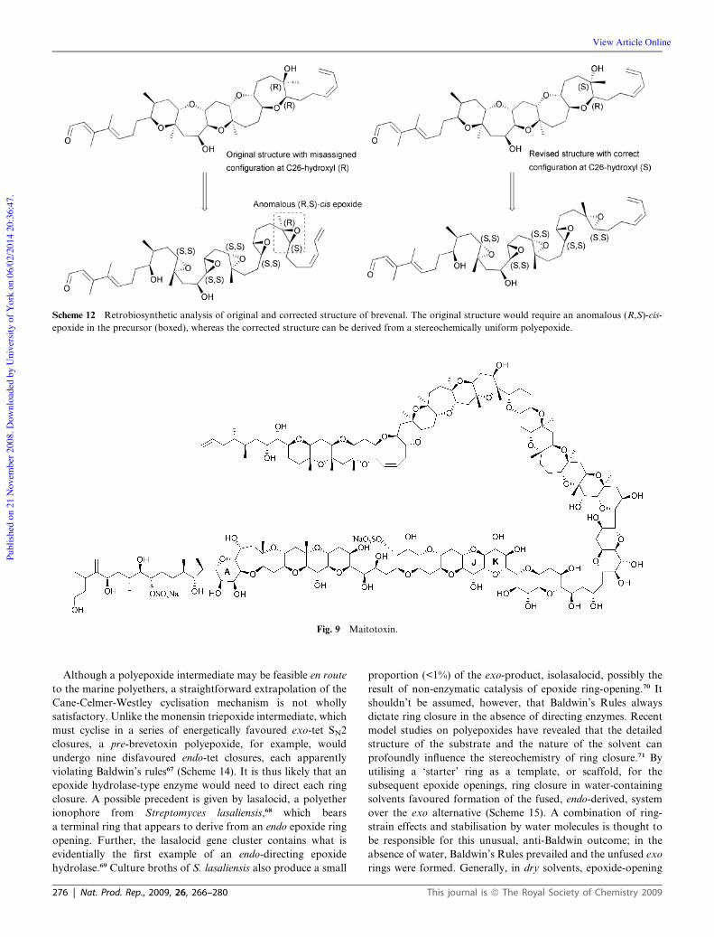
as was predicted by the rule (Scheme 12). Similar arguments
suggested that the structure of maitotoxin, the largest of the
polyethers, might have been misassigned at a single ring junction,
between the J and K rings. The structural elucidation of maito-
toxin (Fig. 9), containing no less than 32 ether rings, was
a Herculean task. However, as with the other, far smaller ladder
polyethers, the ring system could be derived from a set of ster-
eochemically identical epoxides, except for the J–K ring junction.
This was anomalous in that it appeared to derive from an
epoxide with opposite stereochemistry to the others (Scheme 13).
This specific region of the molecule was acknowledged as
particularly challenging to assign.63 Prompted by these concerns,
Nicolaou and his colleagues have synthesised large portions of
the molecule to investigate this issue.64–66 The results so far
appear to be in agreement with the original assignment, but
a crystal structure is needed to settle this matter definitively.
Scheme 10 Shimizu-Nakanishi brevetoxin cyclisation model.
Scheme 11 Proposed formation of okadaic acid fused ether rings from epoxide. Oxygens labelled with 18O2 marked with dots.
274 | Nat. Prod. Rep., 2009, 26, 266–280 This journal is ª The Royal Society of Chemistry 2009
Publ
ished
on
21 N
ovem
ber 2
008.
Dow
nloa
ded
by U
nive
rsity
of Y
ork
on 0
6/02
/201
4 20
:36:
47.
View Article Online

Fig. 8 Stereochemically uniform polyepoxides in ladder polyether biosynthesis.
This journal is ª The Royal Society of Chemistry 2009 Nat. Prod. Rep., 2009, 26, 266–280 | 275
Publ
ished
on
21 N
ovem
ber 2
008.
Dow
nloa
ded
by U
nive
rsity
of Y
ork
on 0
6/02
/201
4 20
:36:
47.
View Article Online
Although a polyepoxide intermediate may be feasible en route
to the marine polyethers, a straightforward extrapolation of the
Cane-Celmer-Westley cyclisation mechanism is not wholly
satisfactory. Unlike the monensin triepoxide intermediate, which
must cyclise in a series of energetically favoured exo-tet SN2
closures, a pre-brevetoxin polyepoxide, for example, would
undergo nine disfavoured endo-tet closures, each apparently
violating Baldwin’s rules67 (Scheme 14). It is thus likely that an
epoxide hydrolase-type enzyme would need to direct each ring
closure. A possible precedent is given by lasalocid, a polyether
ionophore from Streptomyces lasaliensis,68 which bears
a terminal ring that appears to derive from an endo epoxide ring
opening. Further, the lasalocid gene cluster contains what is
evidentially the first example of an endo-directing epoxide
hydrolase.69 Culture broths of S. lasaliensis also produce a small
proportion (<1%) of the exo-product, isolasalocid, possibly the
result of non-enzymatic catalysis of epoxide ring-opening.70 It
shouldn’t be assumed, however, that Baldwin’s Rules always
dictate ring closure in the absence of directing enzymes. Recent
model studies on polyepoxides have revealed that the detailed
structure of the substrate and the nature of the solvent can
profoundly influence the stereochemistry of ring closure.71 By
utilising a ‘starter’ ring as a template, or scaffold, for the
subsequent epoxide openings, ring closure in water-containing
solvents favoured formation of the fused, endo-derived, system
over the exo alternative (Scheme 15). A combination of ring-
strain effects and stabilisation by water molecules is thought to
be responsible for this unusual, anti-Baldwin outcome; in the
absence of water, Baldwin’s Rules prevailed and the unfused exo
rings were formed. Generally, in dry solvents, epoxide-opening
Scheme 12 Retrobiosynthetic analysis of original and corrected structure of brevenal. The original structure would require an anomalous (R,S)-cis-
epoxide in the precursor (boxed), whereas the corrected structure can be derived from a stereochemically uniform polyepoxide.
Fig. 9 Maitotoxin.
276 | Nat. Prod. Rep., 2009, 26, 266–280 This journal is ª The Royal Society of Chemistry 2009
Publ
ished
on
21 N
ovem
ber 2
008.
Dow
nloa
ded
by U
nive
rsity
of Y
ork
on 0
6/02
/201
4 20
:36:
47.
View Article Online

cascades generated unfused land polyethers, whereas in water
they generated fused sea polyethers. It thus can’t be assumed that
an endo-directing enzyme is being employed in ladder polyether
cyclisation.
As an alternative to the endo-cyclisation of a polyepoxide,
Giner has proposed that fused ether rings may be formed by the
rearrangement of an epoxy ester72(Scheme 16). Extrapolating
this synthetic strategy to the biosynthesis of marine polyethers
would require an all-cis polyene precursor. The formation of
selectively positioned cis double-bonds in polyketide chains is
certainly well precedented.73
6 Chain construction in polyether biosynthesis
The polyethers from bacteria, plants and dinoflagellates, are
clearly separated by the manner in which the carbon backbones
are manufactured. As a rule, the biosynthetic sophistication of
the backbone increases as one moves from plants to bacteria to
marine protists, with dinoflagellates employing the most exotic
chemistry. Essentially all annonaceous acetogenins are thought
to derive from either C35 or C37 very long chain fatty acids
(VLCFAs).74 The g-lactone is assumed to be constructed first;
dehydrogenation then yields one or more cis double-bonds.75 It is
at this point that the acetogenin becomes a candidate for
transformation into a polyether, via the appropriate epoxy
derivatives. Depending on the position and number of double-
bonds and subsequent epoxidations, sites of hydroxylation and
varying mechanisms of cyclisation, the range of polyethers
produced from these templates is potentially very large. Many
structures containing both THF and THP rings have already
been characterised,76 often in more than one diastereomeric
form. As the field of annonaceous acetogenins is a relatively
young one, we can assume many structures remain to be
discovered.
Similarly, over 120 natural polyether ionophores are known.
Unlike the plant polyethers, as well as employing olefin-derived
epoxides to construct ether rings, the ionophores commonly
contain rings formed from carbonyl groups; these usually appear
either as single-ring hemiacetals or as spiroketals with two or
three rings joined. Monensin, for example, in addition to the
rings derived from epoxides, contains a 5- and a 6-membered
ring, joined as a spiroketal, as well as a 6-membered ring in the
Scheme 13 Retrobiosynthetic analysis of GHIJK ring section of mai-
totoxin showing exceptional J/K ring junction from anomalous (S,S)
epoxide.
Scheme 14 Alternative modes of cyclisation of a polyepoxide, leading to fused or unfused polycyclic ether.
Scheme 15 Formation of a fused polyether from a polyepoxide cata-
lysed by water.
This journal is ª The Royal Society of Chemistry 2009 Nat. Prod. Rep., 2009, 26, 266–280 | 277
Publ
ished
on
21 N
ovem
ber 2
008.
Dow
nloa
ded
by U
nive
rsity
of Y
ork
on 0
6/02
/201
4 20
:36:
47.
View Article Online

form of a hemiacetal. Salinomycin77 (Fig. 3) has a dispiroketal
system, comprising three connected rings, as well as an anoma-
lous ring that appears to derive from neither epoxide nor
carbonyl. The latter type of ring is also a feature of several of the
marine polyethers, where the mechanism of closure of a terminal
ring is unclear from the structure. Aureothin is an antifungal
polyketide, from Streptomyces thioluteus, that contains a single
THF ring that is constructed by an unusual bifunctional P450
mono-oxygenase without the assistance of carbonyl or epoxide
functionality.78 Some anomalous rings in polyethers might be
made in a similar manner. Barring these exceptions, and unlike
the acetogenins, ionophore biosynthesis utilises the normal
functionalities developed on the polyketide synthase (PKS) in
order to create ether rings—unreduced carbonyls, olefins and
hydroxyls. One might reasonably suppose that the ancestors of
the ionophores were the more straightforward, linear polyenes. It
is thus tempting to suggest the ionophore biosynthetic pathways
diverged from those of the polyenes via the acquisition of
epoxidase genes. Although the oxidative cyclisation of a polyene
to form a polyether is a terrific piece of ‘post-PKS’ engineering, it
is the marine polyether producers that appear to utilise the most
advanced and progressive of polyketide construction methodol-
ogies. Rather than being restricted to the [1,3] substitution
pattern intrinsically built into the polyketide pathway, the
carbon backbones of ladder polyethers, such as the brevetoxins
and ciguatoxins, appear far more sophisticated. As a result, their
biogenesis has proved much more difficult to unravel through
classical feeding studies.79 Feeding exogenous substrates to
dinoflagellates is notoriously difficult, as they are often rejected,
resulting in very low levels of incorporation. This problem is
compounded by the low production levels of polyketide metab-
olites. In spite of this, stable isotope feeding has been successful
in establishing the polyketide origin of the brevetoxins, as well as
related dinoflagellates metabolites, such as okadaic acid80 and
amphidinolide.81 However, when singly-labelled [1-13C] and
[2-13C] and doubly labelled [1,2-13C] acetate was fed to K. brevis,
unexpected patterns of incorporation were observed that cannot
be explained by the usual C2 extension sequence typical of pol-
yketides. A simple polyketide will display the incorporation
pattern, ‘m-c-m-c-m-c-m-c’, etc, where ‘m’is the methyl of acetate
[2-13C] and ‘c’ is the carbonyl [1-13C]. However, brevetoxin B
exhibited numerous consecutive methyls, including six ‘m-m’
moieties, one ‘m-m-m’ and one ‘m-m-m-m’. Similarly, brevetoxin
A was labelled with seven ‘m-m’ moieties, one ‘m-m-m’ and one
‘m-m-m-m’ (Fig. 10). Further, although four of the pendant
methyl groups were labelled with [methyl-13C]-methionine,
the other three were derived from the methyl group of acetate.
This unprecedented labelling pattern was explained by the
Scheme 16 Formation of a fused polycyclic ether via an epoxy-ester
rearrangement.
Fig. 10 Labelling pattern of okadaic acid and brevetoxin B.
Scheme 17 Single carbon excision via a Favorskii-type reaction.
278 | Nat. Prod. Rep., 2009, 26, 266–280 This journal is ª The Royal Society of Chemistry 2009
Publ
ished
on
21 N
ovem
ber 2
008.
Dow
nloa
ded
by U
nive
rsity
of Y
ork
on 0
6/02
/201
4 20
:36:
47.
View Article Online

incorporation of intermediates from the tricarboxylic acid
cycle.82 However, subsequent feeding studies with the okadaic
acid and dinophysistoxin (DTX) producer, Prorocentrum lima,
suggest an alternative explanation.83 The construction of the
okadaic acid backbone mainly involves the usual successive
incorporation of intact acetate units, but, at one point along the
chain, this sequence is interrupted by a single carbon, ‘m’, from
the methyl group of acetate and, at another point, by two
consecutive methyl carbons, ‘m-m’ (Fig. 10). This was regarded
as evidence, not of tricarboxylic acid cycle intermediates, but of
an unusual carbon deletion process. Indeed, the uniform acetate
enrichment observed suggested that unusual intermediates were
not involved. If, during chain extension, a single carboxyl ‘c’
carbon was excised before the chain was passed onto the next
PKS module, the observed lone ‘m’ carbons could be explained.
The mechanism proposed involved a Favorskii-type rearrange-
ment, following mono-oxygenase-mediated oxidation of the ‘m’
carbon. This rearrangement forms a cyclopropanone interme-
diate that collapses with the aid of flavin-derived peroxide,
releasing the carboxyl ‘c’ carbon as carbon dioxide. Overall, this
reaction yields a shortened chain with an oxidised methyl-derived
‘m’ carbon (Scheme 17). This carbon-deletion process would not
interrupt the flow of the polyketide extension process through
detachment and reassembly of the nascent polyketide chain, or
could even take place once the chain has been released from the
PKS. Although this was a purely hypothetical proposal,
a Favorskii-mediated carbon excision process has recently been
shown to occur during the biosynthesis of the antifungal poly-
ketide, ambruticin.84 Ambruticin contains a cyclopropane ring
that is thought to be formed on the growing polyketide chain
with concomitant excision of a single carbon. The Favorskii
reaction leads to closure of the cyclopropane ring through three
conjugated double-bonds (Scheme 18). The cyclopropanone ring
that is also formed is then hydrolysed and a single carbon
released as carbon dioxide. The mechanism explains both
formation of the cyclopropane ring and the unusual lone methyl
‘m’ carbon (as in the brevetoxins and okadaic acid) (Fig. 11),
although the enzymes involved have not yet been identified.
Remarkably, however, an enzyme that performs a Favorskii-
type reaction has been identified as involved in the biosynthesis
of the marine bacteriostatic polyketide, enterocin, from Strep-
tomyces maritimus. EncM is an FAD-dependent oxygenase that
directs the rearrangement of the polyketide chain, through an
oxidative Favorskii reaction, to achieve the unprecedented
carbon skeleton of enterocin.85 The Favorskii-type reactions
occurring in ambruticin and enterocin biosynthesis offer clear
precedents for ladder polyether construction, but more definitive
insights must await progress in finding and defining the genes and
enzymes of these remarkable biosynthetic pathways.
7 Acknowledgements
The author is grateful to Peter Leadlay for advice and helpful
suggestions.
8 References
1 J. Staunton and K. J. Weissman, Nat. Prod. Rep., 2001, 18, 380.2 S. Smith, Faseb J., 1994, 8, 1248.3 J. Cortes, S. F. Haydock, G. A. Roberts, D. J. Bevitt andP. F. Leadlay, Nature, 1990, 348, 176.
4 B. S. Moore and C. Hertweck, Nat. Prod. Rep., 2002, 19, 70.5 O. Ghisalba, H. Fuhrer, W. J. Richter and S. Moss, J. Antibiot., 1981,34, 58.
6 C. J. Dutton, B. J. Banks and C. B. Cooper,Nat. Prod. Rep., 1995, 12,165.
7 B. C. Pressman, Ann. Rev. Biochem., 1976, 45, 501.8 J. B. Russell and A. J. Houlihan, FEMSMicrobiol. Rev., 2003, 27, 65.9 A. Agtarap, J. W. Chamberlin, M. Pinkerton and T. Steinrauf, J. Am.Chem. Soc., 1967, 89, 5737.
10 L. E. Day, J. W. Chamberlin, E. Z. Gordee, S. Chen, M. Gorman,R. L. Hamill, T. Ness, R. E. Weeks and R. Stroshane, Antimicrob.Agents. Chemother., 1973, 4, 410.
11 A. A. Ajaz and J. A. Robinson, J. Chem. Soc., Chem. Commun., 1983,12, 679.
Scheme 18 Formation of cyclopropane ring in ambruticin biosynthesis
with concomitant excision of a single carbon.
Fig. 11 a) Structure of ambruticin; b) Labelling pattern of ambruticin.
Bold bonds indicate intact acetate/propionate units. C5 lone carbon
indicated.
This journal is ª The Royal Society of Chemistry 2009 Nat. Prod. Rep., 2009, 26, 266–280 | 279
Publ
ished
on
21 N
ovem
ber 2
008.
Dow
nloa
ded
by U
nive
rsity
of Y
ork
on 0
6/02
/201
4 20
:36:
47.
View Article Online

12 A. A. Ajaz, J. A. Robinson and D. L. Turner, J. Chem. Soc., Perkin.Trans. 1, 1987, 1, 27.
13 J. W. Westley, R. H. Evans, G. Harvey, R. G. Pitcher andD. L. Pruess, J. Antibiot., 1974, 27, 288.
14 D. E. Cane, W. D. Celmer and J. W. Westley, J. Am. Chem. Soc.,1983, 105, 3594.
15 C. A. Townsend and A. Basak, Tetrahedron, 1991, 47, 2591.16 P. F. Leadlay, J. Staunton, M. Oliynyk, C. Bisang, J. Cortes, E. Frost,
Z. A. Hughes-Thomas, M. A. Jones, S. G. Kendrew, J. B. Lester,P. F. Long, H. A. I. McArthur, E. L. McCormick, Z. Oliynyk,C. B. W. Stark and C. J. Wilkinson, J. Ind. Microbiol. Biotech.,2001, 27, 360.
17 A. Kuliopulos, G. P. Mullen, L. Xue and A. S. Mildvan,Biochemistry, 1991, 30, 3169.
18 H. Cho, G. Choi, K. Y. Choi and B. Oh, Biochemistry, 1998, 37, 8325.19 A. Bhatt, C. B. W. Stark, B. M. Harvey, A. R. Gallimore,
Y. A. Demydchuk, J. B. Spencer, J. Staunton and P. F. Leadlay,Angew. Chem., Int. Ed. Eng., 2005, 44, 7075.
20 T. Lundqvist, J. Rice, C. N. Hodge, G. S. Basarab, J. Pierce andY. Lindqvist, Structure, 1994, 2, 937.
21 T. L. Bullock, W. D. Clarkson, H. M. Kent and M. Stewart, J. Mol.Biol., 1996, 260, 422.
22 M. Arand, B. M. Hallberg, J. Zou, T. Bergfors, F. Oesch, J. van derWerf, J. A. M. de Bont, T. A. Jones and S. L. Mowbray, EMBO J.,2003, 22, 2583.
23 A. R. Gallimore, C. B. W. Stark, A. Bhatt, B. M. Harvey,Y. Demudchuk, V. Bolanos-Garcia, D. J. Fowler, J. Staunton,P. F. Leadlay and J. B. Spencer, Chem. Biol., 2006, 13, 453.
24 Y. H. Sun, X. F. Zhou, H. Dong, G. Q. Tu,M.Wang, B. F.Wang andZ. X. Deng, Chem. Biol., 2003, 10, 431.
25 B. M. Harvey, T. Mironenko, Y. H. Sun, H. Hong, Z. X. Deng,P. F. Leadlay, K. J. Weissman and S. F. Haydock, Chem. Biol.,2007, 14, 703.
26 Y. Demydchuk, Y. H. Sun, H. Hong, J. Staunton, J. B. Spencer andP. F. Leadlay, ChemBiochem, 2008, 9, 1136.
27 M. C. Zafra-Polo, B. Figadere, T. Gallardo, J. R. Tormo andD. Cortes, Phytochemistry., 1998, 48, 1087.
28 S. H. Myint, D. Cortes, A. Laurens, R. Hocquemiller, M. Leboeuf,A. Cave, J. Cotte and A. M. Quero, Phytochemistry, 1991, 30,3335.
29 Y. Hu, A. R. L. Cecil, X. Franck, C. Gleye, B. Figadere andR. C. D. Brown, Org. Biomol. Chem., 2006, 4, 1217.
30 S. Sahpaz, R. Hocquemiller and A. Cave, J. Nat. Prod., 1997, 60, 199.31 C. Gleye, X. Franck, R. Hocquemiller, A. Laurens, O. Laprevote,
S. de Barros and B. Figadere, Eur. J. Org. Chem., 2001, 16, 3161.32 A. Hisham, U. Sreekala, L. Pieters, T. De Bruyne, H. Van den Heuvel
and M. Claeys, Tetrahedron, 1993, 49, 6913.33 C. Gleye, R. Hocquemiller, A. Laurens, C. Forneau, L. Serani,
O. Laprevote, F. Roblot, M. Leboeuf, A. Fournet, A. Rojos DeArias, B. Figadere and A. Cave, Phytochemistry, 1998, 47, 749.
34 Z. M. Gu, X. P. Fang, L. Zeng and J. L. McLaughlin, TetrahedronLett., 1994, 35, 5367.
35 J. R. Tormo, M. C. Zafra-Polo, A. Serrano, E. Estornell andD. Cortes, Planta Medica, 2000, 66, 318.
36 Y. Zhang, L. Zeng, M. H. Woo, Z. M. Gu, Q. Ye, F. E. Wu andJ. L. McLaughlin, Heterocycles, 1995, 41, 1743.
37 S. C. Sinha, A. Sinha, S. C. Sinha and E. Keinan, J. Am. Chem. Soc.,1998, 120, 4017–4018.
38 A. Alkofahi, J. K. Rupprecht, Y. M. Liu, C. J. Chang, D. L. Smithand J. L. McLaughlin, Experientia, 1990, 46, 539.
39 J. Yu, X. E. Hu, D. K. Ho, M. F. Bean, R. E. Stephens andJ. M. Cassady, J. Org. Chem., 1994, 59, 1598.
40 Y. Y. Lin, M. Risk, S. M. Ray, D. VanEngen, J. Clardy, J. Golik,J. C. James and K. Nakanishi, J. Am. Chem. Soc., 1981, 103, 6773.
41 Y. Shimizu, H.-N. Chou, H. Bando, G. Van Duyne and J. C. Clardy,J. Am. Chem. Soc., 1986, 108, 514.
42 B. Kirkpatrick, L. E. Fleming, D. Squicciarini, L. C. Backer, R. Clark,W. Abraham, J. Benson, Y. S. Cheng, D. Johnson, R. Pierce, J. Zaias,G. D. Bossart and D. G. Baden, Harmful Algae, 2004, 3, 99.
43 M. Murata, A. M. Legrand and T. Yasumoto, Tetrahedron Lett.,1989, 30, 3793.
44 M. Murata, A. M. Legrand, Y. Ishibashi and T. Yasumoto, J. Am.Chem. Soc., 1989, 111, 8929.
45 M. Satake, A. Morohashi, H. Oguri, T. Oishi, M. Hirama, N. Haradaand T. Yasumoto, J. Am. Chem. Soc., 1997, 119, 11325.
46 K. Eiki, M. Satake, K. Koike, T. Ogata, T. Mitsuya and Y. Oshima,Fish. Sci., 2005, 71, 633.
47 H. Nagai, K. Torigoe, M. Satake, M. Murata and T. Yasumoto,J. Am. Chem. Soc., 1992, 114, 1102.
48 M. Satake, M. Murata and T. Yasumoto, J. Am. Chem. Soc., 1993,115, 361.
49 M. Satake, M. Shoji, Y. Oshima, H. Naoki, T. Fujita andT. Yasumoto, Tetrahedron Lett., 2002, 43, 5829.
50 M. Satake, Y. Tanaka, Y. Ishikura, Y. Oshima, H. Naoki andT. Yasumoto, Tetrahedron Lett., 2005, 46, 3537.
51 Y. Kishi, Pure. Appl. Chem., 1998, 70, 339.52 M. Sasaki andM. Murata, J. Synth. Org. Chem. Japan, 1997, 55, 535.53 H.-N. Chou and Y. Shimizu, J. Am. Chem.Soc., 1987, 109, 2184.54 M. S. Lee, G.-w. Qin, K. Nakanishi and M. G. Zagorski, J. Am.
Chem. Soc., 1989, 111, 6234.55 Y. Shimizu, Natural Toxins: Animal, plant and microbial (Ed.: J. B.
Harris), Clarendon Press, Oxford, 1986, p. 123.56 K. Nakanishi, Toxicon, 1985, 23, 473.57 M. Murata, M. Izumikawa, K. Tachibana, T. Fujita and H. Naoki,
J. Am. Chem. Soc., 1998, 120, 147.58 M. Izumikawa, M. Murata, K. Tachibana, T. Fujita and H. Naoki,
Eur. J. Biochem., 2000, 267, 5179.59 H.-N. Chou, Y. Shimizu, G. Van Duyne and J. Clardy, Tetrahedron
Lett., 1985, 26, 2865.60 A. R. Gallimore and J. B. Spencer,Angew. Chem., Int. Ed. Eng., 2006,
45, 4406.61 H. Fuwa, M. Ebine and M. Sasaki, J. Am. Chem. Soc., 2006, 128,
9648.62 H. Fuwa, M. Ebine, A. J. Bourdelais, D. G. Baden and M. Sasaki,
J. Am. Chem. Soc., 2006, 128, 16989.63 M. Satake, S. Ishida, T. Yasumoto, M. Murata, H. Utsumi and
T. Hinomoto, J. Am. Chem. Soc., 1995, 117, 7019.64 K. C. Nicolaou and M. O. Frederick, Angew. Chem., Int. Ed. Eng.,
2007, 46, 5278.65 K. C. Nicolaou, K. P. Cole, M. O. Frederick, R. J. Aversa and
R. M. Denton, Angew. Chem., Int. Ed. Eng., 2007, 46, 8875.66 K. C. Nicolaou, M. O. Frederick, A. C. B. Burtoloso, R. M. Denton,
F. Rivas, K. P. Cole, R. J. Aversa, R. Gibe, T. Umezawa andT. Suzuki, J. Am. Chem. Soc., 2008, 130, 7466.
67 J. E. Baldwin, J. Chem. Soc., Chem. Comm., 1976, 11, 734.68 A. Migita, Y. Shichijo, H. Oguri, M. Watanabe, T. Tokiwano and
H. Oikawa, Tetrahedron Lett., 2008, 49, 1021.69 L. Smith, H. Hong, P. F. Leadlay, unpublished data.70 J. W. Westley, J. F. Blount, R. H. Evans, A. Stemple and J. Berger,
J. Antibiot., 1974, 27, 597.71 I. Vilotijevic and T. F. Jamison, Science, 2007, 317, 1189.72 J. Giner, J. Org. Chem., 2005, 70, 721.73 L. Tang, S.Ward, L. Chung, J. R. Carney, Y. Li, R. Reid and L. Katz,
J. Am. Chem. Soc., 2004, 126, 46.74 V. T. Tam, C. Chaboche, B. Figadere, B. Chappe, B. C. Hieu and
A. Cave, Tetrahedron Lett., 1994, 35, 883.75 B. Figadere, Acc. Chem. Res., 1995, 28, 359.76 A. Bermejo, B. Figadere, M. C. Zafra-Polo, I. Barrachina,
E. Estornell and D. Cortes, Nat. Prod. Rep., 2005, 22, 269.77 H. Kinashi, N. Otake, H. Yonehara, S. Sato and Y. Saito,
Tetrahedron Lett., 1973, 49, 4955.78 J. He, M. Muller and C. Hertweck, J. Am. Chem. Soc., 2004, 126,
16742.79 M. S. Lee, G.-W. Qin, K. Nakanishi and M. G. Zagorski, J. Am.
Chem. Soc., 1989, 111, 6234–6241.80 M. Norte, A. Padilla and J. J. Fernandez, Tetrahedron Lett., 1994, 35,
1441.81 J. Kobayashi, M. Takahashi and M. Ishibashi, J. Chem. Soc., Chem.
Comm., 1995, 1639.82 H. N. Chou and Y. Shimizu, J. Am. Chem. Soc., 1987, 109, 2184.83 J. L. C. Wright, T. Hu, J. L. McLachlan, J. Needham and
J. A. Walter, J. Am. Chem. Soc., 1996, 118, 8757.84 B. Julien, Z.-Q. Tian, R. Reid and C. D. Reeves,Chem. Biol., 2006, 13,
1277.85 L. Xiang, J. A. Kalaitzis and B. S. Moore, Proc. Natl. Acad. Sci. USA,
2004, 101, 15609.
280 | Nat. Prod. Rep., 2009, 26, 266–280 This journal is ª The Royal Society of Chemistry 2009
Publ
ished
on
21 N
ovem
ber 2
008.
Dow
nloa
ded
by U
nive
rsity
of Y
ork
on 0
6/02
/201
4 20
:36:
47.
View Article Online
Copyright © 2022 FDOKUMEN


![ChemInform Abstract: Recent Trends in the Chemistry of Aminobenzo[b]thiophenes](https://static.fdokumen.com/doc/165x107/6323ed8c3c19cb2bd106befc/cheminform-abstract-recent-trends-in-the-chemistry-of-aminobenzobthiophenes.jpg)