Impairment of Cellulose Synthases Required for Arabidopsis Secondary Cell Wall Formation Enhances...
15
Impairment of Cellulose Synthases Required for Arabidopsis Secondary Cell Wall Formation Enhances Disease Resistance W Camilo Herna ´ ndez-Blanco, a,1 Dong Xin Feng, b,1,2 Jian Hu, b,3 Andrea Sa ´ nchez-Vallet, a Laurent Deslandes, b Francisco Llorente, a Marta Berrocal-Lobo, c,4 Harald Keller, d Xavier Barlet, b Clara Sa ´ nchez-Rodrı ´guez, a Lisa K. Anderson, e Shauna Somerville, c Yves Marco, b,1,5 and Antonio Molina a,1,5 a Centro de Biotecnologı´a y Geno ´ mica de Plantas, Departamento de Biotecnologı ´a, Universidad Polite ´ cnica de Madrid, Escuela Te ´ cnica Superior Ingenieros Agro ´ nomos, E-28040 Madrid, Spain b Laboratoire de Interactions Plantes-Microorganismes, Centre National de la Recherche Scientifique–Institut National de la Recherche Agronomique, Chemin de Borde Rouge, 31326 Castanet Tolosan Toulouse, France c Department of Plant Biology, Carnegie Institution, Stanford, California 94305 d Unite ´ Mixte de Recherche–Interactions Plantes-Microorganismes et Sante Vegetale, Centre National de la Recherche Scientifique–Institut National de la Recherche Agronomique, 06903 Sophia Antipolis Cedex, France e Developmental, Cell, and Molecular Biology Group, Department of Biology, Duke University, Durham, North Carolina 27708-1000 Cellulose is synthesized by cellulose synthases (CESAs) contained in plasma membrane–localized complexes. In Arabidopsis thaliana, three types of CESA subunits (CESA4/IRREGULAR XYLEM5 [IRX5], CESA7/IRX3, and CESA8/IRX1) are required for secondary cell wall formation. We report that mutations in these proteins conferred enhanced resistance to the soil-borne bacterium Ralstonia solanacearum and the necrotrophic fungus Plectosphaerella cucumerina. By contrast, susceptibility to these pathogens was not altered in cell wall mutants of primary wall CESA subunits (CESA1, CESA3/ISOXABEN RESISTANT1 [IXR1], and CESA6/IXR2) or POWDERY MILDEW–RESISTANT5 (PMR5) and PMR6 genes. Double mutants indicated that irx-mediated resistance was independent of salicylic acid, ethylene, and jasmonate signaling. Comparative transcriptomic analyses identified a set of common irx upregulated genes, including a number of abscisic acid (ABA)–responsive, defense- related genes encoding antibiotic peptides and enzymes involved in the synthesis and activation of antimicrobial secondary metabolites. These data as well as the increased susceptibility of ABA mutants (abi1-1, abi2-1, and aba1-6) to R. solanacearum support a direct role of ABA in resistance to this pathogen. Our results also indicate that alteration of sec- ondary cell wall integrity by inhibiting cellulose synthesis leads to specific activation of novel defense pathways that con- tribute to the generation of an antimicrobial-enriched environment hostile to pathogens. INTRODUCTION The presence of a cell wall confers many of the gross morpho- logical characteristics of plants and is one of the features that distinguish them from animals. The cell wall is a complex com- posite of cellulose, high molecular weight polysaccharides, proteins, and aromatic substances that undergoes dynamic changes. In addition to providing structural support and a pas- sive barrier against invading pathogens and pests, the cell wall controls cell expansion and is involved in the exchange of water and substances throughout plant development (Carpita and McCann, 2000). It also constitutes a reservoir of antimicrobial compounds and is a source of signaling molecules (Carpita and McCann, 2000). Cellulose is a linear polymer of b-(1-4)–linked glucose, which is synthesized at the plasma membrane by a large membrane- bound complex whose only identified components are several structurally similar cellulose synthase (CESA) subunits (Somerville et al., 2004). Arabidopsis thaliana contains 10 CESA genes, six of which encode proteins with known functions (Somerville et al., 2004). The CESA1 (RADIAL SWELLING1 [RSW1]), CESA3 (ISO- XABEN RESISTANT1 [IXR1]), and CESA6 (PRC1/IXR2) subunits are largely responsible for cellulose production during the for- mation of the primary cell wall in most tissues (Fagard et al., 2000; Scheible et al., 2001; Desprez et al., 2002; Somerville et al., 2004), whereas cellulose synthesis for the secondary cell wall, which takes place after the arrest of cell expansion, requires CESA4 1 These authors contributed equally to this work. 2 Current address: Institute of Vegetables and Flowers, Chinese Acad- emy of Agricultural Sciences, Beijing 100081, People’s Republic of China. 3 Current address: Department of Biochemistry and Molecular Biology, College of Biological Sciences, China Agricultural University, Beijing 100094, People’s Republic of China. 4 Current address: Escuela Te ´ cnica Superior Ingenieros Montes, Universidad Polite ´ cnica de Madrid, E-28040 Madrid, Spain. 5 To whom correspondence should be addressed. E-mail yves.marco@ inra.toulouse.fr or [email protected]; fax 33-61285509 or 34- 913365695. The authors responsible for distribution of materials integral to the findings presented in this article in accordance with the policy described in the Instructions for Authors (www.plantcell.org) are: Yves Marco ([email protected]) and Antonio Molina (antonio.molina@ upm.es). W Online version contains Web-only data. www.plantcell.org/cgi/doi/10.1105/tpc.106.048058 The Plant Cell, Vol. 19: 890–903, March 2007, www.plantcell.org ª 2007 American Society of Plant Biologists
-
Upload
independent -
Category
Documents
-
view
1 -
download
0
Transcript of Impairment of Cellulose Synthases Required for Arabidopsis Secondary Cell Wall Formation Enhances...
Impairment of Cellulose Synthases Required for ArabidopsisSecondary Cell Wall Formation Enhances Disease Resistance W
Camilo Hernandez-Blanco,a,1 Dong Xin Feng,b,1,2 Jian Hu,b,3 Andrea Sanchez-Vallet,a Laurent Deslandes,b
Francisco Llorente,a Marta Berrocal-Lobo,c,4 Harald Keller,d Xavier Barlet,b Clara Sanchez-Rodrıguez,a
Lisa K. Anderson,e Shauna Somerville,c Yves Marco,b,1,5 and Antonio Molinaa,1,5
a Centro de Biotecnologıa y Genomica de Plantas, Departamento de Biotecnologıa, Universidad Politecnica de Madrid,
Escuela Tecnica Superior Ingenieros Agronomos, E-28040 Madrid, Spainb Laboratoire de Interactions Plantes-Microorganismes, Centre National de la Recherche Scientifique–Institut
National de la Recherche Agronomique, Chemin de Borde Rouge, 31326 Castanet Tolosan Toulouse, Francec Department of Plant Biology, Carnegie Institution, Stanford, California 94305d Unite Mixte de Recherche–Interactions Plantes-Microorganismes et Sante Vegetale, Centre National de la
Recherche Scientifique–Institut National de la Recherche Agronomique, 06903 Sophia Antipolis Cedex, Francee Developmental, Cell, and Molecular Biology Group, Department of Biology, Duke University, Durham,
North Carolina 27708-1000
Cellulose is synthesized by cellulose synthases (CESAs) contained in plasma membrane–localized complexes. In Arabidopsis
thaliana, three types of CESA subunits (CESA4/IRREGULAR XYLEM5 [IRX5], CESA7/IRX3, and CESA8/IRX1) are required for
secondary cell wall formation. We report that mutations in these proteins conferred enhanced resistance to the soil-borne
bacterium Ralstonia solanacearum and the necrotrophic fungus Plectosphaerella cucumerina. By contrast, susceptibility to
these pathogens was not altered in cell wall mutants of primary wall CESA subunits (CESA1, CESA3/ISOXABEN RESISTANT1
[IXR1], and CESA6/IXR2) or POWDERY MILDEW–RESISTANT5 (PMR5) and PMR6 genes. Double mutants indicated that
irx-mediated resistance was independent of salicylic acid, ethylene, and jasmonate signaling. Comparative transcriptomic
analyses identified a set of common irx upregulated genes, including a number of abscisic acid (ABA)–responsive, defense-
related genes encoding antibiotic peptides and enzymes involved in the synthesis and activation of antimicrobial secondary
metabolites. These data as well as the increased susceptibility of ABA mutants (abi1-1, abi2-1, and aba1-6) to R.
solanacearum support a direct role of ABA in resistance to this pathogen. Our results also indicate that alteration of sec-
ondary cell wall integrity by inhibiting cellulose synthesis leads to specific activation of novel defense pathways that con-
tribute to the generation of an antimicrobial-enriched environment hostile to pathogens.
INTRODUCTION
The presence of a cell wall confers many of the gross morpho-
logical characteristics of plants and is one of the features that
distinguish them from animals. The cell wall is a complex com-
posite of cellulose, high molecular weight polysaccharides,
proteins, and aromatic substances that undergoes dynamic
changes. In addition to providing structural support and a pas-
sive barrier against invading pathogens and pests, the cell wall
controls cell expansion and is involved in the exchange of water
and substances throughout plant development (Carpita and
McCann, 2000). It also constitutes a reservoir of antimicrobial
compounds and is a source of signaling molecules (Carpita and
McCann, 2000).
Cellulose is a linear polymer of b-(1-4)–linked glucose, which is
synthesized at the plasma membrane by a large membrane-
bound complex whose only identified components are several
structurally similar cellulose synthase (CESA) subunits (Somerville
et al., 2004). Arabidopsis thaliana contains 10 CESA genes, six of
which encode proteins with known functions (Somerville et al.,
2004). The CESA1 (RADIAL SWELLING1 [RSW1]), CESA3 (ISO-
XABEN RESISTANT1 [IXR1]), and CESA6 (PRC1/IXR2) subunits
are largely responsible for cellulose production during the for-
mation of the primary cell wall in most tissues (Fagard et al., 2000;
Scheible et al., 2001; Desprez et al., 2002; Somerville et al., 2004),
whereas cellulose synthesis for the secondary cell wall, which
takes place after the arrest of cell expansion, requires CESA4
1 These authors contributed equally to this work.2 Current address: Institute of Vegetables and Flowers, Chinese Acad-emy of Agricultural Sciences, Beijing 100081, People’s Republic ofChina.3 Current address: Department of Biochemistry and Molecular Biology,College of Biological Sciences, China Agricultural University, Beijing100094, People’s Republic of China.4 Current address: Escuela Tecnica Superior Ingenieros Montes,Universidad Politecnica de Madrid, E-28040 Madrid, Spain.5 To whom correspondence should be addressed. E-mail [email protected] or [email protected]; fax 33-61285509 or 34-913365695.The authors responsible for distribution of materials integral to thefindings presented in this article in accordance with the policy describedin the Instructions for Authors (www.plantcell.org) are: Yves Marco([email protected]) and Antonio Molina ([email protected]).W Online version contains Web-only data.www.plantcell.org/cgi/doi/10.1105/tpc.106.048058
The Plant Cell, Vol. 19: 890–903, March 2007, www.plantcell.org ª 2007 American Society of Plant Biologists
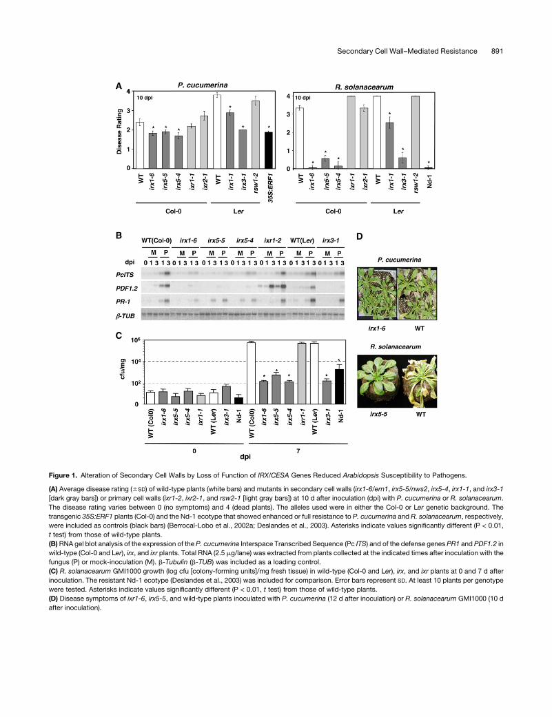
Figure 1. Alteration of Secondary Cell Walls by Loss of Function of IRX/CESA Genes Reduced Arabidopsis Susceptibility to Pathogens.
(A) Average disease rating (6SD) of wild-type plants (white bars) and mutants in secondary cell walls (irx1-6/ern1, irx5-5/nws2, irx5-4, irx1-1, and irx3-1
[dark gray bars]) or primary cell walls (ixr1-2, ixr2-1, and rsw2-1 [light gray bars]) at 10 d after inoculation (dpi) with P. cucumerina or R. solanacearum.
The disease rating varies between 0 (no symptoms) and 4 (dead plants). The alleles used were in either the Col-0 or Ler genetic background. The
transgenic 35S:ERF1 plants (Col-0) and the Nd-1 ecotype that showed enhanced or full resistance to P. cucumerina and R. solanacearum, respectively,
were included as controls (black bars) (Berrocal-Lobo et al., 2002a; Deslandes et al., 2003). Asterisks indicate values significantly different (P < 0.01,
t test) from those of wild-type plants.
(B) RNA gel blot analysis of the expression of the P. cucumerina Interspace Transcribed Sequence (Pc ITS) and of the defense genes PR1 and PDF1.2 in
wild-type (Col-0 and Ler), irx, and ixr plants. Total RNA (2.5 mg/lane) was extracted from plants collected at the indicated times after inoculation with the
fungus (P) or mock-inoculation (M). b-Tubulin (b-TUB) was included as a loading control.
(C) R. solanacearum GMI1000 growth (log cfu [colony-forming units]/mg fresh tissue) in wild-type (Col-0 and Ler), irx, and ixr plants at 0 and 7 d after
inoculation. The resistant Nd-1 ecotype (Deslandes et al., 2003) was included for comparison. Error bars represent SD. At least 10 plants per genotype
were tested. Asterisks indicate values significantly different (P < 0.01, t test) from those of wild-type plants.
(D) Disease symptoms of ixr1-6, irx5-5, and wild-type plants inoculated with P. cucumerina (12 d after inoculation) or R. solanacearum GMI1000 (10 d
after inoculation).
Secondary Cell Wall–Mediated Resistance 891

(IRX5), CESA7 (IRX3), and CESA8 (IRX1) and a diverse set of
recently identified proteins (Taylor et al., 2003; Somerville et al.,
2004; Brown et al., 2005; Persson et al., 2005). Mutations in any
of these six CESA genes lead to a reduced level of cellulose
synthesis and consequently to modifications in the composition
and structure of either the primary or the secondary cell wall
(Fagard et al., 2000; Scheible et al., 2001; Desprez et al., 2002;
Taylor et al., 2003; Somerville et al., 2004; Brown et al., 2005;
Persson et al., 2005).
Initial evidence for a connection between modifications of the
cell wall structure and stress responses came from the finding
that alteration of the primary cell wall of the Arabidopsis ixr1/cev1
(for constitutive expression of VSP1) mutant, caused by partial
loss of CESA3 function, leads to the constitutive activation of the
jasmonate (JA) and ethylene (ET) signaling pathways and to
enhanced resistance to some pathogens (Ellis et al., 2002). Also,
disruption of the Arabidopsis secondary cell wall by inactivation
of the CESA8/IRX1 gene in the leaf wilting2 (lew2)/irx1 mutant
causes an increase of the endogenous abscisic acid (ABA) levels
and an enhanced tolerance to drought and osmotic stress (Chen
et al., 2005). Cell wall polysaccharide composition is also a
determinant of disease development (Vogel et al., 2002, 2004;
Somerville et al., 2004). For example, a search for Arabidopsis
mutants with enhanced resistance to virulent strains of powdery
mildew (Erysiphe cichoracearum) led to the identification of
powdery mildew–resistant5 (pmr5) and pmr6 mutants, which
exhibit an altered cell wall composition (Vogel et al., 2002, 2004).
Our knowledge of the plant molecular mechanisms controlling
infections caused by the necrotrophic fungus Plectosphaerella
cucumerina and the vascular bacterial pathogen Ralstonia sol-
anacearum is limited. Arabidopsis resistance to the fungus
depends on the ET, JA, and salicylic acid (SA) signaling path-
ways, whereas tolerance to the bacterium is just ET-dependent
(Berrocal-Lobo et al., 2002a; Hirsch et al., 2002). Arabidopsis
resistance to various strains of R. solanacearum is monogenic
and depends on RRS1-R, a resistance gene present in the
Niederzenz (Nd-1)–resistant ecotype but truncated in the sus-
ceptible Columbia (Col-0) ecotype (Deslandes et al., 2003). By
contrast, Arabidopsis resistance to P. cucumerina is multigenic
(Llorente et al., 2005). We have shown previously that the receptor-
like kinase ERECTA was required for Arabidopsis resistance to
both P. cucumerina and strain 14-25 of R. solanacearum (Godiard
et al., 2004; Llorente et al., 2005).
To identify additional Arabidopsis genes that control the de-
velopment of the infections caused by these pathogens, we
searched for mutants unable to develop disease symptoms upon
infection. Here, we show that mutations (irx) in any of the three
Arabidopsis CESAs required for secondary cell wall formation
(IRX1/CESA8, IRX3/CESA7, and IRX5/CESA4) conferred en-
hanced resistance to these pathogens as well as to Botrytis
cinerea and powdery mildew fungi. Genetic and transcriptomic
analyses indicated that irx/cesa-mediated resistance was inde-
pendent of SA, ET, and JA signaling and allowed the identifica-
tion in irx plants of a group of constitutively upregulated genes,
which included ABA-responsive, defense-related genes. These
results suggest that modifications of plant cell wall integrity could
activate specific defensive pathways, which could lead to plant
enhanced resistance to different type of pathogens.
RESULTS
Isolation and Characterization of ern1/irx1 and
nws2/irx5 Mutants
To identify Arabidopsis genes that affect the development of the
diseases caused by the necrotroph P. cucumerina and the soil-
borne pathogen R. solanacearum GMI1000, we inoculated an
ethyl methanesulfonate–mutagenized population with the fungus
and T-DNA–tagged mutants with the bacterium, and we
searched for mutants unable to develop disease symptoms. In
these two independent screens, we isolated the ern1 (enhanced
resistance to necrotrophs1) and nws2 (no wilt symptoms2) mu-
tants, which showed reduced disease symptoms or remained
symptomless upon infection with the fungus and the bacterium,
Figure 2. Susceptibility of irx and ixr Mutants to E. cichoracearum USC1
and B. cinerea.
(A) Quantification of E. cichoracearum growth (conidiophores per colony
at 6 d after inoculation) in wild-type plants (Col-0) and irx and ixr mutants.
Mean values 6 SD based on 15 colonies are represented. The resistant
Kashmir (Kas-1) ecotype and the susceptible sid2-1 mutant were in-
cluded for comparison (Vogel et al., 2002).
(B) Percentage of wild-type (Col-0), irx, and ixr decayed plants at several
days after inoculation (dpi) with 5 3 104 spores/mL B. cinerea. Data
values represent averages 6 SD of three independent experiments.
Partially resistant, transgenic 35S:ERF1 plants were included for com-
parison (Berrocal-Lobo et al., 2002a).
892 The Plant Cell

respectively (Figures 1A and 1D). The ern1 and nws2 mutations
were identified by map-based cloning and inverse PCR of T-DNA
flanking sequences (see Supplemental Figure 1 online), and they
were found to be impaired in the CESA8/IRX1 and CESA4/IRX5
proteins, respectively, required for secondary cell wall synthesis
(Taylor et al., 2003; Brown et al., 2005; Chen et al., 2005; Persson
et al., 2005). The ern1 mutant was identical to the lew2 mutant
(Chen et al., 2005), in which a point mutation leads to the
production of a truncated CESA8/IRX1 protein lacking the cat-
alytic domain. The ern1/lew2 mutant was renamed irx1-6, be-
cause it was allelic to the previously described irx1-1 to irx1-5
mutants (Taylor et al., 2003; Brown et al., 2005). In the nws2
mutant, the T-DNA was inserted in the second exon of the
CESA4/IRX5 gene, and no transcript could be detected (see
Supplemental Figure 1 online; data not shown). The nws2 mutant
was renamed irx5-5, because it was allelic to the loss-of-function
mutants irx5-1 to irx5-4 (Taylor et al., 2003; Brown et al., 2005).
To further demonstrate the significance of the impairment of
secondary cell wall CESAs in the reduced susceptibility to both
pathogens, we examined the disease responses of additional
alleles of irx1 (irx1-1) and irx5 (irx5-4) as well as that of irx3-1, a
null mutant of CESA7/IRX3, the third subunit required for the
synthesis of secondary cell wall cellulose (Taylor et al., 2003;
Somerville et al., 2004; Brown et al., 2005). Consistently, all of
these mutants showed reduced susceptibility to P. cucumerina
comparable to that of transgenic 35S:ERF1 plants, in which the
ET and JA defensive pathways are constitutively activated by the
overexpression of the ERF1 transcriptional factor, an integrator
of both pathways (Figure 1A) (Berrocal-Lobo et al., 2002a). The
resistance of these irx mutants to R. solanacearum was similar to
that of the resistant Nd-1 ecotype (Figure 1A) (Deslandes et al.,
2003). Interestingly, mutants of the three CESAs required for
primary cell wall synthesis (ixr1-1/cev1, ixr2-1, and rws1-1)
(Fagard et al., 2000; Scheible et al., 2001; Desprez et al., 2002;
Ellis et al., 2002) were as susceptible to these pathogens as wild-
type plants (Figure 1A). To verify whether the disease symptoms
observed in these mutants correlated with a concomitant dim-
inution of pathogen multiplication in planta, internal growth
curves of R. solanacearum were plotted and determination of
P. cucumerina biomass was performed in the inoculated plants.
These analyses revealed that the reduced disease symptoms
observed in the irx secondary cell wall mutants correlated with
a concomitant decrease in the proliferation of these pathogens
in planta (Figures 1B and 1C). By contrast, in the ixr primary cell
wall mutants, the two pathogens grew to similar levels than in
wild-type plants (Figures 1B and 1C). These results proved that
reduction of cellulose content in secondary cell walls specif-
ically activates Arabidopsis resistance to P. cucumerina and
R. solanacearum.
Besides P. cucumerina and R. solanacearum, we also tested
the responses of the irx1-6 and irx5-5 mutant alleles to a broad
range of pathogens, including oomycetes, fungi, and bacteria.
These mutants were as susceptible as wild-type plants to most of
these pathogens, except for the necrotrophic fungus B. cinerea
and a virulent isolate of powdery mildew (Figure 2, Table 1). How-
ever, the reduced susceptibility to powdery mildew and B. cinerea
may not be specific to secondary cell wall mutants, because the
primary cell wall mutants ixr1/cev1 and ixr2 were also more re-
sistant to these pathogens (Figure 2) (Ellis et al., 2002). To further
determine the specificity of the disease resistance activated by
the impairment of the CESA required for secondary cell wall
formation, we analyzed the susceptibility to P. cucumerina and R.
solanacearum of additional cell wall mutants, such as pmr5 and
pmr6, that showed, like the irx mutants, powdery mildew resis-
tance (Vogel et al., 2002, 2004). Interestingly, the susceptibility
of these cell wall mutants to P. cucumerina, R. solanacearum, and
B. cinerea was similar to that of wild-type plants (Figure 3; data
not shown). These results illustrate the specificity of the resistance
of irx/cesa mutants toward R. solanacearum and P. cucumerina
Figure 3. Susceptibility of pmr5 and pmr6 Cell Wall Mutants to P.
cucumerina.
Average disease rating (6SD) of wild-type plants and mutants at 10 d
after inoculation with P. cucumerina. The disease rating varies between 0
(no symptoms) and 4 (dead plants). At least 10 plants per genotype were
tested. The agb1-1 mutant, which shows enhanced susceptibility to P.
cucumerina, was included as a control (Llorente et al., 2005). Asterisks
indicate values significantly different (P < 0.01, t test) from those of wild-
type plants.
Table 1. Response of irx Mutants to Various Pathogens
Pathogen Wild Type irx5-5 irx1-6
Botrytis cinerea S WR WR
Erysiphe cichoracearum USC1 S WR WR
Hyaloperonospora parasitica Noco2 S S S
H. parasitica Emwa1 HR HR HR
Pseudomonas syringae DC3000 S S S
P. syringae DC3000 (AvrRPM1) HR HR HR
Xanthomonas campestris pv
campestris 8004
S S ND
X. campestris pv campestris 147 HR HR ND
HR, hypersensitive response; ND, not determined; S, susceptible; WR,
weak enhanced resistance.
Secondary Cell Wall–Mediated Resistance 893
and also show that alterations of different components of primary
or secondary cell walls lead to enhanced resistance to different
subsets of pathogens.
irx-Mediated Resistance Is SA-, ET-, and JA-Independent
Distinct mechanisms control infection caused by the necrotroph
P. cucumerina and the vascular pathogen R. solanacearum, two
pathogens whose mode of colonization of host plants differ
widely. Indeed, resistance to the fungus depends on the ET, JA,
and SA signaling pathways, as revealed by the enhanced sus-
ceptibility to the fungus of NahG plants and sid2 mutants, which
are impaired in SA signaling, of the ein2-5 mutant, which is
blocked in the ET pathway, and of the coi1-1 and jar1-1 mutants,
which are defective in JA signaling (Figures 4A and 4B) (Berrocal-
Lobo et al., 2002a; Stein et al., 2006). By contrast, Arabidopsis
tolerance to the bacterium is just ET- and JA-dependent, as
disease development was reduced in ein2-5 and coi1-1 mutants
compared with wild-type plants (Figure 4A) (Hirsch et al., 2002).
To determine the relevance of these defense signaling pathways
(Glazebrook, 2001) in irx-mediated resistance, we disabled each
of them in the irx1-6 mutant by generating irx1-6 NahG lines and
the irx1-6 sid2-1, irx1-6 coi1-1, irx1-6 jar1-1, and irx1-6 ein2-5
double mutants. However, the inactivation of these defense
pathways did not affect the response to R. solanacearum and
P. cucumerina, demonstrating that they did not contribute to the
observed reduced susceptibility of irx plants (Figures 4A and 4B;
data not shown). Similarly, inactivation of the defense regulator
protein NPR1 (Cao et al., 1994; Glazebrook, 2001) in the double
irx1-6 npr1-1 mutant did not affect irx-mediated resistance (data
not shown).
In agreement with the infection data, the SA-responsive gene
PR1 and the ET/JA-associated gene PDF1.2 were not constitu-
tively expressed in uninfected irx plants (Figures 1B and 4C).
Upon infection with the pathogens, the steady state levels of
these genes were weakly increased in irx plants (Figures 1B and
4C). In the case of the R. solanacearum–inoculated plants, PR1
and PDF1.2 expression levels were similar to those found in the
resistant Nd-1 plants (data not shown). Moreover, after fungal
infection, the expression of PDF1.2 and PR1 in irx1-6 NahG,
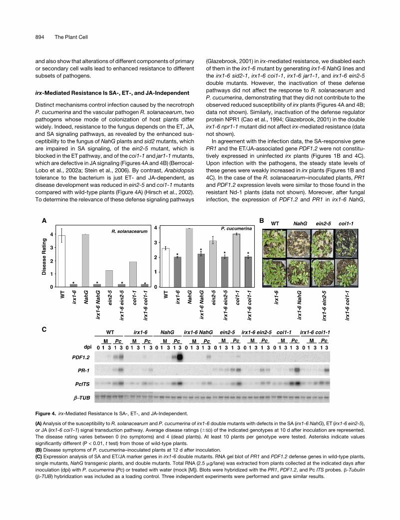
Figure 4. irx-Mediated Resistance Is SA-, ET-, and JA-Independent.
(A) Analysis of the susceptibility to R. solanacearum and P. cucumerina of irx1-6 double mutants with defects in the SA (irx1-6 NahG), ET (irx1-6 ein2-5),
or JA (irx1-6 coi1-1) signal transduction pathway. Average disease ratings (6SD) of the indicated genotypes at 10 d after inoculation are represented.
The disease rating varies between 0 (no symptoms) and 4 (dead plants). At least 10 plants per genotype were tested. Asterisks indicate values
significantly different (P < 0.01, t test) from those of wild-type plants.
(B) Disease symptoms of P. cucumerina–inoculated plants at 12 d after inoculation.
(C) Expression analysis of SA and ET/JA marker genes in irx1-6 double mutants. RNA gel blot of PR1 and PDF1.2 defense genes in wild-type plants,
single mutants, NahG transgenic plants, and double mutants. Total RNA (2.5 mg/lane) was extracted from plants collected at the indicated days after
inoculation (dpi) with P. cucumerina (Pc) or treated with water (mock [M]). Blots were hybridized with the PR1, PDF1.2, and Pc ITS probes. b-Tubulin
(b-TUB) hybridization was included as a loading control. Three independent experiments were performed and gave similar results.
894 The Plant Cell
irx1-6 coi1-1, and irx1-6 ein2-5 plants was similar or lower than
that observed in the NahG, coi1-1, and ein2-5 mutants (Figure
4C). These data demonstrated that these signaling pathways did
not contribute significantly to the reduced susceptibility of the irx
mutants to pathogens.
ABA-Responsive, Defense-Related Genes Are Expressed
Constitutively in the irx1-6 and irx5-5 Mutants
To further investigate the mechanisms controlling irx-mediated
resistance, comparative gene expression analysis of noninocu-
lated wild-type, irx5-5, and irx1-6 plants was performed. Of the
22,000 genes tested, 301 genes were constitutively upregulated
and 265 genes were constitutively downregulated in both irx1-6
and irx5-5 mutants compared with wild-type plants (see Sup-
plemental Tables 1 and 2 online). Most of these differentially ex-
pressed genes seemed to be specific to the secondary cell wall
irx mutants, as quantitative RT-PCR analysis revealed that their
expression in primary cell wall ixr mutants was similar to that in
wild-type plants (see Supplemental Figure 2 online; data not
shown). A significant number of genes encoding cell wall–related
proteins were differentially regulated in the irx mutants (see Sup-
plemental Tables 1 and 2 online), as expected from the substan-
tial changes in secondary cell wall structure and composition
caused by the irx mutations (Taylor et al., 2003; Somerville et al.,
2004; Brown et al., 2005; Persson et al., 2005).
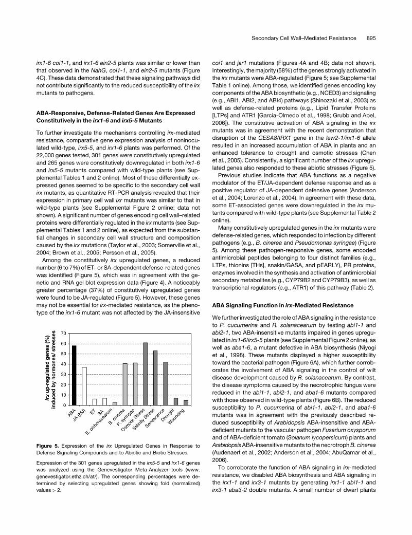
Among the constitutively irx upregulated genes, a reduced
number (6 to 7%) of ET- or SA-dependent defense-related genes
was identified (Figure 5), which was in agreement with the ge-
netic and RNA gel blot expression data (Figure 4). A noticeably
greater percentage (37%) of constitutively upregulated genes
were found to be JA-regulated (Figure 5). However, these genes
may not be essential for irx-mediated resistance, as the pheno-
type of the irx1-6 mutant was not affected by the JA-insensitive
coi1 and jar1 mutations (Figures 4A and 4B; data not shown).
Interestingly, the majority (58%) of the genes strongly activated in
the irx mutants were ABA-regulated (Figure 5; see Supplemental
Table 1 online). Among those, we identified genes encoding key
components of the ABA biosynthetic (e.g., NCED3) and signaling
(e.g., ABI1, ABI2, and ABI4) pathways (Shinozaki et al., 2003) as
well as defense-related proteins (e.g., Lipid Transfer Proteins
[LTPs] and ATR1 [Garcıa-Olmedo et al., 1998; Grubb and Abel,
2006]). The constitutive activation of ABA signaling in the irx
mutants was in agreement with the recent demonstration that
disruption of the CESA8/IRX1 gene in the lew2-1/irx1-6 allele
resulted in an increased accumulation of ABA in planta and an
enhanced tolerance to drought and osmotic stresses (Chen
et al., 2005). Consistently, a significant number of the irx upregu-
lated genes also responded to these abiotic stresses (Figure 5).
Previous studies indicate that ABA functions as a negative
modulator of the ET/JA-dependent defense response and as a
positive regulator of JA-dependent defensive genes (Anderson
et al., 2004; Lorenzo et al., 2004). In agreement with these data,
some ET-associated genes were downregulated in the irx mu-
tants compared with wild-type plants (see Supplemental Table 2
online).
Many constitutively upregulated genes in the irx mutants were
defense-related genes, which responded to infection by different
pathogens (e.g., B. cinerea and Pseudomonas syringae) (Figure
5). Among these pathogen-responsive genes, some encoded
antimicrobial peptides belonging to four distinct families (e.g.,
LTPs, thionins [THs], snakin/GASA, and pEARLY), PR proteins,
enzymes involved in the synthesis and activation of antimicrobial
secondary metabolites (e.g., CYP79B2 and CYP79B3), as well as
transcriptional regulators (e.g., ATR1) of this pathway (Table 2).
ABA Signaling Function in irx-Mediated Resistance
We further investigated the role of ABA signaling in the resistance
to P. cucumerina and R. solanacearum by testing abi1-1 and
abi2-1, two ABA-insensitive mutants impaired in genes upregu-
lated in irx1-6/irx5-5 plants (see Supplemental Figure 2 online), as
well as aba1-6, a mutant defective in ABA biosynthesis (Niyogi
et al., 1998). These mutants displayed a higher susceptibility
toward the bacterial pathogen (Figure 6A), which further corrob-
orates the involvement of ABA signaling in the control of wilt
disease development caused by R. solanacearum. By contrast,
the disease symptoms caused by the necrotrophic fungus were
reduced in the abi1-1, abi2-1, and aba1-6 mutants compared
with those observed in wild-type plants (Figure 6B). The reduced
susceptibility to P. cucumerina of abi1-1, abi2-1, and aba1-6
mutants was in agreement with the previously described re-
duced susceptibility of Arabidopsis ABA-insensitive and ABA-
deficient mutants to the vascular pathogen Fusarium oxysporum
and of ABA-deficient tomato (Solanum lycopersicum) plants and
Arabidopsis ABA-insensitive mutants to the necrotroph B. cinerea
(Audenaert et al., 2002; Anderson et al., 2004; AbuQamar et al.,
2006).
To corroborate the function of ABA signaling in irx-mediated
resistance, we disabled ABA biosynthesis and ABA signaling in
the irx1-1 and irx3-1 mutants by generating irx1-1 abi1-1 and
irx3-1 aba3-2 double mutants. A small number of dwarf plants
Figure 5. Expression of the irx Upregulated Genes in Response to
Defense Signaling Compounds and to Abiotic and Biotic Stresses.
Expression of the 301 genes upregulated in the irx5-5 and irx1-6 genes
was analyzed using the Genevestigator Meta-Analyzer tools (www.
genevestigator.ethz.ch/at/). The corresponding percentages were de-
termined by selecting upregulated genes showing fold (normalized)
values > 2.
Secondary Cell Wall–Mediated Resistance 895
with pronounced developmental abnormalities, corresponding
to the double homozygous mutants, were identified under high-
humidity growth conditions (see Supplemental Figure 3 online;
data not shown). However, these irx1-1 abi1-1 and irx3-1 aba3-2
double mutants died 2 to 3 weeks after sowing, making it
impossible to test their resistance to pathogens. These results
suggested that ABA signaling played a significant role in the
regulation of the irx developmental phenotype.
Function of Secondary Metabolites
in irx-Mediated Resistance
A significant number of defensive genes were constitutively
upregulated in the irx/cesa mutants, including some encoding
antimicrobial peptides (e.g., LTPs and THs) (Table 2; see Sup-
plemental Figure 2 online) and enzymes involved in the synthesis
and activation of secondary metabolites. Secondary metabo-
lites, such as glucosinolates and camalexin, act as antimicrobials
(Grubb and Abel, 2006) that affect the interaction between
Arabidopsis and various pathogens, including the necrotrophic
fungus B. cinerea (Kliebenstein et al., 2005). The synthesis of
these metabolites is regulated by several enzymes (e.g., CYP79B2
and CYP79B3) and transcriptional factors (e.g., ATR1) whose
corresponding genes were constitutively upregulated in the irx
mutants (Table 2). To further determine the relevance of the
secondary metabolites in irx-mediated resistance, we analyzed
the susceptibility to P. cucumerina of the defective cyp79b2 and
cyp79b3 mutants and found that they were slightly, although
significantly, more susceptible to the fungus than wild-type
plants (Figure 7). The susceptibility of cyp79b2 and cyp79b3
was lower than that of the highly susceptible agb1-1 mutant,
which is altered in the b-subunit of the heterotrimeric G protein
(Figure 7) (Llorente et al., 2005). Based on these results, the
simplest hypothesis to explain irx-mediated resistance is that the
secondary metabolites accumulated in the irx mutants may
create a hostile and antimicrobial-enriched environment for the
pathogen, hindering its multiplication.
DISCUSSION
The role of the cell wall in resistance and/or disease development
has been highlighted by several studies (Schulze-Lefert, 2004;
Somerville et al., 2004). Apart from the previously mentioned ixr1/
cev1, pmr5, and pmr6 mutants, which do not support the growth
of two powdery mildew species (Ellis et al., 2002; Vogel et al.,
2002, 2004), inactivation of a cell surface arabinogalactan pro-
tein, RAT1, and of a putative glycosyltransferase, RAT4, confers
enhanced resistance to Agrobacterium tumefaciens infection
(Zhu et al., 2003). In this study, we demonstrated that the irx mu-
tants, impaired in the CESA proteins required for the synthesis
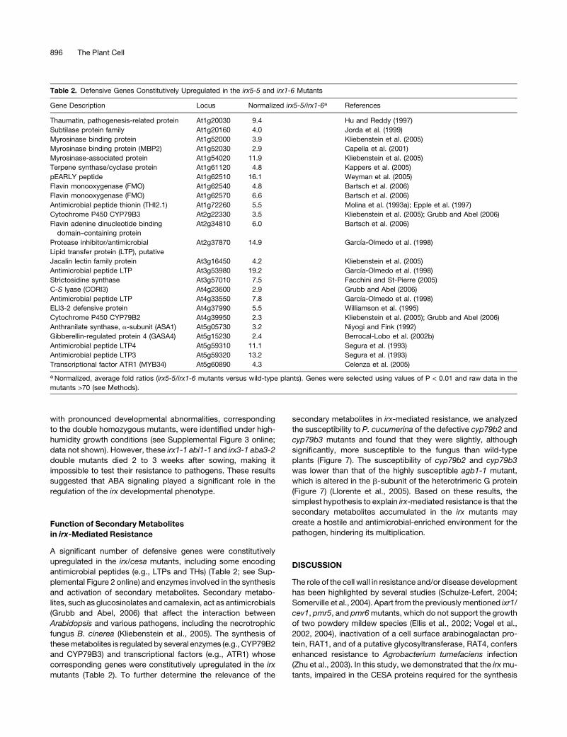
Table 2. Defensive Genes Constitutively Upregulated in the irx5-5 and irx1-6 Mutants
Gene Description Locus Normalized irx5-5/irx1-6a References
Thaumatin, pathogenesis-related protein At1g20030 9.4 Hu and Reddy (1997)
Subtilase protein family At1g20160 4.0 Jorda et al. (1999)
Myrosinase binding protein At1g52000 3.9 Kliebenstein et al. (2005)
Myrosinase binding protein (MBP2) At1g52030 2.9 Capella et al. (2001)
Myrosinase-associated protein At1g54020 11.9 Kliebenstein et al. (2005)
Terpene synthase/cyclase protein At1g61120 4.8 Kappers et al. (2005)
pEARLY peptide At1g62510 16.1 Weyman et al. (2005)
Flavin monooxygenase (FMO) At1g62540 4.8 Bartsch et al. (2006)
Flavin monooxygenase (FMO) At1g62570 6.6 Bartsch et al. (2006)
Antimicrobial peptide thionin (THI2.1) At1g72260 5.5 Molina et al. (1993a); Epple et al. (1997)
Cytochrome P450 CYP79B3 At2g22330 3.5 Kliebenstein et al. (2005); Grubb and Abel (2006)
Flavin adenine dinucleotide binding
domain–containing protein
At2g34810 6.0 Bartsch et al. (2006)
Protease inhibitor/antimicrobial At2g37870 14.9 Garcıa-Olmedo et al. (1998)
Lipid transfer protein (LTP), putative
Jacalin lectin family protein At3g16450 4.2 Kliebenstein et al. (2005)
Antimicrobial peptide LTP At3g53980 19.2 Garcıa-Olmedo et al. (1998)
Strictosidine synthase At3g57010 7.5 Facchini and St-Pierre (2005)
C-S lyase (CORI3) At4g23600 2.9 Grubb and Abel (2006)
Antimicrobial peptide LTP At4g33550 7.8 Garcıa-Olmedo et al. (1998)
ELI3-2 defensive protein At4g37990 5.5 Williamson et al. (1995)
Cytochrome P450 CYP79B2 At4g39950 2.3 Kliebenstein et al. (2005); Grubb and Abel (2006)
Anthranilate synthase, a-subunit (ASA1) At5g05730 3.2 Niyogi and Fink (1992)
Gibberellin-regulated protein 4 (GASA4) At5g15230 2.4 Berrocal-Lobo et al. (2002b)
Antimicrobial peptide LTP4 At5g59310 11.1 Segura et al. (1993)
Antimicrobial peptide LTP3 At5g59320 13.2 Segura et al. (1993)
Transcriptional factor ATR1 (MYB34) At5g60890 4.3 Celenza et al. (2005)
a Normalized, average fold ratios (irx5-5/irx1-6 mutants versus wild-type plants). Genes were selected using values of P < 0.01 and raw data in the
mutants >70 (see Methods).
896 The Plant Cell
of secondary cell wall cellulose, show an enhanced resistance to
fungal (P. cucumerina, B. cinerea, and powdery mildew) and
bacterial (R. solanacearum) pathogens. The reduced suscepti-
bility of irx/cesa mutants to these pathogens was SA-, ET-, and
JA-independent, like that of pmr5 and pmr6 mutants (Vogel et al.,
2002, 2004). By contrast, the enhanced resistance to powdery
mildew of the primary cell wall mutant cev1/ixr1, which is im-
paired in the CESA3 protein, depended on a concomitant acti-
vation of the JA and ET pathways (Ellis et al., 2002). Interestingly,
the susceptibility to P. cucumerina and R. solanacearum of CESA
primary cell wall mutants, including cev1/ixr1, and of pmr5 and
pmr6 mutants was similar to that of wild-type plants (Figures
1 and 3). These results illustrate the high level of specificity and
complexity of the resistance associated with the impairment of
primary and secondary cell wall integrity caused by the irx, ixr,
and pmr mutations.
Most of the cell wall mutants present phenotypic alterations,
which reveal the importance of the corresponding genes for
plant development (Somerville et al., 2004). Developmental alter-
ations have also been described for the majority of Arabidopsis
mutants exhibiting a constitutive activation of disease resistance
(Glazebrook, 2001). In the pmr6 and pmr5 mutants, a pectin
component appears to be altered, and both mutants are reduced
in stature compared with wild-type plants (Vogel et al., 2002,
2004). Similarly, mutations in the CESA/IRX genes cause signif-
icant modifications of the structure and noncellulosic carbohy-
drate composition of the cell wall, which lead to widespread
morphological alterations, including the collapse of xylem ves-
sels, that might affect pathogen attachment and spread within the
plants (Taylor et al., 2003; Brown et al., 2005). This possibility is
rather unlikely, because (1) microscopic examination showed that
pathogen attachment was not impaired in irx mutants (data not
shown); (2) bacteria inoculated by dipping wounded roots in a
microbial suspension could be detected within minutes in the
aerial parts of irx mutants (Figure 1D); (3) the induction of bacterial
hrp genes, which is required for disease development (Aldon
et al., 2000), was unaffected in the irx mutants (data not shown);
and (4) the resistance to P. cucumerina of the gpx1 mutant, which
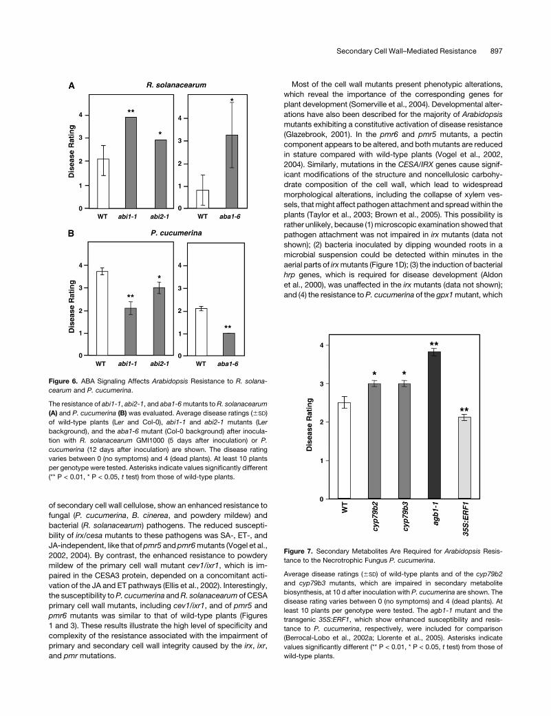
Figure 6. ABA Signaling Affects Arabidopsis Resistance to R. solana-
cearum and P. cucumerina.
The resistance of abi1-1, abi2-1, and aba1-6 mutants to R. solanacearum
(A) and P. cucumerina (B) was evaluated. Average disease ratings (6SD)
of wild-type plants (Ler and Col-0), abi1-1 and abi2-1 mutants (Ler
background), and the aba1-6 mutant (Col-0 background) after inocula-
tion with R. solanacearum GMI1000 (5 days after inoculation) or P.
cucumerina (12 days after inoculation) are shown. The disease rating
varies between 0 (no symptoms) and 4 (dead plants). At least 10 plants
per genotype were tested. Asterisks indicate values significantly different
(** P < 0.01, * P < 0.05, t test) from those of wild-type plants.
Figure 7. Secondary Metabolites Are Required for Arabidopsis Resis-
tance to the Necrotrophic Fungus P. cucumerina.
Average disease ratings (6SD) of wild-type plants and of the cyp79b2
and cyp79b3 mutants, which are impaired in secondary metabolite
biosynthesis, at 10 d after inoculation with P. cucumerina are shown. The
disease rating varies between 0 (no symptoms) and 4 (dead plants). At
least 10 plants per genotype were tested. The agb1-1 mutant and the
transgenic 35S:ERF1, which show enhanced susceptibility and resis-
tance to P. cucumerina, respectively, were included for comparison
(Berrocal-Lobo et al., 2002a; Llorente et al., 2005). Asterisks indicate
values significantly different (** P < 0.01, * P < 0.05, t test) from those of
wild-type plants.
Secondary Cell Wall–Mediated Resistance 897
presents severe alterations in xylem vessels (Turner and Hall,
2000), did not differ from that of wild-type plants (data not shown).
Disruption of the CESA8/IRX1 gene in the irx1-6/ern1/lew2-1
allele leads to increased endogenous ABA levels in planta and to
enhanced tolerance to drought and osmotic stresses (Chen et al.,
2005) (Figure 8). Consistent with these data, transcriptomic anal-
yses revealed that a majority (58%) of the constitutively upregulated
genes in the irx mutants were ABA-responsive (Figure 5). Among
the proteins encoded by these ABA-regulated genes, we identified
enzymes and regulators of the ABA biosynthetic and signaling
pathways (e.g., NECD3, ABI1, ABI2, and ABI4) (Shinozaki et al.,
2003) as well as antimicrobial, defense-related peptides (e.g., LTPs)
(Molina et al., 1993b) and transcriptional regulators of secondary
metabolite biosynthesis (e.g., ATR1) (Grubb and Abel, 2006). ABA
plays a significant role in the control of plant development and the
adaptive response to osmotic stresses (Shinozaki et al., 2003).
Also, ABA modulates disease resistance by interfering with biotic
stress signaling. Thus, ABA can act as a negative regulator of the
ET/JA-dependent defense response and as a positive modulator of
JA-dependent defensive genes (Anderson et al., 2004; Lorenzo
et al., 2004). In agreement with these data, some ET-associated
genes were constitutively downregulated in the irx mutants (see
Supplemental Table 2 online), and upon infection, the induction of
ET/JA-regulated genes, such as PDF1.2, was weaker in the irx
mutants than in wild-type plants (Figures 1B and 4C).
The involvement of ABA in primed callose production at the
site of pathogen penetration is one of the few examples of a
positive function of this hormone in defense and may be the basis
for the described b-aminobutyric acid–induced resistance (Ton
and Mauch-Mani, 2004; Mauch-Mani and Mauch, 2005; Ton et al.,
2005). However, callose deposition at the site of P. cucumerina
infection was similar in irx1-6 mutant and wild-type plants, which
excludes priming for callose production as a mediator of irx-
reduced susceptibility (data not shown). We now provide strong
evidence for a direct involvement of ABA signaling in the control
of Arabidopsis resistance to R. solanacearum. This ABA function
was supported by different observations: (1) the ABA-insensitive
mutants abi1-1 and abi2-1, and the ABA-deficient mutant aba1-6,
were more susceptible to the bacterium than were wild-type
plants (Figure 6); (2) the constitutive expression in the irx mutants
of some ABA signaling regulators, including ABI1-1 and ABI2-1,
was associated with an increased resistance to the bacterium
(Figure 1, Table 2); and (3) the ein2-1 mutant, a phenotypic
suppressor of abi1-1, was shown to be more tolerant to virulent
strains of the bacterium (Baudoin et al., 2000; Hirsch et al., 2002)
(Figure 4A).
ABA was recently implicated as a regulator of defense re-
sponses to necrotrophic pathogens. Thus, ABA-deficient mu-
tants exhibited reduced susceptibility to the necrotroph B.
cinerea and the vascular pathogen F. oxysporum (Audenaert
et al., 2002; Anderson et al., 2004) and ABA-sensitive mutants
showed increased susceptibility to B. cinerea (AbuQamar et al.,
2006). In agreement with these results, we previously demon-
strated that the ABA-hypersensitive mutant agb1-1 (Pandey
et al., 2006) was more susceptible than wild-type plants to these
fungi and P. cucumerina (Llorente et al., 2005). We now demon-
strate that the constitutive activation of ABA signaling in the irx
mutants, or the impairment of the ABA pathway, as it occurs in
the abi and aba mutants, led to an enhanced resistance to P.
cucumerina via the activation of separate pathways, which seem
to regulate distinct sets of defense genes (Figure 8). In the irx
mutants, ABA-regulated genes encoding antimicrobial peptides
(e.g., LTPs and THs) (Molina et al., 1993b) or regulators of sec-
ondary metabolite biosynthesis (e.g., ATR1) (Grubb and Abel,
2006) were constitutively expressed. The accumulation of these
antimicrobial peptides/metabolites in these mutants may explain
their enhanced resistance to pathogens (Figure 8). Additionally,
we have observed that in the abi and aba mutants some ET/JA-
regulated, defense-related genes (e.g., PDF1.2) were overex-
pressed (data not shown), in agreement with the proposed
function of ABA as a negative modulator of the ET/JA-dependent
defense response (Anderson et al., 2004; Lorenzo et al., 2004).
This observation may explain the reduced susceptibility of these
abi and aba mutants to P. cucumerina (Figure 8). Indeed, we have
demonstrated previously that the constitutive activation of the ET
and JA pathways by overexpression of the ERF1 transcriptional
factor was sufficient to confer enhanced resistance to necro-
trophs (Figures 1A and 7) (Berrocal-Lobo et al., 2002a).
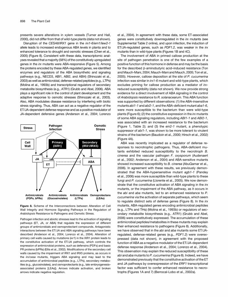
Figure 8. Scheme of the Interconnections between Alteration of Cell
Wall Integrity and Hormone Signaling Pathways in the Regulation of
Arabidopsis Resistance to Pathogens and Osmotic Stress.
Pathogen infection and abiotic stresses lead to the activation of signaling
pathways (ET, JA, or ABA) that regulate the expression of different
groups of antimicrobials and osmoprotectant compounds. Antagonistic
interactions between the ET/JA and ABA signaling pathways have been
described (Anderson et al., 2004; Lorenzo et al., 2004). Alteration of
primary cell walls caused by mutations (ixr1) in the IXR1 protein leads to
the constitutive activation of the ET/JA pathway, which controls the
expression of antimicrobial proteins, such as defensins (PDFs) and basic
PR proteins (bPRs) (Ellis et al., 2002). Modifications of the secondary cell
walls caused by the impairment of IRX1 and IRX5 proteins, as occurs in
the irx/cesa mutants, triggers ABA signaling and may lead to the
accumulation of antimicrobial peptides (e.g., LTPs), secondary metabo-
lites (e.g., glucosinolates), and osmoprotectants (e.g., late embryogenesis–
associated proteins [LEAs]). Arrows indicate activation, and broken
arrows indicate negative regulation.
898 The Plant Cell
The previous observations implicating ABA as a regulator of
defense responses, those reported here, and the recent demon-
stration that stomata have an important role in defense against the
bacterium P. syringae (Melotto et al., 2006) suggest that the ABA
pathway, in addition to mediating the adaptative response to os-
motic stress, may play a significant role in the fine-tuned regulation
of the plant immune system. Examples of positive and negative
crosstalk between biotic and abiotic stresses are emerging (Park
et al., 2001; Mengiste et al., 2003). Interestingly, the enhanced
resistance to biotic and abiotic stresses mediated by irx/cesa
mutations was JA- and ET-independent.
Based on the transcriptomic analysis, the simplest hypothesis
to explain irx-mediated resistance is that the antimicrobial com-
pounds that might accumulate in these mutants (e.g., peptides,
PR proteins, and secondary metabolites) create a hostile envi-
ronment for pathogens, whose progression within the plant is
hindered. Secondary metabolites have been suggested to con-
stitute one of the determinants of nonhost and basal resistance to
several pathogens, including P. cucumerina (Lipka et al., 2005;
Stein et al., 2006), and to affect the interaction between Arabi-
dopsis and the necrotrophic fungus B. cinerea (Kliebenstein et al.,
2005). Also, the defensive functions of some plant antimicrobial
peptides encoded by genes upregulated in the irx mutants are
well established (Table 2) (Garcıa-Olmedo et al., 1998). Indeed,
THs and LTPs, including those encoded by the irx-upregulated
LTP3 and LTP4 genes (Table 2), possess in vitro antibiotic activity,
and their constitutive overexpression in Arabidopsis increases
plant tolerance to various pathogens (Molina et al., 1993a, 1993b;
Segura et al., 1993; Epple et al., 1997; Molina and Garcıa-
Olmedo, 1997; Titarenko et al., 1997). Our hypothesis is sup-
ported by the demonstration of an enhanced susceptibility to
P. cucumerina of the cyp79b2 and cyp79b3 mutants, which are
impaired in the accumulation of antimicrobial secondary metab-
olites (Figure 8). Moreover, we purified and tested the in vitro
antibiotic activity of Arabidopsis LTPs encoded by genes upre-
gulated in the irx mutants and found that they were active against
P. cucumerina and R. solanacearum GMI1000 (data not shown).
The activity of these LTPs was, as described previously for other
pathogens, essentially equivalent to that of other plant LTPs (e.g.,
EC50 values for P. cucumerina of 3.2 6 0.7 mM for barley
[Hordeum vulgare] LTPs [LTP3 þ LTP4] and 6.2 6 0.9 mM for
Arabidopsis LTPs [Molina et al., 1993b; Segura et al., 1993]).
In yeast, there is a mechanism controlling cell wall integrity that
is regulated by the WSC genes (for cell wall integrity and stress
response), which encode integral membrane proteins acting as
surface sensors responding to environmental stress (Philip and
Levin, 2001; Merchan et al., 2004). Our data suggest that a similar
sensing system to that of yeast may exist in plants, because
modifications of the integrity of the primary or secondary cell wall
activate different signaling pathways, at least partially ABA-
dependent in the irx mutants, ET/JA-dependent in the ixr mu-
tants, and still uncharacterized in the pmr5 and pmr6 mutants
(Figure 8). Although the precise functions of most genes exhibit-
ing altered expression in the irx mutants remain to be deter-
mined, some may encode putative sensors/receptors of cell wall
integrity (see Supplemental Table 1 online). The characterization
of plant sensors involved in the detection of pathogen-induced
perturbations of cell wall integrity, and the identification of their
corresponding ligands and downstream effectors, are future
challenges in the plant resistance field.
METHODS
Plant Lines and Growth Conditions
Arabidopsis thaliana accessions used in this study were Col-0, Landsberg
erecta (Ler), Col-5 (a glabrous [gl] derivative of Col-0), Nd-1, and Kas-1.
The primary and secondary cell wall mutants ixr1-1, ixr2-1, rsw2-1, irx1-1,
irx3-1, and irx5-4 analyzed here have been described previously (Fagard
et al., 2000; Scheible et al., 2001; Desprez et al., 2002; Ellis et al., 2002;
Taylor et al., 2003; Brown et al., 2005; Persson et al., 2005). The ABA
mutants aba1-6 (Col-0 background), abi1-1, abi2-1, and aba3-2 (Ler
background) analyzed in this study have been characterized previously
(Niyogi et al., 1998; Gosti et al., 1999; Xiong et al., 2001). Plants were
grown in growth chambers at 20 to 228C with a 10-h photoperiod and a
light intensity of ;150 mE�m�2�s�1, as described previously (Berrocal-
Lobo et al., 2002a; Deslandes et al., 2003). For infection experiments with
Erysiphe cichoracearum (Golovinomyces cichoracearum), plants were
grown in growth chambers at 228C with a 14-h photoperiod of ;125
mE�m�2�s�1 in the 400- to 700-nm range.
Ralstonia solanacearum GMI1000 is a wild-type strain isolated
from tomato (Solanum lycopersicum). R. solanacearum was grown at
288C in B broth medium (Boucher et al., 1985). The ascomycete fungus
Plectosphaerella cucumerina and the necrotroph Botrytis cinerea were
grown at 288C on potato (Solanum tuberosum) dextrose agar and their
spores harvested as reported (Berrocal-Lobo et al., 2002a; Llorente et al.,
2005). Pseudomonas syringae pv tomato DC3000 and P. syringae pv
tomato DC3000 (AvrRPM1) were grown on nutrient broth at 288C as
described previously (Berrocal-Lobo et al., 2002a). Powdery mildew (E.
cichoracearum UCSC1) was cultured and applied to Arabidopsis as
described (Vogel and Somerville, 2000). Hyaloperonospora parasitica
(Peronospora parasitica) isolates Noco-2 and Emwa1 were maintained on
the genetically susceptible Arabidopsis accessions Col-0 and Ler, re-
spectively, as described (Dangl et al., 1992). Xanthomonas campestris pv
campestris bacteria were grown as described previously (Lummerzheim
et al., 2004).
Pathogenicity Assays
The pathogens used in this study were P. cucumerina, P. syringae pv
tomato DC3000 (AvrRPM1), and P. syringae pv tomato DC3000 (Berrocal-
Lobo et al., 2002a), B. cinerea (Llorente et al., 2005), R. solanacearum
GMI1000 (Deslandes et al., 1998), the isolates Noco2 and Emwa1 of H.
parasitica (Dangl et al., 1992), E. cichoracearum UCSC1 (Vogel and
Somerville, 2000), and the strains 8004 and 147 of X. campestris pv
campestris (Lummerzheim et al., 2004).
Plant infection with R. solanacearum was done as reported by inocu-
lating roots of 4-week-old plants with the bacterial suspension (Deslandes
et al., 2003). Bacterial in planta growth curves were measured as de-
scribed (Deslandes et al., 1998). Infection with P. cucumerina was per-
formed by spraying 4-week-old plants with a spore suspension (2 3 106
spores/mL) of the fungus. After inoculation, plants were kept under the
same growth conditions and the average disease rating (6SD) was mea-
sured at different times (days after inoculation) as reported (Llorente et al.,
2005). Disease rating was as follows: 0, no symptoms, 1, 1 to 3 leaves
showing some necrosis; 2, 4 to 8 leaves with some necrosis; 3, 8 to 12
leaves showing necrosis; 4, all leaves showing profuse necrosis or
decayed/dead plant. At least 10 plants per genotype were inoculated in
each of two to three experiments, and the disease rating means and SD
were estimated at different times.
Secondary Cell Wall–Mediated Resistance 899
Plant inoculation with B. cinerea and disease resistance analysis were
done as described (Llorente et al., 2005). Arabidopsis plants were dip-
inoculated with P. syringae pv tomato DC3000 (AvRPM1) or P. syringae pv
tomato DC3000, and in planta bacterial growth was determined at different
times (Berrocal-Lobo et al., 2002a). Disease resistance analysis of H.
parasitica was performed by spraying 2-week-old plants with a conidio-
phore suspension (4 3 104 spores/mL) of the isolates Noco2 or Emwa1, and
the degree of infection was quantified as described (Llorente et al., 2005).
For E. cichoracearum infections, 3-week-old Arabidopsis plants were
inoculated as described above, phenotypes were monitored visually at 4,
5, 6, and 7 d after inoculation, and the number of conidiophores per colony
was determined at the end of the experiment (Vogel and Somerville, 2000).
For X. campestris infection, plants were kept at high humidity for 12 h before
inoculation, and then the abaxial side of the leaves was injected with a
bacterial suspension of 2 3 107 to 108 colony-forming units/mL using a
blunt syringe (Lummerzheim et al., 2004). At least 10 plants per genotype
were inoculated in each of two to three experiments performed.
Statistical analysis of the pathogenicity experiments was performed
using a two-tailed Student’s t test.
In Vitro Inhibition Tests
Arabidopsis LTPs and barley (Hordeum vulgare) LTPs (a mixture of LTP3
and LTP4) used for the in vitro inhibition assays were purified as described
previously (Molina et al., 1993a; Segura et al., 1993). Pathogen inhibition
was done on microtiter plates as reported (Molina et al., 1993b) by mixing
33 mL of the corresponding peptide solution with 66 mL of medium
containing the fungal spores or bacteria (104 colony-forming units or
spores/mL) to give the desired peptide concentrations. The experiment
was repeated at least two times.
Microscopic Analysis
Four-week-old irx1-6 and wild-type plants were mock-inoculated or chal-
lenged, as indicated above, with a spore suspension of P. cucumerina.
Callose staining at different times (0, 1, 3, and 4 d after inoculation) was
performed as reported previously (Llorente et al., 2005). At least 10 plants
per genotype were analyzed, and the experiment was repeated three
times.
Identification and Cloning of the nws2/irx5-5 Gene
Thirty-five thousand T-DNA–tagged Col-5 plants were inoculated with the
virulent GMI1000 bacterium, and plants that did not develop wilt symptoms
were selected and allowed to set seed. Cosegregation analysis in the
F2 population resulting from the nws2 backcross indicated that the resis-
tant phenotype was tightly linked to the T-DNA insertion. The cloning of
the nws2/irx5-5 gene was performed by inverse PCR as described
(http://www.darmouth.edu/tjack/cloning_flanking_DNA_from_.html). Primers
used to amplify the right border were as follows: 59-TCGGGCCTAA-
CTTTTGGTG-39 (adjacent right border) and 59-AGTGCCAAGCTTG-
CATGC-39 (HindIII site in the polylinker). Genomic DNA (200 ng) was
digested with various enzymes (XbaI, SalI, BamHI, and PstI). For each
digestion, 20 ng was used in a ligation reaction. PCR was performed using
the following conditions: 948C for 20 s, 548C for 30 s, and 688C for 10 min
(35 cycles). An 8-kb fragment was obtained using, as a template, the self-
ligated XbaI fragments.
Isolation and Map-Based Cloning of the ern1/irx1-6 Mutant
Approximately 80,000 10-d-old plants of an ethyl methanesulfonate M2
mutagenized population of Arabidopsis Col-0 were sprayed with 2 3 106
spores/mL P. cucumerina fungus for the identification of ern mutants,
which showed reduced disease symptoms compared with Col-0 wild-
type plants. This screen yielded the recessive ern1-1 mutant. The ERN1
gene was mapped using an ern1-1 (Col-0) 3 Ler F2 population of 430
individuals using standard PCR-based marker techniques (Lukowitz
et al., 2000). New markers within the fine-mapping interval on chromo-
some IV were generated using the CEREON database of Col and Ler
polymorphisms (http://www.arabidopsis.org/browse/Cereon/index.jsp). The
derived oligonucleotide sequences used for mapping were as follows:
F15J5A, 59-TCTAAGCGGGTCGGGTCGATTC-39 and 59-AATCAGACCG-
GACCGGACCGG-39; F28J12, 59-CAAATCCATACATCTCTCCCTCTC-39
and 59-GATGGAGTAGATAGAGAAAGGGAG-39; F28A21A, 59-AGGCAT-
TCTTGCTAAGGGC-39 and 59-AATTCACGTGGGACTTGGG-39; F28A21B,
59-AAAAGTTACGAGTCAGTTACATC-39 and 59-GTTGGTGATGAATGATG-
ATG-39; F28A21C, 59-CTTTGAGGTTAGCAAAAACTCAC-39 and 59-CAT-
TGAACTCTTTTCACATGGCAG-39; and T16H5, 59-ATTCTAGCTTTTGA-
GAGGTCAG-39 and 59-CCCAATCCAAATCAAATTCC-39.
Generation and Selection of Double Mutants
The mutant alleles used for the construction of double mutants with irx1-6
were coi1-1 (Feys et al., 1994), npr1-1 (Cao et al., 1994), ein2-5 (Roman
et al., 1995), NahG (Lawton et al., 1995), jar1-1 (Staswick et al., 1992),
sid2-1 (Wildermuth et al., 2001), abi1-1 (Gosti et al., 1999), and aba3-2
(Xiong et al., 2001). All of the mutant lines were in the Col-0 background
except coi1-1, which was in the Col-6 background, and abi1-1 and aba3-2,
which were in the Ler background. We generated the double mutants by
standard genetic crosses, following the mutations in the mutant alleles
(npr1-1, sid2-1, coi1-1, jar1-1, aba3-2, and abi1-1) or the transgene
(NahG) by PCR-based methods, or by selection on Murashige and Skoog
plates containing 10 mM 1-aminocyclopropane-1-carboxylic acid, the ET
precursor (ein2-5 [Roman et al., 1995]), 50 mM JA (coi1-1 and jar1-1 [Feys
et al., 1994]), or 1 mM ABA (abi1-1 [Gosti et al., 1999]). The NahG, coi1-1,
sid2-1, npr1-1, and jar1-1 oligonucleotides used for PCR-based detec-
tion have been described (Stein et al., 2006). Genotyping of the irx1-6
mutation in all of the double mutants was confirmed by PCR amplification
and sequencing of the mutation (oligonucleotides 59-GGTTAATAGGAG-
ACCATTTAG-39 and 59-TCCATCCAAATCTCAATCCC-39). As described
previously (Brown et al., 2005; Chen et al., 2005), the homozygous irx1-6
and irx5-5 plants were sterile; therefore, for the disease resistance and
expression analysis, heterozygous populations were sown on soil and the
corresponding irx1-6 and irx5-5 single or double mutants that showed a
clear distinguishable phenotype were selected for the analyses. For the
study of irx1-6 coi1-1 plants, seeds of heterozygous irx1-6 coi1-1 and
coi1-1 plants were sown on plates containing 50 mM JA for coi1-1 se-
lection (Feys et al., 1994). Plants were then transferred to soil for the
analysis, and the irx1-6 coi1-1 double mutants were selected according
to their phenotype. For the selection of irx3-1 aba3-2 plants, a cleaved
amplified polymorphism was used to identify the irx3-1 mutation (59-CGG-
GAATCTTGGAATTGAGATT-39 and 59-GATCAGCAGTGTTGTCCATTTG-39
and MseI digestion), and the aba3-2 mutation was confirmed by DNA
sequencing (59-CTCAAGTCGCTTACACCTTCTG-39 and 59-GGGATAA-
CTCGAGATACTTTG-39). Genotyping of the irx1-1 abi1-1 double mutant
was confirmed by PCR amplification of abi1-1 (PCR amplification with
oligonucleotides 59-GCCTTTGTATGGTTTTACTTC-39 and 59-CAGCCACG-
TATCACCATCG-39 followed by NcoI digestion) and sequencing of irx1-1
(oligonucleotides 59-GGTTAATAGGAGACCATTTAG-39 and 59-TCC-
ATCCAAATCTCAATCCC-39).
Expression Analysis
Total RNA isolation from plants challenged or not with R. solanacearum
and RNA gel blotting were performed according to the protocol of Hirsch
et al. (2002). RNA extraction of P. cucumerina–infected or mock-inoculated
900 The Plant Cell
plants and RNA gel blot analysis of these RNAs were done as de-
scribed (Llorente et al., 2005). The hybridization probes (PDF1.2, PR1,
Pc ITS1, and b-TUB) used in this study have been described (Llorente
et al. 2005).
For quantitative RT-PCR analyses, 5 mg of total RNA was treated with
DNase and then reverse-transcribed for 90 min at 428C in a 20-mL re-
action volume using the SuperScript II First-Strand Synthesis System kit
(Invitrogen) and oligo(dT). Oligonucleotides used for cDNA amplification
were designed with Primer Express (version 2.0; Applied Biosystems):
At1g18710, 59-GAGCTGCAGATTAAGGTGGCTT-39 and 59-CCGAGAACA-
GCATGAAGTTGG-39; At2g39800, 59-ATGCAGTCTGTGTGTGCACTCC-39
and 59-ACCATGAGTACTGTGCCAAGGC-39; At2g46680, 59-CGATGAT-
GTCAGCAGAGCCAT-39 and 59-CAGTGACTTGATCTGCTCGTCG-39;
At3g1440, 59-ACATCTTTACGGCGATAACCG-39 and 59-TTCCATGTC-
TTCTCGTCGTGA-39; At4g26080, 59-TGTTTTCCCGTCTCACATCTTC-39
and 59-CCGGTTTATGGTCAACGGAT-39; At4g39950, 59-GCCGACCCA-
CTTTGCTTTAAA-39 and 59-GCACAACCTCTTTTCCCGGTA-39; At5g57050,
59-TCAGTGCGGCGAGTAAAAGAA-39 and 59-TCCGTTTCCGAGCCA-
AATC-39; and At3g18780, 59-GACCAGCTCTTCCATCGAGAA-39 and
59-GCAGCTTCCATTCCCACAA-39.
Real-time quantitative PCR amplification and detection was performed
in the 7300 real-time PCR system (Applied Biosystems). Reactions were
done in a final volume of 20 mL and contained 10 mL of 23 SYBR Green
Master Mix (Applied Biosystems), 0.5 mM of each primer, and 8 ng of
cDNA. PCR conditions were as follows: 958C for 10 min, followed by 45
cycles of 958C for 15 s and 608C for 1 min. At the end of each experiment,
a dissociation stage (958C for 15 s, 608C for 30 s, and 958C for 15 s) was
performed to ensure that only single products were formed. Actin2
(At3g18780) expression was used to normalize the transcript level in each
sample. Data analysis was performed using the sequence detector
software (version 1.2.2; Applied Biosystems).
Microarray Experiments and Analysis
Leaves from uninfected 4-week-old homozygous irx5-5 and irx1-6 plants
and their corresponding heterozygous counterparts, which were fully
susceptible to both R. solanacearum and P. cucumerina and showed a
wild-type phenotype, were collected for microarray analysis. Three
biological replicates from irx5-5 and wild-type plants and two replicates
from irx1-6 and wild-type plants were harvested for microarray analysis.
Total RNA was extracted using the NucleoSpin RNAII kit (Macherey-
Nagel). Probes were synthesized from the RNA samples and hybridized to
the Affymetrix GeneChip (ATHI, 22 K) according to the procedures
provided by the manufacturer. The array images were analyzed with the
Affymetrix Microarray Suite 5.0 with the target intensity set to 500. The
expression levels of the genes were analyzed with GeneSpring 7.2
software (Silicon Genetics), and the chip-to-chip signal variation was
minimized by normalizing signal intensities to the averaged intensity
values of wild-type plants using the expression levels of the top 50th
percentile of probe sets. Differentially expressed genes in both the irx5-5
(three replicates) and irx1-6 (two replicates) mutants relative to wild-type
samples (five replicates) were identified using one-way analysis of var-
iance and a Benjamini and Hochberg multiple testing correction (Gene-
Spring 7.2), as described previously (Stein et al., 2006). Genes were
considered differentially expressed at P # 0.01. Upregulated and down-
regulated genes were selected using normalized values (fold) > 2 or < 0.5
relative to wild-type plants, respectively.
Accession Numbers
The data obtained by the microarray hybridizations have been submitted
to NASCArrays (reference number /NASCARRAYS-368/). Arabidopsis
Genome Initiative locus identifiers for the genes impaired in the mutants
characterized in this article are as follows: CESA8/IRX1/ERN1,
At4g18780; CESA4/IRX5/NWS2, At5g44030.
Supplemental Data
The following materials are available in the online version of this article
Supplemental Figure 1. Cloning of the ERN1/IRX1 and NWS2/IRX5
Genes.
Supplemental Figure 2. Quantitative RT-PCR Analysis of Genes
Differentially Upregulated or Downregulated in Secondary Cell Wall
irx Mutants.
Supplemental Figure 3. Developmental Phenotype of the irx3-1
aba3-2 Double Mutant.
Supplemental Table 1. Genes Constitutively Upregulated in the irx5-5
and irx1-6 Mutants.
Supplemental Table 2. Genes Constitutively Downregulated in the
irx5-5 and irx1-6 Mutants.
ACKNOWLEDGMENTS
We thank Gemma Lopez (Centro de Biotecnologıa y Genomica de
Plantas, Universidad Politecnica de Madrid) for technical assistance and
F. Garcıa-Olmedo and F. Garcıa-Arenal (Centro de Biotecnologıa y
Genomica de Plantas) and X. Dong (Duke University) for critical reading
of the manuscript. C.H.-B., A.S.-V., and C.S.-R. were recipients of Ph.D.
fellowships from the Ministerio de Educacion y Ciencia (MEC) of Spain.
F.L. and M.B.-L. were postdoctoral fellows of the Comunidad de Madrid
and MEC (Spain), respectively. This work was supported by MEC (Grant
BIO2003-4424 to A.M.), by Genoplante (Grant AF2001060 to Y.M.), and
by the National Science Foundation (Grant 0114783 to S.S.).
Received October 13, 2006; revised January 17, 2007; accepted Febru-
ary 14, 2007; published March 9, 2007.
REFERENCES
AbuQamar, S., Chen, X., Dhawan, R., Bluhm, B., Salmeron, J., Lam,
S., Dietrich, R.A., and Mengiste, T. (2006). Expression profiling and
mutant analysis reveals complex regulatory networks involved in
Arabidopsis response to Botrytis infection. Plant J. 48: 28–44.
Aldon, D., Brito, B., Boucher, C., and Genin, S. (2000). A bacte-
rial sensor of plant cell contact controls the transcriptional induc-
tion of Ralstonia solanacearum pathogenicity genes. EMBO J. 10:
2304–2314.
Anderson, J.P., Badruzsaufari, E., Schenk, P.M., Manners,
J.M., Desmond, O.J., Ehlert, C., Maclean, D.J., Ebert, P.R., and
Kazan, K. (2004). Antagonistic interaction between abscisic acid
and jasmonate-ethylene signaling pathways modulates defense gene
expression and disease resistance in Arabidopsis. Plant Cell 16:
3460–3479.
Audenaert, K., De Meyer, G.B., and Hofte, M.M. (2002). Abscisic acid
determines basal susceptibility of tomato to Botrytis cinerea and
suppresses salicylic acid-dependent signaling mechanisms. Plant
Physiol. 128: 491–501.
Bartsch, M., Gobbato, E., Bednarek, P., Debey, S., Schultze, J.L.,
Bautor, J., and Parker, J.E. (2006). Salicylic acid-independent
ENHANCED DISEASE SUSCEPTIBILITY1 signaling in Arabidopsis
Secondary Cell Wall–Mediated Resistance 901
immunity and cell death is regulated by the monooxygenase FMO1
and the Nudix hydrolase NUDT7. Plant Cell 18: 1038–1051.
Baudoin, N., Serizet, C., Gosti, F., and Giraudat, J. (2000). Interac-
tions between abscisic acid and ethylene signaling cascades. Plant
Cell 12: 1103–1115.
Berrocal-Lobo, M., Molina, A., and Solano, R. (2002a). Constitutive
expression of ETHYLENE-RESPONSE-FACTOR1 in Arabidopsis con-
fers resistance to several necrotrophic fungi. Plant J. 29: 23–32.
Berrocal-Lobo, M., Segura, A., Moreno, M., Lopez, G., Garcia-
Olmedo, F., and Molina, A. (2002b). Snakin-2, an antimicrobial
peptide from potato whose gene is locally induced by wounding
and responds to pathogen infection. Plant Physiol. 128: 951–961.
Boucher, C.A., Barberis, P.A., Trigalet, A.P., and Demery, D.A.J.
(1985). Transposon mutagenesis of Pseudomonas solanacearum: Isola-
tion of Tn5-induced virulent mutants. J. Gen. Microbiol. 131: 2449–2457.
Brown, D.M., Zeef, L.A., Ellis, J., Goodacre, R., and Turner, S.R.
(2005). Identification of novel genes in Arabidopsis involved in sec-
ondary cell wall formation using expression profiling and reverse
genetics. Plant Cell 17: 2281–2295.
Cao, H., Bowling, S.A., Gordon, A.S., and Dong, X. (1994). Charac-
terization of an Arabidopsis mutant that is nonresponsive to inducers
of systemic acquired resistance. Plant Cell 6: 1583–1592.
Capella, A.N., Menossi, M., Arruda, P., and Benedetti, C.E. (2001).
COI1 affects myrosinase activity and controls the expression of two
flower-specific myrosinase-binding protein homologues in Arabidop-
sis. Planta 213: 691–699.
Carpita, N.C., and McCann, M. (2000). The cell wall. In Biochemistry
and Molecular Biology of Plants, B. Buchanan, W. Gruissem, and R.L.
Jones, eds (Rockville, MD: American Society of Plant Physiologists),
pp. 52–108.
Celenza, J.L., Quiel, J.A., Smolen, G.A., Merrikh, H., Silvestro, A.R.,
Normanly, J., and Bender, J. (2005). The Arabidopsis ATR1 Myb
transcription factor controls indolic glucosinolate homeostasis. Plant
Physiol. 137: 253–262.
Chen, Z., Hong, X., Zhang, H., Wang, Y., Li, X., Zhu, J.K., and Gong,
Z. (2005). Disruption of the cellulose synthase gene, AtCesA8/IRX1,
enhances drought and osmotic stress tolerance in Arabidopsis. Plant
J. 43: 273–283.
Dangl, J.L., Ritter, C., Gibbon, M.J., Mur, L.A., Wood, J.R., Gross, S.,
Mansfield, J., and Taylor, J.D. (1992). Functional homologs of the
Arabidopsis RPM1 disease resistance gene in bean and pea. Plant
Cell 4: 1359–1369.
Deslandes, L., Olivier, J., Peeters, N., Feng, D.X., Khounlotham, M.,
Boucher, C., Somssich, I., Genin, S., and Marco, Y. (2003). Physical
interaction between RRS1-R, a protein conferring resistance to bac-
terial wilt, and PopP2, a type III effector targeted to the plant nucleus.
Proc. Natl. Acad. Sci. USA 100: 8024–8029.
Deslandes, L., Pileur, F., Liaubet, L., Camut, S., Can, C., Williams, K.,
Holub, E, Beynon, J., Arlat, M., and Marco, Y. (1998). Genetic
characterization of RRS1, a recessive locus in Arabidopsis thaliana
that confers resistance to the bacterial soilborne pathogen Ralstonia
solanacearum. Mol. Plant Microbe Interact. 11: 659–667.
Desprez, T., Vernhettes, S., Fagard, M., Refregier, G., Desnos, T.,
Aletti, E., Py, H., Pelletier, S., and Hofte, H. (2002). Resistance
against herbicide isoxaben and cellulose deficiency caused by dis-
tinct mutations in same cellulose synthase isoform CESA6. Plant
Physiol. 128: 482–490.
Ellis, C., Karafyllidis, I., Wasternack, C., and Turner, J.G. (2002). The
Arabidopsis mutant cev1 links cell wall signaling to jasmonate and
ethylene responses. Plant Cell 14: 1557–1566.
Epple, P., Apel, K., and Bohlmann, H. (1997). Overexpression of an
endogenous thionin enhances resistance of Arabidopsis against Fu-
sarium oxysporum. Plant Cell 9: 509–520.
Facchini, P.J., and St-Pierre, B. (2005). Synthesis and trafficking of
alkaloid biosynthetic enzymes. Curr. Opin. Plant Biol. 8: 657–666.
Fagard, M., Desnos, T., Desprez, T., Goubet, F., Refregier, G.,
Mouille, G., McCann, M., Rayon, C., Vernhettes, S., and Hofte,
H. (2000). PROCUSTE1 encodes a cellulose synthase required for
normal cell elongation specifically in roots and dark-grown hypocotyls
of Arabidopsis. Plant Cell 12: 2409–2424.
Feys, B., Benedetti, C.E., Penfold, C.N., and Turner, J.G. (1994).
Arabidopsis mutants selected for resistance to the phytotoxin coro-
natine are male sterile, insensitive to methyl jasmonate, and resistant
to a bacterial pathogen. Plant Cell 6: 751–759.
Garcıa-Olmedo, F., Molina, A., Alamillo, J.M., and Rodriguez-
Palenzuela, P. (1998). Plant defense peptides. Biopolymers 47:
479–491.
Glazebrook, J. (2001). Genes controlling expression of defense re-
sponses in Arabidopsis. Curr. Opin. Plant Biol. 4: 301–308.
Godiard, L., Sauviac, L., Torii, K.U., Grenon, O., Mangin, B., Grimsley,
N.L., and Marco, Y. (2004). ERECTA, an LRR receptor-like kinase
protein controlling development pleiotropically affects resistance to
bacterial wilt. Plant J. 36: 353–365.
Gosti, F., Beaudoin, N., Serizet, C., Webb, A.A., Vartanian, N., and
Giraudat, J. (1999). ABI1 protein phosphatase 2C is a negative
regulator of abscisic acid signaling. Plant Cell 11: 1897–1910.
Grubb, C.D., and Abel, S. (2006). Glucosinolate metabolism and its
control. Trends Plant Sci. 11: 89–100.
Hirsch, J., Deslandes, L., Feng, D.X., Balague, C., and Marco, Y.
(2002). Delayed symptom development in ein2-1, an Arabidopsis
ethylene-insensitive mutant, in response to bacterial wilt caused by
Ralstonia solanacearum. Phytopathology 92: 1142–1148.
Hu, X., and Reddy, A.S. (1997). Cloning and expression of a PR5-like
protein from Arabidopsis: Inhibition of fungal growth by bacterially
expressed protein. Plant Mol. Biol. 34: 949–959.
Jorda, L., Coego, A., Conejero, V., and Vera, P. (1999). A genomic
cluster containing four differentially regulated subtilisin-like pro-
cessing protease genes is in tomato plants. J. Biol. Chem. 274:
2360–2365.
Kappers, I.F., Aharoni, A., van Herpen, T.W., Luckerhoff, L.L., Dicke,
M., and Bouwmeester, H.J. (2005). Genetic engineering of terpe-
noid metabolism attracts bodyguards to Arabidopsis. Science 309:
2070–2072.
Kliebenstein, D.J., Kroymann, J., and Mitchell-Olds, T. (2005). The
glucosinolate-myrosinase system in an ecological and evolutionary
context. Curr. Opin. Plant Biol. 8: 264–271.
Kliebenstein, D.J., Rowe, H.C., and Denby, K.J. (2005). Secondary
metabolites influence Arabidopsis/Botrytis interactions: Variation in
host production and pathogen sensitivity. Plant J. 44: 25–36.
Lawton, K., Weymann, K., Friedrich, L., Vernooij, B., Uknes, S.,
and Ryals, J. (1995). Systemic acquired resistance in Arabidopsis
requires salicylic acid but not ethylene. Mol. Plant Microbe Interact. 8:
863–870.
Lipka, V., et al. (2005). Pre- and postinvasion defenses both contribute
to nonhost resistance in Arabidopsis. Science 310: 1180–1183.
Llorente, F., Alonso-Blanco, C., Sanchez-Rodriguez, C., Jorda, L.,
and Molina, A. (2005). ERECTA receptor-like kinase and heterotri-
meric G protein from Arabidopsis are required for resistance to the ne-
crotrophic fungus Plectosphaerella cucumerina. Plant J. 43: 165–180.
Lorenzo, O., Chico, J.M., Sanchez-Serrano, J.J., and Solano, R.
(2004). JASMONATE-INSENSITIVE1 encodes a MYC transcription
factor essential to discriminate between different jasmonate-regulated
defense responses in Arabidopsis. Plant Cell 16: 1938–1950.
Lukowitz, W., Gillmor, C.S., and Scheible, W.R. (2000). Positional
cloning in Arabidopsis. Why it feels good to have a genome initiative
working for you. Plant Physiol. 123: 795–805.
902 The Plant Cell

Lummerzheim, M., Kroj, T., Ferreira, M., Tronchet, M., Godard, F.,
Van Montagu, M., and Roby, D. (2004). An Arabidopsis mutant with
altered hypersensitive response to Xanthomonas campestris pv.
campestris, hxc1, displays a complex pathophenotype. Mol. Plant
Pathol. 5: 453–464.
Mauch-Mani, B., and Mauch, F. (2005). The role of abscisic acid in
plant-pathogen interactions. Curr. Opin. Plant Biol. 8: 409–414.
Melotto, M., Underwood, W., Koczan, J., Nomura, K., and He, S.Y.
(2006). Plant stomata function in innate immunity against bacterial
invasion. Cell 126: 969–980.
Mengiste, T., Chen, X., Salmeron, J., and Dietrich, R. (2003). The
BOTRYTIS SUSCEPTIBLE1 gene encodes an R2R3MYB transcription
factor protein that is required for biotic and abiotic stress responses in
Arabidopsis. Plant Cell 15: 2551–2565.
Merchan, S., Bernal, D., Serrano, R., and Yenush, L. (2004). Re-
sponse of the Saccharomyces cerevisiae Mpk1 mitogen-activated
protein kinase pathway to increases in internal turgor pressure caused
by loss of Ppz protein phosphatases. Eukaryot. Cell 3: 100–107.
Molina, A., Ahl-Goy, P., Fraile, A., Sanchez-Monge, R., and Garcia-
Olmedo, F. (1993a). Inhibition of bacterial and fungal plant pathogens
by thionins of types I and II. Plant Sci. 92: 169–177.
Molina, A., and Garcıa-Olmedo, F. (1997). Enhanced tolerance to
bacterial pathogens caused by the transgenic expression of barley
lipid transfer protein LTP2. Plant J. 12: 669–675.
Molina, A., Segura, A., and Garcıa-Olmedo, F. (1993b). Lipid transfer
proteins (nsLTPs) from barley and maize leaves are potent inhibitors
of bacterial and fungal plant pathogens. FEBS Lett. 316: 119–122.
Niyogi, K.K., and Fink, G.R. (1992). Two anthranilate synthase genes in
Arabidopsis: Defense-related regulation of the tryptophan pathway.
Plant Cell 4: 721–733.
Niyogi, K.K., Grossman, A.R., and Bjorkman, O. (1998). Arabidopsis
mutants define a central role for the xanthophyll cycle in the regulation
of photosynthetic energy conversion. Plant Cell 10: 1121–1134.
Pandey, S., Chen, J.G., Jones, A.M., and Assmann, S.M. (2006).
G-protein complex mutants are hypersensitive to abscisic acid reg-
ulation of germination and postgermination development. Plant Phys-
iol. 141: 243–256.
Park, J.M., Park, C.-H., Lee, S.-B., Hamd, B.-K., Shin, R., and Paek,
K.-H. (2001). Overexpression of the tobacco Tsi1 gene encoding an
EREBP/AP2-type transcription factor enhances resistance against
pathogen attack and osmotic stress in tobacco. Plant Cell 13: 1035–
1046.
Persson, S., Wei, H., Milne, J., Page, G.P., and Somerville, C.R.
(2005). Identification of genes required for cellulose synthesis by
regression analysis of public microarray data sets. Proc. Natl. Acad.
Sci. USA 102: 8633–8638.
Philip, B., and Levin, D.E. (2001). Wsc1 and Mid2 are cell surface
sensors for cell wall integrity signaling that act through Rom2, a
guanine nucleotide exchange factor for Rho1. Mol. Cell. Biol. 21:
271–280.
Roman, G., Lubarsky, B., Kieber, J.J., Rothenberg, M., and Ecker,
J.R. (1995). Genetic analysis of ethylene signal transduction in
Arabidopsis thaliana: Five novel mutant loci integrated into a stress
response pathway. Genetics 139: 1393–1409.
Scheible, W.R., Eshed, R., Richmond, T., Delmer, D., and Somervillle,
C.R. (2001). Modifications of cellulose synthase confer resistance to
isoxaben and thiazolidinone herbicides in Arabidopsis ixr1 mutants.
Proc. Natl. Acad. Sci. USA 98: 10079–10084.
Schulze-Lefert, P. (2004). Knocking on the heaven’s wall: Pathogenesis
of and resistance to biotrophic fungi at the cell wall. Curr. Opin. Plant
Biol. 7: 377–383.
Segura, A., Moreno, M., and Garcia-Olmedo, F. (1993). Purification
and antipathogenic activity of lipid transfer proteins (LTPs) from the
leaves of Arabidopsis and spinach. FEBS Lett. 332: 243–246.
Shinozaki, K., Yamaguchi-Shinozaki, K., and Seki, M. (2003). Regu-
latory network of gene expression in the drought and cold stress
responses. Curr. Opin. Plant Biol. 6: 410–417.
Somerville, C.R., et al. (2004). Toward a systems approach to under-
standing plant cell walls. Science 306: 2206–2211.
Staswick, P.E., Su, W., and Howell, S.H. (1992). Methyl jasmonate
inhibition of root growth and induction of a leaf protein are decreased
in an Arabidopsis thaliana mutant. Proc. Natl. Acad. Sci. USA 89:
6837–6840.
Stein, M., Dittgen, J., Sanchez-Rodriguez, C., Hou, B.H., Molina, A.,
Schulze-Lefert, P., Lipka, V., and Somerville, S. (2006). Arabidopsis
PEN3/PDR8, an ATP binding cassette transporter, contributes to
nonhost resistance to inappropriate pathogens that enter by direct
penetration. Plant Cell 18: 731–746.
Taylor, N.G., Howells, R.M., Huttly, A.K., Vickers, K., and Turner,
S.R. (2003). Interactions among three distinct CesA proteins essential
for cellulose synthesis. Proc. Natl. Acad. Sci. USA 100: 1450–1455.
Titarenko, E., Lopez-Solanilla, E., Garcia-Olmedo, F., and
Rodriguez-Palenzuela, P. (1997). Mutants of Ralstonia (Pseudomo-
nas) solanacearum sensitive to antimicrobial peptides are altered in
their lipopolysaccharide structure and are avirulent in tobacco. J.
Bacteriol. 179: 6699–6704.
Ton, J., Jakab, G., Toquin, V., Flors, V., Iavicoli, A., Maeder, M.N.,
Metraux, J.P., and Mauch-Mani, B. (2005). Dissecting the beta-
aminobutyric acid-induced priming phenomenon in Arabidopsis. Plant
Cell 17: 987–999.
Ton, J., and Mauch-Mani, B. (2004). Dissecting the beta-aminobutyric
acid-induced priming phenomenon in Arabidopsis. Plant J. 38:
119–130.
Turner, S., and Hall, M. (2000). The gapped xylem mutant identifies a
common regulatory step in secondary cell wall deposition. Plant J. 24:
477–488.
Vogel, J., and Somerville, S. (2000). Isolation and characterization of
powdery mildew-resistant Arabidopsis mutants. Proc. Natl. Acad. Sci.
USA 97: 1897–1902.
Vogel, J.P., Raab, T.K., Schiff, C., and Somerville, S.C. (2002). PMR6,
a pectate lyase-like gene required for powdery mildew susceptibility
in Arabidopsis. Plant Cell 14: 2095–2106.
Vogel, J.P., Raab, T.K., Somerville, C.R., and Somerville, S.C. (2004).
Mutations in PMR5 result in powdery mildew resistance and altered
cell wall composition. Plant J. 40: 968–978.
Weyman, P.D., Pan, Z., Feng, Q., Gilchrist, D.G., and Bostock, R.M.
(2005). A circadian rhythm-regulated tomato gene is induced by
arachidonic acid and Phytophthora infestans infection. Plant Physiol.
140: 235–248.
Wildermuth, M.C., Dewdney, J., Wu, G., and Ausubel, F.M. (2001).
Isochorismate synthase is required to synthesize salicylic acid for
plant defence. Nature 414: 562–565.
Williamson, J.D., Stoop, J.M., Massel, M.O., Conkling, M.A., and
Pharr, D.M. (1995). Sequence analysis of a mannitol dehydrogenase
cDNA from plants reveals a function for the pathogenesis-related
protein ELI3. Proc. Natl. Acad. Sci. USA 92: 7148–7152.
Xiong, L., Ishitani, M., Lee, H., and Zhu, J.K. (2001). The Arabidopsis
LOS5/ABA3 locus encodes a molybdenum cofactor sulfurase and
modulates cold stress- and osmotic stress-responsive gene expres-
sion. Plant Cell 13: 2063–2083.
Zhu, Y., et al. (2003). Identification of Arabidopsis rat mutants. Plant
Physiol. 132: 494–505.
Secondary Cell Wall–Mediated Resistance 903

DOI 10.1105/tpc.106.048058; originally published online March 9, 2007; 2007;19;890-903Plant Cell
K. Anderson, Shauna Somerville, Yves Marco and Antonio MolinaFrancisco Llorente, Marta Berrocal-Lobo, Harald Keller, Xavier Barlet, Clara Sánchez-Rodríguez, Lisa
Camilo Hernández-Blanco, Dong Xin Feng, Jian Hu, Andrea Sánchez-Vallet, Laurent Deslandes,Enhances Disease Resistance
Secondary Cell Wall FormationArabidopsisImpairment of Cellulose Synthases Required for
This information is current as of January 16, 2015
Supplemental Data http://www.plantcell.org/content/suppl/2007/03/09/tpc.106.048058.DC1.html
References http://www.plantcell.org/content/19/3/890.full.html#ref-list-1
This article cites 74 articles, 42 of which can be accessed free at:
Permissions https://www.copyright.com/ccc/openurl.do?sid=pd_hw1532298X&issn=1532298X&WT.mc_id=pd_hw1532298X
eTOCs http://www.plantcell.org/cgi/alerts/ctmain
Sign up for eTOCs at:
CiteTrack Alerts http://www.plantcell.org/cgi/alerts/ctmain
Sign up for CiteTrack Alerts at:
Subscription Information http://www.aspb.org/publications/subscriptions.cfm
is available at:Plant Physiology and The Plant CellSubscription Information for
ADVANCING THE SCIENCE OF PLANT BIOLOGY © American Society of Plant Biologists