Inulin is a promising cryo- and lyoprotectant for PEGylated lipoplexes
Cell Wall Composition of Bacillus subtilis Changes as a Function of pHand Zn2+ Exposure: Insights from Cryo-XPS MeasurementsMadeleine Ramstedt,* Laura Leone, Per Persson,⊥ and Andrey Shchukarev
Department of Chemistry, Umea University, 90187 Umea, Sweden
*S Supporting Information
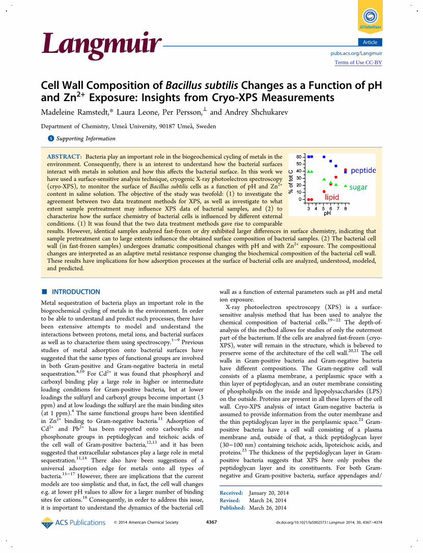
ABSTRACT: Bacteria play an important role in the biogeochemical cycling of metals in theenvironment. Consequently, there is an interest to understand how the bacterial surfacesinteract with metals in solution and how this affects the bacterial surface. In this work wehave used a surface-sensitive analysis technique, cryogenic X-ray photoelectron spectroscopy(cryo-XPS), to monitor the surface of Bacillus subtilis cells as a function of pH and Zn2+
content in saline solution. The objective of the study was twofold: (1) to investigate theagreement between two data treatment methods for XPS, as well as investigate to whatextent sample pretreatment may influence XPS data of bacterial samples, and (2) tocharacterize how the surface chemistry of bacterial cells is influenced by different externalconditions. (1) It was found that the two data treatment methods gave rise to comparableresults. However, identical samples analyzed fast-frozen or dry exhibited larger differences in surface chemistry, indicating thatsample pretreatment can to large extents influence the obtained surface composition of bacterial samples. (2) The bacterial cellwall (in fast-frozen samples) undergoes dramatic compositional changes with pH and with Zn2+ exposure. The compositionalchanges are interpreted as an adaptive metal resistance response changing the biochemical composition of the bacterial cell wall.These results have implications for how adsorption processes at the surface of bacterial cells are analyzed, understood, modeled,and predicted.
■ INTRODUCTION
Metal sequestration of bacteria plays an important role in thebiogeochemical cycling of metals in the environment. In orderto be able to understand and predict such processes, there havebeen extensive attempts to model and understand theinteractions between protons, metal ions, and bacterial surfacesas well as to characterize them using spectroscopy.1−9 Previousstudies of metal adsorption onto bacterial surfaces havesuggested that the same types of functional groups are involvedin both Gram-positive and Gram-negative bacteria in metalsequestration.4,10 For Cd2+ it was found that phosphoryl andcarboxyl binding play a large role in higher or intermediateloading conditions for Gram-positive bacteria, but at lowerloadings the sulfuryl and carboxyl groups become important (3ppm) and at low loadings the sulfuryl are the main binding sites(at 1 ppm).4 The same functional groups have been identifiedin Zn2+ binding to Gram-negative bacteria.11 Adsorption ofCd2+ and Pb2+ has been reported onto carboxylic andphosphonate groups in peptidoglycan and teichoic acids ofthe cell wall of Gram-positive bacteria,12,13 and it has beensuggested that extracellular substances play a large role in metalsequestration.11,14 There also have been suggestions of auniversal adsorption edge for metals onto all types ofbacteria.15−17 However, there are implications that the currentmodels are too simplistic and that, in fact, the cell wall changese.g. at lower pH values to allow for a larger number of bindingsites for cations.18 Consequently, in order to address this issue,it is important to understand the dynamics of the bacterial cell
wall as a function of external parameters such as pH and metalion exposure.X-ray photoelectron spectroscopy (XPS) is a surface-
sensitive analysis method that has been used to analyze thechemical composition of bacterial cells.19−22 The depth-of-analysis of this method allows for studies of only the outermostpart of the bacterium. If the cells are analyzed fast-frozen (cryo-XPS), water will remain in the structure, which is believed topreserve some of the architecture of the cell wall.20,21 The cellwalls in Gram-positive bacteria and Gram-negative bacteriahave different compositions. The Gram-negative cell wallconsists of a plasma membrane, a periplasmic space with athin layer of peptidoglycan, and an outer membrane consistingof phospholipids on the inside and lipopolysaccharides (LPS)on the outside. Proteins are present in all these layers of the cellwall. Cryo-XPS analysis of intact Gram-negative bacteria isassumed to provide information from the outer membrane andthe thin peptidoglycan layer in the periplasmic space.21 Gram-positive bacteria have a cell wall consisting of a plasmamembrane and, outside of that, a thick peptidoglycan layer(30−100 nm) containing teichoic acids, lipoteichoic acids, andproteins.23 The thickness of the peptidoglycan layer in Gram-positive bacteria suggests that XPS here only probes thepeptidoglycan layer and its constituents. For both Gram-negative and Gram-positive bacteria, surface appendages and/
Received: January 20, 2014Revised: March 24, 2014Published: March 26, 2014
Article
pubs.acs.org/Langmuir
© 2014 American Chemical Society 4367 dx.doi.org/10.1021/la5002573 | Langmuir 2014, 30, 4367−4374
Terms of Use CC-BY
or extracellular substances, such as flagella, pili, and capsules,will influence the XPS spectra to some extent depending ontheir quantity. Consequently, bacteria with flagella and pili (orfimbriae) may display higher peptide content, and the presenceof a capsule is expected to increase the polysaccharide contentof the spectra.In this work we have used cryo-XPS to investigate how the
bacterial cell wall of Gram-positive Bacillus subtilis changes withpH and with exposure to Zn(II). B. subtilis is a common soilbacterium that has been reported to tolerate high concen-trations of heavy metals such as Zn(II)24 and is a suitable modelorganism, since it is expected to play a large role in metalbiogeochemical cycling in soil environments. We show that thedramatic changes occurring at the surface of bacterial cells canbe followed using cryo-XPS and that this technique can be usedas a tool to better understand how bacterial surfaces and metalions interact in the environment. We have compared two dataanalysis methods for “unmixing” XPS data to extract thecomposition of the bacterial cell wall with respect to lipids,sugars, and peptides. This comparison aims to establish howwell the two data treatment methods agree for dehydratedsamples, to allow better comparisons between literature studiesusing XPS analyses. Furthermore, we have investigated someconsequences of sample pretreatment in order to study possibleeffects of the dehydration process of bacterial samples and in anattempt to evaluate sample pretreatment methods.
■ EXPERIMENTAL SECTIONReagents. All solutions were prepared by using deionized and
boiled water. NaCl (Merck) was dried overnight at 180 °C and used toadjust the ionic strength to 0.1 M Na(Cl) in all experiments. Stocksolutions of HCl (Fisher p.a.) were standardized against Trizma basetris(hydroxymethyl)aminomethane. NaOH (Merck p.a.) stock sol-utions were standardized against a primary standard HCl solution.ZnCl2 (Merck) and Zn(NO3)2·6H2O (BDH Chemicals) were usedwithout further purification to prepare respectively the solutions usedfor the adsorption experiments and for the atomic absorptionspectroscopy (AAS) calibration curve. Both were standardized withEDTA using xylenol orange as indicator.25
Bacterial Growth. A Bacillus subtilis strain (ATCC 6633) wasgrown at 30 °C in Luria−Bertani broth (composition for 1 L of culturemedium: yeast extract = 5 g, tryptone = 10 g, NaCl = 5 g) underaerobic conditions on an orbital shaker. The cells were collected bycentrifugation at 2880g for 20 min at the end of the exponentialgrowth phase, washed twice with physiological solution (NaCl 0.9%),and resuspended in 0.1 M NaCl ionic medium. The optical density(OD600) of this parent bacterial suspension was measured(spectrophotometer UV-1201 V, Shimadzu), and this value wascorrelated to the dry weight of bacterial cells by using a one-pointcalibration procedure.Preparation of Bacterial Samples at Different pH Values.
Wet paste samples at different pH values were generated by addingHCl and NaOH solutions to resuspended bacterial solutions, andaliquots were removed at desired pH values. Measurement of pH wasdone using a pH electrode that was calibrated with a one-pointcalibration procedure in an acidic solution of known concentration in0.1 M NaCl at 25 °C. The bacterial cells were separated from thesupernatant solutions by centrifugation at 10 000 rpm for 6 min.Zn(II) Adsorption onto Bacillus subtilis. Zn(II) adsorption by
Bacillus subtilis suspensions was studied in batch experimentsperformed at 25 °C ± 1 and in 0.1 M Na(Cl) ionic medium. Samplesfrom the bacterial parent suspensions were transferred into 10 mLtubes, and the pH was varied by adding aliquots of HCl and NaOHsolutions prepared in the same ionic medium from automatic burets.Aliquots of the ZnCl2 solution in 0.1 M Na(Cl) were added at the endin order to have a total Zn(II) concentration in the samplescorresponding to a ratio [Zn2+]/bacterial biomass of 0.12−0.14
mmol/g (dry weight) (corresponding to a total concentration Zn(II)in solution approximately 1 mM). The samples were placed on an end-over-end rotator and centrifuged for 20 min at 2880g after 24 hequilibration time at 25 °C ± 1 and remeasuring of pH. Followingcentrifugation, the supernatants used for AAS analysis were separatedfrom the paste, filtered through 0.22 μm membranes (MILLEX GSfilters, MILLIPORE), acidified, and stored in the refrigerator (4 °C)until analysis.
XPS Analysis. Two series of batch samples were analyzed by XPSin parallel as a function of pH, and they were prepared as follows: fromeach parent suspension of bacterial cells two samples were transferredinto 10 mL tubes. Equal aliquots of acid or base were added to the twosamples in parallel, and an aliquot of the ZnCl2 solution was added toone. The final volume of the two samples was adjusted to the samevalue by using the 0.1 M NaCl solution. While the concentration ofthe bacterial cells was the same in each of the two replicate samples,this value varied from 5.7 to 9.2 g/L dry weight in all the batchsamples prepared at different pH values, and therefore the aliquots ofthe ZnCl2 solution were adjusted in order to obtain a ratio between[Zn(II)]tot/BS(g/L dry weight) in the range of 0.13−0.17. Thesamples were equilibrated for 24 h at 25 °C on an end-over-endrotator and centrifuged at 2880g for 20 min after remeasuring pH. Theequilibrium time was chosen based on previous potentiometric studiesof this system performed in our lab, which showed that equilibriumwas reached before 24 h.26 The supernatant solutions were discarded,and the bacterial pellets were analyzed as fast-frozen wet pastes using aprecooling procedure described elsewhere.20 The XPS spectra werecollected with a Kratos Axis Ultra DLD electron spectrometer usingmonochromated Al Kα source operated at 150 W. An analyzer passenergy of 160 eV was used for acquiring wide spectra and a passenergy of 20 eV for individual photoelectron lines. The surfacepotential was stabilized by the spectrometer charge neutralizationsystem. The binding energy (BE) scale was referenced to the C 1speak aliphatic carbon at 285.0 eV. Thereafter, the compositions of C1s spectra were modeled using the spectral components from themultivariate analysis of C spectra described previously.21 To comparespectra acquired from frozen and dry pastes, some samples were left toslowly warm to room temperature and sublimate the water inside thespectrometer and were subsequently analyzed once more in the sameposition the following day.27 This second measurement will bedenoted as warmed or dry samples in the text.
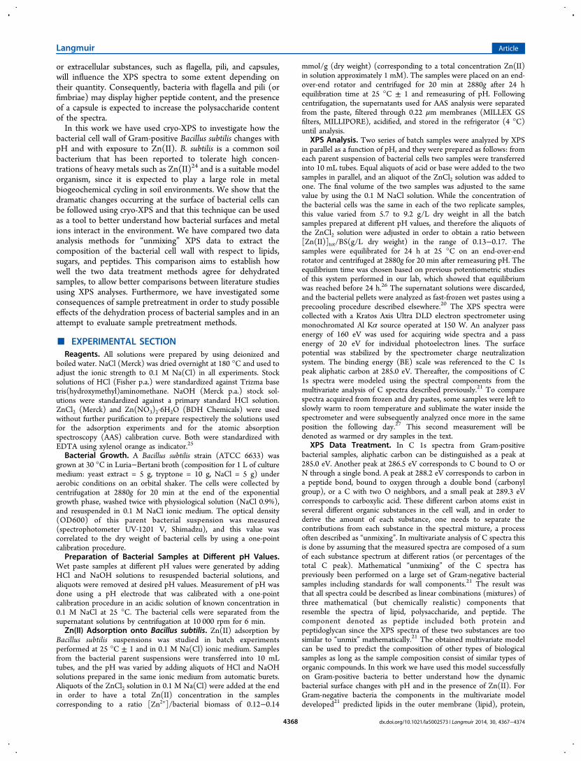
XPS Data Treatment. In C 1s spectra from Gram-positivebacterial samples, aliphatic carbon can be distinguished as a peak at285.0 eV. Another peak at 286.5 eV corresponds to C bound to O orN through a single bond. A peak at 288.2 eV corresponds to carbon ina peptide bond, bound to oxygen through a double bond (carbonylgroup), or a C with two O neighbors, and a small peak at 289.3 eVcorresponds to carboxylic acid. These different carbon atoms exist inseveral different organic substances in the cell wall, and in order toderive the amount of each substance, one needs to separate thecontributions from each substance in the spectral mixture, a processoften described as “unmixing”. In multivariate analysis of C spectra thisis done by assuming that the measured spectra are composed of a sumof each substance spectrum at different ratios (or percentages of thetotal C peak). Mathematical “unmixing” of the C spectra haspreviously been performed on a large set of Gram-negative bacterialsamples including standards for wall components.21 The result wasthat all spectra could be described as linear combinations (mixtures) ofthree mathematical (but chemically realistic) components thatresemble the spectra of lipid, polysaccharide, and peptide. Thecomponent denoted as peptide included both protein andpeptidoglycan since the XPS spectra of these two substances are toosimilar to “unmix” mathematically.21 The obtained multivariate modelcan be used to predict the composition of other types of biologicalsamples as long as the sample composition consist of similar types oforganic compounds. In this work we have used this model successfullyon Gram-positive bacteria to better understand how the dynamicbacterial surface changes with pH and in the presence of Zn(II). ForGram-negative bacteria the components in the multivariate modeldeveloped21 predicted lipids in the outer membrane (lipid), protein,
Langmuir Article
dx.doi.org/10.1021/la5002573 | Langmuir 2014, 30, 4367−43744368
and peptidoglycan (peptide) as well as lipopolysaccharides andpolysaccharides on glycosylated proteins (polysaccharide). ForGram-positive bacteria the cell wall composition differs to someextent, and instead the lipid component in the multivariate modelillustrates changes in the aliphatic part of lipoteichoic acid, the peptidefraction changes in proteins and peptidoglycan (similar as for Gram-negative bacteria), and the polysaccharide component would representpolysaccharides in teichoic and teichuronic acids, polysaccharide onglycosylated proteins, and also polysaccharide in lipoteichoic acid.The multivariate model is not the first attempt described in the
literature to extract the substance composition of the bacterial cell wallfrom XPS data. Previously, Rouxhet and co-workers19,28,29 developedequation systems with ratios between different components in XPSspectra, obtained from curve-fitting, that allow for calculations of theratios of protein (CPr/C), polysaccharide (CPS/C), and hydrocarbon-like compounds (CHC/C) to total carbon using the following equationsystem:
= C C[N/C] 0.279( / )obs Pr (1)
= +C C C C[O/C] 0.325( / ) 0.833( / )obs Pr PS (2)
= = + +C C C C C C[C/C] 1 ( / ) ( / ) ( / )obs Pr PS CH (3)
This scheme works well as long as there are no interfering substancesfor example in the nitrogen or oxygen spectra. Consequently, forfrozen samples where the structural water remains, other methodswere needed. The multivariate “unmixing” of components contributingto the C 1s spectra, used here, was developed in response to the needto estimate and predict the composition of the bacterial cell wall incryo-XPS.21 The unmixing performed using the multivariatecomponents can be seen as a version of eq 3 previously describedby Rouxhet et al.,29 and consequently the outputs from the twomethods should be comparable. However, the multivariate model isexpected to be less subjective to differences in peak-fitting proceduresbetween different experimentalists as the fitting in the multivariatemodel is automatically done using predefined mathematicalcomponents for each substance.21
Statistics. Statistical analysis (ANOVA) was performed usingOrigin 8.1 (Origin Lab Corporation).
■ RESULTS AND DISCUSSIONAnalysis of XPS spectra: Comparison between
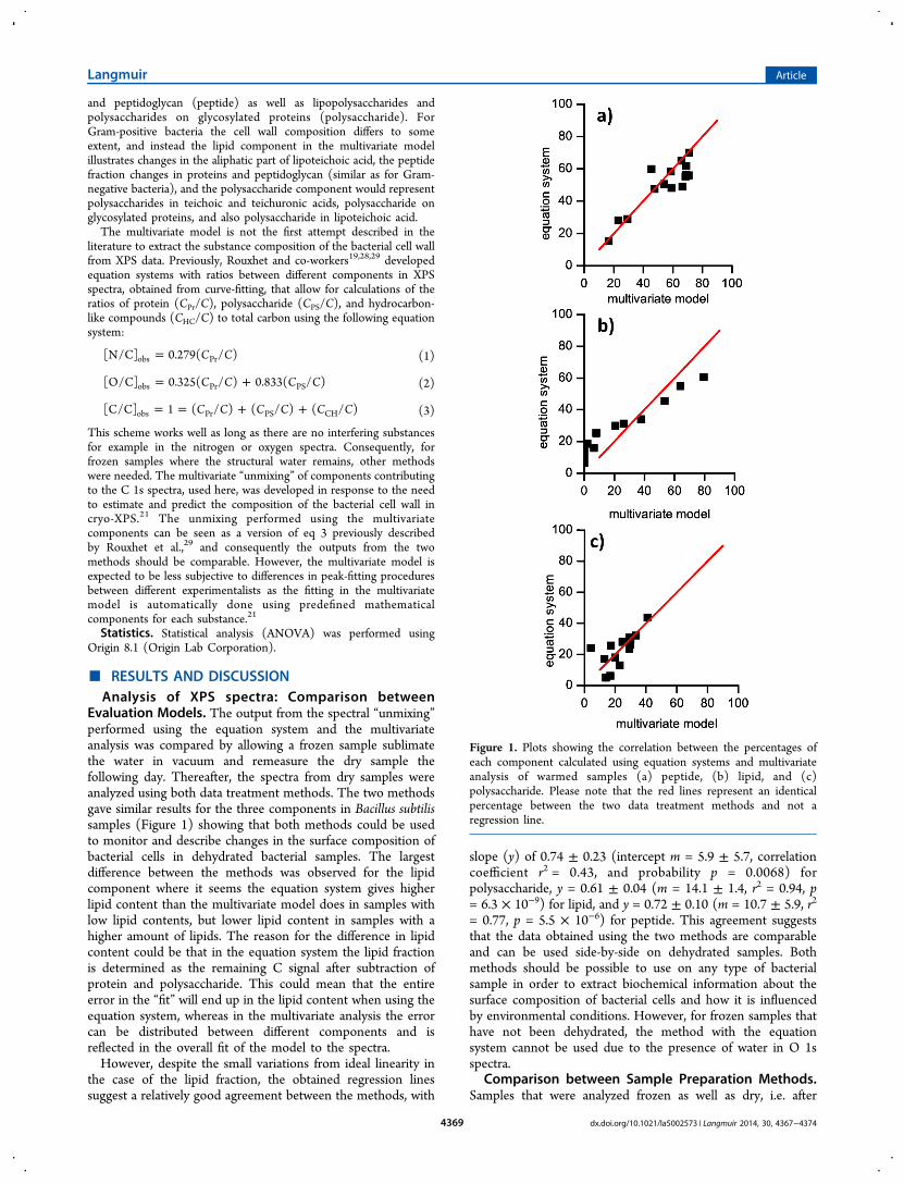
Evaluation Models. The output from the spectral “unmixing”performed using the equation system and the multivariateanalysis was compared by allowing a frozen sample sublimatethe water in vacuum and remeasure the dry sample thefollowing day. Thereafter, the spectra from dry samples wereanalyzed using both data treatment methods. The two methodsgave similar results for the three components in Bacillus subtilissamples (Figure 1) showing that both methods could be usedto monitor and describe changes in the surface composition ofbacterial cells in dehydrated bacterial samples. The largestdifference between the methods was observed for the lipidcomponent where it seems the equation system gives higherlipid content than the multivariate model does in samples withlow lipid contents, but lower lipid content in samples with ahigher amount of lipids. The reason for the difference in lipidcontent could be that in the equation system the lipid fractionis determined as the remaining C signal after subtraction ofprotein and polysaccharide. This could mean that the entireerror in the “fit” will end up in the lipid content when using theequation system, whereas in the multivariate analysis the errorcan be distributed between different components and isreflected in the overall fit of the model to the spectra.However, despite the small variations from ideal linearity in
the case of the lipid fraction, the obtained regression linessuggest a relatively good agreement between the methods, with
slope (y) of 0.74 ± 0.23 (intercept m = 5.9 ± 5.7, correlationcoefficient r2 = 0.43, and probability p = 0.0068) forpolysaccharide, y = 0.61 ± 0.04 (m = 14.1 ± 1.4, r2 = 0.94, p= 6.3 × 10⁻⁹) for lipid, and y = 0.72 ± 0.10 (m = 10.7 ± 5.9, r2
= 0.77, p = 5.5 × 10⁻⁶) for peptide. This agreement suggeststhat the data obtained using the two methods are comparableand can be used side-by-side on dehydrated samples. Bothmethods should be possible to use on any type of bacterialsample in order to extract biochemical information about thesurface composition of bacterial cells and how it is influencedby environmental conditions. However, for frozen samples thathave not been dehydrated, the method with the equationsystem cannot be used due to the presence of water in O 1sspectra.
Comparison between Sample Preparation Methods.Samples that were analyzed frozen as well as dry, i.e. after
Figure 1. Plots showing the correlation between the percentages ofeach component calculated using equation systems and multivariateanalysis of warmed samples (a) peptide, (b) lipid, and (c)polysaccharide. Please note that the red lines represent an identicalpercentage between the two data treatment methods and not aregression line.
Langmuir Article
dx.doi.org/10.1021/la5002573 | Langmuir 2014, 30, 4367−43744369
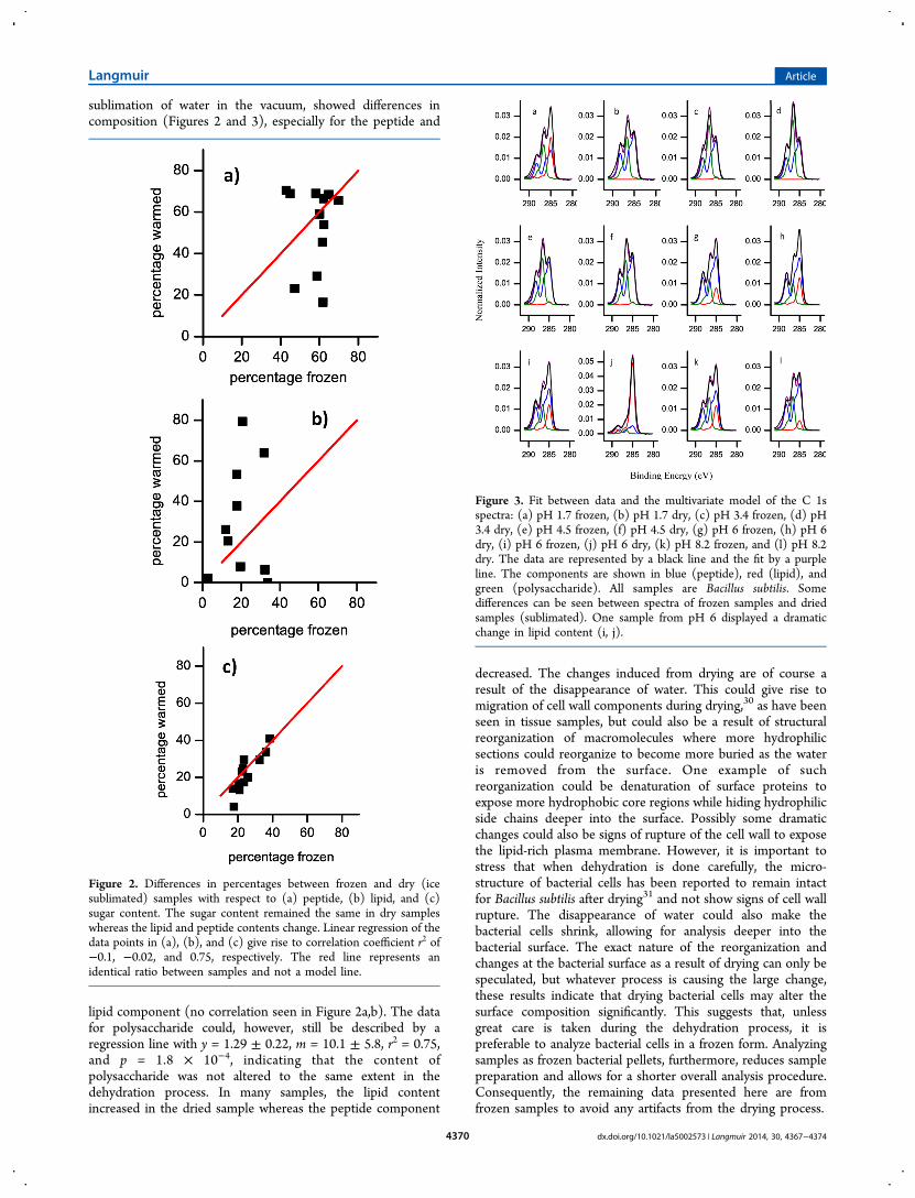
sublimation of water in the vacuum, showed differences incomposition (Figures 2 and 3), especially for the peptide and
lipid component (no correlation seen in Figure 2a,b). The datafor polysaccharide could, however, still be described by aregression line with y = 1.29 ± 0.22, m = 10.1 ± 5.8, r2 = 0.75,and p = 1.8 × 10⁻⁴, indicating that the content ofpolysaccharide was not altered to the same extent in thedehydration process. In many samples, the lipid contentincreased in the dried sample whereas the peptide component
decreased. The changes induced from drying are of course aresult of the disappearance of water. This could give rise tomigration of cell wall components during drying,30 as have beenseen in tissue samples, but could also be a result of structuralreorganization of macromolecules where more hydrophilicsections could reorganize to become more buried as the wateris removed from the surface. One example of suchreorganization could be denaturation of surface proteins toexpose more hydrophobic core regions while hiding hydrophilicside chains deeper into the surface. Possibly some dramaticchanges could also be signs of rupture of the cell wall to exposethe lipid-rich plasma membrane. However, it is important tostress that when dehydration is done carefully, the micro-structure of bacterial cells has been reported to remain intactfor Bacillus subtilis after drying31 and not show signs of cell wallrupture. The disappearance of water could also make thebacterial cells shrink, allowing for analysis deeper into thebacterial surface. The exact nature of the reorganization andchanges at the bacterial surface as a result of drying can only bespeculated, but whatever process is causing the large change,these results indicate that drying bacterial cells may alter thesurface composition significantly. This suggests that, unlessgreat care is taken during the dehydration process, it ispreferable to analyze bacterial cells in a frozen form. Analyzingsamples as frozen bacterial pellets, furthermore, reduces samplepreparation and allows for a shorter overall analysis procedure.Consequently, the remaining data presented here are fromfrozen samples to avoid any artifacts from the drying process.
Figure 2. Differences in percentages between frozen and dry (icesublimated) samples with respect to (a) peptide, (b) lipid, and (c)sugar content. The sugar content remained the same in dry sampleswhereas the lipid and peptide contents change. Linear regression of thedata points in (a), (b), and (c) give rise to correlation coefficient r2 of−0.1, −0.02, and 0.75, respectively. The red line represents anidentical ratio between samples and not a model line.
Figure 3. Fit between data and the multivariate model of the C 1sspectra: (a) pH 1.7 frozen, (b) pH 1.7 dry, (c) pH 3.4 frozen, (d) pH3.4 dry, (e) pH 4.5 frozen, (f) pH 4.5 dry, (g) pH 6 frozen, (h) pH 6dry, (i) pH 6 frozen, (j) pH 6 dry, (k) pH 8.2 frozen, and (l) pH 8.2dry. The data are represented by a black line and the fit by a purpleline. The components are shown in blue (peptide), red (lipid), andgreen (polysaccharide). All samples are Bacillus subtilis. Somedifferences can be seen between spectra of frozen samples and driedsamples (sublimated). One sample from pH 6 displayed a dramaticchange in lipid content (i, j).
Langmuir Article
dx.doi.org/10.1021/la5002573 | Langmuir 2014, 30, 4367−43744370
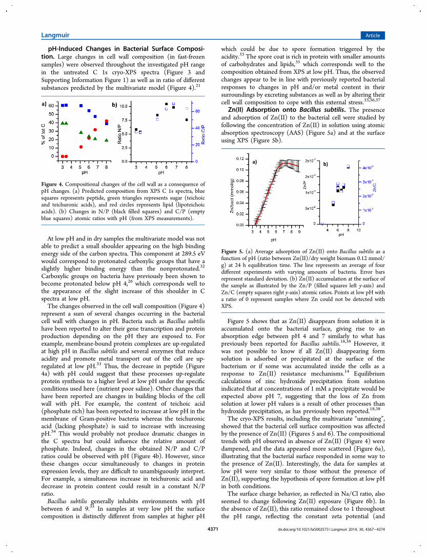
pH-Induced Changes in Bacterial Surface Composi-tion. Large changes in cell wall composition (in fast-frozensamples) were observed throughout the investigated pH rangein the untreated C 1s cryo-XPS spectra (Figure 3 andSupporting Information Figure 1) as well as in ratio of differentsubstances predicted by the multivariate model (Figure 4).21
At low pH and in dry samples the multivariate model was notable to predict a small shoulder appearing on the high bindingenergy side of the carbon spectra. This component at 289.5 eVwould correspond to protonated carboxylic groups that have aslightly higher binding energy than the nonprotonated.32
Carboxylic groups on bacteria have previously been shown tobecome protonated below pH 4,26 which corresponds well tothe appearance of the slight increase of this shoulder in Cspectra at low pH.The changes observed in the cell wall composition (Figure 4)
represent a sum of several changes occurring in the bacterialcell wall with changes in pH. Bacteria such as Bacillus subtilishave been reported to alter their gene transcription and proteinproduction depending on the pH they are exposed to. Forexample, membrane-bound protein complexes are up-regulatedat high pH in Bacillus subtilis and several enzymes that reduceacidity and promote metal transport out of the cell are up-regulated at low pH.33 Thus, the decrease in peptide (Figure4a) with pH could suggest that these processes up-regulateprotein synthesis to a higher level at low pH under the specificconditions used here (nutrient poor saline). Other changes thathave been reported are changes in building blocks of the cellwall with pH. For example, the content of teichoic acid(phosphate rich) has been reported to increase at low pH in themembrane of Gram-positive bacteria whereas the teichuronicacid (lacking phosphate) is said to increase with increasingpH.34 This would probably not produce dramatic changes inthe C spectra but could influence the relative amount ofphosphate. Indeed, changes in the obtained N/P and C/Pratios could be observed with pH (Figure 4b). However, sincethese changes occur simultaneously to changes in proteinexpression levels, they are difficult to unambiguously interpret.For example, a simultaneous increase in teichuronic acid anddecrease in protein content could result in a constant N/Pratio.Bacillus subtilis generally inhabits environments with pH
between 6 and 9.33 In samples at very low pH the surfacecomposition is distinctly different from samples at higher pH
which could be due to spore formation triggered by theacidity.33 The spore coat is rich in protein with smaller amountsof carbohydrates and lipids,35 which corresponds well to thecomposition obtained from XPS at low pH. Thus, the observedchanges appear to be in line with previously reported bacterialresponses to changes in pH and/or metal content in theirsurroundings by excreting substances as well as by altering theircell wall composition to cope with this external stress.33,36,37
Zn(II) Adsorption onto Bacillus subtilis. The presenceand adsorption of Zn(II) to the bacterial cell were studied byfollowing the concentration of Zn(II) in solution using atomicabsorption spectroscopy (AAS) (Figure 5a) and at the surfaceusing XPS (Figure 5b).
Figure 5 shows that as Zn(II) disappears from solution it isaccumulated onto the bacterial surface, giving rise to anabsorption edge between pH 4 and 7 similarly to what haspreviously been reported for Bacillus subtilis.18,38 However, itwas not possible to know if all Zn(II) disappearing formsolution is adsorbed or precipitated at the surface of thebacterium or if some was accumulated inside the cells as aresponse to Zn(II) resistance mechanisms.14 Equilibriumcalculations of zinc hydroxide precipitation from solutionindicated that at concentrations of 1 mM a precipitate would beexpected above pH 7, suggesting that the loss of Zn fromsolution at lower pH values is a result of other processes thanhydroxide precipitation, as has previously been reported.18,38
The cryo-XPS results, including the multivariate “unmixing”,showed that the bacterial cell surface composition was affectedby the presence of Zn(II) (Figures 5 and 6). The compositionaltrends with pH observed in absence of Zn(II) (Figure 4) weredampened, and the data appeared more scattered (Figure 6a),illustrating that the bacterial surface responded in some way tothe presence of Zn(II). Interestingly, the data for samples atlow pH were very similar to those without the presence ofZn(II), supporting the hypothesis of spore formation at low pHin both conditions.The surface charge behavior, as reflected in Na/Cl ratio, also
seemed to change following Zn(II) exposure (Figure 6b). Inthe absence of Zn(II), this ratio remained close to 1 throughoutthe pH range, reflecting the constant zeta potential (and
Figure 4. Compositional changes of the cell wall as a consequence ofpH changes. (a) Predicted composition from XPS C 1s spectra, bluesquares represents peptide, green triangles represents sugar (teichoicand teichuronic acids), and red circles represents lipid (lipoteichoicacids). (b) Changes in N/P (black filled squares) and C/P (emptyblue squares) atomic ratios with pH (from XPS measurements).
Figure 5. (a) Average adsorption of Zn(II) onto Bacillus subtilis as afunction of pH (ratio between Zn(II)/dry weight biomass 0.12 mmol/g) at 24 h equilibration time. The line represents an average of fourdifferent experiments with varying amounts of bacteria. Error barsrepresent standard deviation. (b) Zn(II) accumulation at the surface ofthe sample as illustrated by the Zn/P (filled squares left y-axis) andZn/C (empty squares right y-axis) atomic ratios. Points at low pH witha ratio of 0 represent samples where Zn could not be detected withXPS.
Langmuir Article
dx.doi.org/10.1021/la5002573 | Langmuir 2014, 30, 4367−43744371
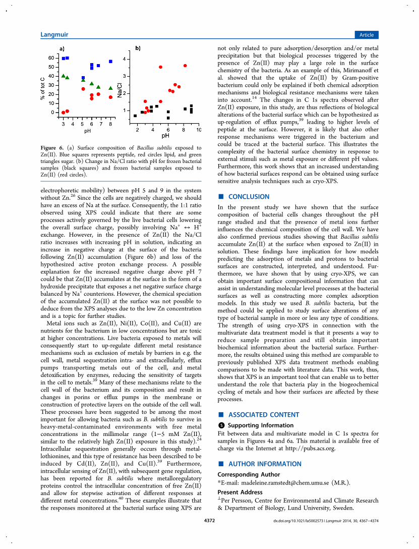
electrophoretic mobility) between pH 5 and 9 in the systemwithout Zn.26 Since the cells are negatively charged, we shouldhave an excess of Na at the surface. Consequently, the 1:1 ratioobserved using XPS could indicate that there are someprocesses actively governed by the live bacterial cells loweringthe overall surface charge, possibly involving Na+ ↔ H+
exchange. However, in the presence of Zn(II) the Na/Clratio increases with increasing pH in solution, indicating anincrease in negative charge at the surface of the bacteriafollowing Zn(II) accumulation (Figure 6b) and loss of thehypothesized active proton exchange process. A possibleexplanation for the increased negative charge above pH 7could be that Zn(II) accumulates at the surface in the form of ahydroxide precipitate that exposes a net negative surface chargebalanced by Na+ counterions. However, the chemical speciationof the accumulated Zn(II) at the surface was not possible todeduce from the XPS analyses due to the low Zn concentrationand is a topic for further studies.Metal ions such as Zn(II), Ni(II), Co(II), and Cu(II) are
nutrients for the bacterium in low concentrations but are toxicat higher concentrations. Live bacteria exposed to metals willconsequently start to up-regulate different metal resistancemechanisms such as exclusion of metals by barriers in e.g. thecell wall, metal sequestration intra- and extracellularly, effluxpumps transporting metals out of the cell, and metaldetoxification by enzymes, reducing the sensitivity of targetsin the cell to metals.39 Many of these mechanisms relate to thecell wall of the bacterium and its composition and result inchanges in porins or efflux pumps in the membrane orconstruction of protective layers on the outside of the cell wall.These processes have been suggested to be among the mostimportant for allowing bacteria such as B. subtilis to survive inheavy-metal-contaminated environments with free metalconcentrations in the millimolar range (1−5 mM Zn(II),similar to the relatively high Zn(II) exposure in this study).24
Intracellular sequestration generally occurs through metal-lothionines, and this type of resistance has been described to beinduced by Cd(II), Zn(II), and Cu(II).39 Furthermore,intracellular sensing of Zn(II), with subsequent gene regulation,has been reported for B. subtilis where metalloregulatoryproteins control the intracellular concentration of free Zn(II)and allow for stepwise activation of different responses atdifferent metal concentrations.40 These examples illustrate thatthe responses monitored at the bacterial surface using XPS are
not only related to pure adsorption/desorption and/or metalprecipitation but that biological processes triggered by thepresence of Zn(II) may play a large role in the surfacechemistry of the bacteria. As an example of this, Mirimanoff etal. showed that the uptake of Zn(II) by Gram-positivebacterium could only be explained if both chemical adsorptionmechanisms and biological resistance mechanisms were takeninto account.14 The changes in C 1s spectra observed afterZn(II) exposure, in this study, are thus reflections of biologicalalterations of the bacterial surface which can be hypothesized asup-regulation of efflux pumps,39 leading to higher levels ofpeptide at the surface. However, it is likely that also otherresponse mechanisms were triggered in the bacterium andcould be traced at the bacterial surface. This illustrates thecomplexity of the bacterial surface chemistry in response toexternal stimuli such as metal exposure or different pH values.Furthermore, this work shows that an increased understandingof how bacterial surfaces respond can be obtained using surfacesensitive analysis techniques such as cryo-XPS.
■ CONCLUSION
In the present study we have shown that the surfacecomposition of bacterial cells changes throughout the pHrange studied and that the presence of metal ions furtherinfluences the chemical composition of the cell wall. We havealso confirmed previous studies showing that Bacillus subtilisaccumulate Zn(II) at the surface when exposed to Zn(II) insolution. These findings have implication for how modelspredicting the adsorption of metals and protons to bacterialsurfaces are constructed, interpreted, and understood. Fur-thermore, we have shown that by using cryo-XPS, we canobtain important surface compositional information that canassist in understanding molecular level processes at the bacterialsurfaces as well as constructing more complex adsorptionmodels. In this study we used B. subtilis bacteria, but themethod could be applied to study surface alterations of anytype of bacterial sample in more or less any type of conditions.The strength of using cryo-XPS in connection with themultivariate data treatment model is that it presents a way toreduce sample preparation and still obtain importantbiochemical information about the bacterial surface. Further-more, the results obtained using this method are comparable topreviously published XPS data treatment methods enablingcomparisons to be made with literature data. This work, thus,shows that XPS is an important tool that can enable us to betterunderstand the role that bacteria play in the biogeochemicalcycling of metals and how their surfaces are affected by theseprocesses.
■ ASSOCIATED CONTENT
*S Supporting InformationFit between data and multivariate model in C 1s spectra forsamples in Figures 4a and 6a. This material is available free ofcharge via the Internet at http://pubs.acs.org.
■ AUTHOR INFORMATION
Corresponding Author*E-mail: [email protected] (M.R.).
Present Address⊥Per Persson, Centre for Environmental and Climate Research& Department of Biology, Lund University, Sweden.
Figure 6. (a) Surface composition of Bacillus subtilis exposed toZn(II). Blue squares represents peptide, red circles lipid, and greentriangles sugar. (b) Change in Na/Cl ratio with pH for frozen bacterialsamples (black squares) and frozen bacterial samples exposed toZn(II) (red circles).
Langmuir Article
dx.doi.org/10.1021/la5002573 | Langmuir 2014, 30, 4367−43744372

NotesThe authors declare no competing financial interest.
■ ACKNOWLEDGMENTS
The Swedish Research Council is acknowledged for funding.
■ REFERENCES(1) Borrok, D.; Fein, J. The impact of ionic strength on theadsorption of protons, Pb, Cd, and Sr onto the surfaces of Gramnegative bacteria: testing non-electro static, diffuse, and triple-layermodels. J. Colloid Interface Sci. 2005, 286, 110−126.(2) Fein, J.; Daughney, C.; Yee, N.; Davis, T. A chemical equilibriummodel for metal adsorption onto bacterial surfaces. Geochim.Cosmochim. Acta 1997, 61, 3319−3328.(3) Fowle, D.; Fein, J. Competitive adsorption of metal cations ontotwo gram positive bacteria: Testing the chemical equilibrium model.Geochim. Cosmochim. Acta 1999, 63, 3059−3067.(4) Mishra, B.; Boyanov, M.; Bunker, B.; Kelly, S.; Kemner, K.; Fein,J. High- and low-affinity binding sites for Cd on the bacterial cell wallsof Bacillus subtilis and Shewanella oneidensis. Geochim. Cosmochim.Acta 2010, 74, 4219−4233.(5) Kenney, J.; Fein, J. Cell wall reactivity of acidophilic andalkaliphilic bacteria determined by potentiometric titrations and Cdadsorption experiments. Environ. Sci. Technol. 2011, 45, 4446−4452.(6) Ojeda, J.; Romero-Gonzalez, M.; Bachmann, R.; Edyvean, R.;Banwart, S. Characterization of the cell surface and cell wall chemistryof drinking water bacteria by combining XPS, FTIR spectroscopy,modeling, and potentiometric titrations. Langmuir 2008, 24, 4032−4040.(7) Heinrich, H.; Bremer, P.; Daughney, C.; McQuillan, A. Acid-basetitrations of functional groups on the surface of the thermophilicbacterium Anoxybacillus flavithermus: Comparing a chemical equili-brium model with ATR-IR spectroscopic data. Langmuir 2007, 23,2731−2740.(8) Johnson, K.; Szymanowski, J.; Borrok, D.; Huynh, T.; Fein, J.Proton and metal adsorption onto bacterial consortia: Stabilityconstants for metal-bacterial surface complexes. Chem. Geol. 2007,239, 13−26.(9) Fein, J.; Martin, A.; Wightman, P. Metal adsorption onto bacterialsurfaces: Development of a predictive approach. Geochim. Cosmochim.Acta 2001, 65, 4267−4273.(10) Toner, B.; Manceau, A.; Marcus, M.; Millet, D.; Sposito, G. Zincsorption by a bacterial biofilm. Environ. Sci. Technol. 2005, 39, 8288−8294.(11) Guine, V.; Spadini, L.; Sarret, G.; Muris, M.; Delolme, C.;Gaudet, J.; Martins, J. Zinc sorption to three gram-negative bacteria:Combined titration, modeling, and EXAFS study. Environ. Sci. Technol.2006, 40, 1806−1813.(12) Johnson, K.; Cygan, R.; Fein, J. Molecular simulations of metaladsorption to bacterial surfaces. Geochim. Cosmochim. Acta 2006, 70,5075−5088.(13) Boyanov, M.; Kelly, S.; Kemner, K.; Bunker, B.; Fein, J.; Fowle,D. Adsorption of cadmium to Bacillus subtilis bacterial cell walls: ApH-dependent X-ray absorption fine structure spectroscopy study.Geochim. Cosmochim. Acta 2003, 67, 3299−3311.(14) Mirimanoff, N.; Wilkinson, K. Regulation of Zn accumulation bya freshwater gram-positive bacterium (Rhodococcus opacus). Environ.Sci. Technol. 2000, 34, 616−622.(15) Yee, N.; Fein, J. Cd adsorption onto bacterial surfaces: Auniversal adsorption edge? Geochim. Cosmochim. Acta 2001, 65, 2037−2042.(16) Ginn, B.; Fein, J. The effect of species diversity on metaladsorption onto bacteria. Geochim. Cosmochim. Acta 2008, 72, 3939−3948.(17) Yee, N.; Fein, J. Quantifying metal adsorption onto bacteriamixtures: A test and application of the surface complexation model.Geomicrobiol. J. 2003, 20, 43−60.
(18) Ginn, B.; Szymanowski, J.; Fein, J. Calibration of a linear freeenergy estimation approach for estimating stability constants for metal-bacterial surface complexes. Geomicrobiol. J. 2010, 27, 321−328.(19) Ahimou, F.; Boonaert, C.; Adriaensen, Y.; Jacques, P.; Thonart,P.; Paquot, M.; Rouxhet, P. XPS analysis of chemical functions at thesurface of Bacillus subtilis. J. Colloid Interface Sci. 2007, 309, 49−55.(20) Leone, L.; Loring, J.; Sjoberg, S.; Persson, P.; Shchukarev, A.Surface characterization of the Gram-positive bacteria Bacillus subtilis -an XPS study. Surf. Interface Anal. 2006, 38, 202−205.(21) Ramstedt, M.; Nakao, R.; Wai, S.; Uhlin, B.; Boily, J. Monitoringsurface chemical changes in the bacterial cell wall - multivariate analysisof cryo-X-ray photoelectron spectroscopy data. J. Biol. Chem. 2011,286, 12389−12396.(22) van der Mei, H.; de Vries, J.; Busscher, H. X-ray photoelectronspectroscopy for the study of microbial cell surfaces. Surf. Sci. Rep.2000, 39, 3−24.(23) Silhavy, T. J.; Kahne, D.; Walker, S. The bacterial cell envelope.Cold Spring Harb Perspect Biol. 2010, 2, a000414.(24) Hookoom, M.; Puchooa, D. Isolation and identification of heavymetals tolerant bacteria from industrial and agricultural areas inmauritius. Curr. Res. Microbiol. Biotechnol. 2013, 1, 119−123.(25) Jeffery, G. H.; Bassett, J.; Mendham, J.; Denney, R. C. Vogel’sTextbook of Quantitative Chemical Analysis; Longman Scientific &Technical: Harlow, England, 1989; p 877.(26) Leone, L.; Ferri, D.; Manfredi, C.; Persson, P.; Shchukarev, A.;Sjoberg, S.; Loring, J. Modeling the acid-base properties of bacterialsurfaces: A combined spectroscopic and potentiometric study of thegram-positive bacterium Bacillus subtilis. Environ. Sci. Technol. 2007,41, 6465−6471.(27) Ramstedt, M.; Shchukarev, A.; Sjoberg, S. Characterization ofhydrous manganite (gamma-MnOOH) surfaces - an XPS study. Surf.Interface Anal. 2002, 632−636.(28) Dufrene, Y.; VanderWal, A.; Norde, W.; Rouxhet, P. X-rayphotoelectron spectroscopy analysis of whole cells and isolated cellwalls of gram-positive bacteria: Comparison with biochemical analysis.J. Bacteriol. 1997, 179, 1023−1028.(29) Rouxhet, P.; Genet, M. XPS analysis of bio-organic systems.Surf. Interface Anal. 2011, 43, 1453−1470.(30) Sjovall, P.; Johansson, B.; Lausmaa, J. Localization of lipids infreeze-dried mouse brain sections by imaging TOF-SIMS. Appl. Surf.Sci. 2006, 252, 6966−6974.(31) Moriwaki, H.; Koide, R.; Yoshikawa, R.; Warabino, Y.;Yamamoto, H. Adsorption of rare earth ions onto the cell walls ofwild-type and lipoteichoic acid-defective strains of Bacillus subtilis.Appl. Microbiol. Biotechnol. 2013, 97, 3721−3728.(32) Moulder, J. F.; Stickle, W. F.; Sobol, P. E.; Bomben, K. D.Handbook of X-ray Photoelectron Spectroscopy; Perkin-Elmer Corpo-ration Physical Electronics Division: Eden Prairie, MN, 1992.(33) Wilks, J. C.; Kitko, R. D.; Cleeton, S. H.; Lee, G. E.; Ugwu, C.S.; Jones, B. D.; BonDurant, S. S.; Slonczewski, J. L. Acid and basestress and transcriptomic responses in Bacillus subtilis. Appl. Environ.Microbiol. 2009, 75, 981−990.(34) Ellwood, D. C.; Tempest, D. W. Influence of culture pH on thecontent and composition of teichoic acids in the walls of Bacillussubtilis. J. Gen Microbiol 1972, 73, 395−402.(35) Driks, A. Bacillus subtilis spore coat. Microbiol Mol. Biol. Rev.1999, 63, 1−20.(36) Slonczewski, J. L.; Fujisawa, M.; Dopson, M.; Krulwich, T. A.Cytoplasmic pH measurement and homeostasis in bacteria andarchaea. Adv. Microb Physiol 2009, 55 (1−79), 317.(37) Thackray, P. D.; Moir, A. SigM, an extracytoplasmic functionsigma factor of Bacillus subtilis, is activated in response to cell wallantibiotics, ethanol, heat, acid, and superoxide stress. J. Bacteriol. 2003,185, 3491−3498.(38) Gorman-Lewis, D. Enthalpies and entropies of Cd and Znadsorption onto Bacillus licheniformis and enthalpies and entropies ofZn adsorption onto Bacillus subtilis from isothermal titrationcalorimetry and surface complexation modeling. Geomicrobiol. J.2014, 31, 383−395.
Langmuir Article
dx.doi.org/10.1021/la5002573 | Langmuir 2014, 30, 4367−43744373

(39) Bruins, M.; Kapil, S.; Oehme, F. Microbial resistance to metalsin the environment. Ecotoxicol. Environ. Saf. 2000, 45, 198−207.(40) Ma, Z.; Gabriel, S.; Helmann, J. Sequential binding and sensingof Zn(II) by Bacillus subtilis Zur. Nucleic Acids Res. 2011, 39, 9130−9138.
Langmuir Article
dx.doi.org/10.1021/la5002573 | Langmuir 2014, 30, 4367−43744374
Copyright © 2022 FDOKUMEN