Bone mineralization proceeds through intracellular calcium phosphate loaded vesicles: A...
9
Bone mineralization proceeds through intracellular calcium phosphate loaded vesicles: A cryo-electron microscopy study Julia Mahamid a,⇑ , Amnon Sharir b,c , Dvir Gur a , Elazar Zelzer b , Lia Addadi a , Steve Weiner a a Department of Structural Biology, Weizmann Institute of Science, 76100 Rehovot, Israel b Department of Molecular Genetics, Weizmann Institute of Science, 76100 Rehovot, Israel c Koret School of Veterinary Medicine, The Robert H. Smith Faculty of Agriculture, Food and Environment, Hebrew University of Jerusalem, 76100 Rehovot, Israel article info Article history: Received 4 January 2011 Received in revised form 18 March 2011 Accepted 18 March 2011 Available online 2 April 2011 Keywords: Biomineralization Mouse model Osteoblast Calvaria Carbonated hydroxyapatite Transient precursor abstract Bone is the most widespread mineralized tissue in vertebrates and its formation is orchestrated by spe- cialized cells – the osteoblasts. Crystalline carbonated hydroxyapatite, an inorganic calcium phosphate mineral, constitutes a substantial fraction of mature bone tissue. Yet key aspects of the mineral formation mechanism, transport pathways and deposition in the extracellular matrix remain unidentified. Using cryo-electron microscopy on native frozen-hydrated tissues we show that during mineralization of devel- oping mouse calvaria and long bones, bone-lining cells concentrate membrane-bound mineral granules within intracellular vesicles. Elemental analysis and electron diffraction show that the intracellular min- eral granules consist of disordered calcium phosphate, a highly metastable phase and a potential precur- sor of carbonated hydroxyapatite. The intracellular mineral contains considerably less calcium than expected for synthetic amorphous calcium phosphate, suggesting the presence of a cellular mechanism by which phosphate entities are first formed and thereafter gradually sequester calcium within the ves- icles. We thus demonstrate that in vivo osteoblasts actively produce disordered mineral packets within intracellular vesicles for mineralization of the extracellular developing bone tissue. The use of a highly disordered precursor mineral phase that later crystallizes within an extracellular matrix is a strategy employed in the formation of fish fin bones and by various invertebrate phyla. This therefore appears to be a widespread strategy used by many animal phyla, including vertebrates. Ó 2011 Elsevier Inc. All rights reserved. 1. Introduction The vertebrate skeleton is a unique organic–inorganic tissue that forms during embryonic development and is dynamically shaped and maintained throughout the animal’s life (Currey, 2002). Bones are complex structures, with a hierarchical organiza- tion encompassing a range from nanometers to centimeters (Wei- ner and Traub, 1992; Weiner and Wagner, 1998). At the fundamental structural level, all bones are comprised of type-I col- lagen fibrils reinforced by co-aligned crystalline platelets of car- bonated hydroxyapatite, a calcium phosphate mineral (Landis et al., 1993; Traub et al., 1989). Various cell types are responsible for the formation as well as the maintenance of the bone integrity and function (Peck and Woods, 1988). Bones of the vertebrate skeleton may develop via two distinct processes: intramembranous bone formation that proceeds through direct mineralization of a newly formed type-I collagen matrix and endochondral bone, involving the formation of a miner- alized cartilaginous precursor that is later replaced by a mineral- ized bone matrix (Caplan, 1987; Streeter, 1949). In both modes osteoblasts orchestrate the bone formation process, initiated by the synthesis of an organic extracellular matrix – the osteoid (Ca- plan, 1987), mainly composed of type-I collagen, but also contain- ing up to 10% non-collagenous proteins (Lowenstam and Weiner, 1989). Mineralization of the collagen fibrils thereafter takes place. Blood serum is the main source of ions in the vertebrate body, and contains significant concentrations of calcium and phosphate, sufficient for the deposition of carbonated hydroxyapatite (Jahnen- Dechent, 2000; Posner et al., 1978). However, the pathways through which the ions are translocated from the serum to the site of deposition within the extracellular matrix, and the precise involvement of cellular processes in mineral deposition are not clear (Gay et al., 2000). Two principal modes have been suggested to describe mineral deposition into collagen matrices: (i) crystals are actively nucleated from solution by charged non-collagenous proteins associated to the collagen gap zones, without intervention of intracellular processes (Glimcher, 1984; Veis and Perry, 1967); 1047-8477/$ - see front matter Ó 2011 Elsevier Inc. All rights reserved. doi:10.1016/j.jsb.2011.03.014 ⇑ Corresponding author. Present address: Department of Molecular Structural Biology, Max Planck Institute of Biochemistry, Am Klopferspitz 18, 82152 Martins- ried, Germany. Fax: +49 89 8578 2641. E-mail address: [email protected] (J. Mahamid). Journal of Structural Biology 174 (2011) 527–535 Contents lists available at ScienceDirect Journal of Structural Biology journal homepage: www.elsevier.com/locate/yjsbi
Transcript of Bone mineralization proceeds through intracellular calcium phosphate loaded vesicles: A...

Journal of Structural Biology 174 (2011) 527–535
Contents lists available at ScienceDirect
Journal of Structural Biology
journal homepage: www.elsevier .com/locate /y jsbi
Bone mineralization proceeds through intracellular calcium phosphateloaded vesicles: A cryo-electron microscopy study
Julia Mahamid a,⇑, Amnon Sharir b,c, Dvir Gur a, Elazar Zelzer b, Lia Addadi a, Steve Weiner a
a Department of Structural Biology, Weizmann Institute of Science, 76100 Rehovot, Israelb Department of Molecular Genetics, Weizmann Institute of Science, 76100 Rehovot, Israelc Koret School of Veterinary Medicine, The Robert H. Smith Faculty of Agriculture, Food and Environment, Hebrew University of Jerusalem, 76100 Rehovot, Israel
a r t i c l e i n f o a b s t r a c t
Article history:Received 4 January 2011Received in revised form 18 March 2011Accepted 18 March 2011Available online 2 April 2011
Keywords:BiomineralizationMouse modelOsteoblastCalvariaCarbonated hydroxyapatiteTransient precursor
1047-8477/$ - see front matter � 2011 Elsevier Inc. Adoi:10.1016/j.jsb.2011.03.014
⇑ Corresponding author. Present address: DepartmBiology, Max Planck Institute of Biochemistry, Am Kloried, Germany. Fax: +49 89 8578 2641.
E-mail address: [email protected] (J. Ma
Bone is the most widespread mineralized tissue in vertebrates and its formation is orchestrated by spe-cialized cells – the osteoblasts. Crystalline carbonated hydroxyapatite, an inorganic calcium phosphatemineral, constitutes a substantial fraction of mature bone tissue. Yet key aspects of the mineral formationmechanism, transport pathways and deposition in the extracellular matrix remain unidentified. Usingcryo-electron microscopy on native frozen-hydrated tissues we show that during mineralization of devel-oping mouse calvaria and long bones, bone-lining cells concentrate membrane-bound mineral granuleswithin intracellular vesicles. Elemental analysis and electron diffraction show that the intracellular min-eral granules consist of disordered calcium phosphate, a highly metastable phase and a potential precur-sor of carbonated hydroxyapatite. The intracellular mineral contains considerably less calcium thanexpected for synthetic amorphous calcium phosphate, suggesting the presence of a cellular mechanismby which phosphate entities are first formed and thereafter gradually sequester calcium within the ves-icles. We thus demonstrate that in vivo osteoblasts actively produce disordered mineral packets withinintracellular vesicles for mineralization of the extracellular developing bone tissue. The use of a highlydisordered precursor mineral phase that later crystallizes within an extracellular matrix is a strategyemployed in the formation of fish fin bones and by various invertebrate phyla. This therefore appearsto be a widespread strategy used by many animal phyla, including vertebrates.
� 2011 Elsevier Inc. All rights reserved.
1. Introduction
The vertebrate skeleton is a unique organic–inorganic tissuethat forms during embryonic development and is dynamicallyshaped and maintained throughout the animal’s life (Currey,2002). Bones are complex structures, with a hierarchical organiza-tion encompassing a range from nanometers to centimeters (Wei-ner and Traub, 1992; Weiner and Wagner, 1998). At thefundamental structural level, all bones are comprised of type-I col-lagen fibrils reinforced by co-aligned crystalline platelets of car-bonated hydroxyapatite, a calcium phosphate mineral (Landiset al., 1993; Traub et al., 1989). Various cell types are responsiblefor the formation as well as the maintenance of the bone integrityand function (Peck and Woods, 1988).
Bones of the vertebrate skeleton may develop via two distinctprocesses: intramembranous bone formation that proceeds
ll rights reserved.
ent of Molecular Structuralpferspitz 18, 82152 Martins-
hamid).
through direct mineralization of a newly formed type-I collagenmatrix and endochondral bone, involving the formation of a miner-alized cartilaginous precursor that is later replaced by a mineral-ized bone matrix (Caplan, 1987; Streeter, 1949). In both modesosteoblasts orchestrate the bone formation process, initiated bythe synthesis of an organic extracellular matrix – the osteoid (Ca-plan, 1987), mainly composed of type-I collagen, but also contain-ing up to 10% non-collagenous proteins (Lowenstam and Weiner,1989). Mineralization of the collagen fibrils thereafter takes place.
Blood serum is the main source of ions in the vertebrate body,and contains significant concentrations of calcium and phosphate,sufficient for the deposition of carbonated hydroxyapatite (Jahnen-Dechent, 2000; Posner et al., 1978). However, the pathwaysthrough which the ions are translocated from the serum to the siteof deposition within the extracellular matrix, and the preciseinvolvement of cellular processes in mineral deposition are notclear (Gay et al., 2000). Two principal modes have been suggestedto describe mineral deposition into collagen matrices: (i) crystalsare actively nucleated from solution by charged non-collagenousproteins associated to the collagen gap zones, without interventionof intracellular processes (Glimcher, 1984; Veis and Perry, 1967);
528 J. Mahamid et al. / Journal of Structural Biology 174 (2011) 527–535
(ii) matrix vesicles that bud from the plasma membrane accumu-late ions extracellularly by virtue of their macromolecular compo-sition (Ali et al., 1970; Anderson et al., 2005). Both modes may beactive in the same tissue. A third alternative mode has been morerecently proposed based on in vitro and in vivo observations,namely that an amorphous mineral precursor is transiently formedand deposited within the gap zones inside the collagen fibril, fol-lowed by crystallization into hydroxyapatite (Mahamid et al.,2008, 2010; Olszta et al., 2007; Weiner, 2008). This strategy iswidely employed by many invertebrates for the formation of theircalcium carbonate mineralized tissues (Weiner et al., 2009). Depo-sition of amorphous precursors, rather than ion sequestration fromsolution, offers major advantages for packaging, transport andextracellular deposition of ions during biomineralization (Weineret al., 2005).
We have previously shown in the continuously forming finbony rays of Tuebingen long fin zebrafish, that mineralization ofthe bone collagenous matrix proceeds via infiltration of a nano-spherulitic amorphous calcium phosphate (ACP) precursor phase(Mahamid et al., 2008, 2010). This precursor later transforms with-in the collagen fibrils into crystalline carbonated hydroxyapatite.The formation and packaging of the ACP particles occur withinbone-lining cells, found in contact with the bone growth zones.The organization of the crystals in the collagenous matrix in thezebrafish fin bones is the same as the mineralized collagen organi-zation in mammals (Mahamid et al., 2008), yet this system stillrepresents a simplified vertebrate model of skeletal bone forma-tion. To assess whether the amorphous precursor phase strategyand our observations of collagen mineralization pathways in thezebrafish model may be a general feature of bone mineralization,we undertook this study on the mineralization processes in devel-oping bones of a mouse model.
Calvarial bones are the classic example of intramembranousbone formation. The first mineralization centers are observed asearly as embryonic day 14.5 (E14.5), in the shape of bone trabecu-lae (Kaufman, 1994). As the bones grow, areas between trabeculaeare filled in by new bone, eventually resulting in a solid plate (Aar-on, 1973). In contrast to calvaria, the long bones of the limbs growin length by endochondral calcification, but thickening is achievedby intramembranous calcification (Caplan, 1987). Mineralizationstarts at late pregnancy (E15–15.5) with the formation of a miner-alized ring around the cartilage core at the mid-diaphysis (Kauf-man, 1994), which undergoes expansion by repetitive centrifugaldeposition of mineralized struts on the outer surface of the previ-ous ring (Caplan, 1987). Previous studies on neonatal mouse cal-varia presented strong indications for the possible involvementof disordered precursor phases during mineralization (Carteret al., 1997; Crane et al., 2006).
Calvaria and long bones from wild type embryonic and neonatalmice were freshly dissected, immediately high pressure frozen andimaged in their native hydrated state using cryogenic electronmicroscopy. By applying cryo-sample preparation methodologyfor high resolution cryo-electron microscopy imaging and analysis,artifacts involving ion diffusion and mineral dissolution/crystalli-zation can be practically eliminated, while enabling observationof intact cellular and extracellular tissues. Cryo-SEM observationswere made to characterize the forming bone tissue morphologyand to detect the earliest mineral phase. Frozen hydrated thin sec-tions of the tissues were visualized by cryo-TEM and subjected toselected area electron diffraction, and freeze-dried for combinedcharacterization of TEM electron diffraction and elemental analysison the mineral. Together, these techniques show that cells of theosteoblast-lineage concentrate calcium and phosphate ions withinintracellular compartments in the form of a disordered mineralprecursor phase. The mineral particles are then extruded into theextracellular collagen matrix, where they eventually crystallize.
2. Materials and methods
2.1. Animals
C57/Bl6 mice were purchased from Harlan Laboratories (Jerusa-lem, Israel). For osteoblast-lineage tracing experiments, Col1a1-Cre(Dacquin et al., 2002) and ROSA-YFP reporter mice (Engleka et al.,2005) were purchased from Jackson Laboratory. In all timed preg-nancies, plug date was defined as E0.5. For harvesting of embryos,pregnant female mice were sacrificed by CO2 intoxication. Thegravid uterus was dissected out and suspended in a bath of coldPBS and the embryos were harvested after amnionectomy and re-moval of the placenta. Tail genomic DNA was used for genotyping.Calvaria and long bones were dissected immediately after the ani-mal was sacrificed and frozen within 30 min from the time ofdeath.
2.2. Micro-computed tomography (l-CT)
The harvested bones were fixed overnight in 4% PFA in PBS, fol-lowed by dehydration to 70% ethanol. Samples were scanned usinga microfocussed X-ray tomographic system (MICRO XCT-400, Xra-dia), at 40 kV and 200 lA. 1000 projection images at a total integra-tion time of 20 ms with a linear magnification of 4�were taken. Thefinal pixel size was 2.1 microns. Images were reconstructed usingthe software provided by the microCT systems and a 3D viewer(Xradia) was used to produce the 3D volume rendering.
2.3. Histology
For charecterization of the cellular tissue, bones were fixedovernight in 4% PFA/PBS and decalcified at 4 �C in 19% EDTA (pH7.4) for 6 days. Then, tissues were dehydrated to 100% ethanol,embedded in paraffin and sectioned at 5 lm. Sections were stainedusing standard hematoxylin and eosin protocol. For detection ofosteoblasts by immunofluorescence, sections from Col1a1-Cre,ROSA-YFP mouse line were incubated overnight at 4 �C with theprimary antibody biotinilated goat anti-GFP (Abcam), diluted1:50 in blocking solution.
Direction and timing of bone deposition was evaluated by intra-peritoneal injections of calcein (2.5 mg/kg body weight, Sigma–Al-drich) at E15.5 and Alizarin complexone (7.5 mg/kg, Sigma–Aldrich) at E16.5 into pregnant females. Before use, all fluorochromesolutions were adjusted to pH values of 7.4, sterilized by filtrationand warmed to 37 �C. The harvested bones were fixed overnightin 4% PFA in PBS, followed by dehydration to 100% ethanol andembedding in Epon. Sections of 4 lm were produced, and eitherstained with Toluidine blue for general tissue histology, or visual-ized using confocal microscopy (Leica DMI 4000B) for characteriza-tion of fluorochrome incorporation into the mineralized tissue.
2.4. Cryo-SEM
Fragments of about 2 mm � 2 mm were cut from freshly dis-sected calvaria samples. Long bones dissected from the limbs wereused whole. The samples were immediately immersed in 10% Dex-tran (Fluka), sandwiched between two metal discs (3 mm diameter,with 0.1 or 0.2 mm cavities) and cryo-immobilized in a high pressurefreezing device HPM10 (Bal-Tec). The frozen samples were mountedon a holder under liquid nitrogen and transferred to a Freeze Fracturedevice BAF 60 (Bal-Tec) using a Vacuum Cryo Transfer device VCT100 (Bal-Tec). Samples were fractured at �140 �C, etched for20 min at �105 �C under a vacuum better than 5 � 10�7 mbar andcoated with 2.5 nm Pt/C by double axis rotary shadowing. Alterna-tively, samples were frozen within a 0.2 mm cavity disc and covered

J. Mahamid et al. / Journal of Structural Biology 174 (2011) 527–535 529
by a flat disc. Frozen samples were transferred to a cryo-ultramicro-tome (Leica) and trimmed using a 20� trimming diamond knife (Dia-tome) for the production of a transverse surface view of the bones.These samples were then transferred to the BAF 60 for etching andcoating as described above. Samples were observed in an Ultra 55SEM (Zeiss) using a secondary electron in-lens detector and a back-scattered electron in-lens detector (operating at 5 kV; ESB grid oper-ating at 500 eV) in the frozen-hydrated state by use of a cryo-stage ata temperature of �120 �C.
2.5. Cryo-TEM
Calvaria samples, high pressure frozen in a 0.2 mm cavity discand covered by a flat disc, were transferred to the cryo-chamberof an ultracryomicrotome (Leica). The two discs were separatedcarefully, while maintaining the frozen tissue within the cavity ofthe metal disc. The sample was then mounted in a clamp sampleholder of the cryo-chamber. The sample block was trimmed witha 20� trimming diamond knife (Diatome) and cryo-sectioned at�160 �C using a 35� diamond knife (Diatome). The 100 nm thicksections were transferred to a copper grid with a thin holey carbonsupport (C-flat, EMS) using an eyelash glued to a wooden stick, andthen pressed with a cryo-tool (composed of two polished ceramicsurfaces) to ensure good attachment to the grid. The grids weretransferred to a Gatan cryo-specimen holder using a Gatan cryo-transfer apparatus and examined in an FEI (Philips) T120-Technai
Fig. 1. A, C: l-CT image of E17 calvaria showing the mineralized portions of thefrontal and parietal paired bones undergoing intramembranous calcification (A) andan image of E18 Femur undergoing endochondral and periosteal calcification (C). B,D: Sequential calcein (E15.5, Green) and alizarin (E16.5, Red) incorporation into themineralizing bones during late pregnancy in calvaria harvested at P1 (B) and femurharvested at E17.5 (D). B: Calvarial bone shown in coronal plane grows rapidly, withmassive formation of new bone after E16.5 (red stain). Each trabecula grows inthickness (inset). D: Cross-section of the femur taken from the mid shaft shows thatthe bone collar (green stain) is the first mineralized structure. The outer miner-alized bone rings form at later stages (red stain).
TEM operating at 120 kV, at a temperature of �180 �C, under lowdose conditions. The electron diffraction patterns were recordedusing a selected area aperture allowing observation of a circulararea with diameter of 200 nm.
2.6. TEM, selected area electron diffraction and energy dispersivespectroscopy (EDS)
Vitrified frozen sections were freeze dried under high vacuumin the BAF 60 and brought to room temperature. Observationsand measurements were performed using a Philips CM120 SuperTwin TEM (120 kV, tungsten/LaB6). EDS spectra were collectedfrom areas of 200 nm in diameter, and calcium/phosphate ratioswere calculated as the calibrated ratio between the atomic per-centages of the two elements. The electron diffraction patternswere also recorded using a selected area aperture with 200 nmdiameter. Calibration of diffraction patterns was done relative todiffraction patterns from a gold standard obtained at the sameworking distance.
3. Results
We examined the process of bone formation in two developingbone types: calvarial parietal bones of mice during the period ofembryonic day 17 (E17) to postnatal day 2 (P2), and long bones (fe-mur and humerus) during the period of E16 to E18. l-CT was usedto characterize the general morphology of the mineralized tissue atthe micrometer scale. E17 calvaria parietal bones appear as thinplates composed of a network of trabeculae (Fig. 1A) that are afew millimeter in length and width and approx. 100 lm in thick-ness. Sequential calcein and alizarin injections during late preg-nancy enabled identification of the bone growth directions andthe extent of mineral deposition. Calvarial bones grow rapidly, withformation of new mineralized tissue both on the top and bottomsurfaces (in the dorso-ventral direction) of each trabecula (Fig. 1Band inset). Mineralized parts of E18 femur examined with l-CTare less than 2 mm long, and about 500 lm in diameter (Fig. 1C).In cross-section, the innermost bone collar is the first to form afterinjection of calcein, as indicated by sequential fluorochrome incor-poration. The bone collar is highly mineralized (Figs. 1D and 2A, B).The outer mineralized bone rings form at later stages and also dis-play lower mineral contents (Figs. 1D and 2A, B).
In agreement with the growth directions observed in the devel-oping bones, histological calvaria sections stained with hematoxylinand eosin show that the forming bone surfaces are lined with an al-most continuous monolayer of cuboidal cells, reminiscent of osteo-blasts (Supplementary Fig. 1A, B) (Aaron, 1973). To unambiguouslyclarify the identity of the bone-lining cells, we performed osteo-blast-lineage tracing experiments utilizing Col1a1-Cre, ROSA-YFPmice. Numerous cells at the bone surface produce specific labelingusing an anti-GFP antibody, confirming their identity as differenti-ated osteoblasts (Supplementary Fig. 1C). Osteocytes residing with-in the mineralized bone also produce fluorescent labeling.
Utilizing cryo-SEM for high resolution characterization of themineralization stages, we were able to identify loci of new bone for-mation in the long bones based on whole bone morphology (Fig. 2A,B). The innermost surfaces – the bone collar – are highly mineral-ized, as shown by the extracellular matrix texture and the strongsignal in backscattered electrons imaging. Trabeculae were foundon the outer surfaces of the bone that were mostly organic, i.e. com-posed of densely packed collagen fibrils, but also contained non-continuous sub-micrometer mineral islands. Lining the formingtrabeculae are well preserved osteoblasts facing a non-mineralizedfibrillar collagen matrix- the osteoid (Fig. 2C). The extracellular ma-trix in each trabecula gradually becomes more mineralized by the
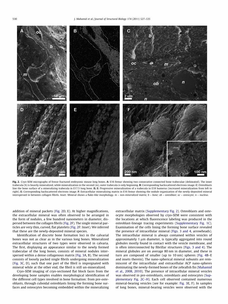
Fig. 2. Cryo-SEM micrographs of freeze fractured embryonic mouse long bones. A: E16 femur showing two consecutive connected bone trabeculae (delineated). The innertrabecula (b) is heavily mineralized, while mineralization in the second (m), outer trabecula is only beginning. B: Corresponding backscattered electrons image. C: Osteoblastsline the bone surface of a mineralizing trabecula in E17.5 long bone. D, E: Progressive mineralization of a trabecula in E18 humerus (increased mineralization from left toright). E: Corresponding backscattered electrons image. F: Extracellular mineralizing matrix in E16 femur showing the nodule organization of the newly-deposited mineralinterspersed in between collagen fibrils. Inset: Mineral shows a flake-like morphology. m – non-mineralized matrix; b – bone; ob – osteoblast; oc – osteocyte; n – nucleus.
530 J. Mahamid et al. / Journal of Structural Biology 174 (2011) 527–535
addition of mineral packets (Fig. 2D, E). At higher magnifications,the extracellular mineral was often observed to be arranged inthe form of nodules, a few hundred nanometers in diameter, dis-persed between the collagen fibrils (Fig. 2F). The single mineral par-ticles are very thin, curved, flat platelets (Fig. 2F: Inset). We inferredthat these are the newly-deposited mineral species.
Identification of discrete bone formation loci in the calvarialbones was not as clear as in the various long bones. Mineralizedextracellular structures of two types were observed in calvaria.The first, displaying an appearance similar to the newly formedtrabeculae of the long bones, consists of mineral nodules inter-spersed within a dense collagenous matrix (Fig. 3A, B). The secondconsists of loosely packed single fibrils undergoing mineralization(Fig. 3C, D), such that one part of the fibril is impregnated withmineral while at the other end, the fibril is still un-mineralized.
Cryo-SEM imaging of cryo-sectioned flat block faces from thedeveloping bone samples enables morphological identification ofthe different cell types involved in bone formation: from pre-oste-oblasts, through cuboidal osteoblasts lining the forming bone sur-faces and osteocytes becoming embedded within the mineralizing
extracellular matrix (Supplementary Fig. 2). Osteoblasts and oste-ocyte morphologies observed by cryo-SEM were consistent withthe locations at which fluorescence labeling was produced in theosteoblast-lineage tracing experiments (Supplementary Fig. 1C).Examination of the cells lining the forming bone surface revealedthe presence of intracellular mineral (Figs. 3 and 4, arrowheads).The intracellular mineral is always contained within vesicles ofapproximately 1 lm diameter, is typically aggregated into roundglobules mostly found in contact with the vesicle membrane, andis often interconnected by fibrillar structures (Figs. 3 and 4). Themineral globules are on average 80 nm in diameter, and these inturn are composed of smaller (up to 10 nm) spheres (Fig. 4B–Eand insets therein). The nano-spherical mineral subunits are rem-iniscent of the intracellular and extracellular ACP nano-spheresdominating the newly-formed bones of the zebrafish fin (Mahamidet al., 2008, 2010). The presence of intracellular mineral vesicleswas observed in pre-osteoblasts, osteoblasts and osteocytes (Sup-plementary Fig. 2C–H). Each cell observed contained numerousmineral-bearing vesicles (see for example: Fig. 3E, F). In samplesof long bones, mineral-bearing vesicles were observed with the

Fig. 3. Cryo-SEM micrographs of freeze fractured (A–D) and cryo-sectioned (E, F) embryonic mouse neonatal calvarial bones. Right panel are the corresponding backscatteredelectrons images. A, B: E17 calvaria showing an extracellular mineralized matrix (b) and intracellular mineral vesicles (arrowheads) in a cell process adjacent to the bonematrix. The bone matrix is impregnated with mineral nodules. C, D: E17 calvaria showing an extracellular loose mineralized matrix (b) and intracellular mineral vesicles(arrowheads). E, F: P1 calvaria showing a dense extracellular mineralized matrix (b) and numerous mineral-containing vesicles (arrowheads) in a cell lining the bone. Highermagnification of the same vesicles is shown in Fig. 4. B, C. The cell’s circumference is clearly delineated by a membrane (highlighted in F). Lineation observed in the cryo-sectioned block face is a sectioning artifact.
J. Mahamid et al. / Journal of Structural Biology 174 (2011) 527–535 531
same characteristic as in calvaria (Fig. 4B, D), but their occurrencewas more sporadic.
Frozen thin sections obtained from equivalent E17 and P1 cal-varial tissue preparations, observed in cryo-TEM, showed that in-deed the bone lining cells often contain electron dense,membrane-bound mineral globules (Fig. 5A: arrowhead, B). Se-lected area electron diffraction of the vesicles resulted only in anamorphous diffuse scatter (Fig. 5G); whereas the adjacent fullymineralized extracellular matrices (Fig. 5A (b)) gave a clear crystal-line hydroxyapatite diffraction pattern (Fig. 5F). Extracellular ma-trix mineral found at the interface between the mineralized andnon-mineralized matrix displayed a thin curved appearance(Fig. 5E). Surprisingly, the mineral produced a diffuse scattering
diffraction pattern consisting of rings around d-spacings expectedfor the strong (0 0 2), (2 1 1), (1 1 2) and (3 0 0) reflections of crys-talline hydroxyapatite. This diffraction pattern is indicative of thepresence of short or medium range order (Fig. 5H).
In order to further characterize the mineral phases observed inthe cryo-preparations, we subjected cryo-sections to freeze-dryingto enable TEM analysis at room temperature. The freeze-dried sec-tions were not completely intact. However areas of interest werestructurally stable enough for imaging and spectroscopic measure-ments (Fig. 5C and Supplementary Fig. 3). Selected area electrondiffraction and elemental analysis by EDS were taken from thesame positions in bone mineralized matrix, in intracellular mineraldeposits and in cellular tissue for background evaluation. Electron

Fig. 4. Cryo-SEM micrographs of cryo-sectioned neonatal calvaria (A–C) and freeze fractured embryonic long bone (D, E). C and E are backscattered electrons imagescorresponding to B and D. A: Cells juxtaposed to a mineralizing bone trabeculae (b) with multiple intracellular mineral-containing vesicles (arrowheads). Nuclei (n) are clearlydelineated by double membranes. B, C: Two intracellular mineral-containing vesicles in a cell process lining a mineralizing bone matrix. Inset: higher magnification ofmineral globules composed of smaller nano-spheres. The micrographs are higher magnification from Fig. 3. E F. D, E: E18 humerus showing an intracellular mineral-containing vesicle. Inset: higher magnification of a mineral globule. The mineral globules are often membrane-bound.
532 J. Mahamid et al. / Journal of Structural Biology 174 (2011) 527–535
diffraction of the intracellular mineral consistently produced a dif-fuse amorphous scatter, even after drying, and retained the appear-ance of nano-spherical amorphous mineral (Fig. 5D andSupplementary Fig. 3). The mineralized matrix of the bone trabec-ulae produced characteristic apatite diffraction patterns. Averagevalues for calcium/phosphate ratios calculated from the measuredatomic percentages were 1.58 ± 0.16 for the mineralized extracel-
lular matrix; 0.75 ± 0.22 for the intracellular mineral-containingvesicles and 0.19 ± 0.16 for the background in the cellular tissue.The values obtained for the extracellular mineral, although lowerthan the expected 1.67 ratio for stoichiometric hydroxyapatite(Christoffersen et al., 1989), are within the range characteristic ofmature bone mineral (Kuhn et al., 2008). Surprisingly, the valueobtained for the intracellular vesicle-bound mineral, 0.75 ± 0.22,

Fig. 5. TEM micrographs of cryo-sections of mouse calvarial bones. A: Cryo-TEM of a vitrified thin section from P1 mouse calvaria showing an extracellular mineralizedmatrix (b) and an intracellular mineral vesicle (arrowhead) in a cell adjacent to the mineralizing bone matrix. Black arrow points to cryo-sectioning linear artifacts. B: Highermagnification of the mineral-containing vesicle in A, showing a number of high electron density, membrane-bound globules. C–E: TEM of a freeze-dried cryo-section fromE17 mouse calvaria. C: Extracellular mineralized matrix (b) and numerous intracellular mineral vesicles (arrowheads) in a cell adjacent to the bone matrix. The cell nucleus isclearly visible. Black arrow points to the holey-carbon film support. D: Higher magnification of part of a mineral-containing vesicle, showing a number of high electrondensity mineral particles, composed of smaller spheres. E: Peripheral newly-deposited extracellular mineral, with highly curved morphologies. F: Selected area electrondiffraction of mineral, corresponding to the area marked by (b) in A. Arrows point to the (0 0 2) reflections of hydroxyapatite. G: Selected area electron diffraction ofintracellular mineral vesicle in A and B. H: Selected area electron diffraction of peripheral extracellular mineral shown in E.
J. Mahamid et al. / Journal of Structural Biology 174 (2011) 527–535 533
is far from the expected value for synthetic amorphous calciumphosphate (namely around 1.5) (Christoffersen et al., 1989).
4. Discussion
Here we show in vivo in a mammalian mouse model that priorto mineralization, cells lining the forming bones, including pre-osteoblasts, osteoblasts and osteocytes, concentrate ions intracel-lularly and produce a membrane-bound disordered calcium-phos-phate phase. The mineral is found in the form of 80 nm granules,which are in turn composed of smaller spherical subunits. We sug-
gest that the mineral-containing vesicles are directly involved ininitiating extracellular mineralization and that the disordered cal-cium-phosphate is a precursor of carbonated hydroxyapatite.
Individual osteoblasts present on surfaces of bone where activebone formation takes place are known to tightly adhere to eachother and form a continuous cell layer (Aaron, 1973; Caplan,1987; Gay et al., 2000). This layer has long been suggested to limitdirect access of ions from the body fluids to the mineralizing bonesurface, and thus the mineral ions must pass through the bone-lin-ing cells (Gay et al., 2000). Our observations clearly demonstrate anactive role of the osteoblast-lineage derived cells in sequesteringions and creating intracellular ‘reservoirs’ of the mineral precursor.

534 J. Mahamid et al. / Journal of Structural Biology 174 (2011) 527–535
These findings are in agreement with previous electron microscopystudies on osteoblasts, both in culture (Bordat et al., 1998, 2004;Rohde and Mayer, 2007) and from frozen tissue preparations (Car-ter et al., 1997; Gay and Schraer, 1975), showing intracellular com-partments containing mineral deposits. Using osmium vaporstaining, Gay and Schraer (1975) concluded that the mineral con-taining compartments are mitochondria, reinforcing previous re-ports of ‘mitochondrial mineralization’ (reviewed in (Lehninger,1970)). Our observations of the mineral-containing vesicles donot show resemblance to the typical appearance of mitochondria,which are easily identified in cryo-SEM (Supplementary Fig. 4).
We suggest that osteoblasts directly secrete the precursor min-eral into the extracellular mineralization site. We were not how-ever able to identify the process of translocation of theintracellular mineral into the extracellular mineralizing matrix. Ina study on osteoblast cell culture, Rohde and Mayer (2007) showedthat intracellular mineral may be translocated to the extracellularenvironment directly by exocytosis.
Non-crystalline calcium phosphates are highly metastable min-eral phases in water and unless stabilized by organic or inorganicadditives or sealed from the surrounding solution, they quicklyconvert into the thermodynamically more stable carbonatedhydroxyapatite crystalline phase (Posner et al., 1978). Indeed, inthe extracellular matrix, inhibition over nucleation is somehowlost and crystallization occurs, giving rise to a mature mineralizedbone matrix.
The amorphous mineral within the intracellular vesicles con-tains considerably less calcium than expected for an ACP phase.This suggests that the intracellular vesicles serve as a confinedspace in which relatively high concentrations of phosphate entitiesare formed. These could in part be in the form of phospholipidsthat compose the vesicle membrane, which were shown in vitroto complex calcium and inhibit crystallization of the forming min-eral (Wuthier and Eanes, 1975). Alternatively, the high phosphateconcentration may be accounted for by the presence of other or-ganic phosphates or polyphosphate. Polyphosphates form strongcomplexes with divalent cations such as calcium, and can lead toa high local accumulation of total phosphate and calcium that ex-ceeds the supersaturation limit of apatite without triggering thespontaneous precipitation of apatite crystals (Omelon and Gryn-pas, 2008). Such complexes, exhibiting Ca/P ratios of approxi-mately 0.5, were recently suggested to be present at the sites ofnew bone formation (Omelon et al., 2009).
We previously reported that amorphous calcium phosphate ispresent in the forming fin bones of zebrafish, where it functionsas a precursor for the mature crystalline mineral (Mahamid et al.,2008, 2010). Following recent findings from studies performedin vitro it appears that the amorphous nature assists infiltrationof the mineral into the confined mineralization spaces within thecollagen fibrils, where it eventually crystallizes (Nudelman et al.,2010; Olszta et al., 2007). Here we confirm that an amorphousmineral precursor phase occurs in mammalian bone and thus con-tributes to the mechanistic understanding of the involvement ofosteoblasts in bone mineralization.
There are several differences in the mineralization processes be-tween the adult zebrafish fin bones and the developing mousebones. Intracellular mineral vesicles observed within bone-liningcells were completely filled with a dense granulated mineral inzebrafish, but the mineral does not appear to be organized into80 nm individual granules within a vesicle as in the mouse bones.Additionally, we observed a curved flake-like mineral form in theextracellular matrix of the developing mouse bones, which didnot produce a crystalline diffraction pattern. The fact that the min-eral particles do not have spherical shapes, but are rather flat andcurved, is reminiscent of the ACP2 phase described by Christoffer-sen et al. (1989), suggesting that the transformation of the intracel-
lular phosphate-rich amorphous phase into the mature crystallinephase may involve additional intermediates. Interestingly, also incalcium carbonate-based mineralization two distinct amorphousphases were observed, representing progressive stages of theamorphous precursor transformation toward the final crystallinephase (Politi et al., 2006, 2008). The morphological differences be-tween mineralization in the two animal models may be a conse-quence of different phase transformation kinetics possibly due tothe differences in body temperature (ambient water temperaturevs. 37 �C). Alternatively, the differences may be related to differentregulatory pathways employed during mature bone growth versusde novo embryonic bone formation.
Deposition of pre-packaged particles of a disordered precursorphases is advantageous for the biological system, insofar as thismechanism facilitates transport of material into the newly formedmatrix and avoids elimination of large amounts of water. Addition-ally, the mineral is provided in a form that can penetrate into theinterior of the collagen fibrils, where it subsequently crystallizes.On the other hand, the finding of intracellular amorphous mineraldeposits only shifts the fundamental questions of how the ions areconcentrated, transiently stabilized as an amorphous phase andpackaged, one step backward – to the intracellular componentsand subcellular compartments, the identity and the function ofwhich still need to be clarified.
5. Conclusions
In this study we extend and expand upon our understanding offish fin bone mineralization mechanisms to mineralization pro-cesses in forming mouse bones. The latter is widely accepted as aleading mammalian model of bone formation. Using cryo-SEMand cryo-TEM we found that in vivo bone-lining cells concentratea non-crystalline calcium phosphate phase within intracellularvesicles that has a surprisingly low Ca/P ratio. These observationshighlight the role of the osteoblasts as being directly responsiblefor the deposition of the first mineral phase in the broader contextof regulating the whole bone formation process.
Acknowledgments
We thank Dr. Vlad Brumfeld for his assistance with fluorescenceand l-CT imaging. The electron microscopy studies were con-ducted at the Irving and Cherna Moskowitz Center for Nano andBio-Nano Imaging, Weizmann Institute of Science. L.A. is theincumbent of the Dorothy and Patrick Gorman Professorial Chairof Biological Ultrastructure, and S.W. is the incumbent of the Dr.Trude Burchardt Professorial Chair of Structural Biology. J.M. wassupported by the Israeli Council for Higher Education. This workwas supported by the Kimmelman Center for Biomolecular Struc-ture and Assembly, Weizmann Institute.
Appendix A. Supplementary data
Supplementary data associated with this article can be found, inthe online version, at doi:10.1016/j.jsb.2011.03.014.
References
Aaron, J.E., 1973. Osteocyte types in the developing mouse calvarium. Calcif. TissueRes. 12, 259–279.
Ali, S.Y., Sajdera, S.W., Anderson, H.C., 1970. Isolation and characterization ofcalcifying matrix vesicles from epiphyseal cartilage. Proc. Natl Acad. Sci. USA 67,1513–1520.
Anderson, H.C., Garimella, R., Tague, S.E., 2005. The role of matrix vesicles in growthplate development and biomineralization. Front. Biosci. 10, 822–837.
Bordat, C., Bouet, O., Cournot, G., 1998. Calcium distribution in high-pressure frozenbone cells by electron energy loss spectroscopy and electron spectroscopicimaging. Histochem. Cell Biol. 109, 167–174.

J. Mahamid et al. / Journal of Structural Biology 174 (2011) 527–535 535
Bordat, C., Guerquin-Kern, J., Lieberherr, M., Cournot, G., 2004. Direct visualizationof intracellular calcium in rat osteoblasts by energy-filtering transmissionelectron microscopy. Histochem. Cell Biol. 121, 131–138.
Caplan, A.I., 1987. Bone development and repair. BioEssays 6, 171–175.Carter, D.H., Hatton, P.V., Aaron, J.E., 1997. The ultrastructure of slam-frozen bone
mineral. Histochem. J. 29, 783–793.Christoffersen, J., Christoffersen, M.R., Kibalczyc, W., Andersen, F.A., 1989. A
contribution to the understanding of the formation of calcium phosphates. J.Cryst. Growth 94, 767–777.
Crane, N.J., Popescu, V., Morris, M.D., Steenhuis, P., Ignelzi, M.A., 2006. Ramanspectroscopic evidence for octacalcium phosphate and other transient mineralspecies deposited during intramembranous mineralization. Bone 39, 434–442.
Currey, J.D., 2002. Bones: Structure and Mechanics. Princeton University Press,Oxford, UK.
Dacquin, R., Starbuck, M., Schinke, T., Karsenty, G., 2002. Mouse alpha1(I)-collagenpromoter is the best known promoter to drive efficient Cre recombinaseexpression in osteoblast. Dev. Dyn. 224, 245–251.
Engleka, K.A., Gitler, A.D., Zhang, M., Zhou, D.D., High, F.A., Epstein, J.A., 2005.Insertion of Cre into the Pax3 locus creates a new allele of Splotch and identifiesunexpected Pax3 derivatives. Dev. Biol. 280, 396–406.
Gay, C.V., Schraer, H., 1975. Frozen thin-sections of rapidly forming bone: Bone cellultrastructure. Calcif. Tissue Res. 19, 39–49.
Gay, C.V., Gilman, V.R., Sugiyama, T., 2000. Perspectives on osteoblast and osteoclastfunction. Poult. Sci. 79, 1005–1008.
Glimcher, M.J., 1984. Recent studies of the mineral phase in bone and its possiblelinkage to the organic matrix by protein-bound phosphate bonds. Philos. Trans.R. Soc. B-Biol. Sci. 304, 479–508.
Jahnen-Dechent, W., 2000. Lot’s wifes problem revisited: How we preventpathological calcification. In: Bäuerlein, E. (Ed.), Biomineralization: Progress inBiology, Molecular Biology and Application. Wiley-VCH, Weinheim, pp. 243–282.
Kaufman, M.H., 1994. The Atlas of Mouse Development. Elsevier Academic Press,London.
Kuhn, L.T., Grynpas, M.D., Rey, C.C., Wu, Y., Ackerman, J.L., Glimcher, M.J., 2008. Acomparison of the physical and chemical differences between cancellous andcortical bovine bone mineral at two ages. Calcif. Tissue Int. 83, 146–154.
Landis, W.J., Song, M.J., McEwen, L.A.L., McEwen, B.F., 1993. Mineral and organicmatrix interaction in normally calcifying tissue visualized in three dimensionsby high voltage electron microscopic tomography and graphic imagereconstruction. J. Struct. Biol. 110, 39–54.
Lehninger, A.L., 1970. Mitochondria and calcium transport. Biochem. J. 119, 129–138.
Lowenstam, H.A., Weiner, S., 1989. On Biomineralization. Oxford University Press,New York.
Mahamid, J., Sharir, A., Addadi, L., Weiner, S., 2008. Amorphous calcium phosphateis a major component of the forming fin bones of zebrafish: Indications for anamorphous precursor phase. Proc. Natl Acad. Sci. USA 105, 12748–12753.
Mahamid, J., Aichmayer, B., Shimoni, E., Ziblat, R., Li, C., Siegel, S., Paris, O., Fratzl, P.,Weiner, S., Addadi, L., 2010. Mapping amorphous calcium phosphate
transformation into crystalline mineral from the cell to the bone in zebrafishfin rays. Proc. Natl Acad. Sci. USA 107, 6316–6321.
Nudelman, F., Pirterse, K., George, A., Bomans, P.H.H., Friedrich, H., Brylka, L.J.,Hilbers, P.A.J., de With, G., Sommerdijk, N.A.J.M., 2010. The role of collagen inbone apatite formation in the presence of hydroxyapatite nucleation inhibitors.Nat. Mater. 9, 1004–1009.
Olszta, M.J., Cheng, X.G., Jee, S.S., Kumar, R., Kim, Y.Y., Kaufman, M.J., Douglas, E.P.,Gower, L.B., 2007. Bone structure and formation: A new perspective. Mater. Sci.Eng. R 58, 77–116.
Omelon, S., Georgiou, J., Henneman, Z.J., Wise, L.M., Sukhu, B., Hunt, T., Wynnyckyj,C., Holmyard, D., Bielecki, R., Grynpas, M.D., 2009. Control of vertebrate skeletalmineralization by polyphosphates. PLoS ONE 4, e5634.
Omelon, S.J., Grynpas, M.D., 2008. Relationships between polyphosphate chemistry,biochemistry and apatite biomineralization. Chem. Rev. 108, 4694–4715.
Peck, W.A., Woods, W.L., 1988. The cells of bone. In: Riggs, L., Melton, L.J. (Eds.),Osteoporosis: Etiology, Diagnosis and Management. Raven Press, New York (pp.1–44).
Politi, Y., Levi-Kalisman, Y., Raz, S., Wilt, F., Addadi, L., Weiner, S., Sagi, I., 2006.Strucutural characterization of the transient calcium carbonate amorphousprecursor phase in sea urchin embryos. Adv. Funct. Mater. 16, 1289–1298.
Politi, Y., Metzler, R.A., Abrecht, M., Gilbert, B., Wilt, F.H., Sagi, I., Addadi, L., Weiner,S., Gilbert, P.U.P.A. , 2008. Transformation mechanism of amorphous calciumcarbonate into calcite in the sea urchin larval spicule. Proc. Natl Acad. Sci. USA105, 20045.
Posner, A.S., Betts, F., Blumenthal, N.C., 1978. Properties of nucleating systems. Me.Bone Dis. & Rel. Res. 1, 179–183.
Rohde, M., Mayer, H., 2007. Exocytotic process as a novel model for mineralizationby osteoblasts in vitro and in vivo determined by electron microscopic analysis.Calcif. Tissue Int. 80, 323–336.
Streeter, G.L., 1949. Developmental horizons in human embryos; a review of thehistogenesis of cartilage and bone. Contrib. Embryol. 33, 149–168.
Traub, W., Arad, T., Weiner, S., 1989. Three dimensional ordered distribution ofcrystals in turkey tendon collagen fibers. Proc. Natl Acad. Sci. USA 86, 9822–9826.
Veis, A., Perry, A., 1967. The phosphoprotein of the dentin matrix. Biochemistry 6,2409–2416.
Weiner, S., 2008. Biomineralization: A structural perspective. J. Struct. Biol. 163,229–234.
Weiner, S., Traub, W., 1992. Bone structure: From angstroms to microns. FASEB J. 6,879–885.
Weiner, S., Wagner, H.D., 1998. The material bone: Structure–mechanical functionrelations. Annu. Rev. Mater. Sci. 28, 271–298.
Weiner, S., Sagi, I., Addadi, L., 2005. Choosing the path less travelled. Science 309,1027–1028.
Weiner, S., Mahamid, J., Politi, Y., Ma, Y., Addadi, L., 2009. Overview of theamorphous precursor phase strategy in biomineralization. Front. Mater. Sci.,China 3, 104–108.
Wuthier, R.E., Eanes, E.D., 1975. Effect of phospholipids on the transformation ofamorphous calcium phosphate to hydroxyapatite in vitro. Calcif. Tissue Res. 19,197–210.