Targeted Nanoparticles That Deliver a Sustained, Specific Release of Paclitaxel to Irradiated Tumors
Upload
independentCategory
view
0download
0
CELL TO CELL I N T E R A C T I O N I N T H E I M M U N E RESPONSE
I I I . CHgOMOSO~AL MARKER ANALYSIS OF SINGLE ANTIBODY-FoRMING CELLS IN RECONSTITUTED, IgRADIAT~D, OR ~ C T O M I Z F _ ~ MICE*,
BY G. J. v. NOSSAL, F.A.A., A. CUNNINGHAM, PH.D., G. F. MITCHELL, J. F. A. P. MILLER, M.B.
(From The Walter and Eliza Hall Institute of Medical Research, Melbourne, Australia)
(Received for publication 3 June 1968)
In the previous two papers of this series (1, 2) evidence has been presented for an interaction between two types of lymphoid cells in mice forming antibody to sheep erythrocytes. To show this interaction, it was necessary to use adoptive transfer models of three basic types: (a) neonatally thymectomized mice, restored with either thymus cells or thoracic duct lymphocytes; (b) lethally irradiated mice, restored with mixtures of bone marrow cells and thoracic duct lymphoeytes; and (c) adult thymecto- mized lethally irradiated hosts protected with bone marrow and injected 2 wk later with thymus cells or thoracic duct lymphoeytes. In the first and third models, allo- geneic as well as syngeneie cell transfers were effective and thus isoantisera could be used to identify the cellular origin of the actual antibody-forming cells. In both cases, most of the plaque formers were derived not from thymus cells or thoracic duct lym- phoeytes, though these were needed in the reaction, but from some host cell, probably a lymphoid or plasma cell precursor of bone marrow origin. In the second model, syngeneic bone marrow cells greatly augmented the adoptive response obtained with thoracic duct lymphoeytes but allogeneic combinations of cells failed to interact. Therefore, isoantisera could not be used to trace cell origins. As the heavily irradiated animal is the standard tool in adoptive transfer work, identification of the source of antibody formers in this model would be of particular value.
In the present paper, we describe two new methods by which a chromosomal squash can be obtained from single cells by micromanipulation. This has been applied to a study of single cells forming antibody against either Salmonella addaide-polymerlzed flagellin or SRBC. Use has been made of the fully histo- compatible strains CBA and CBA/T6T6 developed at Harwell, and the tech- nique has identified the origins of antibody-forming cells in a variety of transfer situations. The general conclusions of the previous papers have been substan- tiated.
* This is publication 1249 from The Walter and Eliza Hall Institute of Medical Research. Supported by the National Health and Medical Research Council of Australia, the
National Institute of Allergy and Infectious Diseases (AI-0-3958), and the United States Atomic Energy Commission AT-D-3695).
839
on March 18, 2013
jem.rupress.org
Dow
nloaded from
Published October 1, 1968
840 CELL TO CELL INTERACTION I N IMMUNE RESPONSE. I I I
Materials and Methods
Animals.--The mice used were CBA and CBA/T6T6 originally obtained through the courtesy of Dr. C. E. Ford, Radiobiological Research Unit, Harwell, and bred at the Hall Institute by strict stem-line mating. Frequent checks by skin grafting showed the two lines to be fully hlstocompafible. In irradiation experiments, both donors and recipients were aged 8 wk when used. Recipients in neonatal thymectomy experiments were 4-6 wk of age.
Chromosome Analysis of Spleen Cell Suspensions.--Mice were given 4 #g/g body weight of colcemid 2 hr (or in some early experiments 4 hr) before killing. Chromosome analysis of spleen cell suspensions was performed in a proportion of the experiments using the method of Ford (3). The cells were spread by an air-drying procedure and stained with aceto-orcein.
Irradiation and Cell Transfer Techniques.--These were exactly as described in the accom- panying papers (i, 2).
[mmunization.--Mice received either 10 /zg of polymerized flagellin from Salmonella adda/de (4) or l0 s sheep erythrocytes intravenously. They were killed 5-7 days after antigen injection.
ldentificaffon of Single Antibody-Forming Cdls.-- Anti-Salmondla antibody formers were detected by the method of bacterial immunocyto-
adherence (5), which in this system is a valid and sensitive test of antibody formation. Briefly, motile bacteria of the Salmonella derby strain SW 721, which shares the H antigen fg but not the O antigen with S. aldda/de (SW 1338) were mixed with well-washed spleen cells in a micromanipulation chamber. Certain cells rapidly became surrounded by a corona of adherent bacteria. These could be identified at 200-fold magnification (phase contrast)and subjected to micromanipulation.
Anti-SRBC antibody formers: Cells forming hemolytic (presumably IgM) antibody against sheep erythrocytes were identified by the method of Cuuningham (6). Spleen cells were washed in cold Eisen's medium (7) containing 10% fetal calf serum and suspended in the same medium at a final concentration of circa 10°/ml. To this were added sheep erythrocytes (final concentration 1% cells, v/v), guinea pig serum (final concentration 5% v/v), and colce- mid (4/~g/mi) to maintain metaphase arrest. Multiple rows of droplets (circa 1/~1 in volume) of this mixture were placed on glass slides under paraffin oil The cells rapidly settled to form a stable monolayer. Batches of 10-20 slides each containing circa 30 droplets were placed on trays into a 37°C incubator for 20 rain. The slides were then placed on the stage of a micro- scope, and with adequate transillumination plaques of hemolysis could readily be seen by the naked eye, and rapidly checked at 100- to 200-fold magnification. This allowed assessment of the size of the antibody-forming cell (PFC) at the center of the plaque. Appropriate ceils were removed from the centers of plaques by micromanipulation.
Choice of Single Antibody-Forming Cells for Chromosome Analysis.--Antibody-forming cells always arise through antigen-induced mitotic division in precursor cells (5, 8) and a stage exists when a cell can simultaneously divide and form antibody. In any population of anti- body-forming cells taken a day or so before the peak of an immune response, there will be a small but significant proportion of cells in mitosis. Through the use of colcemid, which arrests cells in metaphase, this can be considerably increased. In our studies, we endeavored to identify antibody-forming cells in mitosis and to concentrate micromanipulatory study on these. At 200-fold magnification of lymphoid cells in suspension, this was not consistently successful. Unless the cells were artificially flattened, the degree of visualization of structural detail was poor. Cell size was a good guide--the larger the cell, the more likely it was to be a mitotic figure. In a proportion of the cells, vague outlines of chromosomes could be seen giving an effect of ribbing which was highly characteristic and allowed near certain diagnosis of the cell as a mitotic figure. However, frequently this ribbing was not dearly evident, yet the cell turned out to be a mitotic figure. To a degree, our suceess in picking out only mitotic cells
on March 18, 2013
jem.rupress.org
Dow
nloaded from
Published October 1, 1968
NOSSAL~ CUNNINGHAM~ MITCHELL~ AND MILLER 841
for subsequent squashing depended on the intensity of the adoptive immune response. Where there were plenty of antibody formers, one third to one half of the cells selected for study were actually in mitosis. If antibody formers were infrequent, we became less selective and picked out any cell that was slightly larger than the median size of the population, and natur- ally the proportion of mitotic figures obtained was much smaller. In general terms, we found it possible to obtain adequate numbers of mitotic cells from suspensions in which the fre- quency of antibody formers was greater than 1:10,000.
Mioromanipulatory Methods for Obtainia~g Single Call--Chromosome Spreads.-- Desoxycholate method: This technique was used for all of the experiments in the Salmonella
system and most in the SRBC system. I t utilizes the fact that lymphoid cells in microdroplets under paraffin oil burst readily when suspended in serum-free medium, particularly in the presence of low concentrations of detergents. After considerable trial and error, the following conditions were chosen. A single antibody-forming cell believed to be a mitotic figure was removed from its microdrop through a micropipette. In the Salmonella system, the next step was to remove adherent bacteria by vigorousiy drawing the cell in and out through the micro- pipette orifice, subjecting it to shearing stresses which removed the bulk of adherent material. Subsequent steps were similar in both systems. Each cell was washed three times in Eisen's medium containing 1:2000 (v/v) fetal calf serum and 1:10,000 (w/v) sodium desoxycholate. This solution was made up fresh immediately before each experiment. Lymphoid cells are rendered fragile within less than a minute of exposure at room temperature to this medium. The final, critical step was to place the cell, still in the desoxycholate medium, in a small (circa. 5 X 10 -~ ml) hanging microdroplet in the micromauipuiation oil chamber, and then, with a brisk movement, to suck back the bulk of the fluid in the droplet. This created a menis- cus force; the water-oil interface met the cell and forced it against the cover slip. This caused the cell membrane to rupture explosively, liberating the chromosomes which spread apart. These could be examined at 1250-fold magnification (phase contrast) without fixation or st~inlng. In successful cases, excellent spreads were obtained (Figs. 4-9). However, the method has several serious disadvantages. In about half the cases, rupture is not satisfactory, and the chromosomes remain dumped. Such cells must be discarded. Good preparations were not stable. After 2-5 rain, the chromosomes became "thready" and started to disintegrate. We have tried to fax preparations by jetting acetic acid, sometimes containing orcein, into the droplet but found that chromosomes looked worse rather than better after this treatment. However, with unfixed material the time available for observation was quite adequate to allow counting of chromosomes and detection of T6 markers ff these were present. Spreads could also be photographed, though this was not done as a routine in our study. Perhaps the chief disadvantage of the method is that it is very demanding, requiring a person with consider- able experience in micromauipuiation to perform the final step. However, we found that for such an investigator it was a substantially quicker and more convenient method than the method described below.
Mioro-Ford me2hod: This was essentially a microadaptation of the conventional technique (3). The cell was identified and washed in 1% sodium citrate under paraffin oil. Then, it was transferred to a premarked position on a new, clean cover slip not covered with oil, and the droplet in which the cell was contained was considerably enlarged with 1°7o sodium citrate to a final volume of 1-10 pa; i.e., some thousands of times larger than a microdroplet. The cover slip was then placed, right way up, in a moist Petri dish at room temperature for 10-15 min. The cell became moderately firmly adherent to the cover slip. Next, the cover slip was placed on the bottom of a 5 can diameter clean dry Petri dish, held at a slight angle, and observed under a dissecting microscopo at 50-fold magnification. Clarke's fixative (3 parts ethanol to 1 part glacial acetic acid, mixed just prior to use) was gently pipetted into one side of the dish, an amount of about 2 ml being delivered steadily over a period of about 20 sec. As the moving
on March 18, 2013
jem.rupress.org
Dow
nloaded from
Published October 1, 1968
842 CELL TO CELL INTERACTION IN ~ U N E RESPONSE. HI
boundary of the fixative encountered the droplet of citrate solution, the droplet was swept away. However, the cell frequently remained adherent to the glass and could be observed to flatten and spread as the fixative hit it. The cover slip was left in fixative for about 10 rain and then dipped in 40% acetic acid and warmed gently over a tiny gas flame to dry. I t was stained with iactic-acetic orcein. This yielded a stable, conventional preparation (Fig. 10) which was best examined under phase contrast a t 1250-fold magnification. Unfortunately, this method also has its limitations. A substantial proportion of the cells are swept away by the fixative. Sometimes these could be located elsewhere on the cover slip but more often they
u~ 4 LLI
8 O. O4
n" ILl O.
CO ILl ¢D 0 I--
I.L. 0 n- oJ rn
3 0 -
2 0
I0 '
. . 0
Q~
/ /
l /
/
0 ~ ~ ' 0 . . . . . . . . 0
DAYS AFTER IRRADIATION
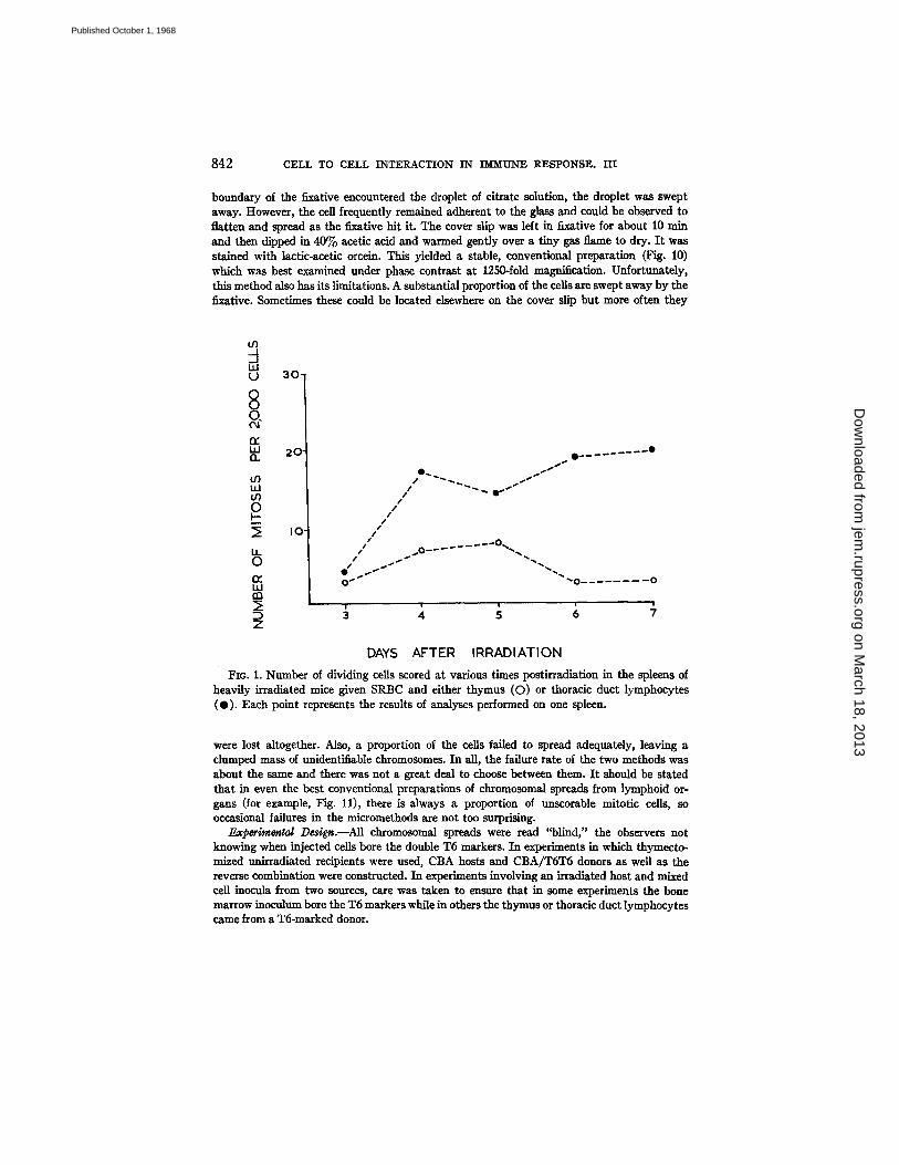
FIo. I. Number of dividing cells scored at various times postirradiation in the spleens of heavily irradiated mice given SRBC and either thymus (O) or thoracic duct lymphocytes (O). Each point represents the results of analyses performed on one spleen.
were lost altogether. Also, a proportion of the cells failed to spread adequately, leaving a clumped mass of unidentiiiable chromosomes. In all, the failure rate of the two methods was about the same and there was not a great deal to choose between them. I t should be stated that in even the best conventional preparations of chromosomal spreads from lymphoid or- gans (for example, Fig. 11), there is always a proportion of unseorable mitotic cells, so occasional failures in the micromethods are not too surprising.
Experimental Design.--AU chromosomal spreads were read "blind," the observers not knowing when injected cells bore the double T6 markers. In experiments in which thymecto- mized unitradiated recipients were used, CBA hosts and CBA/T6T6 donors as well as the reverse combination were constructed. In experiments involving an irradiated host and mixed cell inocula from two sources, care was taken to ensure that in some experiments the bone marrow inoculum bore the T6 markers while in others the thymus or thoracic duct lymphocytes came from a T6-maxked donor.
on March 18, 2013
jem.rupress.org
Dow
nloaded from
Published October 1, 1968
NOSSAL~ CUNNINGHA~, MITCHELL, AND MILLER 843
R E S U L T S
Cytological Analysis of the St~leeus of Heavily Irradiated Mice Injected with SRBC, Thymus, Thoracic Duct, and Bone Marrow Cells.--It was of interest to determine the proportion of mitotic figures of lymphocyte donor-type in irra- diated mice receiving thymus or thoracic duct lymphocytes together with
4 0 - _J
O
~- 30"
n
t t) u J o9 0 2 o I -
LL 0 n- I 0 ' i..iJ rn
Z
DAYS AFTER IRRADIATION
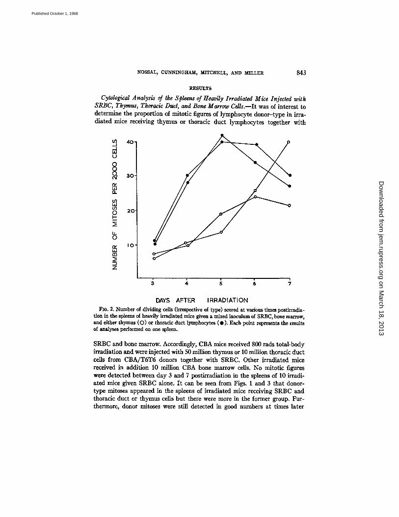
Fxo. 2. Number of dividing cells (irrespective of type) scored at various times postirradia- tion in the spleens of heavily irradiated mice given a mixed inoculum of SRBC, bone marrow, and either thymus (O) or thoracic duct lymphocytes ( • ) . Each point repre-~nts the results of analyses performed on one spleen.
SRBC and bone marrow. Accordingly, CBA mice received 800 rads total-body irradiation and were injected with 50 million thymus or 10 million thoracic duct cells from CBA/TST5 donors together with SRBC. Other irradiated mice received in addition 10 million CBA bone marrow cells. No mitotic figures were detected between day 3 and 7 postirradiation in the spleens of 10 irradi- ated mice given SRBC alone. I t can be seen from Figs. 1 and 3 that donor- type mitoses appeared in the spleens of irradiated mice receiving SRBC and thoracic duct or thymus cells but there were more in the former group. Fur- thermore, donor mitoses were still detected in good numbers at times later
on March 18, 2013
jem.rupress.org
Dow
nloaded from
Published October 1, 1968
844 CELL TO CELL INTERACTION IN IMMUNE R E S P O N S E . I I I
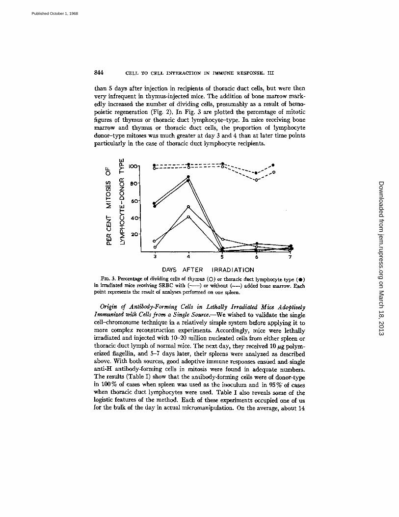
than 5 days after injection in recipients of thoracic duct cells, but were then very infrequent in thymus-injected mice. The addition of bone marrow mark- edly increased the number of dividing cells, presumably as a result of hemo- poietic regeneration (Fig. 2). In Fig. 3 are plotted the percentage of mitotic figures of thymus or thoracic duct lymphocyte-type. In mice receiving bone marrow and thymus or thoracic duct cells, the proportion of lymphocyte donor-type mitoses was much greater at day 3 and 4 than at later time points particularly in the case of thoracic duct lymphocyte recipients.
I.i.J
LL >. I 0 0 " 0 . . . . . . . . u . . . . ~ . . . ~ . . ~ . - ' " " 0
-- i 60"
I-- ~3 40' z 0 la.l
n. ~ 2o, 1.1.1 a
7
DAYS AFTER IRRADIATION
Fzo. 3. Percentage of dividing cells of thymus (O) or thoracic duct lymphocyte type (O) in irradiated mice receiving SRBC with ( ~ ) or without (----) added bone marrow. Each point represents the result of analyses performed on one spleen.
Origin of Antibody-Forming Cells in Lethally Irradiated Mice Adoptivdy Immunized with Cells from a Single Source.--We wished to validate the single cell-chromosome technique in a relatively simple system before applying it to more complex reconstruction experiments. Accordingly, mice were lethally irradiated and injected with 10-20 million nucleated cells from either spleen or thoracic duct lymph of normal mice. The next day, they received 10 ~g polym- erized flageUin, and 5-7 days later, their spleens were analyzed as described above. With both sources, good adoptive immune responses ensued and single anti-H antibody-forming cells in mitosis were found in adequate numbers. The results (Table I) show that the antibody-forming cells were of donor-type in 100 % of cases when spleen was used as the inoculum and in 95 %'of cases when thoracic duct lymphocytes were used. Table I also reveals some of the logistic features of the method. Each of these experiments occupied one of us for the bulk of the day in actual micromanipulation. On the average, about 14
on March 18, 2013
jem.rupress.org
Dow
nloaded from
Published October 1, 1968
NOSSAL, CUNNINGHAM, MITCHELL~ AND M I L L E R 845
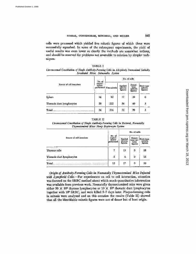
ceils were processed which yielded five mitotic figures of which three were successfully squashed. In some of the subsequent experiments, the yield of useful results was even lower so dearly the methods are somewhat tedious, and should be reserved for problems not amenable to solution by simpler tech- niques.
TABLE I Chromosomal Constitution of Single Antibody-Forming Cells in Adoptivdy Immunized Lethally
Irradiated Mice: Salmonella System
Source of cell inoculum
Spleen
Thoracic duct lymphocytes
T o t a l . . . . . . . . . . . . . . . . . . . . . . . . . . . . . . . . .
No. of experi- ments
performe
14
20
34
No. of cells
Non-mitotic
92
222
314
Spoiled mitotic figures
17
56
73
Donor- Host-ty.pe type mitotic mitotic
figures figures
39 0
6O 3
99 3
TABLE II Chromosomal Constitution of Single Antibody-Forming Cells in Restored, Neonatally
Thymectomized Mice: Sheep Erythrocyte System
Source of cell inoculum
Thymus cells
Thoracic duct lymphocytes
Total . . . . . . . . . . . . . . . . . . . . . . . . . . . . . . . . . . . . . . . . . . .
No. of experi- ments ~rforme~
7
5
12
No. of cells
Spoiled Donor- Host-type type ml- mitotic mitotic totic
figures figures figures
13 0 18
4 0 15
17 0 33
Origin of Antibody-Forming Cells in N eonatally Tkymectomized Mice Injected with Lymphoid Cdls.--For experiments on cell to cell interaction, attention was focused on the SRBC method about which much quantitative information was available from previous work. Neonatally thymectomlzed mice were given either 50 X 106 thymus lymphocytes or 10 X 106 thoracic duct lymphocytes together with l0 s SRBC, and were killed 5-7 days later. Plaque-forming cells in mitosis were analyzed and on this occasion the results (Table I I ) showed that all the identifiable mitotic figures were not of donor but of host origin.
on March 18, 2013
jem.rupress.org
Dow
nloaded from
Published October 1, 1968
846 CELL TO CELL INTERACTION IN ~UIq-E RESPONSE. HI
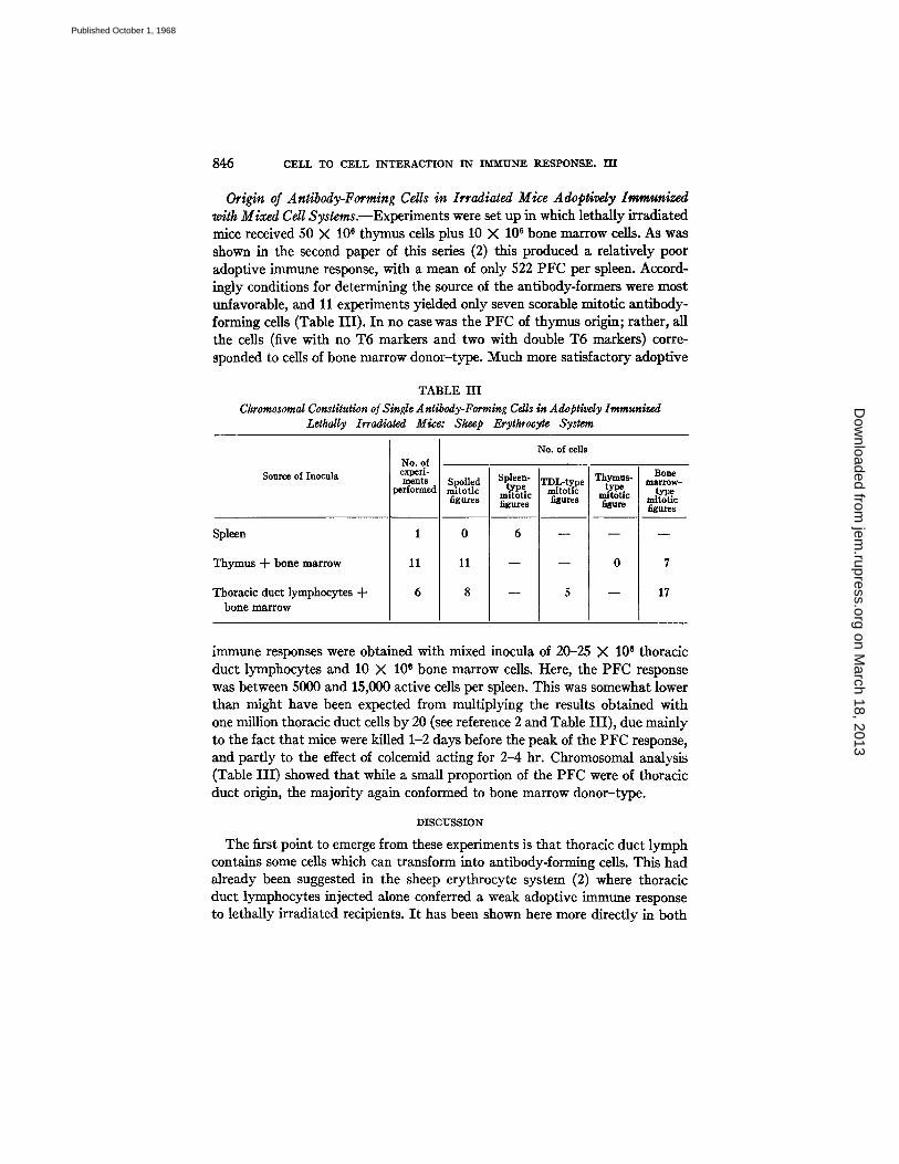
Origin of Antibody-Forming Cells in Irradiated Mice Adopgvdy Immunized with Mixed Cell Systems.--Experiments were set up in which lethally irradiated mice received 50 X 10 e thymus ceils plus 10 X 106 bone marrow cells. As was shown in the second paper of this series (2) this produced a relatively poor adoptive immune response, with a mean of only 522 PFC per spleen. Accord- ingly conditions for determining the source of the antibody-formers were most unfavorable, and 11 experiments yielded only seven scorable mitotic antibody- forming cells (Table III). In no case was the PFC of thymus origin; rather, all the cells (five with no T6 markers and two with double T6 markers) corre- sponded to cells of bone marrow donor-type. Much more satisfactory adoptive
TABLE I I I
Chromosomal Constitution o/Single Antibody-Forming Cells in Adoptivdy Immunized Lethally Irradiated Mice: Sheep Erythrocyte System
Source of Inocula
Spleen
Thymus + bone marrow
Thoracic duct lymphocytes -+- bone marrow
pe~ >" of emx ~erl-
~nts >rmed
No. of cells
Spoiled rmtotlc figures
o
11
8
Spleen- type
mitotic figures
6
rDL-t~pe mitot*c figures
Thymus- type
mitotic f~re
0
Bone marrow- type
mitotic figures
7
17
immune responses were obtained with mixed inocula of 20-25 X 106 thoracic duct lymphocytes and 10 X 106 bone marrow cells. Here, the PFC response was between 5000 and 15,000 active cells per spleen. This was somewhat lower than might have been expected from multiplying the results obtained with one million thoracic duct cells by 20 (see reference 2 and Table III), due mainly to the fact that mice were killed 1-2 days before the peak of the PFC response, and partly to the effect of colcemid acting for 2-4 hr. Chromosomal analysis (Table III) showed that while a small proportion of the PFC were of thoracic duct origin, the majority again conformed to bone marrow donor-type.
DISCUSSION
The first point to emerge from these experiments is that thoracic duct lymph contains some cells which can transform into antibody-forming cells. This had already been suggested in the sheep erythrocyte system (2) where thoracic duct lymphocytes injected alone conferred a weak adoptive immune response to lethally irradiated recipients. I t has been shown here more directly in both
on March 18, 2013
jem.rupress.org
Dow
nloaded from
Published October 1, 1968
NOSSAL, C~N'NING~M, MITCKELL, AND MILLER 847
the Salr~u~ndla (Table I) and SRBC (Table III) systems. The second and more important point is that this does not constitute the only role of thoracic duct lymphocytes. In the SRBC system, thoracic duct lymphocytes interacted synergistically with syngeneic bone marrow-derived cells in the lethally ir- radiated host. Chromosomal analysis showed that over three-quarters of the active cells in animals restored with mixed inocula of thoracic duct lympho- cytes and bone marrow carried the chromosomal marker of the bone marrow donor (Table III). This proportion of bone marrow-type cells is quite impres- sive, as the ratio of thoracic duct to bone marrow cells was 2 or 2.5:1. In some way, the thoracic duct lymphocytes had caused certain bone marrow cells to develop into antibody formers.
The subtle, indirect role of thoracic duct lymphocytes was equally evident in experiments on the reconstitution of immune reactivity in neonatally thy- mectomized animals. Here the results in the present, syngeneic system confirm totally conclusions already reached on the basis of immunoserologic analysis of an allogeneic system (1). Thoracic duct lymphocytes greatly enhance the antibody-forming potential of neonatally thymectomized mice against SRBC, but chromosomal analysis (Table II) shows that this is not through the simple phenomenon of direct transformation into antibody-formers that most workers would have predicted. Rather, the thoracic duct lymphocytes cause some host component, possibly a bone marrow-derived lymphoid cell, to form antibody. Viewed in their entirety, the present results lend strong support to the view already expressed in the preceding paper (2), that tho- racic duct lymphocytes contain both antigen-reactive cells (ARC) and anti- body-forming cell precursors (AFCP), with perhaps a predominance of the former. In view of this, it is not surprising that workers using large numbers of pure thoracic duct lymphocytes can obtain excellent adoptive immune responses with them. In such experiments, cell to cell interaction may well be occurring between heterogeneous elements contained in the one donor population.
The results obtained with thymic cells also fit well with those obtained in the accompanying papers (1, 2). Thymic lymphocytes were very effective in raising the plaque-forming ability of anfigenically challenged neonatally thy- mectomized recipients, though in no case (Table II) did the thymic cells them- selves transform into PFC. Thymic cells were much less effective in collabo- rating with bone marrow cells to achieve acute restoration of antibody-forming potential in lethally irradiated recipients. Nevertheless, they did allow rather feeble responses and chromosomal analysis showed that they did not transform into PFC, but caused bone marrow-derived cells to do so. The concordance of results in these syngeneic and entirely independent systems with conclusions reached in the previous papers is impressive. The thymus appears to contain a small proportion of ARC but no AFCP.
on March 18, 2013
jem.rupress.org
Dow
nloaded from
Published October 1, 1968
848 CELL TO CELL INTERACTION IN" IMMUNE RESPONSE. III
Analysis at the single cell level was necessarily limited to moderate numbers of mitotic figures. Accordingly, it is reassuring to note that the results of chro- mosomal analysis of the total spleen population (Figs. 1-3) fitted in well with the general picture presented here. Both thymus and thoracic duct lymphocytes exhibited a somewhat similar mitotic behavior soon after transfer with bone marrow to irradiated hosts challenged with SRBC. The proportion of dividing cells of lymphocyte donor-type was much higher at 3 4 days postirradiation than later. Since PFC appeared in significant numbers only after 5-6 days (2) and since the majority were of marrow donor-type (Table III), it can be pre- sumed that the cells involved in the early mitotic response were not the pro- genitors of antibody-forming cells but other cell types dividing in response to antigenic stimulation--presumably the antigen-reactive cells. In irradiated recipients not given bone marrow, dividing cells of thymus donor-type were virtually absent after 4 days, whereas cells of thoracic duct continued to divide. Such a behavior would be expected if thymus contained only ARC and tho- racic duct lymph contained both ARC and AFCP.
The new methods described for performing chromosomal analysis on a single cell are tedious but can approach problems not amenable to conven- tional techniques. As a general tool in tracing cell origins in adoptive transfer work, they are far less convenient than the use of isoantisera or allotypic specificity, which in many situations are adequate methods. These, however, are not applicable to experiments involving cell interactions which work only in syngeneic systems, such as the bone marrow-thymus or bone marrow- thoracic duct lymphocyte interactions noted in lethally irradiated acutely restored hosts. Moving away from immunology, it is possible that single cell chromosome analysis might prove a useful tool when one requires to know the origins of a cell of defined morphology in a heterogeneous population and only minor modifications of the present methods would make this practicable. Also, embryological experiments, e.g. on allophenic mice (9), might require methods where only a very small number of mitotic cells were available. I t is our hope that further improvements will see this development established as a useful general tool in cell biology.
SUMMARY
Two new methods are described for making chromosomal spreads of single antibodydorming cells. The first depends on the controlled rupture of cells in small microdroplets through the use of a mild detergent and application of a mechanical stress on the cell. The second is a microadaptation of the conven- tional Ford technique. Both methods have a success rate of over 50%, though the quality of chromosomal spreads obtained is generally not as good as with conventional methods.
These techniques have been applied to an analysis of cell to cell interaction
on March 18, 2013
jem.rupress.org
Dow
nloaded from
Published October 1, 1968
NOSSAL~ CUNNINGHAM, MITCHELL~ AND MILLER 849
in adoptive immune responses, using the full syngeneic transfer system pro- vided by the use of CBA and CBA/T6T6 donor-recipient combinations. When neonatally thymectomized mice were restored to adequate immune responsiveness to sheep erythrocytes by injections of either thymus ceils or thoracic duct lymphocytes, it was shown that all the actual dividing antibody- forming cells were not of donor but of host origin. When lethally irradiated mice were injected with chromosomally marked but syngeneic mixtures of thymus and bone marrow cells, a rather feeble adoptive immune response ensued; all the antibody-forming cells identified were of bone marrow origin. When mixtures of bone marrow cells and thoracic duct lymphocytes were used, immune restoration was much more effective, and over three-quarters of the antibody-forming mitotic figures carried the bone marrow donor chromo- somal marker.
The results were deemed to be consistent with the conclusions derived in the previous paper of this series, namely that thymus contains some, but a small number only of antigen-reactive cells (ARC), bone marrow contains antibody- forming cell precursors (AFCP) but no ARC, and thoracic duct lymph con- rains both ARC and AFCP with a probable predominance of the former. A vigorous immune response to sheep erythrocytes probably requires a collabora- tion between the two cell lineages, involving proliferation first of the ARC and then of the AFCP. The results stressed that the use of large numbers of pure thoracic duct lymphocytes in adoptive transfer work could lead to good adop- tive immune responses, but that such results should not be construed as evi- dence against cell collaboration hypotheses.
Some possible further uses of single cell chromosome techniques were briefly discussed.
We wish to thank Miss Heather Lewis and Miss Ruth Stone for expert technical assistance.
BIBLIOGRAPHY
1. Miller, J. F. A. P., and G. F. Mitchell. 1968. Cell to cell interaction in the immune response. I. Hemolysin-forming cells in neonatally thymectomized mice recon- stituted with thymus or thoracic duct lymphocytes. J. Expg. Med. 128:801.
2. Mitchell, G. F., and J. F. A. P. Miller. 1968. Cell to cell interaction in the immune response. II. The source of hemolysin-forming cells in irradiated mice given bone marrow and thymus or thoracic duct lymphocytes. J. Expg. Med. 128:821.
3. Ford, C. E. 1966. Appendix to Tissue Grafting and Radiation. H. S. Micklem and J. F. Loutit, editors. Academic Press, New York. 197.
4. Ada, G. L., G. J. V. Nossal, J. Pye, and A. Abbot. 1964. Antigens in immunity. I. Preparation and properties of flagellar antigens from Salmonella adelaide. Australian J. Exptl. Biol. Meal. Sci. 42:267.
5. Miikel~i, O., and G. J. V. Nossal. 1962. Autoradingraphic studies on the immune response. II. DNA synthesis amongst single antibody-producing cells. J. Exptl. Med. 115:231.
on March 18, 2013
jem.rupress.org
Dow
nloaded from
Published October 1, 1968
85O CELL TO CELL INTERACTION IN ~ RESPONSE. HI
6. Cunningham, A. J., J. B. Smith, and E. H. Mercer. 1966. Antibody formation by single cells from lymph nodes and efferent lymph of sheep. J. g, xptl. Med. 124:701.
7. Helmreich, E., M. Kern, and H. N. Eisen. 1962. Observations on the mechanism of secretion of "r-globulins by isolated lymph node cells. J. Biol. Chem. ~7:1925.
8. Szenherg, A., and A. J. Cunningham. 1968. DNA synthesis in the development of antibody-forming ceils during the early stages of the immune response. Nature. 217:747.
9. Mint.z, B., and W. K. Silvers. 1967. "Intrinsic" immunological tolerance in silo- phenfc mice. Science. 158:1484.
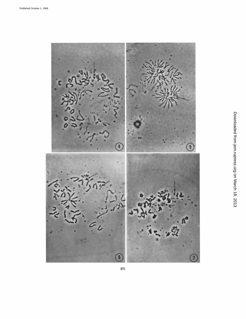
FIGs. 4--7. Single cell chromosome spreads, unfixed and unstained, and viewed with oil-immersion phase-contrast objective as produced by the desoxycholate method, displaying two T6 marker chromosomes (arrowed). These chromosomes are in a watery droplet, and as they are essentially three-dimensional in nature, do not photo- graph well. Better resolution is obtained when the investigator can focus up and down while observing and counting the chromosomes. Many dense bodies (? lysosomes) are present in the droplets but these cause no confusion in the actual analysis.
FIG. 4. Note that several chromosomes towards the bottom of the picture have turned "thready" during the few minutes that it took to transfer the preparation from the micromanipulation microscope to the photomicroscope. The markers are still in good order. X 1250.
FIc. 5.40 chromosomes and two T6 markers are present. × 1000. FIG. 6. In many spreads, the two T6 markers are close together; here they are
separated somewhat. Some distance below the right T6 marker, there is a pattern of lysosomes that slightly resembles a T6 marker, but this type of artefact is easy to recognize after a little experience with the technique. × 1250.
FIG. 7. A poorer degree of spreading, with some dumped chromosomes but two T6 markers still present. This type of spread is amenable to further dispersion by micro- manipulation; e.g., blowing extra fluid on the chromosomes. While this may cause a deterioration in the optical qualities of the chromosomes, it may nevertheless facilitate a confident diagnosis. × 1250.
on March 18, 2013
jem.rupress.org
Dow
nloaded from
Published October 1, 1968
851
on March 18, 2013
jem.rupress.org
Dow
nloaded from
Published October 1, 1968
852 CELL TO CELL I N T E R A C T I O N IN I M M U N E R E S P O N S E . I I I
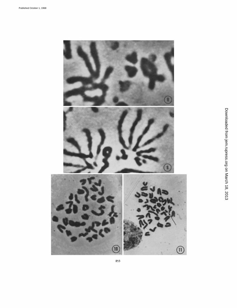
FIGS. 8 and 9. Higher magnifications of the two T6 markers seen in Figs. 4 and 5. Note that though the preparation is unfixed, the appearance of the T6 chromosomes is just as characteristic as in a conventional preparation (Fig. 11). × 5000.
FIG. 10. Single cell chromosome spread as produced by the micro-Ford technique, CBA mouse; 40 chromosomes but no T6 markers are present. X 1500.
FIG. 11. Chromosome spread obtained from whole spleen preparation by the con- ventional Ford technique, displaying two T6 markers (arrowed). The overall quality of the spread is better than can be obtained with single cell material. × 1250.
on March 18, 2013
jem.rupress.org
Dow
nloaded from
Published October 1, 1968
853
on March 18, 2013
jem.rupress.org
Dow
nloaded from
Published October 1, 1968
Copyright © 2022 FDOKUMEN