Cell Therapy for Modification of the Myocardial Electrophysiological Substrate
25
Cell Therapy for Modification of the Myocardial Electrophysiological Substrate Lior Yankelson, BSc*; Yair Feld, MD, PhD*; Tal Bressler-Stramer, PhD; Ilanit Itzhaki, MSc; Irit Huber, PhD; Amira Gepstein, PhD; Doron Aronson, MD; Shimon Marom, MD, PhD†; Lior Gepstein, MD, PhD† Background—Traditional antiarrhythmic pharmacological therapies are limited by their global cardiac action, low efficacy, and significant proarrhythmic effects. We present a novel approach for the modification of the myocardial electrophysiological substrate using cell grafts genetically engineered to express specific ionic channels. Methods and Results—To test the aforementioned concept, we performed ex vivo, in vivo, and computer simulation studies to determine the ability of fibroblasts transfected to express the voltage-sensitive potassium channel Kv1.3 to modify the local myocardial excitable properties. Coculturing of the transfected fibroblasts with neonatal rat ventricular myocyte cultures resulted in a significant reduction (68%) in the spontaneous beating frequency of the cultures compared with baseline values and cocultures seeded with naive fibroblasts. In vivo grafting of the transfected fibroblasts in the rat ventricular myocardium significantly prolonged the local effective refractory period from an initial value of 848 ms (cycle length, 200 ms) to 15413 ms (P0.01). Margatoxin partially reversed this effect (effective refractory period, 1178 ms; P0.01). In contrast, effective refractory period did not change in nontransplanted sites (867 ms) and was only mildly increased in the animals injected with wild-type fibroblasts (735 to 884 ms; P0.05). Similar effective refractory period prolongation also was found during slower pacing drives (cycle length, 350 to 500 ms) after transplantation of the potassium channels expressing fibroblasts (Kv1.3 and Kir2.1) in pigs. Computer modeling studies confirmed the in vivo results. Conclusions—Genetically engineered cell grafts, transfected to express potassium channels, can couple with host cardiomyocytes and alter the local myocardial electrophysiological properties by reducing cardiac automaticity and prolonging refractoriness. (Circulation. 2008;117:720-731.) Key Words: action potentials electrophysiology gene therapy ion channels mapping C ardiac arrhythmias account for significant worldwide morbidity and mortality. Traditional antiarrhythmic ther- apies are aimed at modifying the abnormal electrophysiolog- ical substrate by focal injury (surgery or radiofrequency catheter ablation), 1 implantable devices, 2,3 or pharmacother- apy. The latter approach is hampered by the relatively low efficacy of most antiarrhythmic agents and by their global cardiac action that may result in life-threatening proarrhyth- mic effects. 4 Clinical Perspective p 731 Gene therapy has been suggested as a novel antiarrhythmic strategy. 5,6 The feasibility of this innovative approach was established in several in vitro and in vivo studies demonstrat- ing that manipulation of the expression of different ion channels or their modulators can be used to alter myocardial electrophysiological properties, providing a possible future therapeutic strategy for both tachyarrhythmias and bradyarrhythmias. 5–12 In the present study, we evaluated an alternative approach that focuses on the transplantation of genetically engineered cell grafts. Using detailed in vitro, in vivo, and computer modeling studies, we demonstrated that fibroblasts, trans- fected to express different potassium channels, can couple with host cardiomyocytes and alter the local myocardial electrophysiological properties. This effect was manifested by the ability of the cell grafts to suppress cardiomyocyte automaticity in coculture experiments and to prolong the local myocardial refractory period (RP) in in vivo transplan- tation studies. Received August 24, 2004; accepted November 28, 2007. From the Sohnis Laboratory for Cardiac Electrophysiology and Regenerative Medicine (L.Y., Y.F., T.B.-S., I.I., I.H., A.G., L.G.), Minerva Center for Cell Biophysics (S.M.), and the Cardiology Department (Rambam Medical Center) (D.A., L.G.), Bruce Rappaport Faculty of Medicine, Technion-Israel Institute of Technology, Haifa, Israel. *The first 2 authors contributed equally to this work. †The last 2 authors contributed equally to this work. The online Data Supplement, which contains an expanded Methods section and figures, can be found with this article at http://circ.ahajournals.org/ cgi/content/full/CIRCULATIONAHA.106.671776/DC1. Correspondence to Lior Gepstein, MD, PhD, Technion’s Faculty of Medicine, POB 9649, Haifa, 31096, Israel. E-mail [email protected] © 2008 American Heart Association, Inc. Circulation is available at http://circ.ahajournals.org DOI: 10.1161/CIRCULATIONAHA.106.671776 720 Arrhythmia/Electrophysiology by guest on February 29, 2016 http://circ.ahajournals.org/ Downloaded from by guest on February 29, 2016 http://circ.ahajournals.org/ Downloaded from by guest on February 29, 2016 http://circ.ahajournals.org/ Downloaded from by guest on February 29, 2016 http://circ.ahajournals.org/ Downloaded from by guest on February 29, 2016 http://circ.ahajournals.org/ Downloaded from by guest on February 29, 2016 http://circ.ahajournals.org/ Downloaded from by guest on February 29, 2016 http://circ.ahajournals.org/ Downloaded from by guest on February 29, 2016 http://circ.ahajournals.org/ Downloaded from by guest on February 29, 2016 http://circ.ahajournals.org/ Downloaded from by guest on February 29, 2016 http://circ.ahajournals.org/ Downloaded from by guest on February 29, 2016 http://circ.ahajournals.org/ Downloaded from by guest on February 29, 2016 http://circ.ahajournals.org/ Downloaded from by guest on February 29, 2016 http://circ.ahajournals.org/ Downloaded from by guest on February 29, 2016 http://circ.ahajournals.org/ Downloaded from by guest on February 29, 2016 http://circ.ahajournals.org/ Downloaded from by guest on February 29, 2016 http://circ.ahajournals.org/ Downloaded from by guest on February 29, 2016 http://circ.ahajournals.org/ Downloaded from by guest on February 29, 2016 http://circ.ahajournals.org/ Downloaded from by guest on February 29, 2016 http://circ.ahajournals.org/ Downloaded from
-
Upload
sacklermedicine -
Category
Documents
-
view
1 -
download
0
Transcript of Cell Therapy for Modification of the Myocardial Electrophysiological Substrate

Cell Therapy for Modification of the MyocardialElectrophysiological Substrate
Lior Yankelson, BSc*; Yair Feld, MD, PhD*; Tal Bressler-Stramer, PhD; Ilanit Itzhaki, MSc;Irit Huber, PhD; Amira Gepstein, PhD; Doron Aronson, MD;
Shimon Marom, MD, PhD†; Lior Gepstein, MD, PhD†
Background—Traditional antiarrhythmic pharmacological therapies are limited by their global cardiac action, lowefficacy, and significant proarrhythmic effects. We present a novel approach for the modification of the myocardialelectrophysiological substrate using cell grafts genetically engineered to express specific ionic channels.
Methods and Results—To test the aforementioned concept, we performed ex vivo, in vivo, and computer simulationstudies to determine the ability of fibroblasts transfected to express the voltage-sensitive potassium channel Kv1.3 tomodify the local myocardial excitable properties. Coculturing of the transfected fibroblasts with neonatal rat ventricularmyocyte cultures resulted in a significant reduction (68%) in the spontaneous beating frequency of the culturescompared with baseline values and cocultures seeded with naive fibroblasts. In vivo grafting of the transfectedfibroblasts in the rat ventricular myocardium significantly prolonged the local effective refractory period from an initialvalue of 84�8 ms (cycle length, 200 ms) to 154�13 ms (P�0.01). Margatoxin partially reversed this effect (effectiverefractory period, 117�8 ms; P�0.01). In contrast, effective refractory period did not change in nontransplanted sites(86�7 ms) and was only mildly increased in the animals injected with wild-type fibroblasts (73�5 to 88�4 ms;P�0.05). Similar effective refractory period prolongation also was found during slower pacing drives (cycle length, 350to 500 ms) after transplantation of the potassium channels expressing fibroblasts (Kv1.3 and Kir2.1) in pigs. Computermodeling studies confirmed the in vivo results.
Conclusions—Genetically engineered cell grafts, transfected to express potassium channels, can couple with hostcardiomyocytes and alter the local myocardial electrophysiological properties by reducing cardiac automaticity andprolonging refractoriness. (Circulation. 2008;117:720-731.)
Key Words: action potentials � electrophysiology � gene therapy � ion channels � mapping
Cardiac arrhythmias account for significant worldwidemorbidity and mortality. Traditional antiarrhythmic ther-
apies are aimed at modifying the abnormal electrophysiolog-ical substrate by focal injury (surgery or radiofrequencycatheter ablation),1 implantable devices,2,3 or pharmacother-apy. The latter approach is hampered by the relatively lowefficacy of most antiarrhythmic agents and by their globalcardiac action that may result in life-threatening proarrhyth-mic effects.4
Clinical Perspective p 731Gene therapy has been suggested as a novel antiarrhythmic
strategy.5,6 The feasibility of this innovative approach wasestablished in several in vitro and in vivo studies demonstrat-ing that manipulation of the expression of different ion
channels or their modulators can be used to alter myocardialelectrophysiological properties, providing a possible futuretherapeutic strategy for both tachyarrhythmias andbradyarrhythmias.5–12
In the present study, we evaluated an alternative approachthat focuses on the transplantation of genetically engineeredcell grafts. Using detailed in vitro, in vivo, and computermodeling studies, we demonstrated that fibroblasts, trans-fected to express different potassium channels, can couplewith host cardiomyocytes and alter the local myocardialelectrophysiological properties. This effect was manifestedby the ability of the cell grafts to suppress cardiomyocyteautomaticity in coculture experiments and to prolong thelocal myocardial refractory period (RP) in in vivo transplan-tation studies.
Received August 24, 2004; accepted November 28, 2007.From the Sohnis Laboratory for Cardiac Electrophysiology and Regenerative Medicine (L.Y., Y.F., T.B.-S., I.I., I.H., A.G., L.G.), Minerva Center for
Cell Biophysics (S.M.), and the Cardiology Department (Rambam Medical Center) (D.A., L.G.), Bruce Rappaport Faculty of Medicine, Technion-IsraelInstitute of Technology, Haifa, Israel.
*The first 2 authors contributed equally to this work.†The last 2 authors contributed equally to this work.The online Data Supplement, which contains an expanded Methods section and figures, can be found with this article at http://circ.ahajournals.org/
cgi/content/full/CIRCULATIONAHA.106.671776/DC1.Correspondence to Lior Gepstein, MD, PhD, Technion’s Faculty of Medicine, POB 9649, Haifa, 31096, Israel. E-mail [email protected]© 2008 American Heart Association, Inc.
Circulation is available at http://circ.ahajournals.org DOI: 10.1161/CIRCULATIONAHA.106.671776
720
Arrhythmia/Electrophysiology
by guest on February 29, 2016http://circ.ahajournals.org/Downloaded from by guest on February 29, 2016http://circ.ahajournals.org/Downloaded from by guest on February 29, 2016http://circ.ahajournals.org/Downloaded from by guest on February 29, 2016http://circ.ahajournals.org/Downloaded from by guest on February 29, 2016http://circ.ahajournals.org/Downloaded from by guest on February 29, 2016http://circ.ahajournals.org/Downloaded from by guest on February 29, 2016http://circ.ahajournals.org/Downloaded from by guest on February 29, 2016http://circ.ahajournals.org/Downloaded from by guest on February 29, 2016http://circ.ahajournals.org/Downloaded from by guest on February 29, 2016http://circ.ahajournals.org/Downloaded from by guest on February 29, 2016http://circ.ahajournals.org/Downloaded from by guest on February 29, 2016http://circ.ahajournals.org/Downloaded from by guest on February 29, 2016http://circ.ahajournals.org/Downloaded from by guest on February 29, 2016http://circ.ahajournals.org/Downloaded from by guest on February 29, 2016http://circ.ahajournals.org/Downloaded from by guest on February 29, 2016http://circ.ahajournals.org/Downloaded from by guest on February 29, 2016http://circ.ahajournals.org/Downloaded from by guest on February 29, 2016http://circ.ahajournals.org/Downloaded from by guest on February 29, 2016http://circ.ahajournals.org/Downloaded from

MethodsExpression System andElectrophysiological RecordingsThe NIH-3T3 fibroblast line was stably transfected to express thevoltage-gated potassium channel Kv1.3,13 the inward-rectifier potas-sium channel Kir2.1, or enhanced green fluorescent protein (eGFP).
Details regarding the creation of the lines and their electrophysi-ological characterization can be found in the expanded Methodssection of the online Data Supplement.
Coculturing ExperimentsTo assess the effects of the transfected fibroblasts on the electro-physiological properties of cardiac tissue, we initially performedcoculture experiments using primary cultures of neonatal rat ventric-ular myocytes.13 Details regarding the methodology of these exper-iments, immunostaining analysis, calcein dye transfer,14 and multi-electrode mapping13 studies can be found in the expanded Methodssection of the Data Supplement.
In Vivo Animal StudiesAll studies were approved by the Animal Board and Safety Com-mittee of Technion (Haifa, Israel).
Rat ModelMale Sprague-Dawley rats (200 to 250 g) were anesthetized (keta-mine/xylazine), intubated, and ventilated. After left thoracotomy,6�106 fibroblasts (nontransfected, n�8; or Kv1.3 expressing, n�10)were transplanted into the left ventricular myocardium with a28-gauge needle (volume, 300 �L). A small suture was created at anearby site to identify the site of transplantation. Cells were labeledwith DAPI (Sigma, St Louis, Mo) or with 3,3�-dilinoleyloxa-carbocyanine perchlorate or 5- (and -6)- carboxyfluorescein diacetate(Molecular Probes, Carlsbad, Calif). To prevent graft rejection, allanimals received cyclosporin (10 mg · kg�1 · d�1) and methylpred-nisolone (2 mg · kg�1 · d�1).
Swine ModelFive pigs (20 to 30 kg) underwent right thoracotomy, after which6�106 cells (in 300 �L medium) per injection site were transplantedin 7 different right ventricular locations. To prevent graft rejection,animals were treated with cyclosporine (10 mg · kg�1 · d�1),methylprednisolone (3 mg · kg�1 · d�1), and azathioprim (50 mg/d).
In Vivo Electrophysiological StudyThe myocardial effective RP (ERP) was determined by epicardialventricular pacing. To localize the stimulation effect to the site ofcell grafting, we used custom-made intramyocardial thin electrodes(0.01-in electrodes, 1-mm tip-length exposure) with a bipolar dis-tance of 1 mm. The stimulation protocol consisted of a train of 20consecutive stimuli (S1) at a fixed cycle length, followed by a singlepremature stimulation (S2). The coupling interval of the prema-ture stimulus was reduced in 10-ms steps until the ERP wasreached (note that we did not use a lower limit for the couplinginterval of the extrastimuli). The stimulation amplitude was set totwice the capture threshold, and ERP was determined as the averageof 3 repeated measurements.
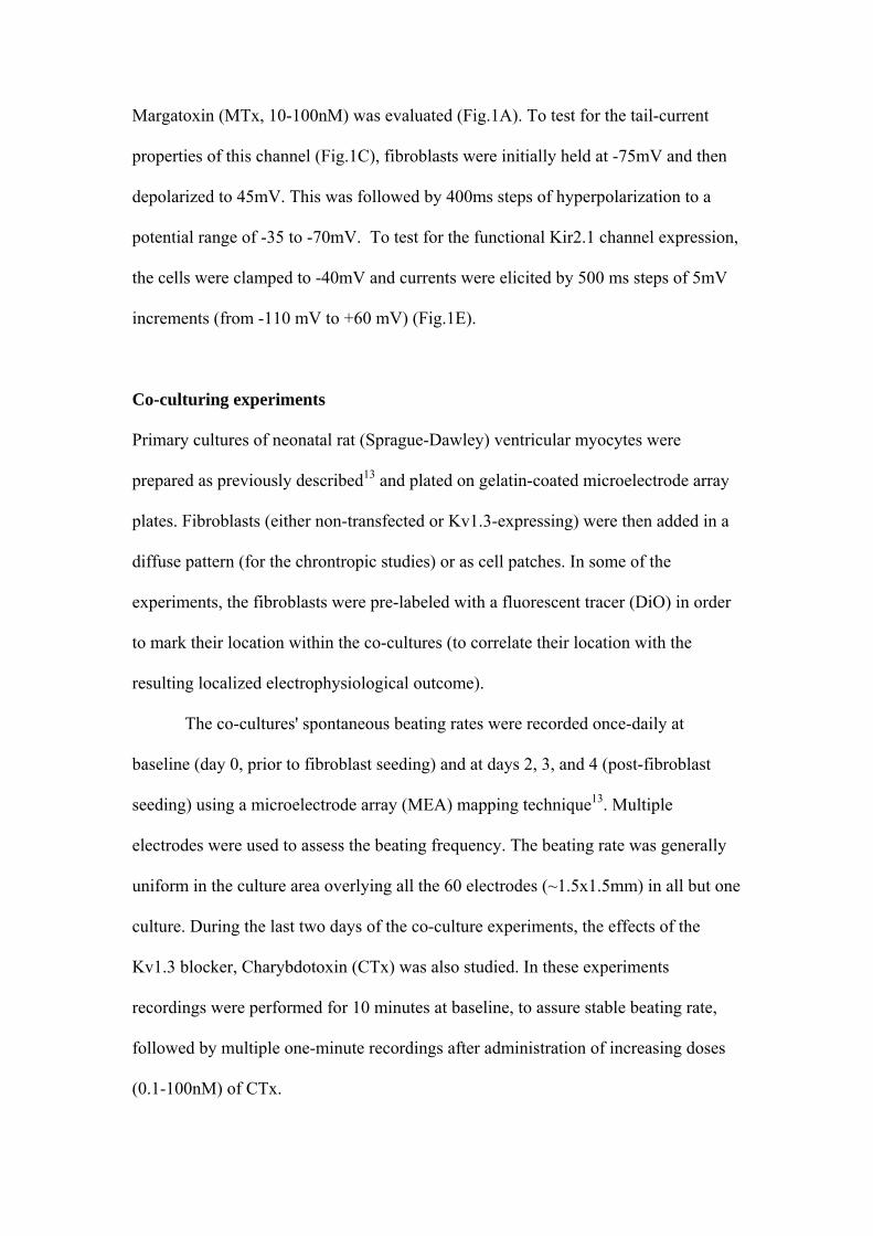
ERP was measured at the site of cell transplantation and at remotesites before and 7 days after cell grafting. Measurements wererepeated after intraperitoneal injection of 9 �g/kg MTx. We chose touse margatoxin for the in vivo studies (instead of charybdotoxin)because of its equivalent Kv1.3 current blocking properties (Figure1A) and its previously established in vivo pharmacokinetic proper-ties.15 The grafting area was identified for the pacing studies by aslight discoloration on the epicardial surface. The stimulation sitewas tagged, and the presence of the transplanted cells was confirmedin histology. In some studies, we coinjected fluorescent microspheresor prelabeled the cells with the fluorescent tracer 3,3�- dilinoleyloxa-carbocyanine perchlorate. The intense fluorescent signal (see Figure
I in the Data Supplement) confirmed the accurate localization of thegrafted area for pacing.
ERP measurements in pigs were performed similarly but withslower pacing cycle lengths (S1, 350 to 500 to ms). In addition to theKv1.3 fibroblasts, we also assessed the effects of Kir2.1- andeGFP-expressing fibroblasts. ERP was measured at the site of celltransplantation, in remote sites, and after intramyocardial injec-tion of MTx.
To assess the potential arrhythmogenic risk of the transplantationprocedure, we performed programmed electric stimulation (n�4)using an electrophysiological catheter positioned in the right ventri-cle. The stimulation protocol consisted of 8 consecutive stimuli (S1)at a fixed cycle length (400 or 300 ms), followed by up to 3consecutive premature stimulations (S2 through S4). The couplinginterval of the 3 extrastimuli was reduced by 10-ms steps until ERPwas reached.
Histological ExaminationDetails can be found in the online expanded Methods section.
Computer SimulationA 1-dimensional numeric model (based on the Luo-Rudy model16 foraction potential simulation) was used to simulate the effects of theengineered fibroblasts on coupled myocytes. Details can be found inthe online Methods.
Statistical AnalysisAll results are expressed as mean�SEM. For the coculture experi-ments, we used general linear model 2-way repeated-measuresANOVA to test the hypothesis that changes in the average beatingrate over time varied among the 2 experimental groups (Kv1.3fibroblasts and nontransfected fibroblasts). The model included theeffects of treatment (representing the nonrepeated factor), time, andtreatment-by-time interaction. The Greenhouse-Geisser adjustmentwas used to correct for the inflated risk of a type I error. TheBonferroni correction was used to assess the significance of pre-defined comparisons at specific time points. For the in vivo rat andswine experiments, repeated-measures ANOVA was used to assessthe effects of fibroblasts injection at different time points and afterMTx application. For the in vivo swine studies comparing the effectsof different engineered fibroblasts on ERP, 1-way ANOVA wasused.
The authors had full access to and take full responsibility for theintegrity of the data. All authors have read and agree to themanuscript as written.
ResultsIn Vitro Studies
Patch-Clamp StudiesVoltage-clamp studies of the transfected fibroblasts con-firmed the robust expression of the Kv1.3 channel and itssensitivity to selective pharmacological blockade (Figure 1Aand 1B). Because we hypothesized that the Kv1.3 fibroblastswould generate significant electrotonic interactions duringthe late repolarization and early postrepolarization phases andtherefore could modify myocardial refractoriness and auto-maticity, we also analyzed the tail current properties of thischannel (Figure 1C). Using a specific protocol, we noted asignificant tail current for up to 100 ms after hyperpolariza-tion. In contrast, we could not detect any significant currentsin wild-type or eGFP-expressing fibroblasts (Figure 1D).
Connexin ImmunostainingA prerequisite for the ability of the engineered fibroblasts tomodulate the electrophysiological properties of neighboringcardiomyocytes is the establishment of functional gap junc-
Yankelson et al Cell Therapy for Cardiac Arrhythmias 721
by guest on February 29, 2016http://circ.ahajournals.org/Downloaded from

tions. To assess for this ability, we first performed immuno-staining analysis in fibroblast-cardiomyocyte cocultures. Ascan be seen in Figure 2A, the connexin43 (Cx43) immunosig-nal could be localized to the junction between the Kv1.3fibroblasts and neighboring cardiomyocytes.
Calcein Dye TransferWe next loaded the engineered fibroblasts with calcein andcocultured them with rat cardiomyocytes. Calcein is anintracellular dye that is diffusible only through gap junctions,and its transfer between cells is a standard technique forstudying gap junction communication.14 Sequential micros-copy revealed that by 3 hours a fluorescent signal could beobserved in the cardiomyocytes adjacent to the loaded fibro-blasts (Figure 2B, right), indicating the formation of func-tional gap junctions between the 2 cell types.
Microelectrode Array Mapping StudiesOn the basis of the encouraging Cx43 immunostaining andcalcein dye transfer results, we continued to assess the abilityof the transfected fibroblasts to modify the excitable proper-ties of cardiac tissue. Initially, possible changes in thespontaneous beating frequency of neonatal rat cardiomyocytecultures were assessed after seeding of the Kv1.3 fibroblasts.
Two-way repeated-measures ANOVA indicated that thetreatment-by-time interaction effect was significant (P�0.04),reflecting the decrease in beating rate in the Kv1.3 fibroblastcocultures (Figure 3A). Although the spontaneous beatingrate did not differ between the control (nontransfected fibro-blasts; n�6) and study (n�6) groups at baseline (beforefibroblast seeding; Figure 3A, day 0), we could start detectinga significant reduction in the beating rate in the Kv1.3fibroblast cocultures by day 2. This negative chronotropiceffect was maintained throughout the experiment (Figure3A). To dissect the contribution of the Kv1.3 channel to theelectrophysiological outcome, we applied the Kv1.3 blockerCTx. Application of CTx had a dose-related effect andresulted in complete reversal of the negative chronotropiceffect when given at a dose of 100 nmol/L (Figure 3B). Incontrast, CTx application had only minimal effects on thebeating frequency in the control group.
We next assessed the spatial extent of the electrophysio-logical changes induced by the engineered fibroblasts. To thisend, we performed coculture studies in which the Kv1.3fibroblasts were labeled with a fluorescent tracer (to followtheir location within the cocultures) and seeded as cellpatches on top of primary cardiomyocyte cultures (Figure 3C
Figure 1. A, Voltage-clamp recordings from the Kv1.3 fibroblast and blockade of the Kv1.3 current with MTx. B, The corresponding I-Vcurve. C, Tail current of the Kv1.3 channel. D, Voltage-clamp recording from a nonstransfected fibroblast. E, Voltage-clamp recordingfrom the Kir2.1 fibroblast and blockade of the current with barium.
722 Circulation February 12, 2008
by guest on February 29, 2016http://circ.ahajournals.org/Downloaded from

and 3D, left). In our previous study,13 we noted that electro-grams recorded from cardiomyocytes located directly beneaththe Kv1.3 fibroblasts were characterized by significantlyreduced amplitudes. Here, we assessed the effects of theKv1.3 fibroblasts on electrograms recorded at increasingdistances from the seeded fibroblasts. To avoid the possiblecompeting effects of a number of nearby cell clusters, theanalysis was performed only in cocultures in which the fieldof interest contained only 1 Kv1.3 fibroblast cell cluster. Ascan be seen in Figure 3C, the Kv1.3 fibroblasts significantlyaffected the electrogram amplitude (note the inverse relation-ship using a logarithmic fit) even at recording electrodeslocated far beyond the fibroblast area (up to 500 �m).Application of CTx resulted in a complete reversal of theseelectrophysiological changes. Note the conversion of theinverse logarithmic relationship into a straight line (Figure3D), reflecting a greater increase in electrogram amplitude atsites closer to the fibroblasts.
In Vivo StudiesWe next continued to evaluate the ability of the Kv1.3-expressing fibroblasts to modify the in vivo myocardialelectrophysiological properties. To this end, possible changesin the ventricular ERP were evaluated in rats. Figure 4 depictschanges in the local ERP before and 1 week after cellgrafting. Note that the ventricular ERP (S1–S1, 200 ms) was60 ms before cell transplantation (Figure 4A) and increased to170 ms at the site of cell grafting 7 days later (Figure 4B).Interestingly, this increase was partially reversed (to 100 ms)after local application of the Kv1.3 blocker MTx (Figure 4C).
Transplantation of the Kv1.3 fibroblasts increased ERP inall animals (n�10) from a baseline value of 84�8 to 154�13ms (Bonferroni adjusted for 3 comparisons, P�0.01) 7 dayslater (Figure 4D). Administration of MTx resulted in asignificant (P�0.01) decrease in ERP at the site of celltransplantation to 117�8 ms (Figure 4D). In contrast, ven-tricular ERP at remote sites did not differ (86�7 ms) frombaseline values (P�0.66).
We next compared the effects of nontransfected fibro-blasts. In this control group (n�8), we observed a mild
increase in the local ERP at the site of cell grafting (from73�5 to 88�4 ms; Bonferroni adjusted for 2 comparisons,P�0.05; Figure 4D). This increase in ERP, however, wasmuch lower than that resulting from the transplantation of theKv1.3 fibroblasts (154�13 ms; P�0.01; Figure 4D). Asexpected, this value did not change after administration ofMTx (P�0.98; Figure 4D).
The presence of the transplanted cells at the pacing siteswas confirmed in histology. The grafted cells were labeledbefore transplantation by the fluorescent tracer 3,3�-dilinoleyloxacarbocyanine diacetate or 5-(and-6)-carboxyfluorescein diacetate (green cells in Figure 5A) orDAPI (Figure 5B) and could later be detected in vivo.Alternatively, the grafted cells also could be detected usinganti-Kv1.3 antibodies (Figure 5C). Gap junctions could oc-casionally be recognized at the interface between the graftedfibroblasts and host cardiomyocytes (Cx43 immunostainingin Figure 5B and 5C). Interestingly, we noted an increase inCx43 expression in fibroblasts located close to hostcardiomyocytes.
In Vivo Effects in the Pig VentricleBecause of the fast beating rate of the rat heart, we couldevaluate the effects of cell grafting only at rapid stimulationrates (cycle length, 200 ms). To assess whether the graftedcells could increase refractoriness at more clinically relevantslower ventricular rates, we injected the Kv1.3 fibroblastsinto sites in the pig right ventricular myocardium. Immuno-staining of these hearts with anti-Kv1.3 antibodies confirmedthe long-term survival of the grafted cells and the continuousexpression of the transgene (Figure 5D).
Electrophysiological studies, conducted 7 days after cellgrafting, revealed significant electrophysiological changesinduced by the Kv1.3 fibroblasts (Figure 6). Note in theexample in Figure 6B the increase in ERP at the site of celltransplantation to 250 ms (S1 cycle length, 400 ms) comparedwith a nontransplanted myocardial site (ERP, 190 ms; Figure6A). This increase was partially reversed (ERP, 210 ms) afterlocal application of MTx (Figure 6C). Grafting of the Kv1.3fibroblasts resulted in a significant increase (P�0.05) in ERP
Figure 2. Fibroblast-cardiomyocyte coupling incocultures. A, Left, Identification of the Kv1.3 fibro-blasts (green) and cardiomyocytes (red) by anti-Kv1.3 and anti-cTnI antibodies, respectively. Right,Positive Cx43 immunosignal (purple; superim-posed on the differential interference contrastimage) at the junction (red arrows) between fibro-blasts (white arrow) and neighboring cardiomyo-cytes (yellow arrows). B, Calcein dye transferbetween the preloaded fibroblasts (FB) and cocul-tured cardiomyocyte.
Yankelson et al Cell Therapy for Cardiac Arrhythmias 723
by guest on February 29, 2016http://circ.ahajournals.org/Downloaded from
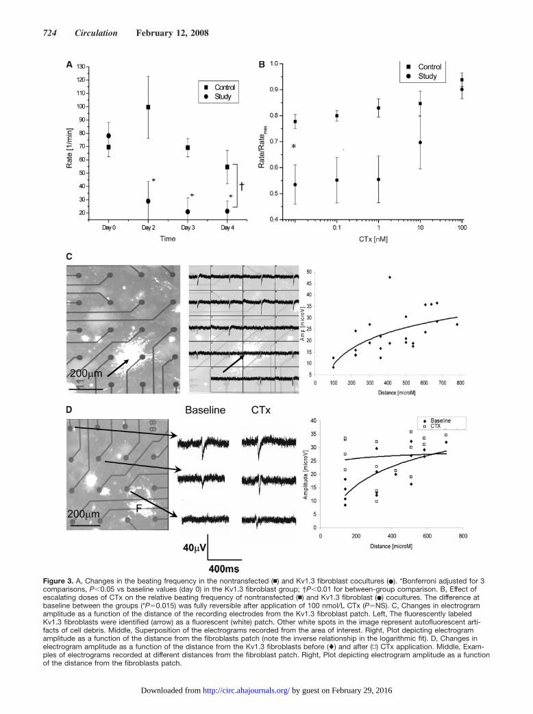
Figure 3. A, Changes in the beating frequency in the nontransfected (�) and Kv1.3 fibroblast cocultures (�). *Bonferroni adjusted for 3comparisons, P�0.05 vs baseline values (day 0) in the Kv1.3 fibroblast group; †P�0.01 for between-group comparison. B, Effect ofescalating doses of CTx on the relative beating frequency of nontransfected (�) and Kv1.3 fibroblast (�) cocultures. The difference atbaseline between the groups (*P�0.015) was fully reversible after application of 100 nmol/L CTx (P�NS). C, Changes in electrogramamplitude as a function of the distance of the recording electrodes from the Kv1.3 fibroblast patch. Left, The fluorescently labeledKv1.3 fibroblasts were identified (arrow) as a fluorescent (white) patch. Other white spots in the image represent autofluorescent arti-facts of cell debris. Middle, Superposition of the electrograms recorded from the area of interest. Right, Plot depicting electrogramamplitude as a function of the distance from the fibroblasts patch (note the inverse relationship in the logarithmic fit). D, Changes inelectrogram amplitude as a function of the distance from the Kv1.3 fibroblasts before (�) and after (▫) CTx application. Middle, Exam-ples of electrograms recorded at different distances from the fibroblast patch. Right, Plot depicting electrogram amplitude as a functionof the distance from the fibroblasts patch.
724 Circulation February 12, 2008
by guest on February 29, 2016http://circ.ahajournals.org/Downloaded from

at 3 different stimulation drives studied (S1 cycle lengths,350, 400, and 500 ms) compared with nontransplanted areas(Figure 6D). The increase in ERP was reversible after localapplication of MTx (P�0.05; Figure 6D).
To assess the arrhythmogenic potential of this procedure,we performed programmed ventricular electrical stimulation(up to 3 premature stimuli) in 4 animals. In none of theseanimals could we induce any ventricular arrhythmias.
We next assessed the electrophysiological effects of 2additional types of genetically engineered fibroblasts. Todetermine the possible contribution of the transfection pro-cess per se, we established a fibroblast line that overexpresseseGFP. Similar to the wild-type fibroblasts in the rat studies,transplantation of the eGFP-expressing cells in pigs had onlya mild effect on ERP (Figure 7C).
Because the Kv1.3 channel may affect different phases of theaction potential (because of its rapid opening kinetics, lack ofinactivation during phases 2 and 3, and significant tail current),we chose also to study fibroblasts overexpressing a potassiumchannel that will be active mainly during late phase 3 and phase
4 of the action potential (in a manner similar to the Kv1.3 tailcurrent). We therefore created a fibroblasts line overexpressingthe Kir2.1 channel.17 Voltage-clamp recordings from thesetransfected fibroblasts confirmed robust functional expression ofthis channel (Figure 1E).
Grafting of the Kir2.1 fibroblasts significantly prolongedthe local ventricular ERP from a baseline value of 219�9 to260�7 ms (P�0.01). Figure 7A and 7B depicts examples ofthe ERP measurements at the site of cell grafting before(Figure 7A) and 7 days after (Figure 7B) transplantation ofthe Kir2.1 fibroblasts. Immunostaining analysis confirmedthe long-term survival of the cells, the continuous expressionof the Kir2.1 transgene, and the formation of gap junctionswith host cardiomyocytes (Figure 5E and 5F).
Figure 7C summarizes the effects of the different fibro-blasts used in the swine study. Note that transplantation of theKir2.1 fibroblasts and Kv1.3 fibroblasts resulted in thegreatest prolongation of the local ERP and that these valueswere significantly higher than control remote sites and sitestransplanted with the eGFP fibroblasts (all P�0.01).
Figure 4. A through C, Measurement ofthe rat ventricular ERP before cell graft-ing (A), 7 days after Kv1.3 fibroblasttransplantation (B), and after MTx appli-cation (C). Top, The shortest coupledS1–S2 interval (arrows show stimulus)that still elicits a ventricular response;bottom, the following S1–S2 interval thatfails to capture the ventricle (arrowheadshows ERP). D, Summary of the meanERP values in the Kv1.3-expressing andthe nontransfected fibroblast groups atbaseline, after 7 days, after MTx applica-tion, and at a remote site. *P�0.01;†P�0.05.
Yankelson et al Cell Therapy for Cardiac Arrhythmias 725
by guest on February 29, 2016http://circ.ahajournals.org/Downloaded from
Modeling ResultsTo attain further mechanistic insights into the electrophysiolog-ical changes induced by the transfected fibroblasts, we used a1-dimensional numeric mathematical model simulating actionpotential propagation along a chain of 5 cardiomyocytes inwhich myocyte C is connected to the engineered fibroblast(online Figure II). The RP in these simulations was defined asthe minimum interval between 2 consecutive stimuli applied tomyocyte A that could be propagated to myocyte E. In the setupwithout a connecting fibroblast, the calculated RP was 375 ms(Figure 8A); it increased slightly to 425 ms (Figure 8B) when anontransfected fibroblast was used. When myocyte C wasconnected to the Kv1.3 fibroblast, a larger increase in the RPwas noted (500 ms; Figure 8C).
Figure 8D shows the simulated action potential morphol-ogy of myocyte C when connected to the nontransfected andKv1.3 fibroblasts. Also shown are the changes in membranepotential of the coupled fibroblast. Note that depolarization inmyocyte C leads to depolarization of the connected fibroblastand opening of the Kv1.3 channels. The resulting outwardcurrent hyperpolarizes the fibroblast. The potential difference
generated between the 2 cell types leads to the developmentof electrotonic currents (the fibroblast acting as a sink) thatultimately modulates different phases of the cardiomyocyteaction potential. As can be seen in Figure 8D, these electro-tonic interactions result in a reduction of the slope of phase 0,in reduced amplitude of the action potential, in shortening ofthe action potential duration (APD), in hyperpolarization ofthe resting membrane potential (RMP) during the earlydiastolic period (Figure 8D, right), and in an increase in theRP (Figure 8C). The latter 2 events stem from the Kv1.3 tailcurrent, which acts to hyperpolarize the RMP and preventsinitiation of a new action potential. Similar hyperpolarizationof the RMP also was identified when simulating the effect ofthe Kir2.1 fibroblast (Figure 8E).
According to the aforementioned paradigm, the electro-physiological changes induced by the transplanted cellsdepend on the active (biophysical properties of the trans-fected channels) and passive properties of grafted and hostcells. To assess the latter, we modeled the effects of changesin the fibroblast surface area (affecting cell capacitance;Figure 8F) and the degree of fibroblast-cardiomyocyte cou-
Figure 5. Histological identification of the graftedcells. A, Identification of the Kv1.3 fibroblasts (pre-labeled with 5-(and-6)-carboxyfluorescein diac-etate; green) and host cardiomyocytes (cTnI immu-nostaining; red). Nuclei were counterstained withToPro3 (blue). B, Cx43 immunostaining (greenpunctuate staining) at the junction (arrows)between host cardiomyocytes (cTnI staining; red)and the Kv1.3 fibroblasts (prelabeled with DAPI;blue). C, Left, High-resolution confocal imageshowing the spatial relationship between host car-diomyocytes (cTnI staining; red) and grafted fibro-blasts (Kv1.3 staining; green). Right, Positive Cx43immunosignal (blue; superimposed on the differen-tial interference contrast image) at the junctionbetween fibroblasts (yellow arrows) and cardio-myocytes (white arrows). D, Identification of theKv1.3 fibroblasts (using anti-Kv1.3 antibodies;blue), cardiomyocytes (cTnI staining; red), and gapjunctions (Cx43 staining; green) within the pigmyocardium. E, Identification of the Kir2.1 fibro-blasts (using anti–His-Tag antibodies; green) withinthe pig myocardium (red). F, Positive Cx43 immu-nosignal (red) at the junction between the graftedfibroblasts (anti–His-Tag; green) and host cardio-myocytes (anti-cTnI; blue). Right, Superposition ofthe left panel (signal intensity was reduced inten-tionally) on top of the corresponding differentialinterference contrast image.
726 Circulation February 12, 2008
by guest on February 29, 2016http://circ.ahajournals.org/Downloaded from

Figure 6. A through C, Measurements of pig ERP at a control remote ventricular site (A), at the site of Kv1.3 fibroblast transplan-tation (B), and after MTx administration (C). Top, Shortest S1–S2 interval that still elicits a ventricular activation; bottom, the fol-lowing S1–S2 interval that fails to capture the ventricle (ERP). D, Summary of the changes in the ventricular ERP before and afterMTx application at the sites of Kv1.3 fibroblast grafting and at control remote sites. Measurements were performed using 3 differ-ent cycle lengths (350, 400, and 500 ms). Bonferroni adjusted for 2 comparisons, *P�0.025 vs control sites; †P�0.025 vs MTxtreatment.
Yankelson et al Cell Therapy for Cardiac Arrhythmias 727
by guest on February 29, 2016http://circ.ahajournals.org/Downloaded from

pling (affecting intercellular resistance; Figure 8G). Thesestudies demonstrated that by increasing the fibroblast surfacearea or improving the fibroblast-cardiomyocyte coupling, thefibroblast acts as a greater sink, further prolonging RP.
DiscussionIn the present study, we evaluated a new approach for themodification of the myocardial electrophysiological proper-ties using genetically engineered cell grafts overexpressingspecific ionic channels. Our results using fibroblasts trans-fected to express different potassium channels (the voltage-gated potassium channel Kv1.3 and the inward-rectifyingKir2.1 channel) demonstrate the validity of this concept byshowing the ability of the cell grafts to reduce excitability and
to prolong local refractoriness in ex vivo, in vivo, andcomputer simulation studies.
The observed changes in the local ventricular electrophys-iological properties indicate the ability of the engraftedengineered fibroblasts to survive, to integrate with host tissue,and to modulate local excitability by generation of electro-tonic currents with neighboring cardiomyocytes. The possi-bility of electric interactions between cardiomyocytes andfibroblasts was previously demonstrated by Rook et al,18 whoshowed morphological and functional coupling in cardiomyo-cyte-fibroblast pairs in vitro; by Fast at al,19 who reported onelectric connections in cultured cardiomyocytes harboringfibroblasts; and by our previous report showing the ability oftransfected fibroblasts to create local conduction blocks in
Figure 7. A and B, Measurements of pig ERP values at baseline (A) and 7 days after transplantation of the Kir2.1 fibroblasts (Fib;B). Top, The shortest S1–S2 interval that still elicits a ventricular activation; bottom, the following S1–S2 interval that fails to cap-ture the ventricle (ERP). C, Summary of the ventricular ERP values after transplantation with the different cell types (Fib-GFP, Fib-Kv1.3, Fib-Kir2.1, and nontransplanted remote sites). *Bonferroni adjusted for 4 comparisons, P�0.01.
728 Circulation February 12, 2008
by guest on February 29, 2016http://circ.ahajournals.org/Downloaded from

coculture experiments.13 More recent studies showed theability of fibroblasts to propagate electrical signals overextended distances (300 �m) in coculture experiments20 andto reduce the spontaneous beating frequency in cocultures21
and suggested an important in vivo role for cardiac fibroblastsin modulating the electrophysiological properties of thesinoatrial node.22
Interestingly, grafting of nontransfected fibroblasts in ourstudy resulted in a mild but statistically significant effect onERP in the in vivo rat studies (as also predicted by themodeling results; Figure 8B). Heterologous gap junction–mediated fibroblast-cardiomyocyte coupling has the potentialto reduce cardiomyocyte excitability by direct membranedepolarization or by an electric sink effect.21,23,24 For exam-ple, Miragoli et al24 showed in a coculture study thatmyofibroblasts can significantly reduce cardiac tissue con-duction velocity and maximal upstroke velocity, most prob-ably because of gradual depolarization of the cardiomyocyte
RMP. Similarly, Kizana et al21 showed a reduction in thespontaneous beating rate of cardiomyocyte cultures whengrown on top of wild-type fibroblast cell layer but not ifcultured on connexin knockout fibroblasts. An alternativeexplanation may be structural changes induced by the graftedwild-type fibroblasts (fibrosis, volume effect of the graftedcells themselves, etc) that may lead to interruption of thenormal myocardial architecture and consequentially tochanges in the local electrophysiological properties. Suchstructural changes may be the underlying mechanism in arecent report showing changes in the atrioventricular nodeproperties after transplantation of transforming growth factor-�–simulated fibroblasts.25
The electrophysiological changes induced by the Kv1.3fibroblasts, in contrast to those of the control fibroblasts, weremuch more pronounced and were manifested by a significantreduction in the spontaneous beating rate in the cocultureexperiments and by marked prolongation of the local ventric-
A
0
20
40Ve [mV]
Stim [nA]
No fibroblast, RP- 375 ms C
0
20
40
Ve [mV] Stim [nA]
Wild type fibroblast, RP- 425 ms
0
20
40Ve [mV]
Stim [nA]
Kv1.3 fibroblast, RP- 500 msB
-60
-40
-20
-60
-40
-20
0
-60
-40
-20
40
0 100 200 300 400 500 600 700-100
-80
time [ms] 0 100 200 300 400 500 600 700
-100
-80
time [ms]
D
0 100 200 300 400 500 600 700-100
-80
time [ms]
-80
-20
0
20
mV
]
Fibroblast overexpressing Kv1.3. MyoC Fibroblast over expressing Kv1.3. MyoC Fibroblast without transfection. F
ms) 440
460
480
500
-82 5
-82
-81.5
-81
-80.5
[mV
]
Fibroblast overexpressing Kv1.3. MyoC Fibroblast over expressing Kv1.3. MyoC Fibroblast without transfection.
-80
-60
-40Vm
[ ER
P (
m
380
400
420
440
-84.5
-84
-83.5
-83
-82.5
Vm
[
0 50 100 150 200 250 300 350 400 450 500-100
time [ms]
E G
Sfib/Smyo
0.00 .05 .10 .15 .20360
500
350 400 450 500-85
time [ms]
0
20
40
MyoC Fibroblast without transfection MyoC Fibroblast overexpressing Kir2.1Fibroblast overexpressing Kir2.1 -74
-72
-70
MyoC Fibroblast without transfectionMyoC Fibroblast overexpressing Kir2.1Fibroblast overexpressing Kir2.1
ER
P (
ms)
420
440
460
480
-60
-40
-20
0
Vm
[m
V]
-84
-82
-80
-78
-76
Vm
[m
V]
Kohm/cm
5 10 15 20 25 30 35 40 45360
380
400
0 50 100 150 200 250 300 350 400 450 500-100
-80
time [ms]350 400 450 500
-90
-88
-86
time [ms]
Figure 8. A through C, Measurements of the RP in the simulation without a coupled fibroblast (A) and with a nontransfected (B) orKv1.3 (C) fibroblast. D, Simulated action potential morphologies in myocyte C (MyoC) and the attached fibroblast when connected to anontransfected or the Kv1.3 fibroblast. Right, Magnification of the boxed area depicting the more hyperpolarized RMP of the cardio-myocyte connected to Kv1.3 fibroblast. E, Simulated action potential morphologies in myocyte C and the attached Kir2.1 fibroblast.Right, Magnification of the boxed area. F, Effect of the fibroblast surface area (Sfib) (normalized to the myocyte surface area [Smyo]) onthe RP. G, Effect of the degree of fibroblast-cardiomyocyte resistance on the simulated RP.
Yankelson et al Cell Therapy for Cardiac Arrhythmias 729
by guest on February 29, 2016http://circ.ahajournals.org/Downloaded from
ular RP in the rat, pig, and modeling studies. The crucial roleof the transfected potassium channels was demonstrated notonly by the significantly greater effects achieved by thetransfected cells (compared with nontransfected fibroblasts)but also by the reversibility of these electrophysiologicalchanges after administration of the specific Kv1.3 channelblockers (CTx or MTx).
We have chosen to use the Kv1.313,26,27 channel in theseproof-of-concept studies for several reasons: the robust ex-pression of this channel in the transfected cells, the absence ofthis current in cardiomyocytes, the high affinity to specificinhibitors that allows selective blockade of this current, andthe rapid opening and relatively slow kinetics from opened toclosed states (the latter property resulting in the significanttail current) that allow modulating effects during variousphases of the action potential.
The mechanism underlying the observed electrophysiolog-ical effects stems from electrotonic interactions formed be-tween the grafted and host cells. In the nontransfectedfibroblast scenario, the fibroblast transmembrane potentialclosely follows that of the coupled myocyte (because of itsrelatively low capacitance), thereby limiting the magnitude ofthe electrotonic interactions. In contrast, depolarization of theKv1.3 fibroblast leads to opening of these channels. Theresulting fibroblast hyperpolarization leads to a sustainedvoltage differences in the myocyte-fibroblast pair throughoutthe action potential, consequentially modulating the cardio-myocyte electrophysiological properties through electrotonicinteractions. Further evidence to support this mechanism canbe found in the coculture studies (Figure 3C and 3D),showing that the electrophysiological effects are not limitedto the fibroblast-attached myocytes but extend (throughelectrotonic interactions) to a distance several cells awayfrom the seeded Kv1.3 fibroblasts.
Although the modeling studies demonstrated shortening ofthe cardiomyocyte APD, both the in vivo and modelingstudies showed significant prolongation of the local ERP afterKv1.3 fibroblast grafting. Such a mismatch between theshortened APD and prolonged RP is the result of thesignificant hyperpolarizing tail current of the Kv1.3 channel.This tail current clamps the myocyte membrane to negativepotential during the late phases of the action potential andearly diastolic period, thereby preventing the initiation of anew action potential. The same early diastolic hyperpolariza-tion mechanism may also underlie the suppression of auto-maticity in the coculture experiments.
To further test the hypothesis that the hyperpolarizating tailcurrent of the Kv1.3 channel is responsible for the prolongedrefractoriness, we created another transgenic fibroblast lineexpressing the Kir2.1 channel. This inwardly rectifying po-tassium current (IK1) is physiologically active at the end ofphase 3 and during phase 4 of the action potential and is themain current in ventricular cells during diastole. Inhibition ofthis current in guinea pig ventricular myocytes resulted inprolongation of APD, depolarization of the RMP, and devel-opment of spontaneous pacemaking activity.11,17 In contrast,overexpression of this channel significantly shortened APD,accelerated phase 3 repolarization, and hyperpolarized theRMP.17 In the present study, transplantation of the Kir2.1-
expressing fibroblasts resulted in a similar prolongation ofERP, further strengthening our hypothesis that hyperpolariz-ing electrotonic interactions during the immediate postrepo-larization stage may prolong local refractoriness.
Study LimitationsThe present study has a number of limitations. First, we didnot record action potential morphologies directly but used anindirect approach of measuring ERP. Second, the electro-physiological effects were assessed during a relatively shorttime period (7 days); a clinically relevant effect would requirelonger follow-up. Thus, ensuring the long-term survival ofthe grafted cells and the continuous expression of the trans-gene is crucial to achieve long-term sustained effect. Third,our study used a fibroblast line and an immunosuppressiveregimen. To overcome these drawbacks, autologous fibro-blasts should be used. Fourth, although programmed electricstimulation failed to induce ventricular arrhythmias in theKv1.3 fibroblast–transplanted hearts, the true arrhythmogenicpotential of this strategy needs to be assessed in furtherstudies.
Finally, as shown in the modeling studies, the Kv1.3fibroblasts may induce both prolongation of the RP andshortening of the APD. One concern is that the shortenedAPD may alter calcium handling and reduce contractility, asdemonstrated previously when the Kir2.1 transgene wasexpressed directly in cardiomyocytes.9 A potential solutionfor this problem may be to design potassium channels thatwill have minimal effects on phases 0 through 3 of the actionpotential but will retain the significant tail current properties,thereby continuing to affect the immediate postrepolarizationphase.
Summary and Potential Clinical ApplicationThe present study provides proof-of-concept evidence for theability to perform a targeted modification of the local cardiacelectrophysiological properties using a combined cell andgene therapy approach. In contrast to most other cell therapyprocedures, which aim to replace dysfunctional tissue, thepresent approach seeks to modify the localized function of thetissue and thus may be more analogous to pharmacotherapy,but it may overcome some of the shortcomings of the latterapproach by inducing a localized rather than a global cardiaceffect.
In the present report, we have provided proof-of-conceptevidence for the ability of fibroblasts transfected to expresspotassium channels to modulate 2 important electrophysio-logical properties that may be important in developing futureantiarrhythmic strategies. These include the ability to prolongthe local RP by a mechanism that does not involve actionpotential prolongation (potentially reducing the proarrhyth-mic risk associated with many antiarrhythmic agents) and theability to reduce local automaticity. The potential of usingcell grafts transfected to express different ionic channels maybe expanded to other cardiac fields such as for the treatmentof bradyarrhythmias (generation of a biological pacemaker28)and to other excitable tissues such as nervous system.
730 Circulation February 12, 2008
by guest on February 29, 2016http://circ.ahajournals.org/Downloaded from

Sources of FundingThis study was supported in part by the Israel Science Foundation (No.520/01), by the Technion R&D unit, and by the Stephen and NancyGrand Foundation.
DisclosuresDrs Feld, Marom, and Gepstein are founders of GeneGrafts, aTechnion University incubator company. Dr Bressler-Stramer is aformer employee of GeneGrafts. The other authors report noconflicts.
References1. Scheinman MM. NASPE survey on catheter ablation. Pacing Clin Elec-
trophysiol. 1995;18:1474–1478.2. Kusumoto FM, Goldschlager N. Cardiac pacing. N Engl J Med. 1996;
334:89–97.3. Passman R, Kadish A. Sudden death prevention with implantable devices.
Circulation. 2007;116:560–571.4. Echt DS, Liebson PR, Mitchell LB, Peters RW, Obias-Manno D, Barker
AH, Arensberg D, Baker A, Friedman L, Greene HL, et al. Mortality andmorbidity in patients receiving encainide, flecainide, or placebo. TheCardiac Arrhythmia Suppression Trial. N Engl J Med. 1991;324:781–788.
5. Donahue JK, Kikuchi K, Sasano T. Gene therapy for cardiac arrhythmias.Trends Cardiovasc Med. 2005;15:219–224.
6. Tomaselli GF, Donahue JK. Somatic gene transfer and cardiac arrhyth-mias: problems and prospects. J Cardiovasc Electrophysiol. 2003;14:547–550.
7. Nuss HB, Marban E, Johns DC. Overexpression of a human potassiumchannel suppresses cardiac hyperexcitability in rabbit ventricularmyocytes. J Clin Invest. 1999;103:889–896.
8. Mazhari R, Nuss HB, Armoundas AA, Winslow RL, Marban E. Ectopicexpression of KCNE3 accelerates cardiac repolarization and abbreviatesthe QT interval. J Clin Invest. 2002;109:1083–1090.
9. Ennis IL, Li RA, Murphy AM, Marban E, Nuss HB. Dual gene therapywith SERCA1 and Kir2.1 abbreviates excitation without suppressingcontractility. J Clin Invest. 2002;109:393–400.
10. Donahue JK, Heldman AW, Fraser H, McDonald AD, Miller JM, RadeJJ, Eschenhagen T, Marban E. Focal modification of electrical conductionin the heart by viral gene transfer. Nat Med. 2000;6:1395–1398.
11. Miake J, Marban E, Nuss HB. Biological pacemaker created by genetransfer. Nature. 2002;419:132–133.
12. Qu J, Plotnikov AN, Danilo P Jr, Shlapakova I, Cohen IS, Robinson RB,Rosen MR. Expression and function of a biological pacemaker in canineheart. Circulation. 2003;107:1106–1109.
13. Feld Y, Melamed-Frank M, Kehat I, Tal D, Marom S, Gepstein L.Electrophysiological modulation of cardiomyocytic tissue by transfected
fibroblasts expressing potassium channels: a novel strategy to manipulateexcitability. Circulation. 2002;105:522–529.
14. Koval M, Geist ST, Westphale EM, Kemendy AE, Civitelli R, Beyer EC,Steinberg TH. Transfected connexin45 alters gap junction permeability incells expressing endogenous connexin43. J Cell Biol. 1995;130:987–995.
15. Koo GC, Blake JT, Talento A, Nguyen M, Lin S, Sirotina A, Shah K,Mulvany K, Hora D Jr, Cunningham P, Wunderler DL, McManus OB,Slaughter R, Bugianesi R, Felix J, Garcia M, Williamson J, KaczorowskiG, Sigal NH, Springer MS, Feeney W. Blockade of the voltage-gatedpotassium channel Kv1.3 inhibits immune responses in vivo. J Immunol.1997;158:5120–5128.
16. Luo CH, Rudy Y. A model of the ventricular cardiac action potential:depolarization, repolarization, and their interaction. Circ Res. 1991;68:1501–1526.
17. Miake J, Marban E, Nuss HB. Functional role of inward rectifier currentin heart probed by Kir2.1 overexpression and dominant-negative sup-pression. J Clin Invest. 2003;111:1529–1536.
18. Rook MB, Jongsma HJ, de Jonge B. Single channel currents of homo- andheterologous gap junctions between cardiac fibroblasts and myocytes.Pflugers Arch. 1989;414:95–98.
19. Fast VG, Darrow BJ, Saffitz JE, Kleber AG. Anisotropic activationspread in heart cell monolayers assessed by high-resolution opticalmapping: role of tissue discontinuities. Circ Res. 1996;79:115–127.
20. Gaudesius G, Miragoli M, Thomas SP, Rohr S. Coupling of cardiacelectrical activity over extended distances by fibroblasts of cardiac origin.Circ Res. 2003;93:421–428.
21. Kizana E, Ginn SL, Smyth CM, Boyd A, Thomas SP, Allen DG, RossDL, Alexander IE. Fibroblasts modulate cardiomyocyte excitability:implications for cardiac gene therapy. Gene Ther. 2006;13:1611–1615.
22. Camelliti P, Green CR, LeGrice I, Kohl P. Fibroblast network in rabbitsinoatrial node: structural and functional identification of homogeneousand heterogeneous cell coupling. Circ Res. 2004;94:828–835.
23. Kleber AG, Rudy Y. Basic mechanisms of cardiac impulse propagationand associated arrhythmias. Physiol Rev. 2004;84:431–488.
24. Miragoli M, Gaudesius G, Rohr S. Electrotonic modulation of cardiacimpulse conduction by myofibroblasts. Circ Res. 2006;98:801–810.
25. Bunch TJ, Mahapatra S, Bruce GK, Johnson SB, Miller DV, Horne BD,Wang XL, Lee HC, Caplice NM, Packer DL. Impact of transforminggrowth factor-beta1 on atrioventricular node conduction modification byinjected autologous fibroblasts in the canine heart. Circulation. 2006;113:2485–2494.
26. Marom S, Levitan IB. State-dependent inactivation of the Kv3 potassiumchannel. Biophys J. 1994;67:579–589.
27. Marom S. Slow changes in the availability of voltage-gated ion channels:effects on the dynamics of excitable membranes. J Membr Biol. 1998;161:105–113.
28. Rosen MR, Brink PR, Cohen IS, Robinson RB. Biological pacemakersbased on I(f). Med Biol Eng Comput. 2007;45:157–166.
CLINICAL PERSPECTIVECardiac arrhythmias account for significant worldwide morbidity and mortality. Preventive antiarrhythmic therapies areaimed at modifying the abnormal electrophysiological substrate by either focal injury (surgery or radiofrequency catheterablation) or pharmacological therapy. Antiarrhythmic drug therapy has been hampered by often low efficacy, globalcardiac and systemic actions that often lead to poorly tolerated systemic side effects, and most important, life-threateningproarrhythmic effects. In this study, we present a novel combined cell and gene therapy approach for the modification ofthe myocardial electrophysiological substrate. Using fibroblast cell grafts genetically engineered to express specificpotassium ionic channels, we aimed to perform a targeted modification of local cardiac electrophysiological properties.Detailed in vitro, in vivo, and computer modeling studies demonstrated the feasibility of this approach by showing that theengineered cells could couple with host cardiac cells and that this modified their electrophysiological properties. The cellgrafts modulated important properties that are of interest for future antiarrhythmic strategies. These include prolongationof the local refractory period by a mechanism that does not involve action potential prolongation (potentially reducing theproarrhythmic risk associated with many antiarrhythmic agents that prolong the QT interval) and reduction in localautomaticity (decreasing the spontaneous firing rate).
Yankelson et al Cell Therapy for Cardiac Arrhythmias 731
by guest on February 29, 2016http://circ.ahajournals.org/Downloaded from

Doron Aronson, Shimon Marom and Lior GepsteinLior Yankelson, Yair Feld, Tal Bressler-Stramer, Ilanit Itzhaki, Irit Huber, Amira Gepstein,
Cell Therapy for Modification of the Myocardial Electrophysiological Substrate
Print ISSN: 0009-7322. Online ISSN: 1524-4539 Copyright © 2008 American Heart Association, Inc. All rights reserved.
is published by the American Heart Association, 7272 Greenville Avenue, Dallas, TX 75231Circulation doi: 10.1161/CIRCULATIONAHA.106.671776
2008;117:720-731; originally published online January 22, 2008;Circulation.
http://circ.ahajournals.org/content/117/6/720World Wide Web at:
The online version of this article, along with updated information and services, is located on the
http://circ.ahajournals.org/content/suppl/2008/01/22/CIRCULATIONAHA.106.671776.DC1.htmlData Supplement (unedited) at:
http://circ.ahajournals.org//subscriptions/
is online at: Circulation Information about subscribing to Subscriptions:
http://www.lww.com/reprints Information about reprints can be found online at: Reprints:
document. Permissions and Rights Question and Answer this process is available in the
click Request Permissions in the middle column of the Web page under Services. Further information aboutOffice. Once the online version of the published article for which permission is being requested is located,
can be obtained via RightsLink, a service of the Copyright Clearance Center, not the EditorialCirculationin Requests for permissions to reproduce figures, tables, or portions of articles originally publishedPermissions:
by guest on February 29, 2016http://circ.ahajournals.org/Downloaded from

A B
Fbs
CMs
C
Figure I

Myo BMyo A Myo C Myo D Myo E
Fibroblast
Figure II

Online Expanded Methods Section
Expression system and electrophysiological recordings
The NIH-3T3 fibroblast line was stably transfected to express either the voltage-gated
potassium channel Kv1.3, the human inward-rectifier potassium channel Kir2.1, or
eGFP.
The plasmid pRC/CMV/Kv1.3 was kindly provided by Dr. I. Levitan. NIH
3T3 fibroblasts were grown in Dulbeco’s modified Eagle’s medium (DMEM)
supplemented with 10% FCS, 4 mM L-glutamine, penicillin and streptomycin. Stable
transfection was achieved by electroporation with a single pulse of 200V, 960μF
delivered from a gene pulser transfection apparatus (Bio-Rad). After 48 h, 400μg/ml
of G-418 (Gibco) was added to select for cells expressing neomycin resistance. Two
weeks later, colonies were picked and tested for channel expression. Colonies
demonstrating expression of the channel (by patch-clamp recordings) were further
expanded using neomycin selection.
Human Kir2.1 was purchased from GeneCopeiaTM LTD. The gene is
embedded in the pReceiver-M01a commercial plasmid, under the regulation of the
CMV promoter, and is His-tagged. The eGFP gene was obtained in an expression
plasmid (pEGFP-N1, Clontech) under the transcriptional control of the CMV
promoter. The NIH3T3 fibroblasts were transfected with the human Kir2.1 or eGFP
plasmids, using lipofectin reagent (Invitrogen) according the manufactures
instruction.
Whole-cell patch-clamp recordings were performed at room temperature using
the Axopatch-200B system and pClamp-9.0 software. The Kv1.3-expressing
fibroblasts were clamped to -80mV and currents were elicited by 500ms steps of
+5mV (Fig.1A). Next, the susceptibility of the Kv1.3 current to the specific blocker,
Margatoxin (MTx, 10-100nM) was evaluated (Fig.1A). To test for the tail-current
properties of this channel (Fig.1C), fibroblasts were initially held at -75mV and then
depolarized to 45mV. This was followed by 400ms steps of hyperpolarization to a
potential range of -35 to -70mV. To test for the functional Kir2.1 channel expression,
the cells were clamped to -40mV and currents were elicited by 500 ms steps of 5mV
increments (from -110 mV to +60 mV) (Fig.1E).
Co-culturing experiments
Primary cultures of neonatal rat (Sprague-Dawley) ventricular myocytes were
prepared as previously described13 and plated on gelatin-coated microelectrode array
plates. Fibroblasts (either non-transfected or Kv1.3-expressing) were then added in a
diffuse pattern (for the chrontropic studies) or as cell patches. In some of the
experiments, the fibroblasts were pre-labeled with a fluorescent tracer (DiO) in order
to mark their location within the co-cultures (to correlate their location with the
resulting localized electrophysiological outcome).
The co-cultures' spontaneous beating rates were recorded once-daily at
baseline (day 0, prior to fibroblast seeding) and at days 2, 3, and 4 (post-fibroblast
seeding) using a microelectrode array (MEA) mapping technique13. Multiple
electrodes were used to assess the beating frequency. The beating rate was generally
uniform in the culture area overlying all the 60 electrodes (~1.5x1.5mm) in all but one
culture. During the last two days of the co-culture experiments, the effects of the
Kv1.3 blocker, Charybdotoxin (CTx) was also studied. In these experiments
recordings were performed for 10 minutes at baseline, to assure stable beating rate,
followed by multiple one-minute recordings after administration of increasing doses
(0.1-100nM) of CTx.

Calcein dye transfer
Fibroblasts were loaded for 45 min with 10 μmol/L Calcein-AM (Molecular Probes),
which can diffuse through gap junctions14. The Calcein-loaded fibroblasts were added
to the plated cardiomyocytes at a 1:50 ratio and dye transfer was assessed by
sequential fluorescent microscopy.
Histological examination
Hearts were harvested, frozen in liquid-nitrogen, and cryo-sectioned (8μm). Sections
were permeabilized with 1%-Triton and blocked with 3%NGS. Immunostaining was
performed using mouse anti-cardiac troponin I (cTnI), rabbit anti-Kv1.3, rabbit anti-
His-Tag (inserted in the Kir2.1 cassette), anti-Kv1.3, and rabbit anti-Cx43 antibodies
(Chemicon). Preparations were incubated with secondary antibodies at 1:100 dilutions
and analyzed by confocal microscopy (Nikon and Bio-Rad scanning system).
Computer simulation:
A one-dimensional numeric mathematical model was utilized to simulate action-
potential propagation along a chain of five cardiomyocytes connected to the
engineered fibroblast (see online supplement Fig.2). We followed the L-R model16 for
action-potential simulation and the cable theory for intercellular conduction
simulation. The simulation was programmed using Matlab6.5 software (Mathworks).
Gap junction resistance between two myocytes was set at 10 K /cm and
between myocyte to fibroblast at 10/√S [K /cm] (S-fibroblast surface area). The
myocyte surface area was arbitrary set to 1cm2. The biophysical parameters of the

Kv1.3 channel were derived from previous studies27 and confirmed by our patch-
clamp studies. For example, the maximal fibroblast's current amplitude
[125nA/(mV∗cm2)] in the model was derived from the patch-clamp experiments of
the Kv1.3-fibroblasts.
A B
Fbs
CMs
C
Figure I

Myo BMyo A Myo C Myo D Myo E
Fibroblast
Figure II
Online Expanded Methods Section
Expression system and electrophysiological recordings
The NIH-3T3 fibroblast line was stably transfected to express either the voltage-gated
potassium channel Kv1.3, the human inward-rectifier potassium channel Kir2.1, or
eGFP.
The plasmid pRC/CMV/Kv1.3 was kindly provided by Dr. I. Levitan. NIH
3T3 fibroblasts were grown in Dulbeco’s modified Eagle’s medium (DMEM)
supplemented with 10% FCS, 4 mM L-glutamine, penicillin and streptomycin. Stable
transfection was achieved by electroporation with a single pulse of 200V, 960μF
delivered from a gene pulser transfection apparatus (Bio-Rad). After 48 h, 400μg/ml
of G-418 (Gibco) was added to select for cells expressing neomycin resistance. Two
weeks later, colonies were picked and tested for channel expression. Colonies
demonstrating expression of the channel (by patch-clamp recordings) were further
expanded using neomycin selection.
Human Kir2.1 was purchased from GeneCopeiaTM LTD. The gene is
embedded in the pReceiver-M01a commercial plasmid, under the regulation of the
CMV promoter, and is His-tagged. The eGFP gene was obtained in an expression
plasmid (pEGFP-N1, Clontech) under the transcriptional control of the CMV
promoter. The NIH3T3 fibroblasts were transfected with the human Kir2.1 or eGFP
plasmids, using lipofectin reagent (Invitrogen) according the manufactures
instruction.
Whole-cell patch-clamp recordings were performed at room temperature using
the Axopatch-200B system and pClamp-9.0 software. The Kv1.3-expressing
fibroblasts were clamped to -80mV and currents were elicited by 500ms steps of
+5mV (Fig.1A). Next, the susceptibility of the Kv1.3 current to the specific blocker,

Margatoxin (MTx, 10-100nM) was evaluated (Fig.1A). To test for the tail-current
properties of this channel (Fig.1C), fibroblasts were initially held at -75mV and then
depolarized to 45mV. This was followed by 400ms steps of hyperpolarization to a
potential range of -35 to -70mV. To test for the functional Kir2.1 channel expression,
the cells were clamped to -40mV and currents were elicited by 500 ms steps of 5mV
increments (from -110 mV to +60 mV) (Fig.1E).
Co-culturing experiments
Primary cultures of neonatal rat (Sprague-Dawley) ventricular myocytes were
prepared as previously described13 and plated on gelatin-coated microelectrode array
plates. Fibroblasts (either non-transfected or Kv1.3-expressing) were then added in a
diffuse pattern (for the chrontropic studies) or as cell patches. In some of the
experiments, the fibroblasts were pre-labeled with a fluorescent tracer (DiO) in order
to mark their location within the co-cultures (to correlate their location with the
resulting localized electrophysiological outcome).
The co-cultures' spontaneous beating rates were recorded once-daily at
baseline (day 0, prior to fibroblast seeding) and at days 2, 3, and 4 (post-fibroblast
seeding) using a microelectrode array (MEA) mapping technique13. Multiple
electrodes were used to assess the beating frequency. The beating rate was generally
uniform in the culture area overlying all the 60 electrodes (~1.5x1.5mm) in all but one
culture. During the last two days of the co-culture experiments, the effects of the
Kv1.3 blocker, Charybdotoxin (CTx) was also studied. In these experiments
recordings were performed for 10 minutes at baseline, to assure stable beating rate,
followed by multiple one-minute recordings after administration of increasing doses
(0.1-100nM) of CTx.

Calcein dye transfer
Fibroblasts were loaded for 45 min with 10 μmol/L Calcein-AM (Molecular Probes),
which can diffuse through gap junctions14. The Calcein-loaded fibroblasts were added
to the plated cardiomyocytes at a 1:50 ratio and dye transfer was assessed by
sequential fluorescent microscopy.
Histological examination
Hearts were harvested, frozen in liquid-nitrogen, and cryo-sectioned (8μm). Sections
were permeabilized with 1%-Triton and blocked with 3%NGS. Immunostaining was
performed using mouse anti-cardiac troponin I (cTnI), rabbit anti-Kv1.3, rabbit anti-
His-Tag (inserted in the Kir2.1 cassette), anti-Kv1.3, and rabbit anti-Cx43 antibodies
(Chemicon). Preparations were incubated with secondary antibodies at 1:100 dilutions
and analyzed by confocal microscopy (Nikon and Bio-Rad scanning system).
Computer simulation:
A one-dimensional numeric mathematical model was utilized to simulate action-
potential propagation along a chain of five cardiomyocytes connected to the
engineered fibroblast (see online supplement Fig.2). We followed the L-R model16 for
action-potential simulation and the cable theory for intercellular conduction
simulation. The simulation was programmed using Matlab6.5 software (Mathworks).
Gap junction resistance between two myocytes was set at 10 K /cm and
between myocyte to fibroblast at 10/√S [K /cm] (S-fibroblast surface area). The
myocyte surface area was arbitrary set to 1cm2. The biophysical parameters of the

Kv1.3 channel were derived from previous studies27 and confirmed by our patch-
clamp studies. For example, the maximal fibroblast's current amplitude
[125nA/(mV∗cm2)] in the model was derived from the patch-clamp experiments of
the Kv1.3-fibroblasts.