Cell Structures and Function
18
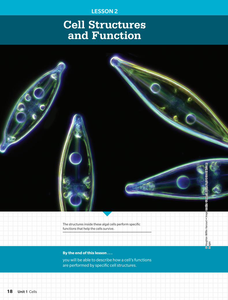
© Houghton Mifflin Harcourt • Image Credits: ©Laguna Design/Oxford Scientific/Getty Images LESSON 2 Cell Structures and Function The structures inside these algal cells perform specific functions that help the cells survive. By the end of this lesson . . . you will be able to describe how a cell’s functions are performed by specific cell structures. Unit 1 Cells 18
-
Upload
khangminh22 -
Category
Documents
-
view
0 -
download
0
Transcript of Cell Structures and Function
© H
ough
ton
Miff
lin H
arco
urt •
Imag
e Cr
edits
: ©La
guna
Des
ign/
Oxfo
rd S
cien
tific
/Get
ty
Imag
es
LESSON 2
Cell Structures and Function
The structures inside these algal cells perform specific functions that help the cells survive.
By the end of this lesson . . .
you will be able to describe how a cell’s functions are performed by specific cell structures.
Unit 1 Cells18
DO NOT EDIT--Changes must be made through "File info"LONumber=6L1_0130; CorrectionKey=NL-A
CAN YOU EXPLAIN IT?
© H
ough
ton
Miff
lin H
arco
urt •
Imag
e Cr
edits
: (l)
©Da
vid
Sucs
y/E+
/Get
ty Im
ages
; (r)
©Ca
llist
a Im
ages
/Sci
ence
Sou
rce
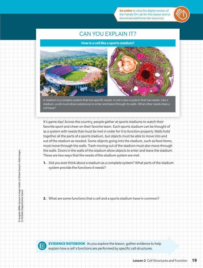
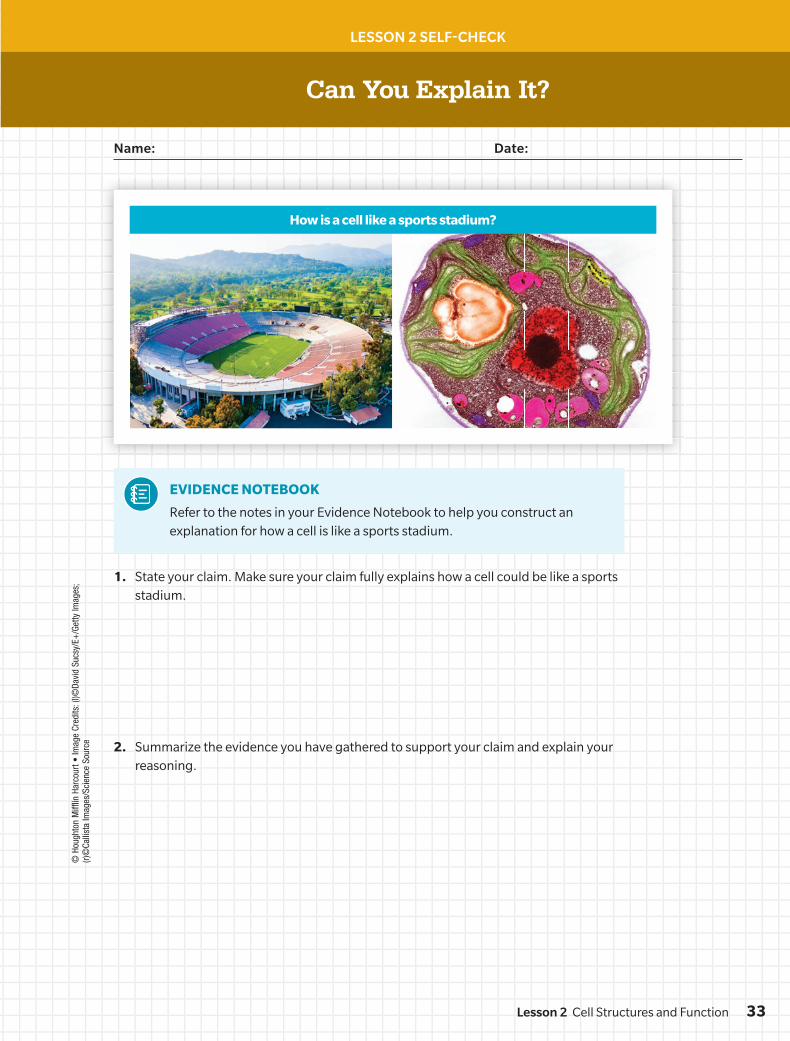
A stadium is a complex system that has specific needs. A cell is also a system that has needs. Like a stadium, a cell must allow substances to enter and leave through its walls. What other needs does a cell have?
How is a cell like a sports stadium?
It’s game day! Across the country, people gather at sports stadiums to watch their favorite sport and cheer on their favorite team. Each sports stadium can be thought of as a system with needs that must be met in order for it to function properly. Walls hold together all the parts of a sports stadium, but objects must be able to move into and out of the stadium as needed. Some objects going into the stadium, such as food items, must move through the walls. Trash moving out of the stadium must also move through the walls. Doors in the walls of the stadium allow objects to enter and leave the stadium. These are two ways that the needs of the stadium system are met.
1. Did you ever think about a stadium as a complete system? What parts of the stadium system provide the functions it needs?
2. What are some functions that a cell and a sports stadium have in common?
EVIDENCE NOTEBOOK As you explore the lesson, gather evidence to help explain how a cell's functions are performed by specific cell structures.
Go online to view the digital version of the Hands-On Lab for this lesson and to download additional lab resources.
19Lesson 2 Cell Structures and Function
DO NOT EDIT--Changes must be made through "File info"LONumber=6L1_0130; CorrectionKey=NL-A
© H
ough
ton
Miff
lin H
arco
urt •
Imag
e Cr
edits
: (l)
©Sc
ienc
e So
urce
/Col
oriz
atio
n by
Mar
y M
artin
; (r)
©Fr
ank
Fox/
Scie
nce
Sour
ce
EXPLORATION 1
Comparing Cell Structures
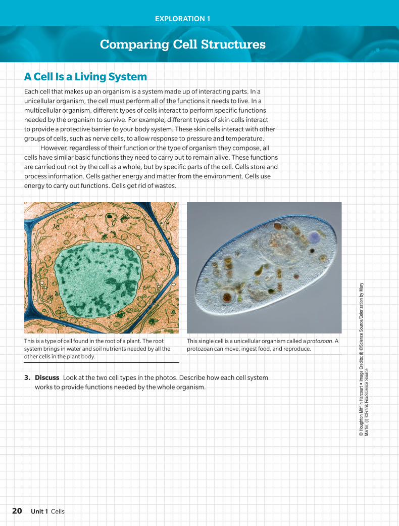
A Cell Is a Living SystemEach cell that makes up an organism is a system made up of interacting parts. In a unicellular organism, the cell must perform all of the functions it needs to live. In a multicellular organism, different types of cells interact to perform specific functions needed by the organism to survive. For example, different types of skin cells interact to provide a protective barrier to your body system. These skin cells interact with other groups of cells, such as nerve cells, to allow response to pressure and temperature.
However, regardless of their function or the type of organism they compose, all cells have similar basic functions they need to carry out to remain alive. These functions are carried out not by the cell as a whole, but by specific parts of the cell. Cells store and process information. Cells gather energy and matter from the environment. Cells use energy to carry out functions. Cells get rid of wastes.
3. Discuss Look at the two cell types in the photos. Describe how each cell system works to provide functions needed by the whole organism.
This is a type of cell found in the root of a plant. The root system brings in water and soil nutrients needed by all the other cells in the plant body.
This single cell is a unicellular organism called a protozoan. A protozoan can move, ingest food, and reproduce.
Unit 1 Cells20
DO NOT EDIT--Changes must be made through “File info” LONumber=6L1_0130; CorrectionKey=NL-B
cell membrane
flagellum
cell wall
cytoplasm
ribosome
genetic material
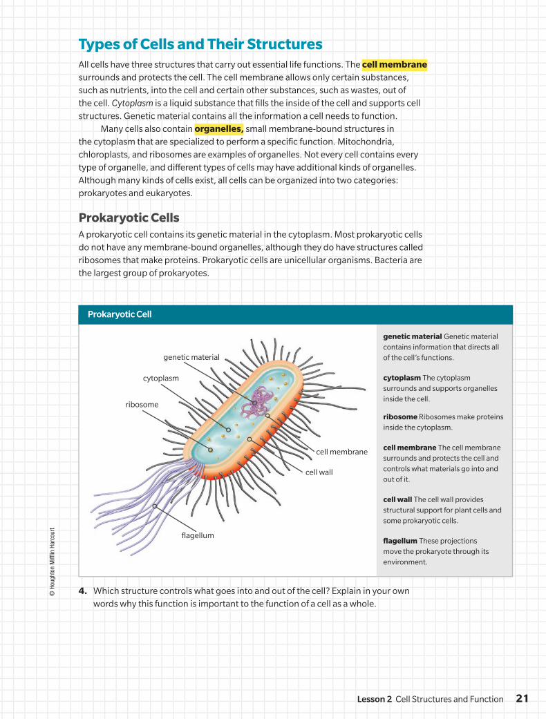
Types of Cells and Their StructuresAll cells have three structures that carry out essential life functions. The cell membrane surrounds and protects the cell. The cell membrane allows only certain substances, such as nutrients, into the cell and certain other substances, such as wastes, out of the cell. Cytoplasm is a liquid substance that fills the inside of the cell and supports cell structures. Genetic material contains all the information a cell needs to function.
Many cells also contain organelles, small membrane-bound structures in the cytoplasm that are specialized to perform a specific function. Mitochondria, chloroplasts, and ribosomes are examples of organelles. Not every cell contains every type of organelle, and different types of cells may have additional kinds of organelles. Although many kinds of cells exist, all cells can be organized into two categories: prokaryotes and eukaryotes.
Prokaryotic CellsA prokaryotic cell contains its genetic material in the cytoplasm. Most prokaryotic cells do not have any membrane-bound organelles, although they do have structures called ribosomes that make proteins. Prokaryotic cells are unicellular organisms. Bacteria are the largest group of prokaryotes.
Prokaryotic Cell
4. Which structure controls what goes into and out of the cell? Explain in your own words why this function is important to the function of a cell as a whole.
© H
ough
ton
Miff
lin H
arco
urt
genetic material Genetic material contains information that directs all of the cell’s functions.
cytoplasm The cytoplasm surrounds and supports organelles inside the cell.
ribosome Ribosomes make proteins inside the cytoplasm.
cell membrane The cell membrane surrounds and protects the cell and controls what materials go into and out of it.
cell wall The cell wall provides structural support for plant cells and some prokaryotic cells.
flagellum These projections move the prokaryote through its environment.
21Lesson 2 Cell Structures and Function
DO NOT EDIT--Changes must be made through “File info”LONumber=6L1_0130; CorrectionKey=NL-B
nucleus
lysosome
Golgi apparatus
cell membrane
cytoplasm
mitochondrion
ribosome
roughendoplasmicreticulum
smoothendoplasmicreticulum
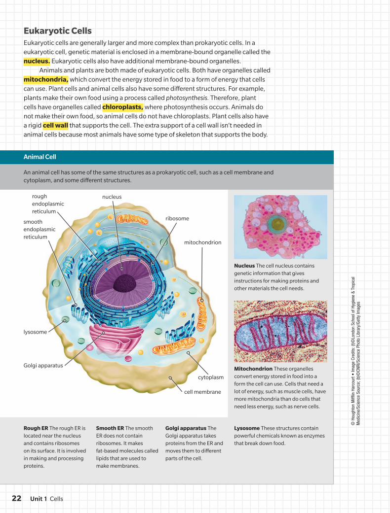
Eukaryotic CellsEukaryotic cells are generally larger and more complex than prokaryotic cells. In a eukaryotic cell, genetic material is enclosed in a membrane-bound organelle called the nucleus. Eukaryotic cells also have additional membrane-bound organelles.
Animals and plants are both made of eukaryotic cells. Both have organelles called mitochondria, which convert the energy stored in food to a form of energy that cells can use. Plant cells and animal cells also have some different structures. For example, plants make their own food using a process called photosynthesis. Therefore, plant cells have organelles called chloroplasts, where photosynthesis occurs. Animals do not make their own food, so animal cells do not have chloroplasts. Plant cells also have a rigid cell wall that supports the cell. The extra support of a cell wall isn’t needed in animal cells because most animals have some type of skeleton that supports the body.
Nucleus The cell nucleus contains genetic information that gives instructions for making proteins and other materials the cell needs.
Rough ER The rough ER is located near the nucleus and contains ribosomes on its surface. It is involved in making and processing proteins.
Smooth ER The smooth ER does not contain ribosomes. It makes fat-based molecules called lipids that are used to make membranes.
Golgi apparatus The Golgi apparatus takes proteins from the ER and moves them to different parts of the cell.
Lysosome These structures contain powerful chemicals known as enzymes that break down food.
Mitochondrion These organelles convert energy stored in food into a form the cell can use. Cells that need a lot of energy, such as muscle cells, have more mitochondria than do cells that need less energy, such as nerve cells.
Animal Cell
An animal cell has some of the same structures as a prokaryotic cell, such as a cell membrane and cytoplasm, and some different structures.
© H
ough
ton
Miff
lin H
arco
urt •
Imag
e Cr
edits
: (t)©
Lond
on S
choo
l of H
ygie
ne &
Tro
pica
l M
edic
ine/
Scie
nce
Sour
ce; (
b)©
CNRI
/Sci
ence
Pho
to L
ibra
ry/G
etty
Imag
es
Unit 1 Cells22
DO NOT EDIT--Changes must be made through “File info”LONumber=6L1_0130; CorrectionKey=NL-B
nucleus
ribosome
vacuole
Golgi apparatus
mitochondrion
cell membranecell wall
chloroplast
smoothendoplasmicreticulum
rough endoplasmicreticulum
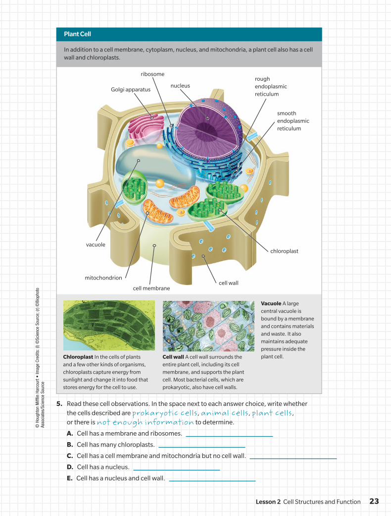
Chloroplast In the cells of plants and a few other kinds of organisms, chloroplasts capture energy from sunlight and change it into food that stores energy for the cell to use.
Cell wall A cell wall surrounds the entire plant cell, including its cell membrane, and supports the plant cell. Most bacterial cells, which are prokaryotic, also have cell walls.
Vacuole A large central vacuole is bound by a membrane and contains materials and waste. It also maintains adequate pressure inside the plant cell.
5. Read these cell observations. In the space next to each answer choice, write whether the cells described are prokaryotic cells, animal cells, plant cells, or there is not enough information to determine.
A. Cell has a membrane and ribosomes.
B. Cell has many chloroplasts.
C. Cell has a cell membrane and mitochondria but no cell wall.
D. Cell has a nucleus.
E. Cell has a nucleus and cell wall.
In addition to a cell membrane, cytoplasm, nucleus, and mitochondria, a plant cell also has a cell wall and chloroplasts.
Plant Cell©
Hou
ghto
n M
ifflin
Har
cour
t • Im
age
Cred
its: (
l) ©
Scie
nce
Sour
ce; (
r) ©
Biop
hoto
As
soci
ates
/Sci
ence
Sou
rce
23Lesson 2 Cell Structures and Function
DO NOT EDIT--Changes must be made through “File info”LONumber=6L1_0130; CorrectionKey=NL-B
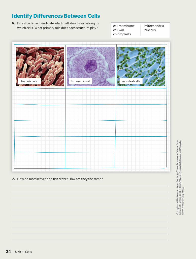
Identify Differences Between Cells6. Fill in the table to indicate which cell structures belong to
which cells. What primary role does each structure play?
7. How do moss leaves and fish differ? How are they the same?
cell membranecell wallchloroplasts
mitochondrianucleus
© H
ough
ton
Miff
lin H
arco
urt •
Imag
e Cr
edits
: (l)
©St
eve
Gsch
mei
ssne
r/Sc
ienc
e Ph
oto
Libr
ary/
Getty
Imag
es; (
c) ©
Ed R
esch
ke/O
xfor
d Sc
ient
ific/
Getty
Imag
es; (
r) ©
Alan
Joh
n La
nder
Phi
llips
/E+
/Get
ty Im
ages
bacteria cells fish embryo cell moss leaf cells
Unit 1 Cells24
DO NOT EDIT--Changes must be made through “File info”LONumber=6L1_0130; CorrectionKey=NL-B
© H
ough
ton
Miff
lin H
arco
urt •
Imag
e Cr
edits
: (tl)
©Jo
seph
F. G
enna
ro, J
r./Sc
ienc
e So
urce
; (tr
) ©Lo
ndon
Sch
ool o
f Hyg
iene
& T
ropi
cal M
edic
ine/
Scie
nce
Sour
ce; (
bl)©
CNRI
/Sci
ence
Ph
oto
Libr
ary/
Getty
Imag
es; (
br)©
Scie
nce
Sour
ce
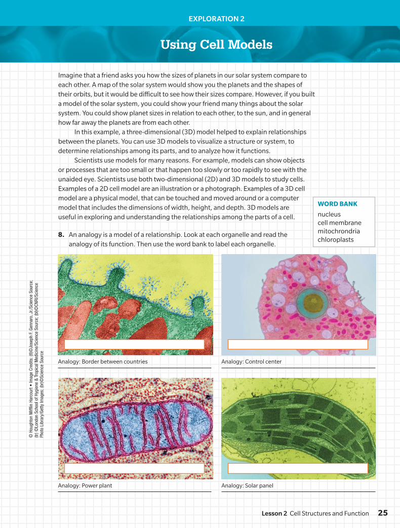
EXPLORATION 2
Imagine that a friend asks you how the sizes of planets in our solar system compare to each other. A map of the solar system would show you the planets and the shapes of their orbits, but it would be difficult to see how their sizes compare. However, if you built a model of the solar system, you could show your friend many things about the solar system. You could show planet sizes in relation to each other, to the sun, and in general how far away the planets are from each other.
In this example, a three-dimensional (3D) model helped to explain relationships between the planets. You can use 3D models to visualize a structure or system, to determine relationships among its parts, and to analyze how it functions.
Scientists use models for many reasons. For example, models can show objects or processes that are too small or that happen too slowly or too rapidly to see with the unaided eye. Scientists use both two-dimensional (2D) and 3D models to study cells. Examples of a 2D cell model are an illustration or a photograph. Examples of a 3D cell model are a physical model, that can be touched and moved around or a computer model that includes the dimensions of width, height, and depth. 3D models are useful in exploring and understanding the relationships among the parts of a cell.
Using Cell Models
8. An analogy is a model of a relationship. Look at each organelle and read the analogy of its function. Then use the word bank to label each organelle.
Analogy: Border between countries
Analogy: Power plant Analogy: Solar panel
Analogy: Control center
WORD BANK
nucleuscell membranemitochrondriachloroplasts
25Lesson 2 Cell Structures and Function
DO NOT EDIT--Changes must be made through “File info”LONumber=6L1_0130; CorrectionKey=NL-B
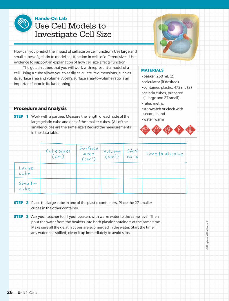
MATERIALS
• beaker, 250 mL (2)
• calculator (if desired)
• container, plastic, 473 mL (2)
• gelatin cubes, prepared (1 large and 27 small)
• ruler, metric
• stopwatch or clock with second hand
• water, warm
Hands-On Lab
Use Cell Models to Investigate Cell Size
How can you predict the impact of cell size on cell function? Use large and small cubes of gelatin to model cell function in cells of different sizes. Use evidence to support an explanation of how cell size affects function.
The gelatin cubes that you will work with represent a model of a cell. Using a cube allows you to easily calculate its dimensions, such as its surface area and volume. A cell’s surface area-to-volume ratio is an important factor in its functioning.
Procedure and Analysis
STEP 1 Work with a partner. Measure the length of each side of the large gelatin cube and one of the smaller cubes. (All of the smaller cubes are the same size.) Record the measurements in the data table.
Surface area
(cm2)Volume (cm3)
SA:V ratio Time to dissolveCube sides
(cm)
Large cube
Smaller cubes
© H
ough
ton
Miff
lin H
arco
urt
STEP 2 Place the large cube in one of the plastic containers. Place the 27 smaller cubes in the other container.
STEP 3 Ask your teacher to fill your beakers with warm water to the same level. Then pour the water from the beakers into both plastic containers at the same time. Make sure all the gelatin cubes are submerged in the water. Start the timer. If any water has spilled, clean it up immediately to avoid slips.
26 Unit 1 Cells
DO NOT EDIT--Changes must be made through “File info”LONumber=6L1_0130; CorrectionKey=NL-B
© H
ough
ton
Miff
lin H
arco
urt
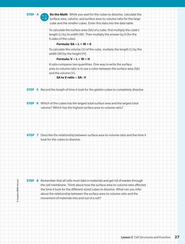
STEP 4 Do the Math While you wait for the cubes to dissolve, calculate the surface area, volume, and surface area-to-volume ratio for the large cube and the smaller cubes. Enter this data into the data table.
To calculate the surface area (SA) of a cube, first multiply the cube’s length (L) by its width (W). Then multiply the answer by 6 (for the 6 sides of the cube).
Formula: SA = L × W × 6
To calculate the volume (V) of the cube, multiply the length (L) by the width (W) by the height (H).
Formula: V = L × W × H
A ratio compares two quantities. One way to write the surface area-to-volume ratio is to use a colon between the surface area (SA) and the volume (V).
SA to V ratio = SA : V
STEP 5 Record the length of time it took for the gelatin cubes to completely dissolve.
STEP 6 Which of the cubes has the largest total surface area and the largest total volume? Which has the highest surface area-to-volume ratio?
STEP 7 Describe the relationship between surface area-to-volume ratio and the time it took for the cubes to dissolve.
STEP 8 Remember that all cells must take in materials and get rid of wastes through the cell membrane. Think about how the surface area-to-volume ratio affected the time it took for the different-sized cubes to dissolve. What can you infer about the relationship between the surface area-to-volume ratio and the movement of materials into and out of a cell?
27Lesson 2 Cell Structures and Function
DO NOT EDIT--Changes must be made through “File info” LONumber=6L1_0130; CorrectionKey=NL-B
© H
ough
ton
Miff
lin H
arco
urt •
Imag
e Cr
edits
: (r)
©Sp
ence
r Sut
ton/
Scie
nce
Sour
ce
EVIDENCE NOTEBOOK
10. Think about the model you are developing that compares a sports stadium to a cell. Describe the primary role each organelle plays in a cell and identify a similar role that is filled in a sports stadium. How do these components contribute to the function of the whole system for a cell and a sports stadium? Record your evidence.
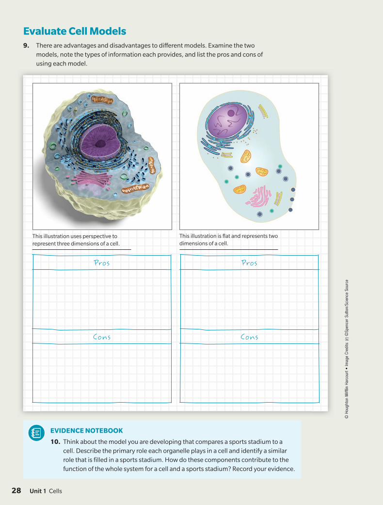
Evaluate Cell Models9. There are advantages and disadvantages to different models. Examine the two
models, note the types of information each provides, and list the pros and cons of using each model.
This illustration uses perspective to represent three dimensions of a cell.
This illustration is flat and represents two dimensions of a cell.
Pros Pros
Cons Cons
28 Unit 1 Cells
DO NOT EDIT--Changes must be made through “File info”LONumber=6L1_0130; CorrectionKey=NL-B
EXPLORATION 3
Explaining Limits to Cell Size
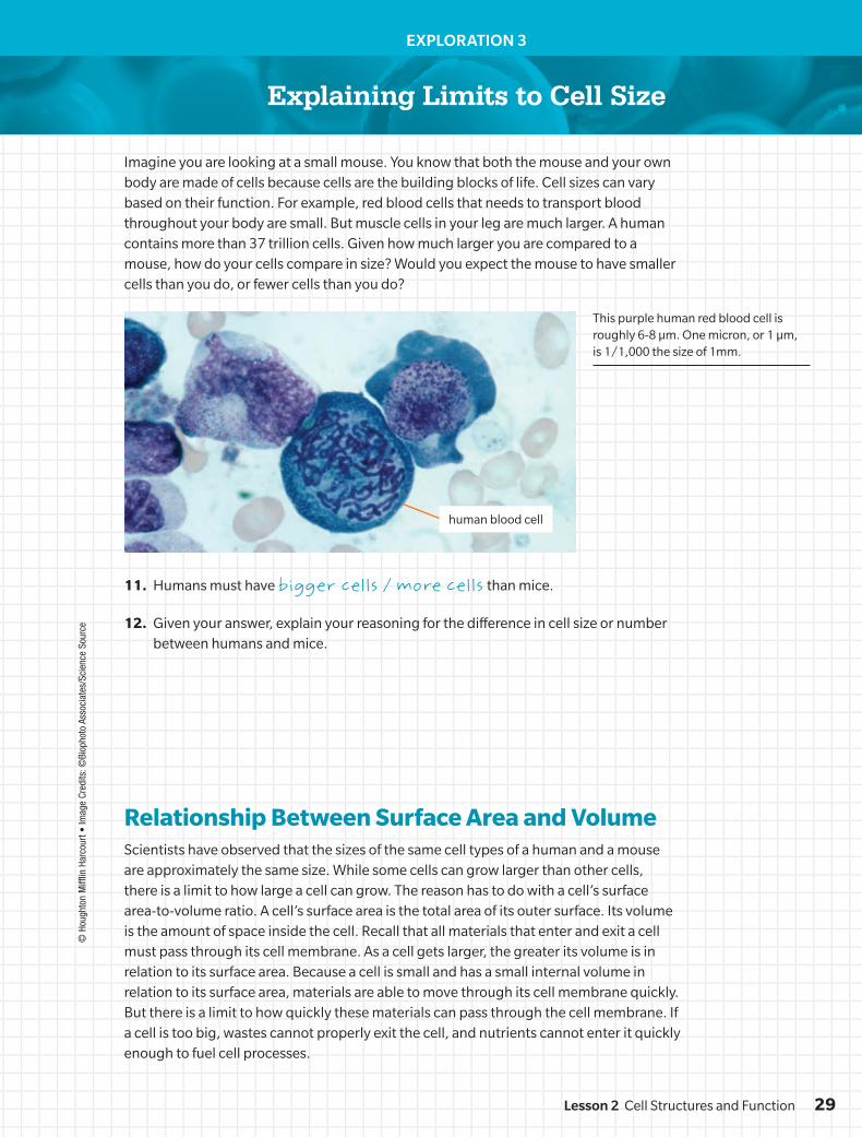
Imagine you are looking at a small mouse. You know that both the mouse and your own body are made of cells because cells are the building blocks of life. Cell sizes can vary based on their function. For example, red blood cells that needs to transport blood throughout your body are small. But muscle cells in your leg are much larger. A human contains more than 37 trillion cells. Given how much larger you are compared to a mouse, how do your cells compare in size? Would you expect the mouse to have smaller cells than you do, or fewer cells than you do?
11. Humans must have bigger cells / more cells than mice.
12. Given your answer, explain your reasoning for the difference in cell size or number between humans and mice.
Relationship Between Surface Area and VolumeScientists have observed that the sizes of the same cell types of a human and a mouse are approximately the same size. While some cells can grow larger than other cells, there is a limit to how large a cell can grow. The reason has to do with a cell’s surface area-to-volume ratio. A cell’s surface area is the total area of its outer surface. Its volume is the amount of space inside the cell. Recall that all materials that enter and exit a cell must pass through its cell membrane. As a cell gets larger, the greater its volume is in relation to its surface area. Because a cell is small and has a small internal volume in relation to its surface area, materials are able to move through its cell membrane quickly. But there is a limit to how quickly these materials can pass through the cell membrane. If a cell is too big, wastes cannot properly exit the cell, and nutrients cannot enter it quickly enough to fuel cell processes.
This purple human red blood cell is roughly 6-8 µm. One micron, or 1 µm, is 1/1,000 the size of 1mm.
© H
ough
ton
Miff
lin H
arco
urt •
Imag
e Cr
edits
: ©Bi
opho
to A
ssoc
iate
s/Sc
ienc
e So
urce
human blood cell
29Lesson 2 Cell Structures and Function
DO NOT EDIT--Changes must be made through “File info”LONumber=6L1_0130; CorrectionKey=NL-B
© H
ough
ton
Miff
lin H
arco
urt
EVIDENCE NOTEBOOK
13. While a sports stadium is a system much like a cell is, it does not have the same limits on size that cells do. How do other parts of a cell depend on the function of the cell membrane and how is this relationship different between the components of a sports stadium? Record your evidence.
Relate Structure of Cell Membrane to Cell Size14. What problems might result if a cell gets too large? Circle all that apply.
A. Wastes will not be able to leave the cell quickly enough.
B. Nutrients will not be able to reach all parts of the cell quickly.
C. Nutrients will move into the cell too quickly for the cell to use.
D. Wastes will leave the cell quickly and take necessary nutrients with them.
15. Language SmArts Citing evidence from the text and from your calculations, explain why cells are unable to perform important functions if they become too large.
16. Engineer It You are designing a building that must minimize energy transfer in the form of heat loss. Describe a building shape that you think would minimize heat loss through the surface of the building, and explain how that shape relates to the surface area-to-volume ratio.
Unit 1 Cells30
DO NOT EDIT--Changes must be made through “File info”LONumber=6L1_0130; CorrectionKey=NL-B
TAKE IT FURTHER
Name: Date:
© H
ough
ton
Miff
lin H
arco
urt •
Imag
e Cr
edits
: ©Na
ncy
R. S
chiff
/Arc
hive
Pho
tos/
Getty
Im
ages
Go online to choose one of these other paths.
People in Science
Check out the path below or go online to choose one of the other paths shown.
Continue Your Exploration
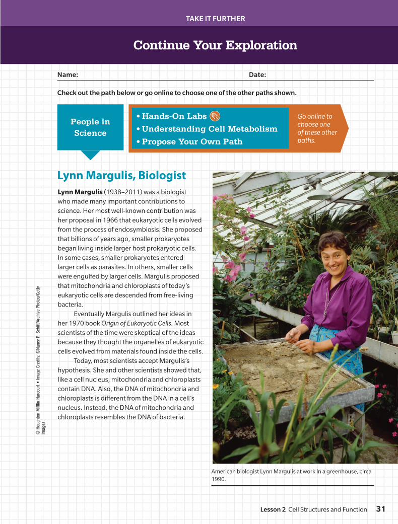
Lynn Margulis, BiologistLynn Margulis (1938–2011) was a biologist who made many important contributions to science. Her most well-known contribution was her proposal in 1966 that eukaryotic cells evolved from the process of endosymbiosis. She proposed that billions of years ago, smaller prokaryotes began living inside larger host prokaryotic cells. In some cases, smaller prokaryotes entered larger cells as parasites. In others, smaller cells were engulfed by larger cells. Margulis proposed that mitochondria and chloroplasts of today’s eukaryotic cells are descended from free-living bacteria.
Eventually Margulis outlined her ideas in her 1970 book Origin of Eukaryotic Cells. Most scientists of the time were skeptical of the ideas because they thought the organelles of eukaryotic cells evolved from materials found inside the cells.
Today, most scientists accept Margulis’s hypothesis. She and other scientists showed that, like a cell nucleus, mitochondria and chloroplasts contain DNA. Also, the DNA of mitochondria and chloroplasts is different from the DNA in a cell’s nucleus. Instead, the DNA of mitochondria and chloroplasts resembles the DNA of bacteria.
• Hands-On Labs
• Understanding Cell Metabolism
• Propose Your Own Path
American biologist Lynn Margulis at work in a greenhouse, circa 1990.
31Lesson 2 Cell Structures and Function
DO NOT EDIT--Changes must be made through “File info”LONumber=6L1_0130; CorrectionKey=NL-B
TAKE IT FURTHER
© H
ough
ton
Miff
lin H
arco
urt
1. Which statements provide evidence to support Lynn Margulis’s hypothesis of endosymbiosis? Arrange the statements below into the order that accurately shows the sequence of events described in Margulis’s hypothesis by writing the number 1, 2, 3, or 4 next to each statement.
Prokaryotes inside other prokaryotes evolved into organelles.
Prokaryotes lived inside other prokaryotes in a symbiotic relationship.
Prokaryotes that had engulfed other prokaryotes evolved into eukaryotes.
Free-living prokaryotes engulfed other free-living prokaryotes.
2. How do the findings of Margulis and other scientists—that mitochondria and chloroplasts have their own DNA, like a cell’s nucleus—support the hypothesis of endosymbiosis?
3. Explain how Lynn Margulis’s hypothesis changed scientific ideas about cell development.
Continue Your Exploration
4. Collaborate Research the prokaryotic organism that most likely evolved into a chloroplast. Develop a model of a chloroplast and the prokaryote and describe the similarities and differences in the structure and function of individual components and the whole system.
32 Unit 1 Cells
DO NOT EDIT--Changes must be made through “File info”LONumber=6L1_0130; CorrectionKey=NL-B
© H
ough
ton
Miff
lin H
arco
urt •
Imag
e Cr
edits
:
Name: Date:
LESSON 2 SELF-CHECK
EVIDENCE NOTEBOOK
Refer to the notes in your Evidence Notebook to help you construct an explanation for how a cell is like a sports stadium.
How is a cell like a sports stadium?
Can You Explain It?
1. State your claim. Make sure your claim fully explains how a cell could be like a sports stadium.
2. Summarize the evidence you have gathered to support your claim and explain your reasoning.
© H
ough
ton
Miff
lin H
arco
urt •
Imag
e Cr
edits
: (l)©
Davi
d Su
csy/
E+/G
etty
Imag
es;
(r)©
Calli
sta
Imag
es/S
cien
ce S
ourc
e
33Lesson 2 Cell Structures and Function
DO NOT EDIT--Changes must be made through “File info”LONumber=6L1_0130; CorrectionKey=NL-B
© H
ough
ton
Miff
lin H
arco
urt •
Imag
e Cr
edits
: ©
Hou
ghto
n M
ifflin
Har
cour
t • Im
age
Cred
its: ©
Alan
Joh
n La
nder
Phi
llips
/E+
/Get
ty Im
ages
LESSON 2 SELF-CHECK
Checkpoints
Answer the following questions to check your understanding of the lesson.
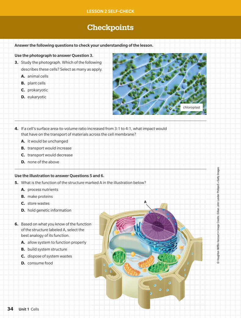
Use the photograph to answer Question 3.
3. Study the photograph. Which of the following
describes these cells? Select as many as apply.
A. animal cells
B. plant cells
C. prokaryotic
D. eukaryotic
4. If a cell’s surface area-to-volume ratio increased from 3:1 to 4:1, what impact would that have on the transport of materials across the cell membrane?
A. it would be unchanged
B. transport would increase
C. transport would decrease
D. none of the above
Use the illustration to answer Questions 5 and 6.
5. What is the function of the structure marked A in the illustration below?
A. process nutrients
B. make proteins
C. store wastes
D. hold genetic information
6. Based on what you know of the function of the structure labeled A, select the best analogy of its function.
A. allow system to function properly
B. build system structure
C. dispose of system wastes
D. consume food
chloroplast
A
34 Unit 1 Cells
DO NOT EDIT--Changes must be made through “File info”LONumber=6L1_0130; CorrectionKey=NL-B
LESSON 2 SELF-CHECK
Interactive Review
Complete this section to review the main concepts of the lesson.
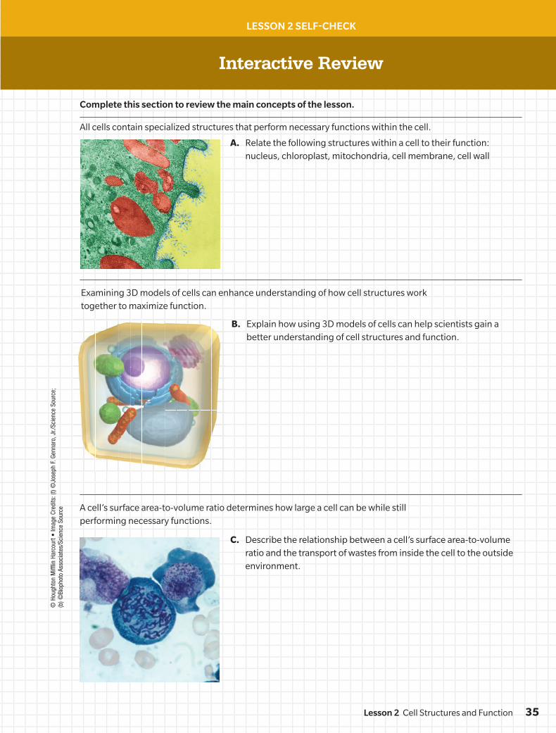
A. Relate the following structures within a cell to their function: nucleus, chloroplast, mitochondria, cell membrane, cell wall
C. Describe the relationship between a cell’s surface area-to-volume ratio and the transport of wastes from inside the cell to the outside environment.
B. Explain how using 3D models of cells can help scientists gain a better understanding of cell structures and function.
All cells contain specialized structures that perform necessary functions within the cell.
A cell’s surface area-to-volume ratio determines how large a cell can be while still performing necessary functions.
Examining 3D models of cells can enhance understanding of how cell structures work together to maximize function.
© H
ough
ton
Miff
lin H
arco
urt •
Imag
e Cr
edits
: (t)
©Jo
seph
F. G
enna
ro, J
r./Sc
ienc
e So
urce
; (b
) ©Bi
opho
to A
ssoc
iate
s/Sc
ienc
e So
urce
35Lesson 2 Cell Structures and Function
DO NOT EDIT--Changes must be made through “File info”LONumber=6L1_0130; CorrectionKey=NL-B