Terminal osteoblast differentiation, mediated by runx2 and p27KIP1, is disrupted in osteosarcoma
Upload
independentCategory
view
0download
0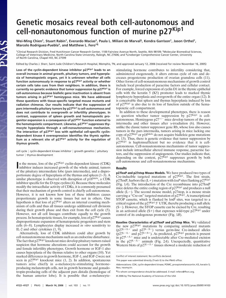
Genetic mosaics reveal both cell-autonomous andcell-nonautonomous function of murine p27Kip1
Wei-Ming Chien†, Stuart Rabin†, Everardo Macias‡, Paula L. Miliani de Marval§, Kendra Garrison†, Jason Orthel†,Marcelo Rodriguez-Puebla‡, and Matthew L. Fero†¶
†Clinical Research Division, Fred Hutchinson Cancer Research Center, 1100 Fairview Avenue North, Seattle, WA 98109; ‡Molecular Biomedical Sciences,College of Veterinary Medicine, North Carolina State University, Raleigh, NC 27606; and §Lineberger Comprehensive Cancer Center, Universityof North Carolina, Chapel Hill, NC 27599
Edited by Charles J. Sherr, Saint Jude Children’s Research Hospital, Memphis, TN, and approved January 12, 2006 (received for review November 16, 2005)
Loss of the cyclin-dependent kinase inhibitor p27Kip1 leads to anoverall increase in animal growth, pituitary tumors, and hyperpla-sia of hematopoietic organs, yet it is unknown whether all cellsfunction autonomously in response to p27Kip1 activity or whethercertain cells take cues from their neighbors. In addition, there iscurrently no genetic evidence that tumor suppression by p27Kip1 iscell-autonomous because biallelic gene inactivation is absent fromtumors arising in p27Kip1 hemizygous mice. We have addressedthese questions with tissue-specific targeted mouse mutants andradiation chimeras. Our results indicate that the suppression ofpars intermedia pituitary tumors by p27Kip1 is cell-autonomous anddoes not contribute to overgrowth or infertility phenotypes. Incontrast, suppression of spleen growth and hematopoietic pro-genitor expansion is a consequence of p27Kip1 function external tothe hematopoietic compartment. Likewise, p27Kip1 suppresses thy-mocyte hyperplasia through a cell-nonautonomous mechanism.The interaction of p27Kip1 loss with epithelial cell-specific cyclin-dependent kinase 4 overexpression identifies the thymic epithe-lium as a relevant site of p27Kip1 activity for the regulation ofthymus growth.
cell cycle � cyclin-dependent kinase inhibitor � growth genetics � pituitarytumor � thymus development
In the mouse, loss of the p27Kip1 cyclin-dependent kinase (CDK)inhibitor induces increased growth of the whole animal, tumors
of the pituitary intermediate lobe (pars intermedia), and a dispro-portionate degree of hyperplasia of the thymus and spleen (1–3). Asimilar phenotype is observed with disruption of p18Ink4c, a CDKinhibitor specific for the D-type cyclins (4). Because these moleculesmodify the intracellular activity of CDKs, it is commonly presumedthat their mechanism of growth control is chiefly cell-autonomous.However, it is not known how loss of these inhibitors causesproportionate growth in some tissues but not in others. Onehypothesis is that loss of p27Kip1 alters an internal counting mech-anism of cells and thus all tissues undergo additional cell divisionsduring their growth phase and then exit from the cell cycle (5).However, not all cell lineages contribute equally to the growthprocess. In hematopoietic tissues, for example, loss of p27Kip1 causesdisproportionate expansion of hematopoietic progenitors and stemcells (6–8). Lymphocytes display increased in vitro sensitivity toIL-2 and other cytokines (1, 9).
Alternatively, loss of CDK inhibitors could alter growth bycell-nonautonomous mechanisms such as an endocrine disturbance.The fact that p27Kip1 knockout mice develop pituitary tumors raisedsuspicion that hormone alterations could account for the growthand female infertility phenotypes. Growth hormone or IGF-1 alsocauses hyperplasia of the thymus relative to other organs (10). Yetmarked differences in growth hormone, IGF-1, and IGF-2 were notseen in p27Kip1 knockout mice (1, 2). In addition, spontaneoustumors arise chief ly in �-melanocyte-stimulating hormone-producing melanotroph cells, not in growth hormone and gonado-tropin-producing cells of the adjacent pars distalis (homologue ofthe human anterior lobe). It is possible that �-melanocyte-
stimulating hormone contributes to infertility considering that,administered exogenously, it alters estrous cycle of rats and de-creases progesterone production of ovarian granulosa cells (11).Other forms of cell-nonautonomous mechanisms of growth controlinclude local production of paracrine factors and cellular contact.For example, forced expression of cyclin D1 in the thymic epithelialcells with the keratin 5 (K5) promoter leads to marked thymiclymphocyte hyperplasia and overgrowth of the entire organ (12). Itis conceivable that spleen and thymus hyperplasia induced by lossof p27Kip1 is also due to its loss of function outside of the hema-topoietic cell compartment.
In addition to these developmental phenotypes, there is reasonto question whether tumor suppression by p27Kip1 is cell-autonomous. Hemizygous p27�/� mice develop tumors of the parsintermedia and other tissues after �-irradiation (6). However,unlike the classic tumor suppressor gene Rb, whose loss also causestumors in the pars intermedia, tumors arising in mice lacking onecopy of p27Kip1 or p18Ink4c do not acquire bialleleic gene mutations(6, 13). Thus, there is genetic evidence that tumor suppression byp27Kip1 is haploinsufficient but no evidence that it is cell-autonomous. Cell-nonautonomous mechanisms of tumor suppres-sion include intracellular altered immune response, paracrine fac-tors, and the suppression of angiogenesis. Our studies indicate that,depending on the context, p27Kip1 suppresses growth by bothcell-autonomous and cell-nonautonomous mechanisms.
Resultsp27loxP and p27stop Mouse Models. We have produced two types ofCre-inducible targeted mutations of p27Kip1. The first strain,p27loxP, harbors the (L�) mutation with loxP sites flanking p27Kip1
(Fig. 1A). The introduction of the Cre recombinase into p27loxPmice deletes the entire coding region of p27Kip1 and produces a nullallele (L�). The second mouse model, p27stop, is a mouse straincarrying a ‘‘Cre-on’’ targeted mutation of p27Kip1. A transcriptionalSTOP cassette, which is flanked by loxP sites, was targeted to acritical region of the p27Kip1 5� UTR, thereby producing a null allele(S�). However, the STOP cassette can be excised by Cre, resultingin an activated allele (S�) that expresses wild-type p27Kip1 undercontrol of its endogenous promoter (Fig. 1B).
Baseline Characteristics of p27loxP and p27stop Mice. We validatedthe new p27Kip1 mutations by comparing uninduced mutants(p27L�/L� and p27S�/S�) versus germ-line Cre-induced alleles(p27L�/L� and p27S�/S�). As predicted, p27Kip1 protein is presentin p27L�/L� mice and is undetectable after Cre-mediated excisionin the p27L�/L� animals (Fig. 2A). Unexpectedly, quantitativeWestern blots of p27L�/L� tissues showed a moderate reduction of
Conflict of interest statement: No conflicts declared.
This paper was submitted directly (Track II) to the PNAS office.
Abbreviations: CDK, cyclin-dependent kinase; POMC, proopiomelanocortin; K5, keratin 5;Neo, neomycin.
¶To whom correspondence should be addressed. E-mail: [email protected].
© 2006 by The National Academy of Sciences of the USA
4122–4127 � PNAS � March 14, 2006 � vol. 103 � no. 11 www.pnas.org�cgi�doi�10.1073�pnas.0509514103
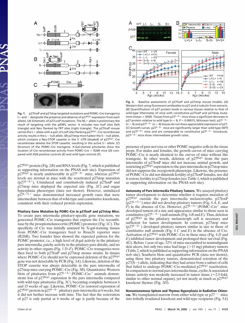
p27Kip1 protein (Fig. 2B) and RNA levels (Fig. 7, which is publishedas supporting information on the PNAS web site). Expression ofp27Kip1 is nearly undetectable in p27S�/S� mice, whereas p27Kip1
levels are normal in mice with the reactivated p27stop mutation(p27S�/S�). Uninduced and constitutively induced p27loxP andp27stop mice displayed the expected size (Fig. 2C) and organhyperplasia phenotypes (data not shown). However, uninducedp27L�/L� mice demonstrated increased growth rates that wereintermediate between that of wild-type and constitutive knockouts,consistent with their reduced protein expression.
Pituitary Gene Mutation by POMC–Cre in p27loxP and p27stop Mice.To create pars intermedia pituitary-specific gene mutations, wegenerated POMC–Cre transgenics that express the Cre recombi-nase by the proopiomelanocortin (POMC) promoter (Fig. 1C). Thespecificity of Cre was initially assessed by X-gal-staining tissuesfrom POMC–Cre transgenics bred to Rosa26 reporter mice(R26R). Two founder lines showed the expected pattern for thePOMC promoter, i.e., a high level of �-gal activity in the pituitarypars intermedia, patchy activity in the pituitary pars distalis, and noactivity in other organs (Fig. 1 D–F). POMC–Cre transgenics werethen bred to both p27loxP and p27stop mouse strains. In tissueswhere POMC–Cre should not be expressed deletion of the p27Kip1
gene was not detectable by PCR (Fig. 3A). Likewise, deletion of theSTOP cassette was always detectable in the pars intermedia ofp27stop mice carrying POMC–Cre (Fig. 3B). Quantitative Westernblots of pituitaries from p27L�/L�;POMC–Cre� animals demon-strate loss of p27Kip1 expression in the pars intermedia comparedwith wild-type pituitaries (Fig. 3C), becoming complete between 6and 15 weeks of age. Likewise, POMC–Cre restored expression ofp27Kip1 protein in p27S�/S� pituitary pars intermedia at 6 weeks, butit did not further increase with time. The fact that the restorationof p27 is only partial at 6 weeks of age is partly because of the
presence of pars nervosa or other POMC-negative cells in the tissuepreps. For males and females, the growth curves of mice carryingPOMC–Cre is nearly identical to the curves of mice without thistransgene. In other words, deletion of p27Kip1 from the parsintermedia of p27loxP mice did not increase animal growth, andrestricting p27Kip1 expression to the pars intermedia in p27stop micedid not suppress the overgrowth phenotype. Likewise, the presenceof POMC–Cre did not diminish fertility of p27loxP females, nor didit restore fertility to p27stop females (see Table 1, which is publishedas supporting information on the PNAS web site).
Autonomy of Pars Intermedia Pituitary Tumors. We assayed pituitarytumor development to determine whether it requires mutation ofp27Kip1 outside the pars intermedia melanotrophs. p27loxP(p27L�/L�) mice did not develop pituitary tumors (Fig. 4 A, E, andF) in the absence of Cre. However, p27L�/L�;POMC–Cre� micedeveloped tumors of a size and appearance similar to those seen inconstitutive (p27L�/L�) null animals (Fig. 4 B and E). Thus, deletionof p27Kip1 in the pituitary melanotroph cell is necessary andsufficient for tumor development. In contrast, p27stop mice(p27S�/S�) developed pituitary tumors similar in size to those ofconstitutive null animals (Fig. 4 C and E) in the absence of Cre.Activation of p27Kip1 with POMC–Cre in these mice (Fig. 4 D andE) inhibited tumor development and prolonged their survival (Fig.4G). Before 1 year of age, 32% of mice succumbed to nonmalignantskin ulcers, but only two mice had large (�15 mg) pituitary tumors(Table 2, which is published as supporting information on the PNASweb site). Southern blots and quantitative PCR (data not shown),using these two pituitary tumors, demonstrated retention of thep27(S�) allele, indicating that they had arisen from a subset of cellsthat failed to undergo POMC–Cre-mediated p27Kip1 reactivation.In comparison to normal pars intermedia tissue, cyclin A-associatedkinase activity was modestly increased in tumor tissue (�2.5-fold,similar to other normal organs), yet not nearly as much as p27Kip1
knockout thymus (Fig. 3D).
Nonautonomous Spleen and Thymus Hyperplasia in Radiation Chime-ras. We transplanted marrow from either wild-type or p27�/� miceinto lethally irradiated knockout and wild-type recipients (Fig. 5A)
Fig. 2. Baseline assessments of p27loxP and p27stop mouse models. (A)Western blot using fluorescent antibodies to p27 and �-tubulin from extracts.(B) Quantification of p27 protein levels in various tissues relative to that ofwild-type littermates of mice with constitutive p27loxP and p27stop muta-tions (mean � SEM). Tissues from p27L�/L� mice show a significant decrease inp27 protein relative to wild type (n � 8, P � 0.00015, Wilcoxon test). p27L�/L�
(n � 4) and p27S�/S� (n � 4) tissues do not show appreciable expression of p27.(C) Growth curves. p27S�/S� mice are significantly larger than wild-type (WT)and p27S�/S� mice and are comparable to constitutive p27L�/L� knockouts.p27L�/L� mice show intermediate growth rates.
Fig. 1. p27loxP and p27stop targeted mutations and POMC–Cre transgenics(� and � designate the presence and absence of p27Kip1 expression from eachallele). (A) Schematic of p27LoxP mutations. The NL� allele is preliminary theresult of targeting with the pKNFL vector. It includes two loxP sites (lefttriangle) and Neo flanked by FRT sites (right triangle). The p27loxP mousecarries the L� allele with a pair of LoxP sites flanking p27Kip1. Cre recombinaseactivity results in the L� null allele. (B) p27stop mice harbor the S� null allele,which contains a Neo-STOP cassette in the 5� UTR (shaded) of p27Kip1. Crerecombinase deletes the STOP cassette, resulting in the active S� allele. (C)Structure of the POMC–Cre transgene. X-Gal-stained pituitaries show thelocation of Cre recombinase activity from POMC–Cre � R26R mice (D) com-pared with R26-positive controls (E) and wild-type controls (F).
Chien et al. PNAS � March 14, 2006 � vol. 103 � no. 11 � 4123
CELL
BIO
LOG
Y

to test whether p27Kip1 suppresses hematopoietic cell growth by acell-autonomous mechanism. Recipients received transplants at 3weeks of age, before the pubertal growth phase, and were necrop-sied after full donor engraftment, 4–5 weeks later. Flow cytometryusing CD45 cell surface markers, which distinguished donor fromrecipient hematopoietic cells, confirmed that the recipients hadachieved �98% donor chimerism. The spleens of p27�/� recipientanimals were enlarged and hypercellular compared with p27�/�
recipients, regardless of the genotype of the donor cells with whichthey had been transplanted (Fig. 5B). In addition, the spleens ofp27�/� recipients harbored increased proportions of hematopoieticprogenitor cells, even if reconstituted with p27�/� hematopoieticcells (Fig. 5C). This result indicates that loss of p27Kip1 causesexpansion of hematopoietic progenitors by a mechanism involvingcells outside the hematopoietic cell compartment.
Thymuses of p27�/� recipient animals were hyperplastic com-pared with wild-type recipients regardless of the genotype of thetransplanted hematopoietic cells (Fig. 5D). The presence or ab-sence of p27 protein in the thymuses was a function of the genotypeof the donor mice, which is expected because lymphocytes are thepredominant cell type in this organ. In addition, both cyclin A (Fig.5E) and Cdk2-associated histone H1 kinase activity are increasedin the mice transplanted with p27�/� donor cells. Flow cytometryof transplanted animals demonstrated that all four groups hadsimilar profiles of lymphocyte differentiation as determined byCD3, CD4, and CD8 subsets (data not shown). However, mice thatreceived p27�/� donor marrow had a 2-fold relative increase in cellsharboring the ��� T cell receptor (Fig. 5F), which others haveshown to be characteristic of p27Kip1 knockout mice (14).
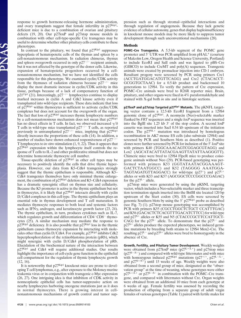
It has been reported that the K5 promoter is active in the thymicepithelium and that forced expression of cyclin D1 by the K5promoter causes marked thymus hyperplasia. However, the thymiclymphocyte expansion was cell-nonautonomous because cyclin Dwas not overexpressed in T cells (12). To determine whether forcedexpression of Cdk4, a catalytic partner of cyclin D1, has a similareffect, we assessed thymuses of K5–Cdk4 transgenic mice (15).Western blots for Cdk4 (Fig. 6A) indicate that it is overexpressedin thymic stromal tissues but not in thymocyte suspensions. How-ever, unlike K5–cyclin D1 or K5–cyclin D2 transgenics, K5–Cdk4mice did not develop progressive thymus hyperplasia (Fig. 6B) (12,16). This finding is consistent with the hypothesis that cyclin D islimiting in the thymic epithelium and that forced expression of Cdk4in this tissue is not sufficient to induce organ hyperplasia. To test thehypothesis that p27Kip1 loss cooperates with overexpression of Cdk4in the thymic epithelium, we bred K5–Cdk4 mice to p27Kip1
knockouts. Remarkably, p27�/�;K5–Cdk4� compound mutant an-
Fig. 3. Effects of POMC–Cre expression in p27loxP and p27stop mice. (A) PCRof tissues from p27L�/L�;POMC–Cre� mice for the uninduced (L�) and induced(L�) null version of the p27loxP allele. (B) PCR of similar tissues from ap27S�/S�;POMC–Cre� mouse for the uninduced null (S�) and activated (S�)alleles. (C) Mean levels of p27Kip1 protein in the pituitary pars intermedia ofp27L�/L� (L�) and p27S�/S� (S�) (all POMC-Cre�) mice plotted relative to wildtype is shown for mice at 6, 15, and 30 weeks of age (n � 6 �SEM). (D) CyclinA-associated histone H1 kinase activity (HH1) from pooled wild-type (WT) parsintermedia tissue and individual pars intermedia tumors arising in p27L�/L�,p27L�/L�;POMC–Cre�, and p27S�/S� mice is shown in comparison to thymusextracts. (E) Growth of p27�/� and p27S�/S� mice is shown both with (Cre�) andwithout (Cre�) POMC–Cre. (F) Growth curves of p27L�/L� mice compared withp27L�/L�;POMC–Cre� mice.
Fig. 4. Pituitary tumors and overall animal survival. In situ appearance ofpituitaries from p27L�/L� (A) and p27L�/L�;POMC–Cre� (B) mice shows thatPOMC–Cre induces pituitary tumor formation. (C) In contrast, p27S�/S� micedevelop spontaneous tumors. (D) POMC–Cre largely suppressed tumors inp27stop mice, but some p27S�/S�. POMC–Cre� mice developed smaller tumorsat 1 year of age. (E) Box plot of pars intermedia weights at 1 year of age orwhen morbid. Median values (horizontal bar) are plotted with the first andthird quartiles (box), ranges (whiskers), and outliers (circles). (F) Percentsurvival of p27L�/L� mice with POMC–Cre is shortened compared with p27L�/L�
mice without Cre (P � 3 � 10�5, log-rank test) and is shown in comparison top27L�/L� and p27�/�;POMC–Cre� controls. (G) Survival of p27S�/S�;POMC–Cre�
mice is significantly prolonged compared with p27S�/S� mice without Cre (P �3 � 10�16) and is shown in comparison with mice with constitutively reacti-vated allele (p27S�/S�) and wild-type p27 (WT).
4124 � www.pnas.org�cgi�doi�10.1073�pnas.0509514103 Chien et al.
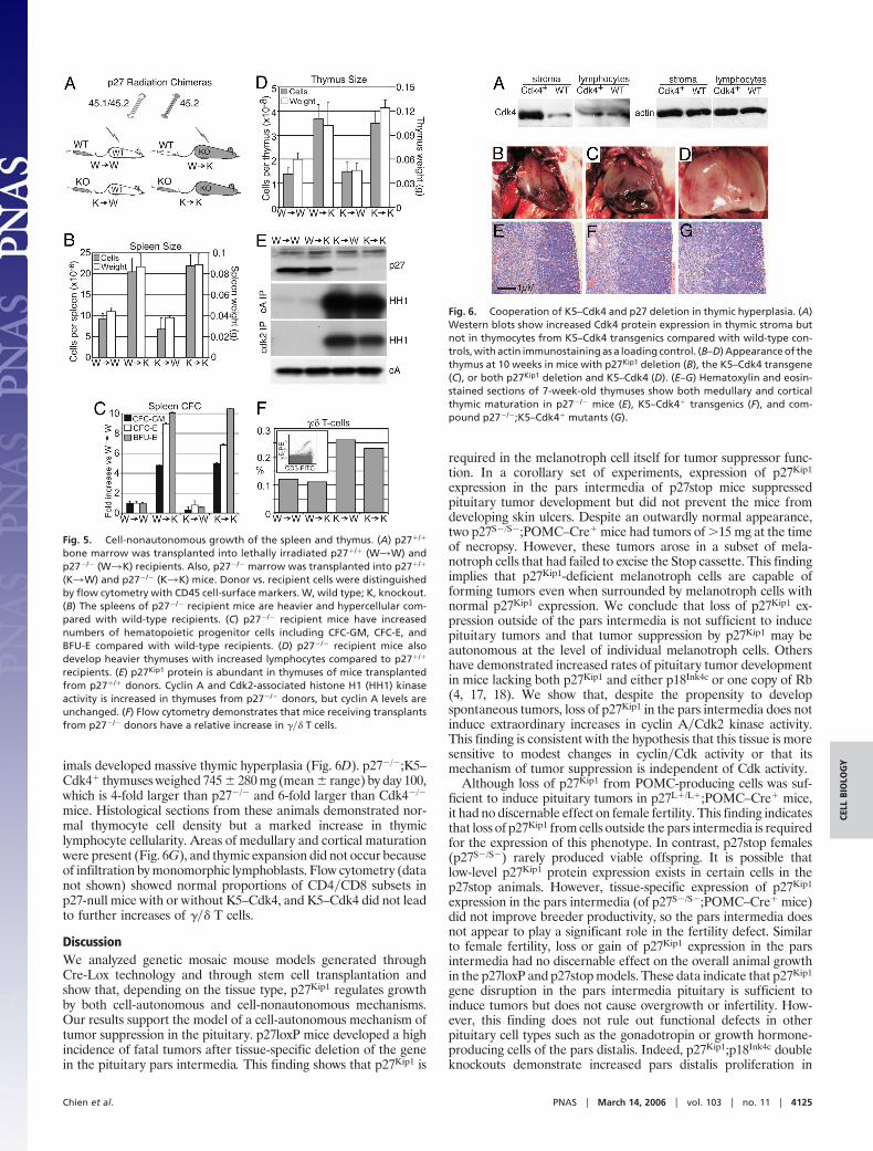
imals developed massive thymic hyperplasia (Fig. 6D). p27�/�;K5–Cdk4� thymuses weighed 745 � 280 mg (mean � range) by day 100,which is 4-fold larger than p27�/� and 6-fold larger than Cdk4�/�
mice. Histological sections from these animals demonstrated nor-mal thymocyte cell density but a marked increase in thymiclymphocyte cellularity. Areas of medullary and cortical maturationwere present (Fig. 6G), and thymic expansion did not occur becauseof infiltration by monomorphic lymphoblasts. Flow cytometry (datanot shown) showed normal proportions of CD4�CD8 subsets inp27-null mice with or without K5–Cdk4, and K5–Cdk4 did not leadto further increases of ��� T cells.
DiscussionWe analyzed genetic mosaic mouse models generated throughCre-Lox technology and through stem cell transplantation andshow that, depending on the tissue type, p27Kip1 regulates growthby both cell-autonomous and cell-nonautonomous mechanisms.Our results support the model of a cell-autonomous mechanism oftumor suppression in the pituitary. p27loxP mice developed a highincidence of fatal tumors after tissue-specific deletion of the genein the pituitary pars intermedia. This finding shows that p27Kip1 is
required in the melanotroph cell itself for tumor suppressor func-tion. In a corollary set of experiments, expression of p27Kip1
expression in the pars intermedia of p27stop mice suppressedpituitary tumor development but did not prevent the mice fromdeveloping skin ulcers. Despite an outwardly normal appearance,two p27S�/S�;POMC–Cre� mice had tumors of �15 mg at the timeof necropsy. However, these tumors arose in a subset of mela-notroph cells that had failed to excise the Stop cassette. This findingimplies that p27Kip1-deficient melanotroph cells are capable offorming tumors even when surrounded by melanotroph cells withnormal p27Kip1 expression. We conclude that loss of p27Kip1 ex-pression outside of the pars intermedia is not sufficient to inducepituitary tumors and that tumor suppression by p27Kip1 may beautonomous at the level of individual melanotroph cells. Othershave demonstrated increased rates of pituitary tumor developmentin mice lacking both p27Kip1 and either p18Ink4c or one copy of Rb(4, 17, 18). We show that, despite the propensity to developspontaneous tumors, loss of p27Kip1 in the pars intermedia does notinduce extraordinary increases in cyclin A�Cdk2 kinase activity.This finding is consistent with the hypothesis that this tissue is moresensitive to modest changes in cyclin�Cdk activity or that itsmechanism of tumor suppression is independent of Cdk activity.
Although loss of p27Kip1 from POMC-producing cells was suf-ficient to induce pituitary tumors in p27L�/L�;POMC–Cre� mice,it had no discernable effect on female fertility. This finding indicatesthat loss of p27Kip1 from cells outside the pars intermedia is requiredfor the expression of this phenotype. In contrast, p27stop females(p27S�/S�) rarely produced viable offspring. It is possible thatlow-level p27Kip1 protein expression exists in certain cells in thep27stop animals. However, tissue-specific expression of p27Kip1
expression in the pars intermedia (of p27S�/S�;POMC–Cre� mice)did not improve breeder productivity, so the pars intermedia doesnot appear to play a significant role in the fertility defect. Similarto female fertility, loss or gain of p27Kip1 expression in the parsintermedia had no discernable effect on the overall animal growthin the p27loxP and p27stop models. These data indicate that p27Kip1
gene disruption in the pars intermedia pituitary is sufficient toinduce tumors but does not cause overgrowth or infertility. How-ever, this finding does not rule out functional defects in otherpituitary cell types such as the gonadotropin or growth hormone-producing cells of the pars distalis. Indeed, p27Kip1;p18Ink4c doubleknockouts demonstrate increased pars distalis proliferation in
Fig. 5. Cell-nonautonomous growth of the spleen and thymus. (A) p27�/�
bone marrow was transplanted into lethally irradiated p27�/� (W3W) andp27�/� (W3K) recipients. Also, p27�/� marrow was transplanted into p27�/�
(K3W) and p27�/� (K3K) mice. Donor vs. recipient cells were distinguishedby flow cytometry with CD45 cell-surface markers. W, wild type; K, knockout.(B) The spleens of p27�/� recipient mice are heavier and hypercellular com-pared with wild-type recipients. (C) p27�/� recipient mice have increasednumbers of hematopoietic progenitor cells including CFC-GM, CFC-E, andBFU-E compared with wild-type recipients. (D) p27�/� recipient mice alsodevelop heavier thymuses with increased lymphocytes compared to p27�/�
recipients. (E) p27Kip1 protein is abundant in thymuses of mice transplantedfrom p27�/� donors. Cyclin A and Cdk2-associated histone H1 (HH1) kinaseactivity is increased in thymuses from p27�/� donors, but cyclin A levels areunchanged. (F) Flow cytometry demonstrates that mice receiving transplantsfrom p27�/� donors have a relative increase in ��� T cells.
Fig. 6. Cooperation of K5–Cdk4 and p27 deletion in thymic hyperplasia. (A)Western blots show increased Cdk4 protein expression in thymic stroma butnot in thymocytes from K5–Cdk4 transgenics compared with wild-type con-trols, with actin immunostaining as a loading control. (B–D) Appearance of thethymus at 10 weeks in mice with p27Kip1 deletion (B), the K5–Cdk4 transgene(C), or both p27Kip1 deletion and K5–Cdk4 (D). (E–G) Hematoxylin and eosin-stained sections of 7-week-old thymuses show both medullary and corticalthymic maturation in p27�/� mice (E), K5–Cdk4� transgenics (F), and com-pound p27�/�;K5–Cdk4� mutants (G).
Chien et al. PNAS � March 14, 2006 � vol. 103 � no. 11 � 4125
CELL
BIO
LOG
Y
response to growth hormone-releasing hormone administration,and ovary transplants suggest that female infertility in p27Kip1-deficient mice is due to a combination of ovarian and pituitarydefects (19, 20). Our p27loxP and p27stop mouse models incombination with other cell-type-specific Cre transgenes may beuseful to determine whether other pituitary cells contribute to thesephenotypes.
In contrast to the pituitary, we found that p27Kip1 suppresseshyperplasia of hematopoietic cells of the thymus and spleen by acell-nonautonomous mechanism. In radiation chimeras, thymusand spleen overgrowth occurred in only p27�/� recipient animals,but it was not affected by the genotype of the donor cells. Likewise,expansion of hematopoietic progenitors occurred by a cell-nonautonomous mechanism, but we have not identified the cellsresponsible for this phenotype. We examined cyclin�CDK activityfrom the thymuses of radiation chimeras because p27�/� micedisplay the most dramatic increase in cyclin�CDK activity in thistissue, perhaps because of a lack of compensatory function ofp21Cip1 (21). Interestingly, p27�/� lymphocytes continue to displaymarked increases in cyclin A and Cdk2 kinase activity even iftransplanted into wild-type recipients. These data indicate that lossof p27Kip1 within thymocytes is sufficient to activate cyclin�CDKcomplexes but does not account for the overgrowth of the organ.The fact that loss of p27Kip1 increases thymic lymphocyte numbersby a cell-nonautonomous mechanism does not mean that p27Kip1
has no direct effects in T cells. In fact, we observed increased ���T cells in wild-type mice transplanted p27�/� T cells, as reportedpreviously in untransplanted p27�/� mice, implying that p27Kip1
directly increases the proportions of these cells (14). In addition, anumber of studies have shown enhanced responsiveness of p27�/�
T lymphocytes to in vitro stimulation (1, 9, 22). Thus it appears thatp27Kip1 expression within the lymphocyte itself controls the re-sponse of T cells to IL-2 and other proliferative stimuli but does notdetermine homeostatic thymocyte cell number.
Tissue-specific deletion of p27Kip1 in other cell types may benecessary to positively identify the cells that drive thymic hyper-plasia. However, our data from K5–Cdk4 transgenics stronglysuggest that the thymic epithelium is responsible. Although K5–Cdk4 transgenics themselves have only minimal thymic enlarge-ment, the combination of p27Kip1 deletion and K5–Cdk4 expressionhas a dramatic synergistic effect on thymus size and cellularity.Because the K5 promoter is active in the thymic epithelium but notin thymocytes, it is likely that p27Kip1 interacts directly with cyclinD�Cdk4 complexes in this cell type. The thymic epithelium plays anessential role in thymus development and T cell maturation. Itmediates thymocyte responses to both local and systemic factorssuch as IFN�, androgen, and keratinocyte growth factor (23, 24).The thymic epithelium, in turn, produces cytokines such as IL-7,which regulates growth and differentiation of CD4�CD8� thymo-cytes (25). A similar mechanism may mediate the response top27Kip1 deficiency. It is also possible that p27Kip1 loss in the thymicepithelium causes thymocyte expansion by interacting with mole-cules other than cyclin D�Cdk4. For example, p27Kip1 inhibits Cdk2hyperphosphorylation of the retinoblastoma protein (pRb), whichmight synergize with cyclin D�Cdk4 phosphorylation of pRb.Elucidation of the biochemical nature of the interaction betweenp27Kip1 and Cdk4 will require additional studies. Our resultshighlight the importance of cell-cycle gene function in the epithelialcell compartment for the regulation of thymic lymphocyte growth(12, 16).
It is noteworthy that p27Kip1 knockout mice are prone to devel-oping T cell lymphomas, e.g., after exposure to the Moloney murineleukemia virus or in conjunction with transgenic c-Myc expression(26, 27). One intriguing idea is that inhibition of CDK activity innonneoplastic epithelial cells has tumor-suppressive action onnearby lymphocytes harboring oncogene mutations just as it doesin normal thymocytes. There is growing interest in cell-nonautonomous mechanisms of growth control and tumor sup-
pression such as through stromal–epithelial interactions andthrough regulation of angiogenesis. Because they lack geneticevidence of cellular autonomy, genes that display haploinsufficiencyin knockout mouse models may be more likely to suppress tumordevelopment through such unconventional mechanisms (28).
MethodsPOMC–Cre Transgenics. A 5.5-kb segment of the POMC genepromoter and 5� UTR was PCR-amplified from pHAL* (courtesyof Malcolm Low, Oregon Health and Science University, Portland)to include EcoRI and SalI ends and was ligated to pBS–Cre(GIBCO) to include CreMT and poly(A) sequences. Transgenicanimals were created by pronuclear injection into CD-1 embryos.Resultant progeny were screened by PCR using primers Cre1(ACCTGATGGACATGTTCAGG) and Cre2 (CTACACCT-GCGGTGCTAAC) for a 0.5-kb product and backcrossed 10generations to 129S4. To verify the pattern of Cre expression,POMC–Cre animals were bred to R26R reporter mice. Brain,pituitary, and other organs were fixed in 4% paraformaldehyde andstained with X-gal both in situ and in histologic sections.
p27loxP and p27stop Targeted p27Kip1 Mutants. The pKNFL target-ing vector contains a 12.5-kb EcoRI fragment from a 129S4genomic clone of p27Kip1. A neomycin (Neo)-selectable markerflanked by FRT sequences and a single loxP sequence was insertedinto the BglII site 1.25 kb 3� of the stop codon. A second loxPcassette was inserted into SacI 0.54 kb upstream of the initiationcodon. The p27NL� mutation was introduced by homologousrecombination in AK7 mouse ES cells (also substrain 129S4) andscreened by PCR and Southern blots as described (1). Positiveclones were further screened by PCR for inclusion of the 5� loxP sitewith primers K40 (TGGCAAACAGTCGGAGCGTAGG) andLox2 (AGCATACATTATACGAAGTTATATTAAGGGTT).Chimeric mice were bred to 129S4 FlpeR mice to produce coiso-genic animals without Neo (29). PCR-based genotyping was per-formed with primers K53 (GGTATAATACGGAAAGT-GACTCTAATGGCC) and K52 (TAGGGG AAATGGA-TAGTAGATGTTAGGACC) for wild-type (p27�) and p27L�
alleles or with K53 and K57 (AGCGGCTCCCGGCCCGAGAC)for the p27L� allele.
p27stop mice were generated by using the pKSNL targetingvector, which includes a Neo-selectable marker and three transcrip-tion termination signals inserted into the BstBI site located 120 bpupstream of the Start codon (30). ES cells were screened bygenomic Southern blots by using the 5� p27Kip1 probe as described(see Fig. 7) (1). p27stop mouse genotyping was accomplished byPCR with primers K55 (CGCCTGGCTCTGCT CCATTTGAC)and K56 (GACACTCTCACGTTTGACATCTTCC) for wild-typeand p27S� alleles or K55 and N5 (CTACCCGCTTCCATTGCT-CAG) for the p27S� allele. To demonstrate that the p27L� andp27S� alleles produce the expected phenotypes, we induced germ-line mutations by breeding both strains to 129S4 Mox2–Cre. Theresulting p27L� and p27S� alleles were bred to homozygosity in theabsence of Cre.
Growth, Fertility, and Pituitary Tumor Development. Weekly weightswere obtained from p27loxP mice (p27L�/L�) and p27stop mice(p27S�/S�) and compared with wild-type littermates and with micewith homozygous induced p27Kip1 mutations (p27�/�, p27L�/L�,and p27S�/S�) until 15 weeks of age. Weekly weights were alsoobtained from a second group of mice, designated as the ‘‘obser-vation group’’ at the time of weaning, whose genotypes were eitherp27L�/L� or p27S�/S� in combination with the POMC–Cre trans-gene, and compared with littermates without Cre. Organ weightswere obtained from an additional 10 mice from each genotype at6 weeks of age. Female fertility was assessed by recording theproduction of offspring from a separate group of adult virginfemales of various genotypes (Table 1) paired with fertile males for
4126 � www.pnas.org�cgi�doi�10.1073�pnas.0509514103 Chien et al.
an average of 3 months. Necropsies were performed in the obser-vational cohort after development of morbid behavior or at 1 yearof age to determine causes of death and morbidity. Survival wascalculated with the R program (v.2.1; www.r-project.org) using thesurvdiff function, pituitary weights were plotted with BOXPLOT, and�2 tests used CHISQ.TEST. Intact pituitaries were photographed insitu on the skull base after excision of the brain and meninges. Parsintermedia together with pars nervosa tissue was dissected free ofthe pars distalis under a stereomicroscope, weighed, and then fixedin 4% paraformaldehyde for histology, frozen for RNA and DNA,or homogenized in RIPA buffer with protease and phosphataseinhibitors for protein studies.
p27Kip1 Expression and CDK Activity. Western blots were stained withrabbit anti-p27Kip1 antibody (1:2,000) and anti-�-tubulin monoclo-nal antibody (1:10,000, T-0198, Sigma) followed by fluorescent goatanti-mouse IgG–Alexa Fluor 680 (Molecular Probes) or goatanti-rabbit IgG-IRDye800 (Rockland) and scanning on a Li-CorOdyssey. For qualitative assessment, horseradish peroxidase-conjugated secondary antibodies were detected by ECL (Amer-sham Pharmacia). p27Kip1 protein standards were generated fromadmixtures of p27�/� and p27�/� tissue extracts. Protein loadingwas normalized by adjustment to �-tubulin staining in the samelane. CDK activity using histone H1 substrate was determined afterimmunoprecipitation with cyclin A or Cdk2 antibodies (Santa CruzBiotechnology) from 100 �g of protein extract on Protein-ASepharose beads (Amersham Pharmacia) as described (31). RNAfrom mouse tissues (n � 4 for each genotype) was purified withTRIzol (Invitrogen), and p27Kip1 quantitative RT-PCR was per-formed by using SYBR green (Applied Biosystems) with primersK25 (GCTGTTTACGTCTGGCGTCGA) and K66 (AGG-AGAGCCAGGATGTCAGCG). Each sample was run in tripli-cate and normalized to mouse ribosomal protein S16 (AGGAGC-GATTTGCTGGTGTGGA and GCTACCAGGCCTTTG-AGATGGA).
p27Kip1 Radiation Chimeras. p27Kip1 knockout mice, previously de-scribed, were backcrossed (N15) to C57BL�6J and then bred top27�/� mice coisogenic to 129S4 to create p27�/� F1 hybrids (1).Control mice were F1 hybrids of 129S4 mice crossed to C57BL�6Jmice or to C57 mice with the CD45.1 cell-surface polymorphism(The Jackson Laboratory, stock catalog no. 2014, B6.SJL-Ptprca
Pep3b�BoyJ). p27�/� and p27�/� F1 hybrid recipients (3.5 weeksold, n � 5 per group) were lethally irradiated (11 Gy, Linac) andthen transplanted with bone marrow pooled from either p27�/� orp27�/� F1 hybrid donors (n � 2–3) to create four groups as in Fig.5A. Five weeks later mice were killed, and donor cell engraftmentof the blood, thymus, and splenocytes was assessed by flow cytom-etry by using CD45.1 and CD45.2 antibodies (Pharmingen). Organswere weighed and cell counts were obtained after suspension inIscove’s modified Dulbecco’s medium or RPMI medium 1640.Splenic hematopoietic progenitor cells were quantified with colony-forming assays for granulocyte-monocyte colony-forming cells(CFC-GM), erythroid colony-forming cells (CFC-E), and erythroidburst-forming units (BFU-E) as described (32). Plates were incu-bated in 5% O2 plus 5% CO2, and colonies were scored on day 2(CFU-E), days 5–6 (BFU-E), and day 7 (CFU-GM) by a researcherblinded to the experimental group. The transplant procedure wasreplicated to confirm the splenic colony-forming assays and thymusflow cytometry�kinase assays. FITC- or phycoerythrin-conjugatedrat monoclonal antibodies (CD4, CD8, CD3, TCR ���, Pharmin-gen) were diluted 1:100 and incubated for 30 min with 105 (CD4 andCD8) or 106 (CD3 and TCR ���) thymocytes. Live cells were gatedby propidium iodide (10 �g�ml) exclusion.
K5–Cdk4 Transgenics. The K5–Cdk4 transgene combines humanCdk4 with the bovine K5 promoter (15). K5–Cdk4 transgenics werebackcrossed five generations to the SENCAR strain and then bredfor two generations to p27�/� mice (C57BL�6J) to generatep27�/�;K5–Cdk4� compound mutants. Thymuses from youngadult animals (6–7 weeks) were minced and suspended in RPMImedium 1640. Lymphocytes were separated from stromal materialby differential sedimentation. Four mice carrying various combi-nations of p27�/� and K5–Cdk4 were killed at 7 weeks of age forhistology to determine thymus size and for flow cytometry. Anadditional four p27�/�;K5Cdk4� mice were assessed at 14–15weeks.
We thank Philippe Soriano (Fred Hutchinson Cancer Research Center,Seattle) for providing R26R, Mox2–Cre, and FlpeR mice and JimRoberts and Steve Collins for critical comments on the manuscript. Wealso acknowledge the excellent technical support of Erin Randel, JuttaFero, and Emily Caufield. This work was supported by grants from theNational Institutes of Health and the National Cancer Institute (toM.L.F.).
1. Fero, M. L., Rivkin, M., Tasch, M., Porter, P., Carow, C. E., Firpo, E., Polyak,K., Tsai, L. H., Broudy, V., Perlmutter, R. M., et al. (1996) Cell 85, 733–744.
2. Kiyokawa, H., Kineman, R. D., Manova-Todorova, K. O., Soares, V. C.,Hoffman, E. S., Ono, M., Khanam, D., Hayday, A. C., Frohman, L. A. & Koff,A. (1996) Cell 85, 721–732.
3. Nakayama, K., Ishida, N., Shirane, M., Inomata, A., Inoue, T., Shishido, N., Horii,I. & Loh, D. Y. (1996) Cell 85, 707–720.
4. Franklin, D. S., Godfrey, V. L., Lee, H., Kovalev, G. I., Schoonhoven, R.,Chen-Kiang, S., Su, L. & Xiong, Y. (1998) Genes Dev. 12, 2899–2911.
5. Durand, B., Gao, F. B. & Raff, M. (1997) EMBO J. 16, 306–317.6. Fero, M. L., Randel, E., Gurley, K. E., Roberts, J. M. & Kemp, C. J. (1998) Nature
396, 177–180.7. Cheng, T., Rodrigues, N., Dombkowski, D., Stier, S. & Scadden, D. T. (2000) Nat.
Med. 6, 1235–1240.8. Walkley, C. R., Fero, M. L., Chien, W. M., Purton, L. E. & McArthur, G. A. (2005)
Nat. Cell Biol. 7, 172–178.9. Zhang, S., Lawless, V. A. & Kaplan, M. H. (2000) J. Immunol. 165, 6270–6277.
10. Guler, H. P., Zapf, J., Scheiwiller, E. & Froesch, E. R. (1988) Proc. Natl. Acad.Sci. USA 85, 4889–4893.
11. Casalino-Matsuda, S. M., Durando, P. E. & Celis, M. E. (2002) J. Physiol.Biochem. 58, 25–31.
12. Robles, A. I., Larcher, F., Whalin, R. B., Murillas, R., Richie, E., Gimenez-Conti,I. B., Jorcano, J. L. & Conti, C. J. (1996) Proc. Natl. Acad. Sci. USA 93, 7634–7638.
13. Bai, F., Pei, X. H., Godfrey, V. L. & Xiong, Y. (2003) Mol. Cell. Biol. 23, 1269–1277.14. Shen, R. & Kaplan, M. H. (2002) J. Immunol. 169, 714–721.15. Miliani de Marval, P. L., Gimenez-Conti, I. B., LaCava, M., Martinez, L. A.,
Conti, C. J. & Rodriguez-Puebla, M. L. (2001) Am. J. Pathol. 159, 369–379.16. Rodriguez-Puebla, M. L., LaCava, M., Miliani De Marval, P. L., Jorcano, J. L.,
Richie, E. R. & Conti, C. J. (2000) Am. J. Pathol. 157, 1039–1050.
17. Carneiro, C., Jiao, M. S., Hu, M., Shaffer, D., Park, M., Pandolfi, P. P.,Cordon-Cardo, C. & Koff, A. (2003) Oncogene 22, 361–369.
18. Park, M. S., Rosai, J., Nguyen, H. T., Capodieci, P., Cordon-Cardo, C. & Koff,A. (1999) Proc. Natl. Acad. Sci. USA 96, 6382–6387.
19. Teixeira, L. T., Kiyokawa, H., Peng, X. D., Christov, K. T., Frohman, L. A. &Kineman, R. D. (2000) Oncogene 19, 1875–1884.
20. Tong, W., Kiyokawa, H., Soos, T. J., Park, M. S., Soares, V. C., Manova, K.,Pollard, J. W. & Koff, A. (1998) Cell Growth Differ. 9, 787–794.
21. Coats, S., Whyte, P., Fero, M. L., Lacy, S., Chung, G., Randel, E., Firpo, E. &Roberts, J. M. (1999) Curr. Biol. 9, 163–173.
22. Mohapatra, S., Agrawal, D. & Pledger, W. J. (2001) J. Biol. Chem. 276,21976–21983.
23. Olsen, N. J., Olson, G., Viselli, S. M., Gu, X. & Kovacs, W. J. (2001) Endocrinology142, 1278–1283.
24. Erickson, M., Morkowski, S., Lehar, S., Gillard, G., Beers, C., Dooley, J., Rubin,J. S., Rudensky, A. & Farr, A. G. (2002) Blood 100, 3269–3278.
25. Yarilin, A. A. & Belyakov, I. M. (2004) Curr. Med. Chem. 11, 447–464.26. Martins, C. P. & Berns, A. (2002) EMBO J. 21, 3739–3748.27. Hwang, H. C., Martins, C. P., Bronkhorst, Y., Randel, E., Berns, A., Fero, M. &
Clurman, B. E. (2002) Proc. Natl. Acad. Sci. USA 99, 11293–11298.28. Payne, S. R. & Kemp, C. J. (2005) Carcinogenesis 26, 2031–2045.29. Farley, F. W., Soriano, P., Steffen, L. S. & Dymecki, S. M. (2000) Genesis 28,
106–110.30. Soriano, P. (1999) Nat. Genet. 21, 70–71.31. Sheaff, R. J., Groudine, M., Gordon, M., Roberts, J. M. & Clurman, B. E. (1997)
Genes Dev. 11, 1464–1478.32. Testa, N. G. & Molineux, G. (1993) Haemopoiesis: A Practical Approach (Oxford
Univ. Press, Oxford).
Chien et al. PNAS � March 14, 2006 � vol. 103 � no. 11 � 4127
CELL
BIO
LOG
Y
Copyright © 2022 FDOKUMEN