Cell-Autonomous Requirement for Rx Function in the Mammalian Retina and Posterior Pituitary
7
Cell-Autonomous Requirement for Rx Function in the Mammalian Retina and Posterior Pituitary Olga Medina-Martinez 1 , Felipe Amaya-Manzanares 2 , Chaomei Liu 1 , Marisela Mendoza 2 , Rina Shah 2 , Li Zhang 2 , Richard R. Behringer 3 , Kathleen A. Mahon 2. , Milan Jamrich 1,2. * 1 Department of Molecular and Human Genetics, Baylor College of Medicine, Houston, Texas, United States of America, 2 Department of Molecular and Cellular Biology, Baylor College of Medicine, Houston, Texas, United States of America, 3 Department of Molecular Genetics, University of Texas, M. D. Anderson Cancer Center, Houston, Texas, United States of America Abstract Rx is a paired-like homeobox gene that is required for vertebrate eye formation. Mice lacking Rx function do not develop eyes or the posterior pituitary. To determine whether Rx is required cell autonomously in these tissues, we generated embryonic chimeras consisting of wild type and Rx2/2 cells. We found that in the eye, Rx-deficient cells cannot participate in the formation of the neuroretina, retina pigment epithelium and the distal part of the optic stalk. In addition, in the ventral forebrain, Rx function is required cell autonomously for the formation of the posterior pituitary. Interestingly, Rx2/2 and wild type cells segregate before the morphogenesis of these two tissues begins. Our observations suggest that Rx function is not only required for the morphogenesis of the retina and posterior pituitary, but also prior to morphogenesis, for the sorting out of cells to form distinct fields of retinal/pituitary cells. Citation: Medina-Martinez O, Amaya-Manzanares F, Liu C, Mendoza M, Shah R, et al. (2009) Cell-Autonomous Requirement for Rx Function in the Mammalian Retina and Posterior Pituitary. PLoS ONE 4(2): e4513. doi:10.1371/journal.pone.0004513 Editor: Patrick Callaerts, Katholieke Universiteit Leuven, Belgium Received October 15, 2008; Accepted January 8, 2009; Published February 20, 2009 Copyright: ß 2009 Medina-Martinez et al. This is an open-access article distributed under the terms of the Creative Commons Attribution License, which permits unrestricted use, distribution, and reproduction in any medium, provided the original author and source are credited. Funding: This research was sponsored by NIH/NEI grants EY12505 and EY12163 to M.J., NIH/NEI training grant T32 EY07102 to O.M. and by the Retina Research Foundation to M.J. The funders had no role in study design, data collection and analysis, decision to publish, or preparation of the manuscript. Competing Interests: The authors have declared that no competing interests exist. * E-mail: [email protected] . These authors contributed equally to this work. Introduction Several homeobox genes play important roles in the patterning of the vertebrate forebrain. One of these is the paired-like homeobox gene Rx [1–3]. Rx plays a critical role in vertebrate eye development, as mutations in this gene cause the loss of or abnormal formation of the eye in several species [2,4–11]. Rx transcription begins in the anterior neural plate of gastrula embryos [1–3], in an area that gives rise to the retina, optic stalk and ventral hypothalamus. Retinal formation begins with the evagination of retinal progenitor cells. These cells initially form the optic vesicle and later the optic cup, which consists of the neuroretina and the retina pigment epithelium (RPE). The retina remains connected to the brain through the optic stalk. The evaginating neuroectoderm induces lens formation in the overlying surface ectoderm. Mice lacking Rx function do not develop optic vesicles or optic cups and consequently do not develop a neuroretina, a RPE or an optic stalk [2]. Since ocular neuroectoderm is necessary for lens formation, Rx-deficient embryos do not form a lens, despite the fact that Rx is not expressed in the surface ectoderm [2,10–12]. In medaka, an intronic insertion in Rx3 is the cause of the temperature sensitive eyeless (el) mutant [13]. This mutation results in the transcriptional repression of Rx3 and is responsible for the lack of evagination of the optic vesicle [6,13]. In zebrafish, the eyeless phenotype in the chokh mutant is caused by a mutation in the homeobox region of Rx3 [5]. Finally, in human, mutations in Rx/RAX lead to anophthalmia [8]. In addition to eye development, Rx function is required for the normal development of the ventral forebrain [14]. During normal development, the ventral forebrain gives rise to the hypothalamo- pituitary axis. The posterior pituitary forms from the ventral hypothalamus through evagination. The ventral hypothalamus and posterior pituitary are instrumental in the induction and correct differentiation of the anterior pituitary that is formed from the head surface ectoderm [15–19]. We have observed previously that the ventral neuroectoderm is very thin [14] and does not undergo proper morphogenesis in Rx- deficient mouse embryos. The posterior pituitary cannot be detected in these animals at the time of birth and Rathke’s pouch, the precursor of the anterior pituitary, begins to form, but does not differentiate properly [14]. The ventral hypothalamus is a rich source of signaling molecules and it has been shown that signaling from this region is necessary for the formation of the anterior pituitary. Bmp4 and Fgfs are expressed in the ventral hypothalamus and are required for induction of the pouch primordium and formation of a definitive Rathke’s pouch, respectively [20–23]. Members of the Sonic hedgehog (SHH), Wnt, and Notch signaling pathways have also been implicated in these processes. Therefore, it is likely that abnormal differentiation of the anterior pituitary in Rx mutant mice is the consequence of abnormal development/ signaling from the hypothalamus. Interestingly, the developing retina (optic cup) and the ventral diencephalon that gives rise to the hypothalamus/posterior pituitary, display similarities in morphogenesis and gene expression. The retina, as well as the posterior pituitary, evaginates from the neuroectoderm and both PLoS ONE | www.plosone.org 1 February 2009 | Volume 4 | Issue 2 | e4513
-
Upload
independent -
Category
Documents
-
view
0 -
download
0
Transcript of Cell-Autonomous Requirement for Rx Function in the Mammalian Retina and Posterior Pituitary
Cell-Autonomous Requirement for Rx Function in theMammalian Retina and Posterior PituitaryOlga Medina-Martinez1, Felipe Amaya-Manzanares2, Chaomei Liu1, Marisela Mendoza2, Rina Shah2, Li
Zhang2, Richard R. Behringer3, Kathleen A. Mahon2., Milan Jamrich1,2.*
1 Department of Molecular and Human Genetics, Baylor College of Medicine, Houston, Texas, United States of America, 2 Department of Molecular and Cellular Biology,
Baylor College of Medicine, Houston, Texas, United States of America, 3 Department of Molecular Genetics, University of Texas, M. D. Anderson Cancer Center, Houston,
Texas, United States of America
Abstract
Rx is a paired-like homeobox gene that is required for vertebrate eye formation. Mice lacking Rx function do not developeyes or the posterior pituitary. To determine whether Rx is required cell autonomously in these tissues, we generatedembryonic chimeras consisting of wild type and Rx2/2 cells. We found that in the eye, Rx-deficient cells cannot participatein the formation of the neuroretina, retina pigment epithelium and the distal part of the optic stalk. In addition, in theventral forebrain, Rx function is required cell autonomously for the formation of the posterior pituitary. Interestingly, Rx2/2and wild type cells segregate before the morphogenesis of these two tissues begins. Our observations suggest that Rxfunction is not only required for the morphogenesis of the retina and posterior pituitary, but also prior to morphogenesis,for the sorting out of cells to form distinct fields of retinal/pituitary cells.
Citation: Medina-Martinez O, Amaya-Manzanares F, Liu C, Mendoza M, Shah R, et al. (2009) Cell-Autonomous Requirement for Rx Function in the MammalianRetina and Posterior Pituitary. PLoS ONE 4(2): e4513. doi:10.1371/journal.pone.0004513
Editor: Patrick Callaerts, Katholieke Universiteit Leuven, Belgium
Received October 15, 2008; Accepted January 8, 2009; Published February 20, 2009
Copyright: � 2009 Medina-Martinez et al. This is an open-access article distributed under the terms of the Creative Commons Attribution License, which permitsunrestricted use, distribution, and reproduction in any medium, provided the original author and source are credited.
Funding: This research was sponsored by NIH/NEI grants EY12505 and EY12163 to M.J., NIH/NEI training grant T32 EY07102 to O.M. and by the Retina ResearchFoundation to M.J. The funders had no role in study design, data collection and analysis, decision to publish, or preparation of the manuscript.
Competing Interests: The authors have declared that no competing interests exist.
* E-mail: [email protected]
. These authors contributed equally to this work.
Introduction
Several homeobox genes play important roles in the patterning
of the vertebrate forebrain. One of these is the paired-like
homeobox gene Rx [1–3]. Rx plays a critical role in vertebrate eye
development, as mutations in this gene cause the loss of or
abnormal formation of the eye in several species [2,4–11]. Rx
transcription begins in the anterior neural plate of gastrula
embryos [1–3], in an area that gives rise to the retina, optic stalk
and ventral hypothalamus.
Retinal formation begins with the evagination of retinal
progenitor cells. These cells initially form the optic vesicle and
later the optic cup, which consists of the neuroretina and the retina
pigment epithelium (RPE). The retina remains connected to the
brain through the optic stalk. The evaginating neuroectoderm
induces lens formation in the overlying surface ectoderm. Mice
lacking Rx function do not develop optic vesicles or optic cups and
consequently do not develop a neuroretina, a RPE or an optic
stalk [2]. Since ocular neuroectoderm is necessary for lens
formation, Rx-deficient embryos do not form a lens, despite the
fact that Rx is not expressed in the surface ectoderm [2,10–12]. In
medaka, an intronic insertion in Rx3 is the cause of the
temperature sensitive eyeless (el) mutant [13]. This mutation results
in the transcriptional repression of Rx3 and is responsible for the
lack of evagination of the optic vesicle [6,13]. In zebrafish, the
eyeless phenotype in the chokh mutant is caused by a mutation in
the homeobox region of Rx3 [5]. Finally, in human, mutations in
Rx/RAX lead to anophthalmia [8].
In addition to eye development, Rx function is required for the
normal development of the ventral forebrain [14]. During normal
development, the ventral forebrain gives rise to the hypothalamo-
pituitary axis. The posterior pituitary forms from the ventral
hypothalamus through evagination. The ventral hypothalamus
and posterior pituitary are instrumental in the induction and
correct differentiation of the anterior pituitary that is formed from
the head surface ectoderm [15–19].
We have observed previously that the ventral neuroectoderm is
very thin [14] and does not undergo proper morphogenesis in Rx-
deficient mouse embryos. The posterior pituitary cannot be
detected in these animals at the time of birth and Rathke’s pouch,
the precursor of the anterior pituitary, begins to form, but does not
differentiate properly [14]. The ventral hypothalamus is a rich
source of signaling molecules and it has been shown that signaling
from this region is necessary for the formation of the anterior
pituitary. Bmp4 and Fgfs are expressed in the ventral hypothalamus
and are required for induction of the pouch primordium and
formation of a definitive Rathke’s pouch, respectively [20–23].
Members of the Sonic hedgehog (SHH), Wnt, and Notch signaling
pathways have also been implicated in these processes. Therefore,
it is likely that abnormal differentiation of the anterior pituitary in
Rx mutant mice is the consequence of abnormal development/
signaling from the hypothalamus. Interestingly, the developing
retina (optic cup) and the ventral diencephalon that gives rise to
the hypothalamus/posterior pituitary, display similarities in
morphogenesis and gene expression. The retina, as well as the
posterior pituitary, evaginates from the neuroectoderm and both
PLoS ONE | www.plosone.org 1 February 2009 | Volume 4 | Issue 2 | e4513
initially express Rx. The optic cup is instrumental in the induction,
invagination and differentiation of the lens from the head surface
ectoderm. Similarly, the ventral hypothalamus is instrumental in
the induction, invagination and the differentiation of Rathke’s
pouch (the anterior/intermediate pituitary), which is also derived
from the surface ectoderm. It is therefore possible that the
formation of both tissue pairs (retina-lens and hypothalamus-
anterior pituitary) is regulated by similar developmental and
molecular mechanisms. Indeed, besides Rx, there are several
homeodomain transcription factors (i.e. Pax6, Six3, Optx2, Lhx2,
Foxl2), as well as signaling molecules (SHH, BMP4, FGF-8, -10,
Wnt and Notch pathway members) expressed during eye
development, that are also expressed in analogous ways in the
development of the hypothalamic-pituitary axis. Furthermore, the
intriguing observation that the adenohypophysis is transformed
into an ectopic lens in the zebrafish midline mutants you-too (yot)
and iguana (igu) by a mutation in Gli2 gene, is additional evidence
for developmental and molecular commonality [24].
While it is known that Rx expression is required for the formation
of the retina and the hypothalamo-pituitary axis in mouse embryos,
it is not known whether Rx function is required cell autonomously in
these tissues. A cell autonomous requirement for Rx3 function for the
morphogenesis of the retina has been reported in medaka and
zebrafish [25–27], but the mechanism of retinal formation is
significantly different in mammals and teleosts [25]. The question of
cell autonomous function of Rx in the retina and the hypothalamo-
pituitary axis has yet to be examined in mammals.
To determine whether Rx expression is required cell autono-
mously for the formation of the retina and the hypothalamo-
pituitary axis in mouse embryos, we analyzed the behavior of Rx-
deficient cells in embryonic chimeras consisting of wild type and
Rx2/2 cells. We also characterized the changes in gene
expression during pituitary development in Rx-deficient embryos.
Results of our studies show that Rx function is required cell-
autonomously for the formation of the retina and the posterior
pituitary and that Optx2 is the common target of Rx during the
development of both tissues.
Results
Our previous experiments have shown that Rx is required for eye
formation in mouse. In the absence of Rx, the optic structures such as
the optic sulci, optic vesicle and optic cup do not form and the
expression of retina and lens specific markers cannot be detected
[2,14]. To determine whether Rx function is required cell
autonomously during retinal development, we generated embryonic
chimeras consisting of wild type and Rx2/2lacZ/+ cells. Chimeric
embryos bearing both wild-type (white) and varying amounts of
mutant cells (blue) were examined to determine whether Rx mutant
cells could contribute to eye structures in a wild type environment.
We analyzed chimeras with a very high contributions of
Rx2/2lacZ/+ cells, but we did not observe any Rx2/2lacZ/+ cells
in retinal structures. As figure 1A shows, at E12.5 the neuroretina is
totally devoid of Rx2/2 cells, while most of the cells surrounding the
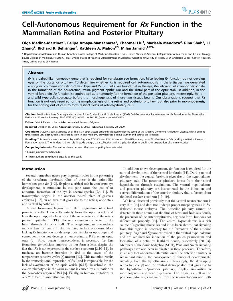
Figure 1. Retinal formation in chimeric embryos. (A) Section through an E12.5 chimeric embryo demonstrating that the neuroretina does notcontain any Rx-deficient (blue) cells. (B) Section through an E10.5 chimeric embryo. Boxed region is enlarged in C. (C) Section through an E10.5chimeric embryo visualizing the absence of Rx-deficient cells in the presumptive neuroretina (arrowhead), RPE and distal optic stalk. Columns of Rx-deficient cells in the proximal optic stalk are labeled with an asterisk. (D) Section through an E9.5 chimeric embryo showing the absence of Rx-deficient cells in the evaginating optic vesicle (arrow). (E) Section through an E8.5 chimeric embryo showing predominantly wild type cells in theevaginating optic neuroectoderm. Two arrows indicate the sharp boundaries between the wild type cells (white) and Rx2/2 cells (blue). (F) Sectionthrough an E8.0 chimeric embryo showing the distribution of wild type and Rx-deficient cells in the optic pit. (G) A control chimera. Section throughan E12.5 chimeric embryo generated from wild type (white) and Gt(ROSA)26Sor/J (blue) cells demonstrating that Gt(ROSA)26Sor/J cells that are wildtype for Rx, participate in retinal formation. (H) Section through an E12.5 chimeric embryo generated from wild type (white) and Rx2/2 cellsdemonstrating that in the presence of only few wild type cells, the entire optic cup develops into an RPE. No lens is present in this case. L – lens, NR –neuroretina, OE – optic evagination, OP – optic pit, POS - presumptive optic stalk, PRPE – presumptive retina pigment epithelium.doi:10.1371/journal.pone.0004513.g001
Rx and Brain Morphogenesis
PLoS ONE | www.plosone.org 2 February 2009 | Volume 4 | Issue 2 | e4513
eye, as well as the lens cells, are Rx2/2. Since the pigmentation of
the retina pigment epithelium (RPE) makes it difficult to determine
whether the cells of the RPE are wild type or Rx2/2, we analyzed
chimeric embryos at E10.5 – before the pigmentation of RPE
appears (Fig. 1B, C). Sections through an E10.5 chimeric embryo
show that Rx2/2 cells do not participate in the formation of the
presumptive neuroretina, RPE or the distal part of the optic stalk
(Fig. 1C). These observations demonstrate that there is a cell-
autonomous requirement for Rx function in these retinal structures.
Interestingly, in the transition zone from the neural tube to the optic
stalk, the wild type cells and Rx-deficient cells can coexist, but they
sort out into columns of cells based on their genetic makeup. The
columns consisting of different cell types display different morphol-
ogy, with Rx-deficient columns cells always being wider than the
columns of wild type cells. The likely explanation for the different
thickness of tissue of different genetic makeup is that the wild type
cells can participate in the convergent extension of the optic stalk,
while the Rx2/2 cells cannot. The lack of Rx2/2 cells in the
evaginating optic vesicle can be observed at E9.5, when the vesicle is
devoid of Rx-deficient cells (Fig. 1D). A relatively sharp boundary
between the evaginating cells of the presumptive optic vesicle and the
surrounding cells can be detected as early as E8.5 (Fig. 1E), the
beginning of evagination of the ocular neuroectoderm. Importantly,
at E8.0, the optic pit contains mostly wild type cells (Fig. 1F),
suggesting that Rx2/2 cells are depleted from this region even
before the morphogenesis of the retinal structures begins. It should
be pointed out that because of the lack of molecular markers
specifically expressed in this region, the optic pit was identified based
on its morphology. In contrast, in control chimeras generated from
wild type (white) and Gt(ROSA)26Sor/J cells that were Rx+/+lacZ/+,
both cell types contributed equally to the formation of the
neuroretina (Fig. 1G).
Interestingly, when the contribution of wild type cells in the
chimeric embryo was very low, the wild type cells developed into
an optic vesicle consisting only of RPE (Fig. 1H). Lenses were not
observed in these embryos, suggesting that this optic vesicle cannot
induce lens formation.
Since our previous results indicated that the formation of the
hypothalamo-pituitary axis was also strongly affected in Rx2/2
embryos [14], we investigated the role of Rx in the formation of the
pituitary. During normal development, the primordium of the
anterior and intermediate lobes of the pituitary gland (adenohy-
pophysis) is first visible as a thickened region of the midline oral
ectoderm that lies adjacent to the floor of the diencephalon or
future hypothalamus. In the mouse embryo, this ectoderm
invaginates at approximately 8.5–9 days to form Rathke’s pouch
(RP) (Fig. 2A), which detaches from the oral ectoderm by 12–12.5
days [for review see reference 18]. At the same time, the
infundibulum, an outpocketing of the diencephalon, evaginates
to give rise to the neurohypophysis or the posterior lobe of the
pituitary (Fig. 2C). The neurohypophysis contains the axon
terminals of the neurosecretory neurons that reside in the
hypothalamus. These two tissues lie in direct contact during this
time, and a large body of evidence indicates that signals from the
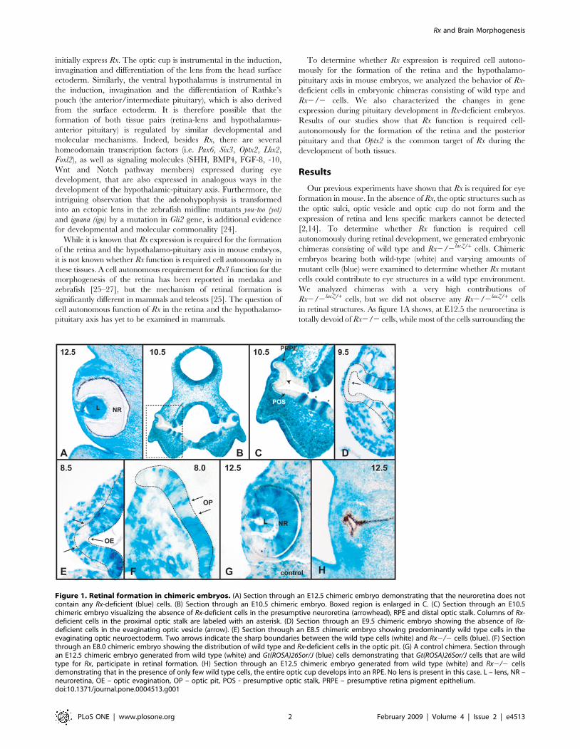
Figure 2. Expression of pituitary specific genes in wild type and Rx-deficient embryos. (A) Lhx3 expression in Rathke’s pouch of an E9.5wild type embryo. (B) Lhx3 expression in Rathke’s pouch of an E9.5 Rx-deficient embryo. (C) Lhx3 expression in the anterior pituitary of an E10.5 wildtype embryo. (D) Lhx3 expression in the dysmorphic anterior pituitary of an E10.5 Rx-deficient embryo. (E) Expression of Fgf10 in the ventralhypothalamus of an E9.5 wild type embryo. (F) Expression of Fgf10 in the ventral hypothalamus of an E9.5 Rx-deficient embryo. (G) Expression ofFgf10 in the posterior pituitary of an E10.5 wild type embryo. (H) Fgf10 expression in an Rx-deficient embryo. (I) Optx2 expression in the head of anE9.5 wild type embryo. (J) Optx2 expression in the head of an E9.5 Rx2/2 embryo. (K) Optx2 expression in the neuroectoderm and head surfaceectoderm of an E8.5 wild type embryo. (L) Optx2 expression in the head surface ectoderm of an E8.5 Rx2/2 embryo. (M) Optx2 expression in theventral hypothalamus and optic vesicle of an E10.5 wild type embryo. (N) Lack of Optx2 expression in an E10.5 Rx2/2 embryo. a – anterior pituitary, p– posterior pituitary, OV – optic vesicle, RP- Rathke’s pouch, VH – ventral hypothalamus.doi:10.1371/journal.pone.0004513.g002
Rx and Brain Morphogenesis
PLoS ONE | www.plosone.org 3 February 2009 | Volume 4 | Issue 2 | e4513
neuroectoderm of the ventral diencephalon (hypothalamus) are
required for the induction, specification and commitment of
Rathke’s pouch and pituitary cell lineages [for review see 19]. In
Rx2/2 embryos, there is no evidence for evagination of the
ventral neuroectoderm to form the posterior pituitary (Fig. 2D).
Rathke’s pouch initially forms in these mutants [14], but it
becomes severely dysmorphic (Fig. 2D) and cannot be visually
identified at birth (not shown). Despite the presence of clefts, the
pouch tissue remains as a multilayered sheet; it does not ever
detach from the oral ectoderm and remains in contact with the
oral cavity (Fig. 2D). Ultimately, the pituitary and ventral
hypothalamic tissue are extruded into the oral cavity about the
same time that the palate would normally be closing (not shown).
In order to better define the molecular steps that lead from Rx
expression to the proper development of the hypothalamus and
formation of the posterior pituitary and to better understand the
development of the pituitary gland in Rx2/2 embryos, we
examined the expression of several genes during the development
of the anterior and posterior pituitary in wild type and Rx-deficient
embryos. Here we present the comparison of Lhx3, Fgf10 and Optx2
expression in wild type and Rx2/2 embryos, as the changes in the
expression of these three genes best demonstrate the morphological
and molecular consequences of the absence of Rx expression. Lhx3 is
a specific marker of anterior pituitary development. It is a LIM-
homeobox gene, expressed as soon as Rathke’s pouch forms and its
expression is absolutely required for pituitary organogenesis [28].
Expression of Lhx3 is not significantly altered in Rx2/2 embryos
(Fig. 2B, D), when compared to wild type embryos (Fig. 2A, C), in
spite of the fact that the anterior pituitary is severely dysmorphic in
Rx2/2 embryos. This indicates that the program of anterior
pituitary organogenesis has been initiated.
Fgf10 is a diagnostic marker of posterior pituitary development
[23]. In wild type embryos, it is expressed in the ventral
hypothalamus and the infundibulum (Fig. 2E, G). In Rx 2/2
embryos, Fgf10 expression is initially quite normal at E9.5 (Fig. 2F),
but later, at E10.5 is severely downregulated (Fig. 2H). No cells
can be found that express this gene at high levels typical of
posterior pituitary development.
The homeobox gene Optx2 (Six6) is co-expressed with Rx early in
development in both the ventral diencephalon and the optic
vesicle In addition this gene is expressed in the head surface
ectoderm [29–32]. In Rx-deficient embryos, Optx2 expression is
completely lost in the optic vesicle/cup at E9.5 (Fig. 2I,J). Strong
reduction of Optx2 expression in the head of Rx2/2 embryos is
already evident at E8.5 (Fig. 2K,L). In both stages in Rx2/2
embryos Optx2 is expressed only in the surface ectoderm, but not
in the neuroectoderm. At E10.5 (Fig. 2M,N) Optx2 is not
expressed in the optic vesicle or in the ventral hypothalamus.
These experiments, taken together with the observation that Rx is
expressed in Optx2 mutant mice [33], indicate that Optx2 is
genetically downstream of Rx and is potentially a direct regulatory
target for Rx. However, a direct regulation of Optx2 expression by
Rx remains to be demonstrated.
Due to the lack of a visible posterior pituitary and the changes in
gene expression in the ventral hypothalamus of Rx2/2 embryos,
we analyzed the ability of Rx2/2 cells to contribute to the
formation of the posterior pituitary in chimeric embryos. We
found that Rx2/2lacZ/+ cells cannot contribute to the formation of
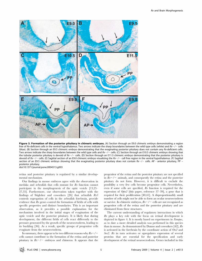
the posterior pituitary. As early as E9.0, there is a distinct region
with sharp boundaries in the ventral hypothalamus that is devoid
of blue, Rx-deficient cells (Fig. 3A,E). This region undergoes
evagination (Fig. 3B,F) and later forms the posterior pituitary,
consisting exclusively of wild type cells (Fig. 3C,D). This
observation suggests that Rx function is required cell-autonomous-
ly for the formation of the field of posterior pituitary progenitor
cells even before the morphogenesis of the posterior pituitary
begins. The similarities between the cell autonomous requirements
for Rx function between the presumptive neuroectoderm of the
retina and posterior pituitary, as well as the lack of Optx2
expression in both tissues in Rx-deficient embryos, gives support to
the hypothesis that the development of the retina and the posterior
pituitary is regulated by similar developmental and molecular
mechanisms.
Discussion
Cell-autonomous requirement for Rx function in theretina and posterior pituitary
We have presented evidence that in mice Rx activity is required
cell autonomously for the formation of the neuroretina, retina
pigment epithelium and the distal optic stalk. Rx-deficient cells
cannot be found in these structures in chimeric embryos. At the
beginning of evagination, the presumptive ocular neuroectoderm of
chimeric embryos contains almost exclusively wild type cells,
indicating that Rx2/2 cells are eliminated from the presumptive
retinal neuroectoderm before the onset of retinal morphogenesis.
This suggests that Rx2/2 cells never behave like, or are recognized,
as retinal progenitor cells. Segregation of wild type and Rx2/2 cells
can be observed in the proximal optic stalk. In this transitional zone
between the optic stalk and the brain, wild type and Rx-deficient cells
can be observed next to each other forming columns of cells
segregated by their genetic makeup. The wild type and Rx2/2
columns of cells have different morphology, presumably because of
different morphogenetic behavior. The wild type columns are
thinner presumably because these cells are undergoing convergent
extension typical of optic stalk formation. The Rx2/2 cells do not
appear to undergo convergent extension [2,11,14].
Retinas in chimeric embryos show a remarkable degree of
autonomy. Even in embryos where most of the surrounding cells are
mutant, retinal development appears to be normal, i.e. the optic cup
forms a normal RPE, neuroretina and optic stalk. Only in cases
when very few wild type cells are available to form a retina, the
differentiation of the optic cup is abnormal. In this case, the entire
retina and optic stalk differentiate into RPE. This is interesting, as it
indicates that the abnormal differentiation of retinal cells is not due
to the genetic makeup of the retinal cells, but rather due to the small
size of the retina. A likely explanation for this phenomenon is
provided by the observations of Cho and Cepko [34]. They found
that during chick eye development, a yet unknown mechanism
induces expression of Wnt2b in the surface ectoderm dorsally to the
evaginating optic cup. Somewhat later, Wnt2b expression spreads to
the adjacent dorsal optic cup, which then becomes RPE. It is possible
that when the evaginating optic cup is very small, the Wnt2b-
inducing signal transverses the entire optic cup and converts it to
RPE. This retina, consisting entirely of RPE cells, cannot induce lens
formation suggesting that neuroretina or neuroretina-specific gene
expression is necessary for lens induction. Alternatively, the
evaginating retinal neuroectoderm is too small to ever contact the
surface ectoderm and therefore the inductive signals from the optic
vesicle never reach the surface ectoderm.
The posterior pituitary gland of chimeric embryos does not
contain Rx-deficient cells showing that Rx function is also required
cell-autonomously for the formation of the posterior pituitary.
Similar to the ocular neuroectoderm, the neuroectoderm of the
presumptive posterior pituitary is depleted of Rx2/2 cells prior to
the evagination of the posterior pituitary. This shows that Rx
function is required for the establishment of a field of posterior
pituitary progenitor cells, indicating that the development of the
Rx and Brain Morphogenesis
PLoS ONE | www.plosone.org 4 February 2009 | Volume 4 | Issue 2 | e4513
retina and posterior pituitary is regulated by a similar develop-
mental mechanism.
Our findings in mouse embryos agree with the observation in
medaka and zebrafish that cells mutant for Rx function cannot
participate in the morphogenesis of the optic vesicle [13,25–
27,35]. Furthermore, our observation taken together with the
findings of Stigloher and coworkers [26] that zebrafish Rx3
controls segregation of cells in the zebrafish forebrain, provide
evidence that Rx genes control the formation of fields of cells with
specific properties and distinct boundaries. This is an important
observation, as it provides a possible explanation for the
mechanism involved in the initial stages of evagination of the
optic vesicle and the posterior pituitary. It is likely that during
development, the different fields of cells react differently to the
pressure generated by the growth of the neuroectoderm, leading to
its buckling [36]. As a result, specific groups of progenitor cells
evaginate from the neuroectoderm.
In summary, there appear to be two different reasons why Rx2/2
cells cannot contribute to the formation of the retina and posterior
pituitary in Rx2/2 embryos and chimeras. It appears that the
progenitor of the retina and the posterior pituitary are not specified
in Rx2/2 animals, and consequently the retina and the posterior
pituitary do not form. However, it is difficult to exclude the
possibility a very few cells become progenitor cells. Nevertheless,
even if some cells are specified, Rx function is required for the
expression of Optx2 [this paper, reference 37–39], a gene that is
required for their proliferation [40,41]. A disproportionably small
number of cells might not be able to form an ocular neuroectoderm
or survive. In chimeric embryos, Rx2/2 cells are not recognized as
progenitor cells of the retina and the posterior pituitary and are
eliminated from these structures.
The current understanding of regulatory interactions in which
Rx plays a key role with the focus on retinal development is
depicted in figure 4. It is mostly based on experiments in Xenopus,
as to date a more detailed analysis was performed in this species
than in mouse. As demonstrated by Danno and coworkers [42], Rx
is activated in the forebrain by the coordinate action of Otx2 and
Sox2. Rx in turn activates or upregulates expression of several
proteins that are essential for the specification and early
development of the retinal neuroectoderm. Genes included in this
Figure 3. Formation of the posterior pituitary in chimeric embryos. (A) Section through an E9.0 chimeric embryo demonstrating a regionfree of Rx-deficient cells in the ventral hypothalamus. Two arrows indicate the sharp boundaries between the wild type cells (white) and Rx2/2 cells(blue). (B) Section through an E9.5 chimeric embryo demonstrating that the evaginating posterior pituitary does not contain any Rx-deficient cells.Two arrows indicate the sharp boundaries between the wild type cells and Rx2/2 cells. (C) Section through an E10.5 chimeric embryo showing thatthe tubular posterior pituitary is devoid of Rx2/2 cells. (D) Section through an E11.5 chimeric embryo demonstrating that the posterior pituitary isdevoid of Rx2/2 cells. (E) Sagittal section of an E9.0 chimeric embryo visualizing the Rx2/2 cell free region in the ventral hypothalamus. (F) Sagittalsection of an E9.5 chimeric embryo showing that the evaginating posterior pituitary does not contain Rx2/2 cells. AP - anterior pituitary, PP –posterior pituitary.doi:10.1371/journal.pone.0004513.g003
Rx and Brain Morphogenesis
PLoS ONE | www.plosone.org 5 February 2009 | Volume 4 | Issue 2 | e4513
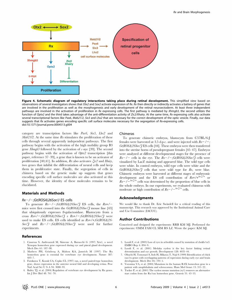
category are transcription factors like Pax6, Six3, Lhx2 and
Mab21l2. At the same time Rx stimulates the proliferation of these
cells through several apparently independent pathways. The first
pathway begins with the activation of the high mobility group B3
gene Xhmgb3 followed by the activation of c-myc [39]. The second
pathway begins with the activation of Optx2 transcription [this
paper, reference 37–39], a gene that is known to be an activator of
proliferation [40,41]. In addition, Rx also activates Zic2 and Hairy,
two genes that inhibit the differentiation of neural cells and keep
them in proliferative state. Finally, the segregation of cells in
chimera based on the genetic make up suggests that genes
encoding specific cell surface molecules are also activated at this
time. However, the identity of these molecules remains to be
elucidated.
Materials and Methods
Rx2/2;Gt(ROSA)26Sor/J ES cellsTo generate Rx2/2;Gt(ROSA)26Sor/J ES cells, the Rx+/2
mice were first crossed into the Gt(ROSA)26Sor/J mouse line [43]
that ubiquitously expresses ß-galactosidase. Blastocysts from a
cross Rx+/2;Gt(ROSA)26Sor/J x Rx+/2;Gt(ROSA)26Sor/J were
used to make ES cells. ES cells identified as Rx+/+;Gt(ROSA)26-
Sor/J and Rx2/2;Gt(ROSA)26Sor/J were used for further
experiments.
ChimerasTo generate chimeric embryos, blastocysts from C57BL/6-J
females were harvested at 3.5 d.p.c. and were injected with Rx2/2;
Gt(ROSA)26Sor/J ES cells [44]. These embryos were then transferred
into the uterine horns of pseudopregnant females [45–47]. Embryos
were analyzed at different developmental stages for the presence of
Rx2/2 cells in the eye. The Rx2/2;Gt(ROSA)26Sor/J cells were
visualized by LacZ staining and appeared blue. The wild type cells
were white. In control embryos, wild type cells were white and the
Gt(ROSA)26Sor/J cells that were wild type for Rx, were blue.
Chimeric embryos were harvested at different stages of embryonic
development and the ES cell contribution of Rx+/+lacZ/+ or
Rx2/2lacZ/+ cells was determined by the proportion of blue cells in
the whole embryo. In our experiments, we evaluated chimeras with
moderate or high contribution of Rx2/2lacZ/+ cells.
Acknowledgments
We would like to thank Dr. Eric Swindell for a critical reading of this
manuscript. This research was approved by the Institutional Animal Care
and Use Committee (IACUC).
Author Contributions
Conceived and designed the experiments: RRB KM MJ. Performed the
experiments: OMM FAM CL MM RS LZ. Wrote the paper: KM MJ.
References
1. Casarosa S, Andreazzoli M, Simeone A, Barsacchi G (1997) Xrx1, a novel
Xenopus homeobox gene expressed during eye and pineal gland development.
Mech Dev 61: 187–98.
2. Mathers PH, Grinberg A, Mahon KA, Jamrich M (1997) The Rx
homeobox gene is essential for vertebrate eye development. Nature 387:
603–7.
3. Furukawa T, Kozak CA, Cepko CL (1997) rax, a novel paired-type homeobox
gene, shows expression in the anterior neural fold and developing retina. Proc
Natl Acad Sci U S A 94: 3088–93.
4. Bailey TJ, et al. (2004) Regulation of vertebrate eye development by Rx genes.
Int J Dev Biol 48: 761–70.
5. Loosli F, et al. (2003) Loss of eyes in zebrafish caused by mutation of chokh/rx3.
EMBO Rep 4: 894–9.
6. Loosli F, et al. (2001) Medaka eyeless is the key factor linking retinal
determination and eye growth. Development 128: 4035–44.
7. Ohuchi H, Tomonari S, Itoh H, Mikawa T, Noji S (1999) Identification of chick
rax/rx genes with overlapping patterns of expression during early eye and brain
development. Mech Dev 85: 193–5.
8. Voronina VA, et al. (2004) Mutations in the human RAX homeobox gene in a
patient with anophthalmia and sclerocornea. Hum Mol Genet 13: 315–22.
9. Tucker P, et al. (2001) The eyeless mouse mutation (ey1) removes an alternative
start codon from the Rx/rax homeobox gene. Genesis 31: 43–53.
Figure 4. Schematic diagram of regulatory interactions taking place during retinal development. This simplified view based onobservations of several investigators shows that Otx2 and Sox2 activate expression of Rx. Rx then directly or indirectly activates a battery of genes thatare involved in the proliferation as well as the morphogenesis and early development of the retinal neuroectoderm. At least three independentpathways are involved in the activation of proliferation in Rx expressing cells. The first pathway is mediated by Xhmgb3, the second utilizes thefunction of Optx2 and the third takes advantage of the anti-differentiation activity of Zic2/Xhairy. At the same time, Rx expressing cells also activateseveral transcriptional factors like Pax6, Mab21/2, Six3 and Lhx2 that are necessary for the correct development of the optic vesicle. Finally, our datasuggests that Rx activates genes encoding specific cell surface molecules necessary for the segregation of Rx-expressing cells.doi:10.1371/journal.pone.0004513.g004
Rx and Brain Morphogenesis
PLoS ONE | www.plosone.org 6 February 2009 | Volume 4 | Issue 2 | e4513
10. Swindell EC, et al. (2008) Eye formation in the absence of retina. Dev Biol 322:
56–64.11. Swindell EC, et al. (2006) Rx-Cre, a tool for inactivation of gene expression in
the developing retina. Genesis 44: 361–363.
12. Brownell I, Dirksen M, Jamrich M (2000) Forkhead Foxe3 maps to thedysgenetic lens locus and is critical in lens development and differentiation.
Genesis 27: 81–93.13. Winkler S, Loosli F, Henrich T, Wakamatsu Y, Wittbrodt J (2000) The
conditional medaka mutation eyeless uncouples patterning and morphogenesis
of the eye. Development 127: 1911–9.14. Zhang L, Mathers PH, Jamrich M (2000) Function of Rx, but not Pax6, is
essential for the formation of retinal progenitor cells in mice. Genesis 28:135–42.
15. Sheng HZ, et al. (1997) Multistep control of pituitary organogenesis. Science278: 1809–12.
16. Rosenfeld MG, et al. (2000) Multistep signaling and transcriptional requirements
for pituitary organogenesis in vivo. Recent Prog Horm Res 55: 1–13; discussion13–4.
17. Hermesz E, Mackem S, Mahon KA (1996) Rpx: a novel anterior-restrictedhomeobox gene progressively activated in the prechordal plate, anterior neural
plate and Rathke’s pouch of the mouse embryo. Development 122: 41–52.
18. Schwind J (1928) The development of the hypophysis cerebri of the albino rat.Amer J Anat 41: 295–315.
19. Scully KM, Rosenfeld MG (2002) Pituitary development: regulatory codes inmammalian organogenesis. Science 295: 2231–5.
20. Ericson J, Norlin S, Jessell TM, Edlund T (1998) Integrated FGF and BMPsignaling controls the progression of progenitor cell differentiation and the
emergence of pattern in the embryonic anterior pituitary. Development 125:
1005–15.21. Takuma N, et al. (1998) Formation of Rathke’s pouch requires dual induction
from the diencephalon. Development 125: 4835–40.22. Treier M, et al. (1998) Multistep signaling requirements for pituitary
organogenesis in vivo. Genes Dev 12: 1691–704.
23. Ohuchi H, et al. (2000) FGF10 acts as a major ligand for FGF receptor 2 IIIb inmouse multi-organ development. Biochem Biophys Res Commun 277: 643–9.
24. Kondoh H, et al. (2000) Zebrafish mutations in Gli-mediated hedgehog signalinglead to lens transdifferentiation from the adenohypophysis anlage. Mechanisms
of Development 96: 165–74.25. Rembold M, Loosli F, Adams RJ, Wittbrodt J (2006) Individual cell migration
serves as the driving force for optic vesicle evagination. Science 313: 1130–4.
26. Stigloher C, et al. (2006) Segregation of telencephalic and eye-field identitiesinside the zebrafish forebrain territory is controlled by Rx3. Development 133:
2925–35.27. Rojas-Munoz A, Dahm R, Nusslein-Volhard C (2005) chokh/rx3 specifies the
retinal pigment epithelium fate independently of eye morphogenesis. Dev Biol
288: 348–62.28. Sheng HZ, et al. (1996) Specification of pituitary cell lineages by the LIM
homeobox gene Lhx3. Science 272: 1004–7.
29. Jean D, Bernier G, Gruss P (1999) Six6 (Optx2) is a novel murine Six3-related
homeobox gene that demarcates the presumptive pituitary/hypothalamic axisand the ventral optic stalk. Mech Dev 84: 31–40.
30. Toy J, Sundin OH (1999) Expression of the optx2 homeobox gene during mouse
development. Mech Dev 83: 183–6.31. Toy J, Yang JM, Leppert GS, Sundin OH (1998) The optx2 homeobox gene is
expressed in early precursors of the eye and activates retina-specific genes. ProcNatl Acad Sci U S A 95: 10643–8.
32. Lopez-Rios J, Gallardo ME, Rodriguez de Cordoba S, Bovolenta P (1999) Six9
(Optx2), a new member of the six gene family of transcription factors, isexpressed at early stages of vertebrate ocular and pituitary development. Mech
Dev 83: 155–9.33. Li X, Perissi V, Liu F, Rose DW, Rosenfeld MG (2002) Tissue-specific
regulation of retinal and pituitary precursor cell proliferation. Science 297:1180–3.
34. Cho SH, Cepko CL (2006) Wnt2b/beta-catenin-mediated canonical Wnt
signaling determines the peripheral fates of the chick eye. Development 133:3167–77.
35. Kennedy BN, et al. (2004) Zebrafish rx3 and mab21l2 are required during eyemorphogenesis. Dev Biol 270: 336–49.
36. Volokh KY (2006) Tissue morphogenesis: a surface buckling mechanism.
Int J Dev Biol 50: 359–65.37. Zuber ME, Gestri G, Viczian AS, Barsacchi G, Harris WA (2003) Specification
of the vertebrate eye by a network of eye field transcription factors. Development130: 5155–67.
38. Andreazzoli M, et al. (2003) Xrx1 controls proliferation and neurogenesis inXenopus anterior neural plate. Development 130: 5143–54.
39. Terada K, Kitayama A, Kanamoto T, Ueno N, Furukawa T (2006) Nucleosome
regulator Xhmgb3 is required for cell proliferation of the eye and brain as adownstream target of Xenopus rax/Rx1. Dev Biol 291: 398–412.
40. Bernier G, et al. (2000) Expanded retina territory by midbrain transformationupon overexpression of Six6 (Optx2) in Xenopus embryos. Mech Dev 93: 59–69.
41. Zuber ME, Perron M, Philpott A, Bang A, Harris WA (1999) Giant eyes in
Xenopus laevis by overexpression of XOptx2. Cell 98: 341–52.42. Danno H, et al. (2008) Molecular links among the causative genes for ocular
malformation: Otx2 and Sox2 coregulate Rax expression. Proc Natl AcadSci U S A 105: 5408–13.
43. Friedrich G, Soriano P (1991) Promoter traps in embryonic stem cells: a geneticscreen to identify and mutate developmental genes in mice. Genes Dev 5:
1513–23.
44. Nagy A (2000) Manipulating the Mouse Embryo. New York: CSHL Press.45. Gardner RL, Davies TJ (2000) Mouse chimeras and the analysis of development.
Methods Mol Biol 135: 397–424.46. Gardner RL (1975) Analysis of determination and differentiation in the early
mammalian embryo using intra- and interspecific chimeras. Symp Soc Dev Biol
33: 207–36.47. Gardner RL (1968) Mouse chimeras obtained by the injection of cells into the
blastocyst. Nature 220: 596–7.
Rx and Brain Morphogenesis
PLoS ONE | www.plosone.org 7 February 2009 | Volume 4 | Issue 2 | e4513