Recombinative events of the T cell antigen receptor delta gene in peripheral T cell lymphomas
Upload
independentCategory
view
1download
0
CD8+ T Cell Priming by Dendritic Cell Vaccines RequiresAntigen Transfer to Endogenous Antigen PresentingCellsAlice W. Yewdall1¤, Scott B. Drutman1, Felecia Jinwala1, Keith S. Bahjat2, Nina Bhardwaj1*
1 Cancer Institute, New York University School of Medicine, New York, New York, United States of America, 2 Earle A. Chiles Research Institute, Providence Cancer Center,
Portland, Oregon, United States of America
Abstract
Background: Immunotherapeutic strategies to stimulate anti-tumor immunity are promising approaches for cancertreatment. A major barrier to their success is the immunosuppressive microenvironment of tumors, which inhibits thefunctions of endogenous dendritic cells (DCs) that are necessary for the generation of anti-tumor CD8+ T cells. To overcomethis problem, autologous DCs are generated ex vivo, loaded with tumor antigens, and activated in this non-suppressiveenvironment before administration to patients. However, DC-based vaccines rarely induce tumor regression.
Methodology/Principal Findings: We examined the fate and function of these DCs following their injection using murinemodels, in order to better understand their interaction with the host immune system. Contrary to previous assumptions, weshow that DC vaccines have an insignificant role in directly priming CD8+ T cells, but instead function primarily as vehiclesfor transferring antigens to endogenous antigen presenting cells, which are responsible for the subsequent activation of Tcells.
Conclusions/Significance: This reliance on endogenous immune cells may explain the limited success of current DCvaccines to treat cancer and offers new insight into how these therapies can be improved. Future approaches should focuson creating DC vaccines that are more effective at directly priming T cells, or abrogating the tumor induced suppression ofendogenous DCs.
Citation: Yewdall AW, Drutman SB, Jinwala F, Bahjat KS, Bhardwaj N (2010) CD8+ T Cell Priming by Dendritic Cell Vaccines Requires Antigen Transfer toEndogenous Antigen Presenting Cells. PLoS ONE 5(6): e11144. doi:10.1371/journal.pone.0011144
Editor: Derya Unutmaz, New York University, United States of America
Received April 7, 2010; Accepted May 25, 2010; Published June 16, 2010
Copyright: � 2010 Yewdall et al. This is an open-access article distributed under the terms of the Creative Commons Attribution License, which permitsunrestricted use, distribution, and reproduction in any medium, provided the original author and source are credited.
Funding: The study was supported by the Cancer Research Institute, the Doris Duke Charitable Foundation, and the National Institutes of Health (NIH) grantsA1044628, A1071078, and A1061684 to N.B. The funders had no role in study design, data collection and analysis, decision to publish, or preparation of themanuscript.
Competing Interests: The authors have declared that no competing interests exist.
* E-mail: [email protected]
¤ Current address: Immunology Institute, Mount Sinai School of Medicine, New York, New York, United States of America
Introduction
Therapeutic vaccines are a considered a realistic approach for
cancer immunotherapy, because animal studies have shown that
recognition of tumor associated antigens (TAAs) by cytotoxic T
lymphocytes can lead to the destruction of tumor cells [1,2,3]. In
order to generate tumor-specific T cells with full effector function
they first must undergo priming by dendritic cells (DCs), the
antigen presenting cell (APC) most efficient at initiating potent
CD8+ T-cell responses [4,5]. Therefore, understanding how to
modulate DC functions may be essential to therapeutically
inducing anti-tumor immune responses [6,7]. The immunomod-
ulatory activity of DCs is regulated by the process of maturation.
In response to environmental signals such as pathogens or
inflammatory cytokines, immature DCs undergo phenotypic and
qualitative changes to become mature DCs, characterized by
presentation of peptides bound to major histocompatibility
complexes (MHC), high surface expression of co-stimulatory
molecules, secretion of cytokines, and potent T cell stimulatory
abilities. Depending upon the maturation stimuli, DCs instruct T
cells towards different types of immune responses [8,9]. Thus, the
development of mature DCs with the essential properties for
induction of effective anti-tumor immunity is critical.
One significant barrier to therapeutic stimulation of anti-tumor
immunity is constituents of the tumor microenvironment [10] such
as secreted immunosuppressive factors [11], myeloid-derived
suppressor cells [12], and regulatory T cells [13], that adversely
affect DC activation and consequently blunt the generation of
anti-tumor immunity. Furthermore, antigen presented to naı̈ve T
cells by DCs that were not properly activated can lead to T cell
tolerance [14,15,16]. To circumvent this problem, many studies
have focused on optimizing the antigen presentation function of
DCs ex vivo such that these DCs can be adoptively transferred back
into the in vivo environment. Ideally, this should yield DCs that,
following injection, traffic to the draining lymph node and
efficiently activate T cells, leading to the generation of an effective
immune response [17,18].
The optimization of ex vivo DC manipulation has focused
primarily on maturation stimuli to enhance the expression of co-
stimulatory molecules, secretion of pro-inflammatory cytokines,
PLoS ONE | www.plosone.org 1 June 2010 | Volume 5 | Issue 6 | e11144

and expression of chemokine receptors important for migration to
lymphoid organs. Additionally, the form of antigen used for
loading DCs is a major consideration. The most commonly used
form of antigens, are pre-determined immunogenic peptide
epitopes derived from TAA, which are restricted to specific
MHC haplotypes; or whole tumor antigens, which are theoreti-
cally more advantageous than peptide antigens because they can
be processed into epitopes that can be presented regardless of
patients’ MHC haplotypes.
Several clinical trials conducted over the past decade have
demonstrated that DC vaccines can prime and boost antigen-specific
CD8+ T cells in humans. However, their clinical efficacy remains to
be definitively demonstrated [6,19,20,21]. The lack of success has
been variously attributed to several factors: administration of
relatively low cell numbers of DCs, suboptimal route of administra-
tion, improper antigen dose, poor choice of antigenic targets,
unsuitable maturation state of DCs, and inappropriate frequency of
injections. However, understanding exactly which of these concerns
represent true problems may be difficult because little is known
regarding the fate and function of ex vivo generated DCs after they
have been injected. Because tracking these events in patients in a
controlled manner is not feasible, we utilized a murine model of DC
vaccination to better understand the events following DC injection.
We show here that ex vivo derived DC vaccines have an insignificant
role in the direct priming of T cells in vivo. Instead, evidence is
provided that DC vaccines indirectly prime naı̈ve CD8+ T cells in vivo
by transferring antigens to endogenous cells, which subsequently
present them to CD8+ T cells.
Results
CD8+ T cell priming by peptide loaded DC vaccinerequires endogenous antigen presenting cells
To investigate the immunogenicity of DC vaccines, we first
established a murine model to monitor the in vivo activation of
antigen-specific CD8+ T cells following vaccination. Mice were
intravenously injected with ex vivo derived DCs pulsed with the
MHC class I-restricted epitope of the ovalbumin (OVA) protein,
OVA257–264 (peptide-DCs). As a control, mice were immunized
with attenuated Listeria monocytogenes (Lm) expressing OVA
(LmOVA). Immunization of wild type C57BL/6 (B6) mice with
either LmOVA or peptide-DCs elicited an OVA257–264-specific
immune response as demonstrated by antigen-specific IFN-cproduction by CD8+ T cells (Fig. 1A). While the T cell response
to LmOVA should depend on antigen presentation by the
endogenous bone-marrow-derived hematopoietic cells [22], the
priming by peptide-DCs in these mice could be a result of either
direct interaction of the injected DCs with endogenous T cells, or
antigens from the injected DCs captured by host cells and
subsequently presented to T cells.
Figure 1. Peptide-pulsed DC vaccines require help from host immune cells to induce a maximal CD8+ T cell response. (A) Wild typeC57BL/6 (B6) mice were vaccinated with either attenuated LmOVA (16106 CFU/mouse), OVA257–264 (100 ng/ml)–pulsed LPS-matured B6 DCs(peptide-DCs, 16106 cells/mouse), or unpulsed LPS-matured B6 DCs (DC alone, 16106 cells/mouse). On day 7, the percentage of IFN-c producingCD8+ T cells was determined (horizontal bar = mean). (B) B6RB6 (B6) and Bm1RB6 (Bm1) marrow chimeras (BMCs) were adoptively transferred with5,000 naı̈ve OT-1 T cells and vaccinated the following day with LmOVA (16106 CFU/mouse), peptide-DCs (16106 cells/mouse), or DCs alone(16106 cells/mouse). The immune response was assessed on days 7 and 18 post-vaccination. (C) Antigen-specific IFN-c production by splenic CD8+ Tcells was measured on day 7. Data are mean + s.d. for 3 mice per group and are representative of 3 experiments. (D) In vivo killing of OVA257–264
peptide coated target cells in the spleen between days 18 and 20 was assessed. Specific killing was normalized to the internal control of target cellscoated with an irrelevant peptide from the Sendai virus nucleoprotein, NP324–332. Data are mean 6 s.d. for 3 mice per group and are representative of2 experiments.doi:10.1371/journal.pone.0011144.g001
Indirect Priming by DC Vaccine
PLoS ONE | www.plosone.org 2 June 2010 | Volume 5 | Issue 6 | e11144

To examine the T cell priming that resulted from only the direct
effect of the injected DCs, we repeated these vaccinations in bone
marrow chimeric mice (BMCs) that lack a hematopoietic
compartment able to present the OVA257–264 peptide. This was
accomplished by reconstituting lethally irradiated wild type mice
with bone marrow from Bm1 mice, which express a mutant H-
2Kb allele (H-2Kbm1) that prevent proper interaction of the
OVA257–264-MHC complex with its cognate CD8+ T cell [23].
We confirmed donor-specific tolerance after allogeneic bone
marrow transplantation (Fig. S1). Wild type and Bm1-derived
bone marrow cells will reconstitute a T cell compartment with
differing repertoires of naive antigen-specific T cells therefore, we
normalized the number of OVA257-264-specific T cells by
adoptively transferring 5,000 naive OT-I CD8+ T cells, into each
mouse one day prior to vaccination (Fig. 1B).
As expected, mice reconstituted with Bm1 bone marrow (Bm1
BMCs) did not elicit an OVA257–264-specific CD8+ T cell
response after vaccination with LmOVA, in contrast to mice
reconstituted with wild type bone marrow (B6 BMCs) (Fig. 1C).
However, following intravenous vaccination with peptide-DCs,
the number of IFN-c producing CD8+ T cells generated was
significantly reduced in Bm1 BMCs (Fig. 1C), and this small
number of T cells primed in the Bm1 BMCs also exhibited a
reduced effector cell function (Fig. 1D). These results suggest
that the T cell priming observed after vaccination with peptide
DCs in Fig. 1A requires a host with a hematopoietic
compartment capable of capturing and re-presenting the
vaccine derived antigen.
Antigen transfer from the injected DCs to endogenous APCs
may occur either by shedding of peptides or transfer of intact
peptide-MHC complexes [24,25], possibly via exchange of plasma
membranes and associated proteins between cells [26]. Contact-
dependent transfer of peptide-MHC complexes from ex vivo
derived DCs to splenic cell populations can occur with great
efficiency in vitro (Fig. S2). However, the inefficient T cell
activation in Bm1 BMCs suggests that translocation of peptide-
MHC complexes to host APCs is unlikely the source responsible
for T cell priming in B6 BMCs.
Priming of CD8+ T cells by DCs loaded with proteinantigen also requires endogenous antigen presentingcells
The heterogeneous nature of most tumor antigens and patient
MHC haplotypes limits the practicality of externally-loaded pre-
determined peptides as an antigen source for vaccines. Conse-
quently, DCs are often loaded with antigens that require
processing. To mimic this vaccine approach, we used DCs
generated ex vivo from mice (Act-mOVA mice) [27] that
constitutively express OVA protein under the actin promoter.
DCs from Act-mOVA mice (ActOVA DCs) process the
endogenous antigen in the MHC class I pathway and express
OVA257–264-MHC class I complexes on their cell surface at a level
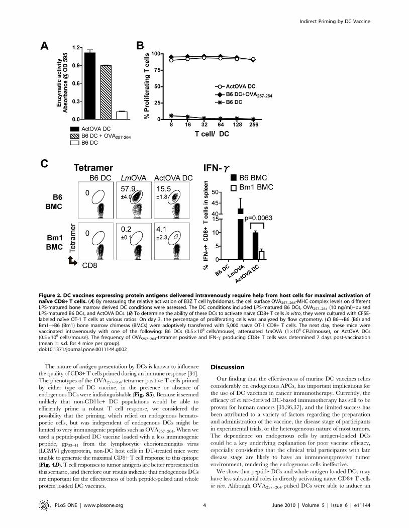
comparable to the peptide-DCs used for vaccination (Fig. 2A),
and both types of DCs stimulate T cells in vitro with comparable
potency (Fig. 2B). When mice were intravenously immunized
with ActOVA DCs using the same strategy described in Fig. 1B,
we again observed the same requirement for host APCs in
generating a maximal CD8+ T cell response as we had with
peptide-DCs (Fig. 2C). Thus, regardless of whether the OVA
antigen was originally expressed in DCs (ActOVA DCs) or
externally loaded on the surface as a pre-determined epitope
(peptide-DCs), maximal priming of OVA-specific T cells requires
host APCs.
The requirement for host antigen presenting cells doesnot depend on the route of DC vaccine delivery
Since the route of injection influences the sites DCs reach and thus
may affect their function, we repeated our vaccination using
subcutaneous injections. We also took this opportunity to extend
our findings in an additional BMC system, in which irradiated wild
type B6 mice were reconstituted with bone marrow from the MHC
class I-deficient KbDb KO mice (KbDb KO BMCs) [28,29]. Similar
to our results with intravenously delivered DCs, transfer of antigens to
host APCs was required for efficient CD8+ T cell priming (Fig. 3A).
The indirect role of the injected DCs in T cell priming was not due to
a lack of exit from the injection site, because the DCs migrated to the
draining lymph node (LN), albeit with a very low frequency of #0.5%
of total injected cells (Fig. 3B). This percentage of DCs to migrate is
consistent with findings from other groups [30,31,32,33]. Addition-
ally, the DCs were viable and had not been internalized by host cells
when detected in the draining LN two days after injection (Fig. 3C).
Our data demonstrate that, irrespective of the route of their delivery,
optimal generation of systemic immune responses by DC vaccines
requires host APCs.
Differential requirements for endogenous DCs bypeptide-loaded DCs versus protein expressing DCs
Because DCs are the APC most efficient at priming CD8+ T-
cells [6] it seemed likely that they might be the cell type responsible
for the presentation of the DC vaccine-derived antigens we
observed. To investigate this possibility, we repeated our
vaccinations in irradiated wild type mice reconstituted with bone
marrow from mice that express the primate diptheria toxin
receptor (DTR) under control of the CD11c promoter (CD11c-
DTR BMCs) [22] (Fig. 4A). Treatment of these mice with
diptheria toxin (DT) throughout the duration of the experiment
selectively eliminated the CD11chigh expressing DCs as confirmed
by flow cytometry (Fig. S3), and the lack of a T cell response in
DT treated mice after infection with LmOVA (Fig. 4B).
Both peptide-DC and ActOVA DC vaccines primed a CD8+ T
cell response efficiently in non-DT treated CD11c-DTR BMCs, as
evidenced by the percentage of cells positive for OVA257–264-tetramer
staining and antigen-specific IFNc and TNF production by CD8+ T
cells 7 days post-vaccination (Fig. 4B). In DT-treated mice,
vaccination with ActOVA DCs induced a poor T cell response,
demonstrating that they rely substantially on host DCs to cross-prime
antigens. However, the frequency of CD8+ T cells primed after
delivery of peptide-DCs was not affected by the DT treatment.
In this model, T cell activation may reflect direct interaction of
peptide-DCs with naive CD8+ T cells. However, in conjunction
with previous results in Bm1 BMCs (Fig. 1C), it is possible that
peptide antigens from the injected peptide-DCs were transferred
to cell types other than CD11c+ DCs, which subsequently
activated naı̈ve CD8+ T cells.
To determine how ActOVA DCs were unable to take
advantage of peptide transfer to endogenous cells for T cell
activation, we compared the stability of surface MHC-bound
peptides on ActOVA DCs to their stability on peptide-DCs. We
found that there is a greater dissociation rate of peptides that are
externally loaded onto surface MHC class I molecules, than of
peptides presented by ActOVA DCs, which are intracellularly
processed and loaded (Fig. 4C). Compared to ActOVA DCs,
peptide-DCs seem to transfer their surface MHC-bound peptides
more readily to host cells. We also confirmed that non-specific
binding of peptides to non-MHC molecules on the peptide-DCs
does not contribute to transfer of antigens to host cells resulting in
T cell activation (Fig. S4).
Indirect Priming by DC Vaccine
PLoS ONE | www.plosone.org 3 June 2010 | Volume 5 | Issue 6 | e11144
The nature of antigen presentation by DCs is known to influence
the quality of CD8+ T cells primed during an immune response [34].
The phenotypes of the OVA257–264-tetramer positive T cells primed
by either type of DC vaccine, in the presence or absence of
endogenous DCs were indistinguishable (Fig. S5). Because it seemed
unlikely that non-CD11c+ DC populations would be able to
efficiently prime a robust T cell response, we considered the
possibility that the priming, which relied on endogenous hemato-
poetic cells, but was independent of endogenous DCs might be
limited to very immunogenic peptides such as OVA257–264. When we
used a peptide-pulsed DC vaccine loaded with a less immunogenic
peptide, gp33–41 from the lymphocytic choriomeningitis virus
(LCMV) glycoprotein, non-DC host cells in DT-treated mice were
unable to generate the maximal CD8+ T cell response to this epitope
(Fig. 4D). T cell responses to tumor antigens are better represented in
this scenario, and therefore our results indicate that endogenous DCs
are important for the effectiveness of both peptide-pulsed and whole
protein loaded DC vaccines.
Discussion
Our finding that the effectiveness of murine DC vaccines relies
considerably on endogenous APCs, has important implications for
the use of DC vaccines in cancer immunotherapy. Currently, the
efficacy of ex vivo-derived DC-based immunotherapy has still to be
proven for human cancers [35,36,37], and the limited success has
been attributed to a variety of factors regarding the preparation
and administration of the vaccine, the disease stage of participants
in experimental trials, or the heterogeneous nature of most tumors.
The dependence on endogenous cells by antigen-loaded DCs
could be a key underlying explanation for poor vaccine efficacy,
especially considering that the clinical trial participants with late
disease stage are likely to have an immunosuppressive tumor
environment, rendering the endogenous cells ineffective.
We show that peptide-DCs and whole antigen-loaded DCs may
have less substantial roles in directly activating naı̈ve CD8+ T cells
in vivo. Although OVA257–264-pulsed DCs were able to induce an
Figure 2. DC vaccines expressing protein antigens delivered intravenously require help from host cells for maximal activation ofnaı̈ve CD8+ T cells. (A) By measuring the relative activation of B3Z T cell hybridomas, the cell surface OVA257–264-MHC complex levels on differentLPS-matured bone marrow derived DC conditions were assessed. The DC conditions included LPS-matured B6 DCs, OVA257–264 (10 ng/ml)–pulsedLPS-matured B6 DCs, and ActOVA DCs. (B) To determine the ability of these DCs to activate naı̈ve CD8+ T cells in vitro, they were cultured with CFSE-labeled naı̈ve OT-1 T cells at various ratios. On day 3, the percentage of proliferating cells was analyzed by flow cytometry. (C) B6RB6 (B6) andBm1RB6 (Bm1) bone marrow chimeras (BMCs) were adoptively transferred with 5,000 naı̈ve OT-1 CD8+ T cells. The next day, these mice werevaccinated intravenously with one of the following: B6 DCs (0.56106 cells/mouse), attenuated LmOVA (16106 CFU/mouse), or ActOVA DCs(0.56106 cells/mouse). The frequency of OVA257–264-tetramer positive and IFN-c producing CD8+ T cells was determined 7 days post-vaccination(mean 6 s.d. for 4 mice per group).doi:10.1371/journal.pone.0011144.g002
Indirect Priming by DC Vaccine
PLoS ONE | www.plosone.org 4 June 2010 | Volume 5 | Issue 6 | e11144

immune response in the absence of endogenous CD11c+ DCs in
DT-treated CD11c-DTR BMCs (Fig. 4B), we hesitate to
conclude that they were directly interacting with T cells in vivo
for two reasons. First, DCs loaded with a less immunogenic
peptide, gp33–41 were unable to induce an optimal antigen-specific
CD8+ T cell response in mice lacking endogenous CD11c+ DCs
(Fig. 4D). Secondly, injection of OVA257–264-pulsed B6 DCs
intravenously or subcutaneously into hosts that lack hematopoietic
cells capable of presenting antigens resulted in a significant
reduction of antigen-specific CD8+ T cells (Fig. 1C and Fig. 3A).
Figure 3. DCs vaccinated subcutaneously require MHC class I expression on the host immune cells to fully activate naı̈ve CD8+ Tcells. (A) B6RB6 (B6) and KbDb KORB6 (KbDb KO) bone marrow chimeras (BMCs) were adoptively transferred with 5,000 naı̈ve OT-1 CD8+ T cells. Thenext day, they were vaccinated subcutaneously in the footpad with one of the following: LPS-matured B6 DCs (0.56106 cells/mouse), attenuated Lm-OVA (16106 CFU/mouse), or LPS-matured ActOVA DCs (0.56106 cells/mouse). The frequencies of OVA257–264-tetramer positive CD8+ T cells in thespleen (individual mice) and draining lymph node (LN, combined value of from mice of each condition) and IFN-c producing CD8+ T cells in thespleen of each mouse were determined 7 days post-vaccination (data are mean 6 s.d. for 4 mice per group). (B) To track migration of DCs to thedraining LN, CFSE-labeled or –unlabeled LPS-matured DCs (16106 cells/mouse) were injected subcutaneously in the foot pad of naı̈ve mice. Thepopliteal LNs were removed 2 and 3 days post-injection. Cells were gated on the live (7-AAD negative) and CD11b+ population using flow cytometryto analyze the percentage (0.1–0.3%) of DCs that migrated. (C) CFSE-labeled CD45.1 donor DCs were injected subcutaneously in the footpad ofCD45.1-CD45.2+ wild type mice. Two days later the popliteal LNs were removed and cells were stained with anti-CD45.1 and anti-CD45.2 antibodies.doi:10.1371/journal.pone.0011144.g003
Indirect Priming by DC Vaccine
PLoS ONE | www.plosone.org 5 June 2010 | Volume 5 | Issue 6 | e11144

Figure 4. Endogenous CD11chigh DCs are recipients of antigens derived from DC vaccines, that are essential for activating naı̈veCD8+ T cells in vivo. (A-B, D) CD11c-DTR BMCs were treated with diptheria toxin (DT) on days 24, 21, and 2 relative to the day of vaccination.Naı̈ve transgenic CD8+ T cells were adoptively transferred intravenously on day 21. Antigen-specific T cell responses were analyzed on day 7 post-
Indirect Priming by DC Vaccine
PLoS ONE | www.plosone.org 6 June 2010 | Volume 5 | Issue 6 | e11144

Peptide-DCs carry antigens in the form of peptides already
bound to their surface MHC molecules, therefore are unlikely to
be used as an antigen source for cross presentation into the MHC
class I antigen presentation pathway, if internalized by a recipient
cell. It is most likely that peptides are being transferred directly
onto the surface MHC class I molecules of host APCs for their
subsequent presentation. In theory, any cell type expressing MHC
class I molecules and co-stimulatory molecules could present the
antigenic peptide donated from peptide-DCs to activate naı̈ve T
cells. Possibly, APCs not eliminated by DT treatment, such as
plasmacytoid DCs [38,39,40] participated in the priming of T
cells. However, we demonstrated with gp33–41 peptides that the
dependence of peptide transfer from injected DCs to non-CD11c+DC endogenous cell populations does not apply for all antigens.
Similar to peptide-DCs, priming of CD8+ T cells by ActOVA
DCs was inefficient in mice with endogenous cells lacking the
appropriate MHC molecules to present the OVA257–264 peptide
(Fig. 2C and Fig. 3A). In the CD11c-DTR model, the OVA
antigen expressed by ActOVA DCs is cross-presented specifically
by endogenous DCs. This data is consistent with previous reports
that DCs are required for the cross-priming of cell-associated
antigens [22]. Protein antigens can be transferred and processed
for MHC class I presentation by host DCs following phagocytosis
of apoptotic [41] or necrotic [42,43] ActOVA DCs. Although
these results are expected, we were surprised that there was a
difference in the requirement of endogenous CD11c+ DCs for T
cell activation by ActOVA DCs versus OVA257–264-pulsed DCs.
An in vitro assay confirmed that peptides loaded externally onto cell
surface bound MHC I molecules (peptide-DCs) are less stable than
those internally processed on loaded onto MHC I molecules, as in
ActOVA DCs (Fig. 4C). We conclude that because ActOVA DCs
may not shed peptides as readily as OVA257–264-pulsed DCs in vivo,
they are dependent on cross-presentation by host DCs.
Under priming conditions where antigen transfer by cross-
presentation or by peptide transfer to MHC class I molecules was
not permitted, there was some residual T cell expansion. The
specific interaction of injected ex vivo derived DCs and antigen-
specific T cells has been visualized in the draining LN of mice [17],
therefore it is possible that the small amount of T-cell activation
may have resulted from direct priming by injected DCs.
Alternatively, several groups have described the transfer of
peptide-MHC complexes to recipient cells from either secreted
exosomes [44,45,46], live cells via the acquisition of plasma
membranes [26], or dead cells in a process called ‘‘cross-dressing’’
[24,25]. In vitro, the transfer of peptide-MHC complexes from ex
vivo derived DCs to splenic cell populations takes place efficiently
(Fig. S2). However, we conclude that this mechanism is not as
effective in vivo, because the expression of an appropriate MHC
haplotype on host cells is essential for a proper T cell response.
More importantly, we showed that the residual T cells primed in
Bm1 BMCs after administering peptide-DCs were not able to
effectively kill target cells (Fig. 1D). In this setting, the initial
number of antigen-specific CD8+ T cells produced early during
immune activation seems to be important for the generation of a
memory response. Altogether our data show that DC vaccines
overcome their inability to directly prime T cells in vivo by
transferring antigens to hematopoietic cells for subsequent antigen
presentation.
In order for endogenous DCs to properly prime naı̈ve CD8+ T
cells, they must undergo maturation. Therefore, in addition to
acting as an antigen source, the injected DCs may also cause
activation of endogenous recipient DCs without further stimula-
tion. It has been shown that cross-presentation of OVA-loaded
irradiated Bm1 or B6 splenocytes by host APCs results in
proliferation of antigen-specific T cells, even in the absence of
additional adjuvants [47,48]. In these circumstances, the activa-
tion of host DCs required for efficient cross-presentation may be
explained by the potential release of TLR ligands during donor
cell death [49]. In our studies, it is possible that the eventual death
of the injected LPS-treated DC vaccines in this study may have
contributed to the successful T cell activation in mice with a wild
type hematopoietic compartment. Phagocytosis of LPS-treated
apoptotic cells has recently been shown to mature DCs in a TLR-4
dependent manner [50]. Indeed, we showed that injection of mice
with DCs coated with LPS caused an upregulation of maturation
markers on a small number of endogenous splenic DCs (Fig. S6),
pointing to a mechanism by which host APCs can undergo
activation following exposure to adoptively administered DCs.
However, the immunosuppressive nature of most cancers is
unfavorable to the proper activation of host DCs [10] and is
therefore an obstacle for any vaccine that relies on these cells to
initiate a T cell response. We speculate that this is one explanation
for the poor efficacy of DC vaccines to eliminate tumors in clinical
trials [20]. In this context, the adjuvant activity of the administered
ex vivo-derived DCs may be inadequate for proper naı̈ve T cell
stimulation, and the benefits of antigen transfer could go
undetected, unless combined with other immunotherapeutic
strategies that focus on inhibiting the factors that contribute to
the immunosuppressive tumor environment [20]. Future anti-
cancer immunotherapeutics might also benefit from activating and
targeting antigens to endogenous DCs directly [51].
Materials and Methods
Mice, bacteria strains, and cellsC57BL/6, Balb/c, and OT-1 Rag KO mice were purchased
from Taconic. CD11c-DTR, Act-mOVA, KbDb KO, and Bm1
mice were purchased from Jackson Laboratory. Mice were 5 weeks
of age at the start of each experiment. All experimental procedures
involving mice were performed with the approval of the New York
University School of Medicine Committee on Animal Research
under the protocol number 070407-02. Attenuated Listeria
monocytogenes (Lm) expressing OVA (LmOVA) and the B3Z T cell
hybridomas were provided by Anza Therapeutics. B3Z assays
were performed as previously described [52]. Mouse bone
marrow-derived DCs were generated as described previously
[53]. Briefly, 26106 bone marrow cells were cultured in media
supplemented with 20ng/ml recombinant murine GM-CSF
vaccination. (B) Mice previously injected with naı̈ve OT-1 CD8+ T cells (5,000 cells/mouse) were left untreated or vaccinated with one of the following:attenuated LmOVA (16106 CFU/mouse), LPS-matured B6 DCs pulsed with 10 ng/ml of OVA257–264 peptide (OVA257+DCs, 0.56106 cells/mouse), orLPS-matured ActOVA DCs (0.56106 cells/mouse). The percentages of OVA256–264-tetramer positive and of IFN-c- and TNF-producing CD8+ T cells areshown (data are mean 6 s.d. for 4 to 9 mice per group as indicated). (C) ActOVA DC and peptide-DCs were cultured in the presence of proteosomeinhibitors and the H-2Kb-restricted NP324–332 peptides (1 ug/ml). Acid washing elutes surface MHC-bound peptides and was used to confirm theinhibition of antigen processing by the inhibitors. Shown are the cell surface levels of OVA257–264-MHC class I complexes. (D) DT-treated or untreatedCD11c-DTR BMCs were injected with 10,000 LCMV P14 T cells, followed by vaccination with B6 bone marrow derived DCs pulsed with 1 ug/ml ofLCMV gp33–41 peptides (peptide-DCs, 0.56106 cells/mouse). On Day 7 post-vaccination, the frequencies of IFN-c producing CD8+ T cells (data aremean 6 s.d. of 5 mouse per group) were determined.doi:10.1371/journal.pone.0011144.g004
Indirect Priming by DC Vaccine
PLoS ONE | www.plosone.org 7 June 2010 | Volume 5 | Issue 6 | e11144
(Invitrogen), and differentiated DCs were used for experiments
between days 8 and 9. To prepare mature DCs for vaccination,
they were cultured with lipopolysaccharide (LPS, 100 ng/ml,
Sigma Aldrich) for 15 hours. Peptide-pulsed DCs were prepared
by incubating LPS-matured DCs with 100 ng/ml of peptides for
1 hour at 4uC.
Peptides and flow cytometry reagentsThe peptides H-2Kb-restricted OVA257–264 (SIINFEKL) and
H-2Db-restricted lymphocytic choriomeningitis virus (LCMV)-
derived gp33–41 (KAVYNFATM, natural epitope), and their
irrelevant peptide controls, H-2Kb-restricted sendai virus-derived
NP324–332 (FAPGNYPAL) and H-2Db-restricted influenza A-
derived NP366–374 (ASNENMETM) were purchased from Sigma
Aldrich. OVA257–264-specific tetramers were generated by the
Vaccine and Cell Therapy facility at New York University School
of Medicine. The antibodies specific for CD3 (clone 145-2C11),
CD8 (clone 53.6.7), CD11c (clone N418), CD11b (clone M1/70),
CD62L (clone MEL-14), CCR7 (clone 4B12), IL-2Rb (clone 5H4),
CXCR3 (clone CXCR3-173), CD45.1 (clone A20), CD45.2 (clone
104), IL-2 (clone JES6-5H4), IFN-c (clone XMG1.2), and TNF
(clone MP6-XT22) were purchased from Biolegend. Antibodies
specific for KLRG-1 (clone 2F1), Bcl-2 (clone 10C4), and
OVA257–264-MHC complex (clone eBio25-D1.16) were purchased
from eBioscience. Flow cytometry was performed using the FACS
Calibur (BD Biosciences). Analysis was done using FlowJo software
(Tree Star).
Peptide stability assayBone marrow derived DCs were incubated with the proteasome
inhibitors epoxomicin (0.5 mM, Sigma Aldrich) and clasto-Lacta-
cystin beta-lactone (1 mM, Sigma Aldrich) for the length of time
indicated. To confirm inhibition of antigen presentation, acid
washing on a portion of the DCs was done after 15 hours of
culture. Briefly, cells were washed with PBS, incubated with
0.131 M sodium citrate and 0.066 M sodium phosphate pH 3.3
for 2 min at room temperature. The cells were washed 3 times and
cultured in the presence of inhibitors again.
Generation of bone marrow chimeras (BMCs)Host mice were supralethally irradiated with a split dose of
1300 rad delivered 3 hours apart using a cesium source (Gamma-
cell 40) at a dose rate of 95 rad/min. Following irradiation the
mice were reconstituted with 26106 bone marrow cells that were
depleted of T cells using anti-CD90 (Thy1.2) (Miltenyi Biotec)
magnetic bead separation. For 2 weeks following irradiation, the
BMCs were maintained on aqueous antibiotics by supplementing
their drinking water with 0.4% Trimethoprim Sulfa (Sigma),
which was changed every 3 days. The mice were used 8–12 weeks
after engrafting.
In vivo killing assayThe generation of an adaptive immune response by vaccination
was analyzed by testing the ability of the immune system to kill
target cells in vivo in an antigen-specific manner. To prepare the
target cells, splenocytes from naı̈ve C57BL/6 mice were divided
into 2 groups, and incubated with 1 mg/ml of either OVA257–264
peptides or an irrelevant peptide for 1 hour at 37uC in serum-free
RPMI. Next, the cells were washed and each group was stained
with a different concentration (5 mM and 1.25 mM) of carboxy-
fluorescein succinimidyl ester (CFSE, Invitrogen) in PBS for 10
minutes. After extensive washing the cells were mixed at equal
proportions and injected intravenously at 5.06106 cells/mouse
into the previously vaccinated mice. After the number of days
indicated, in vivo killing of the target cells in the spleen of each
mouse was evaluated using flow cytometry.
OT-1 CD8+ T cell proliferation assayNaı̈ve CD8+ T cells from OT-1 Rag-/- mice were isolated and
stained with CFSE (5 mM). For the in vitro assay, these cells were
co-cultured with different concentrations of APCs in a U-bottom
96-well plate. On day 3, the cells were stained with antibodies for
the T cell markers, CD3 and CD8. 7-AAD was used to gate out
dead cells. Flow cytometry was used to analyze CFSE dilution that
occurs with each cell division.
Ex vivo T cell stimulation and intracellular stainingSplenocytes from previously vaccinated mice were stimulated
with peptide-pulsed CFSE-labeled naı̈ve splenocytes from wild
type mice in the presence of monensin (Biolegend) for 5 hours at
37uC. Next, cells were surface stained with antibodies against CD3
and CD8, followed by intracellular staining with antibodies against
either IFN-c or TNF. The final percentage of antigen-specific
CD8+ T cell response was determined after gating out CFSE+cells and subtracting the percentage of cytokine producing CD8+T cells in the samples from each mouse that were stimulated with
an irrelevant peptide.
Statistical analysisAll statistical analysis was performed using a two-tailed unpaired
Student’s t test.
Supporting Information
Figure S1 Generation of donor-specific tolerance by allogeneic
bone marrow transplantation. CFSE (5mM)-labeled 5.06106
splenocytes from C57BL/6 (B6) mice were injected into MHC I-
deficient (KbDb KO) mice, and bone marrow chimeric mice
(BMCs) reconstituted with either B6 (B6RB6 BMC) or KbDb KO
(KbDb KORB6 BMC) bone marrow cells. Seven days later,
tolerance of the injected cells was measured by their presence
detected on flow cytometry.
Found at: doi:10.1371/journal.pone.0011144.s001 (4.30 MB TIF)
Figure S2 Peptide-MHC complexes are transferred to splenic
cell populations efficiently in a contact-dependent manner in vitro.
(A) To confirm that MHC class I-deficient cells cannot present
antigens, splenocytes from wild type (B6) mice and from MHC
class I-deficient KbDb KO mice were pulsed with OVA257-264
peptides at 100 ng/ml. Next, CD11c+ DCs and Gr-1+ cells were
sorted and co-cultured with CFSE-labeled naı̈ve OT-1 CD8+ T
cells. Proliferation of the T cells was analyzed 3 days later. (B)
CD45.2+ splenocytes from either wild type B6 or KbDb KO mice
were co-cultured with 10 ng/ml OVA257-264-pulsed DCs
generated from wild type mice (B6.SJL) with a differential
congenic marker, CD45.1. The splenocytes and peptide-pulsed
DCs were co-cultured either directly or separated by a transwell
membrane overnight. Next, different CD45.2+ splenic cell
populations (CD11c+ DCs, B220+ B cells, and Gr-1+ cells) were
sorted and co-cultured with naı̈ve OT-1 CD8+ T cells for 3 days,
after which proliferation of the T cells were analyzed. This
experiment was repeated twice.
Found at: doi:10.1371/journal.pone.0011144.s002 (6.15 MB TIF)
Figure S3 Depletion of endogenous cDCs in CD11c-DTR bone
marrow chimeras by diphteria toxin treatment. To deplete
endogenous cDCs during vaccination, CD11c-DTR bone marrow
chimeras (BMCs) were treated with diphteria toxin (DT) on days
Indirect Priming by DC Vaccine
PLoS ONE | www.plosone.org 8 June 2010 | Volume 5 | Issue 6 | e11144

24, 21, and 2 relative to vaccination days as shown in Fig. 4A.
DC ablation in the spleen was confirmed in DT treated mice on
days 0 and 2 by staining for CD11b+CD11chigh+ cells.
Found at: doi:10.1371/journal.pone.0011144.s003 (4.71 MB TIF)
Figure S4 Peptide transfer is not due to non-specific binding of
peptides on ex vivo generated DCs. DCs generated from either
C57BL/6 or KbDb KO mice were pulsed with OVA257-264
peptides (1 ug/ml) for 1 hour at 4uC. After extensive washing, the
cells were injected (0.56106 cells/mouse) into bone marrow
chimeras generated by reconstituting lethally irradiated KbDb
KO mice with bone marrow from B6 mice (B6R KbDb KO
BMCs). On day 6, antigen specific T cell responses were
determined by measuring the percentage of OVA257-264-
tetramer, IFN-c, or IL-2 positive CD8+ T cells.
Found at: doi:10.1371/journal.pone.0011144.s004 (4.63 MB TIF)
Figure S5 Stage specific phenotype of CD8+ T cells primed in
the presence or absence of endogenous CD11c+ DCs is similar.
Diptheria toxin (DT) treated or untreated CD11c-DTR bone
marrow chimeras (BMCs) were vaccinated with either OVA257-
264-pulsed DCs (peptide-DCs) or ActOVA DCs as in Fig. 4B.
OVA257-264-tetramer positive CD8+ T cells in the spleens on
day 7 were stained with specific markers (CD62L, KLRG-1,
CCR7, CXCR3, Bcl-2, and IL-2Rb) to determine their effector or
memory stage.
Found at: doi:10.1371/journal.pone.0011144.s005 (9.53 MB TIF)
Figure S6 Splenic DCs upregulate maturation markers in mice
injected with LPS-treated in vivo derived DCs. C57BL/6 mice were
injected with either immature DC or LPS-DCs, or left untreated.
Approximately 24 hours later, splenic CD11chigh DCs were
analyzed for expression levels of CD86 and CD40.
Found at: doi:10.1371/journal.pone.0011144.s006 (0.42 MB TIF)
Acknowledgments
We thank Mojca Skoberne for critical review of the manuscript.
Author Contributions
Conceived and designed the experiments: AWY SBD KSB NB. Performed
the experiments: AWY FJ. Analyzed the data: AWY. Contributed
reagents/materials/analysis tools: NB. Wrote the paper: AWY SBD NB.
References
1. Berendt MJ, North RJ, Kirstein DP (1978) The immunological basis of
endotoxin-induced tumor regression. Requirement for T-cell-mediated immu-
nity. J Exp Med 148: 1550–1559.
2. Bui JD, Schreiber RD (2007) Cancer immunosurveillance, immunoediting and
inflammation: independent or interdependent processes? Curr Opin Immunol
19: 203–208.
3. Bhardwaj N (2007) Harnessing the immune system to treat cancer. J Clin Invest
117: 1130–1136.
4. Steinman RM, Witmer MD (1978) Lymphoid dendritic cells are potent
stimulators of the primary mixed leukocyte reaction in mice. Proc Natl Acad
Sci U S A 75: 5132–5136.
5. Banchereau J, Steinman RM (1998) Dendritic cells and the control of immunity.
Nature 392: 245–252.
6. Steinman RM, Banchereau J (2007) Taking dendritic cells into medicine. Nature
449: 419–426.
7. Steinman RM (2007) Dendritic cells: understanding immunogenicity.
Eur J Immunol 37 Suppl 1: S53–60.
8. Munz C, Steinman RM, Fujii S (2005) Dendritic cell maturation by innate
lymphocytes: coordinated stimulation of innate and adaptive immunity. J Exp
Med 202: 203–207.
9. Joffre O, Nolte MA, Sporri R, Reis e Sousa C (2009) Inflammatory signals in
dendritic cell activation and the induction of adaptive immunity. Immunol Rev
227: 234–247.
10. Rabinovich GA, Gabrilovich D, Sotomayor EM (2007) Immunosuppressive
strategies that are mediated by tumor cells. Annu Rev Immunol 25: 267–296.
11. Wang T, Niu G, Kortylewski M, Burdelya L, Shain K, et al. (2004) Regulation
of the innate and adaptive immune responses by Stat-3 signaling in tumor cells.
Nat Med 10: 48–54.
12. Marigo I, Dolcetti L, Serafini P, Zanovello P, Bronte V (2008) Tumor-induced
tolerance and immune suppression by myeloid derived suppressor cells.
Immunol Rev 222: 162–179.
13. Wing K, Onishi Y, Prieto-Martin P, Yamaguchi T, Miyara M, et al. (2008)
CTLA-4 control over Foxp3+ regulatory T cell function. Science 322: 271–275.
14. Hawiger D, Inaba K, Dorsett Y, Guo M, Mahnke K, et al. (2001) Dendritic cells
induce peripheral T cell unresponsiveness under steady state conditions in vivo.
J Exp Med 194: 769–779.
15. Probst HC, McCoy K, Okazaki T, Honjo T, van den Broek M (2005) Resting
dendritic cells induce peripheral CD8+ T cell tolerance through PD-1 and
CTLA-4. Nat Immunol 6: 280–286.
16. Luo X, Tarbell KV, Yang H, Pothoven K, Bailey SL, et al. (2007) Dendritic cells
with TGF-beta1 differentiate naive CD4+CD25- T cells into islet-protective
Foxp3+ regulatory T cells. Proc Natl Acad Sci U S A 104: 2821–2826.
17. Stoll S, Delon J, Brotz TM, Germain RN (2002) Dynamic imaging of T cell-
dendritic cell interactions in lymph nodes. Science 296: 1873–1876.
18. Mempel TR, Henrickson SE, Von Andrian UH (2004) T-cell priming by
dendritic cells in lymph nodes occurs in three distinct phases. Nature 427:
154–159.
19. Lesterhuis WJ, Aarntzen EH, De Vries IJ, Schuurhuis DH, Figdor CG, et al.
(2008) Dendritic cell vaccines in melanoma: from promise to proof? Crit Rev
Oncol Hematol 66: 118–134.
20. Melief CJ (2008) Cancer immunotherapy by dendritic cells. Immunity 29:
372–383.
21. Schadendorf D, Ugurel S, Schuler-Thurner B, Nestle FO, Enk A, et al. (2006)
Dacarbazine (DTIC) versus vaccination with autologous peptide-pulseddendritic cells (DC) in first-line treatment of patients with metastatic melanoma:
a randomized phase III trial of the DC study group of the DeCOG. Ann Oncol17: 563–570.
22. Jung S, Unutmaz D, Wong P, Sano G, De los Santos K, et al. (2002) In vivo
depletion of CD11c(+) dendritic cells abrogates priming of CD8(+) T cells byexogenous cell-associated antigens. Immunity 17: 211–220.
23. Nikolic-Zugic J, Carbone FR (1990) The effect of mutations in the MHC class I
peptide binding groove on the cytotoxic T lymphocyte recognition of the Kb-restricted ovalbumin determinant. Eur J Immunol 20: 2431–2437.
24. Dolan BP, Gibbs KD, Jr., Ostrand-Rosenberg S (2006) Dendritic cells cross-dressed with peptide MHC class I complexes prime CD8+ T cells. J Immunol
177: 6018–6024.
25. Dolan BP, Gibbs KD, Jr., Ostrand-Rosenberg S (2006) Tumor-specific CD4+ Tcells are activated by ‘‘cross-dressed’’ dendritic cells presenting peptide-MHC
class II complexes acquired from cell-based cancer vaccines. J Immunol 176:
1447–1455.
26. Harshyne LA, Watkins SC, Gambotto A, Barratt-Boyes SM (2001) Dendritic
cells acquire antigens from live cells for cross-presentation to CTL. J Immunol166: 3717–3723.
27. Ehst BD, Ingulli E, Jenkins MK (2003) Development of a novel transgenic
mouse for the study of interactions between CD4 and CD8 T cells during graftrejection. Am J Transplant 3: 1355–1362.
28. Vugmeyster Y, Glas R, Perarnau B, Lemonnier FA, Eisen H, et al. (1998) Major
histocompatibility complex (MHC) class I KbDb -/- deficient mice possessfunctional CD8+ T cells and natural killer cells. Proc Natl Acad Sci U S A 95:
12492–12497.
29. Murali-Krishna K, Lau LL, Sambhara S, Lemonnier F, Altman J, et al. (1999)
Persistence of memory CD8 T cells in MHC class I-deficient mice. Science 286:
1377–1381.
30. Labeur MS, Roters B, Pers B, Mehling A, Luger TA, et al. (1999) Generation of
tumor immunity by bone marrow-derived dendritic cells correlates withdendritic cell maturation stage. J Immunol 162: 168–175.
31. Smith AL, Fazekas de St Groth B (1999) Antigen-pulsed CD8alpha+ dendritic
cells generate an immune response after subcutaneous injection without homingto the draining lymph node. J Exp Med 189: 593–598.
32. MartIn-Fontecha A, Sebastiani S, Hopken UE, Uguccioni M, Lipp M, et al.
(2003) Regulation of dendritic cell migration to the draining lymph node: impacton T lymphocyte traffic and priming. J Exp Med 198: 615–621.
33. Baumjohann D, Hess A, Budinsky L, Brune K, Schuler G, et al. (2006) In vivomagnetic resonance imaging of dendritic cell migration into the draining lymph
nodes of mice. Eur J Immunol 36: 2544–2555.
34. Pulendran B, Ahmed R (2006) Translating innate immunity into immunologicalmemory: implications for vaccine development. Cell 124: 849–863.
35. Soruri A, Zwirner J (2005) Dendritic cells: limited potential in immunotherapy.
Int J Biochem Cell Biol 37: 241–245.
36. Figdor CG, de Vries IJ, Lesterhuis WJ, Melief CJ (2004) Dendritic cell
immunotherapy: mapping the way. Nat Med 10: 475–480.
37. O’Neill DW, Adams S, Bhardwaj N (2004) Manipulating dendritic cell biologyfor the active immunotherapy of cancer. Blood 104: 2235–2246.
38. Villadangos JA, Young L (2008) Antigen-presentation properties of plasmacytoiddendritic cells. Immunity 29: 352–361.
Indirect Priming by DC Vaccine
PLoS ONE | www.plosone.org 9 June 2010 | Volume 5 | Issue 6 | e11144
39. Schlecht G, Garcia S, Escriou N, Freitas AA, Leclerc C, et al. (2004) Murine
plasmacytoid dendritic cells induce effector/memory CD8+ T-cell responses invivo after viral stimulation. Blood 104: 1808–1815.
40. Salio M, Palmowski MJ, Atzberger A, Hermans IF, Cerundolo V (2004) CpG-
matured murine plasmacytoid dendritic cells are capable of in vivo priming offunctional CD8 T cell responses to endogenous but not exogenous antigens.
J Exp Med 199: 567–579.41. Albert ML, Sauter B, Bhardwaj N (1998) Dendritic cells acquire antigen from
apoptotic cells and induce class I-restricted CTLs. Nature 392: 86–89.
42. Sauter B, Albert ML, Francisco L, Larsson M, Somersan S, et al. (2000)Consequences of cell death: exposure to necrotic tumor cells, but not primary
tissue cells or apoptotic cells, induces the maturation of immunostimulatorydendritic cells. J Exp Med 191: 423–434.
43. Sancho D, Joffre OP, Keller AM, Rogers NC, Martinez D, et al. (2009)Identification of a dendritic cell receptor that couples sensing of necrosis to
immunity. Nature 458: 899–903.
44. Raposo G, Nijman HW, Stoorvogel W, Liejendekker R, Harding CV, et al.(1996) B lymphocytes secrete antigen-presenting vesicles. J Exp Med 183:
1161–1172.45. Andre F, Chaput N, Schartz NE, Flament C, Aubert N, et al. (2004) Exosomes
as potent cell-free peptide-based vaccine. I. Dendritic cell-derived exosomes
transfer functional MHC class I/peptide complexes to dendritic cells. J Immunol172: 2126–2136.
46. Luketic L, Delanghe J, Sobol PT, Yang P, Frotten E, et al. (2007) Antigen
presentation by exosomes released from peptide-pulsed dendritic cells is not
suppressed by the presence of active CTL. J Immunol 179: 5024–5032.
47. Carbone FR, Bevan MJ (1990) Class I-restricted processing and presentation of
exogenous cell-associated antigen in vivo. J Exp Med 171: 377–387.
48. Li M, Davey GM, Sutherland RM, Kurts C, Lew AM, et al. (2001) Cell-
associated ovalbumin is cross-presented much more efficiently than soluble
ovalbumin in vivo. J Immunol 166: 6099–6103.
49. Apetoh L, Ghiringhelli F, Tesniere A, Obeid M, Ortiz C, et al. (2007) Toll-like
receptor 4-dependent contribution of the immune system to anticancer
chemotherapy and radiotherapy. Nat Med 13: 1050–1059.
50. Blander JM, Medzhitov R (2006) Toll-dependent selection of microbial antigens
for presentation by dendritic cells. Nature 440: 808–812.
51. Bonifaz LC, Bonnyay DP, Charalambous A, Darguste DI, Fujii S, et al. (2004)
In vivo targeting of antigens to maturing dendritic cells via the DEC-205
receptor improves T cell vaccination. J Exp Med 199: 815–824.
52. Shastri N, Gonzalez F (1993) Endogenous generation and presentation of the
ovalbumin peptide/Kb complex to T cells. J Immunol 150: 2724–2736.
53. Lutz MB, Kukutsch N, Ogilvie AL, Rossner S, Koch F, et al. (1999) An
advanced culture method for generating large quantities of highly pure dendritic
cells from mouse bone marrow. J Immunol Methods 223: 77–92.
Indirect Priming by DC Vaccine
PLoS ONE | www.plosone.org 10 June 2010 | Volume 5 | Issue 6 | e11144
Copyright © 2022 FDOKUMEN