
Selective Susceptibility of Human Skin Antigen Presenting Cells to Productive Dengue Virus Infection
15
Selective Susceptibility of Human Skin Antigen Presenting Cells to Productive Dengue Virus Infection Daniela Cerny 1,2 , Muzlifah Haniffa 3 , Amanda Shin 1 , Paul Bigliardi 4,5 , Bien Keem Tan 6 , Bernett Lee 1 , Michael Poidinger 1 , Ern Yu Tan 7 , Florent Ginhoux 1 , Katja Fink 1 * 1 Singapore Immunology Network, Agency for Science, Technology and Research, Singapore, 2 School of Biological Sciences, Nanyang Technological University, Singapore, 3 Institute of Cellular Medicine, Newcastle University, Newcastle upon Tyne, United Kingdom, 4 Institute of Molecular Biology, Agency for Science, Technology and Research, Singapore, 5 Division of Rheumatology, University Medicine Cluster, National University Health System, Singapore, 6 Department of Plastic Surgery, Singapore General Hospital, Singapore, 7 Department of General Surgery, Tan Tock Seng Hospital, Singapore Abstract Dengue is a growing global concern with 390 million people infected each year. Dengue virus (DENV) is transmitted by mosquitoes, thus host cells in the skin are the first point of contact with the virus. Human skin contains several populations of antigen-presenting cells which could drive the immune response to DENV in vivo: epidermal Langerhans cells (LCs), three populations of dermal dendritic cells (DCs), and macrophages. Using samples of normal human skin we detected productive infection of CD14 + and CD1c + DCs, LCs and dermal macrophages, which was independent of DC-SIGN expression. LCs produced the highest viral titers and were less sensitive to IFN-b. Nanostring gene expression data showed significant up- regulation of IFN-b, STAT-1 and CCL5 upon viral exposure in susceptible DC populations. In mice infected intra-dermally with DENV we detected parallel populations of infected DCs originating from the dermis and migrating to the skin-draining lymph nodes. Therefore dermal DCs may simultaneously facilitate systemic spread of DENV and initiate the adaptive anti- viral immune response. Citation: Cerny D, Haniffa M, Shin A, Bigliardi P, Tan BK, et al. (2014) Selective Susceptibility of Human Skin Antigen Presenting Cells to Productive Dengue Virus Infection. PLoS Pathog 10(12): e1004548. doi:10.1371/journal.ppat.1004548 Editor: Richard J. Kuhn, Purdue University, United States of America Received June 30, 2014; Accepted November 1, 2014; Published December 4, 2014 Copyright: ß 2014 Cerny et al. This is an open-access article distributed under the terms of the Creative Commons Attribution License, which permits unrestricted use, distribution, and reproduction in any medium, provided the original author and source are credited. Data Availability: The authors confirm that all data underlying the findings are fully available without restriction. All relevant data are within the paper and its Supporting Information files. Funding: This work was supported by the Agency for Science, Technology and Research, (A*STAR), Singapore. DC is a recipient of the Singapore International Graduate Award (SINGA) and was funded by the A*STAR Graduate Academy. The funders had no role in study design, data collection and analysis, decision to publish, or preparation of the manuscript. Competing Interests: The authors have declared that no competing interests exist. * Email: [email protected] Introduction Aedes mosquitoes are the primary vectors for the transmission of dengue virus (DENV). While probing for blood microvessels from which to feed, the mosquito releases virus-containing saliva into the dermal layer of the skin. Studies using mosquitoes infected with the closely-related West Nile virus showed that more than 99% of the viral particles could be recovered from around the feeding site on mice, indicating that most of the virus is not injected directly into the blood but rather pools in the local tissue [1]. Precisely how such viruses, including West Nile and DENV, then spread to cause systemic infection is currently unknown. Human skin is composed of an epidermal and a dermal layer, separated by the basement membrane. The epidermis contains keratinocytes and Langerhans Cells (LCs), a specialized type of dendritic cell (DC) that constantly probes for antigen in the most exposed, superficial layer of the skin [2]. Upon detection of pathogens during an infection LCs migrate to draining lymph nodes (LNs) where they contribute to the initiation of T cell responses. Although early studies suggested that LCs were the principal migratory DC initiating T cell responses, more recent findings have demonstrated a key role for LCs in Treg activation and skin homeostasis [3–5]. Mice in which LCs have been depleted still generate protective skin-specific T cell responses [6]. The underlying dermis, in contrast, contains fibroblasts as wells as high numbers of immune cells including macrophages, T cells and three subsets of dendritic cells (DCs) [7–9]. Additionally, the dermis harbors a dense network of blood and lymphatic vessels [7], through which immune cells and mediators can both enter and exit the tissue. The three dermal DC subsets are distinguished by positive expression of CD1c (a MHC I-related molecule that presents lipids to T cells), CD14 (co-receptor for bacterial lipopolysaccharide) or CD141 (thrombomodulin). CD1c + DCs are the most abundant amongst the three subsets, and following activation in the skin their functional role is to migrate to draining LNs for the initiation of systemic T cell responses [10]. CD14 + DCs are less abundant than CD1c + DCs, and were recently shown to be monocyte-derived cells transcriptionally related to macro- phages [11] [12]. Skin CD14 + DCs have the capacity to activate CD4 + T cells and drive their differentiation into T follicular helper cells (Tfh) that support the efficient initiation of antibody responses [10,13]. CD14 + DCs also have the capacity to induce tolerance by promoting the generation of Tregs in the presence of Vitamin D3 [5,14]. The third and least abundant subset of skin DCs are the CD141 + DCs, which also migrate to LNs but specialize in cross- presenting antigen to CD8 + T cells [15]. Recently, murine PLOS Pathogens | www.plospathogens.org 1 December 2014 | Volume 10 | Issue 12 | e1004548
-
Upload
independent -
Category
Documents
-
view
4 -
download
0
Transcript of Selective Susceptibility of Human Skin Antigen Presenting Cells to Productive Dengue Virus Infection

Selective Susceptibility of Human Skin AntigenPresenting Cells to Productive Dengue Virus InfectionDaniela Cerny1,2, Muzlifah Haniffa3, Amanda Shin1, Paul Bigliardi4,5, Bien Keem Tan6, Bernett Lee1,
Michael Poidinger1, Ern Yu Tan7, Florent Ginhoux1, Katja Fink1*
1 Singapore Immunology Network, Agency for Science, Technology and Research, Singapore, 2 School of Biological Sciences, Nanyang Technological University,
Singapore, 3 Institute of Cellular Medicine, Newcastle University, Newcastle upon Tyne, United Kingdom, 4 Institute of Molecular Biology, Agency for Science, Technology
and Research, Singapore, 5 Division of Rheumatology, University Medicine Cluster, National University Health System, Singapore, 6 Department of Plastic Surgery,
Singapore General Hospital, Singapore, 7 Department of General Surgery, Tan Tock Seng Hospital, Singapore
Abstract
Dengue is a growing global concern with 390 million people infected each year. Dengue virus (DENV) is transmitted bymosquitoes, thus host cells in the skin are the first point of contact with the virus. Human skin contains several populationsof antigen-presenting cells which could drive the immune response to DENV in vivo: epidermal Langerhans cells (LCs), threepopulations of dermal dendritic cells (DCs), and macrophages. Using samples of normal human skin we detected productiveinfection of CD14+ and CD1c+ DCs, LCs and dermal macrophages, which was independent of DC-SIGN expression. LCsproduced the highest viral titers and were less sensitive to IFN-b. Nanostring gene expression data showed significant up-regulation of IFN-b, STAT-1 and CCL5 upon viral exposure in susceptible DC populations. In mice infected intra-dermallywith DENV we detected parallel populations of infected DCs originating from the dermis and migrating to the skin-draininglymph nodes. Therefore dermal DCs may simultaneously facilitate systemic spread of DENV and initiate the adaptive anti-viral immune response.
Citation: Cerny D, Haniffa M, Shin A, Bigliardi P, Tan BK, et al. (2014) Selective Susceptibility of Human Skin Antigen Presenting Cells to Productive Dengue VirusInfection. PLoS Pathog 10(12): e1004548. doi:10.1371/journal.ppat.1004548
Editor: Richard J. Kuhn, Purdue University, United States of America
Received June 30, 2014; Accepted November 1, 2014; Published December 4, 2014
Copyright: � 2014 Cerny et al. This is an open-access article distributed under the terms of the Creative Commons Attribution License, which permitsunrestricted use, distribution, and reproduction in any medium, provided the original author and source are credited.
Data Availability: The authors confirm that all data underlying the findings are fully available without restriction. All relevant data are within the paper and itsSupporting Information files.
Funding: This work was supported by the Agency for Science, Technology and Research, (A*STAR), Singapore. DC is a recipient of the Singapore InternationalGraduate Award (SINGA) and was funded by the A*STAR Graduate Academy. The funders had no role in study design, data collection and analysis, decision topublish, or preparation of the manuscript.
Competing Interests: The authors have declared that no competing interests exist.
* Email: [email protected]
Introduction
Aedes mosquitoes are the primary vectors for the transmission of
dengue virus (DENV). While probing for blood microvessels from
which to feed, the mosquito releases virus-containing saliva into
the dermal layer of the skin. Studies using mosquitoes infected with
the closely-related West Nile virus showed that more than 99% of
the viral particles could be recovered from around the feeding site
on mice, indicating that most of the virus is not injected directly
into the blood but rather pools in the local tissue [1]. Precisely how
such viruses, including West Nile and DENV, then spread to cause
systemic infection is currently unknown.
Human skin is composed of an epidermal and a dermal layer,
separated by the basement membrane. The epidermis contains
keratinocytes and Langerhans Cells (LCs), a specialized type of
dendritic cell (DC) that constantly probes for antigen in the most
exposed, superficial layer of the skin [2]. Upon detection of
pathogens during an infection LCs migrate to draining lymph
nodes (LNs) where they contribute to the initiation of T cell
responses. Although early studies suggested that LCs were the
principal migratory DC initiating T cell responses, more recent
findings have demonstrated a key role for LCs in Treg activation
and skin homeostasis [3–5]. Mice in which LCs have been
depleted still generate protective skin-specific T cell responses [6].
The underlying dermis, in contrast, contains fibroblasts as wells as
high numbers of immune cells including macrophages, T cells and
three subsets of dendritic cells (DCs) [7–9]. Additionally, the
dermis harbors a dense network of blood and lymphatic vessels
[7], through which immune cells and mediators can both enter
and exit the tissue. The three dermal DC subsets are distinguished
by positive expression of CD1c (a MHC I-related molecule that
presents lipids to T cells), CD14 (co-receptor for bacterial
lipopolysaccharide) or CD141 (thrombomodulin). CD1c+ DCs
are the most abundant amongst the three subsets, and following
activation in the skin their functional role is to migrate to draining
LNs for the initiation of systemic T cell responses [10]. CD14+
DCs are less abundant than CD1c+ DCs, and were recently shown
to be monocyte-derived cells transcriptionally related to macro-
phages [11] [12]. Skin CD14+ DCs have the capacity to activate
CD4+ T cells and drive their differentiation into T follicular helper
cells (Tfh) that support the efficient initiation of antibody responses
[10,13]. CD14+ DCs also have the capacity to induce tolerance by
promoting the generation of Tregs in the presence of Vitamin D3
[5,14]. The third and least abundant subset of skin DCs are the
CD141+ DCs, which also migrate to LNs but specialize in cross-
presenting antigen to CD8+ T cells [15]. Recently, murine
PLOS Pathogens | www.plospathogens.org 1 December 2014 | Volume 10 | Issue 12 | e1004548

homologs of human tissue DC subsets were identified [9,15],
which raises the possibility of drawing new parallels from findings
using murine viral infection models.
In this study we interrogated the host target cells of DENV at
the physiological entry site of infection in human skin, to
understand their functional relevance in the development of
dengue-specific infection and immunity. Our findings demonstrate
heterogeneity in susceptibility to dengue virus infection within skin
APC subsets in both humans and mice. These results enhance our
understanding of the early consequences of dengue virus infection.
Results
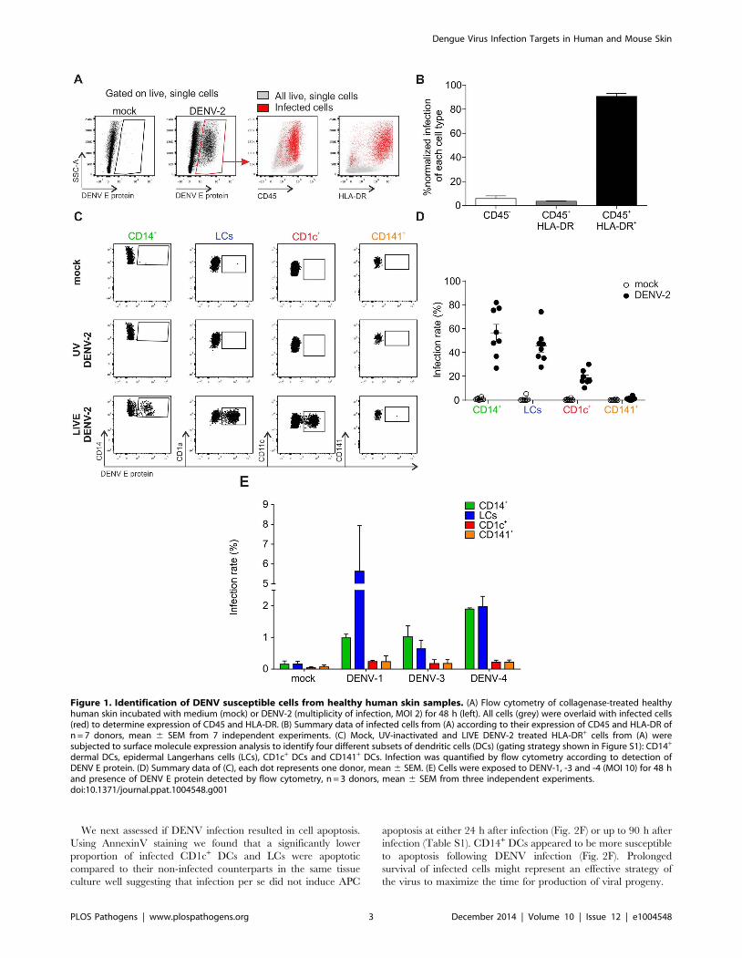
DENV infects LCs, CD1c+ and CD14+ subsets of DCs anddermal macrophages from human skin
To identify the cell types within human skin that are susceptible
to DENV infection we prepared single cell suspensions from
healthy skin obtained from mastectomy or abdominoplasty
surgery, and exposed the cells to DENV-2 strain D2Y98P [16]
at an MOI of 2. After 48 h, flow cytometry was used to
characterize the infected cell types by measuring the percentage of
cells positive for DENV E protein, which forms part of the viral
envelope (Fig. 1). The vast majority (approximately 90%) of cells
positive for E protein expressed CD45 and HLA-DR (Fig. 1A and
B). This finding excluded significant infection of CD452
keratinocytes, fibroblasts and endothelial cells, which had been
reported previously to be possible targets of DENV infection
[17,18]. CD45+ HLA-DR+ cells include all antigen-presenting
cells (APCs) in the skin. To further refine our analysis we employed
a previously described gating strategy to distinguish between
CD14+ DCs, CD1c+ DCs, CD141+ DCs and LCs ([15] and S1A
Figure). Three skin DC subsets were susceptible to DENV-2
infection ex vivo: LCs and CD14+ DCs were infected most
efficiently while CD1c+ cells showed a lower infection rate
(Fig. 1C). To prove that E protein-positive cells were truly infected
and had not only taken up virus particles, cells were also treated
with UV-inactivated virus as a control (Fig. 1C). The infection
profile for skin DC subsets was reproducible and independent of
the skin donor (Fig. 1D). Interestingly, we did not detect infection
of CD141+ DCs. To test whether there were serotype-specific
differences in infection we also exposed single cell suspensions
from skin to DENV-1, -3 and -4. These experiments showed that
LCs and CD14+ DCs were consistently infected at higher rates
than CD1c+ and CD141+ DCs, showing a similar DENV infection
profile in human skin cells to DENV-2 (Fig. 1E). Of note, infection
with DENV-1, -3 and -4 was less efficient than infection with
DENV-2 D2Y98P (Fig. 1A and B), which was expected due to the
enhanced viral RNA synthesis capacity of the latter [19]. A less
virulent DENV-2 strain (TSV01) showed lower infection rates
than D2Y98P, but a similar target cell infection profile (Fig. S1C).
In addition to DCs we identified dermal macrophages by flow
cytometry based on their auto-fluorescence in the FITC channel
[20] (Fig. S1A and B). CD14+ DCs and dermal macrophages were
infected at similar rates 24 h after infection (Figure S1B),
identifying both DCs and macrophages in the skin as potential
DENV targets.
As Aedes mosquitoes deposit virus-containing saliva in the
dermal layer, bypassing the epidermis when probing with their
proboscis to search for blood vessels, we explored infection
susceptibility upon intradermal delivery of DENV. LCs were not
infected when the virus was injected intra-dermally, in contrast to
infection of skin single cell suspension (Figure S1C).
Infected DCs produce high titers of viral progeny andinfection promotes cell survival
Whereas flow cytometry only detects how much viral protein is
expressed in infected cells, production of infectious virus particles
by infected cells can be assessed by using a plaque-forming assay or
by measuring viral RNA by PCR in the cell culture supernatant.
To further characterize the infection kinetics of various skin DC
subsets we infected single cell suspensions of human skin with
DENV-2 strain D2Y98P at an MOI of 2, analyzed the cells by
flow cytometry and cell culture supernatants by plaque assay at
16 h, 24 h, 36 h, 48 h and 72 h after infection (Fig. 2A and B).
We found that LCs were infected most rapidly but that their
infection frequency plateaued after 24 h. The infection kinetics
were delayed for CD14+ DC and CD1c+ DCs but peaked to
similar infection levels as LCs after 36 hrs. CD141+ DCs were
resistant to infection throughout the entire time course (Fig. 2A).
Macrophage and CD14+ DCs showed similar infection kinetics
but overall infection levels were lower for macrophages compared
to CD14+ DCs after 36 hrs. Virus titers measured in the
supernatant of infected total skin cells increased to a peak at
24 h after infection before declining. Only one out of three donors
showed extended virus production until 96 h after infection
(Fig. 2B). To test whether and how much virus was produced by
individual DC subsets, skin cells were sorted, infected, and viral
RNA was extracted from the cell culture supernatants for qRT-
PCR analysis. Macrophages were not viable after sorting and
could not be included in this experiment. 24 h after infection, we
observed the highest titers in LCs compared to the other two
subsets, which showed only little (CD1c+) or no increase in
secreted virus (CD14+) at this time point (Fig. 2C). At 48 h, all
infected DC subsets showed an increase in virus production,
whereby LCs remained the most efficient producers. To determine
the relative viral load contributed by each DC subset we first
determined the relative numbers of each subset in digested whole
skin (Fig. 2D) and then calculated their relative contribution
(Fig. 2E). This analysis revealed that LCs were the main
contributors of viral load produced by DCs, followed by CD1c+
DCs, which were present in higher numbers than CD14+ DCs.
Author Summary
Dengue virus (DENV) is transmitted by mosquitoes withskin as point of entry for the virus. Here, we investigatedDENV infection in primary human skin cells and their initialimmune response. Using skin from normal human donorsfor infection with DENV in vitro we identified antigen-presenting cells (APCs) as main targets of DENV. Furtheranalysis showed that only distinct subsets of dendritic cells(DCs) and macrophages were infected and efficientlyproduced viral progeny. Langerhans cells were mostsusceptible to infection despite lacking DC-SIGN, apreviously described DENV receptor. Infection of the otherDC subsets and macrophages was also independent of DC-SIGN expression. Genes of the interferon pathway andCCL5, a chemokine attracting immune cells to sites ofinflammation, were highly up-regulated in the infected DCsubsets. Using a mouse infection model, we showed thatmurine dermal DCs were also susceptible to DENV andmigrated to draining lymph nodes. At the same timeinfiltrating monocytes differentiated into monocyte-de-rived cells at the site of infection and became an additionaltarget for DENV in vivo. These data demonstrate that DENVdifferentially infects and activates primary human skinAPCs and that infected cell types individually contribute toinflammation and the adaptive response.
Dengue Virus Infection Targets in Human and Mouse Skin
PLOS Pathogens | www.plospathogens.org 2 December 2014 | Volume 10 | Issue 12 | e1004548
We next assessed if DENV infection resulted in cell apoptosis.
Using AnnexinV staining we found that a significantly lower
proportion of infected CD1c+ DCs and LCs were apoptotic
compared to their non-infected counterparts in the same tissue
culture well suggesting that infection per se did not induce APC
apoptosis at either 24 h after infection (Fig. 2F) or up to 90 h after
infection (Table S1). CD14+ DCs appeared to be more susceptible
to apoptosis following DENV infection (Fig. 2F). Prolonged
survival of infected cells might represent an effective strategy of
the virus to maximize the time for production of viral progeny.
Figure 1. Identification of DENV susceptible cells from healthy human skin samples. (A) Flow cytometry of collagenase-treated healthyhuman skin incubated with medium (mock) or DENV-2 (multiplicity of infection, MOI 2) for 48 h (left). All cells (grey) were overlaid with infected cells(red) to determine expression of CD45 and HLA-DR. (B) Summary data of infected cells from (A) according to their expression of CD45 and HLA-DR ofn = 7 donors, mean 6 SEM from 7 independent experiments. (C) Mock, UV-inactivated and LIVE DENV-2 treated HLA-DR+ cells from (A) weresubjected to surface molecule expression analysis to identify four different subsets of dendritic cells (DCs) (gating strategy shown in Figure S1): CD14+
dermal DCs, epidermal Langerhans cells (LCs), CD1c+ DCs and CD141+ DCs. Infection was quantified by flow cytometry according to detection ofDENV E protein. (D) Summary data of (C), each dot represents one donor, mean 6 SEM. (E) Cells were exposed to DENV-1, -3 and -4 (MOI 10) for 48 hand presence of DENV E protein detected by flow cytometry, n = 3 donors, mean 6 SEM from three independent experiments.doi:10.1371/journal.ppat.1004548.g001
Dengue Virus Infection Targets in Human and Mouse Skin
PLOS Pathogens | www.plospathogens.org 3 December 2014 | Volume 10 | Issue 12 | e1004548

In summary, we found that infected human skin DCs were
capable of producing significant amounts of DENV in the absence
of increased levels of apoptosis in infected cells.
Infection of skin APC subsets is independent of DC-SIGNWe next assessed whether the differential infection rates of skin
APCs could be explained by variations in individual cell types’
ability to take up the virus. After confirming that inactivated,
fluorescently labeled virus was still able to bind to host cells (Figure
S2A), virus uptake was measured at 37 degrees, whereas lack of
uptake at 4 degrees served as negative control (Fig. 3A). 2 h after
adding inactivated, fluorescently-labeled virus the highest virus
uptake rate was observed in CD14+ DCs and dermal macrophag-
es, which was in line with efficient infection. LCs, however,
showed a relatively lower viral uptake activity but were still
efficiently infected. This was even more surprising with regards to
Figure 2. Infection characteristics of DENV in DC subsets from human skin. (A) Cells from collagenase-treated healthy human skin wereexposed to DENV-2 at MOI 2 for the indicated times. Presence of DENV E protein in DC subsets was established by flow cytometry. (B) Amount of livevirus in supernatants of cells from (A) was quantified by plaque-forming assay and used to calculate titer in plaque-forming units per ml (pfu/ml),n = 3 donors, mean 6 SEM from three independent experiments. (C) Sorted DC subsets were infected with DENV-2 at MOI 2 and viral RNA wasmeasured in the supernatant by quantitative real time PCR at 0, 24 and 48 hpi. n = 3, mean 6 SEM from three independent experiments. (D) Analysisof skin DC subsets and macrophages by flow cytometry in whole human skin. Each dot represents one donor, mean 6 SEM. (E) Results from (C) and(D) were used to calculate the relative contribution of each DC subset to the total viral load at 24 and 48 hpi. (F) Infected DC subsets from skin werestained with Annexin V and labeled for DENV E protein after 24 h of exposure to virus to determine the extent of apoptosis (one representative donorof three), mean % per quadrant 6 SEM.doi:10.1371/journal.ppat.1004548.g002
Dengue Virus Infection Targets in Human and Mouse Skin
PLOS Pathogens | www.plospathogens.org 4 December 2014 | Volume 10 | Issue 12 | e1004548

the expression pattern of well-described host receptors for viral
binding and infection DC-SIGN (CD209) and mannose receptor
(CD206) [21] [22,23], which were absent on LCs [24] (Figure
S2B). The phosphatidylserine receptor Axl was recently described
as alternative virus-binding receptor [25–27] and is expressed on
LCs [28]. However, we detected negligible levels of Axl on the
surface of skin DC subsets isolated by collagenase-digestion or
spontaneous migration from skin explants ex vivo. DC-SIGN was
expressed on CD14+ DCs but not on CD1c+ or CD141+ DCs,
whereas CD206 was expressed on CD14+ and CD1c+ DCs (Figure
S2B).
At least for dermal DCs, expression of these two receptors could
therefore explain the more efficient infection of CD14+ DCs
compared to CD1c+ DCs and the absence of infection in CD141+
DCs. To further study the relevance of DC-SIGN for infectivity,
we first confirmed that DENV-2 D2Y98P did bind to DC-SIGN
by incubating DC-SIGN-expressing U937 cells in the presence or
absence of DC-SIGN blocking antibody (Figure S2C). Blocking of
DC-SIGN on skin APC subsets had no effect on infection rates,
suggesting that other receptors utilized by DENV might be more
relevant than DC-SIGN on primary skin DCs (Fig. 3B).
Since infection rates by the individual APC subsets might be
affected by differential sensitivity to IFN, we pre-treated total skin
cells with IFN-b and detected infection rates of APC subsets at
24 h by flow cytometry. Increasing concentrations of IFN-b had a
significant inhibitory effect on infection in CD14+ DCs, CD1c+
DCs and macrophages but not in LCs (Fig. 3C). This suggests that
LCs are less sensitive to IFN, allowing the virus to replicate
efficiently.
Overall, infection of skin DC subsets did not strictly correlate
with the expression of DC-SIGN, mannose receptor or Axl, while
the extent of virus particle uptake only correlated with infection in
dermal APCs, and not in LCs. These findings suggested that
additional cell-inherent parameters including IFN-b susceptibility
determined the observed DENV tropism for distinct skin APC
subsets.
DENV infection negatively regulates skin DCallostimulatory potential but efficiently activates type IIFN responses
To evaluate if DENV infection affected T cell stimulatory
function of skin DCs, we tested the capacity of the different skin
DC subsets infected with dengue virus to stimulate proliferation of
allogeneic T cells (Fig. 4A and B). Sorted infected DC subsets were
incubated with allogeneic CD3-sorted CFSE-labeled T cells for 5
days before measurement of CD4+ and CD8+ T cell proliferation
by flow cytometry (Fig. 3A and Table S2). DENV-infected CD14+
DCs were less efficient at inducing CD4+ T cell proliferation
compared to their non-infected counterparts (Fig. 4A). This is in
keeping with previous observations of poor T cell proliferative
responses when PBMCs from dengue infected patients were
stimulated with PHA [29]. The defect could be restored by the
addition of IL-2 or gamma-irradiated PBMCs from healthy
Figure 3. Infection of primary skin DCs is independent of DC-SIGN. (A) DEPC-inactivated, fluorescently-labeled DENV-3 was added to skincells for 2 h at 4uC or 37uC (left) before flow cytometric analysis of viral uptake. Representative results of one donor are shown (right) and summarydata are depicted as mean fluorescence intensity (MFI) at 37uC minus MFI at 4uC (bottom), each dot represents one donor, mean 6 SEM. (B) APCsubsets were incubated with DC-SIGN blocking antibody for 1 h before infection for 24. n = 3 donors, Mean 6 SEM from two independentexperiments. (C) Total skin cells were treated with IFN-b for 24 h before infection for 24 h. Infected APC subsets were analyzed by flow cytometry.n = 4, mean 6 SEM from three independent experiments.doi:10.1371/journal.ppat.1004548.g003
Dengue Virus Infection Targets in Human and Mouse Skin
PLOS Pathogens | www.plospathogens.org 5 December 2014 | Volume 10 | Issue 12 | e1004548

donors, suggesting that APCs but not T cells were impaired in
patients [29]. Moreover, DENV infection of monocyte-derived
DC (moDCs) inhibited their maturation and their capacity to
induce proliferation in allogeneic bulk T cells [30,31], but not
sorted naıve CD4+ T cells [32,33]. In contrast to CD14+ DCs,
infection of CD1c+ DCs and LCs did not impair their capacity to
induce allogeneic CD4+ T cell proliferation, showing that DENV-
mediated inhibition of T cell proliferation was DC subset specific
and not a direct generic effect of the virus either on DCs or T cells
(Fig. 3B). However, infection of DC subsets had no effect on their
capacity to induce CD8+ T cell proliferation (Table S2).
We next tested whether infection of skin DCs was likely to have
an impact on their capacity to migrate towards the chemokine
CCL19, which is expressed in the T cell zones of LN follicles to
attract CCR7-expessing migratory cells [34]. CCR7 expression
levels were comparable between DC subsets exposed to DENV for
24 h and those treated with medium alone (Fig. 4C), and a
functional chemotaxis assay performed at different time points
confirmed that DENV infection did not have an impact on the
migration of skin DCs in vitro as infected cells migrated equally
compared to non-infected DCs (Fig. 4D). CD1c+ and LCs
migrated more efficiently than CD14+ DCs in both conditions
(Fig. 4E).
To assess the effects of DENV infection on skin DC function, we
detected the effects of DENV exposure on the transcription of 184
inflammatory and immune response genes in skin DC subsets.
Figure 4. T cell stimulation capacity of DENV-exposed DC subsets and their migratory response towards the chemokine CCL19. (A)Co-culture of sorted infected DCs with allogeneic, CFSE labeled CD3+ T cells (ratio 1:10). Proliferation was measured after 5 days by flow cytometry,one representative result for CD14+ dermal DCs is shown. (B) Summary data from all three susceptible DC subsets are shown, each line represents onedonor. Statistical analysis was performed using paired two-sided t-test, *p,0.05; ns, not significant, p-values are indicated for each DC subset (C)CCR7 expression on non-treated and DENV-2-exposed DCs at 24 hpi. One representative experiment of three is shown. (D) Skin cell migration wasassessed using a 5 mm pore-sized membrane (see Methods Section) with either medium alone or CCL19 (20 ng/ml) in the bottom chamber. Cellswere allowed to migrate for 2 h at 37uC before CellTiter Glo activity was measured (RLU, relative light units). Composite data of 4–6 donors is shown,mean 6 SEM from 4 independent experiments. (E) Whole skin cells were analyzed by flow cytometry before and after migration towards CCL19.Migrated cells (in the lower well of the chemotaxis plate) were enriched in HLA-DR+ cells compared to input cells (left graph). Migrated HLA-DR+ cellswere enriched in CD1c+ DCs and LCs, but not CD14+ DCs (right graph; non-infected cells are illustrated; similar results were obtained for infectedcells). n = 3 donors, mean 6 SEM from three individual experiments.doi:10.1371/journal.ppat.1004548.g004
Dengue Virus Infection Targets in Human and Mouse Skin
PLOS Pathogens | www.plospathogens.org 6 December 2014 | Volume 10 | Issue 12 | e1004548

Sorted skin DCs were infected with DENV-2 D2Y98P for 24 h
and the mRNA transcripts present in cell lysates were quantified
by Nanostring (Fig. 5A). In these experiments, the mean infection
rates were 28.1% for CD14+ DCs, 39.5% for LCs and 12.5% for
CD1c+ DCs. Transcription of IFN-b, STAT-1 and CCL5 was
significantly up-regulated in all APC subsets upon dengue virus
infection. The greatest changes in expression occurred in the
CD14+ DCs (Fig. 5A). Of note, CD141+ DCs did not up-regulate
early antiviral genes compared to the other subsets, suggesting that
IFN response induction was not responsible for resistance to
DENV infection. Up-regulation of IFNA1 gene expression in
CD141+ cells was not statistically significant and only observed in
two out of four donors studied (Fig. 5A). Expression of IFN-b and
CCL5 48 h after infection was confirmed by ELISA and was not
seen in UV-DENV treated cells, showing that viral replication was
necessary to induce innate immune gene up-regulation (Fig. 5B).
However, these experiments could not distinguish between gene
expression in infected and non-infected cells, which might both
contribute to the total gene up-regulation.
Taken together, our data demonstrated that DENV virus
infection of human skin DC subsets differentially affected their
allogeneic T cell stimulatory capacity. Moreover, primary human
skin DCs had the capacity to initiate IFN transcription upon viral
infection. In addition, the rapid induction of inflammatory genes
such as CCL5 could attract innate immune cells to clear local
infection and possibly increase migration of DCs to draining LNs
for the initiation of the adaptive response.
Infected DC subsets in mouse skin are the counterpartsof infected DCs in human skin
Having identified the cell types in human skin that are able to be
infected by DENV, we next wanted to understand the in vivoconsequences of dengue infection on ensuing functional responses.
We recently identified the functional murine homologs of human
tissue DC subsets [15], which enabled us to exploit a murine
model of dengue infection to interrogate skin APC susceptibility to
DENV infection, their LN migratory capacity and the contribu-
tion of recruited inflammatory myeloid cells upon DENV
infection. As wild-type mice are not susceptible to DENV infection
[35], we used interferon-a/b-receptor knock-out (IFNAR) mice,
which show disease symptoms and clinical parameters comparable
to dengue patients following DENV infection [36]. Mice were
infected intra-dermally with 106 pfu of DENV-2 in a volume of
10 ul into each ear. After two and four days mice were sacrificed
and we prepared single-cell suspensions from their ears for flow
cytometry analysis of infected cell populations staining positively
for DENV E protein. The gating strategy (Figure S3A) allowed us
to differentiate between dermal CD11b+ DCs (homolog of human
CD1c+ DCs), CD11b2 DCs, CD103+ DCs (homolog of human
CD141+ DCs), LCs, MHC class II (IAIE)hi Ly6C+ monocyte-
derived cells and MHC class II (IAIE)2 Ly6C+ inflammatory
monocytes [37,38]. CD11b2 and CD11b+ dermal DCs were
frequently infected, reaching infection levels of approximately
20% and 50% respectively by day 4 post-infection (Fig. 6A and B).
In contrast, by day 4, LCs were not highly infected and similarly
CD103+ DCs showed a low level of infection in the region of 10%
of cells by day 2 (Fig. 6B). In addition to skin-resident DCs, we
found high infection rates in infiltrating Ly6C+ cells in the skin two
days after infection (Fig. 6A), with a marked increase in infection
particularly in the Ly6C+IAIE+ population on day four after
infection (Fig. 6B). In contrast to ex vivo human skin cells, we also
identified a substantial population of infected CD452 cells in the
dermis, but not in the epidermis (Figure S3B). Quantification of
total cell numbers 2 and 4 days after infection showed a decrease,
although not significant, of CD11b2 DCs, CD11b+ DCs and
CD103+ DCs, two days after infection. This was likely due to cell
death rather than LN migration as the increase in numbers of
migrated cells in draining LNs was already observed at day 2 after
infection (see following paragraph). In contrast to dermal DCs, LC
numbers remained constant (Fig. 6C).
The murine in vivo model also permitted analysis of cells
recruited into skin upon intradermal inoculation of DENV. We
observed a more than ten-fold increase in the number of
inflammatory monocytes (Ly6C+IAIE2) and monocyte-derived
cells (Ly6C+IAIE+) in the ears of mice within two days of infection.
This rise was followed by a rapid decline four days after infection,
which may be due to cell death or due to down-regulation of Ly6C
expression on activated monocyte-derived cells. Ly6C2 monocyte-
derived cells could not be distinguished from CD11b+ DCs and it
was difficult to assess whether there was a relatively smaller decline
in this population due to possible parallel effects of cell death and
new formation of monocyte-derived cells (Fig. 6C).
Taken together, functionally-equivalent dermal DC subsets
appeared to be infected in both humans and mice, with the
exception of CD103+ cells, which were infected in mouse skin,
although at much reduced numbers compared to other subsets,
whereas their counterpart in human skin were not infected in our
ex vivo experiments. In addition, in vivo experiments revealed a
massive infiltration of Ly6C+ monocyte-derived cells, identifying
these cells as potentially important infection targets during natural
infection.
Infected skin DCs efficiently migrate to the draininglymph node
It was important to know which cells had the capacity to
migrate to draining LNs for the initiation of adaptive immune
responses and the ensuing immune memory, and whether those
cells carried infectious DENV with them. Cell suspensions were
made from ear-draining LNs of infected mice at days 2 and 4, and
analyzed by flow cytometry for DENV E protein, with immigrant
and resident DC discriminated based on CD11c and MHC Class
II (IAIE) expression [39,40] (Fig. 7A–C). Amongst DCs migrated
from the skin, CD11b2 and CD11b+ DCs, but not LCs were
infected. Despite the low CD103+ DC infection rate compared to
CD11b+ DC in the skin, more infected CD103+ DCs than
CD11b+ DCs were observed in the draining LN at day 4 (Fig. 7C).
The relative abundance of CD103+ DCs in the draining LN
compared to other infected subsets migrating from skin, suggests a
significant role for this subset in T cell activation at later time
points of infection. The LN-resident counterparts of CD103+ cells,
CD8+ DCs, were not infected at the time points tested, suggesting
that virus is transported from the skin via DCs and that little virus
reaches the LN directly via the lymphatics to infect LN-resident
DCs or these cells were not susceptible to infection at this stage
(Fig. 7B and C). Similarly, LN-resident CD11b+ DCs were also
not infected. However, the absolute number of (non-infected) LN-
resident CD11b+ DCs was significantly greater in infected
compared to non-infected mice (Fig. 7D).
In summary, infection of LN-resident DCs was negligible in
contrast to active infection of skin APCs within the first 4 days
after intradermal DENV delivery. This finding suggested that
dermal DCs migrating from the skin to draining LNs were
efficient carriers of infectious DENV and implicates them as likely
initiators of systemic immunity. Our results further indicated that
CD11b+ dermal DCs (the equivalents of human CD1c+ DCs)
might be important to trigger early adaptive anti-DENV T cell
responses.
Dengue Virus Infection Targets in Human and Mouse Skin
PLOS Pathogens | www.plospathogens.org 7 December 2014 | Volume 10 | Issue 12 | e1004548

Dengue Virus Infection Targets in Human and Mouse Skin
PLOS Pathogens | www.plospathogens.org 8 December 2014 | Volume 10 | Issue 12 | e1004548

Discussion
The aim of this study was to characterize the cellular targets of
DENV infection in human skin, and the consequences of APC
infection on systemic infection and the induction of a protective
immune response (see model, Fig. 8). Previous studies have
focused on the role of LCs in DENV infection, but dermal DCs
were not evaluated [41]. DENV is most likely injected into the
dermis by its mosquito vector [42] and we provide evidence that
human dermal APCs can also be efficiently infected by DENV. In
fact, when DENV is injected intradermally ex vivo or in mice, LCs
are not infected efficiently and the functional relevance of natural
LC infection therefore remains unclear. Virus might come into
contact with LCs during the process of probing even though the
mosquito’s proboscis bypasses the epidermis and LCs located
there. Alternatively, LCs that migrate through the dermis towards
draining LNs might be infected en route, although the number of
spontaneously migrating LCs in healthy skin is very small [43].
The suspension cell infection model cannot solve this question and
further experiments, ideally with infected mosquitoes injecting the
virus into the skin of mice or other animal models, will be required
to validate our findings.
Interestingly, we found that DC-SIGN expression did not
correlate with infection and blocking of the receptor did not
reduce the rate of infection in cells expressing DC-SIGN.
Monocyte-derived DCs (moDCs) [44], which represent an easily
accessible and hence useful model to study DCs in vitro, express
high levels of DC-SIGN. The majority of primary DC subsets
found in blood, skin and lymph nodes, however, does not express
DC-SIGN when analyzed ex vivo [45,46]. It was previously shown
that DC-SIGN facilitates attachment to the cell rather than
mediating viral endocytosis per se and that a potentially unknown
bona fide receptor is required for viral entry [47]. We therefore
speculate that DC-SIGN is one of several receptors expressed on
primary DCs that facilitate attachment and viral entry [48].
In mice, CD11b+ DCs, which are the functional homologs of
human CD1c+ DCs, had the highest infection rate. In addition,
recruited inflammatory Ly6C+ monocyte-derived cells into skin
were also efficiently infected (Figure S3, population 6). Based on
the findings in mice, we speculate that human blood monocytes
infiltrate the skin upon infection, similar to Ly6C+ mouse
monocytes, and represent an additional target population for the
virus. Addressing this question in humans in vivo is a challenge, as
skin biopsies from patients from the site of the mosquito bite would
have to be analyzed.
Human steady state CD14+ dermal DCs were efficiently infected.
This subset expressed low levels of CCR7 (Fig. 4C) and is unlikely to
migrate efficiently to draining LNs (Fig. 4E and [12]). However,
CD14+ cells are efficient activators of memory T cells [12]
suggesting a role in local tissue responses, particularly during
secondary infection when DENV-specific T cells may be present in
the skin. Alternatively, infection of CD14+ DCs could affect their
capacity to induce regulatory CD4 T cells in the skin, a function that
has been associated with CD14+ DCs and not with CD1c+ DCs
[5,14]. We were unable to establish in the in vivo model if
infiltrating Ly6C+IAIE+ cells migrated from the skin to the draining
LNs as Ly6C could be downregulated during LN migration as
shown for a West Nile virus murine infection model [49].
Despite the obvious similarities between human and mouse skin
infection targets there were also notable differences: firstly, we
observed a substantial population of CD452 infected cells in
mouse, but not human, skin. This is challenging to interpret, but
the lack of the IFNa/b receptor in these mice could have affected
the susceptibility of non-hematopoietic cells. We showed recently
that the absence of the IFNa/b receptor on CD11c+ and LysM+
expressing cells alone was sufficient to replicate the DENV-
susceptibility phenotype of IFNAR2/2 mice with regards to
viremia and survival [35], though this does not directly exclude the
possibility of some alterations to viral tropism within the model.
The second difference between human and mouse was that
human CD141+ DCs remained uninfected up to 72 h after
infection ex vivo, whereas CD103+ DCs in mice were infected at
day four after infection. It could be that CD141+ DCs may
become susceptible at later time points after infection ex vivo or
that CD141+ DCs are infected in the context of a natural infection.
It remains to be addressed whether the reason for this discrepancy
relates to species-specific differences in the antiviral response. For
human CD141+ DCs, infection-induced up-regulation of IFN-band STAT-1 gene expression was low compared to CD14+ DCs
and CD1c+ DCs (Fig. 4), which might also reflect the lack of
infection of the cells at these time points. The induction of
transcription of the monocyte-attracting cytokine CCL5 in human
cells fits nicely with our observation of infiltrating monocyte and
monocyte-derived cells in mice, making it likely that monocyte-
derived cells are similarly attracted to the site of infection in
humans [50]. IFN signatures and CCL5 were previously found to
be up-regulated in microarrays of dengue patients’ PBMCs [51,52]
and in the serum [50], whereby higher expression seemed to be
associated with less severe disease.
We demonstrate here that human skin DCs are likely to be
important targets for DENV infection in vivo. The observations in
mice suggest that skin dermal DCs were also likely to transport
infectious virus to draining LNs, providing a shuttle for the virus to
potentially establish further sites of infection, and to efficiently
activate a systemic immune response. Our data suggest that intra-
dermal or gene-gun inoculation of live-attenuated dengue vaccine
candidates would likely target the most physiologically relevant
DC populations within the dermis and thereby potentially
stimulate the efficient establishment of a systemic immune
response.
Materials and Methods
Ethics statementHealthy human skin tissue was obtained from mastectomies or
abdominoplastic surgery. The studies were approved by the respective
institutional review boards (National Health Group Domain Specific
Review Board (NHG DSRB 2012/00928) and Singhealth Central-
ized Institutional Review Board (CIRB 2011/327/E), respectively)
and patients gave written informed consent. All skin samples were
processed on the day of surgery. Blood from anonymous healthy
human donors was received from the blood bank at the National
University Hospital of Singapore and blood donors gave written
informed consent. The study was exempted from full IRB review by
the Institutional Review Board of the National University of
Singapore (NUS-IRB) since anonymous samples were used.
Figure 5. Heatmap of differentially regulated genes in DENV infected skin DC subsets. (A) Nanostring gene expression analysis of sortedhuman skin DC subsets exposed to DENV-2 (MOI 2) for 24 h. (B) Summary data of three genes that were significantly elevated by DENV exposure inCD14+, CD1c+ DCs and LCs cultures. Each dot represents one donor. (C) Protein levels of IFN-b and CCL5 produced by dermal cells and by LCsstimulated with UV-inactivated and LIVE DENV-2 for 48 h and measured by ELISA, each dot represents one donor.doi:10.1371/journal.ppat.1004548.g005
Dengue Virus Infection Targets in Human and Mouse Skin
PLOS Pathogens | www.plospathogens.org 9 December 2014 | Volume 10 | Issue 12 | e1004548

Figure 6. In vivo infection of migratory DCs in the skin of IFNAR2/2 mice. (A) Mice were inoculated intra-dermally (i.d) with 16106 pfu ofDENV-2 D2Y98P/ear, and ears were harvested 2 and 4 days post infection. DENV E protein expression was detected by flow cytometry of skin-residentDC subsets and infiltrating monocytes (Gating strategy to identify DC subsets/monocytes shown in Figure S2) (B) Summary data of (A), 4 to 5 mice(taking the average of two ears/mouse) per group pooled from two independent experiments, mean 6 SEM, analyzed with one-way ANOVA followedby Tukey’s multiple comparison test, *p,0.05; **p,0.01; ***p,0.001; ****p,0.0001; non-significant differences are not indicated (C) Quantificationof DCs isolated from ears at 2 and 4 dpi to address infiltration and migration of different cell subsets. Same conditions as in (B).doi:10.1371/journal.ppat.1004548.g006
Dengue Virus Infection Targets in Human and Mouse Skin
PLOS Pathogens | www.plospathogens.org 10 December 2014 | Volume 10 | Issue 12 | e1004548

Figure 7. In vivo infection of resident and migratory DCs in the skin-draining lymph node of IFNAR2/2 mice and their migratorybehavior. (A) Gating strategy to identify lymph node (LN)-resident and migratory DCs in the skin-draining LN of IFNAR2/2 mice. Resident DC subsetsare either CD8+ (1), CD11b2 (2) or CD11b+ (3), while migratory DC subsets incorporate those found in the skin: CD103+ (4), CD11b2 (5), CD11b+ (6)and Langerhans Cells (LCs, 7). (B) Mice were inoculated i.d. with 16106 pfu of DENV-2 D2Y98P/ear and skin-draining lymph nodes (LN) were harvested2 and 4 days post infection (dpi). Presence of DENV E protein was established by flow cytometry in both LN-resident and migratory DC subsets. (C)Summary of data from (B), 4 to 5 mice (average of two LN/mouse) per group pooled from two independent experiments, mean 6 SEM, analyzed withone-way ANOVA followed by Tukey’s multiple comparison test, *p,0.05; **p,0.01; ***p,0.001; ****p,0.0001; non-significant differences are notindicated. (D) Quantification of DCs isolated from LNs at 2 and 4 dpi to address infiltration and migration of different cell subsets. Same conditions asin (C).doi:10.1371/journal.ppat.1004548.g007
Dengue Virus Infection Targets in Human and Mouse Skin
PLOS Pathogens | www.plospathogens.org 11 December 2014 | Volume 10 | Issue 12 | e1004548

Figure 8. Model for skin dengue infection. (A) The mosquito searches for blood vessels in the dermis and thereby releases saliva that containsvirus. The proboscis bypasses the epidermis. The question mark indicates that it is not clear whether during the process of probing virus is alsoreleased into the epidermis. (B) Virus in the dermis infects CD1c+ and CD14+ dermal DCs and macrophages (MP), but not CD141+ DCs. Infected CD1c+
DCs and possibly LCs migrate to draining LNs to initiate the adaptive immune response. Based on their non-migratory behavior ex vivo (Fig. 3E and[12]) CD14+ DCs are not expected to migrate to draining LNs. Based on mouse studies, monocyte-derived cells that infiltrate into the skin are infectedefficiently (Fig. 6) and contribute to the local and systemic immune response. (C) Table summarizing the role of skin APCs during infection with DENVusing single cell suspensions.doi:10.1371/journal.ppat.1004548.g008
Dengue Virus Infection Targets in Human and Mouse Skin
PLOS Pathogens | www.plospathogens.org 12 December 2014 | Volume 10 | Issue 12 | e1004548

Mouse experiments were conducted according to the rules and
guidelines of the Agri-Food and Veterinary Authority (AVA) and
the National Advisory Committee for Laboratory Animal
Research (NACLAR), Singapore. The experiments were reviewed
and approved by the Institutional Review Board of the Biological
Resource Center, Singapore (IACUC protocols 100566 and
120801).
Infection of miceIFN-a/b receptor-deficient mice (IFNAR2/2), on a C57BL/6
background, were infected with 26106 pfu of D2Y98P via the
intradermal (i.d.) route in the ears using a 33-gauge needle and a
microsyringe (Nanofil). Naıve mice served as control.
Cell isolation and cultureFor isolation of human skin cells 300 mm dermatome sections
were incubated in RPMI+10%FCS (BioWest) containing 0.8 mg/
ml collagenase (Type IV, Worthington-Biochemical) and
0.05 mg/ml DNase I (Roche) for 12 h. For nanostring analysis
and T cell proliferation assay skin was treated with 1 mg/ml
dispase (Invitrogen) to separate epidermis and dermis. Dermal
DCs were sorted by fluorescence-activated cell sorting (FACS),
epidermal LCs were isolated using CD1a microbeads (Miltenyi
Biotec) and a magnet (Stemcell techonologies) with a purity of .
90%.
For isolation of mouse skin cells, mice were sacrificed and ears
were cut off at the base. Ear skin was split into dorsal and ventral
halves and incubated in RPMI+10%FCS containing 1 mg/ml
dispase (Invitrogen) for 2 h at 37deg. Epidermis and dermis were
separated and digested in 0.2 mg/ml collagenase (Type IV,
Sigma) for 2 h at 37deg before passing them through a 70 um
filter to obtain a single cell suspension.
Mouse skin-draining auricular lymph nodes were isolated,
incubated in medium+0.2 mg/ml collagenase for 30 min and
passed through 70 um filter.
BHK-21 and C6/36 cells were purchased from the American
Type Culture Collection.
VirusFor infection experiments the following strains of dengue virus
were used: DENV-1 – 08K3126, DENV-2 - TSV-01 or D2Y98P,
DENV-3 – VN32/96, and DENV-4 – 2641Y08. All strains are
patient isolates that have been passaged in C6/36 mosquito cells for
5–20 passages. D2Y98P used here was plaque-purified after passage
20 and derived from an infectious clone [19]. The enhanced viral
RNA synthesis capacity of D2Y98P was mapped to a natural
mutation in NS4b. The mutation had no effect on the IFN-
inhibiting capacity of the virus [19]. All strains viruses used in the
experiments were produced in the C6/36 mosquito cell line. For
phagocytosis assays, DENV-3 - VN32/96 was purified with density
gradient isolation (Opti-Prep, Sigma) according to manufacturer’s
protocol. Viral particles were labeled with Alexa-647 fluorescent
dye using a protein labeling kit (Molecular Probes) and the excess
dye was removed with Amicon protein purification tubes (Millipore)
according to manufacturer’s protocol. The virus was then
inactivated in 2 mM DEPC/PBS-T for 15 min at RT [53].
Flow cytometry and antibodiesFlow cytometry was performed on an LSRII, FACSCanto,
FACS was performed using a FACSAriaII (all Becton Dickinson
[BD]). Software analysis was performed with FlowJo (TreeStar).
The following reagents for labeling of human cells were used:
Carboxyfluorescein succinimidyl ester (CFSE), fixable live/dead
blue dye (Life Science Technologies), anti-CD3 (UCHT1), anti-
CD4 (RPA-T4), anti-CD8 (RPA-T8), anti-CD1a (HI149), anti-
CD209 (9E9A8), anti-CD206 (15-2) (all from Biolegend), anti-
CD11c (B-ly6), AnnexinV Detection Kit, anti-CD45 (HI30), anti-
HLA-DR (L243) (all from BD Biosciences), anti-CD141 PE (AD5-
14H12) (Miltenyi), anti-CD14 (RMO52) (Beckman Coulter), anti-
CCR7 (3D12) (eBioscience), anti-Axl (MAB154) (R&D Systems)
and anti-E protein (4G2) (ATCC).
The following antibodies were purchased from Biolegend to
label mouse cells: anti-CD45 (30-F11), anti-IAIE (M5/114.15.2),
anti-CD11b (M1/70), anti-CD11c (N418), anti-CD326 (EpCAM)
(G8.8), anti-CD103 (2E7), anti-Ly6C (HK1.4).
T cell proliferation assaySorted DCs were infected for 2 h and immediately co-cultured
with allogeneic CD3+ flow-sorted CFSE-labeled T cells from
healthy blood donors in a ratio of 1:10 in 96-well U-bottom plates
for 5 days before proliferation was determined by CFSE dilution.
Chemotaxis assayCell migration was assayed in chemotaxis microchamber plates
(Neuroprobe) containing a membrane with 5 mm pores. Briefly,
medium alone or containing recombinant human CCL19 (20 ng/
ml, R&D Systems) was added to the lower chamber. The
membrane was placed on top and a cell droplet (containing
approximately 250,000 cells) was pipetted on top of the
membrane. Plates were incubated for 2 h at 37 degrees C and
relative cell numbers of migrated cells were determined using a
CellTiter Glo luminescent cell viability assay, read on a GloMax-
96 microplate luminometer (both from Promega).
NanostringNanostring analysis and initial data processing was performed in
the nCounter system according to manufacturer’s instructions.
The human inflammation gene cartridge (GXA-IN1) was used,
and based on the data PGK1, TUBB and GAPDH were used as
housekeeping controls. Differential expression analysis was deter-
mined with a 2-way ANOVA using celltype and infection status as
factors in R v2.15.2/Bioconductor. Multiple testing correction was
performed using the method of Benjamini and Hochberg. Heat
maps were generated using the logarithmically transformed fold
changes of averaged normalized counts for each cell population
using the non-infected samples as the reference. Visualization of
the data and test results were done using TIBCO Spotfire. NCBI
accession numbers of all genes are listed in Table S3.
Quantitation of proteins in culture supernatantsLevels of CCL5 and IFNb in skin cell supernatants were
measured by enzyme-linked immunosorbent assay (ELISA) (both
R&D Systems) following the manufacturer’s instructions.
Determination of virus in cell culture supernatantVirus titer cell culture supernatant was determined by plaque-
forming assay using BHK-21 cells as described elsewhere [35].
Briefly, supernatant of infected cells was diluted 10-fold on BHK-
21 cells. After 1 h, medium was exchanged for 0.8% methylcel-
lulose in RPMI/10%FCS and plates were incubated for 4 days.
Plaque counts were used to calculate viral titer in plaque forming
units per ml.
Viral qPCRViral RNA was extracted from cell supernatants using a viral
RNA extraction (Roche) according to the manufacturer’s protocol
Dengue Virus Infection Targets in Human and Mouse Skin
PLOS Pathogens | www.plospathogens.org 13 December 2014 | Volume 10 | Issue 12 | e1004548

and subsequently quantified by real-time qRT-PCR using primers
and methods reported previously [54]. Forward primer ACAC-
CACAGAGTTCCATCACAGA, reverse primer CATCTCATT-
GAAGTCNAGGCC, probe CGATGGARTGCTCTC.
Binding and DC-SIGN blocking assayBinding experiments were performed with U937 cells stably
expressing DC-SIGN [54]. Virus was incubated with the cells for
1 h at 4uC and cells were subsequently washed with serum-free
medium. Non-fluorescently labelled virus was detected with 4G2-
A647 anti-E antibody. For DC-SIGN blocking experiments cells
were pre-incubated with 20 ug/ml anti-DC-SIGN mAb (clone
120507) or a matched isotype controls (clone 133303) (R&D
Systems) for 1 h at 37uC.
Supporting Information
Figure S1 Human skin DC gating strategy, intradermalinfection and surface molecule expression. (A) Gating
strategy to identify DC subsets after collagenase digestion of
healthy human skin tissue: CD14+ (green), LCs (blue), CD1c+(red) and CD141+ (orange). (B) Macrophage gating strategy,
expression of selected markers and infection rate at 24 hpi
compared to CD14+ dermal DCs. (C) Suspension infection (left)
versus intradermal injection (right) of skin DCs infected with
DENV-2 (TSV01, MOI5) for 48 h. n = 3 and n = 5, respectively,
mean 6 SEM.
(TIF)
Figure S2 Expression of DENV receptors on primarycells and binding of DENV to DC-SIGN. (A) Binding of
DEPC-inactivated fluorescently labeled DENV-3 to DC-SIGN
expressed on U937 cells. Cells were pre-incubated with DC-SIGN
blocking- or a control Ab or left untreated at 37uC for 1 h and
subsequently exposed to the virus at 4uC for 1 h. Mean
fluorescence intensity (MFI) was measured by flow cytometry.
Two independent experiments were performed in triplicates.
mean 6 SD (B) Surface expression of DC-SIGN (CD209), MMR
(CD206) and Axl on skin DC subsets. One representative of three
donors is shown. (C) Binding and blocking of LIVE DENV-2 to
DC-SIGN expressed on U937 cells (as described in (A)), one
experiment was performed in quadruplicates, mean 6 SD.
(TIF)
Figure S3 Murine skin DC gating strategy and infection ofCD452 cells in mouse skin. (A) Gating strategy to identify DC
subsets after collagenase digestion of murine skin tissue in non-treated
or DENV-2-infected IFNAR2/2 mice at 2 or 4 dpi: Infiltrating
monocytes (IAIE2Ly6C+SSClo, gate 1), CD103+ DCs (2), CD11b2
DCs (3), EpCAM+ LCs (4), CD11b+ DCs (5) and monocyte-derived
cells (IAIE+Ly6C+) (6). (B) Presence of DENV E protein was measured
in CD452 cells (see (A)) from the epidermis and dermis, 2 and 4 days
after infection. One representative results (n = 4–5) is shown.
(TIF)
Table S1 DENV-infected cells are not apoptotic. Annexin
V stain 48 and 90 hpi, related to Fig. 2F. Mean percentage of two
donors per time point from four independent experiments.
(PDF)
Table S2 CD8+ T cell proliferation is not altered byinfection of DC subsets. CD8+ T cell proliferation (related to
Fig. 4A and B). Mean of 3–4 donors 6 SEM.
(PDF)
Table S3 List of genes and corresponding accessionnumbers from nanostring analysis in Fig. 5A.(PDF)
Acknowledgments
We would like to thank Anis Larbi and his team from the Flow Cytometry
platform and Josephine Lum and Francesca Zolezzi from the Functional
Genomics platform at SIgN for their valuable contributions to this study.
We also like to thank the research coordinators at Tan Tock Seng Hospital.
We thank Lucy Robinson from Insight Editing for her help with preparing
the manuscript.
Author Contributions
Conceived and designed the experiments: KF FG MH DC. Performed the
experiments: DC. Analyzed the data: KF DC BL MP. Contributed
reagents/materials/analysis tools: AS PB BKT EYT. Wrote the paper: KF
MH DC.
References
1. Styer LM, Kent KA, Albright RG, Bennett CJ, Kramer LD, et al. (2007)
Mosquitoes inoculate high doses of West Nile virus as they probe and feed on
live hosts. PLoS Pathog 3: 1262–1270.
2. Kubo A, Nagao K, Yokouchi M, Sasaki H, Amagai M (2009) External antigen
uptake by Langerhans cells with reorganization of epidermal tight junction
barriers. J Exp Med 206: 2937–2946.
3. Kaplan DH, Jenison MC, Saeland S, Shlomchik WD, Shlomchik MJ (2005)
Epidermal langerhans cell-deficient mice develop enhanced contact hypersen-
sitivity. Immunity 23: 611–620.
4. Seneschal J, Clark RA, Gehad A, Baecher-Allan CM, Kupper TS (2012)
Human epidermal Langerhans cells maintain immune homeostasis in skin by
activating skin resident regulatory T cells. Immunity 36: 873–884.
5. Chu CC, Ali N, Karagiannis P, Di Meglio P, Skowera A, et al. (2012) Resident
CD141 (BDCA3)+ dendritic cells in human skin produce IL-10 and induce
regulatory T cells that suppress skin inflammation. J Exp Med 209: 935–945.
6. Seneschal J, Jiang X, Kupper TS (2014) Langerin+ dermal DC, but not
Langerhans cells, are required for effective CD8-mediated immune responses
after skin scarification with vaccinia virus. J Invest Dermatol 134: 686–694.
7. Wang XN, McGovern N, Gunawan M, Richardson C, Windebank M, et al.
(2014) A three-dimensional atlas of human dermal leukocytes, lymphatics, and
blood vessels. J Invest Dermatol 134: 965–974.
8. Nestle FO, Di Meglio P, Qin JZ, Nickoloff BJ (2009) Skin immune sentinels in
health and disease. Nat Rev Immunol 9: 679–691.
9. Haniffa M, Collin M, Ginhoux F (2013) Ontogeny and functional specialization
of dendritic cells in human and mouse. Adv Immunol 120: 1–49.
10. Klechevsky E, Morita R, Liu M, Cao Y, Coquery S, et al. (2008) Functional
specializations of human epidermal Langerhans cells and CD14+ dermal
dendritic cells. Immunity 29: 497–510.
11. Harman AN, Bye CR, Nasr N, Sandgren KJ, Kim M, et al. (2013) Identification
of lineage relationships and novel markers of blood and skin human dendritic
cells. J Immunol 190: 66–79.
12. McGovern N, Schlitzer A, Gunawan M, Jardine L, Shin A, et al. (2014) Human
Dermal CD14 Cells Are a Transient Population of Monocyte-Derived
Macrophages. Immunity.
13. Matthews K, Chung NP, Klasse PJ, Moore JP, Sanders RW (2012)
Potent induction of antibody-secreting B cells by human dermal-derived
CD14+ dendritic cells triggered by dual TLR ligation. J Immunol 189: 5729–
5744.
14. Bakdash G, Schneider LP, van Capel TM, Kapsenberg ML, Teunissen MB,
et al. (2013) Intradermal application of vitamin D3 increases migration of CD14
(+) dermal dendritic cells and promotes the development of Foxp3 (+) regulatory
T cells. Hum Vaccin Immunother 9.
15. Haniffa M, Shin A, Bigley V, McGovern N, Teo P, et al. (2012) Human tissues
contain CD141hi cross-presenting dendritic cells with functional homology to
mouse CD103+ nonlymphoid dendritic cells. Immunity 37: 60–73.
16. Tan GK, Ng JK, Trasti SL, Schul W, Yip G, et al. (2010) A non mouse-adapted
dengue virus strain as a new model of severe dengue infection in AG129 mice.
PLoS Negl Trop Dis 4: e672.
17. Surasombatpattana P, Hamel R, Patramool S, Luplertlop N, Thomas F, et al.
(2011) Dengue virus replication in infected human keratinocytes leads to
activation of antiviral innate immune responses. Infect Genet Evol 11: 1664–
1673.
18. Bustos-Arriaga J, Garcia-Machorro J, Leon-Juarez M, Garcia-Cordero J,
Santos-Argumedo L, et al. (2011) Activation of the innate immune response
against DENV in normal non-transformed human fibroblasts. PLoS Negl Trop
Dis 5: e1420.
Dengue Virus Infection Targets in Human and Mouse Skin
PLOS Pathogens | www.plospathogens.org 14 December 2014 | Volume 10 | Issue 12 | e1004548

19. Grant D, Tan GK, Qing M, Ng JK, Yip A, et al. (2011) A single amino acid in
nonstructural protein NS4B confers virulence to dengue virus in AG129 micethrough enhancement of viral RNA synthesis. J Virol 85: 7775–7787.
20. Haniffa M, Ginhoux F, Wang XN, Bigley V, Abel M, et al. (2009) Differential
rates of replacement of human dermal dendritic cells and macrophages duringhematopoietic stem cell transplantation. J Exp Med 206: 371–385.
21. Navarro-Sanchez E, Altmeyer R, Amara A, Schwartz O, Fieschi F, et al. (2003)Dendritic-cell-specific ICAM3-grabbing non-integrin is essential for the
productive infection of human dendritic cells by mosquito-cell-derived dengue
viruses. EMBO Rep 4: 723–728.22. Tassaneetrithep B, Burgess TH, Granelli-Piperno A, Trumpfheller C, Finke J,
et al. (2003) DC-SIGN (CD209) mediates dengue virus infection of humandendritic cells. J Exp Med 197: 823–829.
23. Miller JL, de Wet BJ, Martinez-Pomares L, Radcliffe CM, Dwek RA, et al.(2008) The mannose receptor mediates dengue virus infection of macrophages.
PLoS Pathog 4: e17.
24. Soilleux EJ, Coleman N (2001) Langerhans cells and the cells of Langerhans cellhistiocytosis do not express DC-SIGN. Blood 98: 1987–1988.
25. Meertens L, Carnec X, Lecoin MP, Ramdasi R, Guivel-Benhassine F, et al.(2012) The TIM and TAM Families of Phosphatidylserine Receptors Mediate
Dengue Virus Entry. Cell Host Microbe 12: 544–557.
26. Jemielity S, Wang JJ, Chan YK, Ahmed AA, Li W, et al. (2013) TIM-familyproteins promote infection of multiple enveloped viruses through virion-
associated phosphatidylserine. PLoS Pathog 9: e1003232.27. Morizono K, Chen IS (2014) Role of phosphatidylserine receptors in enveloped
virus infection. J Virol 88: 4275–4290.28. Bauer T, Zagorska A, Jurkin J, Yasmin N, Koffel R, et al. (2012) Identification of
Axl as a downstream effector of TGF-beta1 during Langerhans cell
differentiation and epidermal homeostasis. J Exp Med 209: 2033–2047.29. Mathew A, Kurane I, Green S, Vaughn DW, Kalayanarooj S, et al. (1999)
Impaired T cell proliferation in acute dengue infection. J Immunol 162: 5609–5615.
30. Sun P, Celluzzi CM, Marovich M, Subramanian H, Eller M, et al. (2006) CD40
ligand enhances dengue viral infection of dendritic cells: a possible mechanismfor T cell-mediated immunopathology. J Immunol 177: 6497–6503.
31. Palmer DR, Sun P, Celluzzi CM, Bisbing J, Pang S, et al. (2005) Differentialeffects of dengue virus on infected and bystander dendritic cells. Journal of
Virology 79: 2432–2439.32. Chase AJ, Medina FA, Munoz-Jordan JL (2011) Impairment of CD4+ T cell
polarization by dengue virus-infected dendritic cells. J Infect Dis 203: 1763–
1774.33. Nightingale ZD, Patkar C, Rothman AL (2008) Viral replication and paracrine
effects result in distinct, functional responses of dendritic cells following infectionwith dengue 2 virus. J Leukoc Biol 84: 1028–1038.
34. Cyster JG (2000) Leukocyte migration: scent of the T zone. Curr Biol 10: R30–
33.35. Zust R, Toh YX, Valdes I, Cerny D, Heinrich J, et al. (2014) Type I Interferon
Signals in Macrophages and Dendritic Cells Control Dengue Virus Infection:Implications for a New Mouse Model To Test Dengue Vaccines. J Virol 88:
7276–7285.36. Prestwood TR, Prigozhin DM, Sharar KL, Zellweger RM, Shresta S (2008) A
mouse-passaged dengue virus strain with reduced affinity for heparan sulfate
causes severe disease in mice by establishing increased systemic viral loads.J Virol 82: 8411–8421.
37. Getts DR, Terry RL, Getts MT, Muller M, Rana S, et al. (2008) Ly6c+‘‘inflammatory monocytes’’ are microglial precursors recruited in a pathogenic
manner in West Nile virus encephalitis. J Exp Med.
38. Tamoutounour S, Guilliams M, Montanana Sanchis F, Liu H, Terhorst D, et al.
(2013) Origins and functional specialization of macrophages and of conventional
and monocyte-derived dendritic cells in mouse skin. Immunity 39: 925–938.
39. Kissenpfennig A, Henri S, Dubois B, Laplace-Builhe C, Perrin P, et al. (2005)
Dynamics and function of Langerhans cells in vivo: dermal dendritic cells
colonize lymph node areas distinct from slower migrating Langerhans cells.
Immunity 22: 643–654.
40. Ohl L, Mohaupt M, Czeloth N, Hintzen G, Kiafard Z, et al. (2004) CCR7
governs skin dendritic cell migration under inflammatory and steady-state
conditions. Immunity 21: 279–288.
41. Wu SJ, Grouard-Vogel G, Sun W, Mascola JR, Brachtel E, et al. (2000) Human
skin Langerhans cells are targets of dengue virus infection. Nat Med 6: 816–820.
42. Kong XQ, Wu CW (2009) Measurement and Prediction of Insertion Force for
the Mosquito Fascicle Penetrating into Human Skin. Journal of Bionic
Engineering 6: 143–152.
43. Bigley V, Haniffa M, Doulatov S, Wang XN, Dickinson R, et al. (2011) The
human syndrome of dendritic cell, monocyte, B and NK lymphoid deficiency.
J Exp Med 208: 227–234.
44. Sallusto F, Lanzavecchia A (1994) Efficient presentation of soluble antigen by
cultured human dendritic cells is maintained by granulocyte/macrophage
colony-stimulating factor plus interleukin 4 and downregulated by tumor
necrosis factor alpha. J Exp Med 179: 1109–1118.
45. MacDonald KP, Munster DJ, Clark GJ, Dzionek A, Schmitz J, et al. (2002)
Characterization of human blood dendritic cell subsets. Blood 100: 4512–4520.
46. Segura E, Valladeau-Guilemond J, Donnadieu MH, Sastre-Garau X, Soumelis
V, et al. (2012) Characterization of resident and migratory dendritic cells in
human lymph nodes. J Exp Med 209: 653–660.
47. Lozach PY, Burleigh L, Staropoli I, Navarro-Sanchez E, Harriague J, et al.
(2005) Dendritic cell-specific intercellular adhesion molecule 3-grabbing non-
integrin (DC-SIGN)-mediated enhancement of dengue virus infection is
independent of DC-SIGN internalization signals. J Biol Chem 280: 23698–
23708.
48. Perera-Lecoin M, Meertens L, Carnec X, Amara A (2014) Flavivirus entry
receptors: an update. Viruses 6: 69–88.
49. Davison AM, King NJ (2011) Accelerated dendritic cell differentiation from
migrating Ly6C(lo) bone marrow monocytes in early dermal West Nile virus
infection. J Immunol 186: 2382–2396.
50. van de Weg CA, Pannuti CS, de Araujo ES, van den Ham HJ, Andeweg AC,
et al. (2013) Microbial translocation is associated with extensive immune
activation in dengue virus infected patients with severe disease. PLoS Negl Trop
Dis 7: e2236.
51. Simmons CP, Popper S, Dolocek C, Chau TN, Griffiths M, et al. (2007) Patterns
of host genome-wide gene transcript abundance in the peripheral blood of
patients with acute dengue hemorrhagic fever. J Infect Dis 195: 1097–1107.
52. Ubol S, Masrinoul P, Chaijaruwanich J, Kalayanarooj S, Charoensirisuthikul T,
et al. (2008) Differences in global gene expression in peripheral blood
mononuclear cells indicate a significant role of the innate responses in
progression of dengue fever but not dengue hemorrhagic fever. J Infect Dis
197: 1459–1467.
53. Zaitseva E, Yang ST, Melikov K, Pourmal S, Chernomordik LV (2010) Dengue
virus ensures its fusion in late endosomes using compartment-specific lipids.
PLoS Pathog 6: e1001131.
54. Zust R, Dong H, Li XF, Chang DC, Zhang B, et al. (2013) Rational Design of a
Live Attenuated Dengue Vaccine: 29-O-Methyltransferase Mutants Are Highly
Attenuated and Immunogenic in Mice and Macaques. PLoS Pathog 9(8):
e1003521. doi:10.1371/journal.ppat.1003521. PLoS Pathog 9: e1003521.
Dengue Virus Infection Targets in Human and Mouse Skin
PLOS Pathogens | www.plospathogens.org 15 December 2014 | Volume 10 | Issue 12 | e1004548