Estimation of low frequency antigen-presenting cells with a novel RELISPOT assay
8
Research paper Estimation of low frequency antigen-presenting cells with a novel RELISPOT assay Amiran K. Dzutsev a , Igor M. Belyakov a , Dmitry V. Isakov a , Susan J. Gagnon a , David H. Margulies b , Jay A. Berzofsky a, a Vaccine Branch, CCR, NCI, NIH, Bethesda, MD, USA b Laboratory of Immunology, NIAID, NIH, Bethesda, MD, USA Received 7 August 2007; received in revised form 23 December 2007; accepted 9 January 2008 Available online 8 February 2008 Abstract Adequate presentation of self and foreign antigens is a key factor for efficient T-cell immunosurveillance against pathogens and tumors. Cells presenting foreign antigens usually comprise a rare population and are difficult to detect even at the peak of infection. Here we demonstrate a CD8 + T-cell-based approach that allows detection of specific antigen-presenting cells (APC) at a frequency of less than 0.0005%. When T cells are in excess, they form rosettes with rare APCs, which appear as single spots in an IFN-γ ELISPOT assay. Using this RELISPOT (Rosette ELISPOT) method we demonstrate the dynamic interplay between CD8 T cells and professional and non-professional APCs following virus challenge. Published by Elsevier B.V. Keywords: Antigen-presenting cell; T-cell line; ELISPOT; RELISPOT; Dendritic cell; Migration; Vaccinia virus 1. Introduction The efficiency of antigen presentation by MHC molecules and the frequency of antigen-presenting cells (APC) are crucial elements of a successful T-cell immune response to a pathogen. Detailed knowledge of these parameters is critical for a thorough understanding of normal immunological processes as well as for under- standing and combating the subversion of these processes by certain tumors and viruses. Enumeration of profes- sional APCs such as dendritic cells (DCs) bearing a given MHC–peptide complex is particularly difficult, since DCs comprise only a small population of leukocytes and only a minute fraction of these DCs presents a given antigenic peptide to T cells when the T-cell immune response is initiated (Porgador et al., 1998; Norbury et al., 2002). Additionally, specific MHC–peptide complexes expressed on the surface of DCs sometimes number only a few per cell (Demotz et al., 1990), which is below the level of detection of the assays currently in use. Antibody- based detection of cells expressing specific MHC– peptide complexes is possible (Porgador et al., 1997; Polakova et al., 2000; Denkberg et al., 2002), but often presents a significant challenge due to low cell-surface Journal of Immunological Methods 333 (2008) 71 – 78 www.elsevier.com/locate/jim Abbreviations: APC, Antigen-presenting cell; ELISPOT, Enzyme- linked immunosorbent spot; RELISPOT, Rosette ELISPOT; DC, Dendritic cell; LN, Lymph node. Corresponding author. Vaccine Branch, Center for Cancer Research, National Cancer Institute, Bldg. 10, Rm. 6B-04 (MSC- 1578), NIH, Bethesda, Maryland 20892-1578, USA. Tel.: +1 301 496 6874; fax: +1 301 480 0681. E-mail address: [email protected] (J.A. Berzofsky). 0022-1759/$ - see front matter. Published by Elsevier B.V. doi:10.1016/j.jim.2008.01.008
Transcript of Estimation of low frequency antigen-presenting cells with a novel RELISPOT assay
Research paper
Estimation of low frequency antigen-presenting cellswith a novel RELISPOT assay
Amiran K. Dzutsev a, Igor M. Belyakov a, Dmitry V. Isakov a, Susan J. Gagnon a,David H. Margulies b, Jay A. Berzofsky a,!
a Vaccine Branch, CCR, NCI, NIH, Bethesda, MD, USAb Laboratory of Immunology, NIAID, NIH, Bethesda, MD, USA
Received 7 August 2007; received in revised form 23 December 2007; accepted 9 January 2008Available online 8 February 2008
Abstract
Adequate presentation of self and foreign antigens is a key factor for efficient T-cell immunosurveillance against pathogens andtumors. Cells presenting foreign antigens usually comprise a rare population and are difficult to detect even at the peak of infection.Here we demonstrate a CD8+ T-cell-based approach that allows detection of specific antigen-presenting cells (APC) at a frequencyof less than 0.0005%. When T cells are in excess, they form rosettes with rare APCs, which appear as single spots in an IFN-!ELISPOT assay. Using this RELISPOT (Rosette ELISPOT) method we demonstrate the dynamic interplay between CD8 T cellsand professional and non-professional APCs following virus challenge.Published by Elsevier B.V.
Keywords: Antigen-presenting cell; T-cell line; ELISPOT; RELISPOT; Dendritic cell; Migration; Vaccinia virus
1. Introduction
The efficiency of antigen presentation by MHCmolecules and the frequency of antigen-presenting cells(APC) are crucial elements of a successful T-cell immuneresponse to a pathogen. Detailed knowledge of theseparameters is critical for a thorough understanding ofnormal immunological processes as well as for under-
standing and combating the subversion of these processesby certain tumors and viruses. Enumeration of profes-sional APCs such as dendritic cells (DCs) bearing a givenMHC–peptide complex is particularly difficult, sinceDCs comprise only a small population of leukocytes andonly a minute fraction of these DCs presents a givenantigenic peptide to T cells when the T-cell immuneresponse is initiated (Porgador et al., 1998; Norbury et al.,2002). Additionally, specific MHC–peptide complexesexpressed on the surface ofDCs sometimes number only afew per cell (Demotz et al., 1990), which is below thelevel of detection of the assays currently in use. Antibody-based detection of cells expressing specific MHC–peptide complexes is possible (Porgador et al., 1997;Polakova et al., 2000; Denkberg et al., 2002), but oftenpresents a significant challenge due to low cell-surface
Journal of Immunological Methods 333 (2008) 71–78www.elsevier.com/locate/jim
Abbreviations: APC, Antigen-presenting cell; ELISPOT, Enzyme-linked immunosorbent spot; RELISPOT, Rosette ELISPOT; DC,Dendritic cell; LN, Lymph node.! Corresponding author. Vaccine Branch, Center for Cancer
Research, National Cancer Institute, Bldg. 10, Rm. 6B-04 (MSC-1578), NIH, Bethesda, Maryland 20892-1578, USA. Tel.: +1 301 4966874; fax: +1 301 480 0681.
E-mail address: [email protected] (J.A. Berzofsky).
0022-1759/$ - see front matter. Published by Elsevier B.V.doi:10.1016/j.jim.2008.01.008
expression of the specific MHC–peptide complexes.Additionally, antibodies with TCR-like specificities aredifficult to generate. Another option for the detection ofspecific APCs, engineering of high avidity multivalentrecombinant TCR molecules (Plaksin et al., 1997;Subbramanian et al., 2004), also requires significanteffort. Both the antibody and multivalent-TCR methodsof detection require the expression of relatively largenumbers of MHC–peptide complexes on the cell surface,which is not the case for the majority of antigens.
However, in the natural immune response, both CD4+
and CD8+ T cells are capable of recognizing very smallnumbers ofMHC–peptide complexes on the surface of theAPCs (Demotz et al., 1990), and therefore the most logicalchoice would be to use the T cells themselves as themechanism of detection. T-cell lines and clones as well asT cells from TCR-transgenic animals have been used forsome time for the detection of APCs, using cytokineproduction (Belyakov et al., 1998a, 2004) (Somasundaramet al., 2004) and T-cell proliferation (Somasundaram et al.,2004) as readouts for the presence of APCs presenting aspecific peptide. The most sensitive method of APCdetection published to date is in fact based on T cell–APCinteraction (Karttunen et al., 1992). This method uses anantigen-specific T-cell hybridoma that contains a lacZreporter construct that becomes activated upon TCRengagement, thus allowing enumeration of T cells thathave contactedAPCs.However, sincemore than one Tcellcan be in contact with a given APC, the number of lacZpositive cells is not likely to directly represent the numberof APCs. The number of antigen-presenting cells can beestimated indirectly by the generation of a calibrationcurve (Karttunen et al., 1992).
The common limitation of the current assays that use Tcells as a readout is that these assays do not allow thedirect estimation of the exact number of APCs in a testsample, thus providing only relative levels. Here, wedemonstrate a new, higher-sensitivity approach for de-tection of the actual frequency of APCs using a modifiedIFN-! ELISPOT assay and an antigen-specific CD8+ T-cell line. When T cells are in excess, they form rosetteswith theAPCs presenting the cognate antigen and becomeactivated to secrete IFN-!. The resulting spots canprovide a quantitative measure of the number ofpeptide-presenting APC in a given sample.
2. Methods
2.1. Mice, cell isolation and cell lines
BALB/c mice, 4–6 weeks old, were obtained fromFrederick Cancer Research Facility (Frederick, MD).
Mice were used following National Institutes of Healthguidelines and Institutional Animal Care and Use Com-mittee-approved animal protocols.
P815 cells were grown in complete media (CM)consisting of RPMI-1640 supplemented with 10%FCS. Only adherent P815 cells were used for all as-says. DCs were grown from bone marrow by cul-tivating in CM with 1 "g/ml GM-CSF for 5 days. TheP18-I10-specific T-cell line MHb (Derby et al., 2001a)was maintained in CM containing 10% T-Stim Cul-ture Supplement (BD Biosciences, San Jose, CA).Every 10 days T cells were restimulated with addition0.5 nM P18-I10 peptide and irradiated splenocytesfrom BALB/c mice.
2.2. Viruses and peptides
VSC8 (Chakrabarti et al., 1985) is a TK-negativeattenuated vaccinia virus. vPE16 (Earl et al., 1990)expresses the gp160 IIIB determinant of HIV, whichcontains the H-2Dd-restricted CTL epitope RGPGR-AFVTI (P18-I10) (Takahashi et al., 1988, 1989). Thesewere kindly provided byDrs. P. Earl andB.Moss. DCs andP815 cells with greater than 99% viability were infectedwith sonicated viruses at a concentration of 107 cells/ml inPBS/0.5%BSA at a multiplicity of infection of 5 PFU/cellfor 30min. Afterwards, a 10! volume of CMwas added tothe samples and incubated for an additional 15–18 h. TheP18-I10 peptide from gp160HIV IIIBwas purchased fromMultiple Peptide Systems (San Diego, CA) at N95%purity.
2.3. Infections and immunizations
BALB/c mice were infected intraperitoneally (i.p) orin the footpad with 2!107 PFU of virus in 0.1 mlphosphate-buffered saline (PBS). At different timepoints after virus challenge, cells from draining lymphnodes, spleens and ovaries were isolated using asolution of 5 mg/ml collagenase D (Roche AppliedScience, Indianapolis, IN) and 20 "g/ml DNAse I(Sigma, St. Louis, MO) in CM. The erythrocytes in thespleen were lysed with ACK lysing buffer, (Cambrex,Walkersville, MD).
2.4. Ex-vivo ELISPOT assay
HTS plates (Millipore, Billerica, MA) were incubatedovernight with capture anti-IFN-! antibody (MABTECH,Cincinnati, OH). Plates were washed and blocked for 1 hwithCM. P815 target cells were pulsedwith 1"MP18-I10peptide for 1 h and washed twice. Target and effector cells
72 A.K. Dzutsev et al. / Journal of Immunological Methods 333 (2008) 71–78
were transferred to the ELISPOT plate. After 24 h of co-cultivation, spots were developed with a biotinylated anti-IFN-! antibody (MABTECH, Cincinnati, OH), VectastainABC kit (Vector Laboratories, Burlingame, CA), and anAEC, 3-amino-9-ethylcarbazole, substrate kit (VectorLaboratories, Burlingame, CA), according to the manu-facturer's instructions.
2.5. RELISPOT assay
ELISPOT plates were prepared as for the ELISPOTassay described above. Prior to the assay the MHb T-cell line was rested overnight in RPMI 1640 media with1% FCS to prevent non-specific IFN-! release.3!105 MHb T-cells and different number of peptide-pulsed or virus antigen-presenting cells were added toeach well; for initial titration experiments 20 to 3200 ofP815 cells were added per well; for the detection of invitro-infected P815 cells we used 10–10,000 cells perwell; for detection of virus-presenting cells in ovaries500–500,000 cells per well; for lymph node dendriticcell experiment 500–50,000. All APCs were titrateddown from the maximal to minimal number indicatedfor particular experiments. The values used for analysisof the data (except for titration experiments) were fromthose wells, in which numbers of spots did not exceed100 per well. Dendritic cells were either used alive orwere fixed in 0.5% paraformaldehyde (Electron Micro-scopy Sciences, Hatfield, PA) for 10 min. at 10 °C andwashed 3 times in 50 ml of PBS. T cells and APCs wereincubated together for 18–24 h and developed as de-scribed above.
2.6. Antibodies, tetramer assays and flow cytometry
All flow cytometry samples were analyzed on aFACSCalibur (Becton Dickinson, Franklin Lakes, NJ).P18-I10 complexes on the surface of infected cells wereanalyzed with the KP15 mAb, which specifically rec-ognizes the H-2Dd-P18-I10 peptide complex (Polakovaet al., 2000), using a three-step amplification methodwith biotinylated secondary antibody (BD Pharmingen,San Jose, CA) and PE-labeled Streptavidin (BD Phar-mingen, San Jose, CA) as previously described (Dzutsevet al., 2007).
To measure antigen-specific T cells from ovaries,cells were stained with the P18-I10-H-2Dd tetramerprovided by the NIAID Tetramer Core Facility and anti-CD8#MAb (eBiosciences, San Diego, CA) in PBS with1% BSA in the presence of anti-CD16/CD32 MAb (BDPharmingen, San Jose, CA). These T cells were not usedin the RELISPOT assay.
2.7. Microscopy
P815 cells were incubated with 1 "M P18-I10 peptidein CM for 1 h and labeled with 2 "M CFSE (MolecularProbes, Carlsbad, CA) for 15 min at 37 °C in PBS with1% FCS, followed by two washes in CM. P18-I10specific Tcells were labeledwith PKH26Red FluorescentCell Linker (Sigma, St. Louis, MO) according to themanufacturer's instructions. Labeled cells were mixed indifferent ratios to replicate conditions during ELISPOTand RELISPOTassays. Cells were transferred into a clearflat bottom plate and incubated for 1 h at 37 °C Followingthe incubation, undisturbed cells were visualized using aNikon Eclipse TE300 Fluorescence microscope, andimages obtained were analyzed using ImageJ software(NIH).
3. Results
3.1. Antigen-specific CD8+ T cells form clusters aroundantigen-presenting cells and appear as single spots inIFN-! ELISPOT assay
We established the experimental system using apreviously characterized high-avidity CD8+ T cell line,MHb (Derby et al., 2001b), which recognizes the P18-I10epitope from gp160 HIVIIIB in complex with H-2Dd
(Takahashi et al., 1988). This line was previously shown byCTL assay to detect APCs pulsedwith as little as 0.1 nM ofthe P18I10 peptide (Derby et al., 2001b). To establishwhether we could determine the frequency of APC pre-senting this epitope, we co-cultured 25–400 P18-I10peptide-pulsed P815 cells with 3!105 MHb cells per wellin a standard IFN-! ELISPOT assay. In the presence ofexcess antigen-specific T cells, APCs bind to multipleT cells and form rosettes (Fig. 1a). Analysis of the platerevealed that spots are generallymuch larger and of higherintensity than in a traditional ELISPOTassay (Fig. 1b,c vse,f), although there is an overlap in the size distribution(Fig. 1c,f). The spots in the majority of IFN-! ELISPOTassays range in size from, 5000 to 10,000 "m2 (Czerkinskyet al., 1983; Taguchi et al., 1990) while the average sizeobserved in the Rosette ELISPOT (RELISPOT) assayis 10,000–20,000 "m2 (Fig. 1c). Reproducing the condi-tions of the RELISPOT assay in a clear polystyreneplate, we could observe, using a fluorescence microscope,that multiple T cells form large rosettes with the antigen-presenting cells, which likely accounts for the large size ofthe spots (Fig. 1a,d). Whether the T cell/APC rosettesinclude multiple antigen-specific CD8+ T cells or just asingle T-cell–APC conjugate, they should appear as singlespots in the IFN-! RELISPOT assay.
73A.K. Dzutsev et al. / Journal of Immunological Methods 333 (2008) 71–78
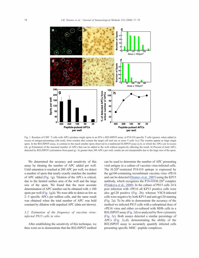
We determined the accuracy and sensitivity of thisassay by titrating the number of APC added per well.Until saturation is reached at 200 APC per well, we detecta number of spots that nearly exactly matches the numberof APC added (Fig. 1g). Titration of the APCs is critical,due to the limited surface area of the well and the largesize of the spots. We found that the most accuratedetermination of APC number can be obtained with!100spots per well (Fig. 1g,h). We were able to detect as few as2–5 specific APCs per million cells, and the same resultwas obtained when the total number of APC was heldconstant by dilution with unpulsed APC (data not shown).
3.2. Estimation of the frequency of vaccinia virus-infected P815 cells in vitro
After establishing the sensitivity of this technique, wethen went on to demonstrate that the RELISPOT method
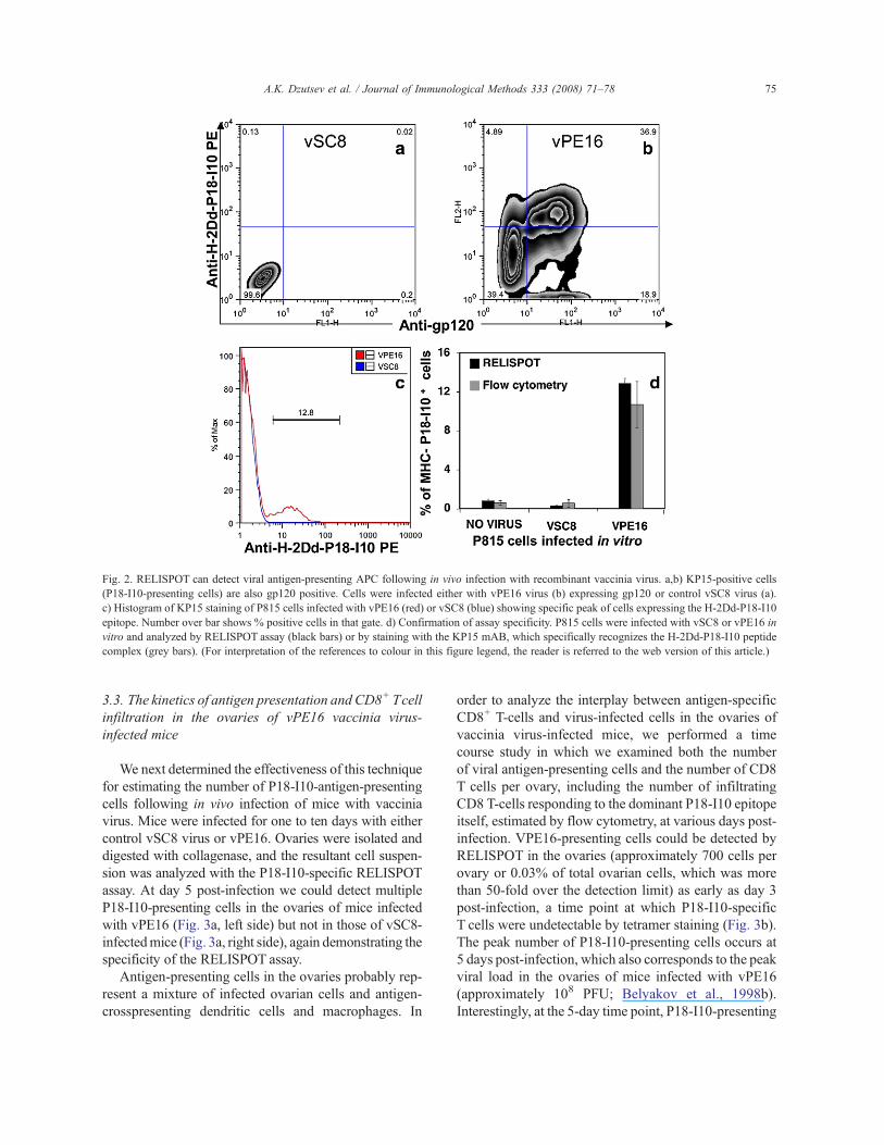
can be used to determine the number of APC presentingviral antigen in a culture of vaccinia virus-infected cells.The H-2Dd-restricted P18-I10 epitope is expressed bythe gp160-containing recombinant vaccinia virus vPE16and can be detected (Dzutsev et al., 2007) using the KP15antibody, which recognizes the P18-I10/H-2Dd complex(Polakova et al., 2000). In the culture of P815 cells 24 hpost infection with vPE16 all KP15 positive cells werealso gp120 positive (Fig. 2b); whereas VSC8-infectedcells were negative by both KP15 and anti-gp120 staining(Fig. 2a). To be able to demonstrate the accuracy of themethod we infected P815 cells with a suboptimal dose ofvPE16 virus and either co-cultured with MHb cells in aRELISPOTassay (Fig. 2d) or analyzed by flow cytometry(Fig. 2c). Both assays detected a similar percentage ofAPCs (Fig. 2c,d), demonstrating the ability of theRELISPOT assay to accurately quantify infected cellspresenting specific MHC–peptide complexes.
Fig. 1. Rosettes of CD8+ T cells with APCs produce single spots in an IFN-! RELISPOT assay. a) P18-I10 specific T cells (green), when added inexcess of antigen-presenting cells (red), form rosettes that contain the target cell and one or more T cells. b,c) The rosettes appear as large singlespots. In the RELISPOT assay, in contrast to the much smaller spots observed in a traditional ELISPOT assay (e,f), in which the APCs are in excess(d). g) Estimation of the maximal number of APCs that can be added to the well without negatively affecting the result. h) Percent of total APCsdetected by RELISPOT (calculation from panel g). At greater than 200 APCs per well, results are not interpretable due to the large size of the spots.
74 A.K. Dzutsev et al. / Journal of Immunological Methods 333 (2008) 71–78
3.3. The kinetics of antigen presentation and CD8+ Tcellinfiltration in the ovaries of vPE16 vaccinia virus-infected mice
We next determined the effectiveness of this techniquefor estimating the number of P18-I10-antigen-presentingcells following in vivo infection of mice with vacciniavirus. Mice were infected for one to ten days with eithercontrol vSC8 virus or vPE16. Ovaries were isolated anddigested with collagenase, and the resultant cell suspen-sion was analyzed with the P18-I10-specific RELISPOTassay. At day 5 post-infection we could detect multipleP18-I10-presenting cells in the ovaries of mice infectedwith vPE16 (Fig. 3a, left side) but not in those of vSC8-infectedmice (Fig. 3a, right side), again demonstrating thespecificity of the RELISPOT assay.
Antigen-presenting cells in the ovaries probably rep-resent a mixture of infected ovarian cells and antigen-crosspresenting dendritic cells and macrophages. In
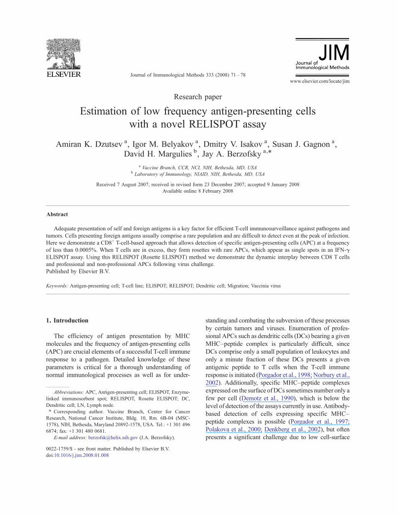
order to analyze the interplay between antigen-specificCD8+ T-cells and virus-infected cells in the ovaries ofvaccinia virus-infected mice, we performed a timecourse study in which we examined both the numberof viral antigen-presenting cells and the number of CD8T cells per ovary, including the number of infiltratingCD8 T-cells responding to the dominant P18-I10 epitopeitself, estimated by flow cytometry, at various days post-infection. VPE16-presenting cells could be detected byRELISPOT in the ovaries (approximately 700 cells perovary or 0.03% of total ovarian cells, which was morethan 50-fold over the detection limit) as early as day 3post-infection, a time point at which P18-I10-specificT cells were undetectable by tetramer staining (Fig. 3b).The peak number of P18-I10-presenting cells occurs at5 days post-infection, which also corresponds to the peakviral load in the ovaries of mice infected with vPE16(approximately 108 PFU; Belyakov et al., 1998b).Interestingly, at the 5-day time point, P18-I10-presenting
Fig. 2. RELISPOT can detect viral antigen-presenting APC following in vivo infection with recombinant vaccinia virus. a,b) KP15-positive cells(P18-I10-presenting cells) are also gp120 positive. Cells were infected either with vPE16 virus (b) expressing gp120 or control vSC8 virus (a).c) Histogram of KP15 staining of P815 cells infected with vPE16 (red) or vSC8 (blue) showing specific peak of cells expressing the H-2Dd-P18-I10epitope. Number over bar shows % positive cells in that gate. d) Confirmation of assay specificity. P815 cells were infected with vSC8 or vPE16 invitro and analyzed by RELISPOT assay (black bars) or by staining with the KP15 mAB, which specifically recognizes the H-2Dd-P18-I10 peptidecomplex (grey bars). (For interpretation of the references to colour in this figure legend, the reader is referred to the web version of this article.)
75A.K. Dzutsev et al. / Journal of Immunological Methods 333 (2008) 71–78
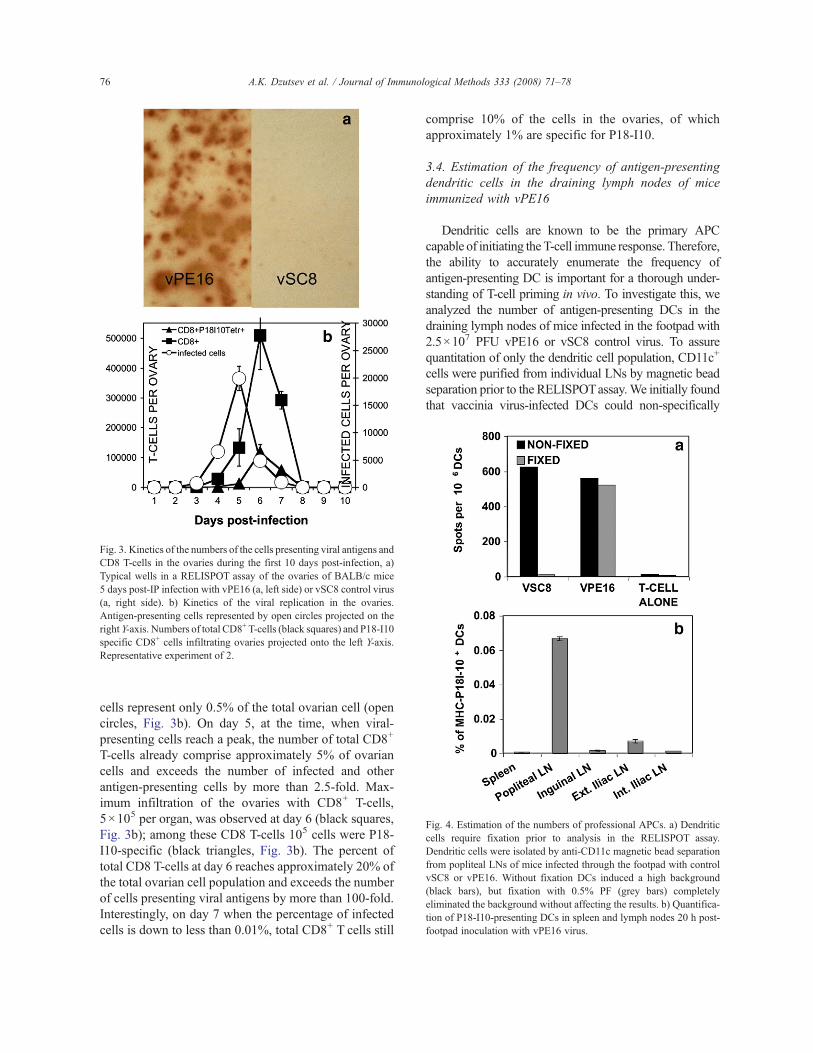
cells represent only 0.5% of the total ovarian cell (opencircles, Fig. 3b). On day 5, at the time, when viral-presenting cells reach a peak, the number of total CD8+
T-cells already comprise approximately 5% of ovariancells and exceeds the number of infected and otherantigen-presenting cells by more than 2.5-fold. Max-imum infiltration of the ovaries with CD8+ T-cells,5!105 per organ, was observed at day 6 (black squares,Fig. 3b); among these CD8 T-cells 105 cells were P18-I10-specific (black triangles, Fig. 3b). The percent oftotal CD8 T-cells at day 6 reaches approximately 20% ofthe total ovarian cell population and exceeds the numberof cells presenting viral antigens by more than 100-fold.Interestingly, on day 7 when the percentage of infectedcells is down to less than 0.01%, total CD8+ T cells still
comprise 10% of the cells in the ovaries, of whichapproximately 1% are specific for P18-I10.
3.4. Estimation of the frequency of antigen-presentingdendritic cells in the draining lymph nodes of miceimmunized with vPE16
Dendritic cells are known to be the primary APCcapable of initiating the T-cell immune response. Therefore,the ability to accurately enumerate the frequency ofantigen-presenting DC is important for a thorough under-standing of T-cell priming in vivo. To investigate this, weanalyzed the number of antigen-presenting DCs in thedraining lymph nodes of mice infected in the footpad with2.5!107 PFU vPE16 or vSC8 control virus. To assurequantitation of only the dendritic cell population, CD11c+
cells were purified from individual LNs by magnetic beadseparation prior to the RELISPOTassay.We initially foundthat vaccinia virus-infected DCs could non-specifically
Fig. 3. Kinetics of the numbers of the cells presenting viral antigens andCD8 T-cells in the ovaries during the first 10 days post-infection, a)Typical wells in a RELISPOT assay of the ovaries of BALB/c mice5 days post-IP infection with vPE16 (a, left side) or vSC8 control virus(a, right side). b) Kinetics of the viral replication in the ovaries.Antigen-presenting cells represented by open circles projected on theright Y-axis. Numbers of total CD8+ T-cells (black squares) and P18-I10specific CD8+ cells infiltrating ovaries projected onto the left Y-axis.Representative experiment of 2.
Fig. 4. Estimation of the numbers of professional APCs. a) Dendriticcells require fixation prior to analysis in the RELISPOT assay.Dendritic cells were isolated by anti-CD11c magnetic bead separationfrom popliteal LNs of mice infected through the footpad with controlvSC8 or vPE16. Without fixation DCs induced a high background(black bars), but fixation with 0.5% PF (grey bars) completelyeliminated the background without affecting the results. b) Quantifica-tion of P18-I10-presenting DCs in spleen and lymph nodes 20 h post-footpad inoculation with vPE16 virus.
76 A.K. Dzutsev et al. / Journal of Immunological Methods 333 (2008) 71–78
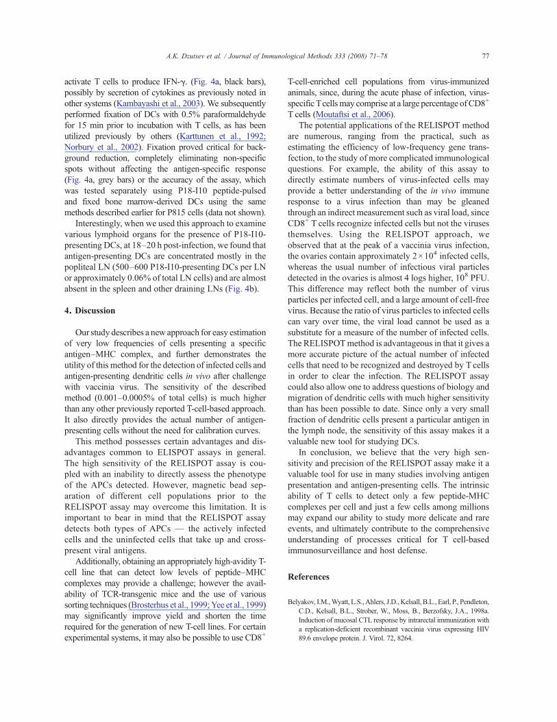
activate T cells to produce IFN-!. (Fig. 4a, black bars),possibly by secretion of cytokines as previously noted inother systems (Kambayashi et al., 2003). We subsequentlyperformed fixation of DCs with 0.5% paraformaldehydefor 15 min prior to incubation with T cells, as has beenutilized previously by others (Karttunen et al., 1992;Norbury et al., 2002). Fixation proved critical for back-ground reduction, completely eliminating non-specificspots without affecting the antigen-specific response(Fig. 4a, grey bars) or the accuracy of the assay, whichwas tested separately using P18-I10 peptide-pulsedand fixed bone marrow-derived DCs using the samemethods described earlier for P815 cells (data not shown).
Interestingly, when we used this approach to examinevarious lymphoid organs for the presence of P18-I10-presenting DCs, at 18–20 h post-infection, we found thatantigen-presenting DCs are concentrated mostly in thepopliteal LN (500–600 P18-I10-presenting DCs per LNor approximately 0.06% of total LN cells) and are almostabsent in the spleen and other draining LNs (Fig. 4b).
4. Discussion
Our study describes a new approach for easy estimationof very low frequencies of cells presenting a specificantigen–MHC complex, and further demonstrates theutility of this method for the detection of infected cells andantigen-presenting dendritic cells in vivo after challengewith vaccinia virus. The sensitivity of the describedmethod (0.001–0.0005% of total cells) is much higherthan any other previously reported T-cell-based approach.It also directly provides the actual number of antigen-presenting cells without the need for calibration curves.
This method possesses certain advantages and dis-advantages common to ELISPOT assays in general.The high sensitivity of the RELISPOT assay is cou-pled with an inability to directly assess the phenotypeof the APCs detected. However, magnetic bead sep-aration of different cell populations prior to theRELISPOT assay may overcome this limitation. It isimportant to bear in mind that the RELISPOT assaydetects both types of APCs — the actively infectedcells and the uninfected cells that take up and cross-present viral antigens.
Additionally, obtaining an appropriately high-avidity T-cell line that can detect low levels of peptide–MHCcomplexes may provide a challenge; however the avail-ability of TCR-transgenic mice and the use of varioussorting techniques (Brosterhus et al., 1999;Yee et al., 1999)may significantly improve yield and shorten the timerequired for the generation of new T-cell lines. For certainexperimental systems, it may also be possible to use CD8+
T-cell-enriched cell populations from virus-immunizedanimals, since, during the acute phase of infection, virus-specific Tcellsmay comprise at a large percentage of CD8+
T cells (Moutaftsi et al., 2006).The potential applications of the RELISPOT method
are numerous, ranging from the practical, such asestimating the efficiency of low-frequency gene trans-fection, to the study of more complicated immunologicalquestions. For example, the ability of this assay todirectly estimate numbers of virus-infected cells mayprovide a better understanding of the in vivo immuneresponse to a virus infection than may be gleanedthrough an indirect measurement such as viral load, sinceCD8+ T cells recognize infected cells but not the virusesthemselves. Using the RELISPOT approach, weobserved that at the peak of a vaccinia virus infection,the ovaries contain approximately 2!104 infected cells,whereas the usual number of infectious viral particlesdetected in the ovaries is almost 4 logs higher, 108 PFU.This difference may reflect both the number of virusparticles per infected cell, and a large amount of cell-freevirus. Because the ratio of virus particles to infected cellscan vary over time, the viral load cannot be used as asubstitute for a measure of the number of infected cells.The RELISPOTmethod is advantageous in that it gives amore accurate picture of the actual number of infectedcells that need to be recognized and destroyed by T cellsin order to clear the infection. The RELISPOT assaycould also allow one to address questions of biology andmigration of dendritic cells with much higher sensitivitythan has been possible to date. Since only a very smallfraction of dendritic cells present a particular antigen inthe lymph node, the sensitivity of this assay makes it avaluable new tool for studying DCs.
In conclusion, we believe that the very high sen-sitivity and precision of the RELISPOT assay make it avaluable tool for use in many studies involving antigenpresentation and antigen-presenting cells. The intrinsicability of T cells to detect only a few peptide-MHCcomplexes per cell and just a few cells among millionsmay expand our ability to study more delicate and rareevents, and ultimately contribute to the comprehensiveunderstanding of processes critical for T cell-basedimmunosurveillance and host defense.
References
Belyakov, I.M.,Wyatt, L.S.,Ahlers, J.D.,Kelsall, B.L., Earl, P., Pendleton,C.D., Kelsall, B.L., Strober, W., Moss, B., Berzofsky, J.A., 1998a.Induction of mucosal CTL response by intrarectal immunization witha replication-deficient recombinant vaccinia virus expressing HIV89.6 envelope protein. J. Virol. 72, 8264.
77A.K. Dzutsev et al. / Journal of Immunological Methods 333 (2008) 71–78
Belyakov, I.M., Derby, M.A., Ahlers, J.D., Kelsall, B.L., Earl, P.,Moss, B., Strober, W., Berzofsky, J.A., 1998b. Mucosal immuni-zation with HIV-1 peptide vaccine induces mucosal and systemiccytotoxic T lymphocytes and protective immunity in mice againstintrarectal recombinant HIV-vaccinia challenge. Proc. Natl. Acad.Sci. U. S. A. 95, 1709.
Belyakov, I.M., Hammond, S.A., Ahlers, J.D., Glenn, G.M., Berzofsky,J.A., 2004. Transcutaneous immunization induces mucosal CTLand protective immunity by migration of primed skin dendriticcells. J. Clin. Invest. 113, 998.
Brosterhus, H., Brings, S., Leyendeckers, J., Manz, R.A., Miltenyi, S.,Radbruch, A., Assenmacher, M., Schmitz, J., 1999. Enrichmentand detection of live antigen-specific CD4+ and CD8+ T cellsbased on cytokine secretion. Eur. J. Immunol. 29, 4053.
Chakrabarti, S., Brechling, K., Moss, B., 1985. Vaccinia virusexpression vector: coexpression of b-galactosidase provides visualscreening of recombinant virus plaques. Mol. Cell Biol. 5, 3403.
Czerkinsky, C.C., Nilsson, L.A., Nygren, H., Ouchterlony, O.,Tarkowski, A., 1983. A solid-phase enzyme-linked immunospot(ELISPOT) assay for enumeration of specific antibody-secretingcells. J. Immunol. Methods 65, 109.
Demotz, S., Grey, H.M., Sette, A., 1990. The minimal number of classII MHC-antigen complexes needed for T cell activation. Science249, 1028.
Denkberg, G., Cohen, C.J., Lev, A., Chames, P., Hoogenboom, H.R.,Reiter, Y., 2002. Direct visualization of distinct T cell epitopesderived from a melanoma tumor-associated antigen by usinghuman recombinant antibodies with MHC-restricted T cellreceptor-like specificity. Proc. Natl. Acad. Sci. U. S. A. 99, 9421.
Derby, M.A., Alexander-Miller, M.A., Tse, R., Berzofsky, J.A., 2001a.High avidity CTL exploit two complementary mechanisms toprovide better protection against viral infection than low avidityCTL. J. Immunol. 166, 1690.
Derby, M.A., Wang, J., Margulies, D.H., Berzofsky, J.A., 2001b. Twointermediate avidity CTL clones with a disparity betweenfunctional avidity and MHC tetramer staining. Int. Immunol. 13,817.
Dzutsev,A.H., Belyakov, I.M., Isakov,D.V.,Margulies, D.H.,Berzofsky,J.A., 2007. Avidity of CD8 T cells sharpens immunodominance. Int.Immunol. 19, 497.
Earl, P.L., Hugin, A.W., Moss, B., 1990. Removal of cryptic poxvirustranscription termination signals from the human immunodefi-ciency virus type 1 envelope gene enhances expression andimmunogenicity of a recombinant vaccinia virus. J. Virol. 64,2448.
Kambayashi, T., Assarsson, E., Lukacher, A.E., Ljunggren, H.G.,Jensen, P.E., 2003. Memory CD8+ T cells provide an early sourceof IFN-gamma. J. Immunol. 170, 2399.
Karttunen, J., Sanderson, S., Shastri, N., 1992. Detection of rareantigen-presenting cells by the lacZ T-cell activation assay
suggests an expression cloning strategy for T-cell antigens. Proc.Natl. Acad. Sci. U. S. A. 89, 6020.
Moutaftsi, M., Peters, B., Pasquetto, V., Tscharke, D.C., Sidney, J.,Bui, H.H., Grey, H., Sette, A., 2006. A consensus epitopeprediction approach identifies the breadth of murine T(CD8+)-cellresponses to vaccinia virus. Nat. Biotechnol. 24, 817.
Norbury, C.C., Malide, D., Gibbs, J.S., Bennink, J.R., Yewdell, J.W.,2002. Visualizing priming of virus-specific CD8+ T cells byinfected dendritic cells in vivo. Nat. Immunol. vol. 3, 265.
Plaksin, D., Polakova, K., McPhie, P., Margulies, D.H., 1997. A three-domain T cell receptor is biologically active and specifically stainscell surface MHC/peptide complexes. J. Immunol. vol. 158, 2218.
Polakova, K., Plaksin, D., Chung, D.H., Belyakov, I.M., Berzofsky, J.A.,Margulies, D.H., 2000. Antibodies directed against the MHC-Imolecule H-2Dd complexed with an antigenic peptide: similarities toa T cell receptor with the same specificity. J. Immunol. 165, 5703.
Porgador, A., Yewdell, J.W., Deng, Y., Bennink, J.R., Germain, R.N.,1997. Localization, quantitation, and in situ detection of specificpeptide–MHC class I complexes using a monoclonal antibody.Immunity 6, 715.
Porgador, A., Irvine, K.R., Iwasaki, A., Barber, B.H., Restifo, N.P.,Germain, R.N., 1998. Predominant role for directly transfecteddendritic cells in antigen presentation to CD8+ T cells after genegun immunization. J. Exp. Med. 188, 1075.
Somasundaram, R., Satyamoorthy, K., Caputo, L., Yssel, H., Herlyn,D., 2004. Detection of HLA class II-dependent T helper antigenusing antigen phage display. Clin. Exp. Immunol. 135, 247.
Subbramanian, R.A., Moriya, C., Martin, K.L., Peyerl, F.W., Hasegawa,A., Naoi, A., Chhay, H., Autissier, P., Gorgone, D.A., Lifton, M.A.,Kuus-Reichel, K., Schmitz, J.E., Letvin, N.L., Kuroda, M.J., 2004.Engineered T-cell receptor tetramers bind MHC–peptide complexeswith high affinity. Nat. Biotechnol. 22, 1429.
Taguchi, T.,McGhee, J.R., Coffman, R.L., Beagley,K.W., Eldridge, J.H.,Takatsu, K., Kiyono,H., 1990.Detection of individual mouse splenicT cells producing IFN-gamma and IL-5 using the enzyme-linkedimmunospot (ELISPOT) assay. J. Immunol. Methods 128, 65.
Takahashi, H., Cohen, J., Hosmalin, A., Cease, K.B., Houghten, R.,Cornette, J., DeLisi, C., Moss, B., Germain, R.N., Berzofsky, J.A.,1988. An immunodominant epitope of the HIV gp160 envelopeglycoprotein recognized by class I MHC molecule-restrictedmurine cytotoxic T lymphocytes. Proc. Natl. Acad. Sci. U. S. A.85, 3105.
Takahashi, H., Merli, S., Putney, S.D., Houghten, R., Moss, B.,Germain, R.N., Berzofsky, J.A., 1989. A single amino acidinterchange yields reciprocal CTL specificities for HIV gp160.Science 246, 118.
Yee, C., Savage, P.A., Lee, P.P., Davis, M.M., Greenberg, P.D., 1999.Isolation of high avidity melanoma-reactive CTL from hetero-geneous populations using peptide-MHC tetramers. J. Immunol.162, 2227.
78 A.K. Dzutsev et al. / Journal of Immunological Methods 333 (2008) 71–78