Cassava Plants with a Depleted Cyanogenic Glucoside Content in Leaves and Tubers. Distribution of...
12
Cassava Plants with a Depleted Cyanogenic Glucoside Content in Leaves and Tubers. Distribution of Cyanogenic Glucosides, Their Site of Synthesis and Transport, and Blockage of the Biosynthesis by RNA Interference Technology 1 Kirsten Jørgensen, Søren Bak, Peter Kamp Busk 2 , Charlotte Sørensen, Carl Erik Olsen, Johanna Puonti-Kaerlas 3 , and Birger Lindberg Møller* Plant Biochemistry Laboratory, Department of Plant Biology, Center for Molecular Plant Physiology (K.J., S.B., P.K.B., C.S., B.L.M.), and Chemistry Department (C.E.O.), Royal Veterinary and Agricultural University, DK–1871 Frederiksberg C, Copenhagen, Denmark; and Laboratory of Plant Biotechnology, Swiss Federal Institute of Technology, Eidgeno ¨ssische Technische Hochschule Zentrum, CH–8092 Zurich, Switzerland (J.P.-K.) Transgenic cassava (Manihot esculenta Crantz, cv MCol22) plants with a 92% reduction in cyanogenic glucoside content in tubers and acyanogenic (,1% of wild type) leaves were obtained by RNA interference to block expression of CYP79D1 and CYP79D2, the two paralogous genes encoding the first committed enzymes in linamarin and lotaustralin synthesis. About 180 independent lines with acyanogenic (,1% of wild type) leaves were obtained. Only a few of these were depleted with respect to cyanogenic glucoside content in tubers. In agreement with this observation, girdling experiments demonstrated that cyanogenic glucosides are synthesized in the shoot apex and transported to the root, resulting in a negative concentration gradient basipetal in the plant with the concentration of cyanogenic glucosides being highest in the shoot apex and the petiole of the first unfolded leaf. Supply of nitrogen increased the cyanogenic glucoside concentration in the shoot apex. In situ polymerase chain reaction studies demonstrated that CYP79D1 and CYP79D2 were preferentially expressed in leaf mesophyll cells positioned adjacent to the epidermis. In young petioles, preferential expression was observed in the epidermis, in the two first cortex cell layers, and in the endodermis together with pericycle cells and specific parenchymatic cells around the laticifers. These data demonstrate that it is possible to drastically reduce the linamarin and lotaustralin content in cassava tubers by blockage of cyanogenic glucoside synthesis in leaves and petioles. The reduced flux to the roots of reduced nitrogen in the form of cyanogenic glucosides did not prevent tuber formation. Cassava (Manihot esculenta Crantz) is the most important root crop in the world and ranks second among African staple crops (Nweke et al., 2002). Cassava is vegetatively propagated through stem cut- tings and produces well on poor soils. The tubers may be kept in the soil for extended time periods. This secures rural farmers a carbohydrate source in years with adverse growth conditions where other crops fail and famine would otherwise prevail. These features and high crop yield contribute to the importance of cassava in Africa, South East Asia, and South America (Nweke et al., 2002). The average annual per capita consumption of cassava in 2002 was 287 kg in the Democratic Republic of Congo and 127 kg in Nigeria (http://faostat.fao.org/ [nutrition data from 2002]). Industrial cassava starch production is important, es- pecially in South East Asia (Bokanga, 1994). Cassava leaves are not widely used as a food source despite their high protein content (Ngudi et al., 2003). Major drawbacks of the cassava crop are the low tuber protein content, rapid tuber perishability follow- ing harvest, and high content of the cyanogenic gluco- sides linamarin and lotaustralin in all tissues. Upon tissue disruption, the cyanogenic glucosides are brought in contact with b-glucosidases and a-hydroxynitrile lyases and degraded into cyanohydrins, hydrogen cy- anide, and ketones (Conn, 1980). When cassava products are used as a primary staple food, careful processing to remove these toxic constituents is required to avoid chronic cyanide intoxication (Onabolu et al., 2002). In- complete processing may result in high cyanide expo- sure and give rise to severe diseases like tropical ataxic 1 This work was supported by the Danish International Develop- ment Agency (grant nos. 104Dan8/503 and 104.Dan.8/91125) and by a grant to the Center for Molecular Plant Physiology (PlaCe) from the Danish National Research Foundation. 2 Present address: Virology and Molecular Toxicology, Novo Nordisk A/S, Novo Nordisk Park, 2760 Malov, Denmark. 3 Present address: European Patent Office, Directorate 2.4.01 Biotechnology, EPA/EPO/OEB, D–80298 Munich, Germany. * Corresponding author; e-mail [email protected]; fax 45–35–28–33–33. Article, publication date, and citation information can be found at www.plantphysiol.org/cgi/doi/10.1104/pp.105.065904. This article is published in Plant Physiology Online, Plant Physiology Preview Section, which publishes manuscripts accepted for publication after they have been edited and the authors have corrected proofs, but before the final, complete issue is published online. Early posting of articles reduces normal time to publication by several weeks. Plant Physiology Preview, www.aspb.org Ó 2005 American Society of Plant Biologists 1 of 12 www.plant.org on March 26, 2016 - Published by www.plantphysiol.org Downloaded from Copyright © 2005 American Society of Plant Biologists. All rights reserved.
Transcript of Cassava Plants with a Depleted Cyanogenic Glucoside Content in Leaves and Tubers. Distribution of...
Cassava Plants with a Depleted Cyanogenic GlucosideContent in Leaves and Tubers. Distribution ofCyanogenic Glucosides, Their Site of Synthesisand Transport, and Blockage of the Biosynthesisby RNA Interference Technology1
Kirsten Jørgensen, Søren Bak, Peter Kamp Busk2, Charlotte Sørensen, Carl Erik Olsen,Johanna Puonti-Kaerlas3, and Birger Lindberg Møller*
Plant Biochemistry Laboratory, Department of Plant Biology, Center for Molecular Plant Physiology(K.J., S.B., P.K.B., C.S., B.L.M.), and Chemistry Department (C.E.O.), Royal Veterinary and AgriculturalUniversity, DK–1871 Frederiksberg C, Copenhagen, Denmark; and Laboratory of Plant Biotechnology,Swiss Federal Institute of Technology, Eidgenossische Technische Hochschule Zentrum, CH–8092 Zurich,Switzerland (J.P.-K.)
Transgenic cassava (Manihot esculenta Crantz, cv MCol22) plants with a 92% reduction in cyanogenic glucoside content intubers and acyanogenic (,1% of wild type) leaves were obtained by RNA interference to block expression of CYP79D1 andCYP79D2, the two paralogous genes encoding the first committed enzymes in linamarin and lotaustralin synthesis. About 180independent lines with acyanogenic (,1% of wild type) leaves were obtained. Only a few of these were depleted with respectto cyanogenic glucoside content in tubers. In agreement with this observation, girdling experiments demonstrated thatcyanogenic glucosides are synthesized in the shoot apex and transported to the root, resulting in a negative concentrationgradient basipetal in the plant with the concentration of cyanogenic glucosides being highest in the shoot apex and the petioleof the first unfolded leaf. Supply of nitrogen increased the cyanogenic glucoside concentration in the shoot apex. In situpolymerase chain reaction studies demonstrated that CYP79D1 and CYP79D2 were preferentially expressed in leaf mesophyllcells positioned adjacent to the epidermis. In young petioles, preferential expression was observed in the epidermis, in the twofirst cortex cell layers, and in the endodermis together with pericycle cells and specific parenchymatic cells around thelaticifers. These data demonstrate that it is possible to drastically reduce the linamarin and lotaustralin content in cassavatubers by blockage of cyanogenic glucoside synthesis in leaves and petioles. The reduced flux to the roots of reduced nitrogenin the form of cyanogenic glucosides did not prevent tuber formation.
Cassava (Manihot esculenta Crantz) is the mostimportant root crop in the world and ranks secondamong African staple crops (Nweke et al., 2002).Cassava is vegetatively propagated through stem cut-tings and produces well on poor soils. The tubers maybe kept in the soil for extended time periods. Thissecures rural farmers a carbohydrate source in yearswith adverse growth conditions where other crops failand famine would otherwise prevail. These featuresand high crop yield contribute to the importance of
cassava in Africa, South East Asia, and South America(Nweke et al., 2002). The average annual per capitaconsumption of cassava in 2002 was 287 kg in theDemocratic Republic of Congo and 127 kg in Nigeria(http://faostat.fao.org/ [nutrition data from 2002]).Industrial cassava starch production is important, es-pecially in South East Asia (Bokanga, 1994). Cassavaleaves are not widely used as a food source despitetheir high protein content (Ngudi et al., 2003).
Major drawbacks of the cassava crop are the lowtuber protein content, rapid tuber perishability follow-ing harvest, and high content of the cyanogenic gluco-sides linamarin and lotaustralin in all tissues. Upontissue disruption, the cyanogenic glucosides are broughtin contact with b-glucosidases and a-hydroxynitrilelyases and degraded into cyanohydrins, hydrogen cy-anide, and ketones (Conn, 1980). When cassava productsare used as a primary staple food, careful processingto remove these toxic constituents is required to avoidchronic cyanide intoxication (Onabolu et al., 2002). In-complete processing may result in high cyanide expo-sure and give rise to severe diseases like tropical ataxic
1 This work was supported by the Danish International Develop-ment Agency (grant nos. 104Dan8/503 and 104.Dan.8/91125) and bya grant to the Center for Molecular Plant Physiology (PlaCe) from theDanish National Research Foundation.
2 Present address: Virology and Molecular Toxicology, NovoNordisk A/S, Novo Nordisk Park, 2760 Malov, Denmark.
3 Present address: European Patent Office, Directorate 2.4.01Biotechnology, EPA/EPO/OEB, D–80298 Munich, Germany.
* Corresponding author; e-mail [email protected]; fax 45–35–28–33–33.Article, publication date, and citation information can be found at
www.plantphysiol.org/cgi/doi/10.1104/pp.105.065904.
This article is published in Plant Physiology Online, Plant Physiology Preview Section, which publishes manuscripts accepted for
publication after they have been edited and the authors have corrected proofs, but before the final, complete issue is published
online. Early posting of articles reduces normal time to publication by several weeks.
Plant Physiology Preview, www.aspb.org � 2005 American Society of Plant Biologists 1 of 12 www.plant.org on March 26, 2016 - Published by www.plantphysiol.orgDownloaded from
Copyright © 2005 American Society of Plant Biologists. All rights reserved.
neuropathy and konzo, especially in populations withpoor nutritional status. Unfortunately, careful process-ing generally results in loss of proteins, vitamins, andminerals, i.e. in products with low nutritional value. Inspite of the availability of efficient processing proce-dures, cyanide exposure from cassava diets prevails(Oluwole et al., 2000). Traditional breeding has gener-ated cassava cultivars with a high to low cyanidepotential, but has not succeeded in providing cassavacultivars totally devoid of cyanogenic glucosides(Nweke et al., 2002).
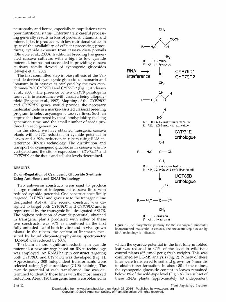
The first committed step in biosynthesis of the Val-and Ile-derived cyanogenic glucosides linamarin andlotaustralin in cassava is catalyzed by the two cyto-chromes P450 CYP79D1 and CYP79D2 (Fig. 1; Andersenet al., 2000). The presence of two CYP79 paralogs incassava is in accordance with cassava being allopoly-ploid (Fregene et al., 1997). Mapping of the CYP79D1and CYP79D2 genes would provide the necessarymolecular tools in a marker-assisted classical breedingprogram to select acyanogenic cassava lines. Such anapproach is hampered by the allopolyploidity, the longgeneration time, and the small number of seeds pro-duced in each generation.
In this study, we have obtained transgenic cassavaplants with .99% reduction in cyanide potential inleaves and a 92% reduction in tubers using RNA in-terference (RNAi) technology. The distribution andtransport of cyanogenic glucosides in cassava was in-vestigated and the site of expression of CYP79D1 andCYP79D2 at the tissue and cellular levels determined.
RESULTS
Down-Regulation of Cyanogenic Glucoside SynthesisUsing Anti-Sense and RNAi Technology
Two anti-sense constructs were used to producea large number of independent cassava lines withreduced cyanide potential. One construct specificallytargeted CYP79D1 and gave rise to the transgenic linedesignated AS17A. The second construct was de-signed to target both CYP79D1 and CYP79D2 and isrepresented by the transgenic line designated AS17B.The highest reduction of cyanide potential, obtainedin transgenic plants produced with either of thesetwo constructs, was 80% as monitored in the firstfully unfolded leaf of both in vitro and in vivo-grownplants. In the tubers, the content of linamarin mea-sured by liquid chromatography-mass spectrometry(LC-MS) was reduced by 60%.
To obtain a more significant reduction in cyanidepotential, a new strategy based on RNAi technologywas employed. An RNAi hairpin construct targetingboth CYP79D1 and CYP79D2 was developed (Fig. 1).Approximately 300 independent transformants wereselected using b-glucuronidase (GUS) staining. Thecyanide potential of each transformed line was de-termined to identify those lines with the most markedreduction. About 180 transgenic lines were obtained in
which the cyanide potential in the first fully unfoldedleaf was reduced to ,1% of the level in wild-typecontrol plants (65 mmol per g fresh weight). This wasconfirmed by LC-MS analysis (Fig. 2). Ninety of theselines were transferred to soil and grown for 6 monthsto obtain tuber formation. In about 80 of these lines,the cyanogenic glucoside content in leaves remainedbelow 1% of the wild-type level (Fig. 2A). In a subset ofthese RNAi plants (approximately 40 independent
Figure 1. The biosynthetic pathway for the cyanogenic glucosideslinamarin and lotaustralin in cassava. The enzymatic step blocked byRNAi technology is indicated.
Jørgensen et al.
2 of 12 Plant Physiology Preview www.plant.org on March 26, 2016 - Published by www.plantphysiol.orgDownloaded from
Copyright © 2005 American Society of Plant Biologists. All rights reserved.
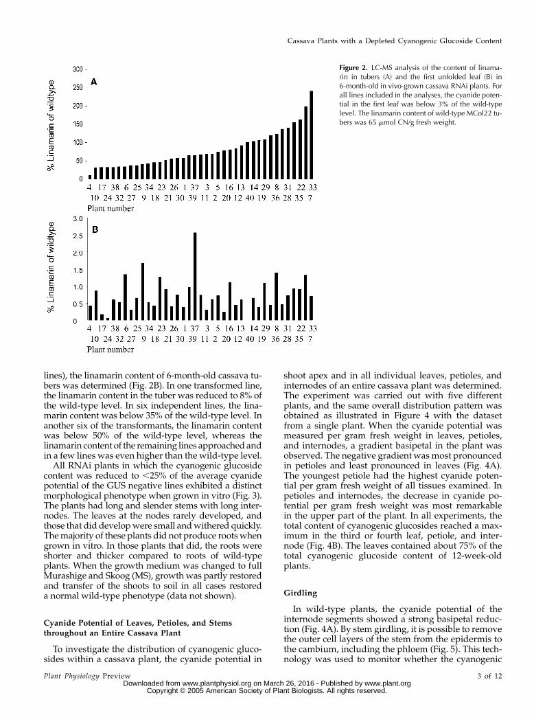
lines), the linamarin content of 6-month-old cassava tu-bers was determined (Fig. 2B). In one transformed line,the linamarin content in the tuber was reduced to 8% ofthe wild-type level. In six independent lines, the lina-marin content was below 35% of the wild-type level. Inanother six of the transformants, the linamarin contentwas below 50% of the wild-type level, whereas thelinamarin content of the remaining lines approached andin a few lines was even higher than the wild-type level.
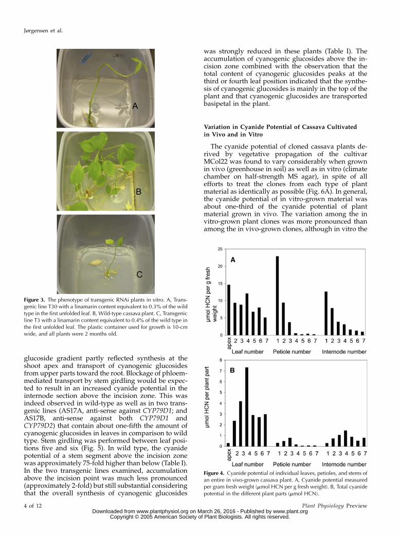
All RNAi plants in which the cyanogenic glucosidecontent was reduced to ,25% of the average cyanidepotential of the GUS negative lines exhibited a distinctmorphological phenotype when grown in vitro (Fig. 3).The plants had long and slender stems with long inter-nodes. The leaves at the nodes rarely developed, andthose that did develop were small and withered quickly.The majority of these plants did not produce roots whengrown in vitro. In those plants that did, the roots wereshorter and thicker compared to roots of wild-typeplants. When the growth medium was changed to fullMurashige and Skoog (MS), growth was partly restoredand transfer of the shoots to soil in all cases restoreda normal wild-type phenotype (data not shown).
Cyanide Potential of Leaves, Petioles, and Stemsthroughout an Entire Cassava Plant
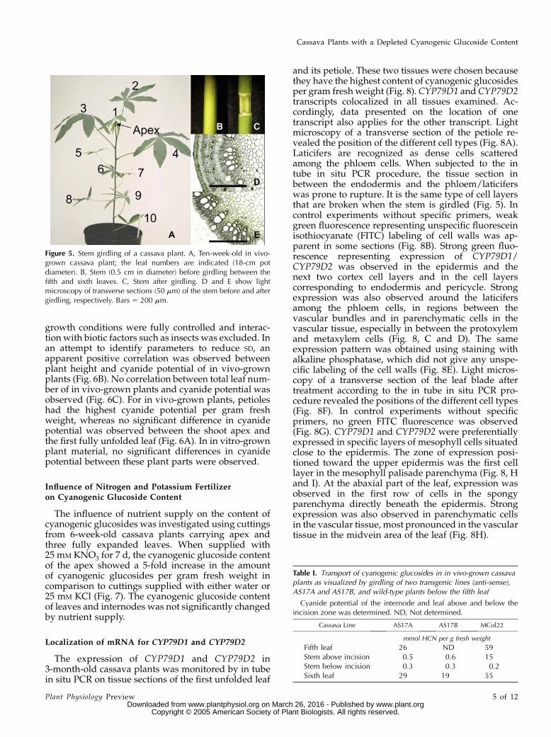
To investigate the distribution of cyanogenic gluco-sides within a cassava plant, the cyanide potential in
shoot apex and in all individual leaves, petioles, andinternodes of an entire cassava plant was determined.The experiment was carried out with five differentplants, and the same overall distribution pattern wasobtained as illustrated in Figure 4 with the datasetfrom a single plant. When the cyanide potential wasmeasured per gram fresh weight in leaves, petioles,and internodes, a gradient basipetal in the plant wasobserved. The negative gradient was most pronouncedin petioles and least pronounced in leaves (Fig. 4A).The youngest petiole had the highest cyanide poten-tial per gram fresh weight of all tissues examined. Inpetioles and internodes, the decrease in cyanide po-tential per gram fresh weight was most remarkablein the upper part of the plant. In all experiments, thetotal content of cyanogenic glucosides reached a max-imum in the third or fourth leaf, petiole, and inter-node (Fig. 4B). The leaves contained about 75% of thetotal cyanogenic glucoside content of 12-week-oldplants.
Girdling
In wild-type plants, the cyanide potential of theinternode segments showed a strong basipetal reduc-tion (Fig. 4A). By stem girdling, it is possible to removethe outer cell layers of the stem from the epidermis tothe cambium, including the phloem (Fig. 5). This tech-nology was used to monitor whether the cyanogenic
Figure 2. LC-MS analysis of the content of linama-rin in tubers (A) and the first unfolded leaf (B) in6-month-old in vivo-grown cassava RNAi plants. Forall lines included in the analyses, the cyanide poten-tial in the first leaf was below 3% of the wild-typelevel. The linamarin content of wild-type MCol22 tu-bers was 65 mmol CN/g fresh weight.
Cassava Plants with a Depleted Cyanogenic Glucoside Content
Plant Physiology Preview 3 of 12 www.plant.org on March 26, 2016 - Published by www.plantphysiol.orgDownloaded from
Copyright © 2005 American Society of Plant Biologists. All rights reserved.
glucoside gradient partly reflected synthesis at theshoot apex and transport of cyanogenic glucosidesfrom upper parts toward the root. Blockage of phloem-mediated transport by stem girdling would be expec-ted to result in an increased cyanide potential in theinternode section above the incision zone. This wasindeed observed in wild-type as well as in two trans-genic lines (AS17A, anti-sense against CYP79D1; andAS17B, anti-sense against both CYP79D1 andCYP79D2) that contain about one-fifth the amount ofcyanogenic glucosides in leaves in comparison to wildtype. Stem girdling was performed between leaf posi-tions five and six (Fig. 5). In wild type, the cyanidepotential of a stem segment above the incision zonewas approximately 75-fold higher than below (Table I).In the two transgenic lines examined, accumulationabove the incision point was much less pronounced(approximately 2-fold) but still substantial consideringthat the overall synthesis of cyanogenic glucosides
was strongly reduced in these plants (Table I). Theaccumulation of cyanogenic glucosides above the in-cision zone combined with the observation that thetotal content of cyanogenic glucosides peaks at thethird or fourth leaf position indicated that the synthe-sis of cyanogenic glucosides is mainly in the top of theplant and that cyanogenic glucosides are transportedbasipetal in the plant.
Variation in Cyanide Potential of Cassava Cultivated
in Vivo and in Vitro
The cyanide potential of cloned cassava plants de-rived by vegetative propagation of the cultivarMCol22 was found to vary considerably when grownin vivo (greenhouse in soil) as well as in vitro (climatechamber on half-strength MS agar), in spite of allefforts to treat the clones from each type of plantmaterial as identically as possible (Fig. 6A). In general,the cyanide potential of in vitro-grown material wasabout one-third of the cyanide potential of plantmaterial grown in vivo. The variation among the invitro-grown plant clones was more pronounced thanamong the in vivo-grown clones, although in vitro the
Figure 3. The phenotype of transgenic RNAi plants in vitro. A, Trans-genic line T30 with a linamarin content equivalent to 0.3% of the wildtype in the first unfolded leaf. B, Wild-type cassava plant. C, Transgenicline T3 with a linamarin content equivalent to 0.4% of the wild type inthe first unfolded leaf. The plastic container used for growth is 10-cmwide, and all plants were 2 months old.
Figure 4. Cyanide potential of individual leaves, petioles, and stems ofan entire in vivo-grown cassava plant. A, Cyanide potential measuredper gram fresh weight (mmol HCN per g fresh weight). B, Total cyanidepotential in the different plant parts (mmol HCN).
Jørgensen et al.
4 of 12 Plant Physiology Preview www.plant.org on March 26, 2016 - Published by www.plantphysiol.orgDownloaded from
Copyright © 2005 American Society of Plant Biologists. All rights reserved.
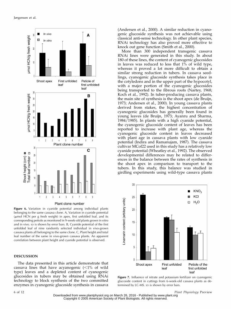
growth conditions were fully controlled and interac-tion with biotic factors such as insects was excluded. Inan attempt to identify parameters to reduce SD, anapparent positive correlation was observed betweenplant height and cyanide potential of in vivo-grownplants (Fig. 6B). No correlation between total leaf num-ber of in vivo-grown plants and cyanide potential wasobserved (Fig. 6C). For in vivo-grown plants, petioleshad the highest cyanide potential per gram freshweight, whereas no significant difference in cyanidepotential was observed between the shoot apex andthe first fully unfolded leaf (Fig. 6A). In in vitro-grownplant material, no significant differences in cyanidepotential between these plant parts were observed.
Influence of Nitrogen and Potassium Fertilizeron Cyanogenic Glucoside Content
The influence of nutrient supply on the content ofcyanogenic glucosides was investigated using cuttingsfrom 6-week-old cassava plants carrying apex andthree fully expanded leaves. When supplied with25 mM KNO3 for 7 d, the cyanogenic glucoside contentof the apex showed a 5-fold increase in the amountof cyanogenic glucosides per gram fresh weight incomparison to cuttings supplied with either water or25 mM KCl (Fig. 7). The cyanogenic glucoside contentof leaves and internodes was not significantly changedby nutrient supply.
Localization of mRNA for CYP79D1 and CYP79D2
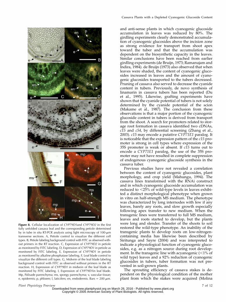
The expression of CYP79D1 and CYP79D2 in3-month-old cassava plants was monitored by in tubein situ PCR on tissue sections of the first unfolded leaf
and its petiole. These two tissues were chosen becausethey have the highest content of cyanogenic glucosidesper gram fresh weight (Fig. 8). CYP79D1 and CYP79D2transcripts colocalized in all tissues examined. Ac-cordingly, data presented on the location of onetranscript also applies for the other transcript. Lightmicroscopy of a transverse section of the petiole re-vealed the position of the different cell types (Fig. 8A).Laticifers are recognized as dense cells scatteredamong the phloem cells. When subjected to the intube in situ PCR procedure, the tissue section inbetween the endodermis and the phloem/laticiferswas prone to rupture. It is the same type of cell layersthat are broken when the stem is girdled (Fig. 5). Incontrol experiments without specific primers, weakgreen fluorescence representing unspecific fluoresceinisothiocyanate (FITC) labeling of cell walls was ap-parent in some sections (Fig. 8B). Strong green fluo-rescence representing expression of CYP79D1/CYP79D2 was observed in the epidermis and thenext two cortex cell layers and in the cell layerscorresponding to endodermis and pericycle. Strongexpression was also observed around the laticifersamong the phloem cells, in regions between thevascular bundles and in parenchymatic cells in thevascular tissue, especially in between the protoxylemand metaxylem cells (Fig. 8, C and D). The sameexpression pattern was obtained using staining withalkaline phosphatase, which did not give any unspe-cific labeling of the cell walls (Fig. 8E). Light micros-copy of a transverse section of the leaf blade aftertreatment according to the in tube in situ PCR pro-cedure revealed the positions of the different cell types(Fig. 8F). In control experiments without specificprimers, no green FITC fluorescence was observed(Fig. 8G). CYP79D1 and CYP79D2 were preferentiallyexpressed in specific layers of mesophyll cells situatedclose to the epidermis. The zone of expression posi-tioned toward the upper epidermis was the first celllayer in the mesophyll palisade parenchyma (Fig. 8, Hand I). At the abaxial part of the leaf, expression wasobserved in the first row of cells in the spongyparenchyma directly beneath the epidermis. Strongexpression was also observed in parenchymatic cellsin the vascular tissue, most pronounced in the vasculartissue in the midvein area of the leaf (Fig. 8H).
Table I. Transport of cyanogenic glucosides in in vivo-grown cassavaplants as visualized by girdling of two transgenic lines (anti-sense),AS17A and AS17B, and wild-type plants below the fifth leaf
Cyanide potential of the internode and leaf above and below theincision zone was determined. ND, Not determined.
Cassava Line AS17A AS17B MCol22
mmol HCN per g fresh weight
Fifth leaf 26 ND 59Stem above incision 0.5 0.6 15Stem below incision 0.3 0.3 0.2Sixth leaf 29 19 55
Figure 5. Stem girdling of a cassava plant. A, Ten-week-old in vivo-grown cassava plant; the leaf numbers are indicated (18-cm potdiameter). B, Stem (0.5 cm in diameter) before girdling between thefifth and sixth leaves. C, Stem after girdling. D and E show lightmicroscopy of transverse sections (50 mm) of the stem before and aftergirdling, respectively. Bars 5 200 mm.
Cassava Plants with a Depleted Cyanogenic Glucoside Content
Plant Physiology Preview 5 of 12 www.plant.org on March 26, 2016 - Published by www.plantphysiol.orgDownloaded from
Copyright © 2005 American Society of Plant Biologists. All rights reserved.
DISCUSSION
The data presented in this article demonstrate thatcassava lines that have acyanogenic (,1% of wildtype) leaves and a depleted content of cyanogenicglucosides in tubers may be obtained using RNAitechnology to block synthesis of the two committedenzymes in cyanogenic glucoside synthesis in cassava
(Andersen et al., 2000). A similar reduction in cyano-genic glucoside synthesis was not achievable usingclassical anti-sense technology. In other plant species,RNAi technology has also proved more effective toknock out gene function (Smith et al., 2000).
More than 300 independent transgenic cassavaRNAi lines were generated in this study. In about180 of these lines, the content of cyanogenic glucosidesin leaves was reduced to less that 1% of wild type,whereas it proved a lot more difficult to obtain asimilar strong reduction in tubers. In cassava seed-lings, cyanogenic glucoside synthesis takes place inthe cotyledons and in the upper part of the hypocotyl,with a major portion of the cyanogenic glucosidesbeing transported to the fibrous roots (Nartey, 1968;Koch et al., 1992). In tuber-producing cassava plants,the main site of synthesis is the shoot apex (de Bruijn,1973; Andersen et al., 2000). In young cassava plantsderived from stakes, the highest concentration ofcyanogenic glucosides has generally been found inyoung leaves (de Bruijn, 1973; Ayanru and Sharma,1984/1985). In plants with a high cyanide potential,the cyanogenic glucoside content of leaves has beenreported to increase with plant age, whereas thecyanogenic glucoside content in leaves decreasedwith plant age in cassava plants with low cyanidepotential (Indira and Ramanujam, 1987). The cassavacultivar MCol22 used in this study has a relatively lowcyanide potential (Wheatley et al., 1992). The observeddevelopmental differences may be related to differ-ences in the balance between the rates of synthesis inthe shoot apex in comparison to transport to thetubers. In this study, this balance was studied ingirdling experiments using wild-type cassava plants
Figure 7. Influence of nitrate and potassium fertilizer on cyanogenicglucoside content in cuttings from 6-week-old cassava plants as de-termined by LC-MS. SD is shown by error bars.
Figure 6. Variation in cyanide potential among individual plantsbelonging to the same cassava clone. A, Variation in cyanide potential(mmol HCN per g fresh weight) in apex, first unfolded leaf, and itscorresponding petiole as monitored in 9-week-old plants grown in vitroand in vivo. SD is shown by error bars. B, Cyanide potential of the firstunfolded leaf of nine randomly selected individual in vivo-growncassava plants all belonging to the same clone. C, Plant height and totalleaf number of the same in vivo-grown cassava plants. An apparentcorrelation between plant height and cyanide potential is observed.
Jørgensen et al.
6 of 12 Plant Physiology Preview www.plant.org on March 26, 2016 - Published by www.plantphysiol.orgDownloaded from
Copyright © 2005 American Society of Plant Biologists. All rights reserved.
and anti-sense plants in which cyanogenic glucosideaccumulation in leaves was reduced by 80%. Thegirdling experiments clearly demonstrated accumula-tion of cyanogenic glucosides above the incision zoneas strong evidence for transport from shoot apextoward the tuber and that the accumulation wasdependent on the biosynthetic capacity in the leaves.Similar conclusions have been reached from earliergirdling experiments (de Bruijn, 1973; Ramanujam andIndira, 1984). de Bruijn (1973) also observed that whenleaves were shaded, the content of cyanogenic gluco-sides increased in leaves and the amount of cyano-genic glucosides transported to the tubers decreased.Pruning of cassava also served to decrease the cyanidecontent in tubers. Previously, de novo synthesis oflinamarin in cassava tubers has been reported (Duet al., 1995). Likewise, grafting experiments haveshown that the cyanide potential of tubers is not solelydetermined by the cyanide potential of the scion(Makame et al., 1987). The conclusion from theseobservations is that a major portion of the cyanogenicglucoside content in tubers is derived from transportfrom the shoot. A search for promoters related to stor-age root formation in cassava identified two cDNAs,c15 and c54, by differential screening (Zhang et al.,2003). c15 may encode a putative CYP71E1 paralog. Itis noticeable that the expression pattern of the c15 pro-moter is strong in cell types where expression of the35S promoter is weak or absent. If c15 turns out toencode a CYP71E1 paralog, the use of the 35S pro-moter may not have resulted in complete suppressionof endogenous cyanogenic glucoside synthesis in thecassava tuber.
Previous studies have not revealed a correlationbetween the content of cyanogenic glucosides, plantmorphology, and crop yield (Mahungu, 1994). Thecassava lines transformed with the RNAi constructand in which cyanogenic glucoside accumulation wasreduced to ,25% of wild-type levels in leaves exhibi-ted a distinct morphological phenotype when grownin vitro on half-strength MS medium. The phenotypewas characterized by long internodes with few if anyleaves, barely any roots, and slow growth especiallyfollowing apex transfer to new medium. When thetransgenic lines were transferred to full MS medium,leaves and roots started to develop, but the plantswere long and slender. Transfer of the shoots to soilrestored the wild-type phenotype. An inability of thetransgenic plants to develop roots on low-nitrogen-containing media has likewise been described bySiritunga and Sayre (2004) and was interpreted toindicate a physiological function of cyanogenic gluco-sides, e.g. as a nitrogen source during root develop-ment. In the transgenic line with acyanogenic (,1% ofwild type) leaves and a 92% reduction of cyanogenicglucosides in tubers, tuber formation was not pre-vented in soil-grown plants.
The sprouting efficiency of cassava stakes is de-pendent on the physiological condition of the motherplant from which the stakes were acquired (Molina
Figure 8. Cellular localization of CYP79D1and CYP79D2 in the firstfully unfolded cassava leaf and the corresponding petiole determinedby in tube in situ RT-PCR analysis using light microscopy of 100-mmtransverse sections. A, Petiole control to visualize the different celltypes. B, Petiole labeling background control with FITC as observed with-out primers in the RT reaction. C, Expression of CYP79D2 in petioleas monitored by FITC labeling. D, Expression of CYP79D1 in petiole asmonitored by FITC labeling. E, Expression of CYP79D1 in petioleas monitored by alkaline phosphatase labeling. F, Leaf blade control tovisualize the different cell types. G, Midvein of the leaf blade labelingbackground control with FITC as observed without primers in the RTreaction. H, Expression of CYP79D1 in midvein of the leaf blade asmonitored by FITC labeling. I, Expression of CYP79D1in leaf blade.Mp, Palisade parenchyma; ms, spongy parenchyma; v, vascular tissue;e, epidermis; p, phloem; l, laticifers; en, endodermis. Bars 5 200 mm.
Cassava Plants with a Depleted Cyanogenic Glucoside Content
Plant Physiology Preview 7 of 12 www.plant.org on March 26, 2016 - Published by www.plantphysiol.orgDownloaded from
Copyright © 2005 American Society of Plant Biologists. All rights reserved.
and El-Sharkawy, 1995). The potassium content in thesoil in which the mother plant was grown has beenreported to exert a stronger effect on stake growthin comparison to direct application of fertilizer to thestakes (Molina and El-Sharkawy, 1995). In this study,we observed that the variability of cyanide potentialwas more pronounced among in vitro plant materialcultivated under sterile conditions on agar in compar-ison to greenhouse-soil-grown in vivo plants. Never-theless, the variability in cyanide potential betweensoil-grown plants derived from stakes was substantial.Differences observed with respect to sprouting periodand plant growth rate may rely on different physio-logical states of the stakes and on the position on themother plant from which the stake was taken. Theseparameters may explain why in this study a close cor-relation was observed between height of plants devel-oped from stakes and cyanide potential.
The cyanide potential of a specific cassava linevaries depending on soil type and nutrient supply(de Bruijn, 1973; Bokanga et al., 1994b). In sorghum(Sorghum bicolor), dhurrin synthesis is induced bynitrogen supply in plants above the age of 8 weeks(Busk and Møller, 2002). In a similar series of experi-ments, we determined that the cyanide potential wasraised in 6-week-old cassava plants following admin-istration of nitrogen. The cyanide potential in leavesand tubers from the same plant may also differ (deBruijn, 1973; Riis et al., 2003). To minimize the exper-imental work to identify a transgenic cassava line thatdoes not contain cyanogenic glucosides in leaves andtubers, it was desirable to be able to define the earliestplant stage and the type of plant tissue suitable forrobust screening to avoid growing all transgenic linesto the tuber stage. A good correlation between leaf andtuber cyanide potential in cassava grown in the fieldhas been reported (Cooke et al., 1978; Mahungu, 1994),but the existence of an association between root andleaf cyanide potential has subsequently been refuted(Ayanru and Sharma, 1984/1985; Makame et al., 1987;Mkong et al., 1990; Bokanga et al., 1994). In tissue cul-ture of cassava, cyanogenesis arises late in the regen-eration process (Joseph et al., 2001). No cyanide potentialwas detectable in the embryos, and the cyanide po-tential rises slowly during the development of plant-lets. In contrast, seedlings derived from cassava seedsare cyanogenic from the onset of germination (Kochet al., 1992). Accordingly, cyanogenic glucoside syn-thesis in shoots is initiated differently whether theshoot arises from a seed or from a stake. In the selec-tion of cassava lines with greatly reduced cyanidepotential, it is pivotal to be certain that cyanogenicglucoside synthesis is not initiated at a subsequentdevelopmental stage. To gain confidence in the iden-tification of lines with low cyanide potential, it wasnecessary to follow a quite laborious approach whereanalysis of the cyanogenic glucoside content was car-ried out at several developmental stages. The firstmeasurements served to identify plantlets with acya-nogenic (,1% of wild type) leaves, the second mea-
surement on leaves was carried out after the presumedacyanogenic (,1% of wild type) lines had grown 1 to 2months in soil, and the final set of measurements wasperformed on tubers of 6-month-old plants. The dataobtained in this study demonstrate that even a low rateof cyanogenic glucoside synthesis in leaves results intransport and accumulation of significant amounts ofcyanogenic glucosides in tubers.
Biosynthetic studies using radiolabeled Val as pre-cursor showed active synthesis of linamarin in pe-tioles, midrib of the leaf, and the shoot apex (Bediakoet al., 1981). In this study, we demonstrated thatmRNAs encoding CYP79D1/D2 are expressed in spe-cific cells situated between the laticifers in the petiole.In the leaves, preferential expression of CYP79D1/D2was observed in specific layers of mesophyll cells andin the parenchyma cells in the vascular bundles. Thus,the cells harboring biosynthetic and catabolic enzymesare located separately but in close proximity. Theprocesses resulting in degradation of linamarin andlotaustralin are well documented (Pancoro and Hughes,1992; White et al., 1998). Degradation proceeds in twosteps. Initially, linamarin and lotaustralin are hydro-lyzed into the corresponding cyanohydrins by theb-glucosidase linamarase (Pancoro and Hughes, 1992).Degradation of the cyanohydrins may proceed non-enzymatically or catalyzed by a-hydroxynitrile lyase,and in both cases results in release of hydrogen cy-anide and ketones (Conn, 1980; White et al., 1998).Linamarase is synthesized in the laticifers (Pancoroand Hughes, 1992) and found in the latex in theapoplast and in the laticifers (Gruhnert et al., 1994;Elias et al., 1997; Santana et al., 2002). a-Hydroxynitrilelyase has primarily been found in the cell walls inthe leaves (White et al., 1998). Overexpression ofa-hydroxynitrile lyase in root tissue of cassava loweredthe amount of acetone cyanohydrin present and helpedto reduce tuber toxicity by enhancing formation ofvolatile degradation products (Siritunga et al., 2004).An advantage of this approach to reduce tuber toxicityis that a putative protective function toward pests thatthe cyanogenic glucosides may exert during plantgrowth would not be lost. However, different rates oftuber tissue drying and different degrees of tissuedisruption are encountered during the vast number ofcassava-processing procedures that are in use. Thisrenders the efficiency of this approach to secureconversion of the cyanogenic glucoside content intovolatile products highly variable because enzymaticdegradation requires an aqueous phase to proceed.Overexpression of the a-hydroxynitrile lyase shouldtherefore not be envisioned as a substitute to tradi-tional cassava processing but as a complementary toolto lower the residual amounts of cyanogens followingtraditional processing.
In a recent study using transgenic cassava plantsharboring an anti-sense construct against CYP79D1/D2 driven by a leaf-specific promoter (CAB1) to spe-cifically down-regulate cyanogenic glucoside synthe-sis in leaves, no cyanide potential was found in the
Jørgensen et al.
8 of 12 Plant Physiology Preview www.plant.org on March 26, 2016 - Published by www.plantphysiol.orgDownloaded from
Copyright © 2005 American Society of Plant Biologists. All rights reserved.
roots of 4-month-old in vitro plants (Siritunga andSayre, 2003). Data on soil-grown cassava plants werenot included. In our studies, we have never been ableto produce tubers in vitro even after extended growthfor 6 months; tuber formation required 3 months ofgrowth in soil in the greenhouse. In the anti-sense linesproduced in this study, the 80% decrease in cyanogenicglucoside content in leaves resulted in a less pro-nounced decrease in roots (approximately 60%). Nu-merous RNAi lines with ,1% of cyanogenicglucosides in leaves were obtained, but some of theselines had wild-type levels of cyanogenic glucosides intheir tubers. Considering these data and the fact thatDu et al. (1995) have demonstrated de novo synthesisof cyanogenic glucosides in cassava tubers, it is dif-ficult to envision that acyanogenic cassava tuberscould be obtained simply by down-regulation of cya-nogenic glucoside synthesis in leaves.
Sorghum is the only plant from which all threegenes (CYP79A1, CYP71E1, and UGT85B1) encodingconversion of a parent amino acid into a cyanogenicglucoside have been identified (Tattersall et al., 2001).Because synthesis of cyanogenic glucosides followsa common biosynthetic scheme, orthologs of CYP71E1and UGT85B1 constitute other target genes to blockcyanogenic glucoside synthesis in cassava. Blockage ofexpression of the CYP71E1 ortholog would result inaccumulation of toxic oximes and detoxification prod-ucts thereof (Bak et al., 2000; Kristensen et al., 2005).Blockage of UGT85B1 expression would result inaccumulation of cyanohydrins that upon dissociationwould liberate hydrogen cyanide and ketones andgive rise to plants with stunted growth (Bak et al.,2000; Kristensen et al., 2005). Thus, even if these geneswere available in cassava, they would not be as goodof targets for blocking of cyanogenic glucoside syn-thesis as CYP79D1 and CYP79D2. An alternativeapproach to block cyanogenic glucoside synthesiswould be to target transcription factors or otherregulatory genes controlling the expression level ofthe biosynthetic genes. In the synthesis of indole-3-acetic acid and indole glucosinolates in Arabidopsis(Arabidopsis thaliana), the Myb transcription factorALTERED TRYPTOPHAN REGULATION1 has beenshown to control indole glucosinolate homeostasisby regulating the transcript level of Trp synthesizinggenes and of CYP79B2/CYP79B3 and CYP83B1 of in-dole glucosinolate synthesis (Bender and Fink, 1998;Celenza et al., 2005). No equivalent regulatory genesare available in cassava, reflecting the fact that cassavais an understudied crop of primary interest only forthe Third World.
Yet, a third approach to reduce the accumulation ofcyanogenic glucosides in tubers would be to blocktransport from the shoot apex. In Hevea braziliensis,linamarin has been suggested to be transported asa linamarase-insensitive transport form, the digluco-side linustatin (Selmar, 1993). The presence of minuteamounts of linustatin has indeed been detected incassava using radiolabeled precursors (Lykkesfeldt
and Møller, 1994), which might suggest the operationof a similar apoplast-based transport system for cya-nogenic glucosides in cassava. The UDP-glycosyl-transferase putatively involved in the conversion oflinamarin to linustatin has not been identified, butknock out of the corresponding gene would be anobvious approach to reduce linamarin accumulationin tubers. In this study, it was not possible to detectlinustatin in the LC-MS spectra (data not shown).This most likely reflects the transient nature of thetransport form.
The role of cyanogenesis in plants has been widelydiscussed. Riis et al. (2003) have investigated pestattack on cassava varieties possessing different cya-nide potential and concluded that a larger pest out-break or damage level in acyanogenic clones was notto be expected. Insect generalists such as the grass-hopper Zonocerus variegatus and the burrowing bugCyrtomenus bergi do not accept high levels of cyano-genic glucosides. The specialist hornworm Erinnyis elloand green mite Mononychellus tanajoa have coevolvedwith cassava and therefore have no preference forhigh- or low-cyanide-containing varieties (Bellotti andRiis, 1994). In the rubber tree H. braziliensis, thepresence of high amounts of cyanogenic glucosideshas been demonstrated to increase the sensitivity ofthe plant to attack by the fungus Microcyclis ulei, mostlikely because the cyanide released inhibits simulta-neous synthesis of protective phytoalexins (Lieberei,1986; Lieberei et al., 1989). White clover (Trifoliumrepens) is polymorphic with respect to cyanogenesis(Corkill, 1942; Hughes, 1991). Acyanogenic whiteclover plants may be divided into three differentgroups: those that lack the cyanogenic glucosides,those that lack linamarase, and those that lack boththe cyanogenic glucosides and the linamarase. Aselective advantage of the cyanogenic white cloverphenotype against feeding by the slug Deroceras retic-ulatum appears to be manifested only when the fre-quency of cyanogenic plants is low (Burgess andEnnos, 1987). On the other hand, acyanogenic whiteclover sets more flowers, especially at lower temper-ature (Daday, 1965). The balancing between metaboliccosts of cyanogenic glucoside formation and benefitsrelated to reduced herbivory and pathogen attack inwhite clover thus remains to be established. Theexistence of the acyanogenic white clover varietiesargues against a basic role of cyanogenic glucosides inprimary metabolism. Some insects preferentially feedon plants producing cyanogenic glucosides. The best-studied example is the moth Zygaena trifolii, which hasacquired the ability to sequester the cyanogenic gluco-sides linamarin and lotaustralin present in its cyano-genic host plant birdsfoot trefoil Lotus corniculatus foruse in its own defense against predators (Zagrobelnyet al., 2004). This insect is also able to de novo syn-thesize the two cyanogenic glucosides (Holzkamp andNahrstedt, 1994). These datasets demonstrate thatattempts to assign a specific biological role to cyano-genic glucosides are not meaningful. The function
Cassava Plants with a Depleted Cyanogenic Glucoside Content
Plant Physiology Preview 9 of 12 www.plant.org on March 26, 2016 - Published by www.plantphysiol.orgDownloaded from
Copyright © 2005 American Society of Plant Biologists. All rights reserved.
varies dependent on plant species, ecosystem, and abi-otic and biotic stress factors. Accordingly, only carefuland extensive field trials can decide on the overallfitness of acyanogenic cassava plants.
MATERIALS AND METHODS
Plant Material
Cassava (Manihot esculenta Crantz) plants of the Columbian cultivar
MCol22, derived from stem cuttings provided by Centro Internacional de
Agricultura Tropical, Cali, Colombia, were grown in a greenhouse at 16-h
light/28�C and 8-h dark/25�C in 2-L pots in Pindstrup soil. The plants were
kept under constant humidity at 70%. For each series of experiments, plants
(1.5–3 months old) derived from stakes obtained from plants of similar age
(4–6 months) and of similar stem diameter (5 mm) were used. Throughout
this study, greenhouse-grown plants are referred to as in vivo plants.
Constructs for Plant Transformation
Anti-Sense Constructs
Two constructs were assembled in the pPZP111 plasmid (Hajdukiewicz
et al., 1994). One contained the first exon of CYP79D1 (accession no. AF140613)
in anti-sense direction positioned between the enhanced 35S cauliflower
mosaic virus promoter and a nos terminator. The second construct contained
the first exon of CYP79D1 (accession no. AF140613) and the second exon of
CYP79D2 (accession no. AF140614), both positioned in anti-sense direction
between an enhanced 35S cauliflower mosaic virus promoter and a nos
terminator (E35S::anti-sense CYP79D1/E35S::anti-sense CYP79D2).
RNAi Construct
A hairpin loop construct was generated from 300 bp of CYP79D1
(accession no. AY834391), position 1329-1629, and 330 bp of CYP79D2
(accession no. AY834390), position 998-1328 (accession no. AF140614), in-
cluding a 345-bp fragment of CYP79D1 intron (S. Bak and P.K. Busk un-
published data) and positioned between the enhanced 35S cauliflower mosaic
virus promoter and 35S terminator (E35S::anti-sense CYP79D1/anti-sense
CYP79D2/intron CYP79D1/sense CYP79D2/sense CYP79D1:35S) in pPS48
harboring GUS and NPTII (Odell et al., 1985; Kay et al., 1987). The hairpin
construct was ligated into pCAMBIA 2301.
Transformation
Primary embryos were obtained by dissection of nodes from plants (3–4
month old) grown under sterile conditions in transparent growth containers.
These plants are referred to as in vitro plants throughout this study. The nodes
were incubated on MS medium (Murashige and Skoog, 1962; Sigma) supple-
mented with 2% Suc, 0.9% agar, and 2,4-dichlorophenoxyacetic acid (10 mg/
L). After 3 weeks, primary embryos were induced. The globular- to torpedo-
shaped embryos were matured on MS medium (Murashige and Skoog, 1962;
Sigma) supplemented with 2% Suc, 0.9% agar, and 6-benzylaminopurine (0.1
mg/L). When embryos reached the cotyledonary stage, the cotyledons were
harvested and placed on MS medium (Murashige and Skoog, 1962; Sigma)
supplemented with 2% Suc, 0.9% agar, and 2,4-dichlorophenoxyacetic acid (6
mg/L) to induce formation of secondary embryos.
Secondary embryos obtained from the cassava cultivar MCol22 were used
for transformation according to Li et al. (1996) with NPTII resistance as the
selection marker gene. The shoots were selected with G418. The selected shoots
were harvested onto medium composed of half-strength MS (Murashige and
Skoog, 1962; Sigma) supplemented with 2% Suc and 0.9% agar. RNAi plantlets
were identified by GUS histochemical staining (Jefferson et al., 1987) and anti-
sense plants by NPTII ELISA assays (Agdia). GUS-positive and NPTII-positive
shoots were analyzed for cyanide potential to detect lines with low content of
cyanogenic glucosides. Transgenic shoots with a cyanide potential below one-
half the average value of the tested wild-type lines were further analyzed by
LC-MS. Shoots with a significantly reduced content of linamarin and lotaus-
tralin were maintained and cultivated in vivo and in vitro.
Linamarase Preparation
A crude preparation of linamarase was obtained from cassava latex
(approximately 500 mL) collected from leaf, petiole, and internode incisions
of MCol22. The latex was diluted (1,000 times) with sodium phosphate buffer
(0.1 M, pH 8.0), filtered, and dialyzed overnight against sodium phosphate
buffer (0.1 M, pH 8.0). The linamarase preparation was divided into aliquots,
frozen in liquid nitrogen, and stored at 280�C.
Determination of Cyanide Potential
A small plant sample (5–10 mg) was submerged in boiling tricine buffer
(200 mL 50 mM, pH 7.9) and boiled (15 min). An aliquot (5–25 mL) was
incubated (28�C, 2 h) with linamarase (20 mL) in tricine buffer (50mM, pH 7.9,
total volume 200 mL) in a closed Eppendorf tube to completely degrade the
cyanogenic glucoside content. The incubation was stopped by freezing the
samples in liquid nitrogen and addition of NaOH (40 mL 6 M) to the frozen
samples. After thawing, samples were left at room temperature (20 min), and
the cyanide potential was determined (Halkier and Møller, 1989) by addition
of glacial HOAc (50 mL) immediately followed by 200 mL reagent A (50 mg
succinimide and 125 mg N-chlorosuccinimide in 50 mL water) and 200 mL
reagent B (3 g barbituric acid and 15 mL pyridine in 35 mL water; Halkier and
Møller, 1989). After thorough mixing and incubation (5 min), the cyanide
content was determined spectrophotometrically (580-nm to 750-nm scan; UV/
VIS spectrometer, Lambda 800; Perkin-Elmer).
LC-MS Analysis of Cyanogenic Glucoside Content
To determine the content of cyanogenic glucosides directly, the plant
samples were immersed into boiling methanol (80%, 1 mL) and boiled (15 min).
The MeOH extract was transferred to a new tube, lyophilized to dryness, re-
suspended in water (total volume 200mL), and filtered through a 0.45-mm filter.
Analytical LC-MS was carried out using an Agilent 1100 Series LC (Agilent
Technologies) coupled to a Bruker Esquire 30001 ion trap mass spectrometer
(Bruker Daltonics) fitted with an XTerra MS C18 column (Waters; 3.5 mM,
2.13100 mm, flow rate 0.2 mL min21). The mobile phases were as follows:
A, 0.1% (v/v) HCOOH and 50 mM NaCl; and B, 0.1% (v/v) HCOOH and 80%
(v/v) MeCN. The gradient program was as follows: 0 to 4 min, isocratic 2%
(v/v) B; 4 to 10 min, linear gradient 2% to 8% B; 10 to 30 min, linear gradient
8% to 50% (v/v) B; 30 to 35 min, linear gradient 50% to 100% (v/v) B; and 35 to
40 min, isocratic 100% B. The mass spectrometer was run in positive ion
mode. Traces of total ion current and of extracted ion currents for specific [M 1
Na]1 adduct ions were used to identify selected peaks. The retention time
for linamarin and for lotaustralin was 5.5 and 15.8 min, respectively.
Variation in Cyanide Potential
To investigate the variation in cyanide potential among vegetatively
propagated cassava plants derived from the same parent plant and grown
under the same greenhouse conditions (in vivo plants), samples (10–20 mg)
were taken from apex, first leaf, and first petiole of genetically identical clones
of the same age (9-week-old plants) and the cyanide potential determined. A
similar series of experiments were carried out using in vitro plants grown on
half-strength MS (Murashige and Skoog, 1962; Sigma), 2% (w/v) Suc, and
0.9% (w/v) agar at 28�C under a 16/8-h light/dark regime in a controlled
climate chamber. To avoid neighbor effects, each transparent growth container
contained a single plant. For each series of experiments, all plants were
initiated the same day using top shoots from 9-week-old plants.
The Cyanide Potential of Leaves, Petioles, andInternodes throughout an Entire Cassava Plant
Apex, all leaves, petioles, and internode sections were harvested from
greenhouse-grown MCol22 plants (9 weeks old). Each plant part was weighed
and its total cyanide potential determined from dissected segments (10–20 mg).
Girdling Experiments
Phloem transport of cyanogenic glucosides was monitored by girdling, i.e.
removal of the outer layer of the stem (epidermis, cortex, and phloem; de
Bruijn, 1973). The cassava plants (8–12 weeks old) were girdled above the sixth
Jørgensen et al.
10 of 12 Plant Physiology Preview www.plant.org on March 26, 2016 - Published by www.plantphysiol.orgDownloaded from
Copyright © 2005 American Society of Plant Biologists. All rights reserved.
leaf. After 2 d, stem segments (1 cm) from the internode beneath the first
unfolded leaf, from just above and beneath the incision zone as well as
neighboring leaves, were collected and their cyanide potential determined.
Three wild-type plants and two transgenic anti-sense lines (AS17A and
AS17B) in which the cyanogenic glucoside content of the leaves was reduced
to 20% of the wild-type level were used for the experiments.
Influence of Nitrogen and Potassium Fertilizer
on Cyanogenic Glucoside Content
Cuttings from three MCol22 plants (8–12 weeks old) carrying apex and the
three upper leaves were kept for 1 week in water, 25 mM KNO3, or 25 mM KCl
in 50-mL Falcon tubes. The content of cyanogenic glucosides in apex, first leaf,
and first petiole was determined by LC-MS. The cuttings were replenished
with fresh growth medium every day.
In Tube in Situ RT-PCR on Tissue Sections
RT-PCR on plant tissue (leaf, petiole, or stem) was carried out using
a modification of the protocol described by Koltai and Bird (2000). The tissue
was fixed in FAA (2% formalin, 60% ethanol, and 5% HOAc in PBS [8 g NaCL,
0.2 g KCl, 1.44 g Na2 HPO4, 0.24 g KH2PO4 in 1 L water]; 4–16 h, 4�C), washed
(2 3 5 min) with washing buffer (60% ethanol, 5% HOAc in PBS), and
transferred to PBS. Samples of fixed tissue (approximately 1 cm) were
embedded in 5% low-melting-point agarose in PBS, and cut into small blocks
with known orientation of the tissue and sectioned on a vibratome (Leica VT
1000S; Leica; 80–100 mm thickness). The sections (5–10 sections per tube) were
immediately placed in sterile water (20 mL) containing RNase inhibitor (4 U)
and DNase (40 U). After incubation (37�C, overnight), washing in sterile water
(23 10 min), and treatment (15 min) with pepsin (2 ng in 1 mL 10 mM HCl), the
sections were washed twice in sterile water. The pepsin treatment served to
remove FAA-mediated cross-linking and thus to ease subsequent access of
primers and polymerase.
After the sections were washed with sterile water, the reverse transcriptase
reaction mixture (13 RT buffer, 2 mM dNTP, 1 mM specific primer [CYP79D1,
rev, 5#-CTT CTT CAG GAT TTC TGG TTG ATT-3#; CYP79D2, rev, 5#-AGA
TTA GGG ATG TCA GAT TCT TGC-3#]) was added (total volume 20 mL).
After heat treatment (65�C, 5 min) and lowering of the temperature (4�C),
RNase inhibitor (10 U/100 mL) and Sensiscript (Qiagen; 1 mL) were added to
each tube, followed by incubation (45�C, 60 min) before heat treatment (97�C,
1 min) and completion of the cycle (4�C).
To initiate the PCR reaction, the RT reaction mixture was removed and the
PCR reaction mixture added (13 ExTaQ buffer, 0.2 mM dNTP, 10 0 nM DIG-11-
dUTP, 0.25 mL ExTaQ, 0.5 mM primer [CYP79D1, rev, 5#-CTT CTT CAG GAT
TTC TGG TTG ATT-3#; for, 5#-AAT TTG TGC TTG ATG CAA ATA AGA-3#;CYP79D2, rev, 5#-AGA TTA GGG ATG TCA GAT TCT TGC-3#; for, 5#-AGA
AGA AAG GAT TCA ACA ATG GAG-3#] [total volume 25 mL]). Thermocy-
cling parameters were as follows: 2 min at 70�C, then 30 cycles at 92�C for 30 s,
60�C for 30 s, and 72�C for 1 min, and final lowering to 4�C.
The sections were washed in PBS and either labeled by fluorescent antibody
enhancer set (1 768 506; Roche Diagnostics GmbH) using FITC as a fluorescent
marker according to the manufacturer’s protocol or by alkaline phophatase as
described by Koltai and Bird (2000). The sections were examined in a Leica
DMR fluorescence microscope and photographed with Leica DC 300F.
Sequence data from this article can be found in the GenBank/EMBL data
libraries under accession numbers AY834390, AY834391, and AF140614.
ACKNOWLEDGMENTS
We thank Christina Mattson, Christine Ratke, and Susanne Jensen for
skilful technical help, and Steen Malmmose for taking care of the cassava
plants in the greenhouse.
Received May 19, 2005; revised June 28, 2005; accepted June 29, 2005;
published August 26, 2005.
LITERATURE CITED
Andersen MD, Busk PK, Svendsen I, Møller BL (2000) Cytochromes P-450
from cassava (Manihot esculenta Crantz) catalyzing the first steps in the
biosynthesis of the cyanogenic glucosides linamarin and lotaustralin.
J Biol Chem 275: 1966–1975
Ayanru DKG, Sharma VC (1984/1985) Changes in total cyanide content
of tissues from cassava plants infested by mites (Mononychellus
tanajoa) and mealybugs (Phenacoccus manihoti). Agric Ecosyst Environ
12: 35–46
Bak S, Olsen CE, Halkier BA, Møller BL (2000) Transgenic tobacco and
Arabidopsis plants expressing the two multifunctional sorghum cyto-
chromes P450, CYP79A1 and CYP71E1, are cyanogenic and accumulate
metabolites derived from intermediates in dhurrin biosynthesis. Plant
Physiol 123: 1437–1448
Bediako MKM, Tapper BA, Pritchard GG (1981) Metabolism, synthetic
site, and translocation of cyanogenic glycosides in cassava. In ER Terry,
KO Oduro, F Caveness, eds, Tropical Root Crops. Proceedings of the
First Triennial Symposium of the International Society for Tropical Root
Crops—African Branch. International Development Research Centre,
Ottawa, pp 143–148
Bellotti AC, Riis L (1994) Cassava cyanogenic potential and resistance to
pests and diseases. Acta Hortic 375: 141–151
Bender J, Fink GF (1998) A Myb homologue, ATR1, activates tryptophan
gene expression in Arabidopsis. Proc Natl Acad Sci USA 95: 5655–5660
Bokanga M (1994) Distribution of cyanogenic potential in cassava germ-
plasm. Acta Hortic 375: 117–123
Bokanga M, Ekanayake IJ, Dixon AGO, Porto MCM (1994) Genotype-
environment interactions for cyanogenic potential in cassava. Acta
Hortic 375: 131–139
Burgess RSL, Ennos RA (1987) Selective grazing of acyanogenic white
clover: variation in behaviour among populations of the slug Deroceras
reticulatum. Oecologia 73: 432–435
Busk PK, Møller BL (2002) Dhurrin synthesis in sorghum is regulated at
the transcriptional level and induced by nitrogen fertilization in older
plants. Plant Physiol 129: 1222–1231
Celenza JL, Quiel JA, Smolen GA, Merrikh H, Silvestro AR, Normanly J,
Bender J (2005) The Arabidopsis ATR1 Myb transcription factor controls
indolic glucosinolate homeostasis. Plant Physiol 137: 253–262
Conn EE (1980) Cyanogenic glucosides. Annu Rev Plant Physiol 31:
433–451
Cooke RD, Howland AK, Hahn SK (1978) Screening cassava for low
cyanide using an enzymatic assay. Exp Agric 14: 367–372
Corkill L (1942) Cyanogenesis in white clover (Trifolium repens L.). V. The
inheritance of cyanogenesis. NZ J Sci Technol B23: 178–193
Daday H (1965) Gene frequencies in wild populations of Trifolium repens
L. IV. Mechanism of natural selection. Heredity 20: 355–365
de Bruijn GH (1973) The cyanogenic character of cassava (Manihot
esculenta). In B Nestel, R MacIntyre, eds, Chronic Cassava Toxicity:
Proceedings of an Interdisciplinary Workshop, January 29–30, 1973,
London. International Development Research Centre, Ottawa, pp 43–48
Du L, Bokanga M, Møller BL, Halkier BA (1995) The biosynthesis of
cyanogenic glucosides in roots of cassava. Phytochemistry 39: 323–326
Elias M, Nambisan B, Sudhakaran PR (1997) Characterization of linamar-
ase of latex and its localization in petioles in cassava. Arch Biochem
Biophys 341: 222–228
Fregene M, Angel F, Gomez R, Rodriguez F, Chavarriaga P, Roca W,
Tohme J, Bonierbale M (1997) A molecular genetic map of cassava
(Manihot esculenta Crantz). Theor Appl Genet 95: 431–441
Gruhnert C, Biehl B, Selmar D (1994) Compartmentation of cyanogenic
glucosides and their degrading enzymes. Planta 195: 36–42
Hajdukiewicz P, Svab Z, Maliga P (1994) The small versatile pPZP family
of Agrobacterium binary vectors for plant transformation. Plant Mol
Biol 25: 989–994
Halkier BA, Møller BL (1989) Biosynthesis of the cyanogenic glucoside
dhurrin in seedlings of Sorghum bicolor (L.) Moench and partial purifi-
cation of the enzyme system involved. Plant Physiol 90: 1552–1559
Holzkamp G, Nahrstedt A (1994) Biosynthesis of cyanogenic glucosides in
the Lepidoptera. Incorporation of [U-14C]-2-methylpropanealdoxime,
2S-[U-14C]-methylbutanealdoxime and D,L-[U-14C]-N-hydroxyisoleu-
cine into linamarin and lotaustralin by the larvae of Zygaena trifolii.
Insect Biochem Mol Biol 24: 161–165
Hughes MA (1991) The cyanogenic polymorphism in Trifolium repens (L.)
(white clover). Heredity 66: 105–115
Indira P, Ramanujam T (1987) Distribution of hydrocyanic acid in high-
cyanide and a low-cyanide variety of cassava in relation to the age of the
plant. Indian J Agric Sci 57: 436–437
Cassava Plants with a Depleted Cyanogenic Glucoside Content
Plant Physiology Preview 11 of 12 www.plant.org on March 26, 2016 - Published by www.plantphysiol.orgDownloaded from
Copyright © 2005 American Society of Plant Biologists. All rights reserved.
Jefferson RA, Kavangh T, Bevan M (1987) GUS fusions: b-glucuronidase
as a sensitive and versatile gene fusion marker in higher plants. EMBO J
6: 3901–3907
Joseph T, Yeoh H-H, Loh C-S (2001) Linamarin content and genetic
stability of cassava plants derived by somatic embryogenesis. Euphytica
120: 7–13
Kay R, Shan A, Daly M, McPherson J (1987) Duplication of CaMV 35S
promoter sequences creates a strong enhancer for plant genes. Science
236: 1299–1302
Koch B, Nielsen VS, Halkier BA, Olsen CE, Møller BL (1992) The
biosynthesis of cyanogenic glucosides in seedlings of cassava (Manihot
esculenta Crantz). Arch Biochem Biophys 292: 141–150
Koltai H, Bird MD (2000) High throughput cellular localization of specific
plant mRNAs by liquid-phase in situ reverse transcription-polymerase
chain reaction of tissue sections. Plant Physiol 123: 1203–1212
Kristensen C, Morant M, Olsen CE, Ekstrøm CT, Galbraith DW, Møller
BL, Bak S (2005) Metabolic engineering of dhurrin in transgenic
Arabidopsis plants with marginal inadvertent effects on the metabo-
lome and transcriptome. Proc Natl Acad Sci USA 102: 1779–1784
Li HQ, Sautter C, Potrykus I, Pounti-Kaerlas J (1996) Genetic trans-
formation of cassava (Manihot esculenta Crantz). Nat Biotechnol 14:
736–740
Lieberei R (1986) Cyanogenesis of Hevea braziliensis during infection with
Microcyclus ulei. J Phytopathol 115: 134–146
Lieberei R, Biehl B, Giesemann A, Junqueira NTV (1989) Cyanogenesis
inhibits active defense reactions in plants. Plant Physiol 90: 33–36
Lykkesfeldt J, Møller BL (1994) Cyanogenic glycosides in cassava, Manihot
esculenta Crantz. Acta Chem Scand 48: 178–180
Mahungu M (1994) Relationships between cyanogenic potential of cassava
and other agronomic traits. Acta Hortic 375: 125–130
Makame M, Akoroda MO, Hahn SK (1987) Effects of reciprocal stem grafts
on cyanide translocation. J Agric Sci Camb 109: 605–608
Mkong OE, Yan H, Chism G, Sayre RT (1990) Purification, characteriza-
tion, and localization of linamarase in cassava. Plant Physiol 93: 176–181
Molina JL, El-Sharkawy MA (1995) Increasing crop productivity of
planting material. Field Crops Res 44: 151–157
Murashige T, Skoog F (1962) A revised medium for rapid growth and
bioassays with tobacco tissue cultures. Physiol Plant 15: 473–497
Nartey F (1968) Studies on cassava, Manihot utilissima Pohl. I. Cyanogen-
esis: the biosynthesis of linamarin and lotaustralin in etiolated seed-
lings. Phytochemistry 7: 1307–1312
Ngudi DD, Kuo YH, Lambein F (2003) Cassava cyanogens and free amino
acids in raw and cooked leaves. Food Chem Toxicol 41: 1193–1197
Nweke FI, Spencer DSC, Lynam JK (2002) The Cassava Transformation.
Africa’s Best Kept Secret. Michigan State University Press, East Lansing,
MI
Odell JT, Nagy F, Chua NH (1985) Identification of DNA sequences
required for activity of the cauliflower mosaic virus 35S promoter.
Nature 313: 810–812
Oluwole OSA, Onabolu AO, Link H, Rosling H (2000) Persistence of
tropical ataxic neuropathy in a Nigerian community. J Neurol Neuro-
surg Psychiatry 69: 96–101
Onabolu AO, Oluwole OSA, Rosling H, Bokanga M (2002) Processing
factors affecting the level of residual cyanohydrins in gari. J Sci Food
Agric 82: 966–969
Pancoro A, Hughes MA (1992) In-situ localization of cyanogenic
b-glucosidase (linamarase) gene expression in leaves of cassava (Man-
ihot esculenta Crantz) using non-isotopic riboprobes. Plant J 2: 821–827
Ramanujam T, Indira P (1984) Effect of girdling on the distribution of total
carbohydrates and hydrocyanic acid in cassava. Indian J Plant Physiol
27: 355–360
Riis L, Bellotti AC, Bonierbale M, O’Brien GM (2003) Cyanogenic
potential in cassava and its influence on a generalist insect herbi-
vore Cyrtomenus bergi (Hemiptera:Cydnidae). J Econ Entomol 96:
1905–1914
Santana MA, Vasquez V, Matehus J, Aldao RR (2002) Linamarase
expression in cassava cultivars with roots of low- and high-cyanide
content. Plant Physiol 129: 1686–1694
Selmar D (1993) Transport of cyanogenic glucosides: linustatin uptake by
Hevea cotyledons. Planta 191: 191–199
Siritunga D, Arias-Garzon D, White W, Sayre RT (2004) Over-expression
of hydroxynitrile lyase in transgenic cassava roots accelerates cyano-
genesis and food detoxification. Plant Biotechnol J 2: 37–43
Siritunga D, Sayre R (2004) Engineering cyanogen synthesis and turnover
in cassava (Manihot esculenta). Plant Mol Biol 56: 661–669
Siritunga D, Sayre RT (2003) Generation of cyanogen-free transgenic
cassava. Planta 217: 367–373
Smith NA, Singh SP, Wang M-B, Stoutjesdjik PA, Green GA, Waterhouse
PM (2000) Total silencing by intron-spliced hairpin RNAs. Nature 407:
319–320
Tattersall DB, Bak S, Jones PR, Olsen CE, Nielsen JK, Hansen ML, Høj
PB, Møller BL (2001) Resistance to an herbivore through engineered
cyanogenic glucoside synthesis. Science 293: 1826–1828
Wheatley CC, Orrego JI, Sanchez T, Granados E (1992) Quality evaluation
of the cassava core collection at CIAT. In WM Roca, AM Thro, eds,
Proceedings of the First International Scientific Meeting of the Cassava
Biotechnology Network, August 25–28, 1992, Cartogana, Colombia.
Centro Internacional de Agricultura Tropical, Cali, Columbia, pp
255–264
White WLB, Arias-Garzon DI, McMahon JM, Sayre RT (1998) Cyanogen-
esis in cassava. The role of hydroxynitrile lyase in root cyanide pro-
duction. Plant Physiol 116: 1219–1225
Zagrobelny M, Bak S, Rasmussen AV, Jørgensen B, Naumann CM, Møller
BL (2004) Cyanogenic glucosides and plant-insect interactions. Phyto-
chemistry 65: 293–306
Zhang P, Bohl-Zenger S, Puonti-Kaerlas J, Potrykus I, Gruissem W (2003)
Two cassava promoters related to vascular expression and storage root
formation. Planta 218: 192–203
Jørgensen et al.
12 of 12 Plant Physiology Preview www.plant.org on March 26, 2016 - Published by www.plantphysiol.orgDownloaded from
Copyright © 2005 American Society of Plant Biologists. All rights reserved.