Disruption-free routing convergence: computing minimal link ...

Cardosins improve neuronal regeneration after celldisruption: a comparative expression study
Ana Sofia Duarte & Emília P. Duarte &
António Correia & Euclides Pires &
Marlene T. Barros
Received: 9 February 2007 /Accepted: 3 January 2008# Springer Science + Business Media B.V. 2008
Abstract The establishment of primary cell culturesis invaluable for studying cell and molecular biolog-ical questions. Although primary cell cultures moreclosely resemble and function like in the nativeenvironment, during the culture establishment thecells undergo several changes including the damagesustained during their removal from original tissue.The resultant cells have to rebalance the expression oftheir processing molecules to ascertain matrix signal-ling that ensure cell adaptation and consequentproliferation. Hence, we used cardosin, a novel plant
enzyme for tissue disaggregation, for isolating andculturing neuronal cells from embryonic rats. Thepresent investigation reports the molecular events,mainly related with matrix metalloproteinases(MMPs)/tissue inhibitor of metalloproteinase (TIMPs)expression, which could substantiate the superiorneurite outgrowth and dendritic extension previouslydescribed. It was observed that 24 h after primaryculture establishment, MMP-2 and MMP-9 messengerRNA (mRNA) are significantly upregulated, whilethe expression of TIMP-1 and TIMP-2 is unaltered.Regarding the role of laminin in neuronal pathfinding,it was found that the use of anti-laminin antibody andarginine–glycine–aspartate (RGD) peptide exertedinhibitory effects on neurite outgrowth after mechan-ical lesion where the expression of MMP-9 andTIMP-1 is upregulated under non-permissive condi-tions in response to mechanical injury.
Keywords Cardosins . Laminin .MMPs .
Neurite outgrowth . Neuronal cultures
Introduction
After neuronal disaggregation, axonal regenerationrequires extensive growth cone motility to generatespecific features of tissue architecture. Extracellularenvironmental signals, also known as guidance cues,can be fixed in place or diffusible; they can attract orrepel axons (Monard 1988; Fambrough et al. 1996).
Cell Biol ToxicolDOI 10.1007/s10565-008-9058-x
A. S. Duarte (*) :A. CorreiaCentre for Environmental and Marine Studies,Department of Biology, University of Aveiro,3810-193 Aveiro, Portugale-mail: [email protected]
E. P. Duarte : E. Pires :M. T. BarrosCentre for Neurosciences and Cell Biology,University of Coimbra,3004-517 Coimbra, Portugal
E. P. DuarteDepartment of Zoology, University of Coimbra,3004-517 Coimbra, Portugal
E. PiresDepartment of Biochemistry, University of Coimbra,3004-517 Coimbra, Portugal
M. T. BarrosCentre for Neurosciences and Cell Biology,Catholic Portuguese University, Viseu,3504-505 Viseu, Portugal

Recent data indicate that extracellular matrix (ECM)molecules may activate intracellular signalling cas-cades and thereby modulate cellular attachment,motility and neuronal outgrowth (Gruenbaum andCarew 1999). ECM signalling is initiated by theinteraction of distinct ECM sequences with specificcellular receptors. The best described group of ECMreceptors is the integrin family that can interact with anumber of ECM components (Schwartz 2001;Schwartz et al. 1995; Campus 2005; Stupack 2005).The interaction can involve different peptide sequenceswithin the ECM molecules, among them the RGDsequence (Arg–Gly–Asp) has been reported to bepresent in laminin, fibronectin and collagen IV(Yamada 1991; Ruoslahti 1996; Tucker et al. 2005).
It has long been conjectured that proteolytic activityon neuronal growth cones controls their migratoryactivity (Aguayo et al. 1981; Dzwonek et al. 2004). Itis believed that the main function of proteases is tocreate penetrable paths for axon extension and tomodulate the activities of receptors and ligandsthrough proteolytic processing (Dzwonek et al.2004). Matrix metalloproteinases (MMP) compriseone of the most important families of proteinases thatparticipate in both the processing of bioactive mole-cules and in the degradative aspects of several diseases(Leppert et al. 2001). The specific inhibition of MMPsby the tissue inhibitor of metalloproteinase (TIMP)family can regulate the extracellular activity of MMPs(Brew et al. 2000). Activation of MMP zymogens isanother critical aspect of the regulation of connectivetissue matrix composition, structure and function.
Enzymatic tissue disaggregation represents a con-ditioned step of cell isolation as ECM digestion isrequired. Hence, it is our purpose to evaluate cellularskills for ECM reexpression after embryonic rat braindissociation. In a previous work we showed thatcardosins, plant aspartic proteinases of Cynara car-dunculus L. with collagenolytic activity, can be usedon the establishment of primary neuronal cultures.These neurons exhibit normal morphology andfunction when compared to cultures prepared by thewidely used trypsin dissociation method. This newprotocol, when compared to the classic protocol,shows an improvement on neuronal recovery aftercell plating, suggesting that cardosin dissociation ofbrain tissue is less aggressive to neurons than trypsin(Duarte et al. 2007). As a matter of fact, we showedthat cells isolated with cardosins appear to recover
faster than those isolated with trypsin, as assessed bythe number and length of neurites. The number ofneurites extending longer than 25 mm was 68.0±5.0in cells that had been isolated using cardosins, whichis significantly larger than in trypsin isolated cells(39.5±4.0, P≤0.01). Their maximal length was 35.0±4.0 mm in trypsin-generated cultures, and 50±6.0 mm(P≤0.01) in cultures obtained with cardosins. Nodifferences were perceptible after 7 days in culture(Duarte et al. 2007). These aspartic proteinases exhibita narrow specificity cleaving peptide bonds betweenhydrophobic amino acids (Sarmento et al. 2004).During the isolation procedure, the collagenolyticactivity of cardosins (Duarte et al. 2005) can promotebrain disaggregation while avoiding neuronal damage.
In the present work, we report the expressionpattern and enzymatic activity of MMP-9 and MMP-2in primary cultures and address the changes that theseenzymes undergo during cell development in culture.Furthermore, we correlated their expression withTIMPs expression. Since laminin constitutes animportant substrate for MMPs and mediates severalimportant cell signalling pathways, we further usefunction-blocking antibodies to address the role oflaminin on neurite outgrowth. Trying to elucidatefurther the mechanisms of the interaction betweencortical neurons and ECM, we additionally used RGDpeptides, which interfere with adhesion and out-growth. This work aims to obtain information aboutmatrix reexpression after cell isolation procedureusing cardosins that could explain the fast neuronalrecovery observed previously.
Material and methods
Reagents Neurobasal medium, B27 supplement andtrypsin (USP grade) were purchased from GIBCO.Mouse anti-MAP-2, glutamine, DNase (DN-25),hydrogen peroxide and trypan blue were purchasedfrom Sigma Chemical Co. Goat anti-mouse IgGantibody conjugated to Alexa Fluor® 594 and goatanti-rabbit IgG antibody conjugated to Alexa Fluor®488 were purchased from Molecular Probes. [3H]GABA was purchased from Amersham, and Univer-sol scintillation cocktail from ICN. Rabbit polyclonalanti-laminin, L9393 was purchased from Sigma andmouse monoclonal anti-MMP-9, Ab-5 (IIA5) wasobtained from NeoMarkers. Rabbit polyclonal anti-
Cell Biol Toxicol

MMP-2 (H-76), sc-10736 and rabbit polyclonal anti-β-actin (H-300) sc-10736 were purchased from SantaCruz Biotechnology. All other reagents were fromSigma Chemical Co. or from Merck KGaA.
Cardosins purification Cardosins were purifiedaccording to Sarmento et al. (2004), by a method thatallows the purification of high amounts of protein.Briefly, stigmas from fresh flowers were homogenisedin a mortar and pestle in sodium citrate 100 mM, pH3.5, and centrifuged. The supernatant was applied to aHiload Superdex 75 semi-prep (Amersham PharmaciaBiotech) equilibrated and eluted with 25 mM Tris–HCl, pH 7.6, at a flow rate of 3 ml/min. Purity ofcardosins was assessed by SDS-polyacrylamide gelelectrophoresis (SDS-PAGE) according to Laemmli(1970) after staining with Comassie Brilliant Blue.Cardosins solutions were concentrated by lyophilisa-tion. Dried protein was stored at −80 °C.
Cell isolation and culture Primary cultures of braincortical cells were prepared from Wistar rat embryos(E15–E16) as described by Duarte et al. (2007).Animals were handled following the approved guide-lines of national ethical requirements for animalresearch and in accordance with the EuropeanConvention for the Protection of Vertebrate AnimalsUsed for Experimental and Other Scientific Purposes.Briefly, pregnant females were sacrificed by cervicaldislocation, the uterus removed under sterile condi-tions and the embryos dissected on a 100-mm Petridish with cold Ca2+- and Mg2+-free Hank’s balancedsalt solution (CMF-HBSS), pH 7.4. The cortices werecarefully removed and placed in CMF-HBSS con-taining 0.3% of BSA in a 35-mm Petri dish forsubsequent removal of the meninges. After slightlyminced, the cortices were digested with 0.1% oftrypsin or cardosins (concentrations indicated in thefigure legends) in CMF-HBSS containing 0.008%Dnase I, for 10 min at 37°C. The digestion wasstopped by adding 10% fetal calf serum (FCS) andwashing. The digested tissue was mechanicallydissociated by gentle forcing through a 5-ml glasspipette. After centrifugation (140×g, 5 min), the cellswere resuspended in Neurobasal Medium supple-mented with 2% B27, 0.5 mM L-glutamine, 100 U/ml penicillin and 100 μg/ml streptomycin. Cellviability was assessed by trypan blue exclusion andcounting on a hemocytometer. The cells were plated
on poly-D-lysine (0.1 mg/ml) coated multi-well plates,at a density of 0.5×10⁶ cells/cm2, or 10-mm glasscoverslips at a density of 0.25×10⁶ cells/cm2. Thecells were maintained at 37°C in a humidified 5%CO2/95% air atmosphere.
Immunocytochemistry Cell identity and morphologywere evaluated after the immunocytochemical label-ling of the cortical neurons with an anti-MAP-2(micotubule-associated protein 2) antibody, and forthe astrocytes with an anti-GFAP (glial fibrillaryacidic protein) antibody. After removal of the culturemedium and washing with phosphate-buffered saline(PBS), the cells were fixed with 4% paraformalde-hyde in PBS for 10 min, followed by permeabilisationwith 0.2% Triton ×-100 in PBS for 10 min. Afterblocking for 90 min with 0.2% gelatine in PBS, thecells were incubated with the anti-MAP-2 (1:500) andanti-GFAP (1:200) antibodies for 90 min. Afterextensive washes in PBS, the cells were incubatedfor 60 min with the secondary antibodies: anti-mouseIgG conjugated to Alexa Fluor 594 (2 mg/ml) andanti-rabbit IgG conjugated to Alexa Fluor 498 (1 mg/ml). To assess the specificity of the immunostaining,the primary antibodies were omitted in some cover-slips. Finally, the coverslips were washed thoroughlyand mounted on glass slides in fluorescent mountingmedium (DAKO). The preparations were observed ina Carl Zeiss fluorescence microscope, and imageswere acquired with the AxioVision software (CarlZeiss Imaging Systems). The number of neurites andtheir maximal length were evaluated in four rando-mised areas, in three independent preparations, withthe AxioVision software, and statistically evaluatedwith Student’s t test.
RNA preparation and RT-PCR Total RNA was pre-pared from neuronal cultures using the TRIzol®method according to the manufacturer’s protocol(Life Technologies). An aliquot (1 mg of RNA) wasused for reverse transcriptase polymerase chainreaction (RT-PCR) using the First Strand cDNASynthesis Kit for RT-PCR (Roche Molecular Bio-chemicals). The reverse transcription (RT) reactionconditions were 25°C for 10 min, 42°C for 60 minand 99°C for 5 min.
Specific primer pairs were designed based onsequences deposited in databases having in mind co-amplification with β-actin (used as an internal
Cell Biol Toxicol

standard). The following oligonucleotide primersequences were used: MMP-2 (sense, 5’-gattgacgctgtgtatgagg-3’; antisense, 5’-agtctgcgatgagcttagg-3’),MMP-9 (sense, 5’-tgtaccgctatggttacac-3’; antisense,5’-tccgcgacaccaaactgg-3’); TIMP-1 (sense, 5’-ccaccttataccagcgttatg-3’; antisense, 5’-gaacagggaaacactgtgca-3’); TIMP-2 (sense, 5’-gcagataaagatgttcaaagg-3’;antisense, 5’-cagtccatccagaggcac-3’) and β-actin(sense, 5’-gactacctcatgaagatcct-3’; antisense, 5’-atcttgatcttcatggtgctg-3’). PCR program for MMP-2, TIMP-1 and TIMP-2 was as follows: 94°C, 30 sec; 55°C,30 sec; 72°C, 1 min in a total of 30 cycles, being thefirst denaturation step of 2 min and the last extensionstep of 5 min. For MMP-9, the program was similarexcept for annealing temperature of 57°C and thenumber of cycles of 34. The annealing temperatureand cycle numbers were chosen such that both thetarget gene and the β-actin PCR products would be inthe linear phase of amplification and of similarintensity. PCR products were electrophoresed in 2%agarose gel in TAE buffer and visualized withethidium bromide luminescence. Results were quan-tified using NIH image software (http://rsb.info.nih.gov/nih-image/download.html) by measuring DNAband intensity from digital images taken on GelDoc(Bio-Rad) with Quantity One program. The nucleo-tide composition of the PCR amplicons was deter-mined by DNA sequence. NCBI databases were usedto verify primer specificity and to check the fidelity ofamplification by comparing the obtained sequences tothe deposited sequences of genes under study.
Western blot analysis Western blots were performedon cell extracts. Cells were collected from threedifferent cultures in each condition and analyzed intriplicates. Each culture dish was rinsed three timeswith Hank’s balanced salt solution (HBSS) at 4°C.The cells were collected by scraping into an extrac-tion buffer containing 25 mM Tris–HCl, 2.5 mMEDTA, 2.5 mM EGTA, 1% Triton ×-100, supple-mented with 1 mM dithiothreitol (DTT), 1 mMphenylmethylsulfonyl fluoride (PMSF), 55 μM leu-peptin, pH 7.4 at 4°C. Protein concentration wasdetermined using a BCA Protein assay kit (Pierce)and 35 μg of extract were boiled for 5 min afteradding 2× concentrated sample buffer [100 mM Tris-Bicine, 2% β-mercaptoethanol, 2% sodium dodecylsulfate (SDS)]. Equal amounts of protein wereseparate by electrophoresis on a 7.5% SDS-polyacryl-
amide gels (SDS-PAGE) and transferred to nitrocel-lulose membranes. These were then blocked for 1 h atroom temperature in 5% skim milk (w/v) in Tris-buffered saline (TBS; 137 mM NaCl, 20 mM Tris–HCl, pH 7.6) containing 0.1% Tween 20 (TBS-T)before immunoblotting. Blocked membranes wereincubated in TBS-T overnight at 4°C with primaryantibodies (rabbit polyclonal anti-laminin 1/500,rabbit polyclonal anti-MMP-2 [H-76] 1/200, mousemonoclonal anti-MMP-9 1/500 and rabbit polyclonalanti-β-actin [H-300] 1/500). Rabbit polyclonal anti-body against β-actin was used as a loading control forWestern blotting. After three washes with TBS-T, themembranes were incubated for 2 h at ambienttemperature with the HRP-labelled second antibody,diluted 1:1000 in TBS-T. The membranes were thenwashed three times with TBS-T and detection wascarried out with enhanced chemiluminescent (ECL)reagents (Amersham Pharmacia Biotech) followingthe manufacturer’s instructions.
MMPs Activities Conditioned media of cell cultureswere collected at 24 h, 4 and 7 days after cultureestablishment and analyzed by gelatin zymography(Liotta and Stetler-Stevenson 1990). Briefly, mediasamples containing 100 μg of protein were mixedwith 2× SDS sample buffer (0.25 M Tris [pH 6.8], 5%[w/v] SDS, 20% glycerol) and electrophoresed direct-ly without boiling, under nonreducing conditions, on10% polyacrylamide gels containing 0.1% gelatin.The gels were renatured at room temperature for30 min in a 2.5% v/v Triton ×-100 solution, beforeincubating at room temperature for 30 min indeveloping buffer (50 mM Tris [pH 7.6], 0.02 MNaCl, 5 mM CaCl2). Gels were incubated in freshdeveloping buffer overnight at 37°C. The gels werestained (1% Coomassie Blue R-250, Sigma), dis-tained in 25% methanol, 10% acetic acid and MMPsactivity demonstrated by the presence of clear bandsupon a blue background.
Quantification of gelatinolytic activities, activeMMP-2 and transcripts expression was performed onat least four independent experiments. mRNA expres-sion and gelatinolytic activities on zymograms wereexpressed as a percentage of the controls ±SEM.Student’s t tests were used for statistical analysis.Rabbit polyclonal anti-MMP-2 (H-76, Santa CruzBiotechnology) 1/200 and mouse monoclonal anti-MMP-9 (Ab5 NeoMarkers) 1/500, were used for
Cell Biol Toxicol

gelatinases identification in parallel western blotanalysis.
Lesion and culture treatments Embryonic rat neuronswere cultured for 7 days to allow neurons to elaborateneurites. Mechanical lesions were carried out by thestylet transection method. Briefly, we used a plasticstylet to scrape adherent cells from a culture dishthereby tearing processes and soma while leaving asignificant proportion of cells intact (Morrison et al.1998). All culture treatments were performed 24 hpost-lesion, by adding a pan-laminin antibody (1:200)or RGD peptide (50 μM) to the culture medium. Theeffects of RGD peptide and anti-laminin antibody onneurite outgrowth were determined both by analysingthe number of neurons with neurites close to thescratched vicinity and by measuring the neurite lengthin four randomised areas, in two independent assays,with the AxioVision software. Neurites of growingneurons were visualized after immunolabelling asdescribed under immunocytochemistry.
Data analysis Expression and activity data areexpressed as means ± SEM. Statistical significancewas determined by using Student’s t tests (*p≤0.05,**p≤0.001 and ***p≤0.001, as indicated in thefigure legends). Neurite length and number wereanalysed for differences with one-way analysis ofvariance (ANOVA), followed by a post-hoc TukeyHSD test, using a significance level of 0.05 (differ-ences between treatments are represented by differentletters in the graph).
Results
Expression and activity of ECM proteins in neuronalcultures
In a previous work we reported that cardosins can beused to dissociate embryonic cortex to prepareneuronal cultures exhibiting normal morphology andfunction compared to cultures prepared by the widelyused trypsin dissociation (Duarte et al. 2007). Actu-ally, as shown in Fig. 1, neurons isolated withcardosins and cultured for 24 h after plating consis-tently exhibited longer neurites (50±6.0 mm, P≤0.01), compared to the cultures of cells dissociated
with trypsin (35.0±4.0 mm, P≤0.01), but the differ-ences were no longer apparent in cells cultured forlonger periods of time (Duarte et al. 2007).
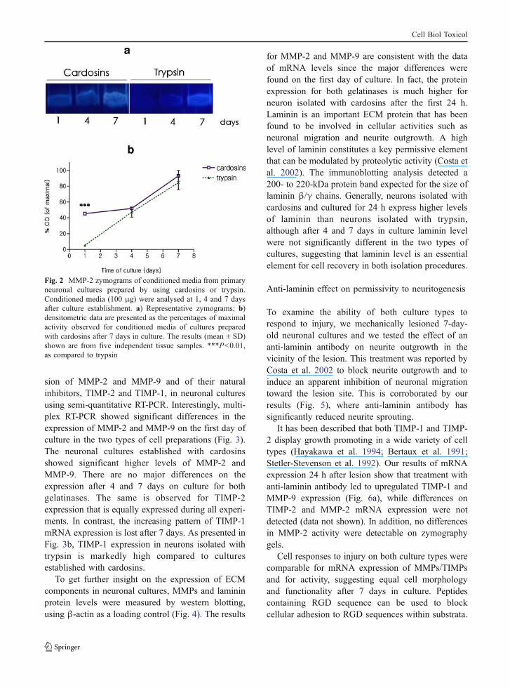
Since neuronal growth and development, in partic-ular neuritogenesis, are closely related with proteo-lytic activity of the ECM, we analysed the activityand expression of MMPs in neuronal cultures pre-pared from embryonic rat cortex. MMPs areexpressed and secreted as inactive precursors thatare activated by the removal of an N-terminalpropeptide (Okada et al. 1988). The latent and activeforms of each enzyme can be differentiated on thebasis of their molecular weights using zymography.We observed that the main gelatinolytic activitypresent in culture media corresponds to the activeform of MMP-2. Figure 2 displays a zymography gelshowing a higher gelatinolytic activity after 24 h inculture in neurons isolated with cardosins in compar-ison with the activity of neurons obtained withtrypsin. On these gels, we did not detect gelatinolyticactivity corresponding to the 92-kDa type IV colla-genase, MMP-9.
In addition to the regulation by zymogene activa-tion, most MMPs are regulated at transcriptionallevel, and their activity is also regulated by specificinhibitors (TIMPs). Hence, we examined the expres-
Fig. 1 Morphological characterisation of cell cultures preparedby cardosins (a) or trypsin (b) dissociation of embryonic braincortex. The cells were cultured for 1 day before fixation andimmunolabelling for MAP-2 (scale bar=50 μM)
Cell Biol Toxicol
sion of MMP-2 and MMP-9 and of their naturalinhibitors, TIMP-2 and TIMP-1, in neuronal culturesusing semi-quantitative RT-PCR. Interestingly, multi-plex RT-PCR showed significant differences in theexpression of MMP-2 and MMP-9 on the first day ofculture in the two types of cell preparations (Fig. 3).The neuronal cultures established with cardosinsshowed significant higher levels of MMP-2 andMMP-9. There are no major differences on theexpression after 4 and 7 days on culture for bothgelatinases. The same is observed for TIMP-2expression that is equally expressed during all experi-ments. In contrast, the increasing pattern of TIMP-1mRNA expression is lost after 7 days. As presented inFig. 3b, TIMP-1 expression in neurons isolated withtrypsin is markedly high compared to culturesestablished with cardosins.
To get further insight on the expression of ECMcomponents in neuronal cultures, MMPs and lamininprotein levels were measured by western blotting,using β-actin as a loading control (Fig. 4). The results
for MMP-2 and MMP-9 are consistent with the dataof mRNA levels since the major differences werefound on the first day of culture. In fact, the proteinexpression for both gelatinases is much higher forneuron isolated with cardosins after the first 24 h.Laminin is an important ECM protein that has beenfound to be involved in cellular activities such asneuronal migration and neurite outgrowth. A highlevel of laminin constitutes a key permissive elementthat can be modulated by proteolytic activity (Costa etal. 2002). The immunoblotting analysis detected a200- to 220-kDa protein band expected for the size oflaminin β/γ chains. Generally, neurons isolated withcardosins and cultured for 24 h express higher levelsof laminin than neurons isolated with trypsin,although after 4 and 7 days in culture laminin levelwere not significantly different in the two types ofcultures, suggesting that laminin level is an essentialelement for cell recovery in both isolation procedures.
Anti-laminin effect on permissivity to neuritogenesis
To examine the ability of both culture types torespond to injury, we mechanically lesioned 7-day-old neuronal cultures and we tested the effect of ananti-laminin antibody on neurite outgrowth in thevicinity of the lesion. This treatment was reported byCosta et al. 2002 to block neurite outgrowth and toinduce an apparent inhibition of neuronal migrationtoward the lesion site. This is corroborated by ourresults (Fig. 5), where anti-laminin antibody hassignificantly reduced neurite sprouting.
It has been described that both TIMP-1 and TIMP-2 display growth promoting in a wide variety of celltypes (Hayakawa et al. 1994; Bertaux et al. 1991;Stetler-Stevenson et al. 1992). Our results of mRNAexpression 24 h after lesion show that treatment withanti-laminin antibody led to upregulated TIMP-1 andMMP-9 expression (Fig. 6a), while differences onTIMP-2 and MMP-2 mRNA expression were notdetected (data not shown). In addition, no differencesin MMP-2 activity were detectable on zymographygels.
Cell responses to injury on both culture types werecomparable for mRNA expression of MMPs/TIMPsand for activity, suggesting equal cell morphologyand functionality after 7 days in culture. Peptidescontaining RGD sequence can be used to blockcellular adhesion to RGD sequences within substrata.
Fig. 2 MMP-2 zymograms of conditioned media from primaryneuronal cultures prepared by using cardosins or trypsin.Conditioned media (100 μg) were analysed at 1, 4 and 7 daysafter culture establishment. a) Representative zymograms; b)densitometric data are presented as the percentages of maximalactivity observed for conditioned media of cultures preparedwith cardosins after 7 days in culture. The results (mean ± SD)shown are from five independent tissue samples. ***P<0.01,as compared to trypsin
Cell Biol Toxicol

Similar to integrins, RGD sequences may be involvednot only in adhesion, but also in the promotion ofneurite outgrowth (Tashiro et al. 1991). Therefore,parallel assays were performed by mechanically
treating injury of neuronal cultures with RGD peptide,instead of anti-laminin antibody. Figure 5 shows thatafter a mechanical injury, RGD effect on neuronalregrowth is similar to the effect observed for the anti-
Fig . 3 Express ion o fMMPs and TIMPs on neu-ronal cultures prepared withcardosins or trypsin. a)mRNA of MMP-2 andTIMP-2; b) mRNA ofMMP-9 and TIMP-1. Thevalues are the ratios of themRNA band densities ofthe target gene/β-actin; c)Representative gel electro-foresis of TIMP-2 mRNAperformed as described under“Material and methods”section; the multiplex RT-PCR produced a 308-basepair TIMP-2 fragment and a490-base pair β-actinfragment
Fig. 4 Levels of laminin,MMP-2 and MMP-9 inextracts of neuronal culturesprepared with cardosins ortrypsin. a) Representativewestern blots; b) quantifica-tion of laminin and MMP-2using β-actin as a loadingcontrol
Cell Biol Toxicol

laminin antibody. Similarly, RGD treatment inducedMMP-9 and TIMP-1 mRNA expression (Fig. 6a) andno additional differences on MMP-2 were detectableby zymography. In addition, an increase of extracel-lular laminin level assessed by western blot is clear(Fig. 6b).
Discussion
Extracellular matrix (ECM) plays a key role inmediating neurite outgrowth following brain tissuedissociation for the establishment of primary neuronalcultures. It is well-known that ECM regeneration isachieved by the effect of MMPs activity on matrix ornon-matrix proteins performing directly or indirectlyon the rebuilding of the new network. Matrix metal-loproteinase (MMP) enzymatic activity plays an
Fig. 6 MMP-9, TIMP-1 and laminin expression in mechanicallesioned neuronal cultures. a) Densitometric results from semi-quantitative RT-PCR analysis (mean ± standard deviation).Effect of anti-laminin addition to conditioned medium onmRNA expression of MMP-9 and TIMP-1. b) Lamininquantification assessed by western blot analysis. Open bars,cultures established with cardosins; patterned bars, neuronalcultures established with trypsin (n=3). **P<0.01, *P<0.05.All the described analysis were performed in parallel on bothculture types (cardosins versus trypsin) showing the sameresults
Fig. 5 Neurite outgrowth evaluation after mechanical injury.a) Representative immunoscytochemistry analysis: a) initiallesion on a 7-day culture; b) 24 h postlesion untreated culture.Observe the neurite outgrowth inside the lesion site comparedwith the control; c) 24 h postlesion treated culture with anti-laminin antibody (1/200); d) 24 h postlesion treated culturewith RGD peptide (500 μM). In c) and d) the reduction ofneurites projecting toward the lesion (scale bar=100 μm) isclear. b) Morphometric analysis of neurite length (μm) inlesioned neuronal cultures (left axis), and of the number ofneurites, extending longer than 5 μm away from the neuronalborder into the lesioned area (right axis), 24 h postinjury. L –lesioned cultures (0 h); UL (24 h) – untreated lesions (control);L + AL (24 h) – treated culture with laminin antibody (1/200);L + RGD – treated culture with RGD peptide (500 μM).Neurite length and number were analysed for differences withone-way analysis of variance (ANOVA), followed by a TukeyHSD multiple comparison test, using a significance level of0.05 for all analyses. Statistically significant differencesbetween treatments are represented by different letters (a, b, c)
Cell Biol Toxicol

important role in brain physiology, possibly consistingof pivotal significance for neuronal plasticity. Thefocus of our investigation consisted of the study of theexpression pattern and enzymatic activity of thesemolecules in neuronal cultures established with cardo-sins, to elucidate the molecular events responsible forthe observed faster neuronal recovery, compared totrypsin. Previous studies by Reeves and colleaguessuggested a potential functional role of MMP-2 inpost-lesion axonal sprouting and neuronal plasticity.They show that differentiation and subsequent func-tional plasticity recovery is correlated to an increasedMMP-2 activity (Reeves et al. 2003). Similarly, duringbrain disaggregation neurons are strained because ofdisruption of connections to the neighbouring cellstriggering a recovery-like cell response (Pérez-Martínezand Jaworski 2005). In the present work we show thatafter 1 day in culture, MMP-2 expression and activitylevels are higher for neurons isolated with cardosinscompared to trypsin. These observations substantiateprevious remarks about early cell recovery as assessedby morphological evaluation.
Moreover, the primary function of TIMPs is toinhibit MMP activity, consequently TIMP-1 and TIMP-2 expression may be upregulated simply in response toincreased MMP expression. TIMP-1 expression isassociated with neuronal plasticity (Dzwonek et al.2004) and neurons isolated with trypsin have a lessprompt recovery to the isolation procedure, in contrastto those isolated with cardosins (Duarte et al. 2007).This may be the reason for the huge TIMP-1 mRNAexpression observed at the fourth day of culture forneurons obtained with trypsin.
It is generally acknowledged that neuronal re-sponse to CNS insults operates by selective enhancingof MMPs activities that promote recovery mecha-nisms and initiate neuroprotective pathways. In thisstudy, the neuronal response to mechanical injury,correlated with MMP-9 and TIMP-1 upregulation.Although it is not yet clear whether MMP-9 isexpressed by the neurons prone to death and/or thoseattempting to recover from the damage, a potentialcorrelation has been proposed between neuronalsurvival, synaptogenesis and MMP-9 (Dzwonek etal. 2004) supporting the presented data.
Matrix metalloproteinase (MMPs) are regulatedafter some types of insult, such as cell isolation ormechanical lesion (Yong et al. 2001). With respect tocell migration, neural cell precursors may require a
rebalance of MMPs activities to migrate to the scarredarea. New axonal growth and synapse formation needto be established and their extension through the brainmatrix may also require a new MMPs/TIMPs ratio.This may be the basis for an increase of MMP-9 andTIMP-1 expression evident in this work. This newbalance must contribute directly or indirectly to theincreased laminin expression, which assists regrowth.
The effects of the pan-laminin antibody and theRGD peptides on the morphology of cortical neuronswere also investigated. The anti-laminin antibodymodulated neuronal morphology in a pattern verysimilar to the effects of RGD sequence: both treat-ments led to a decrease of neurite outgrowth in thescarred area (Fig. 5). Moreover, as shown in Fig. 6b,RGD treatment promoted laminin overexpression.One speculation arising from these observations isthat both types of culture (established with cardosinsor with trypsin) respond to stress/injury conditions vialaminin expression, a well-known mechanism thatfavour permissivity. Finally, these data suggest thatcortical neurons use RGD-dependent mechanisms foradhesion and outgrowth. These mechanisms providepossible sites of modulation by specific growthfactors, as ECM components can act not only aspassive substrates for neuronal attachment and out-growth, but also as active sites for signal transduction.
In conclusion, present results indicate that cellsrespond to isolation procedure by upregulating bothgelatinases, MMP-2 and MMP-9, agreeing with ourprior observation of a faster neuronal outgrowth.After mechanical injury, it was also shown aninhibition of neuronal recovery, as assessed bymorphological analysis, together with an increase ofTIMP-1 and MMP-9 mRNA levels under non-permissive conditions (AL treatment). RGD-containingpeptide had significant influence in the upregulationof TIMP-1 and laminin. Taken together, theseapproaches indicate that the regulation of MMPslevels can help to predict the cellular state, suggestingthat MMPs should be viewed, not only as proteolyticenzymes acting on ECM remodelling but, overall,performing as pruning shears, playing sophisticatedroles in modulating normal cellular behaviour, cell–cell communication and tumor progression. Moreover,the results emphasise the significance of the enzymaticinfluence evaluation on isolation procedures, giventhat different enzymes can produce divergent effects oncell morphology and protein expression by stimulating
Cell Biol Toxicol

or inhibiting cell response. However, after 7 dayscardosins and trypsin produce equivalent cell prepara-tions; cardosins certainly improve neuronal regenera-tion at early stages of culture after cell isolation. Thisfeature might be particularly useful for researchpurposes requiring fast cell manipulation.
Acknowledgements The authors are grateful to Bruno B.Castro, who has been a great help in discussing statistical issuesof proper image analysis. In addition, we would like to thankDr Sukalyan Chatterjee for his comments on the manuscript.This work was supported by a postdoctoral fellowship fromFundação para a Ciência e Tecnologia (FCT), Portugal –SFRH/BPD/25162/2005.
References
Aguayo AJ, David S, Bray GM. Influences of the glial environ-ment on the elongation of axons after injury: transplantationstudies in adult rodents. J Exp Biol. 1981;95:231–40.
Bertaux B, Hornbeck W, Eisen AZ, Dubertret L. Growthstimulation of human keratinocytes by tissue inhibitor ofmetalloproteinases. J Invest Dermatol. 1991;97:679–85.
Brew K, Dinakarpandian D, Nagase H. Tissue inhibitors ofmetalloproteinases: evolution, structure and function.Biochim Biophys Acta. 2000;1477:267–83.
Campos LS. Β1 integrins and neural stem cells: making senseof the extracellular environment. BioEssays. 2005;27(7):698–707.
Costa S, Planchenault T, Charriere-Bertrand C, Mouchel Y,Fages C, Juliano S, Lefrancois T, Barlovatz-Meimon G,Tardy M. Astroglial permissivity for neuritic outgrowth inneuron-astrocyte cocultures depends on regulation oflaminin bioavailability. Glia. 2002;37(2):105–13.
Duarte A, Rosa N, Duarte E, Pires E, Barros M. Cardosins: anew and efficient plant enzymatic tool to dissociateneuronal cells for the establishment of cell cultures.Biotechnol Bioeng. 2007;97(4):991–6.
Duarte AS, Pereira A, Cabrita AS, Moir A, Pires E, Barros M.The characterisation of the collagenolytic activity ofcardosin A demonstrates its potential application forextracellular matrix degradative processes. Curr DrugDiscov Technol. 2005;2(1):37–44.
Dzwonek J, Rylski M, Kaczmarek L. Matrix metalloproteinasesand their endogenous inhibitors in neuronal physiology ofthe adult brain. FEBS Lett. 2004;567(1):129–35. Review.
Fambrough D, Pan D, Rubin GM, Goodman CS. The cellsurface metalloprotease/ disintegrin Kuzbanian is requiredfor axonal extension in Drosophila. Proc Natl Acad Sci US A. 1996;93:13233–8.
Gruenbaum LM, Carew TJ. Growth factor modulation ofsubstrate-specific morphological patterns in Aplysia bagcell neurons. Learn Mem. 1999;6:292–306.
Hayakawa T, Yamashita K, Ohuchi E, Shinagawa A. Cellgrowth-promoting activity of tissue inhibitor of metal-loproteinase-2 (TIMP-2). J Cell Sci. 1994;107:2373–9.
Laemmli UK. Cleavage of structural proteins during theassembly of the head of bacteriophage T4. Nature.1970;227:680–5.
Leppert D, Lindberg RL, Kappos L, Leib SL. Matrix metal-loproteinases: multifunctional effectors of inflammation inmultiple sclerosis and bacterial meningitis. Brain ResBrain Res Rev. 2001;36(2–3):249–57.
Liotta LA, Stetler-Stevenson WG. Metalloproteinases andcancer invasion. Semin Cancer Biol. 1990;1(2):99–106.Review.
Monard D. Cell-derived proteases and protease inhibitors asregulators of neurite outgrowth. Trends Neurosci. 1988;11:541–4.
Morrison B 3rd, Saatman KE, Meaney DF, McIntosh TK. Invitro central nervous system models of mechanically inducedtrauma: a review. J Neurotrauma. 1998;15(11):911–28.
NIH image 2004 [http://rsb.info.nih.gov/nih-image/download.html]
Okada Y, Harris ED Jr, Nagase H. The precursor of ametalloendopeptidase from human rheumatoid synovialfibroblasts. Purification and mechanisms of activation byendopeptidases and 4-aminophenylmercuric acetate. Bio-chem J. 1988;254:731–41.
Pérez-Martínez L, Jaworski DM. Tissue Inhibitor of metal-loproteinase-2 promotes neuronal differentiation by acting asan anti-mitogenic signal. J Neurosci. 2005;25(20):4917–29.
Reeves TM, Prins ML, Zhu J, Povlishock JT, Phillips LL.Matrix metalloproteinase inhibition alters functional andstructural correlates of differentiation-induced sprouting inthe dentate gyrus. J Neurosci. 2003;23(32):10182–9.
Ruoslahti E. RGD and other recognition sequences forintegrins. Annu Rev Cell Dev Biol. 1996;12:697–715.
Sarmento AC, Oliveira CS, Pires EM, Amado F, Barros M.Reverse hydrolysis by cardosin A: specificity consider-ations. J Mol Catal B Enzym. 2004;28:33–7.
Schwartz MA. Integrin signaling revisited. Trends Cell Biol.2001;11(12):466–70. Review.
Schwartz MA, Schaller MD, Ginsberg MH. Integrins: emergingparadigms of signal transduction. Annu Rev Cell DevBiol. 1995;11:549–99.
Stetler-Stevenson WG, Bersch N, Golde DW. Tissue inhibitorof metalloproteinase-2 (TIMP-2) has erythroid-potentiat-ing activity. FEBS Lett. 1992;296:231–4.
Stupack DG. Integrins as a distinct subtype of dependencereceptors. Cell Death Differ. 2005;12(8):1021–30.
Tashiro K, Sephel GC, Greatorex D, Sasaki M, Shirashi N,Martin GR, Kleinman HK, Yamada Y. The RGD contain-ing site of the mouse laminin A chain is active for cellattachment, spreading, migration and neurite outgrowth. JCell Physiol. 1991;146(3):451–9.
Tucker BA, Rahimtula M, Mearow KM. Integrin activation andneurotrophin signaling cooperate to enhance neuriteoutgrowth in sensory neurons. J Comp Neurol. 2005;486(3):267–80.
Yamada KM. Adhesive recognition sequences. J Biol Chem.1991;266:12809–12.
Yong VW, Power C, Forsyth P, Edwards DR. Metalloprotein-ases in biology and pathology of the nervous system. NatRev Neurosci. 2001;2(7):502–11.
Cell Biol Toxicol
All in-text references underlined in blue are linked to publications on ResearchGate, letting you access and read them immediately.
Copyright © 2022 FDOKUMEN