Calcification_in_ostracodes
11
Revista Española de Micropaleontología, 36(1), 2004, pp. 1-11 © Instituto Geológico y Minero de España ISSN: 0556-655X 1 CALCIFICATION IN OSTRACODES D. KEYSER AND R. WALTER Zoologisches Institut und Museum, Martin-Luther-King-Platz 3, D-20146 Hamburg, Germany. E-mail: [email protected] Abstract Ostracodes are often heavily calcified and their shell is mainly built of calcite. Investigations of the carapace reveal a crystalline structure which differs from the structure of chemically precipitated calci- te. The calcium in the epidermal layer is concentrated in small globules which appear concentric when sectioned. Analyses have shown that the main component of these globules is calcium phosphate. When released from the globules, the calcium penetrates the epidermal membrane to form tiny granules of amorphous calcite outside the membrane. These gradually give rise to calcite crystals which are the main constituent of the shell of an ostracode. In some weakly calcified ostracodes such as Cypria ophtalmica the amorphous calcite is rarely transformed into calcite crystallites. Amorphous calcite, which dissolves easily, is usually not substantial enough to fossilise. Therefore, shells of these animals are rare in fossil samples. Keywords: Biomineralisation, Ostracode, amorphous calcite, carapace ultrastructure, SEM. Resumen Los ostrácodos están en general fuertemente calcificados y su concha está básicamente com- puesta por calcita. Investigaciones realizadas sobre el caparazón revelan una estructura cristali- na que difiere de la estructura de la calcita químicamente precipitada. El calcio de la capa epi- dérmica presenta una concentración de pequeños glóbulos que exhiben una estructura interna concéntrica. Los análisis realizados muestran que el componente mayoritario de dichos glóbu- los es el fosfato cálcico. Al liberarse de los glóbulos, el calcio penetra en la membrana epidér- mica para formar minúsculos gránulos de calcita amorfa en el exterior de la membrana. Esto da lugar al crecimiento gradual de cristales de calcita, que son los constituyentes mayoritarios de la concha de un ostrácodo. En algunos ostrácodos débilmente calcificados como Cypria oph- talmica, la calcita amorfa es raramente transformada en cristales de calcita. Esta calcita amor- fa, al disolverse fácilmente, fosiliza raramente. Como consecuencia, las conchas de estos ani- males son raros en muestras fósiles. Palabras clave: Biomineralización, Ostrácodo, calcita amorfa, ultraestructura del caparazón, MEB. INTRODUCTION Ostracodes are small crustaceans encased in a calcified shell. They can be found in a variety of different habi- tats from terrestrial such as wet moss and bromeliads to freshwater and marine environments including the deep sea. But in all cases they are characterised by the possession of a calcified carapace. Even the nauplius, the first instar with its three pairs of appendages, pos- sesses a slightly calcified duplicature of the dorsal cuticle which encases the whole body. The ostracode carapace is shed by moulting up to eight times during development to the adult animal. As in other crustace- ans, the cuticle of the carapace is mineralised with low magnesium calcium carbonate in the form of calcite (Kesling, 1951; Sohn, 1958). This differs from the molluscan shell which contains aragonite. In addition, the calcite is not recycled during moulting as happens
-
Upload
uni-hamburg -
Category
Documents
-
view
1 -
download
0
Transcript of Calcification_in_ostracodes

Revista Española de Micropaleontología, 36(1), 2004, pp. 1-11© Instituto Geológico y Minero de EspañaISSN: 0556-655X
1
CALCIFICATION IN OSTRACODESD. KEYSER AND R. WALTER
Zoologisches Institut und Museum, Martin-Luther-King-Platz 3, D-20146 Hamburg, Germany. E-mail: [email protected]
Abstract
Ostracodes are often heavily calcified and their shell is mainly built of calcite. Investigations of thecarapace reveal a crystalline structure which differs from the structure of chemically precipitated calci-te. The calcium in the epidermal layer is concentrated in small globules which appear concentric whensectioned. Analyses have shown that the main component of these globules is calcium phosphate. Whenreleased from the globules, the calcium penetrates the epidermal membrane to form tiny granules ofamorphous calcite outside the membrane. These gradually give rise to calcite crystals which are the mainconstituent of the shell of an ostracode. In some weakly calcified ostracodes such as Cypria ophtalmicathe amorphous calcite is rarely transformed into calcite crystallites. Amorphous calcite, which dissolveseasily, is usually not substantial enough to fossilise. Therefore, shells of these animals are rare in fossilsamples.
Keywords: Biomineralisation, Ostracode, amorphous calcite, carapace ultrastructure, SEM.
Resumen
Los ostrácodos están en general fuertemente calcificados y su concha está básicamente com-puesta por calcita. Investigaciones realizadas sobre el caparazón revelan una estructura cristali-na que difiere de la estructura de la calcita químicamente precipitada. El calcio de la capa epi-dérmica presenta una concentración de pequeños glóbulos que exhiben una estructura internaconcéntrica. Los análisis realizados muestran que el componente mayoritario de dichos glóbu-los es el fosfato cálcico. Al liberarse de los glóbulos, el calcio penetra en la membrana epidér-mica para formar minúsculos gránulos de calcita amorfa en el exterior de la membrana. Esto dalugar al crecimiento gradual de cristales de calcita, que son los constituyentes mayoritarios dela concha de un ostrácodo. En algunos ostrácodos débilmente calcificados como Cypria oph-talmica, la calcita amorfa es raramente transformada en cristales de calcita. Esta calcita amor-fa, al disolverse fácilmente, fosiliza raramente. Como consecuencia, las conchas de estos ani-males son raros en muestras fósiles.
Palabras clave: Biomineralización, Ostrácodo, calcita amorfa, ultraestructura del caparazón,MEB.
INTRODUCTION
Ostracodes are small crustaceans encased in a calcifiedshell. They can be found in a variety of different habi-tats from terrestrial such as wet moss and bromeliadsto freshwater and marine environments including thedeep sea. But in all cases they are characterised by thepossession of a calcified carapace. Even the nauplius,the first instar with its three pairs of appendages, pos-
sesses a slightly calcified duplicature of the dorsalcuticle which encases the whole body. The ostracodecarapace is shed by moulting up to eight times duringdevelopment to the adult animal. As in other crustace-ans, the cuticle of the carapace is mineralised with lowmagnesium calcium carbonate in the form of calcite(Kesling, 1951; Sohn, 1958). This differs from themolluscan shell which contains aragonite. In addition,the calcite is not recycled during moulting as happens
in many malacostracean crustaceans, but is discardedwith the old carapace and thus has to be formedagain during calcification of the new shell (Turpenand Angel, 1971).
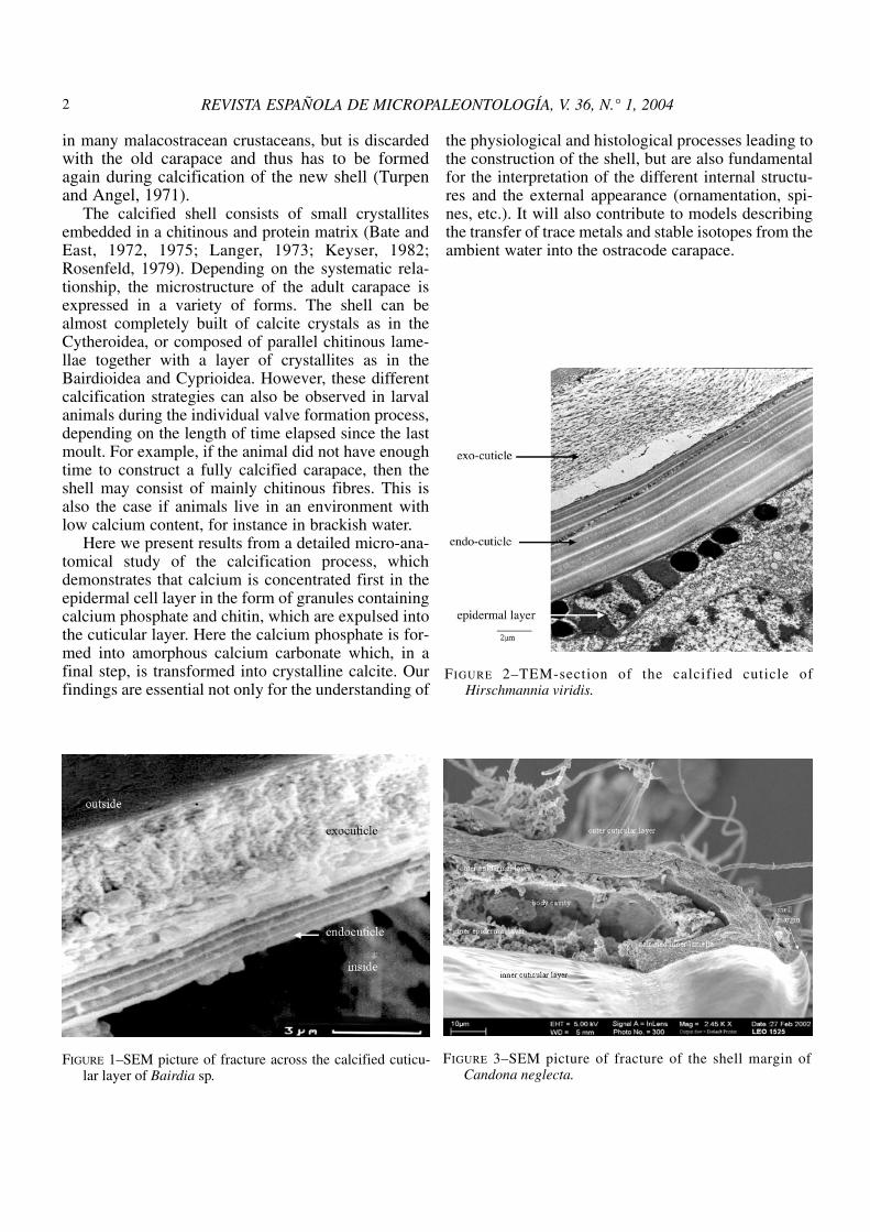
The calcified shell consists of small crystallitesembedded in a chitinous and protein matrix (Bate andEast, 1972, 1975; Langer, 1973; Keyser, 1982;Rosenfeld, 1979). Depending on the systematic rela-tionship, the microstructure of the adult carapace isexpressed in a variety of forms. The shell can bealmost completely built of calcite crystals as in theCytheroidea, or composed of parallel chitinous lame-llae together with a layer of crystallites as in theBairdioidea and Cyprioidea. However, these differentcalcification strategies can also be observed in larvalanimals during the individual valve formation process,depending on the length of time elapsed since the lastmoult. For example, if the animal did not have enoughtime to construct a fully calcified carapace, then theshell may consist of mainly chitinous fibres. This isalso the case if animals live in an environment withlow calcium content, for instance in brackish water.
Here we present results from a detailed micro-ana-tomical study of the calcification process, whichdemonstrates that calcium is concentrated first in theepidermal cell layer in the form of granules containingcalcium phosphate and chitin, which are expulsed intothe cuticular layer. Here the calcium phosphate is for-med into amorphous calcium carbonate which, in afinal step, is transformed into crystalline calcite. Ourfindings are essential not only for the understanding of
the physiological and histological processes leading tothe construction of the shell, but are also fundamentalfor the interpretation of the different internal structu-res and the external appearance (ornamentation, spi-nes, etc.). It will also contribute to models describingthe transfer of trace metals and stable isotopes from theambient water into the ostracode carapace.
REVISTA ESPAÑOLA DE MICROPALEONTOLOGÍA, V. 36, N.° 1, 20042
FIGURE 3–SEM picture of fracture of the shell margin ofCandona neglecta.
FIGURE 1–SEM picture of fracture across the calcified cuticu-lar layer of Bairdia sp.
FIGURE 2–TEM-section of the calcified cuticle ofHirschmannia viridis.

KEYSER-WALTER — CALIFICATION IN OSTRACODES 3
MATERIAL AND METHODS
Ostracodes of the species: Bairdia sp., Boreostomavariabile (Baird, 1835), Hirschmannia viridis (Müller,1785), Cyprideis torosa (Jones, 1950), Candona neglecta(Sars, 1887), Cypria ophtalmica (Jurine, 1820) andHeterocypris salina (Brady, 1868) have been investigated.They were chosen on the basis of their preferred biotope,representing marine, brackish, saline and fresh water envi-ronments.
The animals were either taken directly from their bioto-pe or were kept in aquaria with original water prior to study.
For SEM and TEM investigations the animals werefixed in 3% glutardialdehyde in 0.05% phosphate buffer forone to two hours, followed by three 15-minute washings inbuffer alone. In addition, fixation with 2% osmium-tetra-oxide in the same buffer was performed. Animals were thendehydrated in a graded series of ethanol from 30% to 100%in 10% steps.
At this point the animals for SEM were critical pointdried in a Balzers CPT Dryer with CO2. The animals werebroken in the dried stage with needles or with razor blades.After a shading procedure with carbon in an evaporationunit PD170AZ from Leybold-Heraeus the shells wereobserved and photographed in a LEO 1525 SEM equippedwith an EDAX energy spectrometer.
TEM samples were decalcified in a drop ofAcetylchloride in 100% acetone (10 ml), embedded inSpurr’s resin and cured for two days at 60ºC. Sectionswere cut on a Reichert-Jung Ultracut E. After staining inuranyl-acetate and lead-citrate the sections were viewedin a Zeiss TEM 902 with an electron-filtering-system.
In some cases ostracodes were frozen alive in cold pro-pane at -150ºC. The frozen animals were cut in the UltracutE equipped with a freeze sectioning unit FC 4E manufactu-red by Reichert-Jung. After that they were kept in 100%acetone at -80ºC for four days and then slowly warmed toroom temperature with an extra change of 100% acetone.They were dried in the CPT dryer, carbon coated and vie-wed in the Leo 1525.
FIGURE 4–SEM picture of fracture through the shell ofCyprideis torosa, showing the cells of the outer and innerepidermal layer. The outer calcified layer is removed, rem-nants of the chitin sheets are still visible.
FIGURE 5–Cyprideis torosa: SEM picture of the top of outerepidermal cells just after moulting. (Calcified layer remo-ved.)
FIGURE 6–SEM picture of fracture of a shell of a moultingHeterocypris salina with calcified layer removed.
Chemical analyses were performed on the bulk animalsin the SEM with the help of the energy dispersive microanalysing system EDAX with the software Falcon. Electronspectroscopic imaging (ESI) was performed on sections inthe TEM.
RESULTS
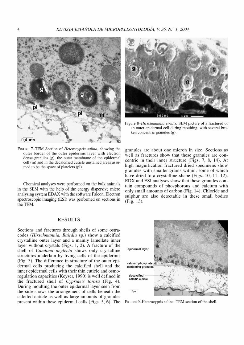
Sections and fractures through shells of some ostra-codes (Hirschmannia, Bairdia sp.) show a calcifiedcrystalline outer layer and a mainly lamellate innerlayer without crystals (Figs. 1, 2). A fracture of theshell of Candona neglecta shows only crystallinestructures underlain by living cells of the epidermis(Fig. 3). The difference in structure of the outer epi-dermal cells producing the calcified shell and theinner epidermal cells with their thin cuticle and osmo-regulation capacities (Keyser, 1990) is well defined inthe fractured shell of Cyprideis torosa (Fig. 4).During moulting the outer epidermal layer seen fromthe side shows the arrangement of cells beneath thecalcifed cuticle as well as large amounts of granulespresent within these epidermal cells (Figs. 5, 6). The
granules are about one micron in size. Sections aswell as fractures show that these granules are con-centric in their inner structure (Figs. 7, 8, 14). Athigh magnification fractured dried specimens showgranules with smaller grains within, some of whichhave dried to a crystalline shape (Figs. 10, 11, 12).EDX and ESI analyses show that these granules con-tain compounds of phosphorous and calcium withonly small amounts of carbon (Fig. 14). Chloride andsulphur are also detectable in these small bodies(Fig. 13).
REVISTA ESPAÑOLA DE MICROPALEONTOLOGÍA, V. 36, N.° 1, 20044
FIGURE 7–TEM Section of Heterocypris salina, showing theouter border of the outer epidermis layer with electrondense granules (g), the outer membrane of the epidermalcell (m) and in the decalcified cuticle unstained areas assu-med to be the space of platelets (pl).
Figure 8–Hirschmannia viridis: SEM picture of a fractured ofan outer epidermal cell during moulting, with several bro-ken concentric granules (g).
FIGURE 9–Heterocypris salina: TEM section of the shell.

Fig. 15 shows what might be the transition of thecontents of two granules across the cell membrane intothe calcified shell. Between the sectioned granule andthe outer membrane is an electron dense area which isalso visible outside the membrane and which appearssimilar to parts of the decalcified areas in the sameregion.
At high magnification fractures of epidermal cellsshow the outer structure of intact granules covered with
short fibres (Figs. 16, 17). In some cases these granulesseem to release a homogenous substance (Fig. 17).Outside the external cell membrane platelets are detec-table, which, from their size and fine surface structure,appear to be derived directly from the material in thegranules of the epidermal cells (Figs. 18, 19). A similarfine structure is also present on the surface of eachcrystal in the calcified shell (Fig. 20). Chemical analy-ses reveal that these crystals have a calciting origin
KEYSER-WALTER — CALIFICATION IN OSTRACODES 5
FIGURE 10–SEM picture of broken epidermal layer of a moul-ting Heterocypris salina showing the open granules (g)and some crystals within (c).
FIGURE 11–SEM picture of broken epidermal granules of amoulting Heterocypris salina with rounded crystals (c).
FIGURE 12–SEM picture of dried crystal (c) in the epidermalgranule in Heterocypris salina resembling a macroscopicocto-calcium-phosphate crystal
FIGURE 13–Heterocypris salina: EDX-Analysis of a granule inthe outer epidermal cell.

REVISTA ESPAÑOLA DE MICROPALEONTOLOGÍA, V. 36, N.° 1, 20046
Figure 15–Heterocypris salina: TEM section through the bor-der of the outer epidermal cells with the calcified layershowing granules and part of the platelets (pl). (Pleasenote the electron dense area between the calcified cuticleand the granule, bridging the cell membrane area).
FIGURE 16–SEM picture of granules (g) of the cells in theouter epidermal layer of Heterocypris salina.
FIGURE 14–ESI-Analysis of a granule in Cypria ophtalmica.

(Fig. 21). However, their appearance differs from thatof mineral calcite which has a smooth surface wit-hout fine structures (Fig. 23). The transition from theplatelets just outside the epidermal cell membrane tothe final crystalline arrangement is achieved by atemporary formation of very small (<30 nm) calciticgranules (Figs. 19, 22).
In Boreostoma variabile the crystals adjacent tothe epidermal cells are very small and show fine
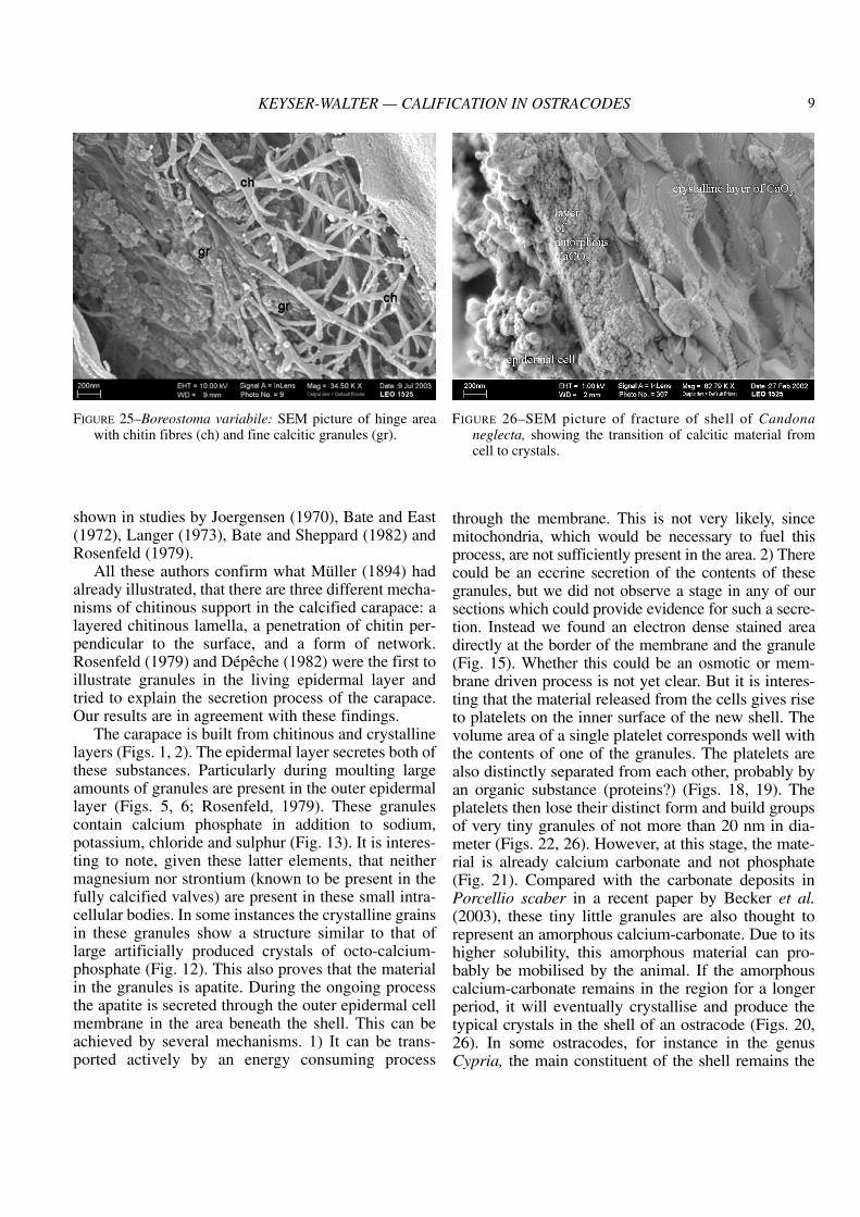
granular features on the side facing the epidermalcells (Fig. 24). In the hinge region in Boreostomathis fine granular substance is present between thechitinous fibres which are the main component inthis region (Fig. 25). The size of these granules isabout 20 to 30 nm. Small granular substances arealso present just above the cell membrane of theouter epidermal layer in Candona (Fig. 26),Cyprideis and Cypria.
KEYSER-WALTER — CALIFICATION IN OSTRACODES 7
FIGURE 17–SEM picture of calcification granules (g) of theouter epidermal layer of Heterocypris salina with chitinfibres and the presumably liquid substance (lq) emergingfrom the granule.
FIGURE 18–SEM picture of inside view of outer calcified cuti-cle with platelets (pl) of granule material in Heterocyprissalina
FIGURE 19–SEM picture of surface of the inside of the calci-fied outer cuticle, showing platelets (pl) and parallel chitinfibres (st) in Heterocypris salina.
FIGURE 20–SEM picture of calcite crystals (c) in the calcifiedcuticle of the shell of Heterocypris salina.

DISCUSSION
The calcified shell of ostracodes has been studiedby many authors. G.W.Müller (1894) separated thechitinous layer from the outer calcified layer and foundseveral lamellar striated chitinous shells in myodoco-pids. Schreiber (1922) mentioned an inner chitinouslayer and an outer calcified layer. Kesling (1951)determined the crystalline structure in the shell as cal-cite. Sohn (1958) reported the different chemical cons-tituents within the shell. He cited Kinser who found
12,8 % protein, 2,2% chitin, 82,7% calcium carbonateand 1,9% trace elements in Chlamydotheca. Sohn(1958) found 80-90% calcium carbonate and 2-15%organic material together with many trace elementssuch as K, Mg, Si, Al, Sr and Ba. Hartmann (1966)cited Dudich (1931), Zalanyi (1944) and Kesling(1951) who showed that the main mineral in ostracodeshells is calcite and not aragonite as in molluscs.
Hartmann’s (1966) explanation regarding the orga-nic support of the calcified carapace was based mainlyon work done by Fassbinder (1912) and is incorrect, as
REVISTA ESPAÑOLA DE MICROPALEONTOLOGÍA, V. 36, N.° 1, 20048
FIGURE 21–Heterocypris salina: EDX-Analysis of the fractureface of crystal.
FIGURE 22–SEM picture of transition of platelet material to thecalcitic shell cuticle in Heterocypris salina.
FIGURE 24–Boreostoma variabile: SEM inside view of the cal-citic layer with the epidermal cells removed.(Please notethe extremely fine globules on the crystals).
FIGURE 23–SEM picture of calcite crystals from the EuropeanAlps.
shown in studies by Joergensen (1970), Bate and East(1972), Langer (1973), Bate and Sheppard (1982) andRosenfeld (1979).
All these authors confirm what Müller (1894) hadalready illustrated, that there are three different mecha-nisms of chitinous support in the calcified carapace: alayered chitinous lamella, a penetration of chitin per-pendicular to the surface, and a form of network.Rosenfeld (1979) and Dépêche (1982) were the first toillustrate granules in the living epidermal layer andtried to explain the secretion process of the carapace.Our results are in agreement with these findings.
The carapace is built from chitinous and crystallinelayers (Figs. 1, 2). The epidermal layer secretes both ofthese substances. Particularly during moulting largeamounts of granules are present in the outer epidermallayer (Figs. 5, 6; Rosenfeld, 1979). These granulescontain calcium phosphate in addition to sodium,potassium, chloride and sulphur (Fig. 13). It is interes-ting to note, given these latter elements, that neithermagnesium nor strontium (known to be present in thefully calcified valves) are present in these small intra-cellular bodies. In some instances the crystalline grainsin these granules show a structure similar to that oflarge artificially produced crystals of octo-calcium-phosphate (Fig. 12). This also proves that the materialin the granules is apatite. During the ongoing processthe apatite is secreted through the outer epidermal cellmembrane in the area beneath the shell. This can beachieved by several mechanisms. 1) It can be trans-ported actively by an energy consuming process
through the membrane. This is not very likely, sincemitochondria, which would be necessary to fuel thisprocess, are not sufficiently present in the area. 2) Therecould be an eccrine secretion of the contents of thesegranules, but we did not observe a stage in any of oursections which could provide evidence for such a secre-tion. Instead we found an electron dense stained areadirectly at the border of the membrane and the granule(Fig. 15). Whether this could be an osmotic or mem-brane driven process is not yet clear. But it is interes-ting that the material released from the cells gives riseto platelets on the inner surface of the new shell. Thevolume area of a single platelet corresponds well withthe contents of one of the granules. The platelets arealso distinctly separated from each other, probably byan organic substance (proteins?) (Figs. 18, 19). Theplatelets then lose their distinct form and build groupsof very tiny granules of not more than 20 nm in dia-meter (Figs. 22, 26). However, at this stage, the mate-rial is already calcium carbonate and not phosphate(Fig. 21). Compared with the carbonate deposits inPorcellio scaber in a recent paper by Becker et al.(2003), these tiny little granules are also thought torepresent an amorphous calcium-carbonate. Due to itshigher solubility, this amorphous material can pro-bably be mobilised by the animal. If the amorphouscalcium-carbonate remains in the region for a longerperiod, it will eventually crystallise and produce thetypical crystals in the shell of an ostracode (Figs. 20,26). In some ostracodes, for instance in the genusCypria, the main constituent of the shell remains the
KEYSER-WALTER — CALIFICATION IN OSTRACODES 9
FIGURE 25–Boreostoma variabile: SEM picture of hinge areawith chitin fibres (ch) and fine calcitic granules (gr).
FIGURE 26–SEM picture of fracture of shell of Candonaneglecta, showing the transition of calcitic material fromcell to crystals.

amorphous material (as in Fig. 25), so that these ani-mals only rarely become fossilised. In shells of adultspecimens of other genera all the amorphous materialhas already crystallised and so no amorphous material is left. In the larval stages on the other handcrystallisation is not complete and the animals haveweaker shells.
Summing up, prior to moulting the ostracodes beginproducing shells by storing a huge amount of calciumphosphate granules together with chitin precursors inthe outer epidermal cells. These granules release theircontents into the extra-cellular space directly outside theepidermal cells. This material is transformed into smallplatelets, each about the size of one granule. The plate-lets are no longer made of calcium phosphate but of cal-cium carbonate. These platelets disintegrate into smallgranular structures, which appear to be amorphous cal-cite. This granular substance then forms the crystals,which, in connection with chitin and proteins, build theshell of the ostracode. These statements are true for allinvestigated animals whether they are freshwater ormarine organisms.
Several questions regarding the secretion of the cal-cified cuticle in ostracodes remain:
1. How are the contents of the intracellular granulesreleased to the outside?
2. Where and when are the environmentally inducedtrace elements such as Mg, Sr, etc., incorporated inthe shell crystals? Can the higher abundance ofnano-granular amorphous calcium carbonateexplain the usually higher Mg-concentration injuvenile shell material?
3. Is there an acidic micro milieu area present in thecrystallisation process as suggested by Keatings(2002) and von Grafenstein (EOM V, abstract) toexplain the positive O18-offset of most ostracodevalve carbonate compared to a theoretical calcitein isotopic equilibrium?
These points must be discussed and investigated bea-ring in mind the observation of Turpen and Angel(1971) that all the calcium secreted by the animal origi-nates from the surrounding water and not from a stora-ge within the animal. Therefore, another important stepinvolved in the ostracode calcification process, thetransport of those large amounts of calcium into the epi-dermal cells – has to be investigated.
ACKNOWLEDGEMENTS
We are indebted to our colleagues on the EOM Vfor the very interesting and stimulating discussions
which helped to clarify some of our opinions. For therevision of the manuscript we express our thanks toMrs. Carol Schöning, Hamburg and the referee of themanuscript (Ulli von Grafenstein), who made veryhelpful suggestions.
REFERENCES
Bate, R. H., and East, B. A. 1972. The structure of the ostra-code carapace. Lethaia, 5, 177-194.
—. 1975. The ultrastructure of the Ostracode (Crustacea) inte-gument. Bulletin of American Paleontology, 65, 529-548.
Bate, R. H., and Sheppard, L. M. 1982. The shell structure ofHalocypris inflata (Dana, 1849). Fossil and RecentOstracods (eds. R. H. Bate, E. Robinson, and L. M.Sheppard), 25-50. Ellis Horwood Limited, Chichester.
Becker, A.; Bismayer, U.; Epple, M.; Fabritius, H.; Hasse, B.;Shi, Jianmin, and Ziegler, A. 2003. Structural characteri-sation of X-ray amorphous calcium carbonate (ACC) inexternal deposits of the crustacea Porcellio scaber. DaltonTrans. (Royal Soc. Chemistry), 551-555.
Depeche, F. 1982. Ultrastructure of the wall of two livingostracods, Herpetocypris chevreuxi (Sars) andPontocythere elongata (Brady), in comparison with fos-sil ostracods from the Middle Jurassic of Normandy.Fossil and Recent Ostracods (eds R. H. Bate, E.Robinson and L. M. Sheppard), 61-74. Ellis HorwoodLimited, Chichester.
Dudich, E. 1931. Systematische und biologischeUntersuchungen über Kalke in lagerungen desCrustaceenpanzers in polarisiertem Licht. Zoologica, 30,1-154.
Fassb inder, K. 1912 . Bei t räge zur Kenntn is derSüßwasserostracoden. Zool. Jahrb. 32, 533-576.
Hartmann, G. 1966. Ostracoda. Klassen und Ordnungen desTierreiches 5:Arthropoda, Abt. 1: Crustacea. 2. Buch, 4.Teil, Ostracoda: 1. Lieferung (ed. Bronn), 1-216.
Jorgensen, N.O. 1970. Ultrastructure of some ostracods.Bulletin of the Geological Society of Denmark; 20, 79-92.
Keatings K. W.; Heaton T. H. E.; and Holmes J.A. 2002.Carbon and oxygen isotope fractionation in non-marineostracods: Results from a ‘natural culture’ environment.Geochimica et Cosmochimica Acta 66, no. 10, p. 1701-1711
Kesling, R. V. 1951. Morphology of ostracod molt stages.Illinois biol. Monogr. 21, 1-126.
Keyser, D. 1982. Development of the sieve pores inHirschmannia viridis (O. F. Müller, 1785). Fossil andRecent Ostracods (eds. R. E. Bate, E. Robinson and L. M.Sheppard), 51-60. Ellis Horwood Ltd.
—. 1990. Morphological changes and function of the innerlamellar layer of podocopid Ostracoda (Crustacea).Proceedings of the 10th international Symposium onOstracoda: Ostracoda and Global Events (ed R. C.
REVISTA ESPAÑOLA DE MICROPALEONTOLOGÍA, V. 36, N.° 1, 200410

Whatley & C. Maybury), 401-410. Chapman & Hall,London.
Langer, W. 1973. Zur Ultrastruktur, Mikromorphologie undTaxonomie des Ostracoda- Carapax. PaleontographicaAbt. A, 144, 1-54.
Müller, G. W. 1894. Die Ostracoden des Golfes von Neapelund der angrenzenden Meeresabschnitte. Fauna undFlora Golf von Neapel, 21, 1-403.
Rosenfeld, A. 1979. Structure and secretion of the carapacein some living ostracodes. Lethaia, 12, 353-379.
Schreiber, E. 1922. Beiträge zur Kenntnis der Morphologie,Entwicklung und Lebensweise der Süsswasser-Ostracoden. Zool. Jahrbuch (Anatomie und Ontogenieder Tiere) 43, 485-538.
Sohn, I. G. 1958. Chemical constituents of Ostracodes someapplications to paleontology and paleoecology. Journalof Paleontology, 4, 730-736.
Turpen, J. B., and Angell, R. W. 1971. Aspects of moltingand calcification in the ostracod Heterocypris. TheBiological Bulletin, 140, 331-338.
Zalanyi, B. 1944. Neogene Ostracoden aus Ungarn. 1. Teil.Geol. Hungar. (Paleont.), 21, 1-183.
MANUSCRITO RECIBIDO: 12 noviembre, 2003MANUSCRITO ACEPTADO: 19 enero, 2004
KEYSER-WALTER — CALIFICATION IN OSTRACODES 11