Plant extracellular ATP signalling by plasma membrane NADPH oxidase and Ca 2+ channels
Upload
independentCategory
view
0download
0
pubs.acs.org/Biochemistry Published on Web 11/24/2009 r 2009 American Chemical Society
12062 Biochemistry 2009, 48, 12062–12080
DOI: 10.1021/bi901739t
Ca2þ Channels on the Move†
Colin W. Taylor,* David L. Prole, and Taufiq Rahman
Department of Pharmacology, University of Cambridge, Cambridge CB2 1PD, U.K.
Received October 8, 2009; Revised Manuscript Received November 24, 2009
ABSTRACT: The versatility of Ca2þ as an intracellular messenger derives largely from the spatial organizationof cytosolic Ca2þ signals, most of which are generated by regulated openings of Ca2þ-permeable channels.Most Ca2þ channels are expressed in the plasma membrane (PM). Others, including the almost ubiquitousinositol 1,4,5-trisphosphate receptors (IP3R) and their relatives, the ryanodine receptors (RyR), arepredominantly expressed in membranes of the sarcoplasmic or endoplasmic reticulum (ER). Targeting ofthese channels to appropriate destinations underpins their ability to generate spatially organized Ca2þ signals.All Ca2þ channels begin life in the cytosol, and the vast majority are then functionally assembled in the ER,where they may either remain or be dispatched to other membranes. Here, by means of selective examples, wereview two issues related to this trafficking of Ca2þ channels via the ER. How do cells avoid wayward activityof Ca2þ channels in transit as they pass from the ER via other membranes to their final destination? Howand why do some cells express small numbers of the archetypal intracellular Ca2þ channels, IP3R and RyR, inthe PM?
LOCAL AND GLOBAL CA2þ SIGNALS
The plasma membrane (PM)1 and the membranes of manyintracellular organelles separate the cytosol, with its low restingfree Ca2þ concentration of ∼100 nM, from environments withvery different free Ca2þ concentrations and electrical potentials.The resulting steep Ca2þ gradients are poised to allow Ca2þ toflow rapidly down its electrochemical gradient whenever Ca2þ-permeable channels within these membranes are open. For thePM and most organelles, notably the endoplasmic reticulum(ER) and organelles derived from it, the gradients are directedtoward the cytosol. Regulated opening of Ca2þ channels withinthese membranes is the means by which most extracellular andintracellular signals evoke the increases in cytosolic Ca2þ con-centration that regulate almost every aspect of cellular activity (1,2). For other membranes, the inner membrane of mitochondriaand perhaps of chloroplasts (3), the Ca2þ gradients are directedaway from the cytosol so that channels within these membranes,MiCawithin the innermitochondrial membrane, for example (4),mediate uptake of Ca2þ from the cytosol. Each of these mem-branes is also home to proteins that transport Ca2þ in the
opposite direction, against its electrochemical gradient, theCa2þ-ATPases of the PM (PMCA), ER (SERCA), and the Golgiand secretory vesicles (SPCA) (5-7), for example. The competingactivities of Ca2þ channels and the pumps and exchangers thatmove Ca2þ up its electrochemical gradient ultimately determinethe cytosolic Ca2þ concentration, but it is the Ca2þ-permeablechannels that mediate the most rapid Ca2þ exchanges and whichare most commonly acutely regulated by signaling pathways.
Each of these Ca2þ-permeable channels allows Ca2þ to passthrough a central pore traversing a biological membrane. Theydiffer, however, in whether under physiological conditions theyeffectively allow only Ca2þ to pass [e.g., voltage-gated Ca2þ
channels (Cav) and the Orai proteins that mediate store-operatedCa2þ entry] or also allow other cations to pass [e.g., IP3 receptors(IP3R), ryanodine receptors (RyR), and nicotinic acetylcholinereceptors]. The difference, defined by the structure of theselectivity filter (8), is significant because it determines whetherthe channels, in addition to mediating Ca2þ fluxes, can alsoregulate membrane potential (most important at the PM) andconduct the counterions required to allow electrogenicmovementof Ca2þ (9).
Because the cytosol of all cells contains high concentrations ofCa2þ buffers, Ca2þ diffuses more slowly in cytosol than in freesolution (10, 11). An important consequence is that as Ca2þ flowsrapidly through an open Ca2þ channel, it creates a local cloudwith a high cytosolic Ca2þ concentration: each active channelcreates its own local Ca2þ signal (12). These spatially organizedCa2þ signals are important because different Ca2þ-bindingproteins selectively associate with different Ca2þ channels, sothat Ca2þ passing through one channel may regulate differentevents to Ca2þ passing through another (2). Store-operated Ca2þ
entry (SOCE) (13), for example, has been reported to regulateselectively the Ca2þ-sensitive adenylyl cyclases (14), nitric oxidesynthase (15), and gene expression in rat basophilic leukemiacells (16). Cardiac IP3R associate with a Ca2þ-regulated proteinkinase that also regulates IP3R activity [Ca2þ-calmodulin-dependent
†Supported by grants from the Wellcome Trust [085295], and theBiotechnology and Biological Sciences Research Council, U.K.*To whom correspondence should be addressed: Department of
Pharmacology, Tennis Court Road, Cambridge CB2 1PD, U.K.E-mail: [email protected]. Telephone: þ44 1223 334058. Fax: þ441223 334100.
1Abbreviations: AKAP, A-kinase-anchoring protein; BCR, B-cellreceptor; CaMKII, Ca2þ-calmodulin-dependent protein kinase II;CaR, Ca2þ-sensing receptor; Cav, voltage-gated Ca2þ (channel);4CmC, 4-chloro-m-cresol; CNG, cyclic nucleotide-gated (channel);ER, endoplasmic reticulum; ERAD, ER-associated degradation; γ,single-channel conductance; IP3, inositol 1,4,5-trisphosphate; IP3R,IP3 receptor(s); PIP2, phosphatidylinositol 4,5-bisphosphate; PKA,cyclic AMP-dependent protein kinase; PM, plasma membrane; PMCA,plasmamembrane Ca2þ-ATPase; RyR, ryanodine receptor(s); SERCA,SR/ER Ca2þ-ATPase; SPCA, secretory pathway Ca2þ-ATPase; SOCE,store-operated Ca2þ entry; SR, sarcoplasmic reticulum; SRP, signalrecognition particle; STIM, stromal interaction protein; TGN, trans-Golgi network; TRP, transient receptor potential.
Current Topic/Perspective Biochemistry, Vol. 48, No. 51, 2009 12063
protein kinase IIδ (CaMKIIδ)], so that release of Ca2þ via IP3Rmay selectively activate an enzyme that then feeds back to inhibitIP3R activity (17). Ca2þ entry via Cav channels (Cav1 and Cav2)selectively regulates the activity of the channel itself (18). Thelatter highlights another key feature of many Ca2þ channels,namely their regulation by cytosolic Ca2þ. This provides feed-back regulation of Ca2þ signaling, and it allows Ca2þ channels toevoke regenerative Ca2þ signals (19). The latter are importantbecause they underpin the versatility of Ca2þ as an intracellularmessenger, permitting it to function either locally or globally (2).It follows from this discussion that the versatility of Ca2þ as aubiquitous intracellular messenger derives in large part fromputting Ca2þ-permeable channels into the right place; only thencan they deliver spatially organized Ca2þ signals.
PUTTINGCA2þCHANNELS INTHERIGHTPLACE
A few proteins, mostly those related to transcription andtranslation, but also components of a Hþ channel (ATPsynthase), are encoded by DNA within mitochondria andchloroplasts. These proteins are synthesized within these orga-nelles and so reach their final destination without traversing thecytoplasm. All other proteins are encoded by nuclear DNA.These proteins are synthesized in the cytosol fromwhere they aredispatched to other destinations. Targeting of proteins hasattracted the most attention (20, 21), but many proteins areguided toward their final destination before translation begins byselective targeting of mRNA (22).
After transcription and mRNA processing within the nucleus,mature mRNA is exported via nuclear pores to the cytosol. Eventhis step may contribute to mRNA targeting. Synthesis ofmRNA encoding some subunits of nicotinic acetylcholine recep-tors, for example, is restricted to the nuclei lying immediatelybeneath the neuromuscular junction of multinucleate musclefibers (23). More generally, transport of mRNA within thecytosol, mediated by binding of proteins to sequences withinthe 30-untranslated region, probably plays the major role inselective trafficking of mRNA (22, 24, 25). Such transport candirect mRNA to associate with the membranes of specificorganelles. The mRNAs for approximately half of all mitochon-drial proteins, for example, including the mRNA for anothersubunit of the ATP synthase, are directed to the outer mitochon-drial membrane (25). Other mRNAs are selectively addressed tonuclearmembranes, the ER (26), and even to specific subdomainsof the ER (27). Transport ofmRNAalso occurs overmuch largerdistances, allowing its selective targeting to specific cytosolicdestinations such as dendrites (24), immature axons (28), andpolarized regions of developing embryos (22). The mRNAs forIP3R1 (29), glutamate receptors, and a major neuronal Ca2þ
sensor (CaMKIIR) are transported to dendrites by microtubuleswithin “mRNA granules” that include ribosomes and additionalproteins. Within these “ready-to-translate” granules (27), trans-lation is probably repressed until the complex reaches its destina-tion, where protein synthesis can proceed close to the finaldestination (24, 28). The advantages of selectively transportingmRNA, rather than protein, have been much discussed(22, 30, 31). Clearly, mRNA transport can complement theadditional targeting provided by proteins, but other advantagesinclude cost (cheaper to move mRNA than all the proteins towhich it will give rise), restricting production of potentially toxicproteins to their target site, speeding the rate at which proteinsynthesis can adapt to local needs (e.g., within postsynaptic
structures) (24), facilitating cotranslational assembly of multi-subunit proteins, and establishing protein gradients, notablyduring development. For Ca2þ channels destined to function inmembranes derived from the ER, mRNA targeting may alsoprovide a means of ensuring that functional channels areassembled near their final destination and so spend less timeen route via organelles within which they may function inappro-priately.
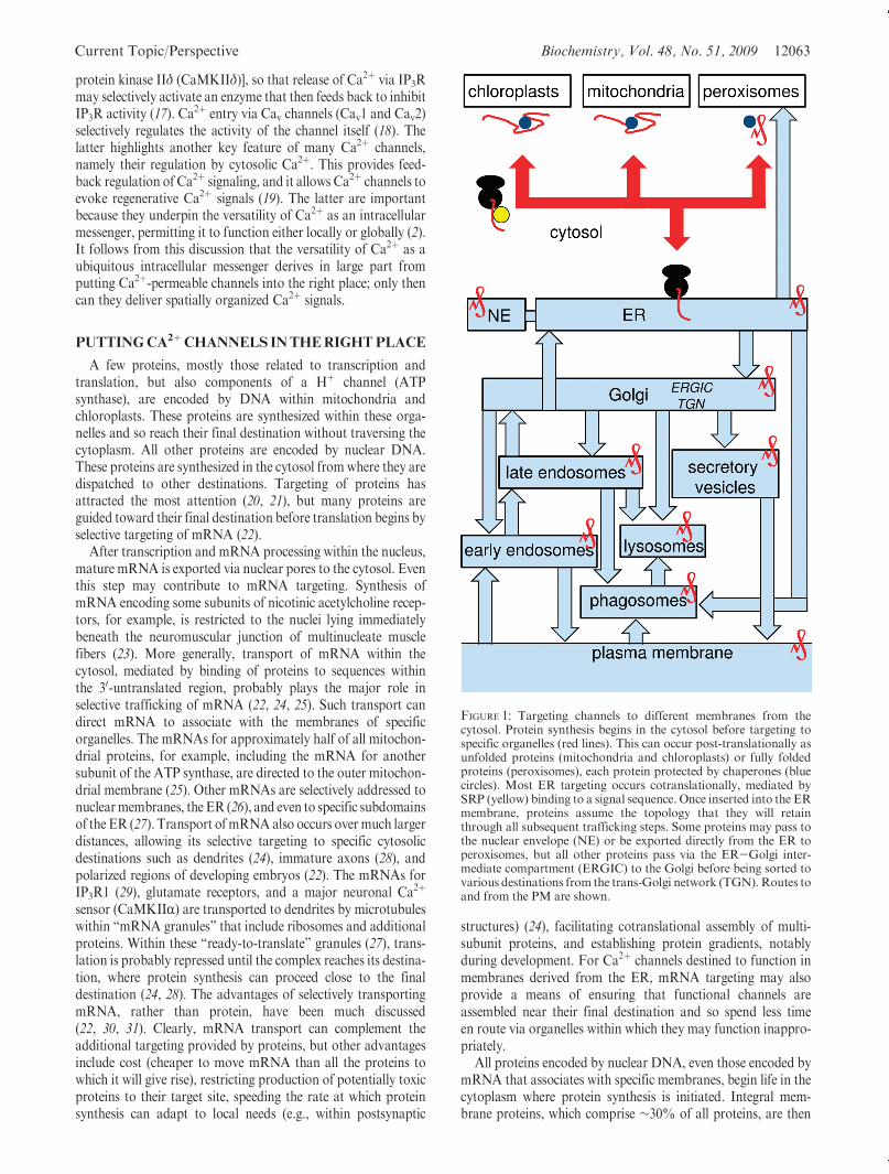
All proteins encoded by nuclear DNA, even those encoded bymRNA that associates with specific membranes, begin life in thecytoplasm where protein synthesis is initiated. Integral mem-brane proteins, which comprise ∼30% of all proteins, are then
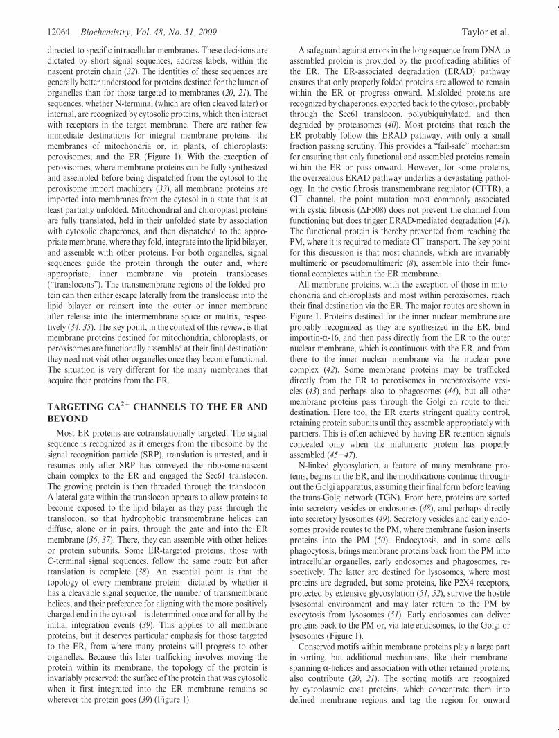
FIGURE 1: Targeting channels to different membranes from thecytosol. Protein synthesis begins in the cytosol before targeting tospecific organelles (red lines). This can occur post-translationally asunfolded proteins (mitochondria and chloroplasts) or fully foldedproteins (peroxisomes), each protein protected by chaperones (bluecircles). Most ER targeting occurs cotranslationally, mediated bySRP (yellow) binding to a signal sequence. Once inserted into the ERmembrane, proteins assume the topology that they will retainthrough all subsequent trafficking steps. Some proteins may pass tothe nuclear envelope (NE) or be exported directly from the ER toperoxisomes, but all other proteins pass via the ER-Golgi inter-mediate compartment (ERGIC) to the Golgi before being sorted tovarious destinations from the trans-Golgi network (TGN).Routes toand from the PM are shown.
12064 Biochemistry, Vol. 48, No. 51, 2009 Taylor et al.
directed to specific intracellular membranes. These decisions aredictated by short signal sequences, address labels, within thenascent protein chain (32). The identities of these sequences aregenerally better understood for proteins destined for the lumen oforganelles than for those targeted to membranes (20, 21). Thesequences, whether N-terminal (which are often cleaved later) orinternal, are recognized by cytosolic proteins, which then interactwith receptors in the target membrane. There are rather fewimmediate destinations for integral membrane proteins: themembranes of mitochondria or, in plants, of chloroplasts;peroxisomes; and the ER (Figure 1). With the exception ofperoxisomes, where membrane proteins can be fully synthesizedand assembled before being dispatched from the cytosol to theperoxisome import machinery (33), all membrane proteins areimported into membranes from the cytosol in a state that is atleast partially unfolded. Mitochondrial and chloroplast proteinsare fully translated, held in their unfolded state by associationwith cytosolic chaperones, and then dispatched to the appro-priate membrane, where they fold, integrate into the lipid bilayer,and assemble with other proteins. For both organelles, signalsequences guide the protein through the outer and, whereappropriate, inner membrane via protein translocases(“translocons”). The transmembrane regions of the folded pro-tein can then either escape laterally from the translocase into thelipid bilayer or reinsert into the outer or inner membraneafter release into the intermembrane space or matrix, respec-tively (34, 35). The key point, in the context of this review, is thatmembrane proteins destined for mitochondria, chloroplasts, orperoxisomes are functionally assembled at their final destination:they need not visit other organelles once they become functional.The situation is very different for the many membranes thatacquire their proteins from the ER.
TARGETING CA2þ CHANNELS TO THE ER AND
BEYOND
Most ER proteins are cotranslationally targeted. The signalsequence is recognized as it emerges from the ribosome by thesignal recognition particle (SRP), translation is arrested, and itresumes only after SRP has conveyed the ribosome-nascentchain complex to the ER and engaged the Sec61 translocon.The growing protein is then threaded through the translocon.A lateral gate within the translocon appears to allow proteins tobecome exposed to the lipid bilayer as they pass through thetranslocon, so that hydrophobic transmembrane helices candiffuse, alone or in pairs, through the gate and into the ERmembrane (36, 37). There, they can assemble with other helicesor protein subunits. Some ER-targeted proteins, those withC-terminal signal sequences, follow the same route but aftertranslation is complete (38). An essential point is that thetopology of every membrane protein—dictated by whether ithas a cleavable signal sequence, the number of transmembranehelices, and their preference for aligning with the more positivelycharged end in the cytosol—is determined once and for all by theinitial integration events (39). This applies to all membraneproteins, but it deserves particular emphasis for those targetedto the ER, from where many proteins will progress to otherorganelles. Because this later trafficking involves moving theprotein within its membrane, the topology of the protein isinvariably preserved: the surface of the protein that was cytosolicwhen it first integrated into the ER membrane remains sowherever the protein goes (39) (Figure 1).
A safeguard against errors in the long sequence from DNA toassembled protein is provided by the proofreading abilities ofthe ER. The ER-associated degradation (ERAD) pathwayensures that only properly folded proteins are allowed to remainwithin the ER or progress onward. Misfolded proteins arerecognized by chaperones, exported back to the cytosol, probablythrough the Sec61 translocon, polyubiquitylated, and thendegraded by proteasomes (40). Most proteins that reach theER probably follow this ERAD pathway, with only a smallfraction passing scrutiny. This provides a “fail-safe” mechanismfor ensuring that only functional and assembled proteins remainwithin the ER or pass onward. However, for some proteins,the overzealous ERAD pathway underlies a devastating pathol-ogy. In the cystic fibrosis transmembrane regulator (CFTR), aCl- channel, the point mutation most commonly associatedwith cystic fibrosis (ΔF508) does not prevent the channel fromfunctioning but does trigger ERAD-mediated degradation (41).The functional protein is thereby prevented from reaching thePM, where it is required to mediate Cl- transport. The key pointfor this discussion is that most channels, which are invariablymultimeric or pseudomultimeric (8), assemble into their func-tional complexes within the ER membrane.
All membrane proteins, with the exception of those in mito-chondria and chloroplasts and most within peroxisomes, reachtheir final destination via the ER. The major routes are shown inFigure 1. Proteins destined for the inner nuclear membrane areprobably recognized as they are synthesized in the ER, bindimportin-R-16, and then pass directly from the ER to the outernuclear membrane, which is continuous with the ER, and fromthere to the inner nuclear membrane via the nuclear porecomplex (42). Some membrane proteins may be traffickeddirectly from the ER to peroxisomes in preperoxisome vesi-cles (43) and perhaps also to phagosomes (44), but all othermembrane proteins pass through the Golgi en route to theirdestination. Here too, the ER exerts stringent quality control,retaining protein subunits until they assemble appropriately withpartners. This is often achieved by having ER retention signalsconcealed only when the multimeric protein has properlyassembled (45-47).
N-linked glycosylation, a feature of many membrane pro-teins, begins in the ER, and the modifications continue through-out the Golgi apparatus, assuming their final form before leavingthe trans-Golgi network (TGN). From here, proteins are sortedinto secretory vesicles or endosomes (48), and perhaps directlyinto secretory lysosomes (49). Secretory vesicles and early endo-somes provide routes to the PM, where membrane fusion insertsproteins into the PM (50). Endocytosis, and in some cellsphagocytosis, brings membrane proteins back from the PM intointracellular organelles, early endosomes and phagosomes, re-spectively. The latter are destined for lysosomes, where mostproteins are degraded, but some proteins, like P2X4 receptors,protected by extensive glycosylation (51, 52), survive the hostilelysosomal environment and may later return to the PM byexocytosis from lysosomes (51). Early endosomes can deliverproteins back to the PM or, via late endosomes, to the Golgi orlysosomes (Figure 1).
Conserved motifs within membrane proteins play a large partin sorting, but additional mechanisms, like their membrane-spanning R-helices and association with other retained proteins,also contribute (20, 21). The sorting motifs are recognizedby cytoplasmic coat proteins, which concentrate them intodefined membrane regions and tag the region for onward
Current Topic/Perspective Biochemistry, Vol. 48, No. 51, 2009 12065
movement to a specific membrane. A cytosolic K(X)KXXsequence, for example, serves both to retain proteins within theER and, after binding COPI coat protein, to return escapees tothe ER from the cis-Golgi. Budding of the membrane from thedonor organelle, transport of the resulting vesicle along micro-tubules or the actin cytoskeleton (50, 53), and fusion with thetarget organelle deliver the membrane protein to its next destina-tion. Details of these processes, the essential role of smallGTPases (54), the identities of the many sorting sequences (32),the role of lipids in these sorting events (55), and the behavior ofthe SNAREs that mediate vesicle fusion (56) are describedelsewhere (20, 21, 32).
Even proteins destined to function in a specific membrane aredynamically shuffled between compartments: escaped ER pro-teins are trafficked back from the Golgi, a PM protein may beendocytosed and then reappear at the PM, and some proteinstake very circuitous routes to their final destination. In polarizedepithelia, for example, many proteins are directly dispatched tothe apical or basolateral PM after being sorted at the TGN (53),but in hepatocytes, all PMproteins are first sent to the basolateralPM, from where apical proteins are selectively endocytosed andsent to the apical PM (57). These observations highlight the factthat most ion channels must pass through several differentmembranes to reach their destination and even then are likelytomake periodic excursions into othermembranes.Howdoes thecell ensure effective control of Ca2þ channels as they pass throughthese different cellular compartments?
GETTING CA2þ CHANNELS HARMLESSLY TO
THEIR DESTINATIONS
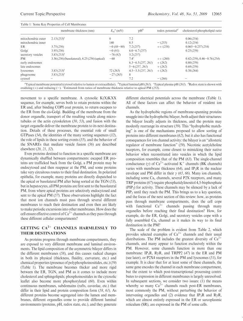
As proteins progress through membrane compartments, theyare exposed to very different membrane and luminal environ-ments. The lipid composition of the asymmetric bilayer differs inthe different membranes (58), and this causes radical changesin both its physical (thickness, fluidity, curvature, etc.) andchemical properties (presence of polyphosphoinositides, etc.) (59)(Table 1). The membrane becomes thicker and more rigidbetween the ER, TGN, and PM as it comes to include morecholesterol and sphingolipids; phosphoinositides in the cytosolicleaflet also become more phosphorylated (60). Even withincontinuous membranes, subdomains (rafts, caveolae, etc.) thatdiffer in their lipid and protein composition form (58, 61). Asdifferent proteins become segregated into the lumen and mem-branes, different organelles come to provide different luminalenvironments (proteins, pH, redox state, etc.), and they generate
different electrical potentials across the membrane (Table 1).All of these factors can affect the behavior of resident ionchannels.
As the hydrophobic regions of membrane-spanning proteinssnuggle into the hydrophobic bilayer, both adjust their structures:the bilayer locally adjusts its thickness, and the protein maymodestly rearrange its structure (59). This “hydrophobic match-ing” is one of the mechanisms proposed to allow sorting ofproteins into different membranes (62), but it also has functionalconsequences for ion channel activity: the bilayer is an “allostericregulator of membrane function” (59). Nicotinic acetylcholinereceptors, for example, come closest to mimicking their nativebehavior when reconstituted into vesicles in which the lipidcomposition resembles that of the PM (63). The single-channelconductance (γ) of Ca2þ-activated Kþ channels (BK channels)varies with membrane thickness (64), and IP3R in the nuclearenvelope and PM differ in their γ (65, 66). Many ion channels,including some Cav channels, several P2X receptors, and manyTRP proteins (67) require phosphatidylinositol 4,5-bisphosphate(PIP2) for activity. These channels may be silenced by a lack ofPIP2 until they reach the PM. This brings us to a key question,and the focus of the next section of this review: how, as proteinspass through membrane compartments, does the cell copewith functional Ca2þ channels passing through manyorganelles before reaching their final destination? How, forexample, do the ER, Golgi, and secretory vesicles cope with afully assembled Cav channel as it makes its way to its finaldestination in the PM?
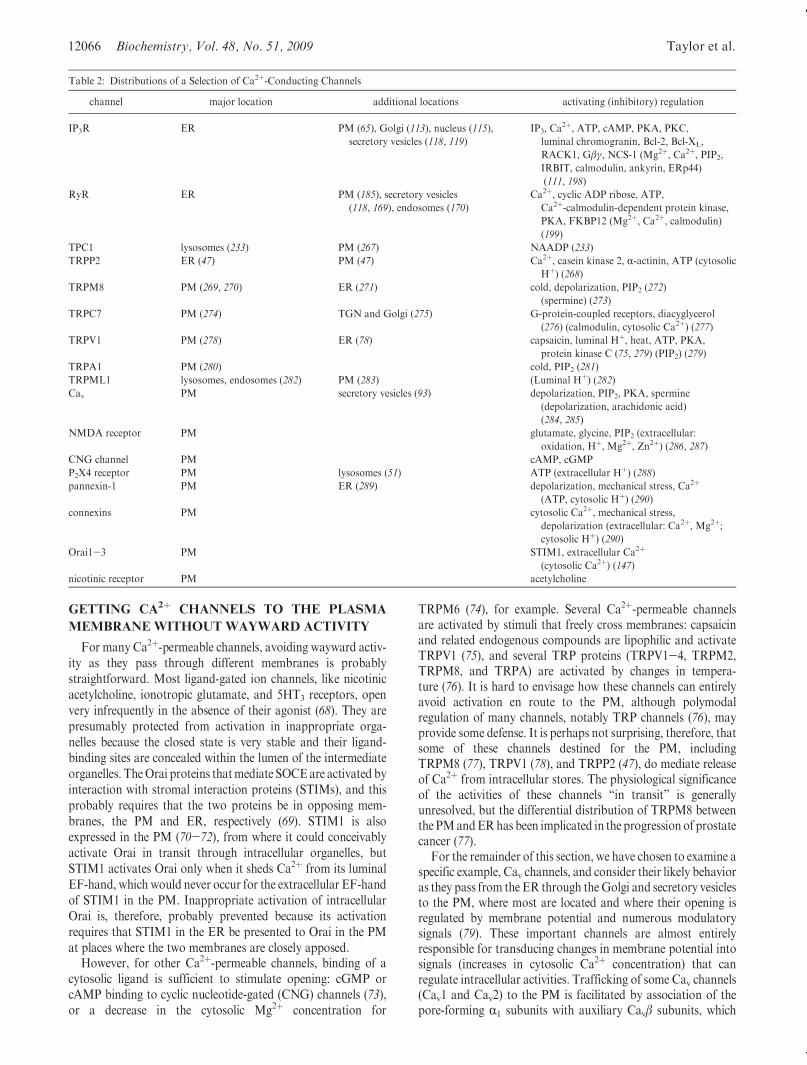
The scale of the problem is evident from Table 2, whichprovides selected examples of Ca2þ channels and their usualdistributions. The PM includes the greatest diversity of Ca2þ
channels, and many appear to function exclusively within thePM. However, some channels function in more than onemembrane: IP3R, RyR, and TRPP2 (47) in the ER and PM(see later), or P2X4 receptors in the PM and lysosomes (51), forexample. It is clear that for at least some of these channels, thesame gene encodes the channel in each membrane compartment,but the extent to which post-transcriptional processing contri-butes to expression in different membranes is largely unresolved.In subsequent sections, we consider two issues: (1) the meanswhereby so many Ca2þ channels reach post-ER membranes,most commonly the PM, without perturbing the behavior ofintervening organelles and (2) evidence that IP3R and RyR,which are almost entirely expressed in the ER or sarcoplasmicreticulum (SR), are expressed in the PM of some cells.
Table 1: Some Key Properties of Cell Membranes
membrane thickness (nm) Ema (mV) pHb redox potentiald cholesterol:phospholipid ratio
mitochondria outer 2.13 (253)e 0 7.2 0.06 (254)
mitochondria inner 140 8.5 - (255) 0.12 (254)
ER 3.75 (256) ∼0 (88-90) 7.2 (257) þþ (258) 0.065-0.237 (254)
Golgi 3.95 (256) ∼0 (91) 6.0-6.7 (257) 0.25 (259)
secretory vesicles 3.83 (253)e -76 (92) 5.2 (257)
PM 3.56 (256) (basolateral), 4.25 (256) (apical) -60 7.4c þþ (260) 0.42 (259), 0.46-0.76 (254)
early endosomes 6.3-6.5 (257, 261) þ (262) 0.80 (259)
late endosomes 5-6 (257, 261) þ (262) 0.69 (259)
lysosomes 3.83 (253)e 72 (263) 4.5-5.5 (257, 261) þ (262) 0.38 (264)
phagosome 3.83 (253)e -27 (265) 8
cytosol - - 7.2 - (266) -aTypical membrane potential (cytosol relative to lumen or extracellular). bTypical luminal pH (261). cTypical plasma pH (261). dRedox state is shownwith
oxidizing (þ) and reducing (-). eEstimated from ratios of membrane thickness relative to apical PM (253).
12066 Biochemistry, Vol. 48, No. 51, 2009 Taylor et al.
GETTING CA2þ CHANNELS TO THE PLASMA
MEMBRANE WITHOUT WAYWARD ACTIVITY
For many Ca2þ-permeable channels, avoiding wayward activ-ity as they pass through different membranes is probablystraightforward. Most ligand-gated ion channels, like nicotinicacetylcholine, ionotropic glutamate, and 5HT3 receptors, openvery infrequently in the absence of their agonist (68). They arepresumably protected from activation in inappropriate orga-nelles because the closed state is very stable and their ligand-binding sites are concealed within the lumen of the intermediateorganelles. TheOrai proteins thatmediate SOCEare activated byinteraction with stromal interaction proteins (STIMs), and thisprobably requires that the two proteins be in opposing mem-branes, the PM and ER, respectively (69). STIM1 is alsoexpressed in the PM (70-72), from where it could conceivablyactivate Orai in transit through intracellular organelles, butSTIM1 activates Orai only when it sheds Ca2þ from its luminalEF-hand, which would never occur for the extracellular EF-handof STIM1 in the PM. Inappropriate activation of intracellularOrai is, therefore, probably prevented because its activationrequires that STIM1 in the ER be presented to Orai in the PMat places where the two membranes are closely apposed.
However, for other Ca2þ-permeable channels, binding of acytosolic ligand is sufficient to stimulate opening: cGMP orcAMP binding to cyclic nucleotide-gated (CNG) channels (73),or a decrease in the cytosolic Mg2þ concentration for
TRPM6 (74), for example. Several Ca2þ-permeable channelsare activated by stimuli that freely cross membranes: capsaicinand related endogenous compounds are lipophilic and activateTRPV1 (75), and several TRP proteins (TRPV1-4, TRPM2,TRPM8, and TRPA) are activated by changes in tempera-ture (76). It is hard to envisage how these channels can entirelyavoid activation en route to the PM, although polymodalregulation of many channels, notably TRP channels (76), mayprovide some defense. It is perhaps not surprising, therefore, thatsome of these channels destined for the PM, includingTRPM8 (77), TRPV1 (78), and TRPP2 (47), do mediate releaseof Ca2þ from intracellular stores. The physiological significanceof the activities of these channels “in transit” is generallyunresolved, but the differential distribution of TRPM8 betweenthe PMandERhas been implicated in the progression of prostatecancer (77).
For the remainder of this section, we have chosen to examine aspecific example, Cav channels, and consider their likely behavioras they pass from the ER through theGolgi and secretory vesiclesto the PM, where most are located and where their opening isregulated by membrane potential and numerous modulatorysignals (79). These important channels are almost entirelyresponsible for transducing changes in membrane potential intosignals (increases in cytosolic Ca2þ concentration) that canregulate intracellular activities. Trafficking of some Cav channels(Cav1 and Cav2) to the PM is facilitated by association of thepore-forming R1 subunits with auxiliary Cavβ subunits, which
Table 2: Distributions of a Selection of Ca2þ-Conducting Channels
channel major location additional locations activating (inhibitory) regulation
IP3R ER PM (65), Golgi (113), nucleus (115),
secretory vesicles (118, 119)
IP3, Ca2þ, ATP, cAMP, PKA, PKC,
luminal chromogranin, Bcl-2, Bcl-XL,
RACK1, Gβγ, NCS-1 (Mg2þ, Ca2þ, PIP2,
IRBIT, calmodulin, ankyrin, ERp44)
(111, 198)
RyR ER PM (185), secretory vesicles
(118, 169), endosomes (170)
Ca2þ, cyclic ADP ribose, ATP,
Ca2þ-calmodulin-dependent protein kinase,
PKA, FKBP12 (Mg2þ, Ca2þ, calmodulin)
(199)
TPC1 lysosomes (233) PM (267) NAADP (233)
TRPP2 ER (47) PM (47) Ca2þ, casein kinase 2, R-actinin, ATP (cytosolic
Hþ) (268)TRPM8 PM (269, 270) ER (271) cold, depolarization, PIP2 (272)
(spermine) (273)
TRPC7 PM (274) TGN and Golgi (275) G-protein-coupled receptors, diacyglycerol
(276) (calmodulin, cytosolic Ca2þ) (277)TRPV1 PM (278) ER (78) capsaicin, luminal Hþ, heat, ATP, PKA,
protein kinase C (75, 279) (PIP2) (279)
TRPA1 PM (280) cold, PIP2 (281)
TRPML1 lysosomes, endosomes (282) PM (283) (Luminal Hþ) (282)Cav PM secretory vesicles (93) depolarization, PIP2, PKA, spermine
(depolarization, arachidonic acid)
(284, 285)
NMDA receptor PM glutamate, glycine, PIP2 (extracellular:
oxidation, Hþ, Mg2þ, Zn2þ) (286, 287)CNG channel PM cAMP, cGMP
P2X4 receptor PM lysosomes (51) ATP (extracellular Hþ) (288)pannexin-1 PM ER (289) depolarization, mechanical stress, Ca2þ
(ATP, cytosolic Hþ) (290)connexins PM cytosolic Ca2þ, mechanical stress,
depolarization (extracellular: Ca2þ, Mg2þ;cytosolic Hþ) (290)
Orai1-3 PM STIM1, extracellular Ca2þ
(cytosolic Ca2þ) (147)nicotinic receptor PM acetylcholine
Current Topic/Perspective Biochemistry, Vol. 48, No. 51, 2009 12067
may mask an ER retention signal within the R1 subunits (80).Mutations within the R1A subunit of the Cav2.1 channel trapassociated subunits within the ER and gives rise to episodicataxia type 2 (81). Whether the pathology reflects aberrantactivity of Cav2.1 within the ER or loss of activity at the PM isunresolved, but in either case, the importance of effectivetrafficking to the PM is clear.
Members of each of the three major families of Cav channels[Cav1 (L-type), Cav2 (N-, P/Q-, and R-type), and Cav3 (T-type)]are inactivated (at very different rates) by sustained depolariza-tion to potentials of approximately-60 to-10mV (Cav1),-120to -20 mV (Cav2), and -100 to -60 mV (Cav3) (8, 82). At theresting membrane potential of the PM (typically approximately-60 mV), most of these channels will not be inactivated and canopen during transient depolarizations. Many ion channels,including Cav channels (83), CNG channels (84), and glutamatereceptors (85, 86), are blocked by polyamines, like spermine, andcanmediate ion fluxes only when changes in membrane potentialdislodge the polyamine. For Ca2þ-permeable AMPA receptors,an accessory protein, stargazin, both guides the receptors to thePM and relieves the block by polyamines (87), perhaps therebyensuring effective block of the channels until they leave the ER.The important point is that activation of Cav channels requirestransient depolarization from a hyperpolarized membrane po-tential for activation. However, the potential across the mem-branes of the ER (88-90) and Golgi (91) is probably close to0 mV, sufficient to inactivate most Cav channels. However,secretory vesicles appear tomaintain a membrane potential morelike that of the PM (-76 mV) (92), and with individual vesiclesshowing considerable variability in membrane potential (-11 to-160 mV), it seems likely that Cav channels within secretoryvesicles may be exposed to potential changes that would allowtheir activation. Because secretory vesicles, in commonwith everyother intracellular Ca2þ store, contain only a limited pool ofCa2þ, Cav within them could contribute to Ca2þ signaling only ifbouts of activation were interspersed with periods of closure toallow the vesicles to refill with Ca2þ. The possibility that Cavchannels might be activated within secretory vesicles assumesfurther significance in light of evidence that a large reservoir offunctional Cav channels may be retained within the secretoryvesicles of neuroblastoma cells, fromwhere trafficking to the PMis dynamically regulated (93). Whether other factors, like low pHor the absence of PIP2 (see below), silence Cav channels insecretory vesicles or whether they are active and responsive tochanges in vesicle membrane potential deserves further study.
The pH to which the luminal/extracellular surface of mem-brane proteins is exposed also changes dramatically as they aretransported from the ER to the PM (Table 1). Many Ca2þ
channels, including Cav1 (94), CNG channels (95), TRPV5, andTRPV6 (96), are inhibited by the low pH encountered within thesecretory pathway. Other Ca2þ channels, such as TRPV1 (97),TRPM6 (98), and TRPM7 (99), are stimulated by this low pH,although this may be accompanied by a reduced Ca2þ flux as Hþ
within the permeation pathway attenuates Ca2þ binding andincreases the relative permeability to monovalent cations (99).
The different lipid compositions of the PM and earliermembranes (Table 1) may also restrain the activity of Ca2þ
channels in transit. PIP2 is enriched in the inner leaflet of the PMand is essential for the activity of Cav2 channels (100). Theactivity of Cav1 channels is enhanced by cholesterol (101), theconcentration of which increases as membranes progress towardthe PM (Table 1). Similar interactionsmay inhibit the activities of
other Ca2þ channels until they reach the PM: many TRPchannels are stimulated by PIP2 (102) or the diacylglycerolproduced by its hydrolysis (103), and the activities of CNGchannels (104) and TRPC3 (105) are enhanced by cholesterol.
Finally, Cav and many other Ca2þ channels have theirresponses tuned by association with additional proteins, suchthat successful targeting of these proteins to the PM can unmasklatent channel activity. We have already discussed the role ofCavβ subunits in coordinating PM targeting and the activity ofCav channels (80). Palmitoylation of Cav channels likewise bothregulates their trafficking to the PM and enhances theiractivity (106-108). Anchoring of cyclic AMP-dependent proteinkinase (PKA) at the PM by A-kinase-anchoring protein(AKAP15) positions PKA close to Cav1 channels, allowingphosphorylation to increase their activity (109).
Clearly, Cav channels can reach the PM without overlyperturbing cytosolic or luminal Ca2þ regulation as they passthrough intervening organelles. A few general themes emerge.Cav (and other) channels in transit may be inactive because theintervening environment is hostile (e.g., inactivation by sustaineddepolarization, lipids, pH, etc.). A single modification (e.g., asso-ciation with a β-subunit or palmitoylation) may both enhanceactivity and simultaneously provide an express ticket to thePM, thereby ensuring that the most active channels reach thePM without lingering in the ER. Additional PM proteins(e.g., AKAP-anchored PKA) may enhance the activity ofjuxtaposed channels. Whether these, or additional mechanisms,are wholly effective in silencing Cav channels in transit seems tobe unresolved. Do Cav channels, for example, mediate voltage-gated Ca2þ fluxes in secretory vesicles? Do they, or other Ca2þ
channels in transit, contribute to the unresolved leak of basalCa2þ from the ER? What are the physiological roles of thoseCa2þ channels in transit that are not completely silenced as theymake their way to the PM?
COUNTING IP3 RECEPTORS INTO THE PLASMA
MEMBRANE
Most IP3R in most cells are expressed within ER membranes,or the nuclear envelope, which is continuous with them (110,111). This pattern of expression is maintained by several ERretention signals within the six C-terminal transmembranedomains of IP3R (112). Nevertheless, there is persuasive evidencethat functional IP3R can also be expressed within the Golgiapparatus (113, 114), the nucleoplasmic reticulum (115), and,more contentiously, secretory vesicles (116-119). In this section,we consider evidence that IP3R, the archetypal intracellular Ca2þ
channels, are also functionally expressed in the PMof some cells,notably DT40 cells, a prelymphocyte cell line (120), and mam-malian B-lymphocytes (65).
Several studies had suggested that IP3R might be expressedwithin the PM (reviewed in refs 65 and 121), but the results wereinconclusive and in many cases likely to reflect the activities ofintracellular IP3R. Subcellular fractionation showed IP3R tocopurify with PM markers (122-124), probably because IP3Rwithin the ER are closely associated with the PM (123, 125). Thesame explanation might account for the presence of IP3Rimmunostaining close to the PM of oviductal ciliated cells (126).Cell surface labeling suggested the presence of IP3R within thePM (127-130). Patch-clamp recording identified IP3-activated,Ca2þ-permeable channels in the PM of some cells (131-136),and a protein purified from a liver PM fraction behaved as an
12068 Biochemistry, Vol. 48, No. 51, 2009 Taylor et al.
IP3-gated channel when reconstituted into lipid bilayers (137).However, the electrophysiological properties of these channelswere very different from those of IP3R either in nuclear mem-branes (66, 110, 111) or after reconstitution of intracellular IP3Rinto bilayers (138, 139). With the exception of IP3R in olfactorycilia (131), which differ from intracellular IP3R in their sensitivityto ruthenium red, insensitivity to ATP, and lower conductance,the other reports of IP3-activated PM channels failed to establishwhether the channels were actually IP3R within the PM or morelikely other channels indirectly activated by stimulation of IP3Rwithin the ER. Our work with DT40 cells (65, 140) andsubsequent work by others (141) have provided conclusiveevidence that functional IP3R can be expressed in the PM of atleast some cells.
Stimulation of DT40 cells with either anti-IgM to activate theB-cell receptor (BCR) or trypsin to activate the protease-acti-vated receptor (PAR) leads to activation of phospholipase C,formation of IP3, and thereby release of Ca2þ from intracellularstores mediated by IP3R (120, 142). DT40 cells are uniquelysuited to analyses of these signaling pathways because Kurosakiand his colleagues generated a DT40 cell line (DT40-KO cells) inwhich the genes for all three IP3R subtypes are disrupted (142).DT40-KO cells are the only vertebrate cells unequivocally devoidof functional IP3R and therefore the only null background inwhich recombinant IP3R can be functionally expressed free of thecomplexity arising from association with residual nativeIP3R (120, 143). The inability of either anti-IgM or trypsin toevoke Ca2þ release in DT40-KO cells and restoration of thatresponse by expression of recombinant IP3R firmly establishesthe role of the IP3R in mediating Ca2þ release (65).
The same stimuli that evoke release of Ca2þ also evoke entry ofCa2þ across the PMof DT40 cells. In most cells, such Ca2þ entryis commonly mediated by SOCE (144, 145). The defining featureof SOCE is that depletion of intracellular Ca2þ stores provides asufficient stimulus for its activation (144). Thapsigargin, whichinhibits SERCA and thereby causes loss of Ca2þ from the ERwithout engaging the signaling pathways used by receptors, isoften used to activate SOCE (Figure 2A). In many cells, theelectrophysiological manifestation of SOCE is the Ca2þ release-activated current (ICRAC), which is characterized by its inwardrectification, Ca2þ selectivity, and low unitary conductance,block by low concentrations of Gd3þ, and activation by depletedCa2þ stores (146, 147). The molecular architecture of the SOCEpathway has recently been established (13), with STIM1 emer-ging as the luminal Ca2þ sensor in the ER membrane (148, 149),and Orai proteins as the pore-forming subunits of the channel inthe PM (150). After Ca2þ is lost from the ER, the luminal EF-hand of STIM1 loses Ca2þ, causing it to cluster. The clusteredpolybasic C-termini of STIM1 can then interact with PIP2 in thePM, allowing a stretch of conserved residues within the cytosolictail of STIM1, the CRACactivation domain (CAD), to stimulateopening of Orai (69, 151). Further details of the mechanisms ofactivation of SOCE (13), including the possible involvement ofTRP proteins (152), the possibility that biochemical signalsmightlink STIM1 to activation of Orai (153), the relative roles ofSTIM1 and STIM2 (154, 155), and the possibility that STIMmight activate Orai by causing its assembly into a tetramericchannel (156), are discussed elsewhere.
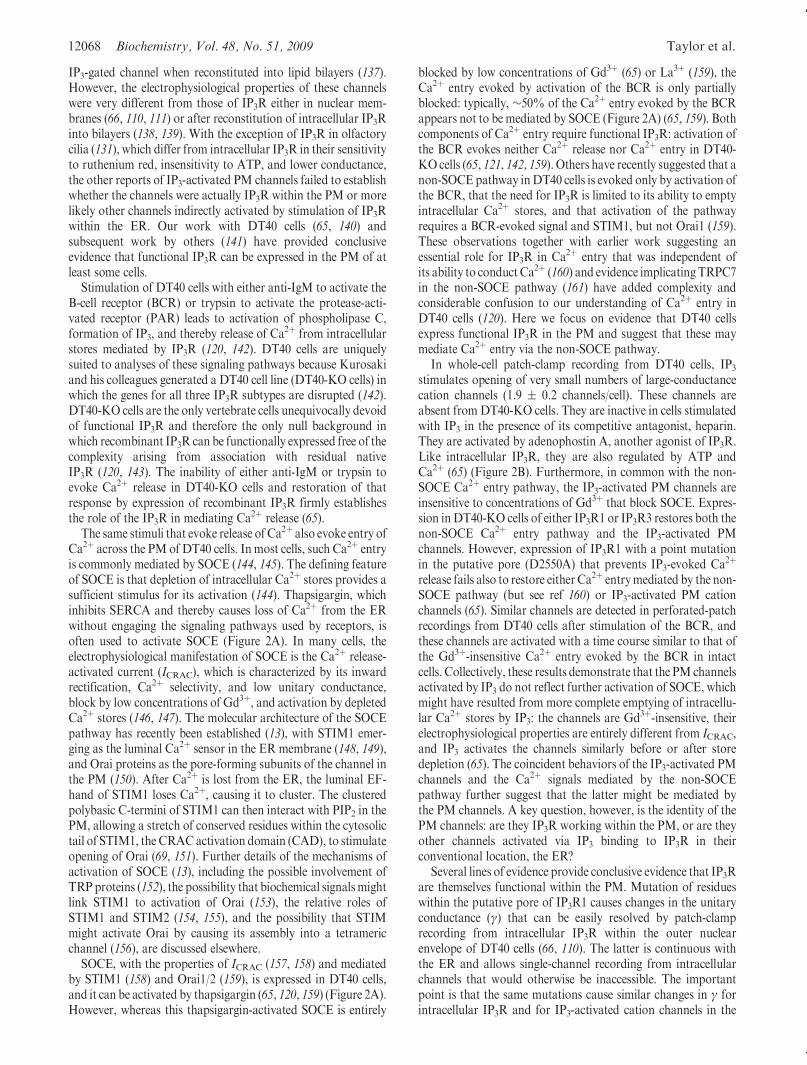
SOCE, with the properties of ICRAC (157, 158) and mediatedby STIM1 (158) and Orai1/2 (159), is expressed in DT40 cells,and it can be activated by thapsigargin (65, 120, 159) (Figure 2A).However, whereas this thapsigargin-activated SOCE is entirely
blocked by low concentrations of Gd3þ (65) or La3þ (159), theCa2þ entry evoked by activation of the BCR is only partiallyblocked: typically, ∼50% of the Ca2þ entry evoked by the BCRappears not to be mediated by SOCE (Figure 2A) (65, 159). Bothcomponents of Ca2þ entry require functional IP3R: activation ofthe BCR evokes neither Ca2þ release nor Ca2þ entry in DT40-KO cells (65, 121, 142, 159). Others have recently suggested that anon-SOCE pathway inDT40 cells is evoked only by activation ofthe BCR, that the need for IP3R is limited to its ability to emptyintracellular Ca2þ stores, and that activation of the pathwayrequires a BCR-evoked signal and STIM1, but not Orai1 (159).These observations together with earlier work suggesting anessential role for IP3R in Ca2þ entry that was independent ofits ability to conductCa2þ (160) and evidence implicatingTRPC7in the non-SOCE pathway (161) have added complexity andconsiderable confusion to our understanding of Ca2þ entry inDT40 cells (120). Here we focus on evidence that DT40 cellsexpress functional IP3R in the PM and suggest that these maymediate Ca2þ entry via the non-SOCE pathway.
In whole-cell patch-clamp recording from DT40 cells, IP3stimulates opening of very small numbers of large-conductancecation channels (1.9 ( 0.2 channels/cell). These channels areabsent from DT40-KO cells. They are inactive in cells stimulatedwith IP3 in the presence of its competitive antagonist, heparin.They are activated by adenophostin A, another agonist of IP3R.Like intracellular IP3R, they are also regulated by ATP andCa2þ (65) (Figure 2B). Furthermore, in common with the non-SOCE Ca2þ entry pathway, the IP3-activated PM channels areinsensitive to concentrations of Gd3þ that block SOCE. Expres-sion inDT40-KO cells of either IP3R1 or IP3R3 restores both thenon-SOCE Ca2þ entry pathway and the IP3-activated PMchannels. However, expression of IP3R1 with a point mutationin the putative pore (D2550A) that prevents IP3-evoked Ca2þ
release fails also to restore either Ca2þ entrymediated by the non-SOCE pathway (but see ref 160) or IP3-activated PM cationchannels (65). Similar channels are detected in perforated-patchrecordings from DT40 cells after stimulation of the BCR, andthese channels are activated with a time course similar to that ofthe Gd3þ-insensitive Ca2þ entry evoked by the BCR in intactcells. Collectively, these results demonstrate that the PMchannelsactivated by IP3 do not reflect further activation of SOCE, whichmight have resulted from more complete emptying of intracellu-lar Ca2þ stores by IP3: the channels are Gd3þ-insensitive, theirelectrophysiological properties are entirely different from ICRAC,and IP3 activates the channels similarly before or after storedepletion (65). The coincident behaviors of the IP3-activated PMchannels and the Ca2þ signals mediated by the non-SOCEpathway further suggest that the latter might be mediated bythe PM channels. A key question, however, is the identity of thePM channels: are they IP3R working within the PM, or are theyother channels activated via IP3 binding to IP3R in theirconventional location, the ER?
Several lines of evidence provide conclusive evidence that IP3Rare themselves functional within the PM. Mutation of residueswithin the putative pore of IP3R1 causes changes in the unitaryconductance (γ) that can be easily resolved by patch-clamprecording from intracellular IP3R within the outer nuclearenvelope of DT40 cells (66, 110). The latter is continuous withthe ER and allows single-channel recording from intracellularchannels that would otherwise be inaccessible. The importantpoint is that the same mutations cause similar changes in γ forintracellular IP3R and for IP3-activated cation channels in the
Current Topic/Perspective Biochemistry, Vol. 48, No. 51, 2009 12069
PM (65, 141) (Figure 2C). Clearly, if the effects of IP3 on the PMwere mediated solely by its interaction with intracellular IP3R,pore mutations within the IP3R would not be expected to affectthe γ of the PMchannels.We also introduced a binding site forR-bungarotoxin (RBgtx) into the luminal loop linking the last pairof transmembrane domains (TMD5-6) of IP3R1 and expressedit in DT40-KO cells (Figure 2D). From the known topology ofthe IP3R, we expected the RBgtx-binding site to be luminal forIP3R within the ER, but extracellular for IP3R expressed in thePM. In whole-cell patch-clamp recordings from DT40 cellsexpressing these IP3R, intracellular IP3 again activated cationchannels in the PM, but their γ and open probability (Po) wereboth increased by addition of extracellular, but not intracellular,RBgtx (65). These results establish that the recombinant IP3Rmust straddle the PM. Finally, others have expressed IP3R with
mutations in sites that are phosphorylated by PKA and shownthat the Po of the PM cation channels activated by adenophostinA is increased by forskolin (to activate PKA) for wild-type IP3R;IP3R with phospho-mimetic mutations are hyperactive, andthose with mutations that prevent phosphorylation have muchreduced activity (141). Collectively, these results demonstrateunequivocally that functional IP3R are expressed in the PM ofDT40 cells.We stress, however, that IP3R are not expressed in thePMof all cells; we have not, for example, detected IP3R in the PMof either Sf9 or human embryonic kidney (HEK) cells.
Functional IP3R in the PM of DT40 cells are expressed at anexceptionally low density, just 1.9( 0.2 channels/cell (65), andwehave never, even in cells massively overexpressing intracellularIP3R, detected more than five functional IP3R in the PM. Thereare some additional examples of cells expressing such tiny
FIGURE 2: Expressionof IP3 receptors in the plasmamembraneofDT40cells. (A)Thapsigargin (top) stimulatesCa2þ release (palest line, inCa2þ-free medium) and Ca2þ entry (black line, in Ca2þ-containing medium). The latter, SOCE, is entirely blocked by 300 nMGdCl3 (gray line). Thebottompanel shows that activationof theBCRwith anti-IgMstimulates bothCa2þ release andCa2þ entry, but the latter is onlypartially inhibitedbyGdCl3. (B)Whole-cell patch-clamp recoding fromDT40 cells with IP3, IP3 with heparin, or adenophostin A in the patch pipette. The holdingpotential was -100 mV; arrowheads denote the closed state. (C) Current-voltage (i-V) relationships for the IP3-stimulated currents recordedfrom the PM or nuclear envelope of DT40-KO cells stably transfected with wild-type IP3R1 (R1) or IP3R1 with mutants in the putative pore(G2547A, R1GA; V2548I, R1VI). The point mutations similarly affected γK of the IP3-activated currents in both settings. (D) The six TMDs of asingle IP3R subunit are shown to highlight the putative selectivity filter (sf) and the engineered RBgtx-binding site. In whole-cell patch-clamprecordings fromDT40 cells expressing IP3R1with thisRBgtx-binding site, intracellular IP3 stimulated channel openings, and bothPo andγKwereincreased by extracellularRBgtx. Reproduced from ref 65with permission. Copyright 2006. AmericanAcademy for theAdvancement of Science.
12070 Biochemistry, Vol. 48, No. 51, 2009 Taylor et al.
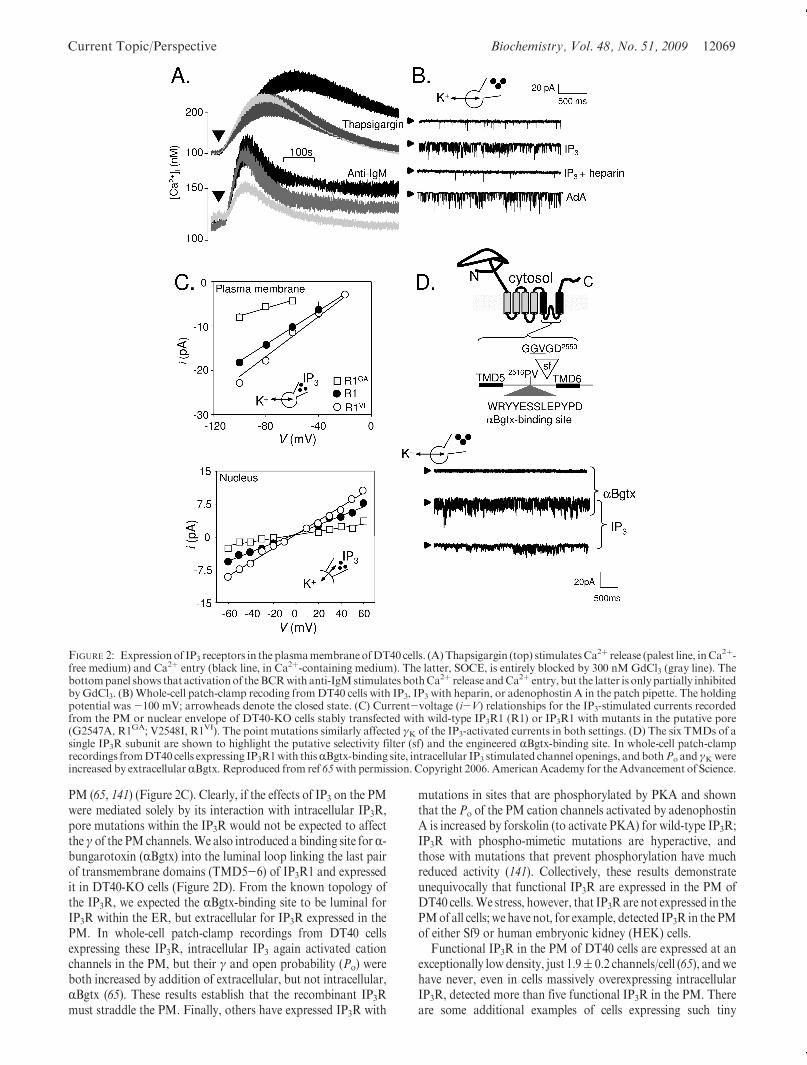
numbers of channels in the PM, ∼10 intermediate conductanceCa2þ-activated Kþ channels (IKCa1) in a resting T-cell (162), forexample, or two or three RyR-like channels in rat portal veinmyocytes (163) (see later), butmost channels expressed in the PMoccur at much higher densities (typically >100 channels/μm2 orseveral thousand channels per cell) (8). An intriguing feature ofIP3R expression in the PM is that in several hundred recordings,we never failed to detect at least one IP3R in the PM (65, 140). Ifthese IP3Rhad been randomly inserted into the PMwith very lowprobability, perhaps reflecting “leakage” from the ER, forexample, we would have expected (on the basis of the Poissondistribution) ∼28% of cells to lack PM IP3R. These resultssuggest that functional IP3R are reliably counted into the PM.This may reflect either counting of proteins into the PM or aregulatory process that only ever allows very few of a largernumber or resident channels to be active within the PM.
In seeking to address mechanisms that might allow suchreliable counting of PM IP3R, we considered that feedbackregulation of IP3R trafficking to the PM from active IP3R withinthe PMwas the most likely possibility. An IP3R in which a singleresidue (D2550A) was mutated to create a pore that is blockedwhenever its luminal surface is bathed inCa2þ provides a channelthat is effectively “pore-dead” throughout its normal life cycle asit passes from ER to PM, yet these channels were effectivelycounted in appropriate numbers (∼2 IP3R/cell) into the PM(Figure 3A). Another possibility was that a feedback signal arosefrom an earlier step in the activation of IP3R by IP3, aconformational change in the IP3-binding site, for example. Wetherefore expressed a mutant IP3R (R568Q) in which the affinityfor IP3 was reduced by ∼10-fold, arguing that at resting levels ofIP3 such IP3R would now be only 10% as likely as normal IP3R
to bind IP3, but these IP3R, like those that were pore-dead, werealso reliably counted into the PM with ∼2 IP3R/cell(Figure 3B) (140). These rather puzzling results suggest that verysmall numbers of functional IP3R are reliably counted into thePM in the apparent absence of any obvious feedback regulatorymechanism.
FUNCTIONAL RYANODINE RECEPTORS IN THE
PLASMA MEMBRANE
RyR are the closest relatives of IP3R, with which they shareboth structural (164, 165) and functional properties (166), andlike IP3R, they are expressed predominantly within the mem-branes of the ER (or SR in muscle), where they are retained byER retention signals within the TMDs (167). However, as withIP3R, evidence that RyR may also be expressed in post-ERmembranes, and in the PM of some cells, is accumulating.
Within pancreatic β-cells, for example, or insulinoma cellsderived from them, functionalRyRappear to be expressedwithinthe membranes of insulin-containing secretory vesicles (168, 169)and/or endosomes (170), whereas neither organelle expressesIP3R (171). In chromaffin cells, both RyR and IP3R appear toescape the ER and function as Ca2þ release channels withinsecretory vesicles (118). Perhaps more contentious is the possi-bility that RyRmay be expressed in the PM (172). In this section,we briefly review the evidence that RyR are functionally ex-pressed in the PMof some cells, and in the concluding section, weconsider the possible physiological significance of RyR and IP3Rin the PM.
An early suggestion that RyR might be expressed in the PMcame from studies of osteoclasts (173). Osteoclasts probably
FIGURE 3: Counting IP3 receptors into the plasma membrane. (A) A point mutation within the putative pore region of IP3R1 (D2550A,highlighted) causes luminal/extracellular Ca2þ to block the channel, but it does not prevent cells from reliably counting IP3R into the PM. Thehistogram shows the observed and predicted (from the Poisson distribution) numbers of functional IP3R detected in each cell and establishes thatIP3R are not randomly inserted into the PM. (B) A point mutation within the IP3-binding core (R568Q, IP3R1RQ) reduces the binding affinity ofthe IP3R for IP3 by 10-fold, evidencedby radioligandbinding analyses (not shown) and the 10-fold decrease in the sensitivity ofCa
2þ release to IP3(left). The reduced sensitivity to IP3 does not impair the reliability withwhich IP3R are functionally expressed in the PM (right). Reproduced withpermission from ref 140. Copyright 2008. American Society for Biochemistry and Molecular Biology.
Current Topic/Perspective Biochemistry, Vol. 48, No. 51, 2009 12071
express functional RyR. These may, as they do elsewhere,amplify, by Ca2þ-induced Ca2þ release, the Ca2þ signals gener-ated by activation of other Ca2þ channels within intracellularstores or the PM (174, 175). However, the evidence that RyR arealso expressed in the PM of osteoclasts deserves close examina-tion because the conclusion is important. Extracellular ruthe-nium red, an antagonist of RyR, blocked Ni2þ-evoked Ca2þ
signals (173). Ni2þ is known to activate the extracellular Ca2þ-sensing receptor (CaR), a G-protein-coupled receptor (176),although the role of CaR in osteoclasts has been contentious.The inhibition by ruthenium red of Ni2þ-evoked Ca2þ signalsneed not reflect an action at PM RyR, because ruthenium red ismembrane-impermeant and its most substantial effects on RyRappear to be mediated by binding to its cytosolic surface (177). Itis perhaps more likely that ruthenium red, a polycation, interactswith the Ca2þ-sensing receptor (CaR), which is known to beregulated by polyvalent metal ions and polyamines (176). Thisinterpretation would also be consistent with evidence thatruthenium red itself evoked Ca2þ signals in osteoclasts (178).Ni2þ was suggested, although without quantitative analysis, toinhibit binding of [3H]ryanodine to osteoblasts (173), but thesebinding assays do not distinguish intracellular RyR from those inthe PM. Because ryanodine binding is use-dependent, any effectof agents that increase the cytosolic Ca2þ concentration on[3H]ryanodine binding might simply reflect regulation of intra-cellular RyR by Ca2þ. Confocal microscopy identified peripheralimmunostaining for RyR (173), but the resolution is insufficientto resolve whether RyR were in the PM or within ER lying closeto it. An anti-peptide antiserum (Ab129) raised to 22 residueswithin a region toward the C-terminus of RyR2 immunostainedintact osteoclasts (173). Because the N-terminus of the RyR iscytosolic (179), the limited information provided in the originalpublication (173), namely that the epitope lies between TMD6and TMD7 of the originally proposed 10-TMD model ofRyR (180), would place the epitope within the cytosol. Subse-quent work revealed that Ab129 was raised to residues 4676-4699of rabbit RyR2 (181), and the revised six-TMD models ofRyR (167, 179) would place the sequence between TMD2 andTMD3 (181), which again places the epitope within the cytosol.Immunostaining of intact cells with this antiserum is thereforeunlikely to reflect its reaction with a PM RyR. Subsequentstudies, using valinomycin to manipulate membrane potential,indicated that hyperpolarization increased the susceptibility toblockade by ryanodine of the Ca2þ release evoked byNi2þ (182).This was interpreted as evidence that the RyR might sensemembrane potential (182) either directly (because it was residentwithin the PM) (175, 178) or indirectly via conformationalcoupling to another PM voltage sensor. Evidence that other G-protein-coupled receptors are regulated by membrane poten-tial (183) raises the possibility that the voltage sensor might evenbe the CaR itself. The significance of the latter findings (182),which provide no decisive evidence of a functional RyR in thePM, is diminished by the observation that in osteoclasts nottreated with valinomycin, responses to Ni2þ were insensitive toryanodine (182). Finally, patch-clamp recordings from excisedpatches of the PMof osteoclasts resolved small numbers of Ba2þ-permeable channels with unexpectedly high Po values (0.5-0.95)in the absence of any known agonists of RyR, but that activitywas massively attenuated by application of cytosolic rutheniumred or an antiserum to a cytosolic epitope of the RyR (175).Neither agent is likely to be specific, although the observationthat both inhibit the active channels is suggestive of a RyR, but
the results are perplexing. The authors show that with symme-trical Ba2þ-containing solutions, the reversal potential is (asmight be expected) 0 mV, but most traces, including those withmassive channel activity, show excised patches at a holdingpotential of 0 mV using symmetrical Ba2þ-containing solutions.Under these conditions, there should be no currents. Theinescapable conclusion is that the electrophysiological evidencein support of RyR in the PMof osteoclasts lacks credibility (175).We suggest that although Zaidi and his colleagues were amongthe first to suggest that RyRmight be expressed in the PM (173),neither the original nor subsequent reports provide compellingevidence.
High-conductance PM cation channels resembling RyR havebeen detected in other cell types. In smooth muscle from portalvein, for example, caffeine, ryanodine, Ca2þ, or extracellularstimuli that evoke an increase in cytosolic Ca2þ concentrationstimulated opening of high-conductance (γK ∼ 190 pS) cationchannels that were permeable to Ca2þ and Ba2þ, and onlymodestly selective between cations (PCa/PNa ∼ 21) [althoughmore so than conventional RyR (184)] (163). It is also note-worthy that the γ of these PMchannels is substantially lower thanthat of RyR reconstituted into lipid bilayers (184) but similar tothat of RyR in the PM of insulinoma cells (γK ∼ 169 pS) (185)(see later). In whole-cell recordings, no more than three simulta-neous openings of these channels were detected, suggesting thatvery few were expressed in the PM. These authors may be correctin cautiously concluding that the PM channels are not RyRthemselves, but other Ca2þ-sensitive channels activated in re-sponse to Ca2þ released from intracellular RyR (163), but itremains entirely plausible that these channels result from RyRwithin the PM that are regulated by physiological stimuli(norepinephrine or acetylcholine) (163). Other studies of gas-tric (186) or arterial (187) smooth muscle detected caffeine-stimulated nonselective cation channels in the PM, but theirinsensitivity to ryanodine (186), low conductance, or lack ofpermeability to bivalent cations (187) suggests that these areprobably not RyR in the PM. In cardiac myocytes, too, caffeineor an increased cytosolic Ca2þ concentration stimulated openingof cation channels with high conductance (γNa ∼ 400 pS) andpermeability to Ba2þ (188, 189). The most persuasive evidencethat thesemight beRyR in the PM is the ability of ryanodine, as itdoes for RyR in bilayers (172, 177), to lock the channels into asubconducting state (189). Collectively, these observations pro-vide persuasive evidence that cardiac myocytes may express justone to four functional RyR within the PM (189). The possibilitythat ventricular myocytes express small numbers of RyR in thePM clearly deserves further study both to strengthen the conclu-sion and to address likely physiological roles.
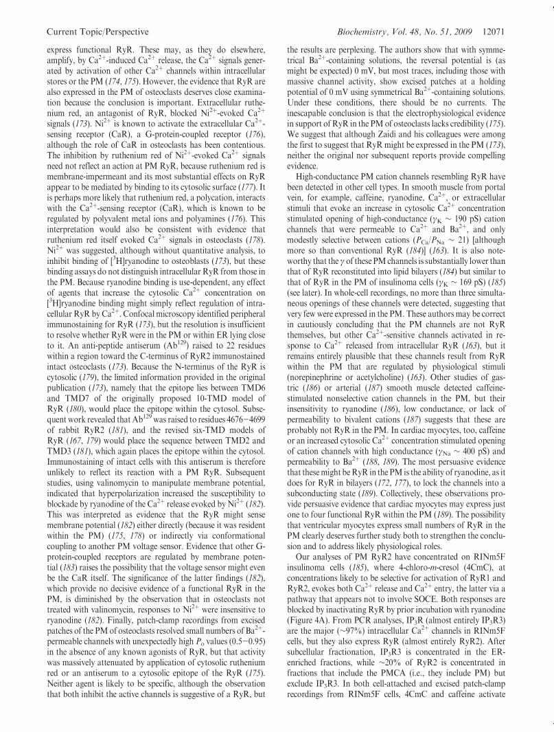
Our analyses of PM RyR2 have concentrated on RINm5Finsulinoma cells (185), where 4-chloro-m-cresol (4CmC), atconcentrations likely to be selective for activation of RyR1 andRyR2, evokes both Ca2þ release and Ca2þ entry, the latter via apathway that appears not to involve SOCE. Both responses areblocked by inactivating RyR by prior incubation with ryanodine(Figure 4A). From PCR analyses, IP3R (almost entirely IP3R3)are the major (∼97%) intracellular Ca2þ channels in RINm5Fcells, but they also express RyR (almost entirely RyR2). Aftersubcellular fractionation, IP3R3 is concentrated in the ER-enriched fractions, while ∼20% of RyR2 is concentrated infractions that include the PMCA (i.e., they include PM) butexclude IP3R3. In both cell-attached and excised patch-clamprecordings from RINm5F cells, 4CmC and caffeine activate
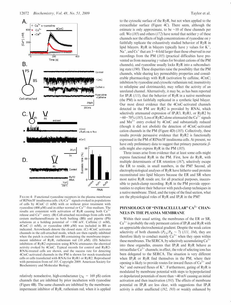
12072 Biochemistry, Vol. 48, No. 51, 2009 Taylor et al.
relatively nonselective, high-conductance (γK = 169 pS) cationchannels that are inhibited by prior incubation with ryanodine(Figure 4B). The same channels are inhibited by the membrane-impermeant inhibitor of RyR, ruthenium red, when it is applied
to the cytosolic surface of the RyR, but not when applied to theextracellular surface (Figure 4C). There seem, although theestimate is only approximate, to be ∼10 of these channels percell. We (185) and others (172) have noted that neither γ of thesechannels nor the effects of high concentrations of ryanodine on γfaithfully replicate the exhaustively studied behavior of RyR inlipid bilayers. RyR in bilayers typically have γ values for Kþ,Naþ, andCsþ that are 3-4-fold larger than those observed in ourrecordings from the PM (185) (practical difficulties have pre-vented us frommeasuring γ values for bivalent cations of the PMchannels), and ryanodine usually locks RyR into a subconduct-ing state (190). These disparities raise the possibility that the PMchannels, while sharing key permeability properties and consid-erable pharmacology with RyR (activation by caffeine, 4CmC;inhibition by ryanodine and cytosolic ruthenium red; insensitivityto nifedipine and clotrimazole), may reflect the activity of anunrelated channel. Alternatively, it may be, as has been reportedfor IP3R (111), that the behavior of RyR in a native membrane(the PM) is not faithfully replicated in a synthetic lipid bilayer.Our most direct evidence that the 4CmC-activated channelsdetected in the PM are RyR2 is provided by RNAi, whichselectively attenuated expression of IP3R3, RyR1, or RyR2 by∼60-70% (185). Loss ofRyR2alone attenuated theCa2þ signalsand Mn2þ entry evoked by 4CmC and substantially reduced(though it did not abolish) the detection of 4CmC-activatedcation channels in the PM (Figure 4D) (185). Collectively, theseresults provide persuasive evidence that RyR2 is functionallyexpressed in the PMof RINm5F insulinoma cells. At present, wehave only preliminary data to suggest that primary pancreatic β-cells might also express RyR in the PM (185).
Three issues arise from evidence that at least some cells mightexpress functional RyR in the PM. First, how do RyR, withmultiple determinants of ER retention (167), selectively escapethe ER to reside, in small numbers, in the PM? Second, allelectrophysiological analyses of RyR have hitherto used proteinsreconstituted into lipid bilayers because the ER and SR wheremost native RyR reside are, for all practical purposes, inacces-sible to patch-clamp recording. RyR in the PM provide oppor-tunities to explore their behavior with patch-clamp techniques ina nativemembrane. Third, and the topic of the final section, whatare the physiological roles of RyR and IP3R in the PM?
PHYSIOLOGYOF“INTRACELLULAR”CA2þCHAN-
NELS IN THE PLASMAMEMBRANE
Within their usual setting, the membranes of the ER or SR,Ca2þ is probably the only permeant cation of IP3R andRyRwithan appreciable electrochemical gradient. Despite the weak cationselectivity of both channels (PCa/PK ∼ 7) (111, 184), they aretherefore likely to conduct mainly Ca2þ when they open withinthesemembranes. The SERCA, by selectively accumulatingCa2þ
into these organelles, ensures that IP3R and RyR behave asintracellular Ca2þ channels; in effect, the role of selecting ions hasbeen delegated to the SERCA. The situation is very differentwhen IP3R or RyR find themselves in the PM, where theiropening is likely to provide routes for inward fluxes of Ca2þ andNaþ and outward fluxes of Kþ. Furthermore, gating of RyR ismodulated by membrane potential with steps to hyperpolarizedor depolarized potentials of more than∼40 mV causing an initialactivation and then inactivation (191). The effects of membranepotential on IP3R are less clear, with suggestions that IP3Ractivity is either unaffected (192, 193) or weakly enhanced by
FIGURE 4: Functional ryanodine receptors in the plasma membraneofRINm5F insulinoma cells. (A) Ca2þ signals evoked in populationsof cells by 4CmC (1 mM) with or without prior treatment withryanodine (400 μM) and in either normal or Ca2þ-free medium. Theresults are consistent with activation of RyR causing both Ca2þ
release and Ca2þ entry. (B) Cell-attached recordings from cells withcesium methanesulfonate in both bathing (BS) and pipette (PS)solutions at a holding potential of -100 mV. Caffeine (1 mM),4CmC (1 mM), or ryanodine (400 μM) was included in BS asindicated. Arrowheads denote the closed state. (C) 4CmC activateschannels in the cell-attached mode, which are then rapidly inhibitedwhen the patch is excised into BS containing the membrane-imper-meant inhibitor of RyR, ruthenium red (10 μM). (D) Selectiveinhibition of RyR2 expression using RNAi attenuates the electricalactivity evoked by 4CmC. Typical records for control and RyR2-RNAi-treated cells are shown, and the success rate for detecting4CmC-activated channels in the PM is shown for mock-transfectedcells or cells transfected with RNAi for RyR1 or RyR2. Reproducedwith permission from ref 185. Copyright 2009. American Society forBiochemistry and Molecular Biology.
Current Topic/Perspective Biochemistry, Vol. 48, No. 51, 2009 12073
depolarization (194, 195). Depolarization and hyperpolarizationhave also been reported to increase γ by relieving theMg2þ blockof the channel (196). These effects ofmembrane potential onRyRand perhaps IP3R have been rather neglected because within theER/SR such regulation is unlikely to be significant, but it may bevery important when IP3R or RyR are expressed in the PM.Within the PM, therefore, IP3R and RyR may regulate bothmembrane potential and Ca2þ entry, and their activity may alsobe regulated by membrane potential.
The behavior of IP3R (111, 197, 198) and RyR (199, 200) ismodulated by their association with many different accessoryproteins. These provide additional levels of regulation of channelgating, directly by the associated proteins (199, 201, 202), viaphosphorylation (203, 204), or via the proteins serving as sensorsof, for example, redox potential (205) or Ca2þ (206). Otherassociated proteins, for example, IRBIT (207, 208) and CaM-KII (17), are directly regulated by the active channels. Theseaccessory proteins include those, like chromogranin (209),ERp44 (205), and calsequestrin (200), that are expressed withinthe ER/SR lumen and others, like calnexin (210), junctin, andtriadin (200, 211), that are expressed only in ER/SR membranes.Each of these channels therefore provides a scaffold aroundwhich a signaling complex is assembled, which then defines thecomplex integrative behavior of IP3R andRyR. The componentsof these signaling complexes must be different for channels in theER/SR andPM, not least because the latter are devoid of luminalproteins. As proteins pass through post-ER compartments,attached carbohydrates are further processed, and these mod-ifications may also affect the behavior of IP3R and RyR thatprogress to the PM. It has, for example, been suggested that PMIP3R in lymphocytes are enriched in sialic acid and differ fromintracellular IP3R in their affinity for IP3 (129). The key pointsare that IP3R and RyR within the PM may, by assembling intodifferent signaling complexes, differ from their intracellularcounterparts both in their regulation and in the downstreamproteins to which they signal.
Ca2þ channels within the ER/SR access a finite Ca2þ store,whereas those within the PM have access to an unlimited pool ofextracellular Ca2þ. The difference is likely to affect the durationof the Ca2þ signals evoked by IP3R and RyR in the two settings,and the impact of feedback regulation by luminal Ca2þ concen-tration on channel gating (206, 212). Finally, because Ca2þ
signals can be locally decoded, the Ca2þ released by channelswithin the ER/SR is likely to recruit the activity of different Ca2þ
sensors to those arising from PM channels. Ca2þ signals emanat-ing from IP3R (or RyR) within the PM will, therefore, havedifferent spatiotemporal profiles compared to those arising fromthe same channels with the ER/SR, and each may therebyregulate different cellular responses.
In the remainder of this final section, we consider the likelyphysiological significance of these effects of RyR and IP3R in thePM.
Bone mass in adults is maintained by the counteractingactivities of the osteoblasts that deposit bone and the osteoclaststhat resorb it. Numerous feedback loops coordinate the activitiesof these two cell types (213), one of which is inhibition of theresorptive activity of osteoclasts by high local extracellular Ca2þ
concentrations, which trigger an increase in cytosolic Ca2þ
concentration (214). CaR, with its huge extracellular region thatbinds Ca2þ (176), mediates the responses of many cells, likeosteoblasts (215) and parathyroid chief cells (216), to changes inextracellular Ca2þ concentration. The role of the CaR in
osteoclasts has been more contentious, although osteoclastsexpress CaR (217, 218) and loss of CaR severely compromisestheir responses to extracellular Ca2þ concentration (218). Theseobservations are consistent with CaR, via activation of phos-pholipase C and an increase in cytosolic Ca2þ concentration,playing a role in feedback inhibition of osteoclast activity byextracellular Ca2þ (219, 220). Zaidi and his colleagues havesuggested that RyR2 within the PM of osteoclasts may furthercontribute to these Ca2þ-regulated pathways, minimally byproviding a route for entry of Ca2þ across the PM, but perhapsalso by providing an additional sensor for extracellularCa2þ (221, 222). The latter suggestion derives from the observa-tion that RyR within the more typical setting, the SR, areregulated by luminal Ca2þ (223). Several arguments suggest thatthe luminal surface of RyR2 is unlikely to serve as a sensor ofextracellular Ca2þ in osteoclasts. First, the affinity of the luminalCa2þ-binding site onRyR2 (KD
Ca∼ 40 μM) (212) is far too high,even allowing for some competition with extracellular Mg2þ, torespond to the changes in the extracellular Ca2þ concentration(several millimolar) to which osteoclasts respond. Second, itseems likely though perhaps not proven that accessory proteins,such as luminal calsequestrin (224), mediate regulation ofRyR2 byluminalCa2þ. Such proteinswould be unlikely to associatewith theextracellular surface of RyR in the PM. Finally, the contributionsfrom intracellular Ca2þ stores and Ca2þ entry (182, 225) and thepharmacology of the intracellular Ca2þ signals evoked in osteo-clasts (stimulation by extracellular Ca2þ, Cd2þ, and Ni2þ) (226)seem more likely to reflect initiation of the Ca2þ signals byCaR (227, 228) rather than RyR. It seems very unlikely, therefore,that a PM RyR serves as an extracellular Ca2þ sensor inosteoclasts. Indeed, until there is more compelling evidence insupport of a functional RyR in the PM of osteoclasts, whether theRyR plays any direct role in mediating Ca2þ entry, rather thansimply fulfilling its more conventional role as an intracellular Ca2þ
channel, in osteoclasts remains an open question.Secretion of insulin from pancreatic β-cells is regulated by the
synergistic actions of glucose and gut hormones (incretins) thatstimulate cAMP formation. Glucose metabolism causes anincrease in cytosolic ATP concentration, which closes KATP
channels, leading, via an unidentified leak channel, to depolar-ization of the PM and thereby activation of Cav channels, anincrease in cytosolic Ca2þ concentration, and exocytosis ofinsulin-containing vesicles (229). Treatment of type 2 diabetesmellitus with sulfonylureas, which close KATP channels, affirmsthe importance of this pathway in insulin secretion, but it is clearthat glucose can also stimulate insulin release via pathways,presently ill-defined, that do not require closure of KATP
channels (230-232). One of several possibilities (233) is that thissecond pathway requires RyR. Functional RyR, most likelyRyR2, are certainly expressed in insulinoma and pancreatic β-cells (170, 234), and stimulation of RyR can evoke insulinrelease (170, 235, 236). Whether RyR are required for glucose-evoked insulin release is less clear (168, 170, 236-242). There is,however, evidence that development of type 2 diabetes isassociated with a loss of functional RyR2 (170, 243, 244).
Minimally, RyR within β-cells seem able, via Ca2þ-inducedCa2þ release, to amplify the Ca2þ signals evoked by glucose-stimulated Ca2þ entry (234, 245-247). Because the sensitivity ofRyR to Ca2þ is modulated by many additional signals, includingcyclic ADP ribose, ATP, cAMP, and redox state, other signalingpathways, the incretins, for example, may regulate the gain onthis relationship between Ca2þ entry and its amplification by
12074 Biochemistry, Vol. 48, No. 51, 2009 Taylor et al.
intracellular Ca2þ stores. RyR within the PM might fulfill asimilar role by coordinating responses from different signalingpathways and transducing them into opening of a channel thatwould both depolarize the PM (by allowing Naþ entry) andprovide a direct route for Ca2þ entry. Expressing a very high-conductance, nonselective cation channel (the RyR) in the PMofan electrically excitable cell (the β-cell) might seem to be adangerous undertaking, but it is worth recalling that RyR arealso regulated by membrane potential, such that they rapidlyinactivate after step changes to either hyperpolarizing or depo-larizing potentials (248). It may therefore be that within the PM,RyR transiently open only when provided with coincidentstimuli: a step change in membrane potential and delivery ofan appropriate cytosolic signal, such as Ca2þ or cyclic ADPribose. Similar considerations might be important determinantsof RyR activity in cardiac myocytes (188, 189) or vascularsmoothmuscle (163). A further level of control might be imposedby dynamic trafficking of RyR to and from the PM of β-cells.Within insulinoma cells, RyR are expressed within insulin-containing vesicles (249) and/or endosomes (170), suggestingthat both secretion of insulin and the subsequent retrieval of themembrane by endocytosis (250) may be intimately associatedwith regulated expression of RyR in the PM. Further work isrequired to extend results with insulinoma cells to primary β-cellsand so establish whether they too express functional RyR2 in thePM, to determine whether PM expression of RyR2 is dynami-cally regulated, and to establish the consequences for β-cellphysiology of gating RyR within the PM.
The evidence that IP3R are expressed in the PM of DT40 cellsis compelling (65, 120, 140, 141, 143, 251). Rather less secure,because it rests more on correlative evidence (see above), is oursuggestion that IP3R in the PM are entirely responsible for theBCR-evoked Ca2þ entry that occurs via a non-SOCE pathway(Figure 2A). That conclusion implies that the two or three IP3Rfound in the PM of each DT40 cell are responsible for approxi-mately half the Ca2þ entry evoked by the BCR (Figure 2A) (120).Our earlier analysis (65), in which we used the measured Ca2þ
conductance of the PM IP3R (γCa ∼ 9 pS), its open probabilitywhen maximally activated by IP3 (Po∼ 0.24), and the number ofIP3R expressed in the PM (∼2) to estimate the likely flux of Ca2þ
(∼4 � 105 Ca2þ/s) through the PM IP3R, suggested that twoIP3R in the PM are sufficient to mediate the Gd3þ-insensitiveCa2þ entry evoked by activation of the BCR (Figure 2A). Theremaining Ca2þ entry, via SOCE, occurs via some 10000 or moreOrai channels (65, 252). We speculate, although without specificevidence yet, that similar amounts of Ca2þ gushing into the cellvia just two or three IP3R is likely to generate very different localCa2þ signals and regulate a different response to that dribblinginto the cell via 10000 low-conductance Orai channels (16). Theeffects of the two pathways onmembrane potential are also likelyto differ: opening of IP3R within the PM is likely to causedepolarization, whereas Orai channels are exquisitely Ca2þ-selective and unlikely to regulate membrane potential directly.Whether IP3R in the PM, where they may be closely associatedwith the signaling machinery that generates IP3, respond differ-ently to intracellular IP3R when cells are stimulated is anotherissue that needs to be resolved.
REFERENCES
1. Berridge, M. J., Bootman, M. D., and Roderick, H. L. (2003)Calcium signalling: Dynamics, homeostasis and remodelling. Nat.Rev. Mol. Cell Biol. 4, 517–529.
2. Berridge, M. J., Lipp, P., and Bootman, M. D. (2000) The versatilityand universality of calcium signalling. Nat. Rev. Mol. Cell Biol. 1,11–21.
3. Xiong, T. C., Bourque, S., Lecourieux, D., Amelot, N., Grat, S.,Briere, C., Mazars, C., Pugin, A., and Ranjeva, R. (2006) Calciumsignaling in plant cell organelles delimited by a double membrane.Biochim. Biophys. Acta 1763, 1209–1215.
4. Kirichok, Y., Krapavinsky, G., and Clapham, D. E. (2004) Themitochondrial calcium uniporter is a highly selective ion channel.Nature 427, 360–364.
5. Toyoshima, C. (2008) Structural aspects of ion pumping by Ca2þ-ATPase of sarcoplasmic reticulum. Arch. Biochem. Biophys. 476,3–11.
6. Brini, M. (2009) Plasma membrane Ca2þ-ATPase: From a house-keeping function to a versatile signaling role. Pfluegers Arch. 457,657–664.
7. Van Baelen, K., Dode, L., Vanoevelen, J., Callewaert, G., De Smedt,H., Missiaen, L., Parys, J. B., Raeymaekers, L., and Wuytack, F.(2004) The Ca2þ/Mn2þ pumps in the Golgi apparatus. Biochim.Biophys. Acta 1742, 103–112.
8. Hille, B. (2001) Ionic Channels of Excitable Membranes , 3rd ed.,Sinauer Associates, Inc., Sunderland, MA.
9. Gillespie, D., and Fill, M. (2008) Intracellular calcium releasechannels mediate their own countercurrent: The ryanodine receptorcase study. Biophys. J. 95, 3706–3714.
10. Allbritton, N. L., Meyer, T., and Stryer, L. (1992) Range ofmessenger action of calcium ion and inositol 1,4,5-trisphosphate.Science 258, 1812–1815.
11. Zhou, Z., and Neher, I. (1993)Mobile and immobile calcium buffersin bovine adrenal chromaffin cells. J. Physiol. 469, 245–273.
12. Falcke, M. (2004) Reading patterns in living cells: The physics ofCa2þ signaling. Adv. Phys. 53, 255–440.
13. Lewis, R. S. (2007) The molecular choreography of a store-operatedcalcium channel. Nature 446, 284–287.
14. Willoughby, D., and Cooper, D. M. (2007) Organization and Ca2þ
regulation of adenylyl cyclases in cAMP microdomains. Physiol.Rev. 87, 965–1010.
15. Dudzinski, D. M., Igarashi, J., Greif, D., and Michel, T. (2006) Theregulation and pharmacology of endothelial nitric oxide synthase.Annu. Rev. Pharmacol. Toxicol. 46, 235–276.
16. Di Capite, J., Ng, S. W., and Parekh, A. B. (2009) Decoding ofcytoplasmic Ca2þ oscillations through the spatial signature drivesgene expression. Curr. Biol. 19, 853–858.
17. Bare, D. J., Kettlun, C. S., Liang, M., Bers, D. M., and Mignery,G. A. (2005) Cardiac type-2 inositol 1,4,5-trisphosphate receptor:Interaction and modulation by CaMKII. J. Biol. Chem. 280, 15912–15920.
18. Tadross, M. R., Dick, I. E., and Yue, D. T. (2008) Mechanism oflocal and global Ca2þ sensing by calmodulin in complex with a Ca2þ
channel. Cell 133, 1228–1240.19. Marchant, J. S., and Parker, I. (2001) Role of elementary Ca2þ puffs
in generating repetitive Ca2þ oscillations. EMBO J. 20, 65–76.20. Dalbey, R. E., and Von Heijne, G. (2002) Protein targeting, trans-
port and translocation , Academic Press, New York.21. Dalbey, R. E., Koehler, C. M., and Tamanoi, F., Eds. (2007) The
enzymes. Molecular machines involved in protein transport acrosscellular membranes , Vol. 25, Academic Press, London.
22. St Johnston, D. (2005) Moving messages: The intracellular localiza-tion of mRNAs. Nat. Rev. Mol. Cell Biol. 6, 363–375.
23. Simon, A.M., Hoppe, P., and Burden, S. J. (1992) Spatial restrictionof AChR gene expression to subsynaptic nuclei. Development 114,545–553.
24. Dahm, R., Kiebler, M., and Macchi, P. (2007) RNA localisation inthe nervous system. Semin. Cell Dev. Biol. 18, 216–223.
25. Margeot, A., Garcia, M., Wang, W., Tetaud, E., di Rago, J. P., andJacq, C. (2005) Why are many mRNAs translated to the vicinity ofmitochondria: A role in protein complex assembly?Gene 354, 64–71.
26. Schmid, M., Jaedicke, A., Du, T. G., and Jansen, R. P. (2006)Coordination of endoplasmic reticulum and mRNA localization tothe yeast bud. Curr. Biol. 16, 1538–1543.
27. Choi, S. B.,Wang, C.,Muench,D.G., Ozawa,K., Franceschi, V. R.,Wu, Y., and Okita, T. W. (2000) Messenger RNA targeting ofrice seed storage proteins to specific ER subdomains. Nature 407,765–767.
28. Hengst, U., and Jaffrey, S. R. (2007) Function and translationalregulation of mRNA in developing axons. Semin. Cell Dev. Biol. 18,209–215.
29. Bannai, H., Fukatsu, K., Mizutani, A., Natsume, T., Iemura, S.,Ikegami, T., Inoue, T., and Mikoshiba, K. (2004) An RNA-inter-
Current Topic/Perspective Biochemistry, Vol. 48, No. 51, 2009 12075
acting protein, SYNCRIP (heterogeneous nuclear ribonuclear pro-tein Q1/NSAP1) is a component of mRNA granule transported withinositol 1,4,5-trisphosphate receptor type 1 mRNA in neuronaldendrites. J. Biol. Chem. 279, 53427–53434.
30. Du, T. G., Schmid, M., and Jansen, R. P. (2007) Why cells movemessages: The biological functions of mRNA localization. Semin.Cell Dev. Biol. 18, 171–177.
31. Jansen, R. P. (2001) mRNA localization:Message on the move.Nat.Rev. Mol. Cell Biol. 2, 247–256.
32. van Vliet, C., Thomas, E. C.,Merino-Trigo, A., Teasdale, R. D., andGleeson, P. A. (2003) Intracellular sorting and transport of proteins.Prog. Biophys. Mol. Biol. 83, 1–45.
33. Platta, H. W., and Erdmann, R. (2007) The peroxisomal proteinimport machinery. FEBS Lett. 581, 2811–2819.
34. Benz, J. P., Soll, J., and Bolter, B. (2009) Protein transport inorganelles: The composition, function and regulation of the Ticcomplex in chloroplast protein import. FEBS Lett. 276, 1166–1176.
35. Neupert, W., and Herrmann, J. M. (2007) Translocation of proteinsinto mitochondria. Annu. Rev. Biochem. 76, 723–749.
36. Hessa, T., Meindl-Beinker, N. M., Bernsel, A., Kim, H., Sato, Y.,Lerch-Bader,M., Nilsson, I.,White, S. H., and vonHeijne, G. (2007)Molecular code for transmembrane-helix recognition by the Sec61translocon. Nature 450, 1026–1030.
37. White, S. H., and von Heijne, G. (2008) How translocons selecttransmembrane helices. Annu. Rev. Biophys. Bioeng. 37, 23–42.
38. Abell, B. M., Pool, M. R., Schelenker, O., Sinning, I., and High, S.(2004) Signal recognition particle mediates post-translational target-ing in eukaryotes. EMBO J. 23, 2755–2764.
39. von Heijne, G. (2006) Membrane-protein topology. Nat. Rev. Mol.Cell Biol. 7, 909–918.
40. Vembar, S. S., and Brodsky, J. L. (2008) One step at a time:Endoplasmic reticulum-associated degradation. Nat. Rev. Mol. CellBiol. 9, 944–957.
41. Grove, D. E., Rosser, M. F., Ren, H. Y., Naren, A. P., and Cyr, D.M. (2009) Mechanisms for rescue of correctable folding defects inCFTRΔF508. Mol. Biol. Cell 20, 4059–4069.
42. Saksena, S., Summers, M. D., Burks, J. K., Johnson, A. E., andBraunagel, S. C. (2006) Importin-R-16 is a translocon-associatedprotein involved in sorting membrane proteins to the nuclearenvelope. Nat. Struct. Mol. Biol. 13, 500–508.
43. Tabak, H. F., van der Zand, A., and Braakman, I. (2008) Peroxi-somes: Minted by the ER. Curr. Opin. Cell Biol. 20, 393–400.
44. Groves, E., Dart, A. E., Covarelli, V., and Caron, E. (2008)Molecular mechanisms of phagocytic uptake in mammalian cells.Cell. Mol. Life Sci. 65, 1957–1976.
45. Dong, C., Filipeanu, C. M., Duvernay, M. T., and Wu, G. (2007)Regulation of G protein-coupled receptor export trafficking. Bio-chim. Biophys. Acta 1768, 853–870.
46. Murrell-Lagnado, R. D., and Qureshi, O. S. (2008) Assembly andtrafficking of P2X purinergic receptors. Mol. Membr. Biol. 25,321–331.
47. Giamarchi, A., Padilla, F., Coste, B., Raoux,M., Crest,M., Honore,E., and Delmas, P. (2006) The versatile nature of the calcium-permeable cation channel TRPP2. EMBO Rep. 7, 787–793.
48. De Matteis, M. A., and Luini, A. (2008) Exiting the Golgi complex.Nat. Rev. Mol. Cell Biol. 9, 273–284.
49. Blott, E. J., and Griffiths, G. M. (2002) Secretory lysosomes. Nat.Rev. Mol. Cell Biol. 3, 122–131.
50. Seaman, M. N. (2008) Endosome protein sorting: Motifs andmachinery. Cell. Mol. Life Sci. 65, 2842–2858.
51. Qureshi, O. S., Paramasivam, A., Yu, J. C., and Murrell-Lagnado,R. D. (2007) Regulation of P2X4 receptors by lysosomal targeting,glycan protection and exocytosis. J. Cell Sci. 120, 3838–3849.
52. Kundra, R., and Kornfeld, S. (1999) Asparagine-linked oligosac-charides protect Lamp-1 and Lamp-2 from intracellular proteolysis.J. Biol. Chem. 274, 31039–31046.
53. Rodriguez-Boulan, E., Kreitzer, G., and Musch, A. (2005) Organi-zation of vesicular trafficking in epithelia.Nat. Rev.Mol. Cell Biol. 6,233–247.
54. Fukuda, M. (2008) Regulation of secretory vesicle traffic by Rabsmall GTPases. Cell. Mol. Life Sci. 65, 2801–2813.
55. Cullen, P. J. (2008) Endosomal sorting and signalling: An emergingrole for sorting nexins. Nat. Rev. Mol. Cell Biol. 9, 574–582.
56. Sudhof, T. C., and Rothman, J. E. (2009) Membrane fusion:Grappling with SNARE and SM proteins. Science 323, 474–447.
57. Mostov, K. E., Verges, M., and Altschuler, Y. (2000) Membranetraffic in polarized epithelial cells. Curr. Opin. Cell Biol. 12,483–490.
58. Munro, S. (2003) Lipid rafts: Elusive or illusive? Cell 115, 377–388.
59. Andersen, O. S., and Koeppe, R. E.II (2007) Bilayer thickness andmembrane protein function: An energetic perspective. Annu. Rev.Biophys. Biomol. Struct. 36, 107–130.
60. Vicinanza, M., D’Angelo, G., Di Campli, A., and DeMatteis, M. A.(2008) Phosphoinositides as regulators of membrane trafficking inhealth and disease. Cell. Mol. Life Sci. 65, 2833–2841.
61. Allen, J. A., Halverson-Tamboli, R. A., and Rasenick, M.M. (2007)Lipid raft microdomains and neurotransmitter signalling. Nat. Rev.Neurosci. 8, 128–140.
62. Pelham, H. R. B., and Munro, S. (1993) Sorting of membraneproteins in the secretory pathway. Cell 75, 603–605.
63. Criado, M., Eibl, H., and Barrantes, F. J. (1984) Functional proper-ties of the acetylcholine receptor incorporated in model lipid mem-branes. Differential effects of chain length and head group ofphospholipids on receptor affinity states and receptor-mediatedion translocation. J. Biol. Chem. 259, 9188–9198.
64. Yuan, C., O’Connell, R. J., Feinberg-Zadek, P. L., Johnston, L. J.,and Treistman, S. N. (2004) Bilayer thickness modulates the con-ductance of the BK channel in model membranes. Biophys. J. 86,3620–3633.
65. Dellis, O., Dedos, S., Tovey, S. C., Rahman, T.-U., Dubel, S. J., andTaylor, C. W. (2006) Ca2þ entry through plasma membrane IP3
receptors. Science 313, 229–233.66. Rahman, T., and Taylor, C. W. (2009) Dynamic regulation of IP3
receptor clustering and activity by IP3. Channels 3, 336–332.67. Suh, B. C., and Hille, B. (2008) PIP2 is a necessary cofactor for
ion channel function: How and why? Annu. Rev. Biophys. 37,175–195.
68. Lape, R., Colquhoun,D., and Sivilotti, L. G. (2008) On the nature ofpartial agonism in the nicotinic receptor superfamily. Nature 454,722–727.
69. Park, C. Y., Hoover, P. J., Mullins, F. M., Bachhawat, P., Covington,E. D., Raunser, S., Walz, T., Garcia, K. C., Dolmetsch, R. E., andLewis, R. S. (2009) STIM1 clusters and activates CRAC channels viadirect binding of a cytosolic domain to Orai1. Cell 136, 876–890.
70. Hewavitharana, T., Deng, X., Soboloff, J., and Gill, D. L. (2007)Role of STIM and Orai proteins in the store-operated calciumsignaling pathway. Cell Calcium 42, 173–182.
71. Mignen, O., Thompson, J. L., and Shuttleworth, T. J. (2007) STIM1regulates Ca2þ entry via arachidonate-regulated Ca2þ-selective(ARC) channels without store depletion or translocation to theplasma membrane. J. Physiol. 579, 703–715.
72. Jardin, I., Lopez, J. J., Redondo, P. C., Salido,G.M., andRosado, J.A. (2009) Store-operated Ca2þ entry is sensitive to the extracellularCa2þ concentration through plasma membrane STIM1. Biochim.Biophys. Acta 1793, 1614–1622.
73. Craven, K. B., and Zagotta, W. N. (2006) CNG and HCN channels:Two peas, one pod. Annu. Rev. Physiol. 68, 375–401.
74. Voets, T., Nilius, B., Hoefs, S., van der Kemp, A. W., Droogmans,G., Bindels, R. J., and Hoenderop, J. G. (2004) TRPM6 forms theMg2þ influx channel involved in intestinal and renal Mg2þ absorp-tion. J. Biol. Chem. 279, 19–25.
75. Vriens, J., Appendino, G., and Nilius, B. (2009) Pharmacology ofvanilloid transient receptor potential cation channels.Mol. Pharma-col. 75, 1262–1279.
76. Nilius, B., Owsianik, G., Voets, T., and Peters, J. A. (2007) Transientreceptor potential cation channels in disease. Physiol. Rev. 87,165–217.
77. Bidaux,G., Flourakis,M., Thebault, S., Zholos, A., Beck, B.,Gkika,D., Roudbaraki, M., Bonnal, J. L., Mauroy, B., Shuba, Y., Skryma,R., and Prevarskaya, N. (2007) Prostate cell differentiation statusdetermines transient receptor potential melastatin member 8channel subcellular localization and function. J. Clin. Invest. 117,1647–1657.
78. Turner, H., Fleig, A., Stokes, A., Kinet, J.-P., and Penner, R. (2003)Discimination of intracellular calcium store subcompartments usingTRPV1 (transient receptor potential channel, vanilloid subfamilymember 1) release channel activity. Biochem. J. 371, 341–350.
79. Catterall, W. A. (2000) Structure and regulation of voltage-gatedCa2þ channels. Annu. Rev. Cell Dev. Biol. 16, 521–555.
80. Dolphin, A. C. (2003) β subunits of voltage-gated calcium channels.J. Bioenerg. Biomembr. 35, 599–620.
81. Jeng, C. J., Sun, M. C., Chen, Y. W., and Tang, C. Y. (2008)Dominant-negative effects of episodic ataxia type 2 mutationsinvolve disruption of membrane trafficking of human P/Q-typeCa2þ channels. J. Cell. Physiol. 214, 422–433.
82. Talavera, K., and Nilius, B. (2006) Biophysics and structure-function relationship of T-type Ca2þ channels. Cell Calcium 40,97–114.
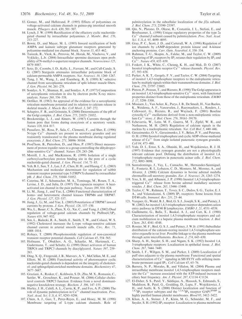
12076 Biochemistry, Vol. 48, No. 51, 2009 Taylor et al.
83. Gomez, M., and Hellstrand, P. (1995) Effects of polyamines onvoltage-activated calcium channels in guinea-pig intestinal smoothmuscle. Pfluegers Arch. 430, 501–507.
84. Lynch, J. W. (1999) Rectification of the olfactory cyclic nucleotide-gated channel by intracellular polyamines. J. Membr. Biol. 170,213–227.
85. Bowie, D., and Mayer, M. L. (1995) Inward rectification of bothAMPA and kainate subtype glutamate receptors generated bypolyamine-mediated ion channel block. Neuron 15, 453–462.
86. Turecek, R., Vlcek, K., Petrovic, M., Horak, M., Vlachova, V., andVyklicky, L.Jr. (2004) Intracellular spermine decreases open prob-ability ofN-methyl-D-aspartate receptor channels.Neuroscience 125,8879–8887.
87. Soto, D., Coombs, I. D., Kelly, L., Farrant, M., and Cull-Candy, S.G. (2007) Stargazin attenuates intracellular polyamine block ofcalcium-permeable AMPA receptors. Nat. Neurosci. 10, 1260–1267.
88. Tang, J. M., Wang, J., and Eisenberg, R. S. (1989) Kþ-selectivechannel from sarcoplasmic reticulum of split lobster muscle fibers.J. Gen. Physiol. 94, 261–278.
89. Somlyo, A. V., Shuman, H., and Somlyo, A. P. (1977) Compositionof sarcoplasmic reticulum in situ by electron probe X-ray micro-analysis. Nature 268, 556–558.
90. Oetliker, H. (1982) An appraisal of the evidence for a sarcoplasmicreticulum membrane potential and its relation to calcium release inskeletal muscle. J. Muscle Res. Cell Motil. 3, 247–272.
91. Schapiro, F. B., and Grinstein, S. (2000) Determinants of the pH ofthe Golgi complex. J. Biol. Chem. 275, 21025–21032.
92. Breckenridge, L. J., and Almers, W. (1987) Currents through thefusion pore that forms during exocytosis of a secretory vesicle.Nature 328, 814–817.
93. Passafaro, M., Rosa, P., Sala, C., Clementi, F., and Sher, E. (1996)N-type Ca2þ channels are present in secretory granules and aretransiently translocated to the plasma membrane during regulatedexocytosis. J. Biol. Chem. 271, 30096–30104.
94. Prod’hom, B., Pietrobon, D., and Hess, P. (1987) Direct measure-ment of proton transfer rates to a group controlling the dihydropyr-idine-sensitive Ca2þ channel. Nature 329, 243–246.
95. Morrill, J. A., and MacKinnon, R. (1999) Isolation of a singlecarboxyl-carboxylate proton binding site in the pore of a cyclicnucleotide-gated channel. J. Gen. Physiol. 114, 71–83.
96. Yeh, B. I., Sun, T. J., Lee, J. Z., Chen,H.H., andHuang, C. L. (2003)Mechanism and molecular determinant for regulation of rabbittransient receptor potential type 5 (TRPV5) channel by extracellularpH. J. Biol. Chem. 278, 51044–51052.
97. Caterina, M. J., Schumacher, M. A., Tominaga, M., Rosen, T. A.,Levine, J. D., and Juliu, D. (1997) The capsaicin receptor: A heat-activated ion channel in the pain pathway. Nature 389, 816–824.
98. Li, M., Jiang, J., and Yue, L. (2006) Functional characterization ofhomo- and heteromeric channel kinases TRPM6 and TRPM7.J. Gen. Physiol. 127, 525–537.
99. Jiang, J., Li, M., and Yue, L. (2005) Potentiation of TRPM7 inwardcurrents by protons. J. Gen. Physiol. 126, 137–150.
100. Wu, L., Bauer, C. S., Zhen, X. G., Xie, C., and Yang, J. (2002) Dualregulation of voltage-gated calcium channels by PtdIns(4,5)P2.Nature 419, 947–952.
101. Sen, L., Bialecki, R. A., Smith, E., Smith, T. W., and Colucci, W. S.(1992) Cholesterol increases the L-type voltage-sensitive calciumchannel current in arterial smooth muscle cells. Circ. Res. 71,1008–1014.
102. Rohacs, T. (2009) Phosphoinositide regulation of non-canonicaltransient receptor potential channels. Cell Calcium 45, 554–565.
103. Hofmann, T., Obukhov, A. G., Schaefer, M., Harteneck, C.,Gudermann, T., and Schultz, G. (1999) Direct activaton of humanTRPC6 and TRPC3 channels by diacylglycerol. Nature 397, 259–263.
104. Ding, X. Q., Fitzgerald, J. B.,Matveev, A. V.,McClellan,M. E., andElliott, M. H. (2008) Functional activity of photoreceptor cyclicnucleotide-gated channels is dependent on the integrity of cholester-ol- and sphingolipid-enriched membrane domains. Biochemistry 47,3677–3687.
105. Graziani, A., Rosker, C., Kohlwein, S.D., Zhu,M.X., Romanin, C.,Sattler, W., Groschner, K., and Poteser, M. (2006) Cellular choles-terol controls TRPC3 function: Evidence from a novel dominant-negative knockdown strategy. Biochem. J. 396, 147–155.
106. Hurley, J. H., Cahill, A. L., Currie, K. P., and Fox, A. P. (2000) Therole of dynamic palmitoylation in Ca2þ channel inactivation. Proc.Natl. Acad. Sci. U.S.A. 97, 9293–9298.
107. Chien, A. J., Gao, T., Perez-Reyes, E., and Hosey, M. M. (1998)Membrane targeting of L-type calcium channels. Role of
palmitoylation in the subcellular localization of the β2a subunit.J. Biol. Chem. 273, 23590–25597.
108. Qin, N., Platano, D., Olcese, R., Costantin, J. L., Stefani, E., andBirnbaumer, L. (1998) Unique regulatory properties of the type 2aCa2þ channel β subunit caused by palmitoylation. Proc. Natl. Acad.Sci. U.S.A. 95, 4690–4695.
109. Gray, P. C., Scott, J. D., and Catterall, W. A. (1998) Regulation ofion channels by cAMP-dependent protein kinase and A-kinaseanchoring proteins. Curr. Opin. Neurobiol. 8, 330–334.
110. Rahman, T.-U., Skupin, A., Falcke, M., and Taylor, C. W. (2009)Clustering of IP3 receptors by IP3 retunes their regulation by IP3 andCa2þ. Nature 458, 655–659.
111. Foskett, J. K., White, C., Cheung, K. H., and Mak, D. O. (2007)Inositol trisphosphate receptor Ca2þ release channels. Physiol. Rev.87, 593–658.
112. Parker, A. K. T., Gergely, F. V., and Taylor, C.W. (2004) Targetingof inositol 1,4,5-trisphosphate receptors to the endoplasmic reticu-lum bymultiple signals within their transmembrane domains. J. Biol.Chem. 279, 23797–23805.
113. Pinton, P., Pozzan, T., andRizzuto, R. (1998) TheGolgi apparatus isan inositol 1,4,5-trisphosphate-sensitive Ca2þ store, with functionalproperties distinct from those of the endoplasmic reticulum. EMBOJ. 17, 5298–5308.
114. Missiaen, L., Van Acker, K., Parys, J. B., De Smedt, H., Van Baelen,K., Weidema, A. F., Vanoevelen, J., Raeymaekers, L., Renders, J.,Callewaert, G., Rizzuto, R., and Wuytack, F. (2001) Baselinecytosolic Ca2þ oscillations derived from a non-endoplasmic reticu-lum Ca2þ store. J. Biol. Chem. 276, 39161–39170.
115. Echevarria, W., Leite, M. F., Guerra, M. T., Zipfel, W. R., andNathanson, M. H. (2003) Regulation of calcium signals in thenucleus by a nucleoplasmic reticulum. Nat. Cell Biol. 5, 440–446.
116. Gerasimenko, O. V., Gerasimenko, J. V., Belan, P. V., and Petersen,O. H. (1996) Inositol trisphosphate and cyclic ADP-ribose-mediatedrelease of Ca2þ from single isolated pancreatic zymogen granules.Cell 84, 473–480.
117. Yule, D. I., Ernst, S. A., Ohnishi, H., and Wojcikiewicz, R. J. H.(1997) Evidence that zymogen granules are not a physiologicallyrelevant calcium pool. Defining the distribution of inositol 1,4,5-trisphosphate receptors in pancreatic acinar cells. J. Biol. Chem.272, 9093–9098.
118. Santodomingo, J., Vay, L., Camacho, M., Hernandez-Sanmiguel,E., Fonteriz, R. I., Lobaton, C. D., Montero, M., Moreno, A., andAlvarez, J. (2008) Calcium dynamics in bovine adrenal medullachromaffin cell secretory granules. Eur. J. Neurosci. 28, 1265–1274.
119. Yoo, S. H., and Albanesi, J. P. (1990) Inositol 1,4,5-trisphosphate-triggered Ca2þ release from bovine adrenal medullary secretoryvesicles. J. Biol. Chem. 265, 13446–13448.
120. Taylor, C. W., Rahman, T., Tovey, S. C., Dedos, S. G., Taylor, E. J.A., and Velamakanni, S. (2009) IP3 receptors: Some lessons fromDT40 cells. Immunol. Rev. 231, 23–44.
121. Vazquez, G.,Wedel, B. J., Bird, G. S. J., Joseph, S. K., and Putney, J.W. (2002) An inositol 1,4,5-trisphosphate receptor-dependent cationentry pathway in DT40 B lymphocytes. EMBO J. 21, 4531–4538.
122. Guillemette, G., Balla, T., Baukal, A. J., and Catt, K. J. (1988)Characterization of inositol 1,4,5-trisphosphate receptors and cal-cium mobilization in a hepatic plasma membrane fraction. J. Biol.Chem. 263, 4541–4548.
123. Rossier,M. F., Bird, G. S. J., and Putney, J.W.Jr. (1991) Subcellulardistribution of the calcium-storing inositol 1,4,5-trisphosphate-sen-sitive organelle in rat liver. Possible linkage to the plasma membranethrough actin microfilaments. Biochem. J. 274, 643–650.
124. Sharp, A. H., Snyder, S. H., and Nigam, S. K. (1992) Inositol 1,4,5-trisphosphate receptors: Localization in epithelial tissue. J. Biol.Chem. 267, 7444–7449.
125. Smith, I. F., Wiltgen, S. M., and Parker, I. (2009) Localization ofpuff sites adjacent to the plasma membrane: Functional and spatialcharacterization of Ca2þ signaling in SH-SY5Y cells utilizing mem-brane-permeant caged IP3. Cell Calcium 45, 65–76.
126. Barrera, N. P., Morales, B., and Villalon, M. (2004) Plasma andintracellular membrane inositol 1,4,5-trisphosphate receptors med-iate the Ca2þ increase associated with the ATP-induced increase inciliary beat frequency. Am. J. Physiol. 287, C1114–C1124.
127. El-Daher, S. S., Patel, Y., Siddiqua, A., Hassock, S., Edmunds, S.,Maddison, B., Patel, G., Goulding, D., Lupu, F., Wojcikiewicz, J.H., and Authi, K. S. (2000) Distinct localization and function of1,4,5IP3 receptor subtypes and the 1,3,4,5IP4 receptor GAP1IP4BP inhighly purified human platelet membranes. Blood 95, 3412–3422.
128. Khan, A. A., Steiner, J. P., Klein, M. G., Schneider, M. F., andSnyder, S. H. (1992) IP3 receptor: Localization to plasma membrane
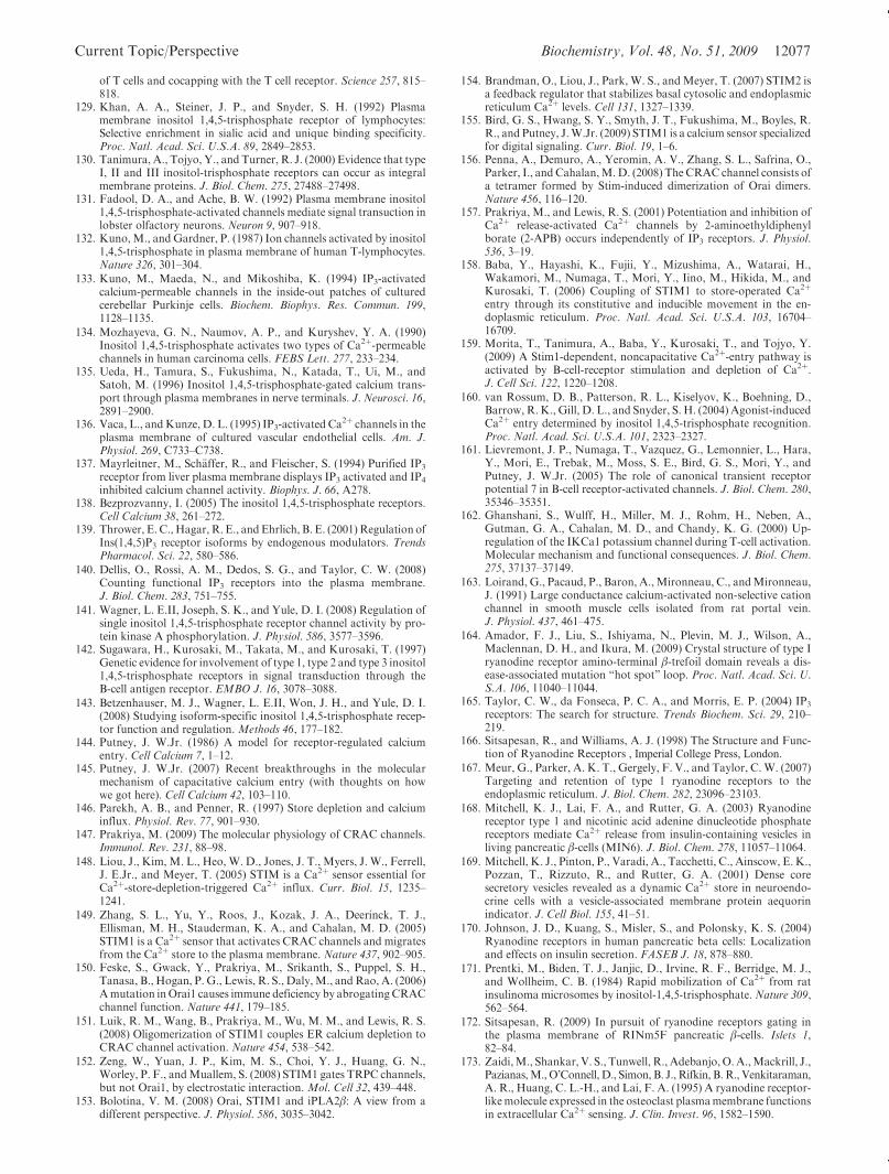
Current Topic/Perspective Biochemistry, Vol. 48, No. 51, 2009 12077
of T cells and cocapping with the T cell receptor. Science 257, 815–818.
129. Khan, A. A., Steiner, J. P., and Snyder, S. H. (1992) Plasmamembrane inositol 1,4,5-trisphosphate receptor of lymphocytes:Selective enrichment in sialic acid and unique binding specificity.Proc. Natl. Acad. Sci. U.S.A. 89, 2849–2853.
130. Tanimura, A., Tojyo, Y., and Turner, R. J. (2000) Evidence that typeI, II and III inositol-trisphosphate receptors can occur as integralmembrane proteins. J. Biol. Chem. 275, 27488–27498.
131. Fadool, D. A., and Ache, B. W. (1992) Plasma membrane inositol1,4,5-trisphosphate-activated channels mediate signal transuction inlobster olfactory neurons. Neuron 9, 907–918.
132. Kuno,M., andGardner, P. (1987) Ion channels activated by inositol1,4,5-trisphosphate in plasma membrane of human T-lymphocytes.Nature 326, 301–304.
133. Kuno, M., Maeda, N., and Mikoshiba, K. (1994) IP3-activatedcalcium-permeable channels in the inside-out patches of culturedcerebellar Purkinje cells. Biochem. Biophys. Res. Commun. 199,1128–1135.
134. Mozhayeva, G. N., Naumov, A. P., and Kuryshev, Y. A. (1990)Inositol 1,4,5-trisphosphate activates two types of Ca2þ-permeablechannels in human carcinoma cells. FEBS Lett. 277, 233–234.
135. Ueda, H., Tamura, S., Fukushima, N., Katada, T., Ui, M., andSatoh, M. (1996) Inositol 1,4,5-trisphosphate-gated calcium trans-port through plasma membranes in nerve terminals. J. Neurosci. 16,2891–2900.
136. Vaca, L., andKunze, D. L. (1995) IP3-activated Ca2þ channels in the
plasma membrane of cultured vascular endothelial cells. Am. J.Physiol. 269, C733–C738.
137. Mayrleitner, M., Sch€affer, R., and Fleischer, S. (1994) Purified IP3
receptor from liver plasma membrane displays IP3 activated and IP4
inhibited calcium channel activity. Biophys. J. 66, A278.138. Bezprozvanny, I. (2005) The inositol 1,4,5-trisphosphate receptors.
Cell Calcium 38, 261–272.139. Thrower, E. C., Hagar, R. E., andEhrlich, B. E. (2001)Regulation of
Ins(1,4,5)P3 receptor isoforms by endogenous modulators. TrendsPharmacol. Sci. 22, 580–586.
140. Dellis, O., Rossi, A. M., Dedos, S. G., and Taylor, C. W. (2008)Counting functional IP3 receptors into the plasma membrane.J. Biol. Chem. 283, 751–755.
141. Wagner, L. E.II, Joseph, S. K., and Yule, D. I. (2008) Regulation ofsingle inositol 1,4,5-trisphosphate receptor channel activity by pro-tein kinase A phosphorylation. J. Physiol. 586, 3577–3596.
142. Sugawara, H., Kurosaki, M., Takata, M., and Kurosaki, T. (1997)Genetic evidence for involvement of type 1, type 2 and type 3 inositol1,4,5-trisphosphate receptors in signal transduction through theB-cell antigen receptor. EMBO J. 16, 3078–3088.
143. Betzenhauser, M. J., Wagner, L. E.II, Won, J. H., and Yule, D. I.(2008) Studying isoform-specific inositol 1,4,5-trisphosphate recep-tor function and regulation. Methods 46, 177–182.
144. Putney, J. W.Jr. (1986) A model for receptor-regulated calciumentry. Cell Calcium 7, 1–12.
145. Putney, J. W.Jr. (2007) Recent breakthroughs in the molecularmechanism of capacitative calcium entry (with thoughts on howwe got here). Cell Calcium 42, 103–110.
146. Parekh, A. B., and Penner, R. (1997) Store depletion and calciuminflux. Physiol. Rev. 77, 901–930.
147. Prakriya, M. (2009) The molecular physiology of CRAC channels.Immunol. Rev. 231, 88–98.
148. Liou, J., Kim, M. L., Heo, W. D., Jones, J. T., Myers, J. W., Ferrell,J. E.Jr., and Meyer, T. (2005) STIM is a Ca2þ sensor essential forCa2þ-store-depletion-triggered Ca2þ influx. Curr. Biol. 15, 1235–1241.
149. Zhang, S. L., Yu, Y., Roos, J., Kozak, J. A., Deerinck, T. J.,Ellisman, M. H., Stauderman, K. A., and Cahalan, M. D. (2005)STIM1 is a Ca2þ sensor that activates CRAC channels and migratesfrom the Ca2þ store to the plasma membrane. Nature 437, 902–905.
150. Feske, S., Gwack, Y., Prakriya, M., Srikanth, S., Puppel, S. H.,Tanasa, B., Hogan, P. G., Lewis, R. S., Daly,M., andRao, A. (2006)Amutation inOrai1 causes immune deficiency by abrogating CRACchannel function. Nature 441, 179–185.
151. Luik, R. M., Wang, B., Prakriya, M., Wu, M. M., and Lewis, R. S.(2008) Oligomerization of STIM1 couples ER calcium depletion toCRAC channel activation. Nature 454, 538–542.
152. Zeng, W., Yuan, J. P., Kim, M. S., Choi, Y. J., Huang, G. N.,Worley, P. F., andMuallem, S. (2008) STIM1 gates TRPC channels,but not Orai1, by electrostatic interaction. Mol. Cell 32, 439–448.
153. Bolotina, V. M. (2008) Orai, STIM1 and iPLA2β: A view from adifferent perspective. J. Physiol. 586, 3035–3042.
154. Brandman, O., Liou, J., Park, W. S., andMeyer, T. (2007) STIM2 isa feedback regulator that stabilizes basal cytosolic and endoplasmicreticulum Ca2þ levels. Cell 131, 1327–1339.
155. Bird, G. S., Hwang, S. Y., Smyth, J. T., Fukushima, M., Boyles, R.R., and Putney, J.W.Jr. (2009) STIM1 is a calcium sensor specializedfor digital signaling. Curr. Biol. 19, 1–6.
156. Penna, A., Demuro, A., Yeromin, A. V., Zhang, S. L., Safrina, O.,Parker, I., andCahalan,M.D. (2008) The CRAC channel consists ofa tetramer formed by Stim-induced dimerization of Orai dimers.Nature 456, 116–120.
157. Prakriya, M., and Lewis, R. S. (2001) Potentiation and inhibition ofCa2þ release-activated Ca2þ channels by 2-aminoethyldiphenylborate (2-APB) occurs independently of IP3 receptors. J. Physiol.536, 3–19.
158. Baba, Y., Hayashi, K., Fujii, Y., Mizushima, A., Watarai, H.,Wakamori, M., Numaga, T., Mori, Y., Iino, M., Hikida, M., andKurosaki, T. (2006) Coupling of STIM1 to store-operated Ca2þ
entry through its constitutive and inducible movement in the en-doplasmic reticulum. Proc. Natl. Acad. Sci. U.S.A. 103, 16704–16709.
159. Morita, T., Tanimura, A., Baba, Y., Kurosaki, T., and Tojyo, Y.(2009) A Stim1-dependent, noncapacitative Ca2þ-entry pathway isactivated by B-cell-receptor stimulation and depletion of Ca2þ.J. Cell Sci. 122, 1220–1208.
160. van Rossum, D. B., Patterson, R. L., Kiselyov, K., Boehning, D.,Barrow, R. K., Gill, D. L., and Snyder, S. H. (2004) Agonist-inducedCa2þ entry determined by inositol 1,4,5-trisphosphate recognition.Proc. Natl. Acad. Sci. U.S.A. 101, 2323–2327.
161. Lievremont, J. P., Numaga, T., Vazquez, G., Lemonnier, L., Hara,Y., Mori, E., Trebak, M., Moss, S. E., Bird, G. S., Mori, Y., andPutney, J. W.Jr. (2005) The role of canonical transient receptorpotential 7 in B-cell receptor-activated channels. J. Biol. Chem. 280,35346–35351.
162. Ghanshani, S., Wulff, H., Miller, M. J., Rohm, H., Neben, A.,Gutman, G. A., Cahalan, M. D., and Chandy, K. G. (2000) Up-regulation of the IKCa1 potassium channel during T-cell activation.Molecular mechanism and functional consequences. J. Biol. Chem.275, 37137–37149.
163. Loirand, G., Pacaud, P., Baron, A.,Mironneau, C., andMironneau,J. (1991) Large conductance calcium-activated non-selective cationchannel in smooth muscle cells isolated from rat portal vein.J. Physiol. 437, 461–475.
164. Amador, F. J., Liu, S., Ishiyama, N., Plevin, M. J., Wilson, A.,Maclennan, D. H., and Ikura, M. (2009) Crystal structure of type Iryanodine receptor amino-terminal β-trefoil domain reveals a dis-ease-associated mutation “hot spot” loop. Proc. Natl. Acad. Sci. U.S.A. 106, 11040–11044.
165. Taylor, C. W., da Fonseca, P. C. A., and Morris, E. P. (2004) IP3
receptors: The search for structure. Trends Biochem. Sci. 29, 210–219.
166. Sitsapesan, R., and Williams, A. J. (1998) The Structure and Func-tion of Ryanodine Receptors , Imperial College Press, London.
167. Meur, G., Parker, A. K. T., Gergely, F. V., and Taylor, C.W. (2007)Targeting and retention of type 1 ryanodine receptors to theendoplasmic reticulum. J. Biol. Chem. 282, 23096–23103.
168. Mitchell, K. J., Lai, F. A., and Rutter, G. A. (2003) Ryanodinereceptor type 1 and nicotinic acid adenine dinucleotide phosphatereceptors mediate Ca2þ release from insulin-containing vesicles inliving pancreatic β-cells (MIN6). J. Biol. Chem. 278, 11057–11064.
169. Mitchell, K. J., Pinton, P., Varadi, A., Tacchetti, C., Ainscow, E. K.,Pozzan, T., Rizzuto, R., and Rutter, G. A. (2001) Dense coresecretory vesicles revealed as a dynamic Ca2þ store in neuroendo-crine cells with a vesicle-associated membrane protein aequorinindicator. J. Cell Biol. 155, 41–51.
170. Johnson, J. D., Kuang, S., Misler, S., and Polonsky, K. S. (2004)Ryanodine receptors in human pancreatic beta cells: Localizationand effects on insulin secretion. FASEB J. 18, 878–880.
171. Prentki, M., Biden, T. J., Janjic, D., Irvine, R. F., Berridge, M. J.,and Wollheim, C. B. (1984) Rapid mobilization of Ca2þ from ratinsulinoma microsomes by inositol-1,4,5-trisphosphate. Nature 309,562–564.
172. Sitsapesan, R. (2009) In pursuit of ryanodine receptors gating inthe plasma membrane of RINm5F pancreatic β-cells. Islets 1,82–84.
173. Zaidi,M., Shankar, V. S., Tunwell, R., Adebanjo,O.A.,Mackrill, J.,Pazianas,M.,O’Connell,D., Simon, B. J., Rifkin, B. R., Venkitaraman,A. R., Huang, C. L.-H., and Lai, F. A. (1995) A ryanodine receptor-likemolecule expressed in the osteoclast plasmamembrane functionsin extracellular Ca2þ sensing. J. Clin. Invest. 96, 1582–1590.
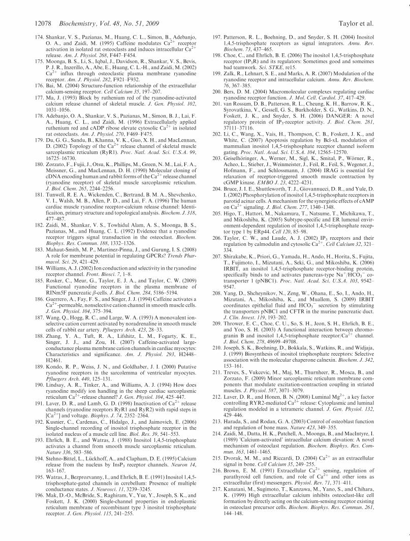
12078 Biochemistry, Vol. 48, No. 51, 2009 Taylor et al.
174. Shankar, V. S., Pazianas, M., Huang, C. L., Simon, B., Adebanjo,O. A., and Zaidi, M. (1995) Caffeine modulates Ca2þ receptoractivation in isolated rat osteoclasts and induces intracellular Ca2þ
release. Am. J. Physiol. 268, F447–F454.175. Moonga, B. S., Li, S., Iqbal, J., Davidson, R., Shankar, V. S., Bevis,
P. J. R., Inzerillo, A., Abe, E., Huang, C. L.-H., and Zaidi,M. (2002)Ca2þ influx through osteoclastic plasma membrane ryanodinereceptor. Am. J. Physiol. 282, F921–F932.
176. Bai, M. (2004) Structure-function relationship of the extracellularcalcium-sensing receptor. Cell Calcium 35, 197–207.
177. Ma, J. (1993) Block by ruthenium red of the ryanodine-activatedcalcium release channel of skeletal muscle. J. Gen. Physiol. 102,1031–1056.
178. Adebanjo, O. A., Shankar, V. S., Pazianas, M., Simon, B. J., Lai, F.A., Huang, C. L., and Zaidi, M. (1996) Extracellularly appliedruthenium red and cADP ribose elevate cytosolic Ca2þ in isolatedrat osteoclasts. Am. J. Physiol. 270, F469–F475.
179. Du, G. G., Sandu, B., Khanna, V. K., Guo, X. H., andMacLennan,D. (2002) Topology of the Ca2þ release channel of skeletal musclesarcoplasmic reticulum (RyR1). Proc. Natl. Acad. Sci. U.S.A. 99,16725–16730.
180. Zorzato, F., Fujii, J., Otsu,K., Phillips,M.,Green, N.M., Lai, F.A.,Meissner, G., and MacLennan, D. H. (1990) Molecular cloning ofcDNAencoding human and rabbit forms of theCa2þ release channel(ryanodine receptor) of skeletal muscle sarcoplasmic reticulum.J. Biol. Chem. 265, 2244–2256.
181. Tunwell, R. E. A., Wickenden, C., Bertrand, B. M. A., Shevchenko,V. I., Walsh, M. B., Allen, P. D., and Lai, F. A. (1996) The humancardiac muscle ryanodine receptor-calcium release channel: Identi-ficaiton, primary structure and topological analysis.Biochem. J. 318,477–487.
182. Zaidi, M., Shankar, V. S., Towhidul Alam, A. S., Moonga, B. S.,Pazianas, M., and Huang, C. L. (1992) Evidence that a ryanodinereceptor triggers signal transduction in the osteoclast. Biochem.Biophys. Res. Commun. 188, 1332–1326.
183. Mahaut-Smith, M. P., Martinez-Pinna, J., and Gurung, I. S. (2008)A role for membrane potential in regulating GPCRs? Trends Phar-macol. Sci. 29, 421–429.
184. Williams, A. J. (2002) Ion conduction and selectivity in the ryanodinereceptor channel. Front. Biosci. 7, 1–8.
185. Rosker, C., Meur, G., Taylor, E. J. A., and Taylor, C. W. (2009)Functional ryanodine receptors in the plasma membrane ofRINm5F pancreatic β-cells. J. Biol. Chem. 284, 5186–5194.
186. Guerrero, A., Fay, F. S., and Singer, J. J. (1994) Caffeine activates aCa2þ-permeable, nonselective cation channel in smoothmuscle cells.J. Gen. Physiol. 104, 375–394.
187. Wang, Q., Hogg, R. C., and Large, W. A. (1993) A monovalent ion-selective cation current activated by noradrenaline in smooth musclecells of rabbit ear artery. Pfluegers Arch. 423, 28–33.
188. Zhang, Y. A., Tuft, R. A., Lifshitz, L. M., Fogarty, K. E.,Singer, J. J., and Zou, H. (2007) Caffeine-activated large-conductance plasmamembrane cation channels in cardiacmyocytes:Characteristics and significance. Am. J. Physiol. 293, H2448–H2461.
189. Kondo, R. P., Weiss, J. N., and Goldhaber, J. I. (2000) Putativeryanodine receptors in the sarcolemma of ventricular myocytes.Pfluegers Arch. 440, 125–131.
190. Lindsay, A. R., Tinker, A., and Williams, A. J. (1994) How doesryanodine modify ion handling in the sheep cardiac sarcoplasmicreticulum Ca2þ-release channel? J. Gen. Physiol. 104, 425–447.
191. Laver, D. R., and Lamb, G. D. (1998) Inactivation of Ca2þ releasechannels (ryanodine receptors RyR1 and RyR2) with rapid steps in[Ca2þ] and voltage. Biophys. J. 74, 2352–2364.
192. Kusnier, C., Cardenas, C., Hidalgo, J., and Jaimovich, E. (2006)Single-channel recording of inositol trisphosphate receptor in theisolated nucleus of a muscle cell line. Biol. Res. 39, 541–553.
193. Ehrlich, B. E., and Watras, J. (1988) Inositol 1,4,5-trisphosphateactivates a channel from smooth muscle sarcoplasmic reticulum.Nature 336, 583–586.
194. Stehno-Bittel, L., L€uckhoff, A., and Clapham, D. E. (1995) Calciumrelease from the nucleus by InsP3 receptor channels. Neuron 14,163–167.
195. Watras, J., Bezprozvanny, I., andEhrlich, B. E. (1991) Inositol 1,4,5-trisphosphate-gated channels in cerebellum: Presence of multipleconductance states. J. Neurosci. 11, 3239–3245.
196. Mak, D.-O., McBride, S., Raghiram, V., Yue, Y., Joseph, S. K., andFoskett, J. K. (2000) Single-channel properties in endoplasmicreticulum membrane of recombinant type 3 inositol trisphosphatereceptor. J. Gen. Physiol. 115, 241–255.
197. Patterson, R. L., Boehning, D., and Snyder, S. H. (2004) Inositol1,4,5-trisphosphate receptors as signal integrators. Annu. Rev.Biochem. 73, 437–465.
198. Choe, C., and Ehrlich, B. E. (2006) The inositol 1,4,5-trisphosphatereceptor (IP3R) and its regulators: Sometimes good and someimesbad teamwork. Sci. STKE, re15.
199. Zalk, R., Lehnart, S. E., andMarks, A. R. (2007) Modulation of theryanodine receptor and intracellular calcium. Annu. Rev. Biochem.76, 367–385.
200. Bers, D. M. (2004) Macromolecular complexes regulating cardiacryanodine receptor function. J. Mol. Cell. Cardiol. 37, 417–429.
201. van Rossum, D. B., Patterson, R. L., Cheung, K. H., Barrow, R. K.,Syrovatkina, V., Gessell, G. S., Burkholder, S. G., Watkins, D. N.,Foskett, J. K., and Snyder, S. H. (2006) DANGER: A novelregulatory protein of IP3-receptor activity. J. Biol. Chem. 281,37111–37116.
202. Li, C., Wang, X., Vais, H., Thompson, C. B., Foskett, J. K., andWhite, C. (2007) Apoptosis regulation by Bcl-xL modulation ofmammalian inositol 1,4,5-trisphosphate receptor channel isoformgating. Proc. Natl. Acad. Sci. U.S.A. 104, 12565–12570.
203. Geiselh€oringer, A., Werner, M., Sigl, K., Smital, P., W€orner, R.,Acheo, L., Stieber, J., Weinmeister, J., Feil, R., Feil, S., Wegener, J.,Hofmann, F., and Schlossmann, J. (2004) IRAG is essential forrelaxation of receptor-triggered smooth muscle contraction bycGMP kinase. EMBO J. 23, 4222–4231.
204. Bruce, J. I. E., Shuttleworth, T. J., Giovannucci, D. R., and Yule, D.I. (2002) Phosphorylation of inositol 1,4,5-trisphosphate receptors inparotid acinar cells. Amechanism for the synergistic effects of cAMPon Ca2þ signaling. J. Biol. Chem. 277, 1340–1348.
205. Higo, T., Hattori, M., Nakamura, T., Natsume, T., Michikawa, T.,and Mikoshiba, K. (2005) Subtype-specific and ER lumenal envir-onment-dependent regulation of inositol 1,4,5-trisphosphate recep-tor type 1 by ERp44. Cell 120, 85–98.
206. Taylor, C. W., and Laude, A. J. (2002) IP3 receptors and theirregulation by calmodulin and cytosolic Ca2þ. Cell Calcium 32, 321–334.
207. Shirakabe, K., Priori, G., Yamada, H., Ando, H., Horita, S., Fujita,T., Fujimoto, I., Mizutani, A., Seki, G., and Mikoshiba, K. (2006)IRBIT, an inositol 1,4,5-trisphosphate receptor-binding protein,specifically binds to and activates pancreas-type Naþ/HCO3
- co-transporter 1 (pNBC1). Proc. Natl. Acad. Sci. U.S.A. 103, 9542–9547.
208. Yang, D., Shcheynikov, N., Zeng, W., Ohana, E., So, I., Ando, H.,Mizutani, A., Mikoshiba, K., and Muallem, S. (2009) IRBITcoordinates epithelial fluid and HCO3
- secretion by stimulatingthe transporters pNBC1 and CFTR in the murine pancreatic duct.J. Clin. Invest. 119, 193–202.
209. Thrower, E. C., Choe, C. U., So, S. H., Jeon, S. H., Ehrlich, B. E.,and Yoo, S. H. (2003) A functional interaction between chromo-granin B and inositol 1,4,5-trisphosphate receptor/Ca2þ channel.J. Biol. Chem. 278, 49699–49708.
210. Joseph, S. K., Boehning, D., Bokkala, S., Watkins, R., andWidjaja,J. (1999) Biosynthesis of inositol trisphosphate receptors: Selectiveassociation with the molecular chaperone calnexin. Biochem. J. 342,153–161.
211. Treves, S., Vukcevic, M., Maj, M., Thurnheer, R., Mosca, B., andZorzato, F. (2009) Minor sarcoplasmic reticulum membrane com-ponents that modulate excitation-contraction coupling in striatedmuscles. J. Physiol. 587, 3071–3079.
212. Laver, D. R., and Honen, B. N. (2008) Luminal Mg2þ, a key factorcontrolling RYR2-mediated Ca2þ release: Cytoplasmic and luminalregulation modeled in a tetrameric channel. J. Gen. Physiol. 132,429–446.
213. Harada, S., and Rodan, G. A. (2003) Control of osteoblast functionand regulation of bone mass. Nature 423, 349–355.
214. Zaidi, M., Datta, H. K., Patchell, A., Moonga, B., andMacIntyre, I.(1989) ’Calcium-activated’ intracellular calcium elevation: A novelmechanism of osteoclast regulation. Biochem. Biophys. Res. Com-mun. 163, 1461–1465.
215. Dvorak, M. M., and Riccardi, D. (2004) Ca2þ as an extracellularsignal in bone. Cell Calcium 35, 249–255.
216. Brown, E. M. (1991) Extracellular Ca2þ sensing, regulation ofparathyroid cell function, and role of Ca2þ and other ions asextracellular (first) messengers. Physiol. Rev. 71, 371–411.
217. Kanatani, M., Sugimoto, T., Kanzawa, M., Yano, S., and Chihara,K. (1999) High extracellular calcium inhibits osteoclast-like cellformation by directly acting on the calcium-sensing receptor existingin osteoclast precursor cells. Biochem. Biophys. Res. Commun. 261,144–148.
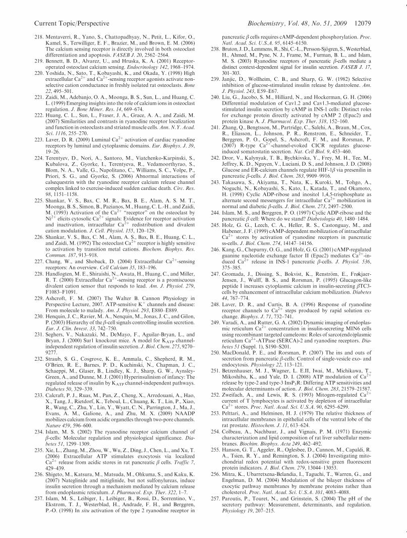
Current Topic/Perspective Biochemistry, Vol. 48, No. 51, 2009 12079
218. Mentaverri, R., Yano, S., Chattopadhyay, N., Petit, L., Kifor, O.,Kamel, S., Terwilliger, E. F., Brazier, M., and Brown, E. M. (2006)The calcium sensing receptor is directly involved in both osteoclastdifferentiation and apoptosis. FASEB J. 20, 2562–2564.
219. Bennett, B. D., Alvarez, U., and Hruska, K. A. (2001) Receptor-operated osteoclast calcium sensing. Endocrinology 142, 1968–1974.
220. Yoshida, N., Sato, T., Kobayashi, K., and Okada, Y. (1998) Highextracellular Ca2þ and Ca2þ-sensing receptor agonists activate non-selective cation conductance in freshly isolated rat osteoclasts. Bone22, 495–501.
221. Zaidi, M., Adebanjo, O. A., Moonga, B. S., Sun, L., and Huang, C.L. (1999) Emerging insights into the role of calcium ions in osteoclastregulation. J. Bone Miner. Res. 14, 669–674.
222. Huang, C. L., Sun, L., Fraser, J. A., Grace, A. A., and Zaidi, M.(2007) Similarities and contrasts in ryanodine receptor localizationand function in osteoclasts and striatedmuscle cells.Ann. N.Y. Acad.Sci. 1116, 255–270.
223. Laver, D. R. (2009) Luminal Ca2þ activation of cardiac ryanodinereceptors by luminal and cytoplasmic domains. Eur. Biophys. J. 39,19–26.
224. Terentyev, D., Nori, A., Santoro, M., Viatchenko-Karpinski, S.,Kubalova, Z., Gyorke, I., Terentyeva, R., Vedamoorthyrao, S.,Blom, N. A., Valle, G., Napolitano, C., Williams, S. C., Volpe, P.,Priori, S. G., and Gyorke, S. (2006) Abnormal interactions ofcalsequestrin with the ryanodine receptor calcium release channelcomplex linked to exercise-induced sudden cardiac death. Circ. Res.98, 1151–1158.
225. Shankar, V. S., Bax, C. M. R., Bax, B. E., Alam, A. S. M. T.,Moonga, B. S., Simon, B., Pazianos,M.,Huang, C. L.-H., andZaidi,M. (1993) Activation of the Ca2þ “receptor” on the osteoclast byNi2þ elicits cytosolic Ca2þ signals: Evidence for receptor activationand inactivation, intracellular Ca2þ redistribution and divalentcation modulation. J. Cell. Physiol. 155, 120–129.
226. Shankar, V. S., Bax, C. M., Alam, A. S., Bax, B. E., Huang, C. L.,and Zaidi, M. (1992) The osteoclast Ca2þ receptor is highly sensitiveto activation by transition metal cations. Biochem. Biophys. Res.Commun. 187, 913–918.
227. Chang, W., and Shoback, D. (2004) Extracellular Ca2þ-sensingreceptors: An overview. Cell Calcium 35, 183–196.
228. Handlogten, M. E., Shiraishi, N., Awata, H., Huang, C., andMiller,R. T. (2000) Extracellular Ca2þ-sensing receptor is a promiscuousdivalent cation sensor that responds to lead. Am. J. Physiol. 279,F1083–F1091.
229. Ashcroft, F. M. (2007) The Walter B. Cannon Physiology inPerspective Lecture, 2007. ATP-sensitive Kþ channels and disease:From molecule to malady. Am. J. Physiol. 293, E880–E889.
230. Henquin, J. C., Ravier, M. A., Nenquin,M., Jonas, J. C., and Gilon,P. (2003) Hierarchy of the β-cell signals controlling insulin secretion.Eur. J. Clin. Invest. 33, 742–750.
231. Seghers, V., Nakazaki, M., DeMayo, F., Aguilar-Bryan, L., andBryan, J. (2000) Sur1 knockout mice. A model for KATP channel-independent regulation of insulin secretion. J. Biol. Chem. 275, 9270–9277.
232. Straub, S. G., Cosgrove, K. E., Ammala, C., Shepherd, R. M.,O’Brien, R. E., Barnes, P. D., Kuchinski, N., Chapman, J. C.,Schaeppi, M., Glaser, B., Lindley, K. J., Sharp, G. W., Aynsley-Green, A., and Dunne, M. J. (2001) Hyperinsulinism of infancy: Theregulated release of insulin by KATP channel-independent pathways.Diabetes 50, 329–339.
233. Calcraft, P. J., Ruas, M., Pan, Z., Cheng, X., Arredouani, A., Hao,X., Tang, J., Rietdorf, K., Teboul, L., Chuang, K. T., Lin, P., Xiao,R., Wang, C., Zhu, Y., Lin, Y., Wyatt, C. N., Parrington, J., Ma, J.,Evans, A. M., Galione, A., and Zhu, M. X. (2009) NAADPmobilizes calcium from acidic organelles through two-pore channels.Nature 459, 596–600.
234. Islam, M. S. (2002) The ryanodine receptor calcium channel ofβ-cells: Molecular regulation and physiological significance. Dia-betes 51, 1299–1309.
235. Xie, L., Zhang,M., Zhou,W.,Wu, Z., Ding, J., Chen, L., andXu, T.(2006) Extracellular ATP stimulates exocytosis via localizedCa2þ release from acidic stores in rat pancreatic β cells. Traffic 7,429–439.
236. Shigeto, M., Katsura, M., Matsuda, M., Ohkuma, S., and Kaku, K.(2007) Nateglinide and mitiglinide, but not sulfonylureas, induceinsulin secretion through a mechanism mediated by calcium releasefrom endoplasmic reticulum. J. Pharmacol. Exp. Ther. 322, 1–7.
237. Islam, M. S., Leibiger, I., Leibiger, B., Rossi, D., Sorrentino, V.,Ekstrom, T. J., Westerblad, H., Andrade, F. H., and Berggren,P.-O. (1998) In situ activation of the type 2 ryanodine receptor in
pancreatic β cells requires cAMP-dependent phosphorylation. Proc.Natl. Acad. Sci. U.S.A. 95, 6145–6150.
238. Bruton, J.D.,Lemmens,R., Shi,C.-L., Persson-Sj€ogren, S.,Westerblad,H., Ahmed, M., Pyne, N. J., Frame, M., Furman, B. L., and Islam,M. S. (2003) Ryanodine receptors of pancreatic β-cells mediate adistinct context-dependent signal for insulin secretion. FASEB J. 17,301–303.
239. Janjic, D., Wollheim, C. B., and Sharp, G. W. (1982) Selectiveinhibition of glucose-stimulated insulin release by dantrolene. Am.J. Physiol. 243, E59–E67.
240. Liu, G., Jacobo, S. M., Hilliard, N., and Hockerman, G. H. (2006)Differential modulation of Cav1.2 and Cav1.3-mediated glucose-stimulated insulin secretion by cAMP in INS-1 cells: Distinct rolesfor exchange protein directly activated by cAMP 2 (Epac2) andprotein kinase A. J. Pharmacol. Exp. Ther. 318, 152–160.
241. Zhang, Q., Bengtsson,M., Partridge, C., Salehi, A., Braun,M., Cox,R., Eliasson, L., Johnson, P. R., Renstrom, E., Schneider, T.,Berggren, P. O., Gopel, S., Ashcroft, F. M., and Rorsman, P.(2007) R-type Ca2þ-channel-evoked CICR regulates glucose-induced somatostatin secretion. Nat. Cell Biol. 9, 453–460.
242. Dror, V., Kalynyak, T. B., Bychkivska, Y., Frey, M. H., Tee, M.,Jeffrey, K. D., Nguyen, V., Luciani, D. S., and Johnson, J. D. (2008)Glucose and ER-calcium channels regulate HIF-1β via presenilin inpancreatic β-cells. J. Biol. Chem. 283, 9909–9916.
243. Takasawa, S., Akiyama, T., Nata, K., Kuroki, M., Tohgo, A.,Noguchi, N., Kobayashi, S., Kato, I., Katada, T., and Okamoto,H. (1998) Cyclic ADP-ribose and inositol 1,4,5-trisphosphate asalternate second messengers for intracellular Ca2þ mobilization innormal and diabetic β-cells. J. Biol. Chem. 273, 2497–2500.
244. Islam, M. S., and Berggren, P. O. (1997) Cyclic ADP-ribose and thepancreatic β cell: Where do we stand? Diabetologia 40, 1480–1484.
245. Holz, G. G., Leech, C. A., Heller, R. S., Castonguay, M., andHabener, J. F. (1999) cAMP-dependent mobilization of intracellularCa2þ stores by activation of ryanodine receptors in pancreaticss-cells. J. Biol. Chem. 274, 14147–14156.
246. Kang, G., Chepurny, O.G., andHolz, G.G. (2001) cAMP-regulatedguanine nucleotide exchange factor II (Epac2) mediates Ca2þ-in-duced Ca2þ release in INS-1 pancreatic β-cells. J. Physiol. 536,375–385.
247. Gromada, J., Dissing, S., Bokvist, K., Renstr€om, E., Froekjaer-Jensen, J., Wulff, B. S., and Rorsman, P. (1995) Glucagon-likepeptide I increases cytoplasmic calcium in insulin-secreting βTC3-cells by enhancement of intracellular calcium mobilization.Diabetes44, 767–774.
248. Laver, D. R., and Curtis, B. A. (1996) Response of ryanodinereceptor channels to Ca2þ steps produced by rapid solution ex-change. Biophys. J. 71, 732–741.
249. Varadi, A., and Rutter, G. A. (2002) Dynamic imaging of endoplas-mic reticulum Ca2þ concentration in insulin-secreting MIN6 cellsusing recombinant targeted cameleons: Roles of sarco(endo)plasmicreticulum Ca2þ-ATPase (SERCA)-2 and ryanodine receptors. Dia-betes 51 (Suppl. 1), S190–S201.
250. MacDonald, P. E., and Rorsman, P. (2007) The ins and outs ofsecretion from pancreatic β-cells: Control of single-vesicle exo- andendocytosis. Physiology 22, 113–121.
251. Betzenhauser, M. J., Wagner, L. E.II, Iwai, M., Michikawa, T.,Mikoshiba, K., and Yule, D. I. (2008) ATP modulation of Ca2þ
release by type-2 and type-3 InsP3R: Differing ATP sensitivities andmolecular determinants of action. J. Biol. Chem. 283, 21579–21587.
252. Zweifach, A., and Lewis, R. S. (1993) Mitogen-regulated Ca2þ
current of T lymphocytes is activated by depletion of intracellularCa2þ stores. Proc. Natl. Acad. Sci. U.S.A. 90, 6295–6299.
253. Pelttari, A., and Helminen, H. J. (1979) The relative thickness ofintracellular membranes in epithelial cells of the ventral lobe of therat prostate. Histochem. J. 11, 613–624.
254. Colbeau, A., Nachbaur, J., and Vignais, P. M. (1971) Enzymiccharacterization and lipid composition of rat liver subcellular mem-branes. Biochim. Biophys. Acta 249, 462–492.
255. Hanson, G. T., Aggeler, R., Oglesbee, D., Cannon, M., Capaldi, R.A., Tsien, R. Y., and Remington, S. J. (2004) Investigating mito-chondrial redox potential with redox-sensitive green fluorescentprotein indicators. J. Biol. Chem. 279, 13044–13053.
256. Mitra, K., Ubarretxena-Belandia, I., Taguchi, T., Warren, G., andEngelman, D. M. (2004) Modulation of the bilayer thickness ofexocytic pathway membranes by membrane proteins rather thancholesterol. Proc. Natl. Acad. Sci. U.S.A. 101, 4083–4088.
257. Paroutis, P., Touret, N., and Grinstein, S. (2004) The pH of thesecretory pathway: Measurement, determinants, and regulation.Physiology 19, 207–215.
12080 Biochemistry, Vol. 48, No. 51, 2009 Taylor et al.
258. Hwang, C., Sinskey, A. J., and Lodish, H. F. (1992) Oxidized redoxstate of glutathione in the endoplasmic reticulum. Science 257, 1496–1502.
259. Evans, W. H., and Hardison, W. G. (1985) Phospholipid, cholester-ol, polypeptide and glycoprotein composition of hepatic endosomesubfractions. Biochem. J. 232, 33–36.
260. Go, Y. M., and Jones, D. P. (2008) Redox compartmentalization ineukaryotic cells. Biochim. Biophys. Acta 1780, 1273–1290.
261. Saroussi, S., andNelson, N. (2009) VacuolarHþ-ATPase-an enzymefor all seasons. Pfluegers Arch. 457, 581–587.
262. Austin, C. D.,Wen, X., Gazzard, L., Nelson, C., Scheller, R. H., andScales, S. J. (2005) Oxidizing potential of endosomes and lysosomeslimits intracellular cleavage of disulfide-based antibody-drug con-jugates. Proc. Natl. Acad. Sci. U.S.A. 102, 17987–17992.
263. Harikumar, P., and Reeves, J. P. (1983) The lysosomal proton pumpis electrogenic. J. Biol. Chem. 258, 10403–10410.
264. Schoer, J. K., Gallegos, A. M., McIntosh, A. L., Starodub, O., Kier,A. B., Billheimer, J. T., and Schroeder, F. (2000) Lysosomalmembrane cholesterol dynamics. Biochemistry 39, 7662–7677.
265. Steinberg, B. E., Touret, N., Vargas-Caballero,M., andGrinstein, S.(2007) In situ measurement of the electrical potential across thephagosomal membrane using FRET and its contribution to theproton-motive force. Proc. Natl. Acad. Sci. U.S.A. 104, 9523–9528.
266. Dooley, C. T., Dore, T. M., Hanson, G. T., Jackson, W. C.,Remington, S. J., and Tsien, R. Y. (2004) Imaging dynamic redoxchanges in mammalian cells with green fluorescent protein indica-tors. J. Biol. Chem. 279, 22284–22293.
267. Moccia, F., Billington, R. A., and Santella, L. (2006) Pharmacolo-gical characterization of NAADP-induced Ca2þ signals in starfishoocytes. Biochem. Biophys. Res. Commun. 348, 329–336.
268. Witzgall, R. (2007) TRPP2 channel regulation. Handb. Exp. Phar-macol., 363–375.
269. Peier,A.M.,Moqrich,A.,Hergarden,A.C., Reeve,A. J., Andersson,D.A., Story,G.M., Earley, T. J., Dragoni, I.,McIntyre, P., Bevan, S.,and Patapoutian, A. (2002) A TRP channel that senses cold stimuliand menthol. Cell 108, 705–715.
270. McKemy, D. D., Neuhausser, W. M., and Julius, D. (2002) Identi-fication of a cold receptor reveals a general role for TRP channels inthermosensation. Nature 416, 52–58.
271. Thebault, S., Lemonnier, L., Bidaux, G., Flourakis, M., Bavencoffe,A., Gordienko, D., Roudbaraki, M., Delcourt, P., Panchin, Y.,Shuba, Y., Skryma, R., and Prevarskaya, N. (2005) Novel role ofcold/menthol-sensitive transient receptor potential melastatine fa-mily member 8 (TRPM8) in the activation of store-operated chan-nels in LNCaP human prostate cancer epithelial cells. J. Biol. Chem.280, 39423–39435.
272. Latorre, R., Brauchi, S., Orta, G., Zaelzer, C., and Vargas, G. (2007)ThermoTRP channels as modular proteins with allosteric gating.Cell Calcium 42, 427–438.
273. Kraft, R., and Harteneck, C. (2005) The mammalian melastatin-related transient receptor potential cation channels: An overview.Pfluegers Arch. 451, 204–211.
274. Okada, T., Inoue, R., Yamazaki, K., Maeda, A., Kurosaki, T.,Yamakuni, T., Tanaka, I., Shimizu, S., Ikenaka, K., Imoto, K., andMori, Y. (1999)Molecular and functional characterization of a novel
mouse transient receptor potential protein homologue TRP7. Ca2þ-permeable cation channel that is constitutively activated andenhanced by stimulation of G protein-coupled receptor. J. Biol.Chem. 274, 27359–27370.
275. Lavender, V., Chong, S., Ralphs, K., Wolstenholme, A. J., andReaves, B. J. (2008) Increasing the expression of calcium-permeableTRPC3 and TRPC7 channels enhances constitutive secretion. Bio-chem. J. 413, 437–446.
276. Abramowitz, J., and Birnbaumer, L. (2009) Physiology and patho-physiology of canonical transient receptor potential channels. FAS-EB J. 23, 297–328.
277. Shi, J., Mori, E., Mori, Y., Mori, M., Li, J., Ito, Y., and Inoue, R.(2004) Multiple regulation by calcium of murine homologues oftransient receptor potential proteins TRPC6 and TRPC7 expressedin HEK293 cells. J. Physiol. 561, 415–432.
278. Caterina, M. J., Rosen, T. A., Tominaga, M., Brake, A. J., andJulius, D. (1999) A capsaicin-receptor homologue with a highthreshold for noxious heat. Nature 398, 436–441.
279. Tominaga, M., and Tominaga, T. (2005) Structure and function ofTRPV1. Pfluegers Arch. 451, 143–150.
280. Story, G. M., Peier, A. M., Reeve, A. J., Eid, S. R., Mosbacher, J.,Hricik, T. R., Earley, T. J., Hergarden, A. C., Andersson,D. A., Hwang, S. W., McIntyre, P., Jegla, T., Bevan, S., andPatapoutian, A. (2003) ANKTM1, a TRP-like channel expressedin nociceptive neurons, is activated by cold temperatures. Cell 112,819–829.
281. Karashima, Y., Prenen, J., Meseguer, V., Owsianik, G., Voets, T.,and Nilius, B. (2008) Modulation of the transient receptor potentialchannel TRPA1 by phosphatidylinositol 4,5-biphosphate manipu-lators. Pfluegers Arch. 457, 77–89.
282. Qian, F., and Noben-Trauth, K. (2005) Cellular and molecularfunction of mucolipins (TRPML) and polycystin 2 (TRPP2). Pflue-gers Arch. 451, 277–285.
283. Dong, X. P.,Wang, X., Shen, D., Chen, S., Liu,M.,Wang, Y.,Mills,E., Cheng, X., Delling, M., and Xu, H. (2009) Activating mutationsof the TRPML1 channel revealed by proline scanning mutagenesis.J. Biol. Chem. 284, 32040–32052.
284. Drouin,H., andHermann,A. (1994) Intracellular action of spermineon neuronal Ca2þ and Kþ currents. Eur. J. Neurosci. 6, 412–419.
285. Roberts-Crowley,M. L.,Mitra-Ganguli, T., Liu, L., andRittenhouse,A. R. (2009) Regulation of voltage-gated Ca2þ channels by lipids.CellCalcium 45, 589–601.
286. Dingledine, R., Borges, K., Bowie, D., and Traynelis, S. F. (1999)The glutamate receptor ion channels. Pharm. Rev. 51, 7–61.
287. Mandal,M., andYan, Z. (2009) PIP2 regulation ofNMDA receptorchannels in cortical neurons. Mol. Pharmacol. 76, 1349–1359.
288. Jarvis, M. F., and Khakh, B. S. (2009) ATP-gated P2X cation-channels. Neuropharmacology 56, 208–215.
289. Vanden Abeele, F., Bidaux, G., Gordienko, D., Beck, B., Panchin,Y. V., Baranova, A. V., Ivanov, D. V., Skryma, R., and Prevarskaya,N. (2006) Functional implications of calcium permeability of thechannel formed by pannexin 1. J. Cell Biol. 174, 535–546.
290. D’Hondt, C., Ponsaerts, R., De Smedt, H., Bultynck, G., andHimpens, B. (2009) Pannexins, distant relatives of the connexinfamily with specific cellular functions? BioEssays 31, 953–974.
Copyright © 2022 FDOKUMEN