C-Terminal Tyrosine of Ferredoxin−NADP + Reductase in Hydride Transfer Processes with NAD(P) + /H...
14
C-Terminal Tyrosine of Ferredoxin-NADP + Reductase in Hydride Transfer Processes with NAD(P) + /H ² Jesu ´s Tejero, ‡ Inmaculada Pe ´rez-Dorado, § Celia Maya, § Marta Martı ´nez-Ju ´lvez, ‡ Julia Sanz-Aparicio, § Carlos Go ´mez-Moreno, ‡ Juan A. Hermoso, § and Milagros Medina* ,‡ Departamento de Bioquı ´mica y Biologı ´a Molecular y Celular, Facultad de Ciencias, and Institute of Biocomputation and Physics of Complex Systems (BIFI), UniVersidad de Zaragoza, E-50009 Zaragoza, Spain, and Grupo de Cristalografı ´a Macromolecular y Biologı ´a Estructural, Instituto Quı ´mica-Fı ´sica Rocasolano, CSIC, Serrano 119, 28006 Madrid, Spain ReceiVed July 4, 2005; ReVised Manuscript ReceiVed August 16, 2005 ABSTRACT: Ferredoxin-NADP + reductase (FNR) catalyzes the reduction of NADP + to NADPH in an overall reversible reaction, showing some differences in the mechanisms between cyanobacterial and higher plant FNRs. During hydride transfer it is proposed that the FNR C-terminal Tyr is displaced by the nicotinamide. Thus, this C-terminal Tyr might be involved not only in modulating the flavin redox properties, as already shown, but also in nicotinamide binding and hydride transfer. FNR variants from the cyanobacterium Anabaena in which the C-terminal Tyr has been replaced by Trp, Phe, or Ser have been produced. All FNR variants show enhanced NADP + and NAD + binding, especially Tyr303Ser, which correlates with a noticeable improvement of NADH-dependent reactions. Nevertheless, the Tyr303Ser variant shows a decrease in the steady-state k cat value with NADPH. Fast kinetic analysis of the hydride transfer shows that the low efficiency observed for this mutant FNR under steady-state conditions is not due to a lack of catalytic ability but rather to the strong enzyme-coenzyme interaction. Three-dimensional structures for Tyr303Ser and Tyr303Trp variants and its complexes with NADP + show significant differences between plant and cyanobacterial FNRs. Our results suggest that modulation of coenzyme affinity is highly influenced by the strength of the C-terminus-FAD interaction and that subtle changes between plant and cyanobacterial structures are able to modify the energy of that interaction. Additionally, it is shown that the C-terminal Tyr of FNR lowers the affinity for NADP + /H to levels compatible with steady-state turnover during the catalytic cycle, but it is not involved in the hydride transfer itself. Ferredoxin-NADP + reductase (FNR, 1 EC 1.18.1.2) is a FAD-dependent enzyme that catalyzes the electron transfer (ET) from reduced protein electron carriers, ferredoxin or flavodoxin, to NADP + in the last step of the photosynthetic electron transfer chain to produce NADPH (1-4). The reaction is highly specific for NADP + /H, the enzyme showing a dramatic decrease in activity when NAD + /H is used as coenzyme (5, 6). Three-dimensional structures of FNRs from different sources, as well as of several mutant FNRs and in complex with its substrates, are available (7- 15). The structure consists of two distinct domains: an N-terminal FAD binding domain and a C-terminal domain that binds the NADP + coenzyme (Figure 1A). This two- domain motif is the building block of a large family of flavoproteins. In many of these enzymes, additional domains have been fused to the FNR building block, thus extending the system ability to a wide range of reactions (7, 16). Coenzyme specificity studies have usually focused in the structural differences between the NADPH and NADH coenzymes, which are reduced to the presence or absence of a phosphate group at the 2′ position of the adenine ribose moiety (2′P-AMP). Although changes limited to the 2′-P or 2′-OH binding pocket in this family of enzymes have proven to be effective in reversing coenzyme specificity in some enzymes (17-19), these changes are usually unable to fully reverse the specificity or cause a dramatic decrease in catalytic efficiency as a side effect (6). This fact soon pointed out that the enzyme residues interacting with regions common to both NADPH and NADH might also be important in conferring coenzyme specificity. Thus, the relevance of the region that binds the pyrophosphate bridge between the 2′P-AMP and nicotinamide mononucleotide (NMN) moieties of NAD(P) + /H has also been demonstrated (6, 20-22). Apart from these common motifs to any NAD(P) + /H binding protein, a new determinant of coenzyme specificity, restricted to the FNR protein family, has been reported. In light of the experimental data and the three-dimensional structures reported for the FNR-NADP + interaction, it is ² This work has been supported by the Comisio ´n Interministerial de Ciencia y Tecnologı ´a (CICYT, Grant BIO2003-00627 to C.G.-M. and Grant BIO2004-00279 to M.M.) and by CONSI+D (DGA, Grant P006/ 2000 to M.M.). * Address correspondence to this author. Fax: +34 976 762123. Phone: +34 976 762476. E-mail: [email protected]. ‡ Universidad de Zaragoza. § Instituto Quı ´mica-Fı ´sica Rocasolano. CSIC. 1 Abbreviations: FNR, ferredoxin-NADP + reductase; FNRox, FNR in the fully oxidized state; FNRrd, FNR in the hydroquinone state; FNRsq, FNR in the semiquinone state; ET, electron transfer; DCPIP, 2,6-dichlorophenolindophenol; WT, wild type; CP450R, cytochrome P450 reductase; NOS, nitric oxide synthase; NMN, nicotinamide mononucleotide portion of NAD(P) + /H; 2′P-AMP, 2′-phospho-AMP portion of NADP + /H; rmsd, root mean square deviation; C-HEGA-10, cyclohexylbutanoyl-N-hydroxyethylglucamide. 13477 Biochemistry 2005, 44, 13477-13490 10.1021/bi051278c CCC: $30.25 © 2005 American Chemical Society Published on Web 09/24/2005
Transcript of C-Terminal Tyrosine of Ferredoxin−NADP + Reductase in Hydride Transfer Processes with NAD(P) + /H...

C-Terminal Tyrosine of Ferredoxin-NADP+ Reductase in Hydride TransferProcesses with NAD(P)+/H†
Jesu´s Tejero,‡ Inmaculada Pe´rez-Dorado,§ Celia Maya,§ Marta Martınez-Ju´lvez,‡ Julia Sanz-Aparicio,§
Carlos Go´mez-Moreno,‡ Juan A. Hermoso,§ and Milagros Medina*,‡
Departamento de Bioquı´mica y Biologı´a Molecular y Celular, Facultad de Ciencias, and Institute of Biocomputation andPhysics of Complex Systems (BIFI), UniVersidad de Zaragoza, E-50009 Zaragoza, Spain, and Grupo de Cristalografı´aMacromolecular y Biologı´a Estructural, Instituto Quı´mica-Fısica Rocasolano, CSIC, Serrano 119, 28006 Madrid, Spain
ReceiVed July 4, 2005; ReVised Manuscript ReceiVed August 16, 2005
ABSTRACT: Ferredoxin-NADP+ reductase (FNR) catalyzes the reduction of NADP+ to NADPH in anoverall reversible reaction, showing some differences in the mechanisms between cyanobacterial and higherplant FNRs. During hydride transfer it is proposed that the FNR C-terminal Tyr is displaced by thenicotinamide. Thus, this C-terminal Tyr might be involved not only in modulating the flavin redoxproperties, as already shown, but also in nicotinamide binding and hydride transfer. FNR variants fromthe cyanobacteriumAnabaenain which the C-terminal Tyr has been replaced by Trp, Phe, or Ser havebeen produced. All FNR variants show enhanced NADP+ and NAD+ binding, especially Tyr303Ser,which correlates with a noticeable improvement of NADH-dependent reactions. Nevertheless, the Tyr303Servariant shows a decrease in the steady-statekcat value with NADPH. Fast kinetic analysis of the hydridetransfer shows that the low efficiency observed for this mutant FNR under steady-state conditions is notdue to a lack of catalytic ability but rather to the strong enzyme-coenzyme interaction. Three-dimensionalstructures for Tyr303Ser and Tyr303Trp variants and its complexes with NADP+ show significantdifferences between plant and cyanobacterial FNRs. Our results suggest that modulation of coenzymeaffinity is highly influenced by the strength of the C-terminus-FAD interaction and that subtle changesbetween plant and cyanobacterial structures are able to modify the energy of that interaction. Additionally,it is shown that the C-terminal Tyr of FNR lowers the affinity for NADP+/H to levels compatible withsteady-state turnover during the catalytic cycle, but it is not involved in the hydride transfer itself.
Ferredoxin-NADP+ reductase (FNR,1 EC 1.18.1.2) is aFAD-dependent enzyme that catalyzes the electron transfer(ET) from reduced protein electron carriers, ferredoxin orflavodoxin, to NADP+ in the last step of the photosyntheticelectron transfer chain to produce NADPH (1-4). Thereaction is highly specific for NADP+/H, the enzymeshowing a dramatic decrease in activity when NAD+/H isused as coenzyme (5, 6). Three-dimensional structures ofFNRs from different sources, as well as of several mutantFNRs and in complex with its substrates, are available (7-15). The structure consists of two distinct domains: anN-terminal FAD binding domain and a C-terminal domainthat binds the NADP+ coenzyme (Figure 1A). This two-
domain motif is the building block of a large family offlavoproteins. In many of these enzymes, additional domainshave been fused to the FNR building block, thus extendingthe system ability to a wide range of reactions (7, 16).
Coenzyme specificity studies have usually focused in thestructural differences between the NADPH and NADHcoenzymes, which are reduced to the presence or absenceof a phosphate group at the 2′ position of the adenine ribosemoiety (2′P-AMP). Although changes limited to the 2′-P or2′-OH binding pocket in this family of enzymes have provento be effective in reversing coenzyme specificity in someenzymes (17-19), these changes are usually unable to fullyreverse the specificity or cause a dramatic decrease incatalytic efficiency as a side effect (6). This fact soon pointedout that the enzyme residues interacting with regionscommon to both NADPH and NADH might also beimportant in conferring coenzyme specificity. Thus, therelevance of the region that binds the pyrophosphate bridgebetween the 2′P-AMP and nicotinamide mononucleotide(NMN) moieties of NAD(P)+/H has also been demonstrated(6, 20-22).
Apart from these common motifs to any NAD(P)+/Hbinding protein, a new determinant of coenzyme specificity,restricted to the FNR protein family, has been reported. Inlight of the experimental data and the three-dimensionalstructures reported for the FNR-NADP+ interaction, it is
† This work has been supported by the Comisio´n Interministerial deCiencia y Tecnologı´a (CICYT, Grant BIO2003-00627 to C.G.-M. andGrant BIO2004-00279 to M.M.) and by CONSI+D (DGA, Grant P006/2000 to M.M.).
* Address correspondence to this author. Fax:+34 976 762123.Phone: +34 976 762476. E-mail: [email protected].
‡ Universidad de Zaragoza.§ Instituto Quı´mica-Fısica Rocasolano. CSIC.1 Abbreviations: FNR, ferredoxin-NADP+ reductase; FNRox, FNR
in the fully oxidized state; FNRrd, FNR in the hydroquinone state;FNRsq, FNR in the semiquinone state; ET, electron transfer; DCPIP,2,6-dichlorophenolindophenol; WT, wild type; CP450R, cytochromeP450 reductase; NOS, nitric oxide synthase; NMN, nicotinamidemononucleotide portion of NAD(P)+/H; 2′P-AMP, 2′-phospho-AMPportion of NADP+/H; rmsd, root mean square deviation; C-HEGA-10,cyclohexylbutanoyl-N-hydroxyethylglucamide.
13477Biochemistry2005,44, 13477-13490
10.1021/bi051278c CCC: $30.25 © 2005 American Chemical SocietyPublished on Web 09/24/2005

proposed that the last step in optimal coenzyme bindingputatively involves displacement of the FNR C-terminalresidue (Tyr303 inAnabaenaFNR, Figure 1B) in order toallow the entrance of the nicotinamide ring into the hydridetransfer site (10, 15). This residue can be Tyr, Phe, or Trpin NADP+/H binding proteins, whereas in NAD+/H-depend-ing enzymes either a Tyr or a different arrangement of theC-terminus is found (5, 23, 24). Accordingly, the structuralfeatures of this C-terminal region in the FNR family ofenzymes show two different patterns (see Figure 6 in ref24). A group of enzymes, such as FNR, cytochrome P450reductase (CP450R), or nitric oxide synthase (NOS), presentan aromatic residue forming a stacking interaction with theisoalloxazine ring of FAD. On the other hand, in mostNAD+/H-dependent enzymes of the FNR family, the aro-matic residue is either absent or occupies a different position,forming a larger cavity around there face of the isoalloxazinering (24). In this latter case, there would be no need of aside chain displacement in order to accommodate thenicotinamide ring of the coenzyme. Involvement of theC-terminal aromatic residue in coenzyme specificity has beenconfirmed in pea FNR (5), as well as in other NADP+/H-dependent enzymes, such as CP450R (25, 26) or NOS (23).
However, site-directed mutagenesis replacement of thisaromatic residue produces different effects in each enzyme,an observation in agreement with the fact that not onlycoenzyme specificity but other effects such as modificationof the reduction potential or inter- and intraprotein ETprocesses can be affected by the mutation (23, 24).
A closer look at the role of this C-terminal residue is takenin the present work, focusing on the differences found inthe interaction with the coenzyme between cyanobacterialFNRs and plant FNRs (27), as well as other members of theFNR family. Thus, structural, spectroscopic, and, especially,fast kinetic (not previously reported for pea FNR variants)studies are here reported for theAnabaenaFNR Tyr303Phe,Tyr303Trp, and Tyr303Ser interaction and hydride transferwith NAD(P)+/H. Such data are discussed in light of previousanalysis on these mutants, indicating a role for Tyr303 inmodulating the rates of electron exchange with the proteinpartners by contributing to the flavin semiquinone stabiliza-tion required for electron splitting (24), and on thosepreviously reported for related enzymes in order to achievea deeper understanding of the role of this C-terminal residuein these systems.
MATERIALS AND METHODS
Production of FNR Variants.The Tyr303Ser, Tyr303Phe,and Tyr303TrpAnabaenaPCC7119 FNR variants wereprepared using the construct pET28a-FNR (22) and theQuikChange mutagenesis kit (Stratagene) as previouslydescribed (24). The pET28a-FNR vectors carrying theselected mutations were used to transformEscherichia coliBL21(DE3) Gold cells (Stratagene).E. coli BL21(DE3) Goldcultures carrying the pET28a-FNR plasmid were grown at30 °C for 24 h. The WT and mutatedAnabaenaFNR formsfrom the pET28a-FNR vectors were purified as describedpreviously (28). UV-vis spectra and SDS-PAGE were usedas purity criteria.
Spectral Analysis.UV-visible spectral measurements wereperformed in a Kontron Uvikon 942 spectrophotometer.Dissociation constants (Kd) for the complexes of oxidizedFNR forms with either NADP+ or NAD+ were estimatedby difference spectroscopy in 50 mM Tris-HCl, pH 8.0, aspreviously described (6). Errors in the estimatedKd valueswere(15%.
Enzymatic Assays.Diaphorase activity was assayed using2,6-dichlorophenolindophenol (DCPIP) as electron acceptorin 50 mM Tris-HCl, pH 8.0 at 25°C (6). Errors in theestimated values ofKm and kcat were (20% and(15%,respectively.
Stopped-Flow Kinetic Measurements.Kinetic measure-ments were performed under anaerobic conditions using anApplied Photophysics SX.17 MV stopped-flow instrumentin 50 mM Tris-HCl, pH 8.0 at 25°C, as previously described(28). Final FNR concentrations were kept between 7 and 9µM. NADPH and NADH concentrations were in the rangeof 175-200µM. Additional measurements were carried outwith ∼2.5 mM NADH. Observed rate constants (kobs) wereobtained by fitting the transients to a mono- or biexponentialequation. Errors in the estimated values were(15%.Reactions were mainly followed at 460 nm, but otherwavelengths, 340 and 600 nm, were also analyzed.
Apparent Equilibrium Constant Calculation.To extractadditional information from the experimental dissociation
FIGURE 1: Overall structure of nativeAnabaena FNR. (A)Molecular surface and ribbon diagram of the FNR, with the FADprosthetic group and sulfate ion represented as balls and sticks.Protein binding domains for each cofactor, FAD and NADP+, arecolored in blue and in orange, respectively. (B) Detail of the FADbinding site showing the FAD, the C-terminal Tyr residue, andsulfate ion in a ball-and-stick representation.
13478 Biochemistry, Vol. 44, No. 41, 2005 Tejero et al.

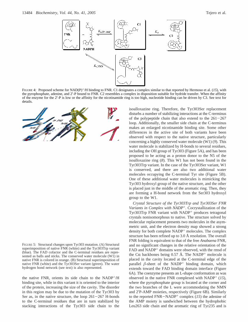
constants, the global equilibrium was fitted to a hypotheticalmodel in which the binding of the 2′P-AMP and NMNmoieties of NADP+ are assumed to be independent (seeFigure 4 below). For the FNR-NADP+ dissociation con-stants, the experimental values were fitted to the hypotheticalscheme which includes both the binding constantKa,2′P-AMP
and a second equilibrium constant,Kin, which takes intoaccount the concerted movement of the C-terminal residueand the NMN moiety of NADP+. The NMN occupancy ofthe Anabaenaand pea FNR-NADP+ complexes was as-sumed from spectroscopy and crystallographic data to be 0%and 14%, respectively (5). Thus, for theAnabaenaWTFNR-NADP+ complex, only the C1 species (see Figure 4below) is formed, and therebyKa,2′P-AMP ) 1/Kd. The bindingof the 2′P-AMP moiety of NADP+ was assumed to be similarin WT and mutant FNRs from the same organism, and thusa constant value forKa,2′P-AMP was used. ExperimentalKd
values (Table 2) were calculated as above-described accord-ing to a mechanism that assumes formation of single species,FNR + NAD(P)+ T FNR-NAD(P)+. To calculateKin
values, the amount of complex formed (calculated using theexperimentalKd constant) was assumed to be equal to thesum of the C1 and C2 complexes (see Figure 4 below). Inthe case of the FNR-NAD+ dissociation constants, only theequilibrium involvingKa,NMN (see Figure 4 below) is assumedto take place, and thenKa,NMN ) 1/Kd. Calculations werecarried out with the Gepasi software, version 3.30 (29).Nicotinamide occupancy calculations are directly derivedfrom the calculatedKin values, as in the equilibriumconditions (excess of NADP+) all of the FNR is in the formof either the C1 or C2 complexes, and, thus, occupancy (%)) 100[Kin/(1 + Kin)].
Crystal Growth, Data Collection, and Structure Refine-ment. Crystals of the Tyr303Ser and Tyr303Trp FNRvariants, and that of the Tyr303Trp and Tyr303Ser FNRvariants complexed with NADP+, were grown by the hangingdrop method. Both Tyr303Ser and Tyr303Trp variants gavehexagonal crystals that grew in 5µL droplets consisting of2 µL of 0.75 mM protein solution buffered with 10 mM Tris-HCl, pH 8.0, 1µL of unbufferedâ-octyl glucoside at 5%(w/v), and 2µL of reservoir solution containing 18-20%polyethyleneglycol 6000, 20 mM ammonium sulfate, and 0.1mM MES/NaOH, pH 5.5, or 0.1 mM sodium acetate, pH5.0. Drops were equilibrated at 20°C against 1 mL ofreservoir solution. Crystals reached their maximum size of0.8 × 0.4 × 0.4 mm in 1-7 days. The Tyr303Trp FNRvariant was crystallized complexed with NADP+, obtainingtetragonal crystals that grew in 5µL droplets formed by 1µL of 0.75 mM protein solution buffered with 10 mM Tris-HCl, pH 8.0, 1µL of unbuffered C-HEGA-10 solution at350 mM, and 2µL of reservoir solution containing 19%polyethyleneglycol 6000 and 0.1 M sodium acetate, pH 5.0.Complex crystals grew to a maximum size of 0.6× 0.02×0.02 mm in about 2 weeks. The complex Tyr303Ser FNR-NADP+ was obtained by soaking the crystals of the freeprotein (crystallized as indicated above) into a 100 mMNADP+ solution for 30 min.
The Tyr303Ser and Tyr303Trp crystals belong to thehexagonalP65 space group with unit cell dimensions thatare indicated in Table 1. TheVM are 2.89 and 2.93 Å3/Dafor Ser and Trp variants with one molecule in the asymmetricunit and a solvent content of 60%. Tyr303Trp-NADP+
complex crystals belong to the tetragonalI4 space group withunit cell dimensionsa ) b ) 221.3 Å andc ) 37.7 Å. TheVM is 3.20 Å3/Da, and there are two molecules in thesymmetric unit with a solvent content of 62%. Tyr303Ser-NADP+ complex crystals present unit cell dimensionsa )b ) 87.15 Å andc ) 96.15 Å. TheVM is 3.0 Å3/Da withone molecule in the asymmetric unit and a solvent contentof 59%. The X-ray data sets were collected using graphite-monochromated Cu KR radiation generated by Bruker-Nonius rotating anode generators, with a Mar Researchimaging plate and Kappa2000 CCD detectors.
Tyr303Ser and Tyr303Ser-NADP+ complex X-ray datasets were processed with Mosflm and scaled and reducedwith SCALA from the CCP4 package (30). Tyr303Trp andTyr303Trp-NADP+ complex data sets were processed andscaled with Mosflm (31). All of the structures were solvedby molecular replacement using the program AMoRe (32)(except that of the Tyr303Ser-NADP+ complex, solvedusing MolRep) on the basis of the native FNR model (PDBcode 1QUE) without the FAD cofactor. An unambiguoussingle solution for the rotation and translation functions wasobtained for all proteins. Models were subjected to alternatecycles of refinement with the program CNS (33) and manualmodel building with the software package O (34).
The Tyr303Ser and Tyr303Trp variant final models consistof residues 9-303 (the first eight residues were not observedin the electron density map), one FAD moiety, one SO4
2-
molecule, and solvent molecules. The Tyr303Ser-NADP+
and Tyr303Trp-NADP+ complexes consist of residues9-303, one FAD molecule, and one NADP+ molecule, and,in the former, also solvent molecules. For the latter, therewere not added solvent molecules in the complex model dueto the low resolution of the data. The quality of the finalstructures was assessed with the PROCHECK (35) andWHATCHECK (36) programs. Statistics for refinement aresummarized in Table 1. Pictures were generated withMOLSCRIPT (37) and RENDER (38). Atomic coordinatesand structure factors for FNR variants have been depositedin the PDB with accession codes 1w34 (Tyr303Ser FNR),1w35 (Tyr303Trp FNR), 1w87 (Tyr303Trp FNR complexedwith NADP+), and 2bsa (Tyr303Ser FNR complexed withNADP+).
RESULTS
Expression, Purification, and Spectral Properties of FNRVariants. The expression of all of the FNR variants wassimilar to that of the WT enzyme. The Tyr303Ser and, to alesser extent, the Tyr303Trp and Tyr303Phe variants werepurified with bound NADP+ as already reported (10, 24).The nucleotide was removed by chromatography on aCibacron blue matrix. As previously indicated, the FNRvariants showed slight changes in the flavin visible bands(24). The fact that theAnabaenaTyr303Ser FNR form ispurified in complex with NADP+, as also reported for thepea enzyme (5, 10), allows analysis of the spectroscopiccharacteristics of the Tyr303Ser FNR-NADP+ interaction.This spectrum shows a peak displacement of bands I and IIof the flavin toward longer wavelengths (not shown), themaxima being shifted from 388 and 456 nm in the NADP+-free variant to 401 and 473 nm, respectively, in theTyr303Ser-NADP+ complex. This agrees with the peak
C-Terminal Tyrosine Residue of FNR Biochemistry, Vol. 44, No. 41, 200513479

displacement reported for the pea Tyr308Ser-NADP+
complex (10). Noticeably, the Tyr303Trp variant shows abroad band I with absorbance far beyond 550 nm (see Figure1 from ref24). A similar effect in band I has been observedin other flavoproteins with a Trp side chain in the closeenvironment of the isoalloxazine ring, as in the Tyr94Trpvariant ofAnabaenaPCC7119 flavodoxin (39), and the WTCP450R, where a tryptophan residue occupies the equivalentposition of Tyr303 inAnabaenaFNR (40). Extension of bandI to longer wavelengths has been assigned to a charge transfercharacter of the interaction between the Trp and the iso-alloxazine moiety of the flavin.
Interaction of the FNR Variants with NADP+ and NAD+.The ability of the different FNR variants to interact withNADP+ and/or NAD+ was analyzed by difference spectros-copy. Addition of both pyridine nucleotides elicited differ-ence spectra for all of the FNR mutants, indicating complexformation with both coenzymes and allowing determinationof the correspondingKd values (Figure 2). The main featureobserved in the difference spectra of WTAnabaenaFNRupon NADP+ titration is a valley around 390 nm (dottedline in Figure 2A-C). Noticeably, different difference spectrawere obtained for the threeAnabaenaFNR variants uponaddition of NADP+, all of them showing a prominent peakin the 500-510 nm region (solid line in Figure 2A-C). Suchpeak has previously been reported as an indication of astacking interaction of the NADP+ nicotinamide ring withthe isoalloxazine ring of FAD in higher plant FNRs but neverbefore seen in the case of cyanobacterial FNRs, includingAnabaena(5, 10, 27). Difference spectra of the Tyr303Serand Tyr303Phe variants elicited by NADP+ show similarmaxima and minima (maxima at 409 and 508 nm, minimaat 373 and 450 nm), with differences of(2 nm (Figure2A,B). The Tyr303Phe difference spectrum shows a smaller
∆ε in the 409 nm peak (Figure 2B) and a better resolutionin the valleys, with additional minima at 390 and 433 nm.The difference spectra of the Tyr303Trp-NADP+ complexshow distinctive and interesting features (Figure 2C). Thus,although binding of NADP+ to this FNR variant is consider-ably tight, as assessed by itsKd value (Table 2), the shapeof the spectrum suggests a different relative arrangement ofthe isoalloxazine and nicotinamide rings. Comparison to thedifference spectra of the Tyr303Ser and Tyr303Phe variantsshows changes in both the intensity and the wavelength ofthe maxima and minima. The spectrum shows maxima at402 and 499 nm and minima at 380, 447, and 542 nm (Figure2C). The peaks are thus displaced to shorter wavelengths,as in the visible spectrum of the free enzyme. The∆ε of thedifference spectrum is much smaller, suggesting a lesseroccupancy of the nicotinamide binding site and/or a differentbinding mode. However, the most remarkable feature in thedifference spectra is the loss of absorbance observed atwavelengths over 525 nm that corresponds to the loss of thebroad band of the visible spectrum of the unbound FNRvariant upon NADP+ binding. Since this band has beenrelated to a Trp/isoalloxazine ring charge transfer interaction(24), its absorbance decrease must be related to a disruptionof such interaction. This strongly suggests a rearrangementin which the Trp303 side chain moves away to allow thenicotinamide ring entrance into the catalytic site, as has beenproposed to happen to the Tyr in the WT enzyme (10, 15).
TheKd values obtained for the complexes of the mutatedforms with NADP+ are in all cases smaller than that reportedfor the WT enzyme, indicating a higher affinity toward thecoenzyme (Table 2). Tyr303Trp and Tyr303Phe showed a3.5- and 5-fold decrease of theKd values, whereas the affinityof the Tyr303Ser variant for NADP+ is extremely high andonly an upper limit for theKd value could be estimated
FIGURE 2: Spectroscopic characterization of the complexes between theAnabaenaFNRox forms and the coenzymes NADP+ and NAD+.(A-C) Difference spectra elicited by the binding of (A) Tyr303Ser and NADP+, (B) Tyr303Phe and NADP+, and (C) Tyr303Trp andNADP+ (solid lines). The difference spectrum elicited by the binding of WT FNR and NADP+ is shown in panels A-C as a dashed line.(D) Spectrophotometric titration of the FNR WT and mutant enzymes with NADP+: WT enzyme (open diamonds, thin line); Tyr303Ser(open circles); Tyr303Phe (open squares); Tyr303Trp (open triangles). (E-G) Difference spectra elicited by the binding of (E) Tyr303Serand NAD+, (F) Tyr303Phe and NAD+, and (G) Tyr303Trp and NAD+. (H) Spectrophotometric titration of the FNR variants with NAD+:Tyr303Ser (open circles); Tyr303Phe (open squares); Tyr303Trp (open triangles).
13480 Biochemistry, Vol. 44, No. 41, 2005 Tejero et al.

(Figure 2D). Extinction coefficients were calculated for thepeak in the 500-510 nm region, which is related to thenicotinamide occupancy of the putative ET site (5, 10) (Table2). The values indicate a very high occupancy for theTyr303Ser and Tyr303Phe variants (100% and∼75%,respectively). The Tyr303Trp variant might present anoccupancy over 60%, as calculated indirectly (see below,Table 5), but the observed spectral changes precluded directcomparison to the Tyr303Ser and Tyr303Phe variants.
Whereas WT FNR is unable to form any complex withNAD+ as judged by difference spectroscopy (6, 27), NAD+
addition elicited difference spectra for all of the FNR variantshere studied (Figure 2E-G), allowing determination of thecorresponding dissociation constants (Figure 2H). As ex-pected, theseKd values were much higher than those obtainedwith NADP+ (Table 2). The spectral features of theTyr303Ser-NAD+ complex (Figure 2E) are almost identicalto those of the complex with NADP+, suggesting a flavin-nicotinamide interaction similar in both cases, albeit weakerwith NAD+ (Table 2). The Tyr303Phe and Tyr303Trpvariants showed weak difference spectra upon NAD+ titration(Figure 2F,G). Both spectra show a broad valley between400 and 475 nm, roughly similar to that reported for theFNR-NAD+ complexes of the FNR variants Leu263Pro andLeu263Ala (see Figure 2 in ref22). The Tyr303Phe variantstill presents a measurable amount of nicotinamide-flavininteraction, as judged by the peak of 504 nm (Figure 2F).NAD+ addition to Tyr303Trp elicited a weak differencespectrum, but the small peak at 494 nm and the absorption
decrease in the long wavelength range indicate that the mainfeatures of the Tyr303Trp-NADP+ interaction are con-served. The calculated parameters indicate a weak, butmeasurable, interaction with NAD+ for the Tyr303Phe andTyr303Trp variants, withKd values in the millimolar rangeand a 6- and 17-fold decrease in the∆ε with regard to theNADP+ interaction. On the other hand, removal of thearomatic residue in the Tyr303Ser variant causes an en-hancement of the NAD+ affinity, with almost completeoccupancy of the putative nicotinamide binding site asindicated by the∆ε value (Table 2).
Steady-State Kinetics of the FNR Variants.The diaphoraseactivity of the different FNR variants was assayed withDCPIP as artificial electron acceptor and either NADPH orNADH as electron donor (Table 3). All variants showed anenhanced affinity for NADPH, withKm decreasing between2- and 8-fold. Replacement of the C-terminal Tyr by Serleads to a variant with a considerably decreasedkcat value,whereas the Tyr303Phe and Tyr303Trp variants showkcat
values similar to that of the WT enzyme. A similar behaviorwas reported for the pea enzyme (5). Therefore, highercatalytic efficiency, as judged by thekcat/Km ratio (2- and6-fold increase for Tyr303Phe and Tyr303Trp, respectively),is achieved, with the only exception of Tyr303Ser (Table3). Thus, in the case of the Tyr303Ser variant, the affinityenhancement causes a slow release of the product, whichbecomes the rate-limiting step (5). The introduced mutationsproduced a considerable enhancement of the NADH-de-pendent activity. Thus, although only the Tyr303Ser variant
Table 1: Experimental Data for Variant and Complexed Crystal Structures
Tyr303Ser Tyr303Trp Tyr303Trp-NADP+ Tyr303Ser-NADP+
crystal dataspace group P65 P65 I4 P65
a, b (Å) 86.3 87.1 221.3 87.2c (Å) 96.7 96.2 37.7 96.8
data collection statisticswavelength (Å) 1.5418 1.5418 1.5418 1.5418resolution (Å)a 23.0 (1.82)-1.73 32.3 (1.97)-1.90 35.0 (3.1)-3.0 27.3 (2.02)-1.92unique data 42423 32488 16780 31825redundancy 4.6 (4.3) 9.6 (5.0) 5.0 (2.6) 12.1 (10.8)completeness (%) 99.4 (97.5) 100 (100) 88.2 (58.2) 99.7 (99.7)I/σ(I) 6.7 (2.9) 14.0 (3.0) 6.8 (1.6) 8.5 (2.6)Rsym 0.06 (0.24) 0.14 (0.35) 0.17 (0.60) 0.071 (0.29)
refinement statisticsresolution range (Å) 23.0-1.73 32.3-1.90 35.0-3.0 27.3-1.92Rwork(Rfree) 0.18 (0.20) 0.17 (0.20) 0.23 (0.27) 0.20 (0.22)rmsd bonds (Å) 0.006 0.007 0.011 0.009rmsd angles (deg) 1.4 1.3 1.7 1.5no. of atoms 2832 2889 4880 2708
protein 2331 2339 4678 2429water 443 492 0 278FAD 53 53 106 53NADP+ 96 48SO4
2- 5 5a Highest resolution shell is shown in parentheses.
Table 2: Dissociation Constants and Extinction Coefficient Changes at Selected Wavelengths for Complex Formation between Different FNRForms in the Oxidized State and NADP+ or NAD+
FNR form KdNADP+ (µM) ∆ε (mM-1 cm-1) λd (nm) Kd
NAD+ (µM) ∆ε (mM-1 cm-1) λd (nm)
WTa 5.7( 0.3 1.2( 0.1 393 NDb NDb NDb
Tyr303Phe 1.2( 0.16 2.5( 0.1 510 10200( 1100 0.4( 0.05 504Tyr303Trp 1.6( 0.6 0.35( 0.02 499 6100( 800 0.02( 0.004 420Tyr303Ser ,0.01c 3.5( 0.1 509 550( 20 3.0( 0.2 507
a Data from ref28. b No difference spectra were detected.c Exact value cannot be accurately determined.d Wavelength at which∆ε is calculated.
C-Terminal Tyrosine Residue of FNR Biochemistry, Vol. 44, No. 41, 200513481

shows a significant decrease (16-fold) of theKm for NADHwith regard to the WT, whereas Tyr303Phe and Tyr303TrppresentKm values similar to WT, thekcat values are increasedby 30-, 250-, and 580-fold in the Tyr303Trp, Tyr303Phe,and Tyr303Ser variants, respectively. These changes lead toa 30-, 260-, and 9500-fold efficiency increase in the reactionwith NADH.
It is interesting to note that the FNR variants with the lowerkcat values in the NADPH-dependent reaction turned out tobe the ones displaying the highest NADH-dependent activi-ties (Table 3). In the latter reaction, the Tyr303Ser variantshowed akcat value 3 orders of magnitude higher than thatof the WT enzyme and comparable with thekcat of the WTFNR with NADPH as a substrate. The Tyr303Phe variant,which was very efficient as an NADPH-dependent catalyst,also showed a remarkably highkcat in the NADH-dependentreaction. Additionally, theKm of FNR for NADH was indeedaffected by mutations of the C-terminal residue with a patternsimilar to that observed for NADPH. Whereas substitutingTyr by Phe or Trp did not produce major changes in theKm
value for NADH, replacement by Ser caused a decrease inthe Km as compared with the WT value (Table 3). Suchbehavior is observed in the case of the Tyr303Ser variantfor both coenzymes, but binding of NADPH is 43-fold muchstronger than binding of NADH. These data, in combinationwith the corresponding ones in Table 2, suggest that, afterthe hydride transfer takes place, release of NAD+ from theTyr303Ser variant to initiate a new cycle is much easier thanrelease of NADP+, being this event, and not the hydridetransfer itself, the rate-limiting step in the catalytic mecha-nism with NADPH (as will be confirmed below by stopped-flow data). These observations strongly support the idea thatthe decrease inkcat for the NADPH-dependent diaphorasereaction of the Ser mutants was caused by tight binding ofthe NADP+ product during enzyme turnover (5).
To assess the relative change in the specificity, the[kcat/Km(NADPH)]/[kcat/Km(NADH)] ratio was calculated (6,20) (Table 3). The large increases in NADH efficiency yielda change of the specificity for NADPH from the 67500 valueof the WT enzyme to a value of just 1.3 for the Tyr303Servariant, which is virtually unable to distinguish between bothcoenzymes.
Stopped-Flow Kinetic Analysis of the Reaction of the FNRVariants with either NADPH or NADH.The fast kinetics ofthe reaction between the FNR variants and either NADPHor NADH was followed using stopped-flow anaerobictechniques (Figure 3 and Table 4). Flavin reduction byhydride transfer was monitored by following the decreasein absorbance at 460 nm, and the observed kinetics werecompared with those reported for the WT enzyme (6, 28).In most cases, reactions were best fit to a biphasic process,with a fast step related to the formation of a charge transfer
complex [FNRox-NADPH] followed by the hydride transferand the fast equilibration of a mixture of both charge transfercomplexes, [FNRox-NADPH] and [FNRrd-NADP+], asreported for the spinach andAnabaenaWT enzymes (28,41, 42).
Reaction of all FNR variants with NADPH was aconsiderably fast process, although the observed rate con-stants (kobs) were slightly lower than those reported for WTFNR (Figure 3A and Table 4). Thus, reaction was very fastfor the Tyr303Ser and Tyr303Phe variants, with most of theabsorbance decrease occurring within the instrumental deadtime, whereas a much longer portion of the process can befollowed for the reaction with the Tyr303Trp variant (Figure3A). The Tyr303Trp variant yielded a slightly slowerkinetics, and its transient was best fit to a monoexponentialprocess. However, thekobsdecrease was no more than 3-fold.Analyses of the kinetic traces at 600 nm (data not shown)showed a slight absorbance decay, but due to the very smallamplitude of the changes, interpretation of these data hasnot been attempted.
The behavior of the variants in the reaction with NADHwas very different from that reported for the WT enzyme.Reaction rates were much higher, withkobs increases of 14-,120-, and up to 1400-fold for Tyr303Trp, Tyr303Phe, andTyr303Ser, respectively (Table 4 and Figure 3B-D). Thus,reduction of Tyr303Ser by NADH reacheskobsvalues in therange of those obtained for the reduction of the WT enzymeby NADPH and shows a small dependency on the NADHconcentration (Figure 3B). The Tyr303Phe and Tyr303Trpvariants show a much higher dependency on the NADHconcentration (Table 4), probably related to the much loweraffinity of these FNR variants toward NADH as comparedto the Tyr303Ser variant. Kinetic traces for the Tyr303Phevariant were best fit to a monoexponential process, butTyr303Trp clearly shows a biphasic character. Reactions ofTyr303Phe and Tyr303Trp with NADH showed a decreaseat 340 nm that resembles the data at 460 nm (insets in Figure3C,D). This strongly indicates that the rates obtained at 460nm indicate hydride transfer from NADH to the flavin ring,whereas this is not usually the case in the reactions withNADPH, where intermediate complex formations mightaccount for changes in absorbance at 460 nm but not at 340nm. The main feature of the traces obtained at 600 nm(Figure 3D, inset) is the absorbance increase for theTyr303Trp variant. As these changes appear at time scalesbeyond 2 s, and there is no concomitant NADH oxidationas assessed by the absence of absorbance changes at 340nm (Figure 3D, inset), it may be related to a secondaryreaction such as deproportionation of FNRox and FNRrd toform FNRsq.
It is worth noting that, unlike the majority of the reactionsof the WT and variants with the coenzymes, reactions of
Table 3: Steady-State Kinetic Parameters for Diaphorase Activity with DCPIP of WT and Mutant FNR Forms
FNR formKm(NADPH)
(µM)kcat(NADPH)
(s-1)kcat/Km(NADPH)
(s-1‚µM-1)Km(NADH)
(µM)kcat(NADH)
(s-1)kcat/Km(NADH)
(s-1‚µM-1) specificitya
WTb 6.0 81.5 13.5 800 0.16 2× 10⁻⁴ 67500Tyr303Phe 3.4 99 29 780 41 5.3× 10⁻² 550Tyr303Trp 0.78 63 81 770 4.9 6.4× 10⁻³ 12600Tyr303Ser 1.1 2.8 2.5 48 93 1.9 1.3a Defined as the ratio [kcat/Km(NADPH)]/[kcat/Km(NADH)]. b Data from ref28.
13482 Biochemistry, Vol. 44, No. 41, 2005 Tejero et al.
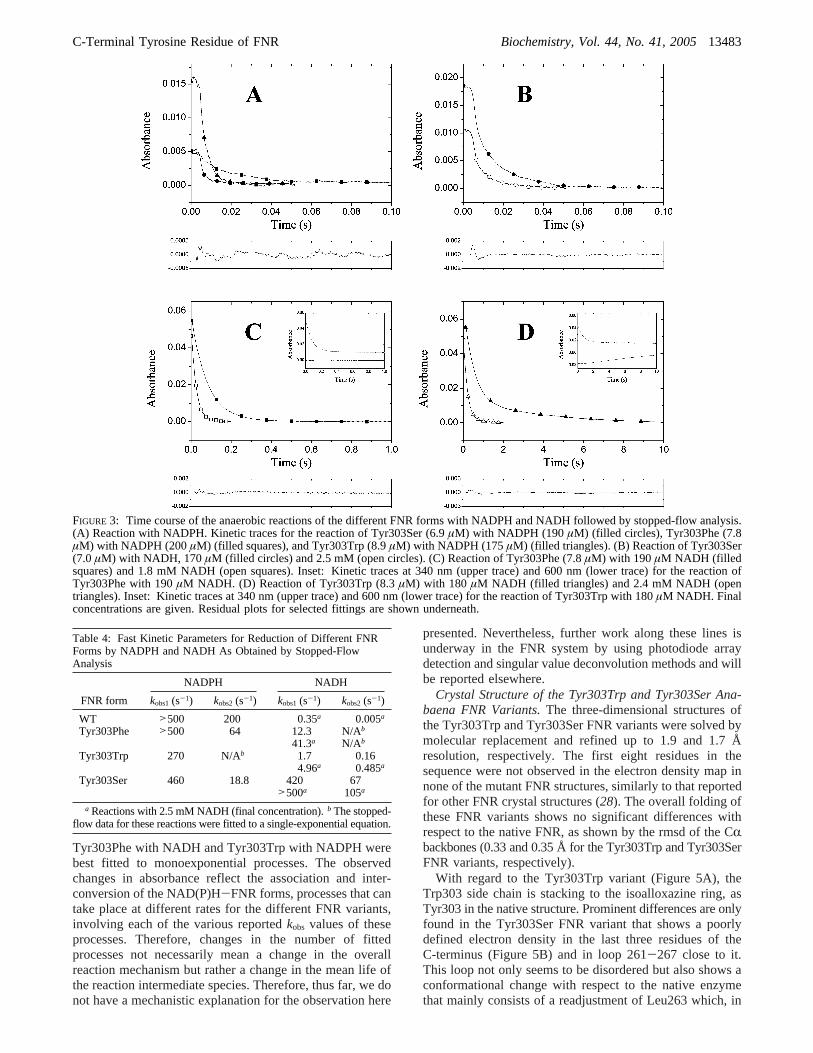
Tyr303Phe with NADH and Tyr303Trp with NADPH werebest fitted to monoexponential processes. The observedchanges in absorbance reflect the association and inter-conversion of the NAD(P)H-FNR forms, processes that cantake place at different rates for the different FNR variants,involving each of the various reportedkobs values of theseprocesses. Therefore, changes in the number of fittedprocesses not necessarily mean a change in the overallreaction mechanism but rather a change in the mean life ofthe reaction intermediate species. Therefore, thus far, we donot have a mechanistic explanation for the observation here
presented. Nevertheless, further work along these lines isunderway in the FNR system by using photodiode arraydetection and singular value deconvolution methods and willbe reported elsewhere.
Crystal Structure of the Tyr303Trp and Tyr303Ser Ana-baena FNR Variants.The three-dimensional structures ofthe Tyr303Trp and Tyr303Ser FNR variants were solved bymolecular replacement and refined up to 1.9 and 1.7 Åresolution, respectively. The first eight residues in thesequence were not observed in the electron density map innone of the mutant FNR structures, similarly to that reportedfor other FNR crystal structures (28). The overall folding ofthese FNR variants shows no significant differences withrespect to the native FNR, as shown by the rmsd of the CRbackbones (0.33 and 0.35 Å for the Tyr303Trp and Tyr303SerFNR variants, respectively).
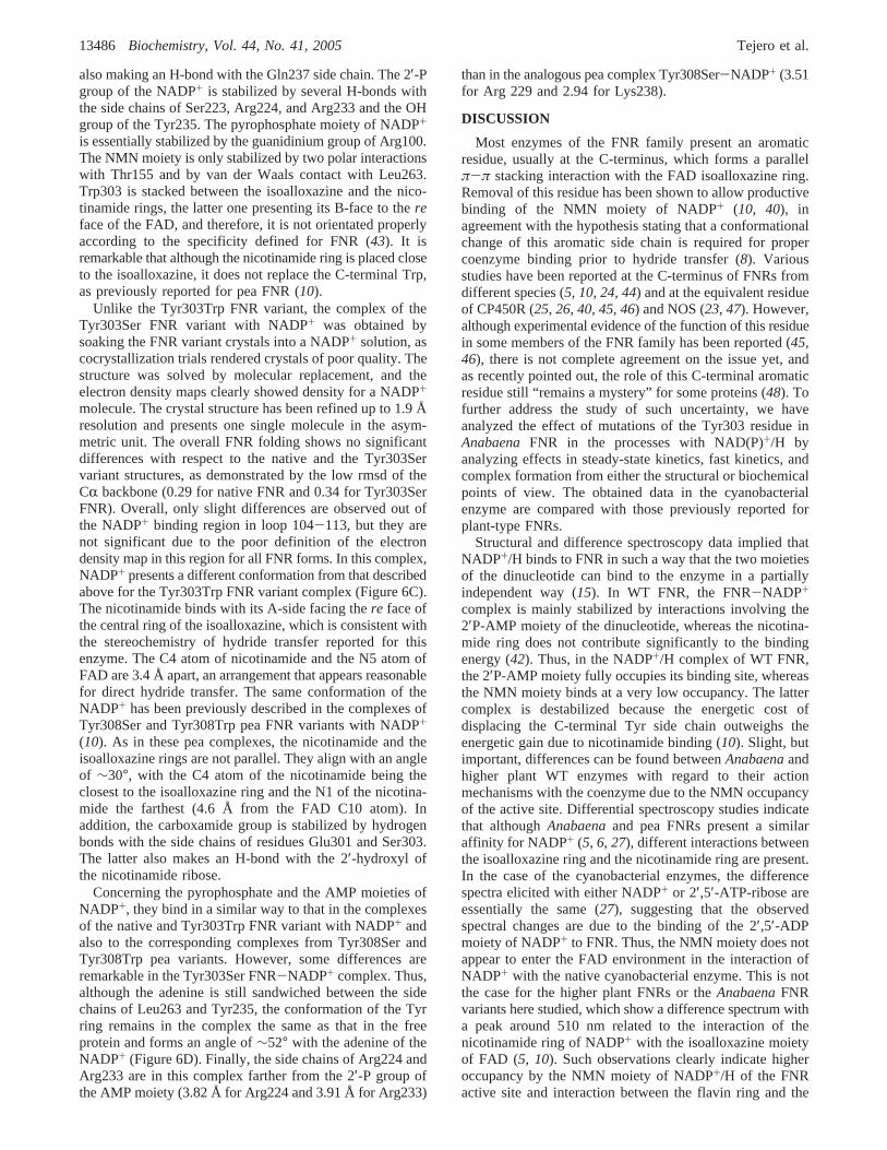
With regard to the Tyr303Trp variant (Figure 5A), theTrp303 side chain is stacking to the isoalloxazine ring, asTyr303 in the native structure. Prominent differences are onlyfound in the Tyr303Ser FNR variant that shows a poorlydefined electron density in the last three residues of theC-terminus (Figure 5B) and in loop 261-267 close to it.This loop not only seems to be disordered but also shows aconformational change with respect to the native enzymethat mainly consists of a readjustment of Leu263 which, in
FIGURE 3: Time course of the anaerobic reactions of the different FNR forms with NADPH and NADH followed by stopped-flow analysis.(A) Reaction with NADPH. Kinetic traces for the reaction of Tyr303Ser (6.9µM) with NADPH (190µM) (filled circles), Tyr303Phe (7.8µM) with NADPH (200µM) (filled squares), and Tyr303Trp (8.9µM) with NADPH (175µM) (filled triangles). (B) Reaction of Tyr303Ser(7.0µM) with NADH, 170 µM (filled circles) and 2.5 mM (open circles). (C) Reaction of Tyr303Phe (7.8µM) with 190 µM NADH (filledsquares) and 1.8 mM NADH (open squares). Inset: Kinetic traces at 340 nm (upper trace) and 600 nm (lower trace) for the reaction ofTyr303Phe with 190µM NADH. (D) Reaction of Tyr303Trp (8.3µM) with 180 µM NADH (filled triangles) and 2.4 mM NADH (opentriangles). Inset: Kinetic traces at 340 nm (upper trace) and 600 nm (lower trace) for the reaction of Tyr303Trp with 180µM NADH. Finalconcentrations are given. Residual plots for selected fittings are shown underneath.
Table 4: Fast Kinetic Parameters for Reduction of Different FNRForms by NADPH and NADH As Obtained by Stopped-FlowAnalysis
NADPH NADH
FNR form kobs1(s-1) kobs2(s-1) kobs1(s-1) kobs2(s-1)
WT >500 200 0.35a 0.005a
Tyr303Phe >500 64 12.3 N/Ab
41.3a N/Ab
Tyr303Trp 270 N/Ab 1.7 0.164.96a 0.485a
Tyr303Ser 460 18.8 420 67>500a 105a
a Reactions with 2.5 mM NADH (final concentration).b The stopped-flow data for these reactions were fitted to a single-exponential equation.
C-Terminal Tyrosine Residue of FNR Biochemistry, Vol. 44, No. 41, 200513483
the native FNR, orients its side chain to the NADP+/Hbinding site, while in this variant it is oriented to the interiorof the protein, increasing the size of the cavity. The disorderin this region may be due to the mutation of the Tyr303 bySer as, in the native structure, the loop 261-267 H-bondsto the C-terminal residues that are in turn stabilized bystacking interactions of the Tyr303 side chain to the
isoalloxazine ring. Therefore, the Tyr303Ser replacementdisturbs a number of stabilizing interactions at the C-terminusof the polypeptide chain that also extend to the 261-267loop. Additionally, the smaller side chain at the C-terminusmakes an enlarged nicotinamide binding site. Some otherdifferences in the active site of both variants have beenobserved with respect to the native structure, particularlyconcerning a highly conserved water molecule (W1) (9). Thiswater molecule is stabilized by H-bonds to several residues,including the OH group of Tyr303 (Figure 5A), and has beenproposed to be acting as a proton donor to the N5 of theisoalloxazine ring (8). This W1 has not been found in theTyr303Trp variant. In the case of the Tyr303Ser variant, W1is conserved, and there are also two additional watermolecules occupying the C-terminal Tyr site (Figure 5B).One of these additional water molecules is mimicking theTyr303 hydroxyl group of the native structure, and the otheris placed just in the middle of the aromatic ring. Then, theyare forming a H-bond network from the Ser303 hydroxylgroup to the W1.
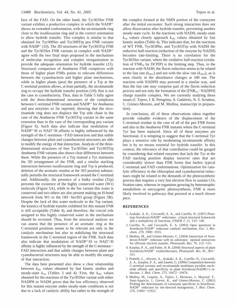
Crystal Structure of the Tyr303Trp and Tyr303Ser FNRVariants in Complex with NADP+. Cocrystallization of theTyr303Trp FNR variant with NADP+ produces tetragonalcrystals nonisomorphous to native. The structure solved bymolecular replacement presents two molecules in the asym-metric unit, and the electron density map showed a strongdensity for both complete NADP+ molecules. The complexstructure has been refined up to 3.0 Å resolution. The overallFNR folding is equivalent to that of the freeAnabaenaFNR,and no significant changes in the relative orientation of theFAD and NADP+ domains were observed, with the rmsd ofthe CR backbones being 0.57 Å. The NADP+ molecule isplaced in the cavity located at the C-terminal edge of theparallel â-sheet of the NADP+ binding domain, whichextends toward the FAD binding domain interface (Figure6A). The coenzyme presents an L-shape conformation as wasobserved in the native FNR complexed with NADP+ (15),where the pyrophosphate group is located at the corner andthe two branches of the L were accommodating the NMNand 2′P-AMP moieties, respectively (Figure 6B). Similarlyto the reported FNR-NADP+ complex (15) the adenine ofthe AMP moiety is sandwiched between the hydrophobicLeu263 side chain and the aromatic ring of Tyr235 and is
FIGURE 4: Proposed scheme for NAD(P)+/H binding to FNR. C1 designates a complex similar to that reported by Hermoso et al. (15), withthe pyrophosphate, adenine, and 2′-P bound to FNR. C2 resembles a complex in disposition suitable for hydride transfer. When the affinityof the enzyme for the 2′-P is low or the affinity for the nicotinamide ring is too high, nucleotide binding can be driven by C3. See text fordetails.
FIGURE 5: Structural changes upon Tyr303 mutation. (A) Structuralsuperimposition of native FNR (white) and the Tyr303Trp variant(blue). The FAD cofactor and the C-terminal residues are repre-sented as balls and sticks. The conserved water molecule (W1) innative FNR is colored in orange. (B) Structural superimposition ofnative FNR (white) and the Tyr303Ser variant (green). The waterhydrogen bond network (see text) is also represented.
13484 Biochemistry, Vol. 44, No. 41, 2005 Tejero et al.

FIGURE 6: Crystal structures of Tyr303Trp FNR-NADP+ and Tyr303Ser FNR-NADP+ complexes. (A) Molecular surface and ribbon diagram of the Tyr303Trp FNR-NADP+ complex. TheFAD prosthetic group (pink), NADP+ coenzyme (cyan), and Trp303 (purple) are represented as balls and sticks. Protein binding domains for each cofactor, FAD and NADP+, are colored palegreen and purple, respectively. (B) Stereo diagram showing the residues lining the NADP+ in the Tyr303Trp FNR-NADP+ complex. Relevant residues are shown in a ball-and-stick representationand colored by atom type. FAD (orange) and NADP+ (green) cofactors are drawn in a ball-and-stick representation. (C) Stereoview representation of the superimposition of Tyr303Trp FNR-NADP+ (white) and Tyr303Ser FNR-NADP+ (green) complexes. FAD cofactor and NADP+ coenzyme are colored in orange for the Tyr303Trp FNR and in blue for the Tyr303Ser FNR. (D)Stereo diagram showing the residues lining the NADP+ in the Tyr303Ser FNR-NADP+ complex. Relevant residues are shown in a ball-and-stick representation and colored by atom type. FAD(orange) and NADP+ (cyan) cofactors are drawn in a ball-and-stick representation.
C-T
erminalT
yrosineR
esidueof
FN
RB
ioch
em
istry,V
ol.
44
,N
o.
41
,2
00
513485
also making an H-bond with the Gln237 side chain. The 2′-Pgroup of the NADP+ is stabilized by several H-bonds withthe side chains of Ser223, Arg224, and Arg233 and the OHgroup of the Tyr235. The pyrophosphate moiety of NADP+
is essentially stabilized by the guanidinium group of Arg100.The NMN moiety is only stabilized by two polar interactionswith Thr155 and by van der Waals contact with Leu263.Trp303 is stacked between the isoalloxazine and the nico-tinamide rings, the latter one presenting its B-face to thereface of the FAD, and therefore, it is not orientated properlyaccording to the specificity defined for FNR (43). It isremarkable that although the nicotinamide ring is placed closeto the isoalloxazine, it does not replace the C-terminal Trp,as previously reported for pea FNR (10).
Unlike the Tyr303Trp FNR variant, the complex of theTyr303Ser FNR variant with NADP+ was obtained bysoaking the FNR variant crystals into a NADP+ solution, ascocrystallization trials rendered crystals of poor quality. Thestructure was solved by molecular replacement, and theelectron density maps clearly showed density for a NADP+
molecule. The crystal structure has been refined up to 1.9 Åresolution and presents one single molecule in the asym-metric unit. The overall FNR folding shows no significantdifferences with respect to the native and the Tyr303Servariant structures, as demonstrated by the low rmsd of theCR backbone (0.29 for native FNR and 0.34 for Tyr303SerFNR). Overall, only slight differences are observed out ofthe NADP+ binding region in loop 104-113, but they arenot significant due to the poor definition of the electrondensity map in this region for all FNR forms. In this complex,NADP+ presents a different conformation from that describedabove for the Tyr303Trp FNR variant complex (Figure 6C).The nicotinamide binds with its A-side facing there face ofthe central ring of the isoalloxazine, which is consistent withthe stereochemistry of hydride transfer reported for thisenzyme. The C4 atom of nicotinamide and the N5 atom ofFAD are 3.4 Å apart, an arrangement that appears reasonablefor direct hydride transfer. The same conformation of theNADP+ has been previously described in the complexes ofTyr308Ser and Tyr308Trp pea FNR variants with NADP+
(10). As in these pea complexes, the nicotinamide and theisoalloxazine rings are not parallel. They align with an angleof ∼30°, with the C4 atom of the nicotinamide being theclosest to the isoalloxazine ring and the N1 of the nicotina-mide the farthest (4.6 Å from the FAD C10 atom). Inaddition, the carboxamide group is stabilized by hydrogenbonds with the side chains of residues Glu301 and Ser303.The latter also makes an H-bond with the 2′-hydroxyl ofthe nicotinamide ribose.
Concerning the pyrophosphate and the AMP moieties ofNADP+, they bind in a similar way to that in the complexesof the native and Tyr303Trp FNR variant with NADP+ andalso to the corresponding complexes from Tyr308Ser andTyr308Trp pea variants. However, some differences areremarkable in the Tyr303Ser FNR-NADP+ complex. Thus,although the adenine is still sandwiched between the sidechains of Leu263 and Tyr235, the conformation of the Tyrring remains in the complex the same as that in the freeprotein and forms an angle of∼52° with the adenine of theNADP+ (Figure 6D). Finally, the side chains of Arg224 andArg233 are in this complex farther from the 2′-P group ofthe AMP moiety (3.82 Å for Arg224 and 3.91 Å for Arg233)
than in the analogous pea complex Tyr308Ser-NADP+ (3.51for Arg 229 and 2.94 for Lys238).
DISCUSSION
Most enzymes of the FNR family present an aromaticresidue, usually at the C-terminus, which forms a parallelπ-π stacking interaction with the FAD isoalloxazine ring.Removal of this residue has been shown to allow productivebinding of the NMN moiety of NADP+ (10, 40), inagreement with the hypothesis stating that a conformationalchange of this aromatic side chain is required for propercoenzyme binding prior to hydride transfer (8). Variousstudies have been reported at the C-terminus of FNRs fromdifferent species (5, 10, 24, 44) and at the equivalent residueof CP450R (25, 26, 40, 45, 46) and NOS (23, 47). However,although experimental evidence of the function of this residuein some members of the FNR family has been reported (45,46), there is not complete agreement on the issue yet, andas recently pointed out, the role of this C-terminal aromaticresidue still “remains a mystery” for some proteins (48). Tofurther address the study of such uncertainty, we haveanalyzed the effect of mutations of the Tyr303 residue inAnabaenaFNR in the processes with NAD(P)+/H byanalyzing effects in steady-state kinetics, fast kinetics, andcomplex formation from either the structural or biochemicalpoints of view. The obtained data in the cyanobacterialenzyme are compared with those previously reported forplant-type FNRs.
Structural and difference spectroscopy data implied thatNADP+/H binds to FNR in such a way that the two moietiesof the dinucleotide can bind to the enzyme in a partiallyindependent way (15). In WT FNR, the FNR-NADP+
complex is mainly stabilized by interactions involving the2′P-AMP moiety of the dinucleotide, whereas the nicotina-mide ring does not contribute significantly to the bindingenergy (42). Thus, in the NADP+/H complex of WT FNR,the 2′P-AMP moiety fully occupies its binding site, whereasthe NMN moiety binds at a very low occupancy. The lattercomplex is destabilized because the energetic cost ofdisplacing the C-terminal Tyr side chain outweighs theenergetic gain due to nicotinamide binding (10). Slight, butimportant, differences can be found betweenAnabaenaandhigher plant WT enzymes with regard to their actionmechanisms with the coenzyme due to the NMN occupancyof the active site. Differential spectroscopy studies indicatethat althoughAnabaenaand pea FNRs present a similaraffinity for NADP+ (5, 6, 27), different interactions betweenthe isoalloxazine ring and the nicotinamide ring are present.In the case of the cyanobacterial enzymes, the differencespectra elicited with either NADP+ or 2′,5′-ATP-ribose areessentially the same (27), suggesting that the observedspectral changes are due to the binding of the 2′,5′-ADPmoiety of NADP+ to FNR. Thus, the NMN moiety does notappear to enter the FAD environment in the interaction ofNADP+ with the native cyanobacterial enzyme. This is notthe case for the higher plant FNRs or theAnabaenaFNRvariants here studied, which show a difference spectrum witha peak around 510 nm related to the interaction of thenicotinamide ring of NADP+ with the isoalloxazine moietyof FAD (5, 10). Such observations clearly indicate higheroccupancy by the NMN moiety of NADP+/H of the FNRactive site and interaction between the flavin ring and the
13486 Biochemistry, Vol. 44, No. 41, 2005 Tejero et al.

NADP+ nicotinamide moiety (Figure 2). This explains why,based on considerations of the bipartite binding model, theAnabaenaFNR C-terminal mutants show enhanced bindingof NADP+, and in the case of these FNR mutants, theincreased affinity for the NMN portion of the coenzymedrives the binding of the whole dinucleotide and producesstronger interactions. Additionally, it is worth noting thedecay in the long-wavelength band observed in the case ofthe Tyr303Trp variant (putatively due to the charge transfercharacter of the interaction between the indole group of Trpand the isoalloxazine ring of FAD) upon complex formationwith both NADP+ and NAD+ (Figure 2). Thus, the NADP+
and NAD+ difference spectra of theAnabaenaTyr303TrpFNR complexes show that nucleotide binding causes thedisruption of this interaction, probably by displacement ofthe Trp residue from the isoalloxazine ring environment.However, this effect has not been reported in CP450R, asthe nicotinamide appears unable to displace the Trp residue(40).
Kd data can be fitted to a simple model to obtain separatebinding constants for the 2′P-AMP (Ka,2′P-AMP) and nicotin-amide (Ka,NMN) moieties (Figure 4 and Table 5). In thismodel, shown in Figure 4, it is assumed that the displacementof the C-terminal residue and the nicotinamide bindingconstitute a concerted event. This is the simplest model toaccount for the observed events; a further complication ofthe mechanism in which the C-terminal residue can switchbetween two conformations, flipping in and out of the pocket,is ignored in the calculations. This movement would createan equilibrium between “in” and “out” conformations, beingthe “out” conformation the reactive species. A slow equili-bration between “in” and “out” conformations would convertthe C1 species (Figure 4) in a bottleneck. As the fullreduction of the WT and mutated enzymes with NADPH isquite fast (Figure 3 and Table 4), it can be assumed that theflipping of the C-terminal residue, if existent, proceeds in ashort time scale and it is not rate-limiting. A secondsimplification assumes that, in the case of NAD+, the bindingis only driven by the NMN moiety, and the influence of the2′P-AMP moiety is negligible. Previous data support thisbipartite binding model (5, 10, 15, 26). Thus, the values ofKa,NMN are directly derived from theKd values obtained forNAD+, whereas the values ofKa,2′P-AMP are derived fromthe Kd values obtained for NADP+, on the followingassumptions: (1) nicotinamide occupancy for theAnabaenaWT FNR-NADP+ complex is 0; (2) nicotinamide oc-
cupancy for the pea WT FNR-NADP+ complex is 14%, aspreviously estimated (5); (3) Ka,2′P-AMP is similar to WT forall of the mutant enzymes.
The calculated equilibrium constants (Table 5) are con-sistent with the proposed scheme. Two binding equilibriacompete when FNR and the coenzyme are mixed (15). WhenNADP+/H is used, the equilibrium is driven through C1(Figure 4), whereas in the case of NAD+/H the formationof C1 is negligible (6). Nevertheless, if the NMN moietycan replace the C-terminal residue, it could provide adifferent binding mode forming species C3 (Figure 4). Toexplain the recognition of NADP+/H, a sequential equilib-rium has to be postulated (15), with an apparent equilibriumconstant,Kin (Table 5). A lowKin value indicates that theinteraction between the isoalloxazine ring and the C-terminalresidue is much stronger than the interaction between theisoalloxazine ring and the nicotinamide moiety of NAD-(P)+/H (stabilization of form C1 in Figure 4), whereas a highKin value indicates the opposite situation (stabilization ofform C3 in Figure 4). In most cases, the values determinedsuggest a strong displacement of the equilibrium in one wayor the other. Only for theAnabaena Tyr303Phe andTyr303Trp variants and the pea WT and Tyr308Trp can avalue ofKin be estimated, thus indicating that the stackinginteraction of the aromatic residue with the isoalloxazine ringof FAD has a comparable energy to that of the nicotina-mide-FAD interaction. The NMN occupancy values esti-mated from theKin (Table 5) show a good agreement withthe experimental values derived from the difference spectrareported in this work (Table 2) and those of ref5. It isremarkable the low % occexp obtained for the interaction ofNADP+ with the AnabaenaTyr303Trp variant, whichcorrelates with our structural studies showing no occupancyof the C-terminal position by the coenzyme NMN portion(Figure 6).
Three-dimensional structures of the Tyr303Trp FNR andthe Tyr303SerAnabaenaFNR variants in complex withNADP+ have been solved (Figure 6). Unexpectedly, bothstructures are largely different. The Tyr303Trp FNR variantexhibits a nonproductive complex, similar to that reportedfor the FNR-NADP+ complex obtained by cocrystallization(15), in which the 2′P-AMP and pyrophosphate portions ofthe NADP+ are perfectly bound. In addition, the NMNmoiety is placed in a new pocket created near the FADcofactor with the ribose being in a tight conformation.Besides, the nicotinamide ring presents its B-face to there
Table 5: Calculated Equilibrium Constants for Bipartite Binding of Nicotinamide Coenzymesa
FNR form Ka,2′P-AMP (µM-1) Kin Ka,NMN (µM-1) % occexpb % occcalc
b
AnabaenaPCC7119WT 0.175 <0.01 ,1.0× 10-4 0 0Tyr303Phe 0.175c 3.7 1.0× 10⁻⁴ 71 79Tyr303Trp 0.175c 2.6 1.6× 10⁻⁴ ∼15 71Tyr303Ser 0.175c >50 1.8× 10⁻³ 100 >98
peaWT 0.093 0.16 ,1.0× 10-4 14d 14e
Tyr308Trp 0.093c 1.0 ,1.0× 10-4 40d 50Tyr308Phe 0.093c >50 1.2× 10⁻⁴ 85d >98Tyr308Gly 0.093c >50 2.0× 10⁻³ 84d >98Tyr308Ser 0.093c >50 5.3× 10⁻³ 100d >98
a Referred to the scheme in Figure 4.b % occexp, nicotinamide occupancy estimated experimentally from the∆ε value of the peak at∼500 nm;% occcalc, nicotinamide occupancy calculated fromKin values.c Value of the WT enzyme assumed for all FNR variants.d Values from ref5.e Value implicit in constant calculation.
C-Terminal Tyrosine Residue of FNR Biochemistry, Vol. 44, No. 41, 200513487
face of the FAD. On the other hand, the Tyr303Ser FNRvariant exhibits a productive complex in which the NADP+
shows an extended conformation, with the nicotinamide ringclose to the isoalloxazine ring and in the correct orientationto allow hydride transfer. This complex is similar to thatobtained for Tyr308Ser and Tyr308Trp pea FNR variantswith NADP+ (10). The 3D structures of the Tyr303Trp FNRand the Tyr303Ser FNR variants in complex with NADP+
agree with the two final steps proposed in the mechanismof molecular recognition and complex reorganization toprovide the adequate orientation for hydride transfer (15).Structural comparison ofAnabaenaFNR complexes withthose of higher plant FNRs points to relevant differencesbetween the cyanobacteria and higher plant mechanisms,while in higher plants (pea) the presence of a Trp at theC-terminal position allows, at least partially, the nicotinamidering to occupy the hydride transfer position (10); that is notthe case in cyanobacteria. Thus, data in Table 5 fully agreewith the three-dimensional structures of the complexesbetween C-terminal FNR variants and NADP+ for Anabaenaand pea enzymes so far reported, showing that the nico-tinamide ring does not displace the Trp side chain in thecase of theAnabaenaFNR Tyr303Trp variant in the sameextension than in the case of the corresponding pea variant(Figure 6). Such data also indicate that modulation ofNADP+/H vs NAD+/H affinity is highly influenced by thestrength of the C-terminus-FAD interaction and that subtlechanges between plant and cyanobacterial structures are ableto modify the energy of that interaction. Analysis of the three-dimensional structures of free Tyr303Ser and Tyr303TrpAnabaenaFNR variants also shows clear differences betweenthem. While the presence of a Trp instead a Tyr maintainsthe 3D arrangement of the FNR, and a similar stackinginteraction between isoalloxazine ring and Trp is produced,deletion of the aromatic residue at the 303 position substan-tially perturbs the structural framework around the C-terminalend. Additionally, the presence of a bulky residue (Trp)prevents the existence of the highly conserved water (W1)molecule (Figure 5A), while in the Ser variant this water isconserved and two others are also present making an H-bondnetwork from W1 to the OH-Ser303 group (Figure 5B).Despite the lack of this water molecule in the Trp variant,the kinetics of hydride transfer exhibited for this mutant FNRis still acceptable (Table 4), and therefore, the crucial roleassigned to this highly conserved water in the mechanismshould be revisited. Thus, from the structural analysis wecan assess that the presence of an aromatic ring at theC-terminal positions seems to be relevant not only in thecatalytic mechanism but also in stabilizing the structuralframework in the C-terminal region of the FNR. Such dataalso indicate that modulation of NADP+/H vs NAD+/Haffinity is highly influenced by the strength of the C-terminus-FAD interaction and that subtle changes between plant andcyanobacterial structures may be able to modify the energyof that interaction.
The data here presented also show a clear relationshipbetweenkobs values obtained by fast kinetic studies andsteady-statekcat (Tables 3 and 4). First, thekobs valuesobtained for the reactions of the Tyr303Ser variant with eitherNADPH or NADH prove that the low efficiency observedfor this mutant enzyme under steady-state conditions is notdue to a lack of catalytic ability but rather to the strength of
the complex formed at the NMN portion of the coenzymeafter the initial encounter. Such strong interaction does notallow dissociation after hydride transfer, preventing anothersteady-state cycle. In the reactions with NADH, steady-statekcat values closely approachkobs values obtained by fastkinetic studies (Table 4). This indicates that, for the reactionsof WT FNR, Tyr303Phe, and Tyr303Trp with NADH thereductive half-reaction (reduction of the enzyme by NADH)becomes rate-limiting. There is no correlation for theTyr303Ser variant, where the oxidative half-reaction (oxida-tion of FNRrd by DCPIP) is the limiting step. Thus, in thereaction with NADH, the flavin reduction seems to be relatedto the fast rate (kobs1) and not with the slow rate (kobs2), as isseen clearly in the absorbance changes at 340 nm. Thereaction with NADPH may proceed in a similar way, andthus the fast rate may comprise part of the flavin reductionprocess and not only the formation of the [FNRox-NADPH]charge transfer complex. We are currently studying theseissues (J. Tejero, J. R. Peregrina, A. Gutie´rrez, N. S. Scrutton,C. Gomez-Moreno, and M. Medina, manuscript in prepara-tion).
In conclusion, all of these observations taken togetherprovide valuable evidence of the displacement of theC-terminal residue in the case of all of the pea FNR formsand also in theAnabaenaFNR mutants where the C-terminalTyr has been replaced. Since all of these enzymes arefunctional, it is tempting to suggest that the C-terminal Tyrplays a sensitive role by modulating nicotinamide affinitybut is by no means essential for hydride transfer. In thiscontext, the relevance of that contribution could be gaugedby considering that related enzymes that lack the Tyr at theFAD stacking position display turnover rates that areconsiderably slower than FNR forms that harbor typicalC-terminal and FAD conformations. Optimization for cata-lytic efficiency in the chloroplast and cyanobacterial reduc-tases might be related to the demands of the photosyntheticprocess that requires a very fast electron flow to sustain CO2
fixation rates, whereas in organisms growing by heterotrophicmetabolism or anoxygenic photosynthesis, FNR is morelikely involved in pathways that proceed at a much slowerpace.
REFERENCES
1. Arakaki, A. K., Ceccarelli, E. A., and Carrillo, N. (1997) Plant-type ferredoxin-NADP+ reductases: a basal structural frameworkand a multiplicity of functions,FASEB J. 11, 133-140.
2. Carrillo, N., and Ceccarelli, E. A. (2003) Open questions inferredoxin-NADP+ reductase catalytic mechanism,Eur. J. Bio-chem. 270, 1900-1915.
3. Medina, M., and Go´mez-Moreno, C. (2004) Interaction of ferre-doxin-NADP+ reductase with its substrates: optimal interactionfor efficient electron transfer,Photosynth. Res. 79, 113-131.
4. Karplus, P. A., and Faber, H. R. (2004) Structural aspects of plantferredoxin:NADP+ oxidoreductases,Photosynth. Res. 81, 303-315.
5. Piubelli, L., Aliverti, A., Arakaki, A. K., Carrillo, N., Ceccarelli,E. A., Karplus, P. A., and Zanetti, G. (2000) Competition betweenC-terminal tyrosine and nicotinamide modulates pyridine nucle-otide affinity and specificity in plant ferredoxin-NADP(+) re-ductase,J. Biol. Chem. 275, 10472-10476.
6. Medina, M., Luquita, A., Tejero, J., Hermoso, J., Mayoral, T.,Sanz-Aparicio, J., Grever, K., and Go´mez-Moreno, C. (2001)Probing the determinants of coenzyme specificity in ferredoxin-NADP+ reductase by site-directed mutagenesis,J. Biol. Chem.276, 11902-11912.
13488 Biochemistry, Vol. 44, No. 41, 2005 Tejero et al.

7. Karplus, P. A., Daniels, M. J., and Herriott, J. R. (1991) Atomicstructure of ferredoxin-NADP+ reductase: prototype for a struc-turally novel flavoenzyme family,Science 251, 60-66.
8. Bruns, C. M., and Karplus, P. A. (1995) Refined crystal structureof spinach ferredoxin reductase at 1.7 Å resolution: oxidized,reduced and 2′-phospho-5′-AMP bound states,J. Mol. Biol. 247,125-145 (doi: 10.1006/jmbi.1994.0127).
9. Serre, L., Vellieux, F. M., Medina, M., Go´mez-Moreno, C.,Fontecilla-Camps, J. C., and Frey, M. (1996) X-ray structure ofthe ferredoxin:NADP+ reductase from the cyanobacteriumAna-baenaPCC 7119 at 1.8 Å resolution, and crystallographic studiesof NADP+ binding at 2.25 Å resolution,J. Mol. Biol. 263, 20-39.
10. Deng, Z., Aliverti, A., Zanetti, G., Arakaki, A. K., Ottado, J.,Orellano, E. G., Calcaterra, N. B., Ceccarelli, E. A., Carrillo, N.,and Karplus, P. A. (1999) A productive NADP+ binding modeof ferredoxin-NADP+ reductase revealed by protein engineeringand crystallographic studies,Nat. Struct. Biol. 6, 847-853.
11. Dorowski, A., Hofmann, A., Steegborn, C., Boicu, M., and Huber,R. (2001) Crystal structure of paprika ferredoxin-NADP+ reduc-tase. Implications for the electron transfer pathway,J. Biol. Chem.276, 9253-9263.
12. Aliverti, A., Faber, R., Finnerty, C. M., Ferioli, C., Pandini, V.,Negri, A., Karplus, P. A., and Zanetti, G. (2001) Biochemicaland crystallographic characterization of ferredoxin-NADP(+)reductase from nonphotosynthetic tissues,Biochemistry 40, 14501-14508.
13. Morales, R., Charon, M.-H., Kachalova, G., Serre, L., Medina,M., Gomez-Moreno, C., and Frey, M. (2000) A redox-dependentinteraction between two electron-transfer partners involved inphotosynthesis,EMBO Rep. 1, 271-276.
14. Kurisu, G., Kusunoki, M., Katoh, E., Yamazaki, T., Teshima, K.,Onda, Y., Kimata-Ariga, Y., and Hase, T. (2001) Structure of theelectron transfer complex between ferredoxin and ferredoxin-NADP(+) reductase,Nat. Struct. Biol. 8, 117-121.
15. Hermoso, J. A., Mayoral, T., Faro, M., Go´mez-Moreno, C., Sanz-Aparicio, J., and Medina, M. (2002) Mechanism of coenzymerecognition and binding revealed by crystal structure analysis offerredoxin-NADP+ reductase complexed with NADP+, J. Mol.Biol. 319, 1133-1142.
16. Karplus, P. A., and Bruns, C. M. (1994) Structure-functionrelations for ferredoxin reductase,J. Bioenerg. Biomembr. 26, 89-99.
17. Chen, Z., Lee, W. R., and Chang, S. H. (1991) Role of asparticacid 38 in the cofactor specificity ofDrosophila alcohol de-hydrogenase,Eur. J. Biochem. 202, 263-267.
18. Clermont, S., Corbier, C., Mely, Y., Gerard, D., Wonacott, A.,and Branlant, G. (1993) Determinants of coenzyme specificity inglyceraldehyde-3-phosphate dehydrogenase: role of the acidicresidue in the fingerprint region of the nucleotide binding fold,Biochemistry 32, 10178-10184.
19. Marohnic, C. C., Bewley, M. C., and Barber, M. J. (2003)Engineering and characterization of a NADPH-utilizing cyto-chromeb5 reductase,Biochemistry 42, 11170-11182.
20. Scrutton, N. S., Berry, A., and Perham, R. N. (1990) Redesign ofthe coenzyme specificity of a dehydrogenase by protein engineer-ing, Nature 343, 38-43.
21. Bocanegra, J. A., Scrutton, N. S., and Perham, R. N. (1993)Creation of an NADP-dependent pyruvate dehydrogenase multi-enzyme complex by protein engineering,Biochemistry 32, 2737-2740.
22. Tejero, J., Martı´nez-Ju´lvez, M., Mayoral, T., Luquita, A., Sanz-Aparicio, J., Hermoso, J. A., Hurley, J. K., Tollin, G., Go´mez-Moreno, C., and Medina, M. (2003) Involvement of the pyro-phosphate and the 2′-phosphate binding regions of ferredoxin-NADP+ reductase in coenzyme specificity,J. Biol. Chem. 278,49203-49214.
23. Adak, S., Sharma, M., Meade, A. L., and Stuehr, D. J. (2002) Aconserved flavin-shielding residue regulates NO synthase electrontransfer and nicotinamide coenzyme specificity,Proc. Natl. Acad.Sci. U.S.A. 99, 13516-13521.
24. Nogue´s, I., Tejero, J., Hurley, J. K., Paladini, D., Frago, S., Tollin,G., Mayhew, S. G., Go´mez-Moreno, C., Ceccarelli, E. A., Carrillo,N., and Medina, M. (2004) Role of the C-terminal tyrosine offerredoxin-nicotinamide adenine dinucleotide phosphate reductasein the electron transfer processes with its protein partnersferredoxin and flavodoxin,Biochemistry 43, 6127-6137.
25. Dohr, O., Paine, M. J., Friedberg, T., Roberts, G. C., and Wolf,C. R. (2001) Engineering of a functional human NADH-dependentcytochrome P450 system,Proc. Natl. Acad. Sci. U.S.A. 98, 81-86.
26. Elmore, C. L., and Porter, T. D. (2002) Modification of thenucleotide cofactor-binding site of cytochrome P-450 reductaseto enhance turnover with NADH in vivo,J. Biol. Chem. 277,48960-48964.
27. Sancho, J., and Gomez-Moreno, C. (1991) Interaction of ferre-doxin-NADP+ reductase fromAnabaenawith its substrates,Arch.Biochem. Biophys. 288, 231-238.
28. Medina, M., Martı´nez-Ju´lvez, M., Hurley, J. K., Tollin, G., andGomez-Moreno, C. (1998) Involvement of glutamic acid 301 inthe catalytic mechanism of ferredoxin-NADP+ reductase fromAnabaenaPCC 7119,Biochemistry 37, 2715-2728.
29. Mendes, P., and Kell, D. B. (1998) Non-linear optimization ofbiochemical pathways: applications to metabolic engineering andparameter estimation,Bioinformatics 14, 869-883.
30. Collaborative Computational Project Number 4 (1994) The CCP4Suite: Programs for Protein Crystallography,Acta Crystallogr.D 50, 760-763.
31. Leslie, A. G. W. (1987) Profile fitting, inProceedings of the CCP4Study Weekend(Machin, J. R., and Papiz, M. Z., Eds.) pp 39-50, SERC Daresbury Laboratory, Warrington, U.K.
32. Navaza, J. (1994) AMoRe: an automated package for molecularreplacement,Acta Crystallogr. A 50, 157-163.
33. Brunger, A. T., Adams, P. D., Clore, G. M., DeLano, W. L., Gros,P., Grosse-Kunstleve, R. W., Jiang, J. S., Kuszewski, J., Nilges,M., Pannu, N. S., Read, R. J., Rice, L. M., Simonson, T., andWarren, G. L. (1998) Crystallography & NMR system: A newsoftware suite for macromolecular structure determination,ActaCrystallogr. D 54, 905-921.
34. Jones, T. A., Zou, J. Y., Cowan, S. W., and Kjeldgaard, M. (1991)Improved methods for building protein models in electron densitymaps and the location of errors in these models,Acta Crystallogr.A 47, 110-119.
35. Laskowski, R. A., MacArthur, M. W., Moss, D. S., and Thornton,J. M. (1993) PROCHECK: a program to check the stereochemicalquality of protein structures,J. Appl. Crystallogr. 26, 283-291.
36. Hooft, R. W. W., Vriend, G., Sander, C., and Abola, E. E. (1996)Errors in protein structures,Nature 381, 272.
37. Kraulis, P. J. (1991) MOLSCRIPT: a program to produce bothdetailed and schematic plots of protein structures,J. Appl.Crystallogr. 24, 946-950.
38. Merritt, E. A., and Bacon, D. J. (1997) Raster3D: PhotorealisticMolecular Graphics,Methods Enzymol. 277, 505-524.
39. Lostao, A., Gomez-Moreno, C., Mayhew, S. G., and Sancho, J.(1997) Differential stabilization of the three FMN redox formsby tyrosine 94 and tryptophan 57 in flavodoxin fromAnabaenaand its influence on the redox potentials,Biochemistry 36, 14334-14344.
40. Hubbard, P. A., Shen, A. L., Paschke, R., Kasper, C. B., and Kim,J. J. (2001) NADPH-cytochrome P450 oxidoreductase. Structuralbasis for hydride and electron transfer,J. Biol. Chem. 276, 29163-29170.
41. Batie, C. J., and Kamin, H. (1984) Electron transfer by ferredoxin:NADP+ reductase. Rapid-reaction evidence for participation of aternary complex,J. Biol. Chem. 259, 11976-11985.
42. Batie, C. J., and Kamin, H. (1986) Association of ferredoxin-NADP+ reductase with NADP(H) specificity and oxidation-reduction properties,J. Biol. Chem. 261, 11214-11223.
43. Krakow, G., Ammeraal, R. N., and Vennesland, B. (1965) Thestereospecificity of the Hill reaction with triphosphopyridinenucleotide,J. Biol. Chem. 240, 1820-1823.
44. Calcaterra, N. B., Pico, G. A., Orellano, E. G., Ottado, J., Carrillo,N., and Ceccarelli, E. A. (1995) Contribution of the FAD bindingsite residue Tyr 308 to the stability of pea ferredoxin-NADP+
oxidoreductase,Biochemistry 34, 12842-12848.45. Gutierrez, A., Doehr, O., Paine, M., Wolf, C. R., Scrutton, N. S.,
and Roberts, G. C. (2000) Trp-676 facilitates nicotinamidecoenzyme exchange in the reductive half-reaction of humancytochrome P450 reductase: properties of the soluble W676H andW676A mutant reductases,Biochemistry 39, 15990-15999.
46. Gutierrez, A., Paine, M., Wolf, C. R., Scrutton, N. S., and Roberts,G. C. (2002) Relaxation kinetics of cytochrome P450 reductase:internal electron transfer is limited by conformational changeand regulated by coenzyme binding,Biochemistry 41, 4626-4637.
C-Terminal Tyrosine Residue of FNR Biochemistry, Vol. 44, No. 41, 200513489

47. Konas, D. W., Zhu, K., Sharma, M., Aulak, K. S., Brudvig, G.W., and Stuehr, D. J. (2004) The FAD-shielding residue Phe1395regulates neuronal nitric oxide synthase catalysis by controllingNADP+ affinity and a conformational equilibrium within theflavoprotein domain,J. Biol. Chem. 279, 35412-35425.
48. Murataliev, M. B., Feyereisen, R., and Walker, F. A. (2004)Electron transfer by diflavin reductases,Biochim. Biophys. Acta1698, 1-26.
BI051278C
13490 Biochemistry, Vol. 44, No. 41, 2005 Tejero et al.
![Structure of a [2Fe–2S] ferredoxin from Rhodobacter capsulatus likely involved in Fe–S cluster biogenesis and conformational changes observed upon reduction](https://static.fdokumen.com/doc/165x107/63363fcacd4bf2402c0b6cfb/structure-of-a-2fe2s-ferredoxin-from-rhodobacter-capsulatus-likely-involved.jpg)