Are plastocyanin and ferredoxin specific electron ... - OSF
40
1 Are plastocyanin and ferredoxin specific electron carriers or generic redox 1 capacitors? Classical and murburn perspectives on two chloroplast proteins 2 Daniel Andrew Gideon 1,2 *, Vijay Nirusimhan 2 & Kelath Murali Manoj 1 * 3 *Corresponding authors 4 Email: [email protected] ; [email protected] 5 6 1 Satyamjayatu: The Science & Ethics Foundation 7 Kulappully, Shoranur-2 (PO), Palakkad District, Kerala State, India-679122. 8 9 2 Department of Biotechnology and Bioinformatics, Bishop Heber College (Autonomous), 10 Vayalur Road, Tiruchirappalli, Tamil Nadu, India-620017. 11 12 ABSTRACT: Within the context of light reaction of photosynthesis, the structure-function 13 correlations of the chloroplast proteins of plastocyanin and ferredoxins (Fd) are analyzed via 14 two perspectives: 1) The Z-scheme, which considers PC/Fd as specific affinity binding-based 15 electron-relay agents, thereby deterministically linking the functions of Cytochrome b 6 f (Cyt. 16 b 6 f) and Photosystem I (PS I) to NADP + reduction by Fd:NADPH oxidoreductase (FNR) via 17 protein-protein contacts and 2) The murburn explanation for oxygenic photophosphorylation, 18 which deems PC/Fd as generic ‘redox capacitors’, temporally accepting and releasing one- 19 electron equivalents in reaction milieu. Amino acid residues located on the surface loci of key 20 patches of PC/Fd vary in electrostatic/contour (topography) signatures. Crystal structures of 21 four different complexes each of cyt.f-PC and Fd-FNR show little conservation in the 22 contact-surfaces, thereby discrediting ‘affinity binding-based electron transfers (ET)’ as an 23 evolutionary logic. Further, thermodynamic and kinetic data on wildtype and mutant proteins 24 interactions do not align well with model 1. Furthermore, micromolar physiological 25 concentrations of PC (when K d values 100 μM) and the non-conducive architecture of 26 chloroplasts render the classical model untenable. In the 2 nd model, PC is optional and higher 27 concentrations of PC (sought by model 1) could inhibit ET, quite like the role of cytochrome 28 c of mitochondria and cytochrome b 5 of cytoplasmic microsomes. Also, PC is found in both 29 lumen and stroma, and plants lacking PC survive and grow. Thus, evidence from structure, 30 interactive dynamics with redox partners and physiological implications of PC/Fd supports 31 the murburn perspective that these proteins serve as generic redox-capacitors in chloroplasts. 32 Keywords: plastocyanin; ferredoxin; Z scheme, murburn concept; oxygenic photosynthesis; 33 light reaction; Q cycle; electron transport chain; chloroplast; 34
-
Upload
khangminh22 -
Category
Documents
-
view
3 -
download
0
Transcript of Are plastocyanin and ferredoxin specific electron ... - OSF
1
Are plastocyanin and ferredoxin specific electron carriers or generic redox 1
capacitors? Classical and murburn perspectives on two chloroplast proteins 2
Daniel Andrew Gideon1,2
*, Vijay Nirusimhan2 & Kelath Murali Manoj
1* 3
*Corresponding authors 4 Email: [email protected]; [email protected] 5
6 1Satyamjayatu: The Science & Ethics Foundation 7
Kulappully, Shoranur-2 (PO), Palakkad District, Kerala State, India-679122. 8 9
2 Department of Biotechnology and Bioinformatics, Bishop Heber College (Autonomous), 10
Vayalur Road, Tiruchirappalli, Tamil Nadu, India-620017. 11
12
ABSTRACT: Within the context of light reaction of photosynthesis, the structure-function 13
correlations of the chloroplast proteins of plastocyanin and ferredoxins (Fd) are analyzed via 14
two perspectives: 1) The Z-scheme, which considers PC/Fd as specific affinity binding-based 15
electron-relay agents, thereby deterministically linking the functions of Cytochrome b6f (Cyt. 16
b6f) and Photosystem I (PS I) to NADP+ reduction by Fd:NADPH oxidoreductase (FNR) via 17
protein-protein contacts and 2) The murburn explanation for oxygenic photophosphorylation, 18
which deems PC/Fd as generic ‘redox capacitors’, temporally accepting and releasing one-19
electron equivalents in reaction milieu. Amino acid residues located on the surface loci of key 20
patches of PC/Fd vary in electrostatic/contour (topography) signatures. Crystal structures of 21
four different complexes each of cyt.f-PC and Fd-FNR show little conservation in the 22
contact-surfaces, thereby discrediting ‘affinity binding-based electron transfers (ET)’ as an 23
evolutionary logic. Further, thermodynamic and kinetic data on wildtype and mutant proteins 24
interactions do not align well with model 1. Furthermore, micromolar physiological 25
concentrations of PC (when Kd values 100 μM) and the non-conducive architecture of 26
chloroplasts render the classical model untenable. In the 2nd
model, PC is optional and higher 27
concentrations of PC (sought by model 1) could inhibit ET, quite like the role of cytochrome 28
c of mitochondria and cytochrome b5 of cytoplasmic microsomes. Also, PC is found in both 29
lumen and stroma, and plants lacking PC survive and grow. Thus, evidence from structure, 30
interactive dynamics with redox partners and physiological implications of PC/Fd supports 31
the murburn perspective that these proteins serve as generic redox-capacitors in chloroplasts. 32
Keywords: plastocyanin; ferredoxin; Z scheme, murburn concept; oxygenic photosynthesis; 33
light reaction; Q cycle; electron transport chain; chloroplast; 34
2
INTRODUCTION 35
Both ferredoxin (Fd) and plastocyanin (PC) are soluble, monomeric, globular, redox-active 36
proteins involved in the light reaction of photosynthesis (1, 2). PC is a ~10 KDa blue 37
coloured Cu-protein of 97-104 amino acids, with linear dimensions of ~3 to 4 nm. It shows 38
an antiparallel fl-barrel structure containing 8 beta sheets and one Cu atom (3-5) (Figure 1 39
and Figure S1, Supplementary Information). Since crystal structures of PC from at least 15 40
organisms (cyanobacteria, algae, pteridophytes, angiosperms, etc.) have been solved using 41
XRD and NMR, PC is one of the most well-studied plant/photosynthetic proteins. Plant type 42
ferredoxins (Fds) are and several crystal structures of diverse Fds from myriad phototroph 43
sources is available. Fds are [2Fe2S] cluster iron-sulphur proteins (~11 KDa, 94-108 amino 44
acids) located on the stromal side of the thylakoid membrane (Figure 2) and they mediate 45
electron transfers between reduced PS I to oxidized FNR (Fd-NADP+ reductase). Several 46
Fds’ crystal structures are revealed and they also interact with other redox proteins (such as 47
thioredoxins and ferredoxin:thioredoxin reductase, FTR; apart from PS I and FNR) (6). 48
49
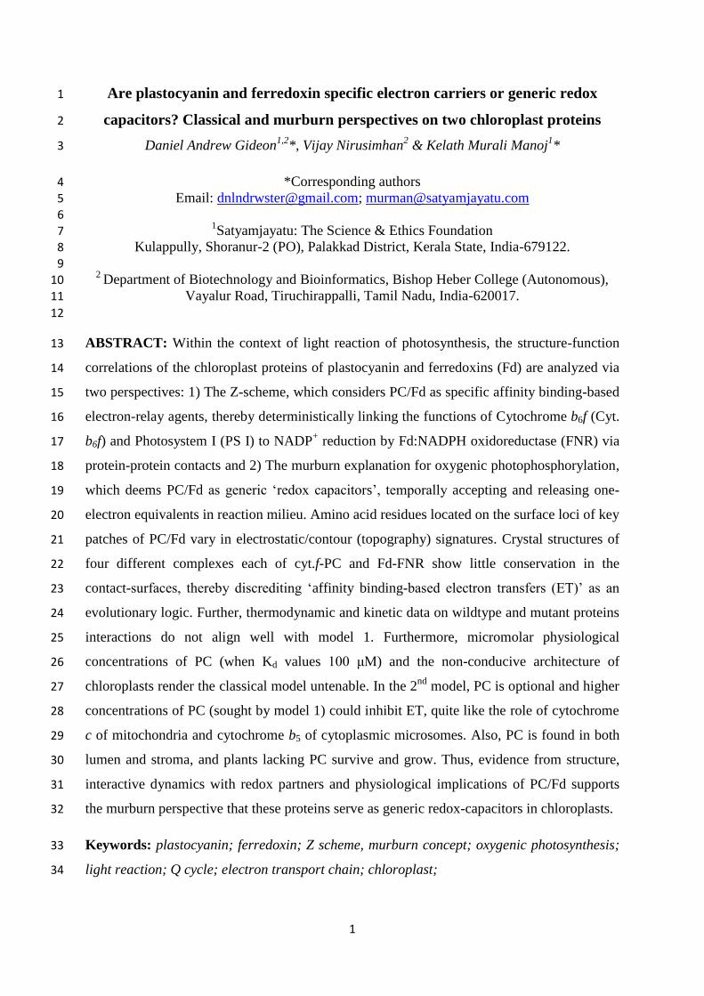
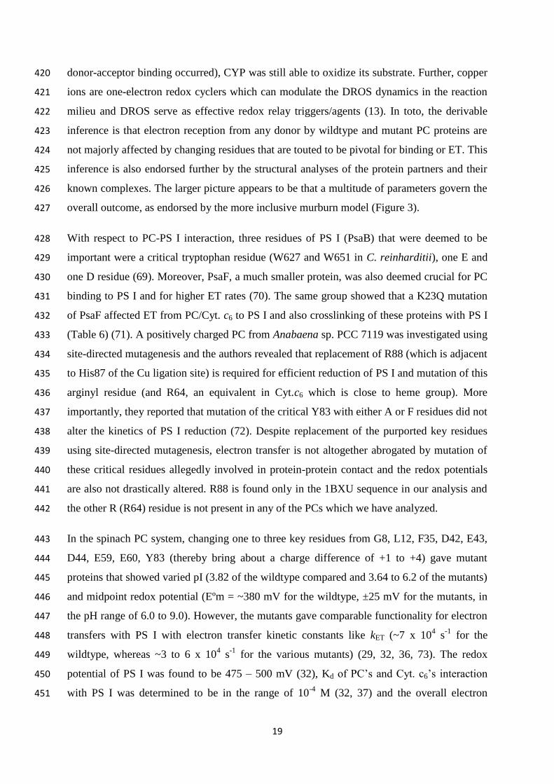
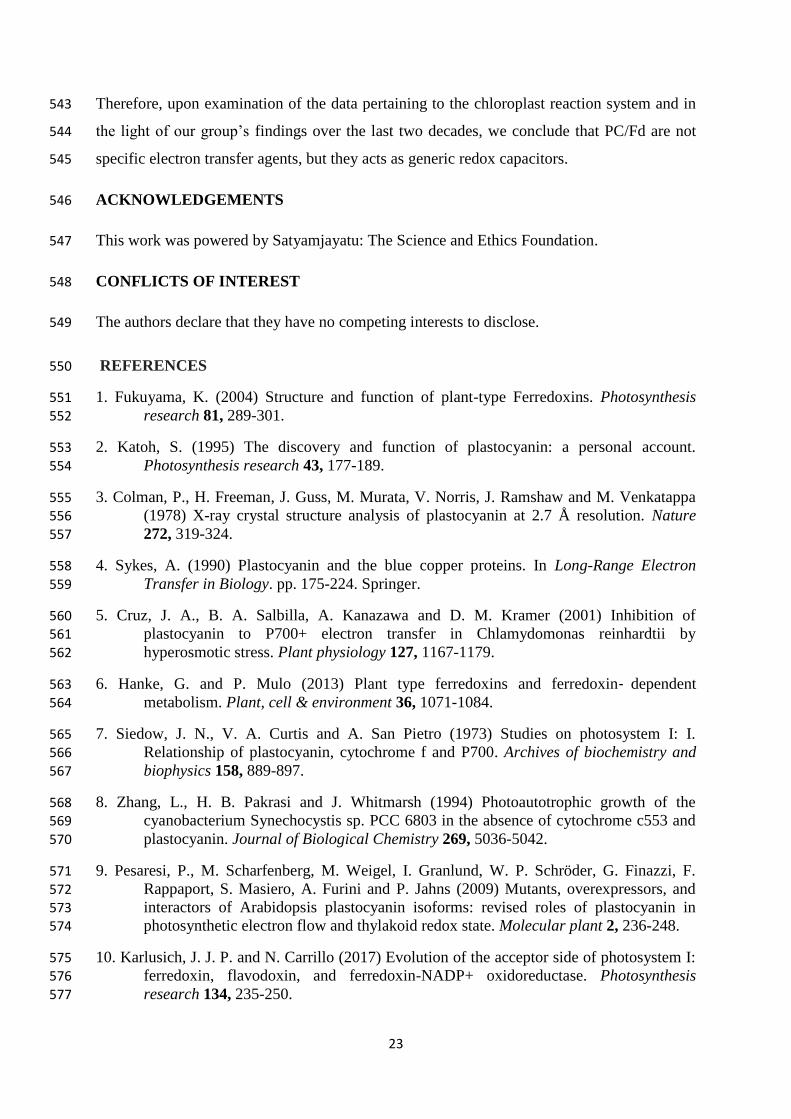
Figure 1: Surface view of spinach plastocyanin (1AG6). Left: H87 and Cu are highlighted. PC looks like a 50 pseudo-cylinder with the Cu atom towards the north side. The aminoacids of the hydrophobic patch (L12, A33, 51 G34, F35, P86 & A90) of the north side are coloured red. Y83 is coloured magenta and the D (42, 44 & 61 - 52 green) and E residues (bottom, 43, 45, 60 & 61 - blue) of the eastern/acidic patch are shown. Right top panel: A 53 schematic of the structure shown in the left is presented for spatial comprehension. Right bottom panel: In the 54 north patch, H37, C84, H87 and M92 form a ‘quasi-tetrahedral’ or ‘distorted trigonal bipyramidal’ coordination 55 sphere with the central Cu atom. The conserved β-barrel structure is shown in Supplementary Information (SI), 56 Figure S1. 57
3
58
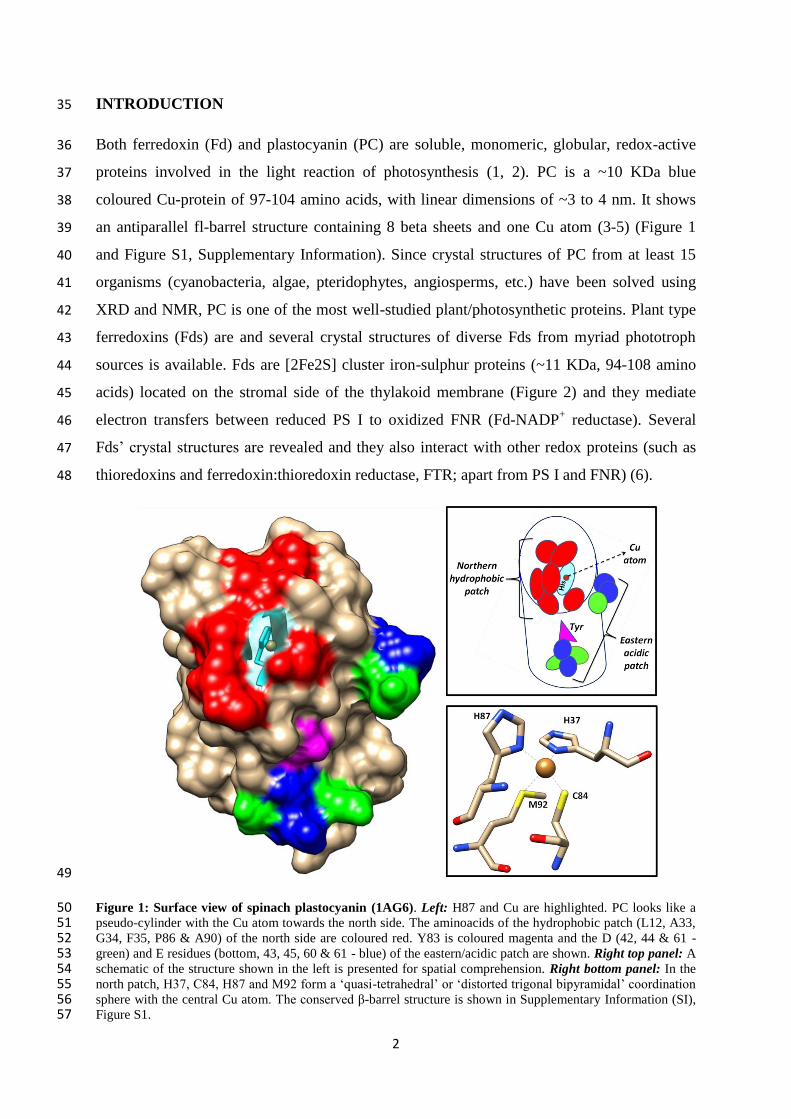
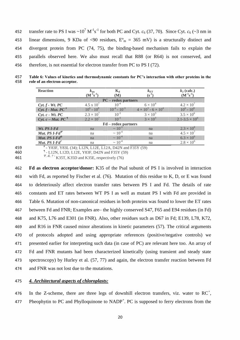
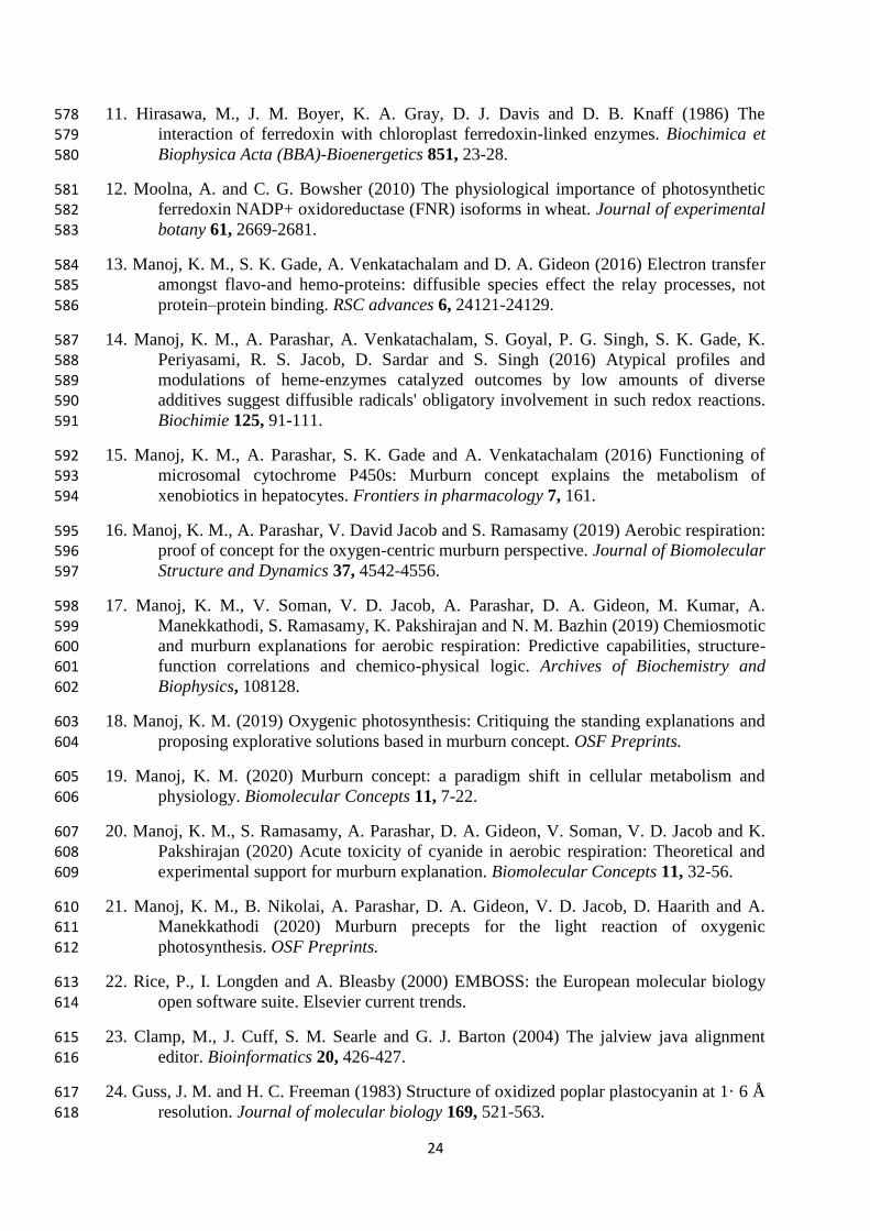
Figure 2: Structure of spinach Fd (1A70). Left: The [2Fe2S] cluster and aminoacid residues which are 59 deemed to be necessary for binding to FNR are highlighted. Fd is globular, with 4 conserved Cys residues that 60 hold the FeS cluster in place towards one end of the protein. When binding to FNR and other redox proteins, 61 this side of the cluster is generally known to interact. Key residues on the surface such as D26, E29, E30, D34, 62 D65 and D66 were found to be crucial for interaction with FNR. D residues are shaded green and E residues are 63 coloured blue. Right: The coordination sphere of the [2Fe2S] cluster is shown, with iron atoms (brown spheres) 64 and sulphur (yellow spheres) of the cluster bound by the four Cys residues of varying positions in Fds. 65
PROBLEM STATEMENT AND METHODOLOGY 66
In spite of PC/Fd being very well-studied proteins, significant confusions and discordance 67
exist regarding their functionality (7-12). We believe that this is so because the perceptions 68
on PC/Fd were “concretized” before the structures and functions of other components of the 69
photosynthetic system were known and the information available at later timeframes were 70
either overlooked or interpreted to fit the acclaimed perspective. Therefore, the current study 71
investigates the structure-function correlations of PC/Fd within the light reaction of 72
photosynthesis (also called oxygenic photolysis-photophosphorylation or Pl-Pp) from a 73
skeptic’s perspective, prioritizing several newly revealed information and concepts. 74
There are essentially two mechanistic proposals regarding biological electron transfers (7), as 75
shown in Figure 3. The classical explanation (depicted in left panel of Figure 3) requires a 76
high affinity binding (lasting millisecond timescales) between the original donor and final 77
acceptor proteins, and the electron transfer occurs as a result of tunneling (Marcus’ outer 78
sphere mechanism) between the redox centers, with the electron following a definite route 79
from the donor to the acceptor. This view would necessitate: i) high mobility and 80
concentrations of both donor and acceptor, & ii) for mutual affinity-based identification and 81
deterministic electron relay with the donor-acceptor complex, both the ligand sphere of 82
redox-metal centers (Cu & 2Fe) and the surface residues of PC/Fd that interact with their 83
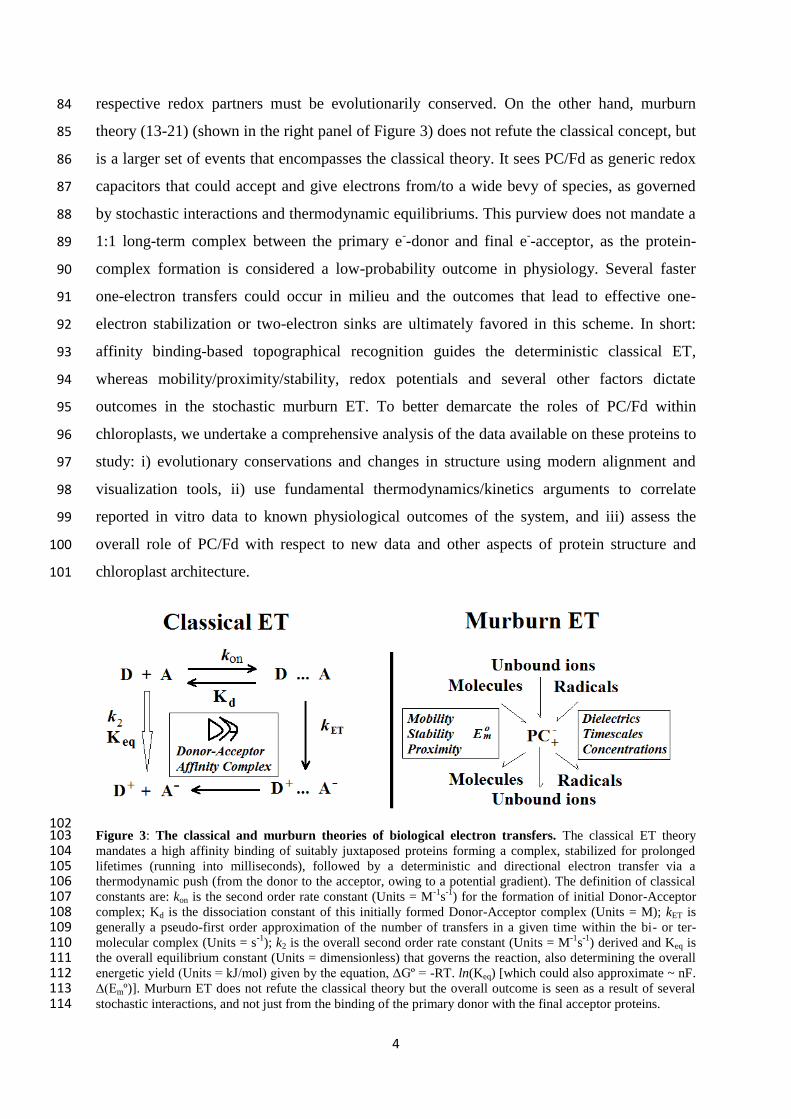
4
respective redox partners must be evolutionarily conserved. On the other hand, murburn 84
theory (13-21) (shown in the right panel of Figure 3) does not refute the classical concept, but 85
is a larger set of events that encompasses the classical theory. It sees PC/Fd as generic redox 86
capacitors that could accept and give electrons from/to a wide bevy of species, as governed 87
by stochastic interactions and thermodynamic equilibriums. This purview does not mandate a 88
1:1 long-term complex between the primary e--donor and final e
--acceptor, as the protein-89
complex formation is considered a low-probability outcome in physiology. Several faster 90
one-electron transfers could occur in milieu and the outcomes that lead to effective one-91
electron stabilization or two-electron sinks are ultimately favored in this scheme. In short: 92
affinity binding-based topographical recognition guides the deterministic classical ET, 93
whereas mobility/proximity/stability, redox potentials and several other factors dictate 94
outcomes in the stochastic murburn ET. To better demarcate the roles of PC/Fd within 95
chloroplasts, we undertake a comprehensive analysis of the data available on these proteins to 96
study: i) evolutionary conservations and changes in structure using modern alignment and 97
visualization tools, ii) use fundamental thermodynamics/kinetics arguments to correlate 98
reported in vitro data to known physiological outcomes of the system, and iii) assess the 99
overall role of PC/Fd with respect to new data and other aspects of protein structure and 100
chloroplast architecture. 101
102 Figure 3: The classical and murburn theories of biological electron transfers. The classical ET theory 103 mandates a high affinity binding of suitably juxtaposed proteins forming a complex, stabilized for prolonged 104 lifetimes (running into milliseconds), followed by a deterministic and directional electron transfer via a 105 thermodynamic push (from the donor to the acceptor, owing to a potential gradient). The definition of classical 106 constants are: kon is the second order rate constant (Units = M
-1s
-1) for the formation of initial Donor-Acceptor 107
complex; Kd is the dissociation constant of this initially formed Donor-Acceptor complex (Units = M); kET is 108 generally a pseudo-first order approximation of the number of transfers in a given time within the bi- or ter- 109 molecular complex (Units = s
-1); k2 is the overall second order rate constant (Units = M
-1s
-1) derived and Keq is 110
the overall equilibrium constant (Units = dimensionless) that governs the reaction, also determining the overall 111 energetic yield (Units = kJ/mol) given by the equation, ΔGº = -RT. ln(Keq) [which could also approximate ~ nF. 112 Δ(Emº)]. Murburn ET does not refute the classical theory but the overall outcome is seen as a result of several 113 stochastic interactions, and not just from the binding of the primary donor with the final acceptor proteins. 114
5
RESULTS AND DISCUSSION 115
1. Structural aspects required for Donor-Acceptor recognition and binding 116
While it is inevitable for protein sequences to remain unaltered during the course of 117
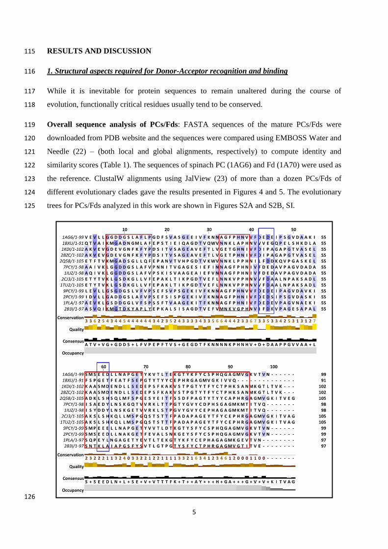
evolution, functionally critical residues usually tend to be conserved. 118
Overall sequence analysis of PCs/Fds: FASTA sequences of the mature PCs/Fds were 119
downloaded from PDB website and the sequences were compared using EMBOSS Water and 120
Needle (22) – (both local and global alignments, respectively) to compute identity and 121
similarity scores (Table 1). The sequences of spinach PC (1AG6) and Fd (1A70) were used as 122
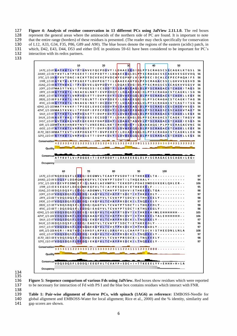
the reference. ClustalW alignments using JalView (23) of more than a dozen PCs/Fds of 123
different evolutionary clades gave the results presented in Figures 4 and 5. The evolutionary 124
trees for PCs/Fds analyzed in this work are shown in Figures S2A and S2B, SI. 125
126
6
Figure 4: Analysis of residue conservation in 13 different PCs using JalView 2.11.1.0. The red boxes 127 represent the general areas where the aminoacids of the northern side of PC are found. It is important to note 128 that the entire range (borders) of these residues is presented. (The reader may check specifically for conservation 129 of L12, A33, G34, F35, P86, G89 and A90). The blue boxes denote the regions of the eastern (acidic) patch, in 130 which, D42, E43, D44, D53 and either D/E in positions 59-61 have been considered to be important for PC’s 131 interaction with its redox partners. 132 133
134 135 Figure 5: Sequence comparison of various Fds using JalView. Red boxes show residues which were reported 136 to be necessary for interaction of Fd with PS I and the blue box contains residues which interact with FNR. 137 138 Table 1: Pair-wise alignment of diverse PCs, with spinach (1AG6) as reference: EMBOSS-Needle for 139 global alignment and EMBOSS-Water for local alignment; Rice et al., 2000) and the % identity, similarity and 140 gap scores are shown. 141

7
142 No. Source of PC
Global alignment (%) Local alignment (%)
identity similarity gaps identity similarity gaps
1 1BXU
(Synechococcus)
39.6 58.4 11.9 40.8 60.2 10.2
2 1KDI
(Adiantum capillus-
veneris)
35.2 53.3 8.6 35.9 54.4 8.7
3 2BZC
(Dryopteris
crassirhizoma)
36.2 54.3 8.6 36.9 55.3 8.7
4 2Q5B
(Phormidium)
38.1 59 5.7 40.4 62.6 3.0
5 7PCY
(Enteromorpha)
58.0 68.0 3.0 60.4 70.8 2.1
6 1IUZ
(Ulva)
58.0 67.0 3.0 60.4 69.8 2.1
7 2CJ3
(Anabaena)
43.4 54.7 7.5 46.0 58.0 5.0
8 1TU2
(Nostoc)
43.4 54.7 7.5 46.0 58.0 5.0
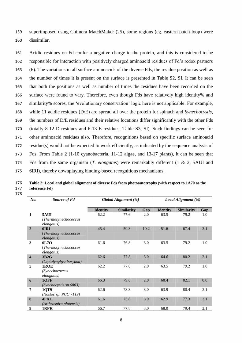
9 9PCY
(French bean)
82.8 88.9 0.0 82.8 88.9 0.0
10 2PCY
(Poplar)
78.8 90.9 0.0 78.8 90.9 0.0
11 1PLA
(parsley)
70.7 79.8 2.0 71.4 80.6 2.0
12 3B3I
(Prochlorothrix)
70.7 79.8 2.0 42.9 62.2 4.1
143
Our findings on PC can be compared to Guss & Freeman et al.’s results (24). They found that 144
52 residues were conserved and 11 were substituted conservatively among a total of 99 amino 145
acid residues and observed an identity score of 62% between algal (Chlamydomonas) and 146
plant (poplar; Populus nigra) PCs. When comparing the evolution of PCs (listed herein) with 147
a higher plant PC (spinach, 1AG6), we can see lower identity scores for fern protein than for 148
algal or cyanobacterial proteins. Interestingly, PC of Prochlorothrix, a photosynthetic 149
prokaryote (2B3I), had better scores than fern, some algal and cyanobacterial varieties. In 150
toto, the identity scores were lower than expected, showing that PC protein sequence is not 151
highly conserved across species. A similar profile was observed when a phylogenetic tree 152
was drawn (Figure S2, SI). In toto, 14 residues were conserved globally (as found with 153
ClustalW alignment; Figure S3, SI). Among them (G6/8, P16/18, G24/26, H37/38/39, 154
N38/41, G78/80, Y80/82, Y83/85, C84/86, P86/88, H87/88/89, A90/92, M92/94,V98/100), 155
the residue numbers are different in various PCs (with gaps in alignment) and only H87/89, 156
P86/88 and A90/92 are part of the hydrophobic patch. Another interesting observation is that 157
while the core region (shape and fold) is almost identical in the different PCs which were 158
8
superimposed using Chimera MatchMaker (25), some regions (eg. eastern patch loop) were 159
dissimilar. 160
Acidic residues on Fd confer a negative charge to the protein, and this is considered to be 161
responsible for interaction with positively charged aminoacid residues of Fd’s redox partners 162
(6). The variations in all surface aminoacids of the diverse Fds, the residue position as well as 163
the number of times it is present on the surface is presented in Table S2, SI. It can be seen 164
that both the positions as well as number of times the residues have been recorded on the 165
surface were found to vary. Therefore, even though Fds have relatively high identity% and 166
similarity% scores, the ‘evolutionary conservation’ logic here is not applicable. For example, 167
while 11 acidic residues (D/E) are spread all over the protein for spinach and Synechocystis, 168
the numbers of D/E residues and their relative locations differ significantly with the other Fds 169
(totally 8-12 D residues and 6-13 E residues, Table S3, SI). Such findings can be seen for 170
other aminoacid residues also. Therefore, recognitions based on specific surface aminoacid 171
residue(s) would not be expected to work efficiently, as indicated by the sequence analysis of 172
Fds. From Table 2 (1-10 cyanobacteria, 11-12 algae, and 13-17 plants), it can be seen that 173
Fds from the same organism (T. elongatus) were remarkably different (1 & 2, 5AUI and 174
6IRI), thereby downplaying binding-based recognitions mechanisms. 175
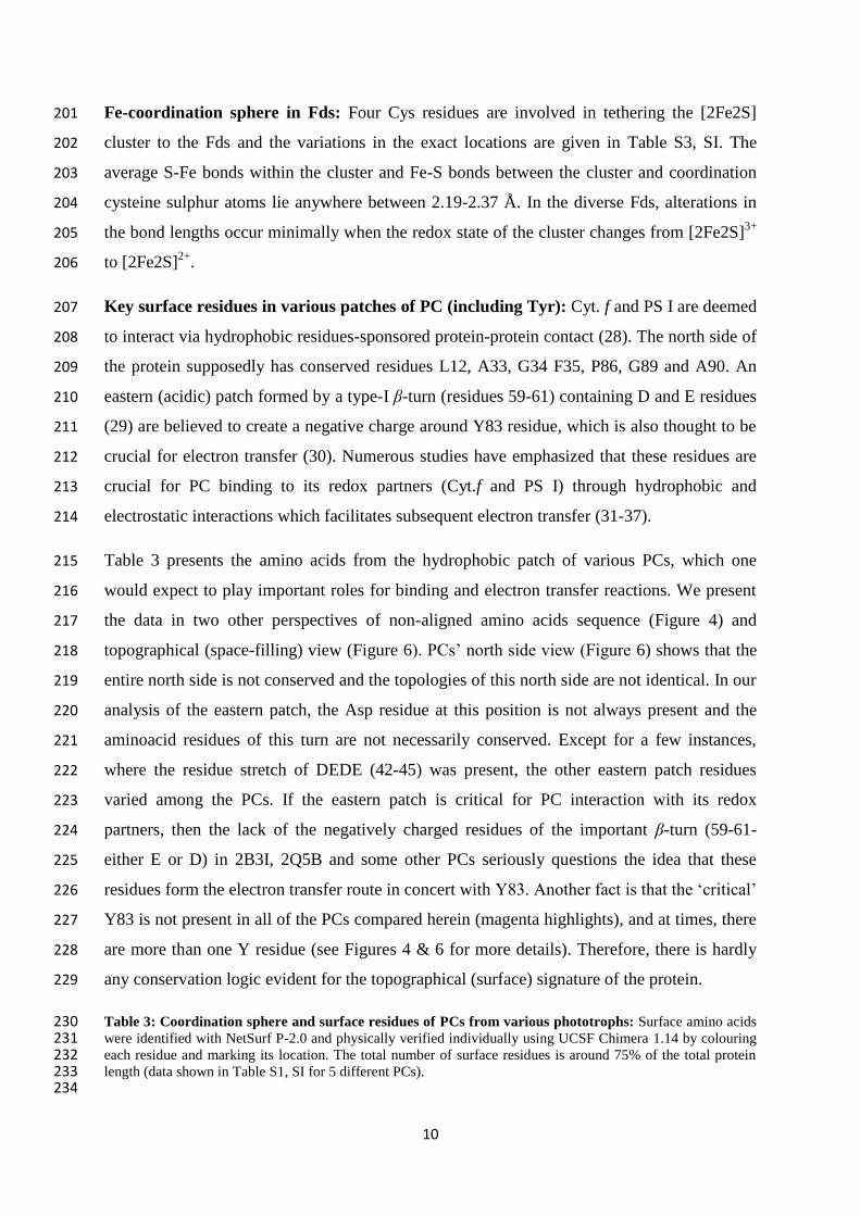
Table 2: Local and global alignment of diverse Fds from photoautotrophs (with respect to 1A70 as the 176 reference Fd) 177 178
No. Source of Fd Global Alignment (%) Local Alignment (%)
Identity Similarity Gap Identity Similarity Gap
1 5AUI
(Thermosynechococcus
elongatus)
62.2 77.6 2.0 63.5 79.2 1.0
2 6IRI
(Thermosynechococcus
elongatus)
45.4 59.3 10.2 51.6 67.4 2.1
3 6L7O
(Thermosynechococcus
elongatus)
61.6 76.8 3.0 63.5 79.2 1.0
4 3B2G
(Leptolyngbya boryana)
62.6 77.8 3.0 64.6 80.2 2.1
5 1ROE (Synechococcus
elongatus)
62.2 77.6 2.0 63.5 79.2 1.0
6 1OFF (Synchocystis sp.6803)
66.3 79.6 2.0 68.4 82.1 0.0
7 1QT9
(Nostoc sp. PCC 7119)
62.6 78.8 3.0 63.9 80.4 2.1
8 4FXC
(Arthrospira platensis)
61.6 75.8 3.0 62.9 77.3 2.1
9 1RFK 66.7 77.8 3.0 68.0 79.4 2.1

9
(Mastigocladus
laminosus)
10 3P63
(Mastigocladus
laminosus)
71.1 82.5 1.0 72.6 84.2 0.0
11 1AWD
(Chlorella fusca)
63.9 79.4 3.1 66.7 82.8 0.0
12 3AV8 (Aphanothece sacrum)
64.3 76.5 2.0 65.6 78.1 1.0
13 1FRR (Equisetum arvense)
57.7 73.2 2.1 59.6 75.5 0.0
14 3B2F
(Zea Mays)
73.5 82.7 1.0 75.0 84.4 0.0
15 5H57
(Zea mays)
61.2 75.5 2.0 62.5 77.1 1.0
16 4ZHO
(Arabidopsis thaliana)
66.7 78.1 7.6 73.7 86.3 0.0
17 4ZHP
(Solanum tuberosum)
67.0 76.4 8.5 73.2 83.5 0.0
179
Cu-coordination sphere in PCs: Two histidine nitrogen atoms and a cysteine sulphur atom 180
are conserved, whereas the fourth ligand could be a sulphur atom from methionine or 181
nitrogen from glutamine. The exact locations of the histidines, cysteine and 182
methionine/glutamine are found to vary slightly within the amino acid sequence, and their 183
immediate neighbours also change among the proteins (see Table 3). This suggests that while 184
there is some conservation of the coordination environment, the ambiance near the Cu atom 185
varies across the proteins. The Cu-N bond lengths were 2.10 and 2.04 Å, the Cu-S(Cys) bond 186
was 2.52 Å and the Cu-S(Met) bond was slightly longer (~2.9 Å), as measured from the 187
crystal structure of the Poplar PC (2PCY) (24). These bond lengths, when measured for all 188
the proteins’ coordination spheres, were slightly different. For example, in Phormidium PC 189
(2Q5B), the Cu-N bond lengths were 2.05 and 2.08 Å and the Cu(Cys) and Cu(Met) bonds 190
were 2.24 and 2.60 Å, respectively. Alterations in these bond lengths can result from 191
differences in the relative positions of the residues involved in copper ligation. The copper 192
site was shown to be more flexible and the H87-M92 loop is considered to regulate electron 193
transfer between Cyt.f and PC (26). The Cu coordination sphere supposedly undergoes 194
modifications to facilitate electron transfer when PC binds to Cyt.f. Counter-intuitively, such 195
a change was minimal when PC bound to PS I. The same authors showed that both reduced 196
and oxidized PC showed no drastic changes in the Cu coordination sphere (27). In short, our 197
analysis shows that the residue numbers, local ambiance and the bond lengths of the Cu 198
ligands were found to vary; and this scenario is not expected for a conserved routing of 199
electrons to and fro the central copper atom. 200
10
Fe-coordination sphere in Fds: Four Cys residues are involved in tethering the [2Fe2S] 201
cluster to the Fds and the variations in the exact locations are given in Table S3, SI. The 202
average S-Fe bonds within the cluster and Fe-S bonds between the cluster and coordination 203
cysteine sulphur atoms lie anywhere between 2.19-2.37 Å. In the diverse Fds, alterations in 204
the bond lengths occur minimally when the redox state of the cluster changes from [2Fe2S]3+
205
to [2Fe2S]2+
. 206
Key surface residues in various patches of PC (including Tyr): Cyt. f and PS I are deemed 207
to interact via hydrophobic residues-sponsored protein-protein contact (28). The north side of 208
the protein supposedly has conserved residues L12, A33, G34 F35, P86, G89 and A90. An 209
eastern (acidic) patch formed by a type-I β-turn (residues 59-61) containing D and E residues 210
(29) are believed to create a negative charge around Y83 residue, which is also thought to be 211
crucial for electron transfer (30). Numerous studies have emphasized that these residues are 212
crucial for PC binding to its redox partners (Cyt.f and PS I) through hydrophobic and 213
electrostatic interactions which facilitates subsequent electron transfer (31-37). 214
Table 3 presents the amino acids from the hydrophobic patch of various PCs, which one 215
would expect to play important roles for binding and electron transfer reactions. We present 216
the data in two other perspectives of non-aligned amino acids sequence (Figure 4) and 217
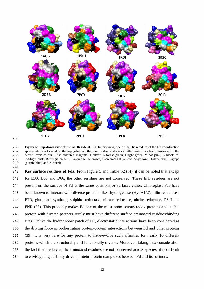
topographical (space-filling) view (Figure 6). PCs’ north side view (Figure 6) shows that the 218
entire north side is not conserved and the topologies of this north side are not identical. In our 219
analysis of the eastern patch, the Asp residue at this position is not always present and the 220
aminoacid residues of this turn are not necessarily conserved. Except for a few instances, 221
where the residue stretch of DEDE (42-45) was present, the other eastern patch residues 222
varied among the PCs. If the eastern patch is critical for PC interaction with its redox 223
partners, then the lack of the negatively charged residues of the important β-turn (59-61-224
either E or D) in 2B3I, 2Q5B and some other PCs seriously questions the idea that these 225
residues form the electron transfer route in concert with Y83. Another fact is that the ‘critical’ 226
Y83 is not present in all of the PCs compared herein (magenta highlights), and at times, there 227
are more than one Y residue (see Figures 4 & 6 for more details). Therefore, there is hardly 228
any conservation logic evident for the topographical (surface) signature of the protein. 229
Table 3: Coordination sphere and surface residues of PCs from various phototrophs: Surface amino acids 230 were identified with NetSurf P-2.0 and physically verified individually using UCSF Chimera 1.14 by colouring 231 each residue and marking its location. The total number of surface residues is around 75% of the total protein 232 length (data shown in Table S1, SI for 5 different PCs). 233 234
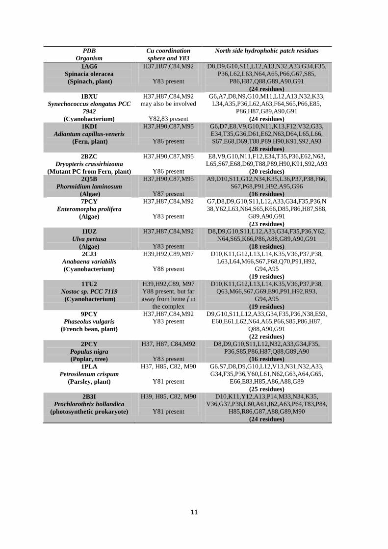
11
PDB
Organism
Cu coordination
sphere and Y83
North side hydrophobic patch residues
1AG6
Spinacia oleracea
(Spinach, plant)
H37,H87,C84,M92
Y83 present
D8,D9,G10,S11,L12,A13,N32,A33,G34,F35,
P36,L62,L63,N64,A65,P66,G67,S85,
P86,H87,Q88,G89,A90,G91
(24 residues)
1BXU
Synechococcus elongatus PCC
7942
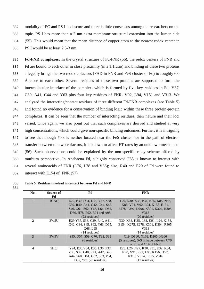
(Cyanobacterium)
H37,H87,C84,M92
may also be involved
Y82,83 present
G6,A7,D8,N9,G10,M11,L12,A13,N32,K33,
L34,A35,P36,L62,A63,F64,S65,P66,E85,
P86,H87,G89,A90,G91
(24 residues)
1KDI
Adiantum capillus-veneris
(Fern, plant)
H37,H90,C87,M95
Y86 present
G6,D7,E8,V9,G10,N11,K13,F12,V32,G33,
E34,T35,G36,D61,E62,N63,D64,L65,L66,
S67,E68,D69,T88,P89,H90,K91,S92,A93
(28 residues)
2BZC
Dryopteris crassirhizoma
(Mutant PC from Fern, plant)
H37,H90,C87,M95
Y86 present
E8,V9,G10,N11,F12,E34,T35,P36,E62,N63,
L65,S67,E68,D69,T88,P89,H90,K91,S92,A93
(20 residues)
2Q5B
Phormidium laminosum
(Algae)
H37,H90,C87,M95
Y87 present
A9,D10,S11,G12,N34,K35,L36,P37,P38,F66,
S67,P68,P91,H92,A95,G96
(16 residues)
7PCY
Enteromorpha prolifera
(Algae)
H37,H87,C84,M92
Y83 present
G7,D8,D9,G10,S11,L12,A33,G34,F35,P36,N
38,Y62,L63,N64,S65,K66,D85,P86,H87,S88,
G89,A90,G91
(23 residues)
1IUZ
Ulva pertusa
(Algae)
H37,H87,C84,M92
Y83 present
D8,D9,G10,S11,L12,A33,G34,F35,P36,Y62,
N64,S65,K66,P86,A88,G89,A90,G91
(18 residues)
2CJ3
Anabaena variabilis
(Cyanobacterium)
H39,H92,C89,M97
Y88 present
D10,K11,G12,L13,L14,K35,V36,P37,P38,
L63,L64,M66,S67,P68,Q70,P91,H92,
G94,A95
(19 residues)
1TU2
Nostoc sp. PCC 7119
(Cyanobacterium)
H39,H92,C89, M97
Y88 present, but far
away from heme f in
the complex
D10,K11,G12,L13,L14,K35,V36,P37,P38,
Q63,M66,S67,G69,E90,P91,H92,R93,
G94,A95
(19 residues)
9PCY
Phaseolus vulgaris
(French bean, plant)
H37,H87,C84,M92
Y83 present
D9,G10,S11,L12,A33,G34,F35,P36,N38,E59,
E60,E61,L62,N64,A65,P66,S85,P86,H87,
Q88,A90,G91
(22 residues)
2PCY
Populus nigra
(Poplar, tree)
H37, H87, C84,M92
Y83 present
D8,D9,G10,S11,L12,N32,A33,G34,F35,
P36,S85,P86,H87,Q88,G89,A90
(16 residues)
1PLA
Petrosilenum crispum
(Parsley, plant)
H37, H85, C82, M90
Y81 present
G6.S7,D8,D9,G10,L12,V13,N31,N32,A33,
G34,F35,P36,Y60,L61,N62,G63,A64,G65,
E66,E83,H85,A86,A88,G89
(25 residues)
2B3I
Prochlorothrix hollandica
(photosynthetic prokaryote)
H39, H85, C82, M90
Y81 present
D10,K11,Y12,A13,P14,M33,N34,K35,
V36,G37,P38,L60,A61,I62,A63,P64,T83,P84,
H85,R86,G87,A88,G89,M90
(24 residues)
12
235
Figure 6: Top-down view of the north side of PC: In this view, one of the His residues of the Cu coordination 236 sphere which is located on the top (while another one is almost always a little buried) has been positioned in the 237 centre (cyan colour). P is coloured magenta, F-silver, L-forest green, I-light green, V-hot pink, G-black, Y-238 red/light pink, R-red (if present), A-orange, K-brown, S-cream/light yellow, M-yellow, D-dark blue, E-grape 239 (purple blue) and N-purple. 240 241 Key surface residues of Fds: From Figure 5 and Table S2 (SI), it can be noted that except 242
for E30, D65 and D66, the other residues are not conserved. These E/D residues are not 243
present on the surface of Fd at the same positions or surfaces either. Chloroplast Fds have 244
been known to interact with diverse proteins like– hydrogenase (HydA1/2), bilin reductases, 245
FTR, glutamate synthase, sulphite reductase, nitrate reductase, nitrite reductase, PS I and 246
FNR (38). This probably makes Fd one of the most promiscuous redox proteins and such a 247
protein with diverse partners surely must have different surface aminoacid residues/binding 248
sites. Unlike the hydrophobic patch of PC, electrostatic interactions have been considered as 249
the driving force in orchestrating protein-protein interactions between Fd and other proteins 250
(39). It is very rare for any protein to have/evolve such affinities for nearly 10 different 251
proteins which are structurally and functionally diverse. Moreover, taking into consideration 252
the fact that the key acidic aminoacid residues are not conserved across species, it is difficult 253
to envisage high affinity driven protein-protein complexes between Fd and its partners. 254
13
2. Analyzing electron donor-acceptor (protein-protein) complexes’ crystal structures: 255
Cyt. f-PC complexes: When the surface residues (northern and eastern patch, in particular) 256
were analyzed for their interactions with Cyt. f in crystal structures of the four reported PC-257
Cyt. f complexes- 1TU2 (40), 2PCF (41), 2JXM (42) and 1TKW (43), not all of the purported 258
PC residues which are deemed mandatory for interaction were found to be involved in the 259
actual complex formation with Cyt. f (Table 4). Supplementary Figure S4 shows that PC 260
binds with different orientations to Cyt. f. Sometimes, while only the north side is bound 261
(2JXM and 1TU2), the north side as well as eastern patch were found to interact with Cyt. f in 262
1TKW. In only 1TKW and 2PCF, D/E residues of the eastern patch of PC featured in the 263
interaction. The distance between Fe (Cyt. f) and Cu (PC) was closer than the distance 264
between Fe (the electron donor) and Y83 (the electron relay). If the electron transfer occurs 265
via relay through critical aminoacids (F and Y), what is the purpose of large cavities and the 266
solvent accessibility of the heme residue? Moreover, the distance between Cu (in PC) and 267
iron in heme f is much lesser in these complexes (11-17 Å) than the alleged round-about and 268
long route involving the F/Y residues in Cyt. f and Y83 of PC surrounded by an acidic patch. 269
That is, why would electrons flow through a prolonged path, when the interatomic distances 270
between the two key metal ions is much shorter? In Cyt.f as well, barring Y1 -which was 271
found in all 4 complexes, the other interacting residues were not the same in all the 272
complexes). Only in 1TU2 and 2JXM, four residues that were involved in direct contact in 273
the crystal structures were conserved in complex formation. In 1TU2, 1TKW and 2PCF, large 274
channels in Cyt. f expose the heme residue, which is highly accessible to the solvent 275
environment. Fedorov et al. concluded from their molecular dynamics studies that the crucial 276
loop of G188, E189 and D190 in Cyt. f was involved in stabilization of the complex (44). 277
While some presume that low salt concentrations facilitate protein-protein associations of 278
these proteins, Ueda et al. showed that the acidic patch residues of PC were not involved in 279
salt bridge formation with either PS I or Cyt. b6f in the electron transfer complexes at lower 280
ionic strengths (45). An important point to consider is that crystallographic complexes are 281
derived by using very high concentrations of proteins, which could give non-specific binding 282
(particularly, with hydrophobic patches), and such interactions could have little physiological 283
significance. Given such a background and the non-positive results, we infer that there is no 284
‘conserved aminoacid stretch’ or motif which is responsible for the PC binding to its redox 285
partners. 286
287
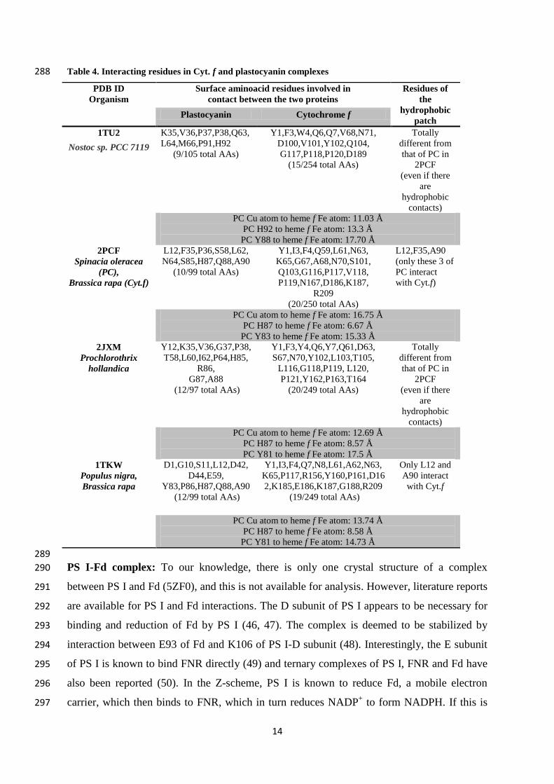
14
Table 4. Interacting residues in Cyt. f and plastocyanin complexes 288
PDB ID
Organism
Surface aminoacid residues involved in
contact between the two proteins
Residues of
the
hydrophobic
patch Plastocyanin Cytochrome f
1TU2
Nostoc sp. PCC 7119
K35,V36,P37,P38,Q63,
L64,M66,P91,H92
(9/105 total AAs)
Y1,F3,W4,Q6,Q7,V68,N71,
D100,V101,Y102,Q104,
G117,P118,P120,D189
(15/254 total AAs)
Totally
different from
that of PC in
2PCF
(even if there
are
hydrophobic
contacts)
PC Cu atom to heme f Fe atom: 11.03 Å
PC H92 to heme f Fe atom: 13.3 Å
PC Y88 to heme f Fe atom: 17.70 Å
2PCF
Spinacia oleracea
(PC),
Brassica rapa (Cyt.f)
L12,F35,P36,S58,L62,
N64,S85,H87,Q88,A90
(10/99 total AAs)
Y1,I3,F4,Q59,L61,N63,
K65,G67,A68,N70,S101,
Q103,G116,P117,V118,
P119,N167,D186,K187,
R209
(20/250 total AAs)
L12,F35,A90
(only these 3 of
PC interact
with Cyt.f)
PC Cu atom to heme f Fe atom: 16.75 Å
PC H87 to heme f Fe atom: 6.67 Å
PC Y83 to heme f Fe atom: 15.33 Å
2JXM
Prochlorothrix
hollandica
Y12,K35,V36,G37,P38,
T58,L60,I62,P64,H85,
R86,
G87,A88
(12/97 total AAs)
Y1,F3,Y4,Q6,Y7,Q61,D63,
S67,N70,Y102,L103,T105,
L116,G118,P119, L120,
P121,Y162,P163,T164
(20/249 total AAs)
Totally
different from
that of PC in
2PCF
(even if there
are
hydrophobic
contacts)
PC Cu atom to heme f Fe atom: 12.69 Å
PC H87 to heme f Fe atom: 8.57 Å
PC Y81 to heme f Fe atom: 17.5 Å
1TKW
Populus nigra,
Brassica rapa
D1,G10,S11,L12,D42,
D44,E59,
Y83,P86,H87,Q88,A90
(12/99 total AAs)
Y1,I3,F4,Q7,N8,L61,A62,N63,
K65,P117,R156,Y160,P161,D16
2,K185,E186,K187,G188,R209
(19/249 total AAs)
Only L12 and
A90 interact
with Cyt.f
PC Cu atom to heme f Fe atom: 13.74 Å
PC H87 to heme f Fe atom: 8.58 Å
PC Y81 to heme f Fe atom: 14.73 Å
289
PS I-Fd complex: To our knowledge, there is only one crystal structure of a complex 290
between PS I and Fd (5ZF0), and this is not available for analysis. However, literature reports 291
are available for PS I and Fd interactions. The D subunit of PS I appears to be necessary for 292
binding and reduction of Fd by PS I (46, 47). The complex is deemed to be stabilized by 293
interaction between E93 of Fd and K106 of PS I-D subunit (48). Interestingly, the E subunit 294
of PS I is known to bind FNR directly (49) and ternary complexes of PS I, FNR and Fd have 295
also been reported (50). In the Z-scheme, PS I is known to reduce Fd, a mobile electron 296
carrier, which then binds to FNR, which in turn reduces NADP+ to form NADPH. If this is 297
15
the path of electron transfer, why would FNR need to bind directly to PS I? Also, what is the 298
need for two different isoforms of Fds (51) and also FNR (12)? Flavodoxins (Flds) are known 299
to be alternative electron carriers in cyanobacteria and algae (but not in higher plants), which 300
perform the function of Fds in iron-limiting conditions (52). Flds are also known to exchange 301
electrons with Fds and also partner redox reactions with both FNR and PS I (53). The 302
structure of Fds and Flds are quite different and it is improbable that both these proteins 303
would have similar binding affinities to the redox partners PS I and FNR. When the two 304
proteins (2V5V-Fld and 1QT9-Fd) from Nostoc sp. PCC 7119 were aligned using ClustalW, 305
only 19 aminoacid residues were conserved between them. Analysis using EMBOSS-Needle 306
(global alignment) gave identity and similarity scores of 12.7 and 19.8%, respectively. How 307
could Flds (an FMN containing redox protein of ~170 amino acids) with remarkably different 308
topology and surface electrostatics substitute for Fds? Such outcomes can effectively be 309
addressed by non-specific interactions espoused by the murburn paradigm. 310
311
PC-PS I complex: To the best of our knowledge, there is no crystal structure of PC-PS I 312
complexes reported till date. However, both northern and eastern patch residues of PC have 313
been reckoned to be involved in PC-PS I interactions. PsaB D612 and D613 of PS I were 314
proposed to be necessary for docking of PC, which also is through hydrophobic contact (37). 315
When we explored the crystal structures of PS I, we found that the residues W622, D619 and 316
E611 (Synechocystis PS I; PDB ID: 5OY0) were far apart from each other and it is not clear 317
which of these residues are deemed to interact with PC. Also, the exact role of PsaF is not 318
clear, because the distance from PsaF to the P700 center is roughly >40 Å in the crystal 319
structures 5OY0 (& 4XK8 of Pisum sativum) and ~35 Å in 6JO6 (C. reinhardtii PS I). In the 320
6JO6 structure, the Y626, D623 and E612 residues are spaced apart from the P700 center (by 321
11, 16 and 24 Å, respectively) and the PsaF chain is slightly elevated above the contour of 322
PsaB. While the Y, D and E residues, although jutting out of the PsaB protein- are still below 323
the PsaF region, which would mean that if PC were to bind to this region spanning over ~35-324
40 Å from PsaF to the P700 center, it would be bound to a slightly elevated PsaF on one side 325
and much lower Y residue on the other side. Besides, in those regions, there are many other 326
Y, D and E residues which could also interact. We wonder why these specific residues have 327
been reckoned to act as electron transfer conduits. PS I-N subunit, which is present only in 328
the lumenal side of PS I in higher plants alone, was shown to be important for PS I-PC 329
interactions. However, even though the ET rates were reduced, the authors admit that 330
photoautotrophic growth remained unaffected (54). These reports show that the interaction 331
16
modality of PC and PS I is obscure and there is little consensus among the researchers on the 332
topic. PS I has more than a 2 nm extra-membrane structural extension into the lumen side 333
(55). This would mean that the mean distance of copper atom to the nearest redox center in 334
PS I would be at least 2.5-3 nm. 335
Fd-FNR complexes: In the crystal structure of Fd-FNR (56), the redox centers of FNR and 336
Fd are bound to each other in close proximity (in a 1:1ratio) and binding of these two proteins 337
allegedly brings the two redox cofactors (FAD in FNR and FeS cluster of Fd) to roughly 6.0 338
Å close to each other. Several residues of these two proteins are supposed to form the 339
intermolecular interface of the complex, which is formed by five key residues in Fd- Y37, 340
C39, A41, C44 and Y63 plus four key residues of FNR- V92, L94, V151 and V313. We 341
analyzed the interacting/contact residues of three different Fd-FNR complexes (see Table 5) 342
and found no evidence for a conservation of binding logic within these three protein-protein 343
complexes. It can be seen that the number of interacting residues, their nature and their loci 344
varied. Once again, we also point out that such complexes are derived and studied at very 345
high concentrations, which could give non-specific binding outcomes. Further, it is intriguing 346
to see that though Y83 is neither located near the FeS cluster nor in the path of electron 347
transfer between the two cofactors, it is known to affect ET rates by an unknown mechanism 348
(56). Such observations could be explained by the non-specific relay scheme offered by 349
murburn perspective. In Anabaena Fd, a highly conserved F65 is known to interact with 350
several aminoacids of FNR (L76, L78 and V36); also, R40 and E29 of Fd were found to 351
interact with E154 of FNR (57). 352
Table 5: Residues involved in contact between Fd and FNR 353 354
No. Source of
Fd
Fd FNR
1 1GAQ
E29, E30, D34, L35, Y37, S38,
C39, R40, A41, G42, C44, S45,
S46, Q61, S62, Y63, L64, D65,
D66, H78, E92, E94 and A98
(23 residues)
T29, N30, K33, P34, K35, K85, N86,
K88, V91, V92, L94, K153, E154,
E278, F297, D298, K301, K304, R305,
V313
(20 residues)
2 3W5U E29,Y37, S38, C39, R40, A41,
G42, C44, S45, S62, Y63, D65,
Q68, L95
(14 residues)
N30, K33, K35, L88, K91, L94, K153,
E154, K275, E278, K301, K304, R305,
V313
(14 residues)
3 3W5V S55, D57, S59, C70, T82, S83
(6 residues)
C19, D160, N162, D265, N266
(5 residues); S-S linkage between C79
of Fd and C19 of FNR
4 5H5J Y24, E30,V34, E35, L36, P37,
Y38, S39, C40, R41, A42, G43,
A44, S60, D61, G62, S63, P64,
D67, Y81 (20 residues)
E23, L26, N27, K30, P31, K32, K84,
N90, V91, R92, L93, K156, I157,
K310, V314, E315, V316
(17 residues)
17
355
3. Thermodynamic and kinetic data for binding and electron transfers of wildtype/mutant 356
PCs/Fds with their redox partners: 357
PC as electron acceptor/donor: The overall experimental equilibrium constant of Cyt. f to 358
PC electron transfer reaction is only 2 (58), and this is because the redox potential gradient is 359
very low. The redox potential gradient from cytochrome c to PC is greater (250 mV to 380 360
mV; when compared to the practically non-existent gradient between cytochrome f and PC), 361
which dictates that thermodynamically, the free energy change (ΔGº = n.Ƒ.ΔEºm) would be 362
higher for the former reaction. Yet, the overall calculated reaction rate of k2 [as defined by 363
(1/k2) = (1/kon) + (Kd/kET)] is higher for the Cyt. f – PC reaction. Now, the relevant question: 364
Is it the higher intrinsic ET rate within the Cyt. f – PC complex and/or the lower Kd of the 365
Cyt. f-PC interaction that determines the favourable Cyt. f – PC reaction kinetics? To answer 366
this question, we must understand the protocol adopted for determining the binding constants 367
and ET rates. While the binding assay is done at several tens of micromolar proteins, the ET 368
reaction stopped flow measurements are done at about two orders lower concentrations. At 369
higher concentrations, the binding agent may also sponsor redox changes, which gets 370
registered into the heme charge transfer Soret band absorption range, where heme f is 371
monitored. It can be noted that though oxidized PC has negligible absorbance at 410 nm (or 372
at 422 nm, wherein binding and reaction rate are measured) (59), reduced copper’s spectral 373
signature is significantly high at 410 nm (60). Since Cyt. f and PC have comparative redox 374
potentials, reduction of PC could also lead to a 410 nm signature. We have pointed out such 375
procedural influences in similar issues faced in other heme enzymes and P450 redox research 376
(14, 15, 61, 62). (However, the following discussion is made over-sighting such issues.) 377
The most important point to remember is that the physiological concentration of PC in 378
chloroplasts is around ~1 μM (63), and at such low concentrations, the major population of 379
PC would be unbound. Therefore, the overall outcomes of kinetics at lower concentrations of 380
the proteins cannot be explained by the ‘erroneous’ binding constants (which are anyway 381
insignificant with respect to the reaction assay concentrations). It is important to point out 382
that even the reaction assay employed is unrealistic: 10X of oxidized PC is taken with respect 383
to reduced Cyt. f (59). The idea that intrinsic ET is preferred over certain routes is disclaimed 384
by the overall conformity of rates in mutants within the Cyt. c6 and PC transfers. Even in Cyt. 385
f – PC transfers, the value changes are only an order lesser or higher than the wildtype. That 386
18
is- if the positive control with wildtype shows 107 M
-1s
-1 and the test reaction with mutant 387
shows 106 M
-1s
-1, the mutant still gives ~10% rate of the wildtype reaction. In kinetics and 388
equilibriums, this value is NOT mechanistically insignificant. If the value of mutant reaction 389
is several orders lower, then the mechanism would be deemed to be more attributed to the 390
‘crucially required factor’ (say, via an obligatory tyrosine). In other words, substituting a 391
tyrosine with another amino acid residue should not give a functional ET between the Cyt. f 392
and mutant PC, whereas we do observe significant ET rates even in this system. In the 393
absence of oxidized plastocyanin, the negative control of reduced Cyt. f taken alone is 394
relatively stable and the absorbance change in the cytochrome Soret is not seen to occur 395
significantly within the reaction times. That is- the mutant still could give 105 to 10
6 times the 396
rates of the negative control and this is mechanistically significant. However, the fact that 397
addition of PC alters absorbance profiles of Cyt. f has been taken as an indication of ONLY 398
direct interactions of PC and Cyt. f. We have corrected such assumptions with our works in 399
heme enzymology and explained the kinetic outcomes for such proteins with murburn 400
concept (13-15, 64, 65). 401
Another flaw is that some workers have reported overall k2 ET rates as high as 2×10⁸ M
-1s
-1 402
(33), which approach diffusion controlled rates. This is because collision rates of 108-10
9 M
-403
1s
-1 have been assumed (66). Such high values for collision and protein-protein interaction-404
mediated electron transfer must be questioned, because we are dealing with proteins such as 405
PS I and Cyt. b6f which are stationary and much larger than the 10 KDa PC. When PC (Cu 406
center) reduction was monitored, lower rates of 102 M
-1 s
-1 were obtained, while inter-protein 407
ET rates (between horse heart Cyt. c and PC) were found to be higher, at 104-10
5 M
-1 s
-1 (67). 408
Moreover, binding in biology would involve collisions and dependence on the ionic strength 409
of the medium for ‘optimal’ interactions and electron relays. Kovalenko et al. simulated the 410
interactions of PC and ferredoxin with PS I and concluded that there was a “non-monotonic 411
dependence of the complex formation at low ionic strength” which was due to “long-range 412
electrostatic interactions” (39). In their analysis, the complex formation depended on ionic 413
strength in order to be successful. In a similar redox metabolic system occurring in 414
cytoplasmic microsomes, we had that protein-protein complexation between cytochrome 415
P450 (CYP) and cytochrome P450 reductase (CPR) is a low probability event (68, 13, 14, 19) 416
and explained outcomes with the application of murburn concept. We had shown that 417
increasing the molarity of phosphate buffer increased the reduction rate of Cyt. c (13). Also, 418
even when CYP and CPR were physically separated using a membrane (in other words, no 419
19
donor-acceptor binding occurred), CYP was still able to oxidize its substrate. Further, copper 420
ions are one-electron redox cyclers which can modulate the DROS dynamics in the reaction 421
milieu and DROS serve as effective redox relay triggers/agents (13). In toto, the derivable 422
inference is that electron reception from any donor by wildtype and mutant PC proteins are 423
not majorly affected by changing residues that are touted to be pivotal for binding or ET. This 424
inference is also endorsed further by the structural analyses of the protein partners and their 425
known complexes. The larger picture appears to be that a multitude of parameters govern the 426
overall outcome, as endorsed by the more inclusive murburn model (Figure 3). 427
With respect to PC-PS I interaction, three residues of PS I (PsaB) that were deemed to be 428
important were a critical tryptophan residue (W627 and W651 in C. reinharditii), one E and 429
one D residue (69). Moreover, PsaF, a much smaller protein, was also deemed crucial for PC 430
binding to PS I and for higher ET rates (70). The same group showed that a K23Q mutation 431
of PsaF affected ET from PC/Cyt. c6 to PS I and also crosslinking of these proteins with PS I 432
(Table 6) (71). A positively charged PC from Anabaena sp. PCC 7119 was investigated using 433
site-directed mutagenesis and the authors revealed that replacement of R88 (which is adjacent 434
to His87 of the Cu ligation site) is required for efficient reduction of PS I and mutation of this 435
arginyl residue (and R64, an equivalent in Cyt.c6 which is close to heme group). More 436
importantly, they reported that mutation of the critical Y83 with either A or F residues did not 437
alter the kinetics of PS I reduction (72). Despite replacement of the purported key residues 438
using site-directed mutagenesis, electron transfer is not altogether abrogated by mutation of 439
these critical residues allegedly involved in protein-protein contact and the redox potentials 440
are also not drastically altered. R88 is found only in the 1BXU sequence in our analysis and 441
the other R (R64) residue is not present in any of the PCs which we have analyzed. 442
In the spinach PC system, changing one to three key residues from G8, L12, F35, D42, E43, 443
D44, E59, E60, Y83 (thereby bring about a charge difference of +1 to +4) gave mutant 444
proteins that showed varied pI (3.82 of the wildtype compared and 3.64 to 6.2 of the mutants) 445
and midpoint redox potential (Eºm = ~380 mV for the wildtype, ±25 mV for the mutants, in 446
the pH range of 6.0 to 9.0). However, the mutants gave comparable functionality for electron 447
transfers with PS I with electron transfer kinetic constants like kET (~7 x 10⁴ s
-1 for the 448
wildtype, whereas ~3 to 6 x 10⁴ s
-1 for the various mutants) (29, 32, 36, 73). The redox 449
potential of PS I was found to be 475 – 500 mV (32), Kd of PC’s and Cyt. c6’s interaction 450
with PS I was determined to be in the range of 10-4
M (32, 37) and the overall electron 451
20
transfer rate to PS I was ~107 M
-1s
-1 for both PC and Cyt. c6 (37, 70). Since Cyt. c6 (~3 nm in 452
linear dimensions, 9 KDa of <90 residues, Eºm = 365 mV) is a structurally distinct and 453
divergent protein from PC (74, 75), the binding-based mechanism fails to explain the 454
parallels observed here. We also must recall that R88 (or R64) is not conserved, and 455
therefore, is not essential for electron transfer from PC to PS I (72). 456
Table 6: Values of kinetics and thermodynamic constants for PC’s interaction with other proteins in the 457 role of an electron-acceptor. 458
Reaction kon
(M-1
s-1
)
Kd
(M)
kET
(s-1
)
k2 (calc.)
(M-1
s-1
)
PC – redox partners
Cyt. f - Wt. PC 4.5 x 10⁷ 10
-4 6 × 10
4 4.2 × 10
7
Cyt. f - Mut. PC # 10
6 - 10
8 10
-4 - 10
-3 4 × 10
3 - 6 × 10
4 10
6 - 10
8
Cyt. c - Wt. PC 2.3 × 10⁷ 10
-3 3 × 10
3 3.5 × 10
6
Cyt. c – Mut. PC ¶ 2.2 × 10
7 10
-3 3 × 10
3 2.1-3.5 × 10
6
Fd – redox partners
Wt. PS I-Fd na ~ 10-5
na 2.3 × 10⁸
Mut. PS I-FdΨ
na ~ 10-4
na 4.5 × 10⁷
Mut. PS I-Fd&
na ~ 10-4
na 6.3 × 10⁶
Mut. PS I-Fd† na ~ 10
-4 na 2.8 × 10
6
# - Y83F, Y83L (34); L12N, L12E, L12A, D42N and F35Y (59) 459
¶ - L12N, L12D, L12E, Y83F, D42N and F35Y (59) 460
Ψ, &, † - K35T, K35D and K35E, respectively (76) 461
Fd as electron acceptor/donor: K35 of the PsaI subunit of PS I is involved in interaction 462
with Fd, as reported by Fischer et al. (76). Mutation of this residue to K, D, or E was found 463
to deleteriously affect electron transfer rates between PS I and Fd. The details of rate 464
constants and ET rates between WT PS I as well as mutant PS I with Fd are provided in 465
Table 6. Mutation of non-canonical residues in both proteins was found to lower the ET rates 466
between Fd and FNR; Examples are– the highly conserved S47, F65 and E94 residues (in Fd) 467
and K75, L76 and E301 (in FNR). Also, other residues such as D67 in Fd; E139, L78, K72, 468
and R16 in FNR caused minor alterations in kinetic parameters (57). The critical arguments 469
of protocols adopted and using appropriate references (positive/negative controls) we 470
presented earlier for interpreting such data (in case of PC) are relevant here too. An array of 471
Fd and FNR mutants had been characterized kinetically (using transient and steady state 472
spectroscopy) by Hurley et al. (57, 77) and again, the electron transfer reaction between Fd 473
and FNR was not lost due to the mutations. 474
4. Architectural aspects of chloroplasts: 475
In the Z-scheme, there are three legs of downhill electron transfers, viz. water to RC+, 476
Pheophytin to PC and Phylloquinone to NADP+. PC is supposed to ferry electrons from the 477
21
photo-activated PS II sponsored second downhill phase to the photo-activated PS I sponsored 478
third downhill phase. The problem is that PS II are primarily found at packing densities in the 479
appressed grana region. That is, PS II complexes are located deep within the interior 480
thylakoids of the grana whereas Cyt. b6f and PS II are located in the peripheral thylakoids and 481
stromal lamellae (78, 79). It is an enigma how PQ would undertake a journey from the 482
appressed thylakoid membranes to the peripheral regions to serve the Q Cycle at Cyt. b6f, to 483
reduce PC. Further, it is also an enigma as to why PC must deterministically undertake 484
journeys from such a Cyt. b6f molecule to a remote PS I, when there are no specific binding 485
or other thermodynamic logic to demand such a motion. Further, PC is distributed across the 486
lumen of thylakoids and stroma, more or less evenly (63). If we accept the Z-scheme as the 487
binding logic, then it is a sheer waste of resources to have the limiting reaction (Q-cycle) 488
component in the stromal phase. If we understand PC/Fd as redox buffers for discrete 489
‘thermodynamic windows’, there are no such incongruities and the chloroplast architecture 490
and relative distributions fit into the overall picture well. 491
5. Overall physiology: 492
Although the ratio of PS I:PC is 1:2-3, such stoichiometries are not a prerequisite for efficient 493
photosynthesis because even a decrease of 20-40% PC levels did not significantly affect the 494
ET rates (9). Angiosperms express two isoforms of PC as a recent evolutionary adaptation 495
(80). Cytochrome c6 is a functional analog of PC, which is deemed to exchange electrons 496
with PC and also transfer electrons from Cyt.b6f to PS I (75). Weigel et al. mutated 2 PETE 497
genes in Arabidopsis thaliana (it has 2 PCs) and found that increasing the dose of the gene 498
encoding for Cyt.c6 did not substitute for PC and then affirmed that due to lack of 499
phototrophic growth in the PC knockout, PC is indispensable for photosynthetic electron flow 500
(81). Some have shown that PC is not the only electron transfer protein in plants and that 501
Cyt.c6 can act as its substitute (82). Also, these two proteins were found to differ strongly in 502
their global electrostatic charge and interestingly, are acidic proteins in eukaryotes and either 503
basic or neutral in cyanobacteria, requiring different purification strategies (83). Very 504
crucially, as the thylakoid lumen is purportedly kept at a positive polarity with respect to the 505
stroma, there is little drive for Fd to carry away electrons from PS I for a deterministic 506
journey to FNR. 507
The murburn explanation is ratified by experiments which show that PC is not absolutely 508
necessary for plant growth under optimal conditions. Zhang et al. showed that neither Cyt.c553 509
22
nor PC was absolutely necessary for optimal photoautotrophic plant growth and dark 510
respiration in Synechocystis sp. PCC 6803 (8). Pesaresi et al. (9) mutated both isoforms of PC 511
in Arabidopsis thaliana which were encoded by genes PETE1 and PETE2 and opined that PC 512
as well as its ‘analog’ (Cyt.c6) may have other possible physiological roles, such as thylakoid 513
redox reactions. They also found no evidence for physical interaction between PC and Cyt.c6 514
using interactions in yeast. Complementation of the PCs by overexpression of the knocked 515
out genes yielded essentially the same photosynthetic performance as the WT plants. With 516
these astonishing studies, we can ascertain that PC is not indispensible for 517
photophosphorylation and plant growth and may act as a generic redox capacitor, rather than 518
a dedicated, binding-based and specific electron relay agent which links Cyt.b6f and PS I in 519
oxygenic photosynthesis. Copper is a transition metal which participates in the dynamics of 520
Diffusible Reactive Oxygen Species (DROS, like superoxide and peroxide) and the protein 521
environment of PC governs the redox potential of PC. DROS are indispensable for life and 522
are routinely produced; while most of the biologists deem that these species are undesirable 523
and toxic waste products, we have shown for more two decades that these entities occupy a 524
central role in the chemistry of heme and flavoprotein-mediated reactions (84, 85, 13, 17). 525
DROS are routinely produced during metabolism in aerobic respiration. Plants utilize 526
superoxide dismutase to scavenge superoxide to form H2O2. Takahashi et al. reported the 527
superoxide-mediated reduction of plastocyanin with a rate constant of ~1×10⁶ M
-1s
-1 (86). 528
Also, they showed that reduced PC reacted with H2O2 through the peroxidase-like reaction of 529
Cu/Zn-SOD. Fenton chemistry of copper in biological systems is well-established (87). 530
Given the simple structure of PC, there is little way it could dictate specific redox transfers 531
alone and hence, its electron transfer ability is dictated by copper's redox state switch 532
between only cupric (Cu2+
) and cuprous (Cu+). 533
CONCLUSIONS 534
From an evolutionary perspective, it is clear that the surface residues of PCs/Fds are not 535
conserved. Crystal structures of binary complexes do not provide any conserved binding 536
logic either. Thermodynamics and kinetics data of WT vs. mutant these redox proteins have 537
shown that electron transfer still occurs and there are no drastic changes in the redox potential 538
of the mutants. Even knocking out the PC genes did not lead to changes in photoautotrophic 539
plant growth. PCs/Fds are one of the most promiscuous redox proteins known, quite like the 540
flavo-reductases and cytochrome b5 interactions with cytochrome P450s of liver microsomal 541
system and such functionalities are better explicated with murburn concept (13-15). 542
23
Therefore, upon examination of the data pertaining to the chloroplast reaction system and in 543
the light of our group’s findings over the last two decades, we conclude that PC/Fd are not 544
specific electron transfer agents, but they acts as generic redox capacitors. 545
ACKNOWLEDGEMENTS 546
This work was powered by Satyamjayatu: The Science and Ethics Foundation. 547
CONFLICTS OF INTEREST 548
The authors declare that they have no competing interests to disclose. 549
REFERENCES 550
1. Fukuyama, K. (2004) Structure and function of plant-type Ferredoxins. Photosynthesis 551
research 81, 289-301. 552
2. Katoh, S. (1995) The discovery and function of plastocyanin: a personal account. 553
Photosynthesis research 43, 177-189. 554
3. Colman, P., H. Freeman, J. Guss, M. Murata, V. Norris, J. Ramshaw and M. Venkatappa 555 (1978) X-ray crystal structure analysis of plastocyanin at 2.7 Å resolution. Nature 556
272, 319-324. 557
4. Sykes, A. (1990) Plastocyanin and the blue copper proteins. In Long-Range Electron 558
Transfer in Biology. pp. 175-224. Springer. 559
5. Cruz, J. A., B. A. Salbilla, A. Kanazawa and D. M. Kramer (2001) Inhibition of 560 plastocyanin to P700+ electron transfer in Chlamydomonas reinhardtii by 561
hyperosmotic stress. Plant physiology 127, 1167-1179. 562
6. Hanke, G. and P. Mulo (2013) Plant type ferredoxins and ferredoxin‐ dependent 563
metabolism. Plant, cell & environment 36, 1071-1084. 564
7. Siedow, J. N., V. A. Curtis and A. San Pietro (1973) Studies on photosystem I: I. 565 Relationship of plastocyanin, cytochrome f and P700. Archives of biochemistry and 566
biophysics 158, 889-897. 567
8. Zhang, L., H. B. Pakrasi and J. Whitmarsh (1994) Photoautotrophic growth of the 568 cyanobacterium Synechocystis sp. PCC 6803 in the absence of cytochrome c553 and 569
plastocyanin. Journal of Biological Chemistry 269, 5036-5042. 570
9. Pesaresi, P., M. Scharfenberg, M. Weigel, I. Granlund, W. P. Schröder, G. Finazzi, F. 571 Rappaport, S. Masiero, A. Furini and P. Jahns (2009) Mutants, overexpressors, and 572 interactors of Arabidopsis plastocyanin isoforms: revised roles of plastocyanin in 573
photosynthetic electron flow and thylakoid redox state. Molecular plant 2, 236-248. 574
10. Karlusich, J. J. P. and N. Carrillo (2017) Evolution of the acceptor side of photosystem I: 575 ferredoxin, flavodoxin, and ferredoxin-NADP+ oxidoreductase. Photosynthesis 576
research 134, 235-250. 577
24
11. Hirasawa, M., J. M. Boyer, K. A. Gray, D. J. Davis and D. B. Knaff (1986) The 578
interaction of ferredoxin with chloroplast ferredoxin-linked enzymes. Biochimica et 579
Biophysica Acta (BBA)-Bioenergetics 851, 23-28. 580
12. Moolna, A. and C. G. Bowsher (2010) The physiological importance of photosynthetic 581 ferredoxin NADP+ oxidoreductase (FNR) isoforms in wheat. Journal of experimental 582
botany 61, 2669-2681. 583
13. Manoj, K. M., S. K. Gade, A. Venkatachalam and D. A. Gideon (2016) Electron transfer 584 amongst flavo-and hemo-proteins: diffusible species effect the relay processes, not 585
protein–protein binding. RSC advances 6, 24121-24129. 586
14. Manoj, K. M., A. Parashar, A. Venkatachalam, S. Goyal, P. G. Singh, S. K. Gade, K. 587
Periyasami, R. S. Jacob, D. Sardar and S. Singh (2016) Atypical profiles and 588
modulations of heme-enzymes catalyzed outcomes by low amounts of diverse 589
additives suggest diffusible radicals' obligatory involvement in such redox reactions. 590
Biochimie 125, 91-111. 591
15. Manoj, K. M., A. Parashar, S. K. Gade and A. Venkatachalam (2016) Functioning of 592 microsomal cytochrome P450s: Murburn concept explains the metabolism of 593
xenobiotics in hepatocytes. Frontiers in pharmacology 7, 161. 594
16. Manoj, K. M., A. Parashar, V. David Jacob and S. Ramasamy (2019) Aerobic respiration: 595 proof of concept for the oxygen-centric murburn perspective. Journal of Biomolecular 596
Structure and Dynamics 37, 4542-4556. 597
17. Manoj, K. M., V. Soman, V. D. Jacob, A. Parashar, D. A. Gideon, M. Kumar, A. 598
Manekkathodi, S. Ramasamy, K. Pakshirajan and N. M. Bazhin (2019) Chemiosmotic 599 and murburn explanations for aerobic respiration: Predictive capabilities, structure-600
function correlations and chemico-physical logic. Archives of Biochemistry and 601
Biophysics, 108128. 602
18. Manoj, K. M. (2019) Oxygenic photosynthesis: Critiquing the standing explanations and 603
proposing explorative solutions based in murburn concept. OSF Preprints. 604
19. Manoj, K. M. (2020) Murburn concept: a paradigm shift in cellular metabolism and 605
physiology. Biomolecular Concepts 11, 7-22. 606
20. Manoj, K. M., S. Ramasamy, A. Parashar, D. A. Gideon, V. Soman, V. D. Jacob and K. 607
Pakshirajan (2020) Acute toxicity of cyanide in aerobic respiration: Theoretical and 608
experimental support for murburn explanation. Biomolecular Concepts 11, 32-56. 609
21. Manoj, K. M., B. Nikolai, A. Parashar, D. A. Gideon, V. D. Jacob, D. Haarith and A. 610
Manekkathodi (2020) Murburn precepts for the light reaction of oxygenic 611
photosynthesis. OSF Preprints. 612
22. Rice, P., I. Longden and A. Bleasby (2000) EMBOSS: the European molecular biology 613
open software suite. Elsevier current trends. 614
23. Clamp, M., J. Cuff, S. M. Searle and G. J. Barton (2004) The jalview java alignment 615
editor. Bioinformatics 20, 426-427. 616
24. Guss, J. M. and H. C. Freeman (1983) Structure of oxidized poplar plastocyanin at 1· 6 Å 617
resolution. Journal of molecular biology 169, 521-563. 618
25
25. Pettersen, E. F., T. D. Goddard, C. C. Huang, G. S. Couch, D. M. Greenblatt, E. C. Meng 619
and T. E. Ferrin (2004) UCSF Chimera—a visualization system for exploratory 620
research and analysis. Journal of computational chemistry 25, 1605-1612. 621
26. Shibata, N., T. Inoue, C. Nagano, N. Nishio, T. Kohzuma, K. Onodera, F. Yoshizaki, Y. 622 Sugimura and Y. Kai (1999) Novel Insight into the Copper-Ligand Geometry in the 623 Crystal Structure of Ulva pertusa Plastocyanin at 1.6-Å Resolution Structural basis for 624 regulation of the copper site by residue 88 Journal of Biological Chemistry 274, 4225-625
4230. 626
27. Díaz-Moreno, I., A. Díaz-Quintana, S. Díaz-Moreno, G. Subías and A. Miguel (2006) 627 Transient binding of plastocyanin to its physiological redox partners modifies the 628
copper site geometry. FEBS letters 580, 6187-6194. 629
28. Cruz-Gallardo, I., I. Díaz-Moreno, A. Díaz-Quintana and A. Miguel (2012) The 630
cytochrome f–plastocyanin complex as a model to study transient interactions 631
between redox proteins. FEBS letters 586, 646-652. 632
29. Young, S., K. Sigfridsson, K. Olesen and Ö. Hansson (1997) The involvement of the two 633 acidic patches of spinach plastocyanin in the reaction with photosystem I. Biochimica 634
et Biophysica Acta (BBA)-Bioenergetics 1322, 106-114. 635
30. Redinbo, M. R., T. O. Yeates and S. Merchant (1994) Plastocyanin: structural and 636 functional analysis. Journal of bioenergetics and biomembranes 26, 49-66. 637
31. Hippler, M., R. Ratajczak and W. Haehnel (1989) Identification of the plastocyanin 638
binding subunit of photosystem I. FEBS letters 250, 280-284. 639
32. Drepper, F., M. Hippler, W. Nitschke and W. Haehnel (1996) Binding dynamics and 640
electron transfer between plastocyanin and photosystem I. Biochemistry 35, 1282-641
1295. 642
33. Ubbink, M., M. Ejdebäck, B. G. Karlsson and D. S. Bendall (1998) The structure of the 643 complex of plastocyanin and cytochrome f, determined by paramagnetic NMR and 644
restrained rigid-body molecular dynamics. Structure 6, 323-335. 645
34. He, S., S. Modi, D. Bendall and J. Gray (1991) The surface‐ exposed tyrosine residue 646
μTyr83 of pea plastocyanin is involved in both binding and electron transfer reactions 647
with cytochrome f. The EMBO journal 10, 4011-4016. 648
35. Crowley, P. B., N. Vintonenko, G. S. Bullerjahn and M. Ubbink (2002) Plastocyanin− 649
cytochrome f interactions: The influence of hydrophobic patch mutations studied by 650
NMR spectroscopy. Biochemistry 41, 15698-15705. 651
36. Jansson, H., M. Ökvist, F. Jacobson, M. Ejdebäck, Ö. Hansson and L. Sjölin (2003) The 652
crystal structure of the spinach plastocyanin double mutant G8D/L12E gives insight 653 into its low reactivity towards photosystem 1 and cytochrome f. Biochimica et 654
Biophysica Acta (BBA)-Bioenergetics 1607, 203-210. 655
37. Kuhlgert, S., F. Drepper, C. Fufezan, F. Sommer and M. Hippler (2012) Residues PsaB 656 Asp612 and PsaB Glu613 of photosystem I confer pH-dependent binding of 657
plastocyanin and cytochrome c 6. Biochemistry 51, 7297-7303. 658
26
38. Mondal, J. and B. Bruce (2018) Ferredoxin: the central hub connecting photosystem I to 659
cellular metabolism. Photosynthetica 56, 279-293. 660
39. Kovalenko, I. B., A. M. Abaturova, G. Y. Riznichenko and A. B. Rubin (2011) Computer 661 simulation of interaction of photosystem 1 with plastocyanin and ferredoxin. 662
BioSystems 103, 180-187. 663
40. Dıaz-Moreno, I., A. Dıaz-Quintana, M. De la Rosa and M. Ubbink (2005) Structure of the 664 complex between plastocyanin and cytochrome f from the cyanobacterium Nostoc sp. 665
PCC 7119 as determined by paramagnetic NMR. J Biol Chem 280, 18908-18915. 666
41. Ubbink, M., J. Gray and D. Bendall (1995) Protein: protein interactions studied by NMR: 667 does cytochrome c bind to plastocyanin on its acidic patch? Journal of Inorganic 668
Biochemistry 59, 282-282. 669
42. Hulsker, R., M. V. Baranova, G. S. Bullerjahn and M. Ubbink (2008) Dynamics in the 670 Transient Complex of Plastocyanin− Cytochrome f from Prochlorothrix hollandica. 671
Journal of the American Chemical Society 130, 1985-1991. 672
43. Lange, C., T. Cornvik, I. Díaz-Moreno and M. Ubbink (2005) The transient complex of 673
poplar plastocyanin with cytochrome f: effects of ionic strength and pH. Biochimica 674
et Biophysica Acta (BBA)-Bioenergetics 1707, 179-188. 675
44. Fedorov, V. A., I. B. Kovalenko, S. S. Khruschev, D. M. Ustinin, T. K. Antal, G. Y. 676 Riznichenko and A. B. Rubin (2019) Comparative analysis of plastocyanin–677
cytochrome f complex formation in higher plants, green algae and cyanobacteria. 678
Physiologia plantarum 166, 320-335. 679
45. Ueda, T., N. Nomoto, M. Koga, H. Ogasa, Y. Ogawa, M. Matsumoto, P. Stampoulis, K. 680
Sode, H. Terasawa and I. Shimada (2012) Structural basis of efficient electron 681 transport between photosynthetic membrane proteins and plastocyanin in spinach 682
revealed using nuclear magnetic resonance. The Plant Cell 24, 4173-4186. 683
46. ZANETTI, G. and G. MERATI (1987) Interaction between photosystem I and ferredoxin: 684 identification by chemical cross‐ linking of the polypeptide which binds ferredoxin. 685
European journal of biochemistry 169, 143-146. 686
47. Cassan, N., B. Lagoutte and P. Sétif (2005) Ferredoxin-NADP+ Reductase kinetics of 687 electron transfer, transient intermediates, and catalytic activities studied by flash-688
absorption spectroscopy with isolated Photosystem I and Ferredoxin. Journal of 689
Biological Chemistry 280, 25960-25972. 690
48. Lelong, C., P. Setif, B. Lagoutte and H. Bottin (1994) Identification of the amino acids 691
involved in the functional interaction between photosystem I and ferredoxin from 692 Synechocystis sp. PCC 6803 by chemical cross-linking. Journal of Biological 693
Chemistry 269, 10034-10039. 694
49. Andersen, B., H. V. Scheller and B. L. Møller (1992) The PSI‐ E subunit of photosystem 695
I binds ferredoxin: NADP+ oxidoreductase. FEBS letters 311, 169-173. 696
50. van Thor, J. J., T. H. Geerlings, H. C. Matthijs and K. J. Hellingwerf (1999) Kinetic 697 evidence for the PsaE-dependent transient ternary complex photosystem 698
27
I/ferredoxin/ferredoxin: NADP+ reductase in a cyanobacterium. Biochemistry 38, 699
12735-12746. 700
51. Winkler, M., A. Hemschemeier, J. Jacobs, S. Stripp and T. Happe (2010) Multiple 701 ferredoxin isoforms in Chlamydomonas reinhardtii–their role under stress conditions 702
and biotechnological implications. European journal of cell biology 89, 998-1004. 703
52. Hurley, J. K., G. Tollin, M. Medina and C. Gómez-Moreno (2006) Electron transfer from 704 ferredoxin and flavodoxin to ferredoxin: NADP+ reductase. In Photosystem I. pp. 705
455-476. Springer. 706
53. Sétif, P. (2001) Ferredoxin and flavodoxin reduction by photosystem I. Biochimica et 707
Biophysica Acta (BBA)-Bioenergetics 1507, 161-179. 708
54. Haldrup, A., Naver, Helle and H. V. Scheller (1999) The interaction between 709 plastocyanin and photosystem I is inefficient in transgenic Arabidopsis plants lacking 710
the PSI‐ N subunit of photosystem. The Plant Journal 17, 689-698. 711
55. Dekker, J. P. and E. J. Boekema (2005) Supramolecular organization of thylakoid 712 membrane proteins in green plants. Biochimica et Biophysica Acta (BBA)-713
Bioenergetics 1706, 12-39. 714
56. Kurisu, G., M. Kusunoki, E. Katoh, T. Yamazaki, K. Teshima, Y. Onda, Y. Kimata-Ariga 715 and T. Hase (2001) Structure of the electron transfer complex between ferredoxin and 716
ferredoxin-NADP+ reductase. Nature structural biology 8, 117-121. 717
57. Hurley, J. K., R. Morales, M. Martınez-Júlvez, T. B. Brodie, M. Medina, C. Gómez-718
Moreno and G. Tollin (2002) Structure–function relationships in Anabaena 719
ferredoxin/ferredoxin: NADP+ reductase electron transfer: insights from site-directed 720
mutagenesis, transient absorption spectroscopy and X-ray crystallography. 721
Biochimica et Biophysica Acta (BBA)-Bioenergetics 1554, 5-21. 722
58. Hope, A., P. Valente and D. Matthews (1994) Effects of pH on the kinetics of redox 723 reactions in and around the cytochromebf complex in an isolated system. 724
Photosynthesis Research 42, 111-120. 725
59. Modi, S., M. Nordling, L. G. Lundberg, Ö. Hansson and D. S. Bendall (1992) Reactivity 726
of cytochromes c and f with mutant forms of spinach plastocyanin. Biochimica et 727
Biophysica Acta (BBA)-Bioenergetics 1102, 85-90. 728
60. Monari, A., T. Very, J.-L. Rivail and X. Assfeld (2012) A QM/MM study on the spinach 729
plastocyanin: redox properties and absorption spectra. Computational and Theoretical 730
Chemistry 990, 119-125. 731
61. Manoj, K. M., A. Baburaj, B. Ephraim, F. Pappachan, P. P. Maviliparambathu, U. K. 732
Vijayan, S. V. Narayanan, K. Periasamy, E. A. George and L. T. Mathew (2010) 733 Explaining the atypical reaction profiles of heme enzymes with a novel mechanistic 734
hypothesis and kinetic treatment. PloS one 5, e10801. 735
62. Manoj, K. M. and L. P. Hager (2008) Chloroperoxidase, a janus enzyme. Biochemistry 736
47, 2997-3003. 737
63. Haehnel, W., R. Ratajczak and H. Robenek (1989) Lateral distribution and diffusion of 738
plastocyanin in chloroplast thylakoids. The Journal of cell biology 108, 1397-1405. 739
28
64. Parashar, A., A. Venkatachalam, D. A. Gideon and K. M. Manoj (2014) Cyanide does 740
more to inhibit heme enzymes, than merely serving as an active-site ligand. 741
Biochemical and biophysical research communications 455, 190-193. 742
65. Manoj, K. M. (2018) The ubiquitous biochemical logic of murburn concept. Biomedical 743
Reviews 29, 89-97. 744
66. Hope, A. (2000) Electron transfers amongst cytochrome f, plastocyanin and photosystem 745 I: kinetics and mechanisms. Biochimica et Biophysica Acta (BBA)-Bioenergetics 746
1456, 5-26. 747
67. Paumann, M., M. Bernroitner, B. Lubura, M. Peer, C. Jakopitsch, P. G. Furtmüller, G. A. 748 Peschek and C. Obinger (2004) Kinetics of electron transfer between plastocyanin and 749
the soluble CuA domain of cyanobacterial cytochrome c oxidase. FEMS microbiology 750
letters 239, 301-307. 751
68. Gideon, D. A., R. Kumari, A. M. Lynn and K. M. Manoj (2012) What is the functional 752 role of N-terminal transmembrane helices in the metabolism mediated by liver 753
microsomal cytochrome P450 and its reductase? Cell biochemistry and biophysics 63, 754
35-45. 755
69. Sommer, F., F. Drepper, W. Haehnel and M. Hippler (2004) The hydrophobic recognition 756
site formed by residues PsaA-Trp651 and PsaB-Trp627 of photosystem I in 757 Chlamydomonas reinhardtii confers distinct selectivity for binding of plastocyanin 758
and cytochrome c6. Journal of Biological Chemistry 279, 20009-20017. 759
70. Hippler, M., F. Drepper, J. Farah and J.-D. Rochaix (1997) Fast electron transfer from 760
cytochrome c 6 and plastocyanin to photosystem I of Chlamydomonas reinhardtii 761
requires PsaF. Biochemistry 36, 6343-6349. 762
71. Hippler, M., F. Drepper, W. Haehnel and J.-D. Rochaix (1998) The N-terminal domain of 763 PsaF: precise recognition site for binding and fast electron transfer from cytochrome 764
c6 and plastocyanin to photosystem I of Chlamydomonas reinhardtii. Proceedings of 765
the National Academy of Sciences 95, 7339-7344. 766
72. Molina-Heredia, F. P., M. Hervás, J. A. Navarro and A. Miguel (2001) A Single Arginyl 767 Residue in Plastocyanin and in Cytochromec 6 from the Cyanobacterium 768
Anabaenasp. PCC 7119 Is Required for Efficient Reduction of Photosystem I. Journal 769
of Biological Chemistry 276, 601-605. 770
73. Sigfridsson, K., S. Young and Ö. Hansson (1996) Structural Dynamics in the 771
Plastocyanin− Photosystem 1 Electron-Transfer Complex as Revealed by Mutant 772
Studies. Biochemistry 35, 1249-1257. 773
74. Kerfeld, C., H. Anwar, R. Interrante, S. Merchant and T. Yeates (1995) structure of 774 chloroplast cytochrome c6 at 1.9 angstroms resolution: evidence for functional 775
oligomerization. Journal of molecular biology. 776
75. Howe, C. J., B. G. Schlarb-Ridley, J. Wastl, S. Purton and D. S. Bendall (2006) The novel 777 cytochrome c 6 of chloroplasts: a case of evolutionary bricolage? Journal of 778
experimental botany 57, 13-22. 779
29
76. Fischer, N., M. Hippler, P. Sétif, J. P. Jacquot and J. D. Rochaix (1998) The PsaC subunit 780
of photosystem I provides an essential lysine residue for fast electron transfer to 781
ferredoxin. The EMBO journal 17, 849-858. 782
77. Batie, C. and H. Kamin (1984) Electron transfer by ferredoxin: NADP+ reductase. Rapid-783 reaction evidence for participation of a ternary complex. Journal of Biological 784
Chemistry 259, 11976-11985. 785
78. Danielsson, R., P.-Å. Albertsson, F. Mamedov and S. Styring (2004) Quantification of 786 photosystem I and II in different parts of the thylakoid membrane from spinach. 787
Biochimica et Biophysica Acta (BBA)-Bioenergetics 1608, 53-61. 788
79. Caffarri, S., T. Tibiletti, R. C Jennings and S. Santabarbara (2014) A comparison between 789
plant photosystem I and photosystem II architecture and functioning. Current Protein 790
and Peptide Science 15, 296-331. 791
80. Abdel-Ghany, S. E. (2009) Contribution of plastocyanin isoforms to photosynthesis and 792 copper homeostasis in Arabidopsis thaliana grown at different copper regimes. Planta 793
229, 767-779. 794
81. Weigel, M., P. Pesaresi and D. Leister (2003) Tracking the function of the cytochrome 795
c6-like protein in higher plants. Trends in plant science 8, 513-517. 796
82. Durán, R. V., M. Hervás, A. Miguel and J. A. Navarro (2004) The efficient functioning of 797 photosynthesis and respiration in Synechocystis sp. PCC 6803 strictly requires the 798
presence of either cytochrome c6 or plastocyanin. Journal of Biological Chemistry 799
279, 7229-7233. 800
83. Navarro, J. A., M. Hervás and A. Miguel (2004) Purification of plastocyanin and 801
cytochrome c 6 from plants, green algae, and cyanobacteria. In Photosynthesis 802
Research Protocols. pp. 79-92. Springer. 803
84. Manoj, K. M., S. K. Gade and L. Mathew (2010) Cytochrome P450 reductase: a 804
harbinger of diffusible reduced oxygen species. PloS one 5, e13272. 805
85. Manoj, K. M. (2006) Chlorinations catalyzed by chloroperoxidase occur via diffusible 806 intermediate (s) and the reaction components play multiple roles in the overall 807
process. Biochimica et Biophysica Acta (BBA)-Proteins and Proteomics 1764, 1325-808
1339. 809
86. Takahashi, M.-a., Y. Kono and K. Asada (1980) Reduction of plastocyanin with O2− and 810
superoxide dismutase-dependent oxidation of plastocyanin by H2O2. Plant and cell 811
physiology 21, 1431-1438. 812
87. Sandmann, G. and P. Böger (1980) Copper-mediated lipid peroxidation processes in 813
photosynthetic membranes. Plant Physiology 66, 797-800. 814
815
816
817
818
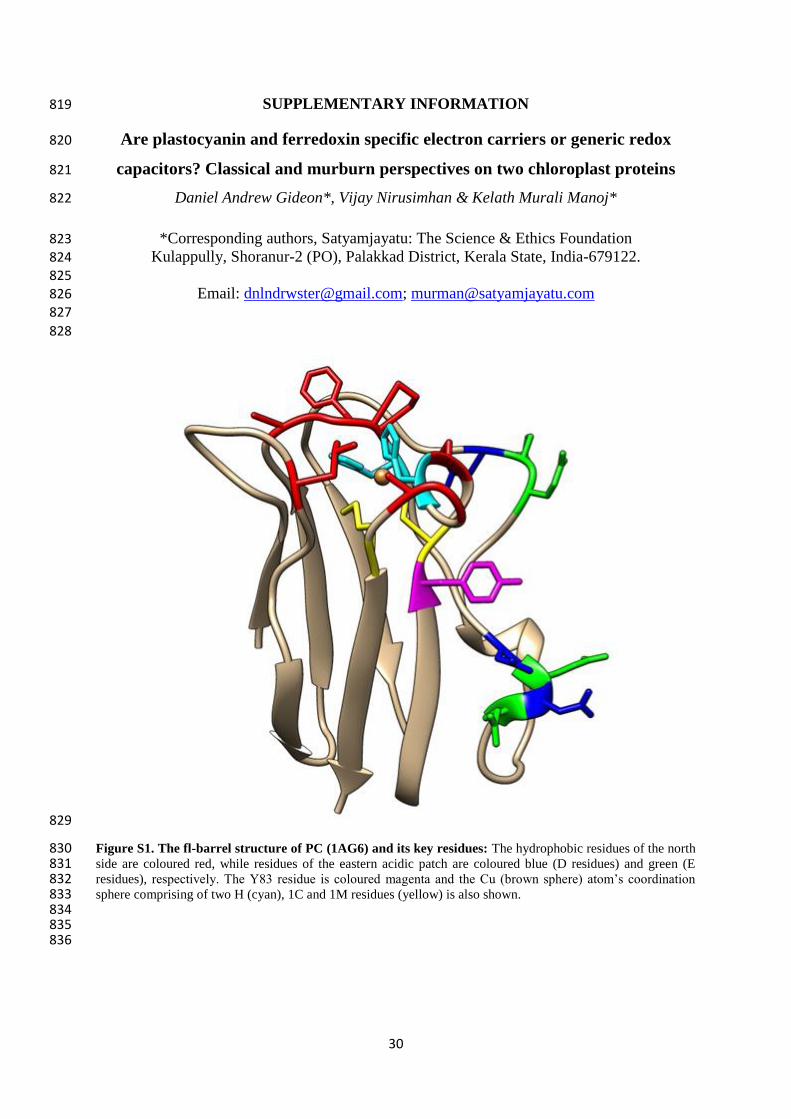
30
SUPPLEMENTARY INFORMATION 819
Are plastocyanin and ferredoxin specific electron carriers or generic redox 820
capacitors? Classical and murburn perspectives on two chloroplast proteins 821
Daniel Andrew Gideon*, Vijay Nirusimhan & Kelath Murali Manoj* 822
*Corresponding authors, Satyamjayatu: The Science & Ethics Foundation 823 Kulappully, Shoranur-2 (PO), Palakkad District, Kerala State, India-679122. 824
825 Email: [email protected]; [email protected] 826
827
828
829
Figure S1. The fl-barrel structure of PC (1AG6) and its key residues: The hydrophobic residues of the north 830 side are coloured red, while residues of the eastern acidic patch are coloured blue (D residues) and green (E 831 residues), respectively. The Y83 residue is coloured magenta and the Cu (brown sphere) atom’s coordination 832 sphere comprising of two H (cyan), 1C and 1M residues (yellow) is also shown. 833 834 835 836
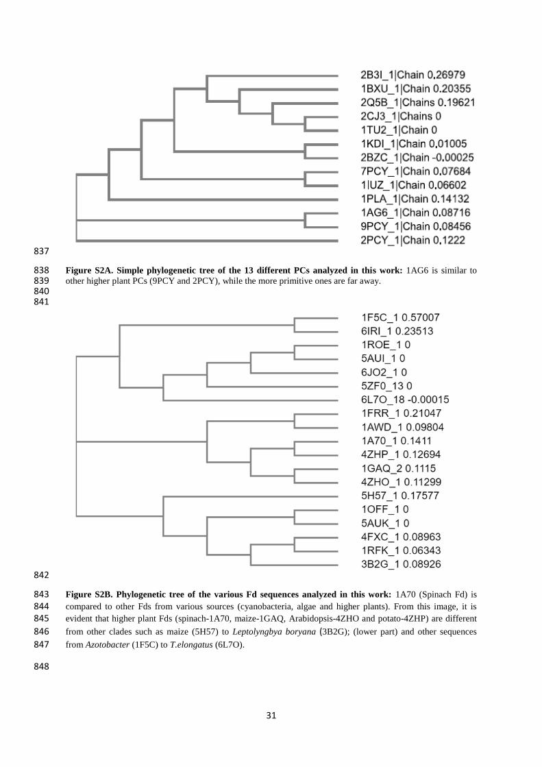
31
837
Figure S2A. Simple phylogenetic tree of the 13 different PCs analyzed in this work: 1AG6 is similar to 838 other higher plant PCs (9PCY and 2PCY), while the more primitive ones are far away. 839 840 841
842
Figure S2B. Phylogenetic tree of the various Fd sequences analyzed in this work: 1A70 (Spinach Fd) is 843 compared to other Fds from various sources (cyanobacteria, algae and higher plants). From this image, it is 844 evident that higher plant Fds (spinach-1A70, maize-1GAQ, Arabidopsis-4ZHO and potato-4ZHP) are different 845
from other clades such as maize (5H57) to Leptolyngbya boryana (3B2G); (lower part) and other sequences 846
from Azotobacter (1F5C) to T.elongatus (6L7O). 847
848
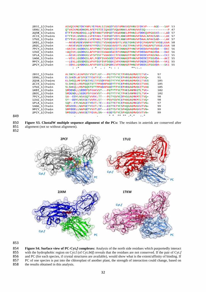
32
849
Figure S3. ClustalW multiple sequence alignment of the PCs: The residues in asterisk are conserved after 850 alignment (not so without alignment). 851 852
853
Figure S4. Surface view of PC-Cyt.f complexes: Analysis of the north side residues which purportedly interact 854 with the hydrophobic region on Cyt.f (of Cyt.b6f) reveals that the residues are not conserved. If the pair of Cyt.f 855 and PC (for each species, if crystal structures are available), would show what is the extent/affinity of binding. If 856 PC of one species is put into the chloroplast of another plant, the strength of interaction could change, based on 857 the results obtained in this analysis. 858
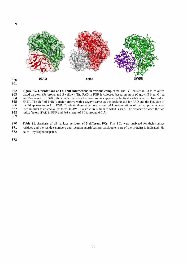
33
859
860 861
Figure S5. Orientations of Fd-FNR interactions in various complexes: The FeS cluster in Fd is coloured 862 based on atom (Fe-brown and S-yellow). The FAD in FNR is coloured based on atom (C-grey, N-blue, O-red 863 and P-orange). In 1GAQ, the contact between the two proteins appears to be tighter (that what is observed in 864 5H5J). The cleft of FNR (a major groove with a cavity) serves as the docking site for FAD and the FeS side of 865 the Fd appears to dock to FNR. To obtain these structures, several μM concentrations of the two proteins were 866 used in order to co-crystallize them. In 3W5U, a structure similar to 5H5J is seen. The distance between the two 867 redox factors (FAD in FNR and FeS cluster of Fd is around 6-7 Å) 868 869
Table S1. Analysis of all surface residues of 5 different PCs: Five PCs were analyzed for their surface 870 residues and the residue numbers and location (north/eastern patch/other part of the protein) is indicated. Hp 871 patch – hydrophobic patch. 872
873
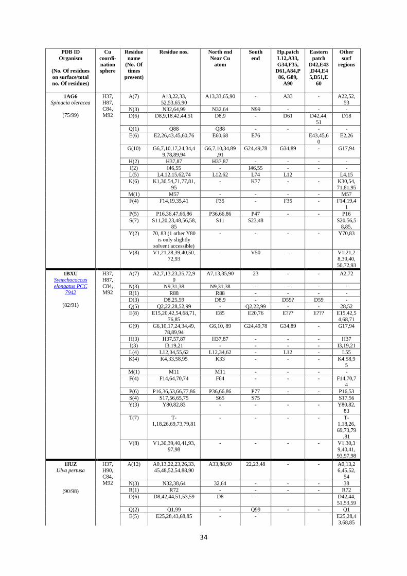
34
PDB ID
Organism
(No. Of residues
on surface/total
no. Of residues)
Cu
coordi-
nation
sphere
Residue
name
(No. Of
times
present)
Residue nos. North end
Near Cu
atom
South
end
Hp.patch
L12,A33,
G34,F35,
D61,A84,P
86, G89,
A90
Eastern
patch
D42,E43
,D44,E4
5,D51,E
60
Other
surf
regions
1AG6
Spinacia oleracea
(75/99)
H37,
H87, C84,
M92
A(7) A13,22,33,
52,53,65,90
A13,33,65,90 - A33 - A22,52,
53
N(3) N32,64,99 N32,64 N99 - - -
D(6) D8,9,18,42,44,51 D8,9 - D61 D42,44,
51
D18
Q(1) Q88 Q88 - - - -
E(6) E2,26,43,45,60,76 E60,68 E76 E43,45,60
E2,26
G(10) G6,7,10,17,24,34,4
9,78,89,94
G6,7,10,34,89
,91
G24,49,78 G34,89 - G17,94
H(2) H37,87 H37,87 - - - -
I(2) I46,55 - I46,55 - - -
L(5) L4,12,15,62,74 L12,62 L74 L12 L4,15
K(6) K1,30,54,71,77,81,
95
- K77 - - K30,54,
71,81,95
M(1) M57 - - - - M57
F(4) F14,19,35,41 F35 - F35 - F14,19,41
P(5) P16,36,47,66,86 P36,66,86 P47 - - P16
S(7) S11,20,23,48,56,58,85
S11 S23,48 S20,56,58,85,
Y(2) 70, 83 (1 other Y80
is only slightly
solvent accessible)
- - - - Y70,83
V(8) V1,21,28,39,40,50,
72,93
- V50 - - V1,21,2
8,39,40,
50,72,93
1BXU
Synechococcus
elongatus PCC 7942
(82/91)
H37,
H87,
C84, M92
A(7) A2,7,13,23,35,72,9
0
A7,13,35,90 23 - - A2,72
N(3) N9,31,38 N9,31,38 - - - -
R(1) R88 R88 - - - -
D(3) D8,25,59 D8,9 - D59? D59 -
Q(5) Q2,22,28,52,99 - Q2,22,99 - - 28,52
E(8) E15,20,42,54,68,71,
76,85
E85 E20,76 E??? E??? E15,42,5
4,68,71
G(9) G6,10,17,24,34,49,78,89,94
G6,10, 89 G24,49,78 G34,89 - G17,94
H(3) H37,57,87 H37,87 - - - H37
I(3) I3,19,21 - - - - I3,19,21
L(4) L12,34,55,62 L12,34,62 - L12 - L55
K(4) K4,33,58,95 K33 - - - K4,58,95
M(1) M11 M11 - - - -
F(4) F14,64,70,74 F64 - - - F14,70,7
4
P(6) P16,36,53,66,77,86 P36,66,86 P77 - - P16,53
S(4) S17,56,65,75 S65 S75 S17,56
Y(3) Y80,82,83 - - - - Y80,82,
83
T(7) T-1,18,26,69,73,79,81
- - - - T-1,18,26,
69,73,79
,81
V(8) V1,30,39,40,41,93,
97,98
- - - - V1,30,3
9,40,41,
93,97,98
1IUZ
Ulva pertusa
(90/98)
H37,
H90,
C84, M92
A(12) A0,13,22,23,26,33,
45,48,52,54,88,90
A33,88,90 22,23,48 - - A0,13,2
6,45,52,
54
N(3) N32,38,64 32,64 - - - 38
R(1) R72 - - - - R72
D(6) D8,42,44,51,53,59 D8 - D42,44,
51,53,59
Q(2) Q1,99 - Q99 - - Q1
E(5) E25,28,43,68,85 - - E25,28,43,68,85
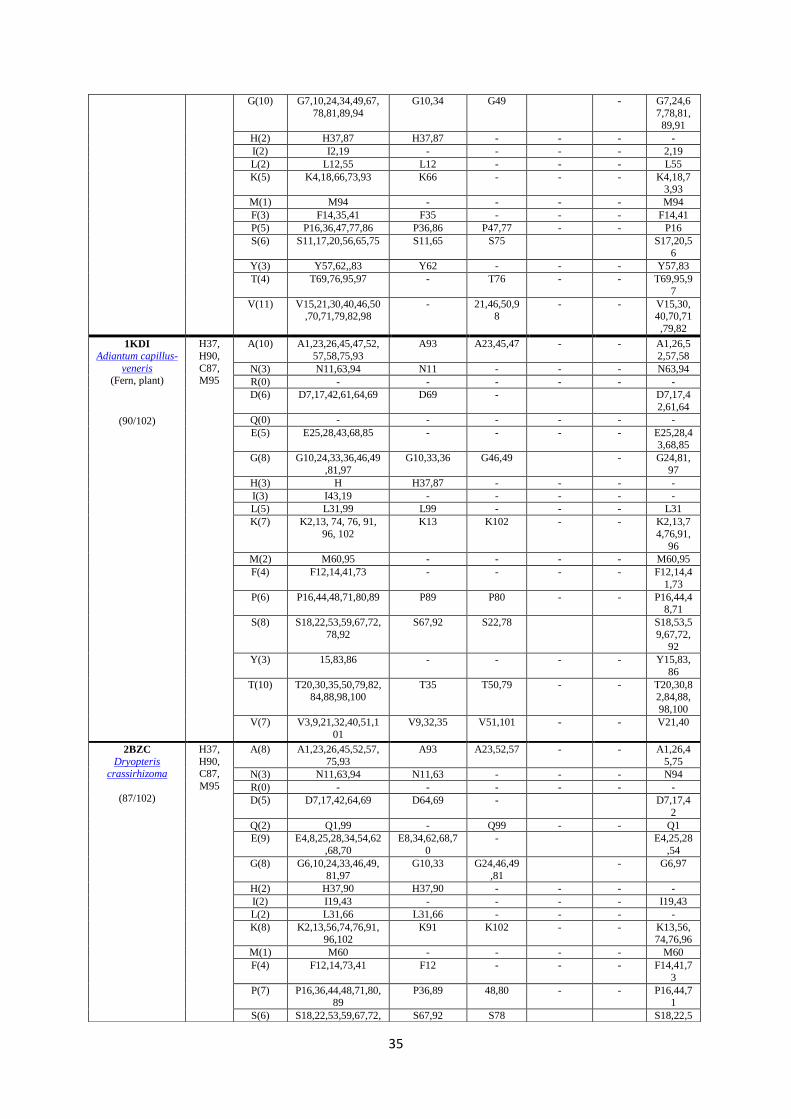
35
G(10) G7,10,24,34,49,67,
78,81,89,94
G10,34 G49 - G7,24,6
7,78,81,89,91
H(2) H37,87 H37,87 - - - -
I(2) I2,19 - - - - 2,19
L(2) L12,55 L12 - - - L55
K(5) K4,18,66,73,93 K66 - - - K4,18,73,93
M(1) M94 - - - - M94
F(3) F14,35,41 F35 - - - F14,41
P(5) P16,36,47,77,86 P36,86 P47,77 - - P16
S(6) S11,17,20,56,65,75 S11,65 S75 S17,20,56
Y(3) Y57,62,,83 Y62 - - - Y57,83
T(4) T69,76,95,97 - T76 - - T69,95,9
7
V(11) V15,21,30,40,46,50,70,71,79,82,98
- 21,46,50,98
- - V15,30,40,70,71
,79,82
1KDI
Adiantum capillus-
veneris (Fern, plant)
(90/102)
H37,
H90,
C87, M95
A(10) A1,23,26,45,47,52,
57,58,75,93
A93 A23,45,47 - - A1,26,5
2,57,58
N(3) N11,63,94 N11 - - - N63,94
R(0) - - - - - -
D(6) D7,17,42,61,64,69 D69 - D7,17,4
2,61,64
Q(0) - - - - - -
E(5) E25,28,43,68,85 - - - - E25,28,43,68,85
G(8) G10,24,33,36,46,49
,81,97
G10,33,36 G46,49 - G24,81,
97
H(3) H H37,87 - - - -
I(3) I43,19 - - - - -
L(5) L31,99 L99 - - - L31
K(7) K2,13, 74, 76, 91,
96, 102
K13 K102 - - K2,13,7
4,76,91,
96
M(2) M60,95 - - - - M60,95
F(4) F12,14,41,73 - - - - F12,14,4
1,73
P(6) P16,44,48,71,80,89 P89 P80 - - P16,44,48,71
S(8) S18,22,53,59,67,72,
78,92
S67,92 S22,78 S18,53,5
9,67,72,92
Y(3) 15,83,86 - - - - Y15,83,
86
T(10) T20,30,35,50,79,82,84,88,98,100
T35 T50,79 - - T20,30,82,84,88,
98,100
V(7) V3,9,21,32,40,51,1
01
V9,32,35 V51,101 - - V21,40
2BZC
Dryopteris crassirhizoma
(87/102)
H37,
H90, C87,
M95
A(8) A1,23,26,45,52,57,
75,93
A93 A23,52,57 - - A1,26,4
5,75
N(3) N11,63,94 N11,63 - - - N94
R(0) - - - - - -
D(5) D7,17,42,64,69 D64,69 - D7,17,4
2
Q(2) Q1,99 - Q99 - - Q1
E(9) E4,8,25,28,34,54,62
,68,70
E8,34,62,68,7
0
- E4,25,28
,54
G(8) G6,10,24,33,46,49,
81,97
G10,33 G24,46,49
,81
- G6,97
H(2) H37,90 H37,90 - - - -
I(2) I19,43 - - - - I19,43
L(2) L31,66 L31,66 - - - -
K(8) K2,13,56,74,76,91,96,102
K91 K102 - - K13,56,74,76,96
M(1) M60 - - - - M60
F(4) F12,14,73,41 F12 - - - F14,41,7
3
P(7) P16,36,44,48,71,80,89
P36,89 48,80 - - P16,44,71
S(6) S18,22,53,59,67,72, S67,92 S78 S18,22,5
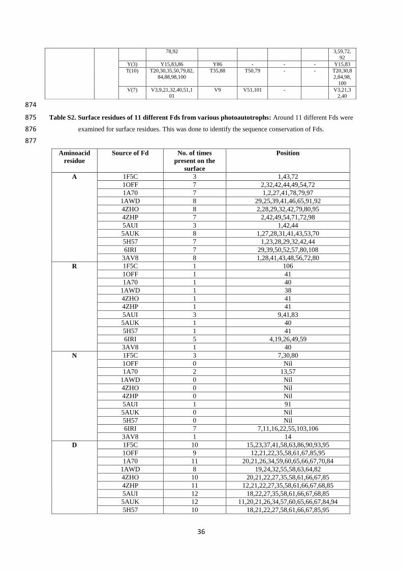
36
874
Table S2. Surface residues of 11 different Fds from various photoautotrophs: Around 11 different Fds were 875
examined for surface residues. This was done to identify the sequence conservation of Fds. 876
877
Aminoacid
residue
Source of Fd No. of times
present on the
surface
Position
A
1F5C 3 1,43,72
1OFF 7 2,32,42,44,49,54,72
1A70 7 1,2,27,41,78,79,97
1AWD 8 29,25,39,41,46,65,91,92
4ZHO 8 2,28,29,32,42,79,80,95
4ZHP 7 2,42,49,54,71,72,98
5AUI 3 1,42,44
5AUK 8 1,27,28,31,41,43,53,70
5H57 7 1,23,28,29,32,42,44
6IRI 7 29,39,50,52,57,80,108
3AV8 8 1,28,41,43,48,56,72,80
R 1F5C 1 106
1OFF 1 41
1A70 1 40
1AWD 1 38
4ZHO 1 41
4ZHP 1 41
5AUI 3 9,41,83
5AUK 1 40
5H57 1 41
6IRI 5 4,19,26,49,59
3AV8 1 40
N 1F5C 3 7,30,80
1OFF 0 Nil
1A70 2 13,57
1AWD 0 Nil
4ZHO 0 Nil
4ZHP 0 Nil
5AUI 1 91
5AUK 0 Nil
5H57 0 Nil
6IRI 7 7,11,16,22,55,103,106
3AV8 1 14
D 1F5C 10 15,23,37,41,58,63,86,90,93,95
1OFF 9 12,21,22,35,58,61,67,85,95
1A70 11 20,21,26,34,59,60,65,66,67,70,84
1AWD 8 19,24,32,55,58,63,64,82
4ZHO 10 20,21,22,27,35,58,61,66,67,85
4ZHP 11 12,21,22,27,35,58,61,66,67,68,85
5AUI 12 18,22,27,35,58,61,66,67,68,85
5AUK 12 11,20,21,26,34,57,60,65,66,67,84,94
5H57 10 18,21,22,27,58,61,66,67,85,95
78,92 3,59,72,
92
Y(3) Y15,83,86 Y86 - - - Y15,83
T(10) T20,30,35,50,79,82,
84,88,98,100
T35,88 T50,79 - - T20,30,8
2,84,98,
100
V(7) V3,9,21,32,40,51,101
V9 V51,101 - V3,21,32,40
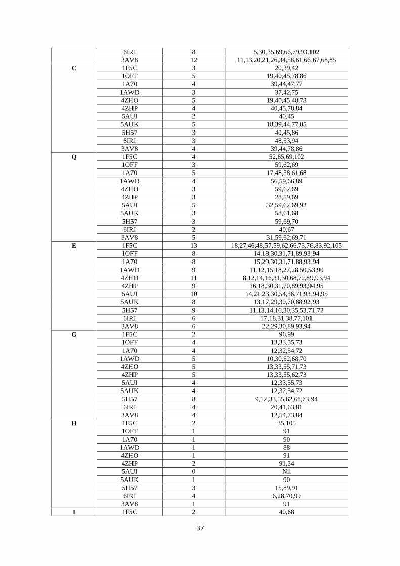
37
6IRI 8 5,30,35,69,66,79,93,102
3AV8 12 11,13,20,21,26,34,58,61,66,67,68,85
C 1F5C 3 20,39,42
1OFF 5 19,40,45,78,86
1A70 4 39,44,47,77
1AWD 3 37,42,75
4ZHO 5 19,40,45,48,78
4ZHP 4 40,45,78,84
5AUI 2 40,45
5AUK 5 18,39,44,77,85
5H57 3 40,45,86
6IRI 3 48,53,94
3AV8 4 39,44,78,86
Q 1F5C 4 52,65,69,102
1OFF 3 59,62,69
1A70 5 17,48,58,61,68
1AWD 4 56,59,66,89
4ZHO 3 59,62,69
4ZHP 3 28,59,69
5AUI 5 32,59,62,69,92
5AUK 3 58,61,68
5H57 3 59,69,70
6IRI 2 40,67
3AV8 5 31,59,62,69,71
E 1F5C 13 18,27,46,48,57,59,62,66,73,76,83,92,105
1OFF 8 14,18,30,31,71,89,93,94
1A70 8 15,29,30,31,71,88,93,94
1AWD 9 11,12,15,18,27,28,50,53,90
4ZHO 11 8,12,14,16,31,30,68,72,89,93,94
4ZHP 9 16,18,30,31,70,89,93,94,95
5AUI 10 14,21,23,30,54,56,71,93,94,95
5AUK 8 13,17,29,30,70,88,92,93
5H57 9 11,13,14,16,30,35,53,71,72
6IRI 6 17,18,31,38,77,101
3AV8 6 22,29,30,89,93,94
G 1F5C 2 96,99
1OFF 4 13,33,55,73
1A70 4 12,32,54,72
1AWD 5 10,30,52,68,70
4ZHO 5 13,33,55,71,73
4ZHP 5 13,33,55,62,73
5AUI 4 12,33,55,73
5AUK 4 12,32,54,72
5H57 8 9,12,33,55,62,68,73,94
6IRI 4 20,41,63,81
3AV8 4 12,54,73,84
H 1F5C 2 35,105
1OFF 1 91
1A70 1 90
1AWD 1 88
4ZHO 1 91
4ZHP 2 91,34
5AUI 0 Nil
5AUK 1 90
5H57 3 15,89,91
6IRI 4 6,28,70,99
3AV8 1 91
I 1F5C 2 40,68
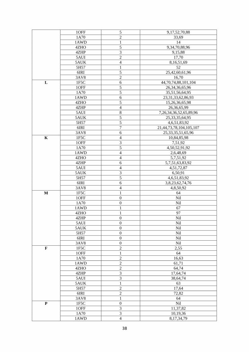
38
1OFF 5 9,17,52,70,88
1A70 2 33,69
1AWD 1 14
4ZHO 5 9,34,70,88,96
4ZHP 3 9,15,88
5AUI 2 17,70
5AUK 4 8,16,51,69
5H57 1 52
6IRI 5 25,42,60,61,96
3AV8 2 16,70
L 1F5C 6 44,70,74,88,101,104
1OFF 5 26,34,36,65,96
1A70 5 35,51,56,64,95
1AWD 6 23,31,33,62,86,93
4ZHO 5 15,26,36,65,98
4ZHP 4 26,36,65,99
5AUI 8 7,26,34,36,52,65,89,96
5AUK 5 25,33,35,64,95
5H57 5 4,6,51,83,92
6IRI 7 21,44,73,78,104,105,107
3AV8 6 25,33,35,51,65,96
K 1F5C 4 10,84,85,98
1OFF 3 7,51,92
1A70 5 4,50,52,91,92
1AWD 4 2,6,48,69
4ZHO 4 5,7,51,92
4ZHP 6 5,7,51,63,83,92
5AUI 4 4,51,72,87
5AUK 3 6,50,91
5H57 5 4,6,51,83,92
6IRI 6 3,8,23,62,74,76
3AV8 4 4,8,50,92
M 1F5C 1 64
1OFF 0 Nil
1A70 0 Nil
1AWD 1 67
4ZHO 1 97
4ZHP 0 Nil
5AUI 0 Nil
5AUK 0 Nil
5H57 0 Nil
6IRI 0 Nil
3AV8 0 Nil
F 1F5C 2 2,55
1OFF 1 64
1A70 2 16,63
1AWD 2 61,71
4ZHO 2 64,74
4ZHP 3 17,64,74
5AUI 3 38,64,74
5AUK 1 63
5H57 2 17,64
6IRI 2 72,82
3AV8 1 64
P 1F5C 0 Nil
1OFF 3 11,37,82
1A70 3 10,19,36
1AWD 4 8,17,34,79
39
4ZHO 3 11,37,82
4ZHP 5 11,14,20,37,82
5AUI 3 10,20,37
5AUK 3 10,36,81
5H57 4 10,20,37,82
6IRI 4 43,45,75,90
3AV8 6 10,19,36,55,57,82
S 1F5C 1 56
1OFF 10 3,15,16,20,39,46,56,60,63,84
1A70 7 38,43,45,46,55,62,83
1AWD 8 9,36,43,44,51,57,60,81
4ZHO 8 44,46,47,54,56,60,63,84
4ZHP 6 3,39,44,46,47,60
5AUI 5 13,39,60,63,84
5AUK 10 2,14,15,19,38,45,55,59,62,83
5H57 7 39,46,54,56,60,63,84
6IRI 5 47,68,71,92,97
3AV8 6 2,38,45,53,60,63
T 1F5C 2 5,82
1OFF 6 5,10,23,53,83,87
1A70 7 6,9,11,53,86,89,96
1AWD 6 4,7,13,20,80,84
4ZHO 5 3,10,83,87,90
4ZHP 6 10,53,56,87,90,97
5AUI 6 2,6,15,16,47,90
5AUK 8 4,9,22,46,52,82,86,89
5H57 3 31,47,90
6IRI 6 2,13,24,64,91,98
3AV8 5 6,9,17,46,83
W 1F5C 2 78,94
1OFF 0 Nil
1A70 0 Nil
1AWD 0 Nil
4ZHO 0 Nil
4ZHP 0 Nil
5AUI 0 Nil
5AUK 0 Nil
5H57 0 Nil
6IRI 0 Nil
3AV8 0 Nil
Y 1F5C 2 13,26
1OFF 4 4,24,38,81
1A70 4 3,23,37,80
1AWD 5 1,21,35,78,94
4ZHO 4 4,24,38,81
4ZHP 4 4,24,38,81
5AUI 4 3,24,81,97
5AUK 6 3,23,37,73,80,96
5H57 6 3,24,38,74,81,97
6IRI 4 10,32,46,89
3AV8 6 3,23,37,74,81,97
V 1F5C 6 17,19,22,60,77,97
1OFF 3 57,75,79
1A70 6 8,14,22,74,82,85
1AWD 4 49,54,76,83
4ZHO 6 17,23,52,53,57,86
4ZHP 5 23,57,52,79,86
5AUI 5 8,19,28,57,79
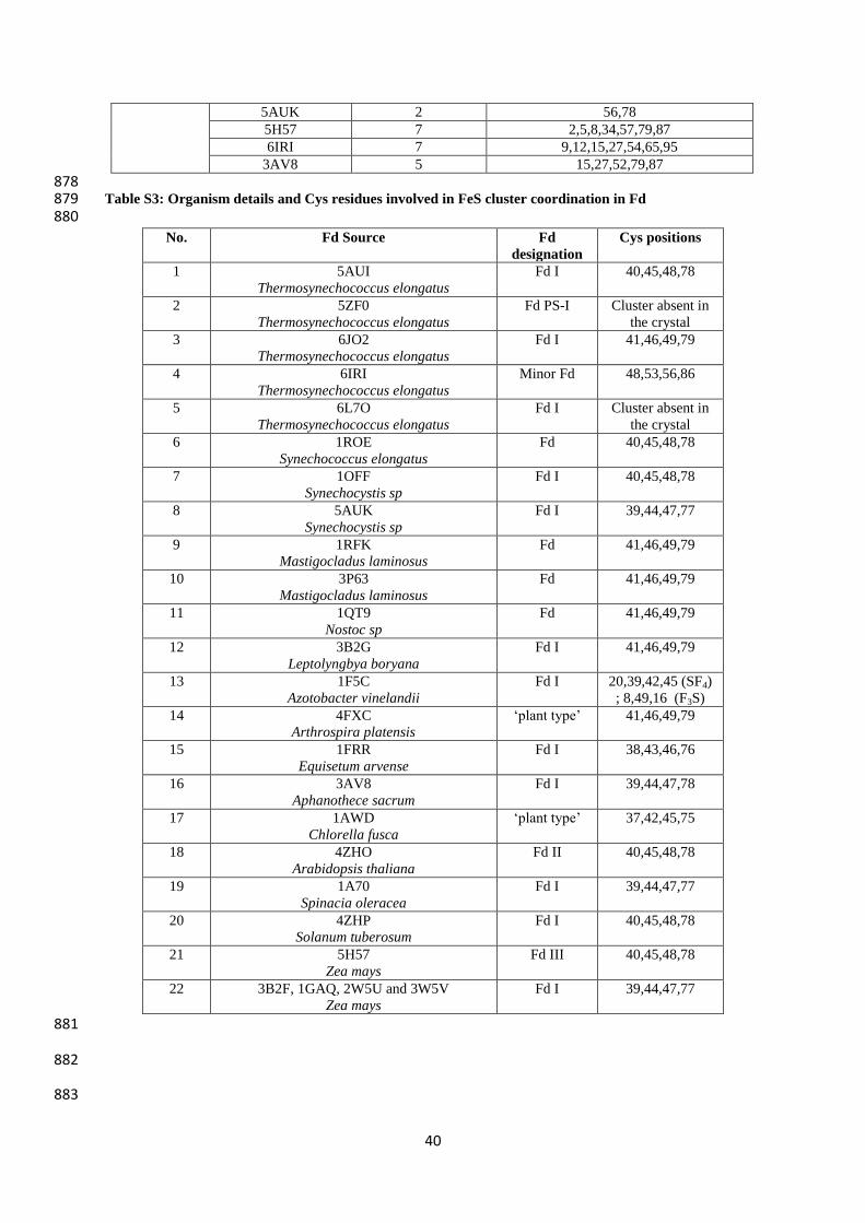
40
5AUK 2 56,78
5H57 7 2,5,8,34,57,79,87
6IRI 7 9,12,15,27,54,65,95
3AV8 5 15,27,52,79,87
878 Table S3: Organism details and Cys residues involved in FeS cluster coordination in Fd 879 880
No. Fd Source Fd
designation
Cys positions
1 5AUI
Thermosynechococcus elongatus
Fd I 40,45,48,78
2 5ZF0
Thermosynechococcus elongatus
Fd PS-I Cluster absent in
the crystal
3 6JO2
Thermosynechococcus elongatus
Fd I 41,46,49,79
4 6IRI
Thermosynechococcus elongatus
Minor Fd 48,53,56,86
5 6L7O
Thermosynechococcus elongatus
Fd I Cluster absent in
the crystal
6 1ROE
Synechococcus elongatus
Fd 40,45,48,78
7 1OFF
Synechocystis sp
Fd I 40,45,48,78
8 5AUK
Synechocystis sp
Fd I 39,44,47,77
9 1RFK
Mastigocladus laminosus
Fd 41,46,49,79
10 3P63
Mastigocladus laminosus
Fd 41,46,49,79
11 1QT9
Nostoc sp
Fd 41,46,49,79
12 3B2G
Leptolyngbya boryana
Fd I 41,46,49,79
13 1F5C
Azotobacter vinelandii
Fd I 20,39,42,45 (SF4)
; 8,49,16 (F3S)
14 4FXC
Arthrospira platensis
‘plant type’ 41,46,49,79
15 1FRR
Equisetum arvense
Fd I 38,43,46,76
16 3AV8
Aphanothece sacrum
Fd I 39,44,47,78
17 1AWD
Chlorella fusca
‘plant type’ 37,42,45,75
18 4ZHO
Arabidopsis thaliana
Fd II 40,45,48,78
19 1A70
Spinacia oleracea
Fd I 39,44,47,77
20 4ZHP
Solanum tuberosum
Fd I 40,45,48,78
21 5H57
Zea mays
Fd III 40,45,48,78
22 3B2F, 1GAQ, 2W5U and 3W5V
Zea mays
Fd I 39,44,47,77
881
882
883