Brown algae species as biomonitors of Zn and Cd at Sepetiba Bay, Rio de Janeiro, Brazil
12
Brown algae species as biomonitors of Zn and Cd at Sepetiba Bay, Rio de Janeiro, Brazil G.M. Amado Filho a, *, L.R. Andrade b , C.S. Karez c , M. Farina b , W.C. Pfeier d a Programa Zona Costeira, Instituto de Pesquisas Jardim Bota ˆnico do Rio de Janeiro, Rua Pacheco Lea ˜o 915, 22460-030 Rio de Janeiro, Brazil b Departmento de Anatomia, Instituto de Cieˆncias Biome ´dicas, CCS/UFRJ, Cidade Universita ´ria, 21941-590, Rio de Janeiro, Brazil c UNESCO Oce, ROSTLAC, Montevideo 11300, Uruguay d Laborato ´rio de Radioiso ´topos, Instituto de Biofi ´ sica Carlos Chagas Filho, CCS/UFRJ, Cidade Universita ´ria, 21949-900 Rio de Janeiro, Brazil Received 14 September 1998; received in revised form 17 February 1999; accepted 15 March 1999 Abstract In order to contribute to monitoring heavy metal contamination of Sepetiba Bay, Rio de Janeiro, Brazil, long term evaluation of Zn and Cd concentration was performed in two brown algae species, Padina gymnospora and Sargassum stenophyllum. In relation to Sepetiba Bay macroalgae community, these species were the most abundant in substrate cover. The algae metal concentration variation from 1990 to 1997 should be related to the inputs of metals released into the bay by industrial process of Zn production in the area. In situ uptake and release transplant experiments with Padina between Sepetiba Bay and a near uncontami- nated area showed that the species could reflect the variation in metal environment availability. A lower reduction in metal concentration by plants transplanted to the uncontaminated area was observed. Analytical electron microscopy showed that the Zn in P. gymnospora from Sepetiba Bay was present as small deposits distributed in the cell walls. # 1999 Elsevier Science Ltd. All rights reserved. Keywords: Pollution monitoring; Sepetiba Bay; Heavy metals; Brown algae, bioaccumulation; Epibenthic community; Transplant experiment; Subcellular localisation Marine Environmental Research 48 (1999) 213–224 www.elsevier.com/locate/marenvrev 0141-1136/99/$ - see front matter # 1999 Elsevier Science Ltd. All rights reserved. PII: S0141-1136(99)00042-2 * Corresponding author. Tel. and fax: +55-21-2947526. E-mail address: gfi[email protected] (G.M. Amado Filho)
Transcript of Brown algae species as biomonitors of Zn and Cd at Sepetiba Bay, Rio de Janeiro, Brazil
Brown algae species as biomonitors of Zn andCd at Sepetiba Bay, Rio de Janeiro, Brazil
G.M. Amado Filho a,*, L.R. Andrade b, C.S. Karez c,M. Farina b, W.C. Pfei�er d
aPrograma Zona Costeira, Instituto de Pesquisas Jardim BotaÃnico do Rio de Janeiro,
Rua Pacheco LeaÄo 915, 22460-030 Rio de Janeiro, BrazilbDepartmento de Anatomia, Instituto de CieÃncias BiomeÂdicas, CCS/UFRJ, Cidade UniversitaÂria,
21941-590, Rio de Janeiro, BrazilcUNESCO O�ce, ROSTLAC, Montevideo 11300, Uruguay
dLaboratoÂrio de RadioisoÂtopos, Instituto de BiofiÂsica Carlos Chagas Filho, CCS/UFRJ,
Cidade UniversitaÂria, 21949-900 Rio de Janeiro, Brazil
Received 14 September 1998; received in revised form 17 February 1999; accepted 15 March 1999
Abstract
In order to contribute to monitoring heavy metal contamination of Sepetiba Bay, Rio de
Janeiro, Brazil, long term evaluation of Zn and Cd concentration was performed in twobrown algae species, Padina gymnospora and Sargassum stenophyllum. In relation to SepetibaBay macroalgae community, these species were the most abundant in substrate cover. The
algae metal concentration variation from 1990 to 1997 should be related to the inputs ofmetals released into the bay by industrial process of Zn production in the area. In situ uptakeand release transplant experiments with Padina between Sepetiba Bay and a near uncontami-
nated area showed that the species could re¯ect the variation in metal environment availability.A lower reduction in metal concentration by plants transplanted to the uncontaminated areawas observed. Analytical electron microscopy showed that the Zn in P. gymnospora from
Sepetiba Bay was present as small deposits distributed in the cell walls. # 1999 Elsevier ScienceLtd. All rights reserved.
Keywords: Pollution monitoring; Sepetiba Bay; Heavy metals; Brown algae, bioaccumulation; Epibenthic
community; Transplant experiment; Subcellular localisation
Marine Environmental Research 48 (1999) 213±224
www.elsevier.com/locate/marenvrev
0141-1136/99/$ - see front matter # 1999 Elsevier Science Ltd. All rights reserved.
PI I : S0141-1136(99 )00042 -2
* Corresponding author. Tel. and fax: +55-21-2947526.
E-mail address: g®[email protected] (G.M. Amado Filho)

1. Introduction
Long-term determination of critical pollutants in speci®c compartments of eco-systems is an essential component of environmental monitoring programs. In thelast 15 years Cd and Zn have been identi®ed as the main contaminants in sedimentsand biota of Sepetiba Bay, Rio de Janeiro, Brazil (Barcellos & Lacerda, 1994; Kur-ita & Pfei�er, 1991; Lacerda, Carvalho, & Gomes, 1989; Lima, Lacerda, Pfei�er, &Fiszman, 1986; Lacerda, Pfei�er, & Fizman, 1985, 1987, 1989; Pfei�er, Lacerda,Fizman, & Lima, 1985). However, few studies have quanti®ed (Barcellos, 1995;Rezende, Lacerda & Pfei�er, 1991) the contamination status in recent years. Lack oflong-term follow-up information makes the interpretation of available data and theapplication of mitigation decisions di�cult.Heavy metal levels have been analysed in organisms from di�erent taxonomic
groups in Sepetiba Bay: ®shes, crustaceans, molluscs, seagrasses and algae (Karez,MagalhaÄ es, Pfei�er, & Amado Filho, 1994; Lacerda & Rezende, 1986; Lima et al.,1986; Pfei�er et al., 1985). Among these groups, the highest levels of Zn and Cd werefound in oysters (Crassostrea brasiliana), followed by macroalgae. Among 10 mac-roalgae analysed, the brown alga Padina gymnospora (Dictyotales) presented thehighest metal concentrations (Karez et al., 1994). The concentrations of Zn and Cdin P. gymnospora from Sepetiba Bay were up to 25 and four times higher, respec-tively, than individuals from a nearby uncontaminated area.Knowledge of factors a�ecting the rate of accumulation and release in brown
algae is of special value when using them to biomonitor heavy metal pollution (Eide,Myklestad & Melson, 1980; Rainbow, 1995). In laboratory assays, Zn accumulatedby P. gymnospora increased in direct proportion to its concentrations in seawaterover 21 days experimental time (Amado Filho, Karez, Andrade, Yoneshigue-Valentin, & Pfei�er, 1997). In short-term experiment of 65Zn kinetics, the release ofZn by P. gymnospora was insigni®cant, suggesting that the metal could be stronglybound into cellular sites (Karez et al., 1994). Although these results indicated thegreat ability of P. gymnospora to accumulate Zn, data on uptake and release of themetal in situ were lacking.Two mechanisms have been associated with the capability of some brown algae to
accumulate high metal concentrations: the presence of cell wall-speci®c polyanionicpolysaccharides that provide the sequestration and precipitation of metals in the cellwalls (Crist et al., 1992; Lignell, Roomans & Pederson, 1982; Mariani, Tolomio, &Braghetta, 1985; Volesky & Holan, 1995) and the a�nity between metals and poly-phenols (Ragan & Glombitza, 1986; Ragan, Smidsrod, & Larsen, 1979). Karez andPereira (1995) showed a low percentage association of Zn and Cd with polyphenolicfraction of P. gymnospora from Sepetiba Bay and suggested that the high Zn and Cdfound in whole algae from polluted areas (like Sepetiba Bay) might thus be greaterthan the capability of polyphenols to bind these metals. Analytical electron micro-scopy of P. gymnospora, submitted to high Zn concentrations under laboratoryexperimental conditions, showed the presence of Zn in dense granules along the cellwalls indicating that these walls could play the main role in the Zn accumulation(Amado Filho, Karez, Pfei�er, Yoneshigue-Valentin, & Farina, 1996).
214 G.M. Amado Filho et al. /Marine Environmental Research 48 (1999) 213±224

In this work, the abundance of macroalgae in the epibenthic community of twostudied sites in Sepetiba Bay is described and the Zn and Cd levels in populationsof brown algae from 1990 to 1997 is evaluated. Also, the accumulation and releaseof metals was determined in the ®eld by transplanting experiments. Metals werelocalised at the subcellular level in individuals of P. gymnospora from SepetibaBay.
2. Materials and Methods
Sepetiba Bay (SB) is a semi-enclosed lagoon, 447 km2 in area, situated 60 km westof Rio de Janeiro City (Fig. 1). In order to describe the abundance of macroalgae,20 m-long transects were placed horizontally at about 1 m below lower astronomictide (LAT) at two sites in SB (Ilha do Gato [IG] and Praia Grande [PG]; Fig. 1), inFebruary, April, June and September 1997. Percentage cover of macroalgae specieswere recorded using 10 0.25 m2 quadrats randomly positioned (a table of randomnumbers was used) along the transects.Zn and Cd analyses were conducted on two selected brown algae species,
P. gymnospora and S. stenophyllum, collected at the sublittoral zone in the twosites of SB. The collections were performed from April 1990 to November 1993(some results were published in Karez et al., 1994) and from August 1995 to July1997.Transplanting experiments were carried out with 25 whole-adult individuals (8±12
cm long) of P. gymnospora. The plants were collected from the sublittoral zone ofSB (PG), ®xed by their holdfast in nylon nets and immediately transported and ®xedto rocky blocks at the same littoral zone (similar depth) in the uncontaminated area,Ribeira Bay (RB) situated 60 km west of SB (Fig. 1). The same procedure was usedto transfer individuals of P. gymnospora from RB to SB. These experiments weredone in three seasons: September 1996, March 1997 and June 1997. After 30 days oftransplantation, the individuals were collected for metal analysis and the resultswere compared with data from natural populations of PG.Only whole, healthy and adult plants were analysed. Samples were initially
cleaned of epiphytes, washed in seawater from the sites of collection and thereafterin distilled water, dried at 50�C, homogenised and ashed at 420�C. Fractions of 0.5 g(dry wt) were digested in 10 ml concentrated HNO3 (65% wt). The resulting solutionwas evaporated and redissolved in 0.1 N HCl. Metal concentrations were measuredby ¯ame atomic absorption spectrophotometry (Varian AA-1475) in triplicates.Results are given in mg. gÿ1 of dry weight. Analytical procedures were tested bycomparative analysis of International Atomic Energy Agency (IAEA) referencematerial IAEA-140 (sea plant homogenate, Fucus reference values: Zn=47.3 mg. gÿ1,Cd=0.537 mg. gÿ1: mean value obtained in this study: Zn=45.8 mg. gÿ1, Cd=0.510mg. gÿ1).Specimens of P. gymnospora were prepared for transmission electron microscopy
according to Amado Filho et al. (1996). The localisation of metals was obtained byelectron spectroscopic imaging (ESI) performed with a ZEISS CEM 902 analytical
G.M. Amado Filho et al. /Marine Environmental Research 48 (1999) 213±224 215

electron microscope, operating at 80 KV. An imaging ®lter integrated in the colummof the microspope allows for the selection of electrons with particular energy lossesto form an electron spectroscopic image. An energy selective slit of variable widthdetermines the energy resolution (~; in the present work ~=30 eV; for details seeBauer, 1988).
Fig.1. Location of sampling areas and collection sites. *Rio de Janeiro City; PG, Praia Grande; and IG,
Ilha do Gato. Arrow indicates the main metal source.
216 G.M. Amado Filho et al. /Marine Environmental Research 48 (1999) 213±224
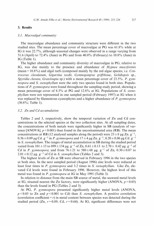
3. Results
3.1. Macroalgal community
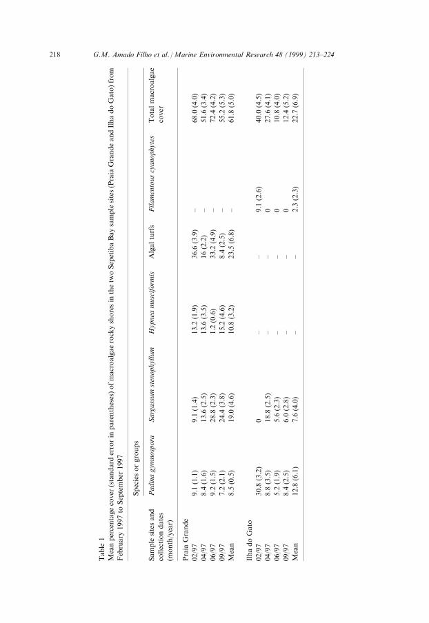
The macroalgae abundance and community structure were di�erent in the twostudied sites. The mean percentage cover of macroalgae at PG was 61.8% while atIG it was 22.7%, although seasonal changes were observed in a range varying from51.6 (April) to 72.4% (June) in PG and from 40.0% (February) to 10.8% (June) inIG (Table 1).The higher abundance and community diversity of macroalgae in PG, relative to
IG, was due mainly to the presence and abundance of Hypnea musciformis(mean=10.8%) and algal turfs (composed mainly by the red algae species, i.e. Cen-troceras clavulatum, Gigartina teedii, Gymnogongrus gri�thsiae, Gelidiopsis sp.,Spyridia clavata, Grateloupia sp.) with a mean percentage cover of 23.5%. P. gym-nospora and S. stenophyllum were the only two species found in both sites. Popula-tions of P. gymnospora were found throughout the sampling study period, showing amean percentage cover of 8.5% at PG and 12.8% at IG. Populations of S. steno-phyllum were not represented in one sampled period (February 1997) at IG when itwas replaced by ®lamentous cyanophytes and a higher abundance of P. gymnospora(30.8%; Table 1).
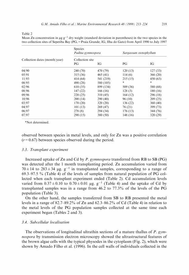
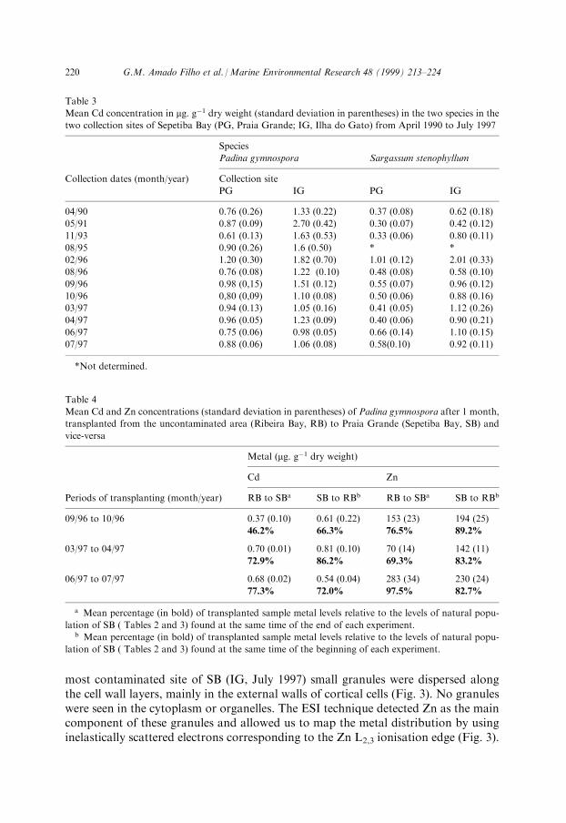
3.2. Zn and Cd accumulation
Tables 2 and 3, respectively, show the temporal variation of Zn and Cd con-centrations in the selected species at the two collection sites. At all sampling dates,the concentrations of both metals were signi®cantly higher in SB (analysis of var-iance [ANOVA], p<0.001) than found in the uncontaminated area (RB). The meanconcentrations at RB (12 analysed samples along the period) were 23�8 mg Zn. gÿ1,0.36�0.09 mg Cd. gÿ1 in P. gymnospora and 17�4 mg Zn. gÿ1, 0.20�0.06 mg Cd. gÿ1
in S. stenophyllum. The ranges of metal accumulation in SB during the studied periodvaried from 101�13 to 899�134 mg. gÿ1 of Zn, 0.61�0.13 to 2.70� 0.42 mg. gÿ1 ofCd in P. gymnospora; and from 76�21 to 580�68 mg. gÿ1 of Zn, 0.30�0.07 to2.01�0.12 mg. gÿ1 of Cd in S. stenophyllum (Tables 2 and 3).The highest levels of Zn at SB were observed in February 1996 in the two species
at both sites. In the next sampled period (August 1996) zinc levels were reduced atleast four times in P. gymnospora and 3.2 times in S. stenophyllum. Like Zn, ele-vated Cd levels were found in February 1996. However, the highest level of thismetal was found in P. gymnospora at IG in May 1991 (Table 3).In relation to distance from the main SB source of metal, the seaweed metal levels
at IG, situated nearest the Zn factory, were signi®cantly higher (ANOVA, p<0.05)than the levels found in PG (Tables 2 and 3).At PG, P. gymnospora presented signi®cantly higher metal levels (ANOVA,
p<0.05 to Zn and p<0.001 to Cd) than S. stenophyllum. A positive correlation(correlation coe�cient=r) in metal content between species was detected during thestudied period (Zn, r=0.89; Cd, r=0.64). At IG, signi®cant di�erences were not
G.M. Amado Filho et al. /Marine Environmental Research 48 (1999) 213±224 217
Table
1
Meanpercentagecover
(standard
errorin
parentheses)ofmacroalgaerockyshoresin
thetw
oSepetibaBaysamplesites(Praia
GrandeandIlhadoGato)from
February
1997to
September
1997
Speciesorgroups
Sample
sitesand
collectiondates
(month/year)
Padinagymnospora
Sargassum
stenophyllum
Hypnea
musciform
isAlgalturfs
Filamentouscyanophytes
Totalmacroalgae
cover
Praia
Grande
02/97
9.1
(1.1)
9.1
(1.4)
13.2
(1.9)
36.6
(3.9)
±68.0
(4.0)
04/97
8.4
(1.6)
13.6
(2.5)
13.6
(3.5)
16(2.2)
±51.6
(3.4)
06/97
9.2
(1.5)
28.8
(2.3)
1.2
(0.6)
33.2
(4.9)
±72.4
(4.2)
09/97
7.2
(2.1)
24.4
(3.8)
15.2
(4.6)
8.4
(2.5)
±55.2
(5.3)
Mean
8.5
(0.5)
19.0
(4.6)
10.8
(3.2)
23.5
(6.8)
±61.8
(5.0)
IlhadoGato
02/97
30.8
(3.2)
0±
±9.1
(2.6)
40.0
(4.5)
04/97
8.8
(3.5)
18.8
(2.5)
±±
027.6
(4.1)
06/97
5.2
(1.9)
5.6
(2.3)
±±
010.8
(4.0)
09/97
8.4
(2.5)
6.0
(2.8)
±±
012.4
(5.2)
Mean
12.8
(6.1)
7.6
(4.0)
±±
2.3
(2.3)
22.7
(6.9)
218 G.M. Amado Filho et al. /Marine Environmental Research 48 (1999) 213±224
observed between species in metal levels, and only for Zn was a positive correlation(r=0.67) between species observed during the period.
3.3. Transplant experiment
Increased uptake of Zn and Cd by P. gymnospora transferred from RB to SB (PG)was detected after the 1 month transplanting period. Zn accumulation varied from70�14 to 283�34 mg. gÿ1 in transplanted samples, corresponding to a range of69.3±97.5 % (Table 4) of the levels of samples from natural population of PG col-lected when each transplant experiment ended (Table 2). Cd accumulation levelsvaried from 0.37�0.10 to 0.70�0.01 mg. gÿ1 (Table 4) and the uptake of Cd bytransplanted samples was in a range from 46.2 to 77.3% of the levels of the PGpopulation (Table 3).On the other hand, the samples transferred from SB to RB presented the metal
levels in a range of 82.7±89.2% of Zn and 62.3±86.2% of Cd (Table 4) in relation tothe metal levels of the PG population samples collected at the same time eachexperiment began (Tables 2 and 3).
3.4. Subcellular localisation
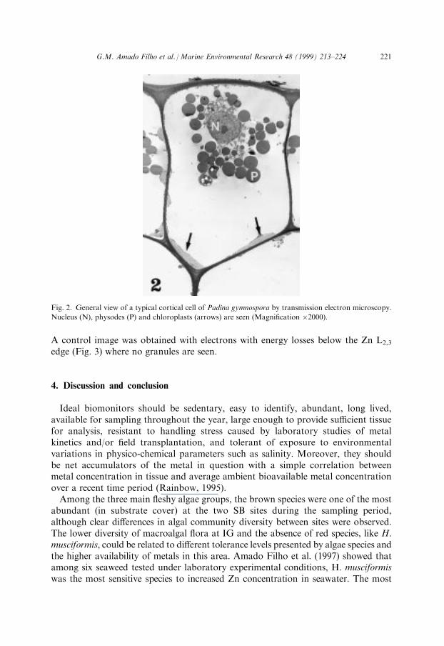
The observations of longitudinal ultrathin sections of a mature thallus of P. gym-nospora by transmission electron microscopy showed the ultrastructural features ofthe brown algae cells with the typical physodes in the cytoplasm (Fig. 2), which wereshown by Amado Filho et al. (1996). In the cell walls of individuals collected in the
Table 2
Mean Zn concentration in mg gÿ1 dry weight (standard deviation in parentheses) in the two species in the
two collection sites of Sepetiba Bay (PG,=Praia Grande; IG, Ilha do Gato) from April 1990 to July 1997
Species
Padina gymnospora Sargassum stenophyllum
Collection dates (month/year) Collection site
PG IG PG IG
04/90 240 (70) 470 (79) 120 (13) 127 (33)
05/91 315 (36) 465 (41) 114 (6) 366 (20)
11/93 414 (64) 541 (219) 215 (15) 450 (63)
08/95 490 (28) 580 (105) * *
02/96 610 (35) 899 (134) 509 (36) 580 (68)
08/96 147 (22) 166 (16) 128 (3) 180 (16)
09/96 220 (25) 310 (45) 164 (12) 296 (18)
10/96 200 (14) 290 (40) 96 (10) 209 (25)
03/97 170 (20) 320 (20) 136 (22) 360 (40)
04/97 101 (13) 269 (47) 76 (21) 399 (73)
06/97 278 (8) 294 (34) 176 (13) 344 (76)
07/97 290 (15) 380 (50) 148 (16) 320 (29)
*Not determined.
G.M. Amado Filho et al. /Marine Environmental Research 48 (1999) 213±224 219
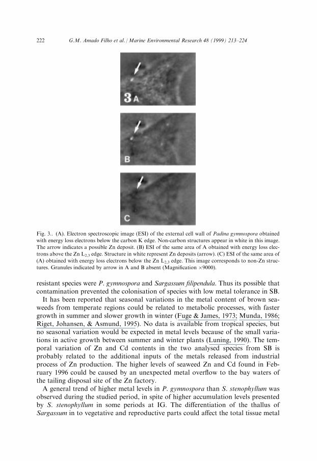
most contaminated site of SB (IG, July 1997) small granules were dispersed alongthe cell wall layers, mainly in the external walls of cortical cells (Fig. 3). No granuleswere seen in the cytoplasm or organelles. The ESI technique detected Zn as the maincomponent of these granules and allowed us to map the metal distribution by usinginelastically scattered electrons corresponding to the Zn L2,3 ionisation edge (Fig. 3).
Table 3
Mean Cd concentration in mg. gÿ1 dry weight (standard deviation in parentheses) in the two species in the
two collection sites of Sepetiba Bay (PG, Praia Grande; IG, Ilha do Gato) from April 1990 to July 1997
Species
Padina gymnospora Sargassum stenophyllum
Collection dates (month/year) Collection site
PG IG PG IG
04/90 0.76 (0.26) 1.33 (0.22) 0.37 (0.08) 0.62 (0.18)
05/91 0.87 (0.09) 2.70 (0.42) 0.30 (0.07) 0.42 (0.12)
11/93 0.61 (0.13) 1.63 (0.53) 0.33 (0.06) 0.80 (0.11)
08/95 0.90 (0.26) 1.6 (0.50) * *
02/96 1.20 (0.30) 1.82 (0.70) 1.01 (0.12) 2.01 (0.33)
08/96 0.76 (0.08) 1.22 (0.10) 0.48 (0.08) 0.58 (0.10)
09/96 0.98 (0,15) 1.51 (0.12) 0.55 (0.07) 0.96 (0.12)
10/96 0,80 (0,09) 1.10 (0.08) 0.50 (0.06) 0.88 (0.16)
03/97 0.94 (0.13) 1.05 (0.16) 0.41 (0.05) 1.12 (0.26)
04/97 0.96 (0.05) 1.23 (0.09) 0.40 (0.06) 0.90 (0.21)
06/97 0.75 (0.06) 0.98 (0.05) 0.66 (0.14) 1.10 (0.15)
07/97 0.88 (0.06) 1.06 (0.08) 0.58(0.10) 0.92 (0.11)
*Not determined.
Table 4
Mean Cd and Zn concentrations (standard deviation in parentheses) of Padina gymnospora after 1 month,
transplanted from the uncontaminated area (Ribeira Bay, RB) to Praia Grande (Sepetiba Bay, SB) and
vice-versa
Metal (mg. gÿ1 dry weight)
Cd Zn
Periods of transplanting (month/year) RB to SBa SB to RBb RB to SBa SB to RBb
09/96 to 10/96 0.37 (0.10) 0.61 (0.22) 153 (23) 194 (25)
46.2% 66.3% 76.5% 89.2%
03/97 to 04/97 0.70 (0.01) 0.81 (0.10) 70 (14) 142 (11)
72.9% 86.2% 69.3% 83.2%
06/97 to 07/97 0.68 (0.02) 0.54 (0.04) 283 (34) 230 (24)
77.3% 72.0% 97.5% 82.7%
a Mean percentage (in bold) of transplanted sample metal levels relative to the levels of natural popu-
lation of SB ( Tables 2 and 3) found at the same time of the end of each experiment.b Mean percentage (in bold) of transplanted sample metal levels relative to the levels of natural popu-
lation of SB ( Tables 2 and 3) found at the same time of the beginning of each experiment.
220 G.M. Amado Filho et al. /Marine Environmental Research 48 (1999) 213±224
A control image was obtained with electrons with energy losses below the Zn L2,3
edge (Fig. 3) where no granules are seen.
4. Discussion and conclusion
Ideal biomonitors should be sedentary, easy to identify, abundant, long lived,available for sampling throughout the year, large enough to provide su�cient tissuefor analysis, resistant to handling stress caused by laboratory studies of metalkinetics and/or ®eld transplantation, and tolerant of exposure to environmentalvariations in physico-chemical parameters such as salinity. Moreover, they shouldbe net accumulators of the metal in question with a simple correlation betweenmetal concentration in tissue and average ambient bioavailable metal concentrationover a recent time period (Rainbow, 1995).Among the three main ¯eshy algae groups, the brown species were one of the most
abundant (in substrate cover) at the two SB sites during the sampling period,although clear di�erences in algal community diversity between sites were observed.The lower diversity of macroalgal ¯ora at IG and the absence of red species, like H.musciformis, could be related to di�erent tolerance levels presented by algae species andthe higher availability of metals in this area. Amado Filho et al. (1997) showed thatamong six seaweed tested under laboratory experimental conditions, H. musciformiswas the most sensitive species to increased Zn concentration in seawater. The most
Fig. 2. General view of a typical cortical cell of Padina gymnospora by transmission electron microscopy.
Nucleus (N), physodes (P) and chloroplasts (arrows) are seen (Magni®cation �2000).
G.M. Amado Filho et al. /Marine Environmental Research 48 (1999) 213±224 221
resistant species were P. gymnospora and Sargassum ®lipendula. Thus its possible thatcontamination prevented the colonisation of species with low metal tolerance in SB.It has been reported that seasonal variations in the metal content of brown sea-
weeds from temperate regions could be related to metabolic processes, with fastergrowth in summer and slower growth in winter (Fuge & James, 1973; Munda, 1986;Riget, Johansen, & Asmund, 1995). No data is available from tropical species, butno seasonal variation would be expected in metal levels because of the small varia-tions in active growth between summer and winter plants (Luning, 1990). The tem-poral variation of Zn and Cd contents in the two analysed species from SB isprobably related to the additional inputs of the metals released from industrialprocess of Zn production. The higher levels of seaweed Zn and Cd found in Feb-ruary 1996 could be caused by an unexpected metal over¯ow to the bay waters ofthe tailing disposal site of the Zn factory.A general trend of higher metal levels in P. gymnospora than S. stenophyllum was
observed during the studied period, in spite of higher accumulation levels presentedby S. stenophyllum in some periods at IG. The di�erentiation of the thallus ofSargassum in to vegetative and reproductive parts could a�ect the total tissue metal
Fig. 3.. (A). Electron spectroscopic image (ESI) of the external cell wall of Padina gymnospora obtained
with energy loss electrons below the carbon K edge. Non-carbon structures appear in white in this image.
The arrow indicates a possible Zn deposit. (B) ESI of the same area of A obtained with energy loss elec-
trons above the Zn L2,3 edge. Structure in white represent Zn deposits (arrow). (C) ESI of the same area of
(A) obtained with energy loss electrons below the Zn L2,3 edge. This image corresponds to non-Zn struc-
tures. Granules indicated by arrow in A and B absent (Magni®cation �9000).
222 G.M. Amado Filho et al. /Marine Environmental Research 48 (1999) 213±224
levels. Munda (1986) showed in Fucus spiralis (Fucales, same taxonomic order ofSargassum) that heavy metal accumulation is lower in receptacles (reproductivestructures) than in vegetative parts of the thalli and, hence, responsible for dimin-ished metal accumulation in fucoids. The relative proportion of vegetative andreproductive parts biomass of S. stenophyllum could have contributed to the varia-tion in metal levels, especially in the most contaminated site (IG) of SB.The results of the transplant experiments show that P. gymnospora is able to
accumulate the studied metals in a short period of time (1 month) re¯ecting theambient bioavailability. The changes in metal level observed among transplantedperiods could be related to the variation of available metals in seawater since, espe-cially in the case of Zn, the metal levels in plants transplanted to SB followed themetal levels variation found in natural population. Eide et al. (1980), in a similartransplant experiment with the temperate brown alga Ascophyllum nodusum, con-cluded that a decrease or increase of heavy metal concentration in the water will give ade®nitive change in the heavy metal accumulation pattern of the plants. The release ofmetals by brown algae has been frequently associated with exudation of metal chelatedto polyphenolic compounds known to be present in this algal group (Eide et al.,1980; Ragan & Glombitza, 1986). The reduction in metal levels observed in P.gymnospora transplanted to the uncontaminated area could be related, not to a tissuemetal loss (or release) to seawater, but to a dilution of metal tissue concentration asso-ciated with the 1-month growth in a seawater free of higher metal levels. This postula-tion is due to the fact that the metals in P. gymnospora were not detected throughanalytical electronmicroscopy in physodes (Amado Filho et al., 1996, and present study).The localisation of Zn in cell walls of individuals from SB, in addition to previous
in-vitro results (Amado Filho et al. 1996), con®rm that cell walls play a main role inmetal accumulation in the case of P. gymnospora. Although Cd was not detected inSB individuals, recent in-vitro experiments performed with P. gymnospora submittedto high Cd concentration in seawater (Andrade, 1998), suggested that cell walls alsoplay the main role in Cd accumulation.
Acknowledgements
Dr. Wanderley de Souza of the Laborato rio de Ultraestrutura Celular HerthaMeyer, IBCCF/UFRJ, kindly provided the electron microscopes for the ultra-structural observation and analytical electron microscopy. This work was supportedby Instituto de Pesquisas Jardim Botaà nico/MMA, PRONEX/MCT, and a researchgrant of Conselho Nacional de Desenvolvimento Cienti ®co e Tecnolo gicoÐCNPqNo. 521688/96-5 OcÐto G.M. Amado Filho.
References
Amado Filho, G. M., Karez, C. S., Pfei�er, W. C., Yoneshigue-Valentin, Y., & Farina, M. (1996). E�ects
on growth, accumulation, and ultrastructural localization of zinc in Padina gymnospora (Phaeophy-
ceae). Hydrobiologia, 326/327, 451±456.
G.M. Amado Filho et al. /Marine Environmental Research 48 (1999) 213±224 223
Amado Filho, G. M., Karez, C. S., Andrade, L. R., Yoneshigue-Valentin, Y., & Pfei�er, W. C. (1997).E�ects on growth and accumulation of zinc in six seaweed species. Ecotoxicology and EnvironmentalSafety, 37, 223±228.
Andrade, L. R. (1998). Aspects ultraestrutursis da parede celular da alga porda Podina qumnosporn e suarelacao com a oeumulacao de metais pesados. Master Thesis, Universidade Federal do Rio de Janeiro,p. 150.
Barcellos, C. (1995). GeodinaÃmica de CaÂdmio e Zinco na Baia de Sepetiba. PhD thesis, Universidade Fed-eral Fluminense, Niteroi, Brazil.
Barcellos, C., & Lacerda, L. D. (1994). Cadmium and zinc source assessment in the Sepetiba Bay andbasin region. Environ. Monitor. and Assess, 29(2), 183±199.
Bauer, R. (1988). Electron spectroscopic imaging: an advanced technique for imaging and analysis intransmission electron microscopy. Methods in Microbiol., 20, 113±146.
Crist, R. H., Oberholser, K., McGarrity, J., Crist, D. R., Johnson, J. K., & Brittsan, J. M. (1992). Inter-action of metals and protons with algae. 3. Marine algae, with emphasis on lead and aluminium.Environ. Sci. Technol., 26, 496±502.
Eide, I., Myklestad, S., & Melson, S. (1980). Long-term uptake and release of heavy metals by Asco-phyllum nodosum (L.) Le Jol. (Phaeophyceae) in situ. Environ. Pollut., 23, 19±28.
Fuge, R., & James, K. H. (1973). Trace metal concentration in brown seaweeds, Cardigan Bay, Wales.Mar. Chem., 1, 281±293.
Karez, C. S., & Pereira, R. C. (1995). Metal content in polyphenolic fractions extracted from the brownalga Padina gymnospora. Bot. Mar., 38, 151±155.
Karez, C. S., MagalhaÄ es, V. F., Pfei�er, W. C., & Amado Filho, G. M. (1994). Trace metal accumulationby algae in Sepetiba Bay, Brazil. Environ. Pollut., 83, 351±356 .
Kurita, M. H., & Pfei�er, W. C. (1991). Heavy metals in sediments and biota of Sepetiba Bay, Rio deJaneiro-Brazil. Proceeding of Heavy metals in the environment (pp. 519±522). Edinburgh, UK.
Lacerda, L. D., & Rezende, C. E. (1986). Metal in the seagrass Halodule wrightii Aschers during onegrowing season. Rev. bras. Bot., 9, 87±90.
Lacerda, L. D., Pfei�er, W. C., & Fizman, M. (1985). Intertidal beach sands as monitors for heavy metalpollution in coastal bodies. Environm. Technol. Lett., 6, 123±128.
Lacerda, L. D., Pfei�er, W. C., & Fizman, M. (1987). Heavy metal distribution, availability and fate inSepetiba Bay, SE Brazil. Sci. Total Environ., 65, 163±173.
Lacerda, L. D., Carvalho, C. E. V., & Gomes, M. P. (1989). Nota sobre a distribuisË aÄ o de Mn, Zn e Cu emsiris da bai a de Sepetiba. Rev. Bras. Biol., 49( 3), 847±849.
Lignell, A., Roomans, G. M., & Pedersen, M. (1982). Localization of adsorbed cadmium in Fucus vesi-culosus L. by X-ray microanalysis. Z. P¯anzenphysiol. Bd., 105, 103±109.
Lima, N. R. W., Lacerda, L. D., Pfei�er, W. C., & Fiszman, M. (1986). Temporal and spatial variabilityin Zn, Cr, Cd and Fe concentrations in oyster tissues (Crassostrea brasiliana L.) from Sepetiba Bay,Brazil. Environ. Technol. Lett., 7, 453±460.
Luning, K. (1990). Seaweeds, their environment, biogeography and ecophysiology. New York: John Wiley.Mariani, P., Tolomio, C., & Braghetta, P. (1985). An ultrastructural approach to the adaptive role of the
cell wall in the intertidal alga Fucus virsoides. Protoplasma, 128, 208±217.Munda, I. M. (1986). Di�erences in heavy metals accumulation between vegetative parts of thalli and
receptacles in Fucus spiralis. L. Bot. Mar., 29, 341±349.Pfei�er, W. C., Lacerda, L. D., Fizman, M., & Lima, R. W. (1985). Metais pesados no pescado da bai a de
Sepetiba. CieÃncia e Cultura, 37, 297±302.Ragan, M. A., & Glombitza, K. W. (1986). Phlorotannins, brown algal polyphenols. In Progress in
phycological research, Round & Chapman (pp. 129±241). Bristol: Biopress.Ragan, M. A., Smidsrod, O., & Larsen, B. Eds. (1979). Chelation of divalent metal ions by brown algal
polyphenols. Mar. Chem., 7, 265±271.Rainbow, P. (1995). Biomonitoring of heavy metal availability in the marine environment. Mar. Pollut.
Bull., 31(4±12), 183±192.Rezende, C. E., Lacerda, L. D., & Pfei�er, W. C. (1991). Evolution of heavy metal contamination (1980±
1989) of Sepetiba Bay determined by using beach sands as monitors. CieÃncia e Cultura, 43(1), 61±63.Riget, F., Johansen, P., & Asmund, G. (1995). Natural seasonal variation of cadmium, copper, lead and
zinc in brown seaweed (Fucus vesiculosus). Mar. Pollut. Bull., 30(6), 409±413.Volesky, B., & Holan, Z. H. (1995). Biosorption of heavy metals. Biotechnol. Prog., 11, 235±250.
224 G.M. Amado Filho et al. /Marine Environmental Research 48 (1999) 213±224