Asymmetric Cell Divisions: Zygotes of Fucoid Algae as a ...
19
Plant Cell Monogr (9) D.P.S. Verma and Z. Hong: Cell Division Control in Plants DOI 10.1007/7089_2007_134/Published online: 21 August 2007 © Springer-Verlag Berlin Heidelberg 2007 Asymmetric Cell Divisions: Zygotes of Fucoid Algae as a Model System Sherryl R. Bisgrove 1 (✉) · Darryl L. Kropf 2 1 Department of Biological Sciences, Simon Fraser University, 8888 University Drive, Burnaby, BC V5A 1S6, Canada [email protected] 2 Department of Biology, University of Utah, 257 South 1400 East, Salt Lake City, UT 84112, USA Abstract Asymmetric cell divisions are commonly used across diverse phyla to generate different kinds of cells during development. Although asymmetric divisions play import- ant roles during development in plants, algae, fungi, and animals, emerging data indicate that there is some variability amongst the mechanisms that are at play in these differ- ent organisms. Zygotes of fucoid algae have long served as models for understanding early developmental processes including cell polarization and asymmetric cell division. In addition, brown algae are phylogenetically distant from other organisms, including plant models, a feature that makes them interesting from a comparative perspective (Andersen 2004; Peters et al. 2004). This monograph focuses on advances made toward under- standing how asymmetric divisions are regulated in fucoid algae and, where appropriate, comparisons are made to higher plant zygotes. 1 Introduction How does a single cell, the zygote, give rise to a complex organism with many different cell and tissue types? The answer to this question lies in the abil- ity of cells in a growing embryo to acquire separate identities, a feat that is often accomplished by asymmetric cell divisions. By definition, asymmetric cell divisions produce nonidentical daughter cells and can thereby initiate the process of cell differentiation. Asymmetric cell divisions are known to play important roles in development across diverse plant and algal phyla. Exam- ples include the first cell division in many zygotes (Brownlee 2004; Gallagher and Smith 1997; Okamoto et al. 2005; Zernicka-Goetz 2004), as well as the production of gonidial and somatic cells in Volvox carteri (Kirk 2004; Schmitt 2003), reproductive initial cells from caulonema filaments in moss (Cove et al. 2006; Schumaker and Dietrich 1998), rhizoids from prothalli cells in ferns (Murata and Sugai 2000), stomata on the epidermal surfaces of leaves (Lucas et al. 2006; Nadeau and Sack 2002, 2003), and microspores during pollen de- velopment (Park et al. 2004; Twell et al. 1998). Because of the importance of asymmetric divisions in development, the mechanisms that regulate the pro-
-
Upload
khangminh22 -
Category
Documents
-
view
5 -
download
0
Transcript of Asymmetric Cell Divisions: Zygotes of Fucoid Algae as a ...
Plant Cell Monogr (9)D.P.S. Verma and Z. Hong: Cell Division Control in PlantsDOI 10.1007/7089_2007_134/Published online: 21 August 2007© Springer-Verlag Berlin Heidelberg 2007
Asymmetric Cell Divisions:Zygotes of Fucoid Algae as a Model System
Sherryl R. Bisgrove1 (�) · Darryl L. Kropf 2
1Department of Biological Sciences, Simon Fraser University,8888 University Drive, Burnaby, BC V5A 1S6, [email protected]
2Department of Biology, University of Utah, 257 South 1400 East,Salt Lake City, UT 84112, USA
Abstract Asymmetric cell divisions are commonly used across diverse phyla to generatedifferent kinds of cells during development. Although asymmetric divisions play import-ant roles during development in plants, algae, fungi, and animals, emerging data indicatethat there is some variability amongst the mechanisms that are at play in these differ-ent organisms. Zygotes of fucoid algae have long served as models for understandingearly developmental processes including cell polarization and asymmetric cell division. Inaddition, brown algae are phylogenetically distant from other organisms, including plantmodels, a feature that makes them interesting from a comparative perspective (Andersen2004; Peters et al. 2004). This monograph focuses on advances made toward under-standing how asymmetric divisions are regulated in fucoid algae and, where appropriate,comparisons are made to higher plant zygotes.
1Introduction
How does a single cell, the zygote, give rise to a complex organism with manydifferent cell and tissue types? The answer to this question lies in the abil-ity of cells in a growing embryo to acquire separate identities, a feat that isoften accomplished by asymmetric cell divisions. By definition, asymmetriccell divisions produce nonidentical daughter cells and can thereby initiate theprocess of cell differentiation. Asymmetric cell divisions are known to playimportant roles in development across diverse plant and algal phyla. Exam-ples include the first cell division in many zygotes (Brownlee 2004; Gallagherand Smith 1997; Okamoto et al. 2005; Zernicka-Goetz 2004), as well as theproduction of gonidial and somatic cells in Volvox carteri (Kirk 2004; Schmitt2003), reproductive initial cells from caulonema filaments in moss (Cove et al.2006; Schumaker and Dietrich 1998), rhizoids from prothalli cells in ferns(Murata and Sugai 2000), stomata on the epidermal surfaces of leaves (Lucaset al. 2006; Nadeau and Sack 2002, 2003), and microspores during pollen de-velopment (Park et al. 2004; Twell et al. 1998). Because of the importance ofasymmetric divisions in development, the mechanisms that regulate the pro-
324 S.R. Bisgrove · D.L. Kropf
cess are under investigation in several model organisms. In this monograph,we focus on advances made toward understanding how asymmetric divisionsare regulated in zygotes of fucoid brown algae.
1.1Asymmetric Divisions and Cell Fate Decisions
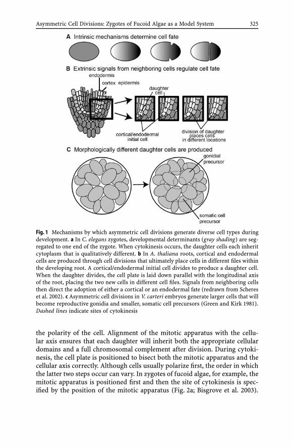
Generally, there are three ways by which the products of an asymmetric divi-sion acquire separate identities (Fig. 1):
1. Developmental determinants can be differentially partitioned betweencells during division. In this case, each cell inherits a different set ofcytoplasmic instructions that lead it down a unique developmental path-way. Because cell fate is controlled by determinants located within thecytoplasm, this type of development is often referred to as intrinsic orcell-autonomous (Fig. 1a). Both the first division of the Caenorhabditis el-egans zygote and the divisions of neuroblasts in Drosophila melanogasterembryos represent examples of asymmetric divisions in which intrinsicfactors control daughter cell fates (Betschinger and Knoblich 2004; Cowanand Hyman 2004).
2. In some cases, the cytokinetic plane is positioned such that the daugh-ter cells are placed in different locations within the developing organism.Each cell then receives a unique set of positional cues from neighboringcells or the environment that dictate its fate (Fig. 1b). Since cell identi-ties are determined by signals received from external sources, this typeof development is known as extrinsic or non-cell-autonomous. In theArabidopsis thaliana root, for example, the decision to become either anendodermal or a cortical cell depends on an asymmetric cell divisionthat places daughter cells in different cell files in the root. Signals fromneighboring cells then direct the daughters down different developmentalpathways (Heidstra et al. 2004; Scheres et al. 2002).
3. An asymmetric division can produce daughters of different sizes and/orshapes, and these morphological differences determine the developmen-tal pathway that each cell will follow (Fig. 1c). In V. carteri, asymmetricdivisions generate small and large daughter cell pairs, and the size of thecell then activates either a somatic or a germline developmental program(Cheng et al. 2005; Kirk et al. 1993; Schmitt 2003).
Asymmetric divisions are commonly regulated in a three-step process. In thefirst step, cells polarize (Fig. 2). Sometimes there are obvious cytological ormorphological changes associated with cell polarization while in other casesthe polarity is more subtle, and may be manifested simply by the fact that theends of the cell lie in different positions in the developing organism. Aftercell polarization, the mitotic apparatus (step 2) and the site of cytokinesis(step 3) must be positioned appropriately with respect to the axis defined by
Asymmetric Cell Divisions: Zygotes of Fucoid Algae as a Model System 325
Fig. 1 Mechanisms by which asymmetric cell divisions generate diverse cell types duringdevelopment. a In C. elegans zygotes, developmental determinants (gray shading) are seg-regated to one end of the zygote. When cytokinesis occurs, the daughter cells each inheritcytoplasm that is qualitatively different. b In A. thaliana roots, cortical and endodermalcells are produced through cell divisions that ultimately place cells in different files withinthe developing root. A cortical/endodermal initial cell divides to produce a daughter cell.When the daughter divides, the cell plate is laid down parallel with the longitudinal axisof the root, placing the two new cells in different cell files. Signals from neighboring cellsthen direct the adoption of either a cortical or an endodermal fate (redrawn from Schereset al. 2002). c Asymmetric cell divisions in V. carteri embryos generate larger cells that willbecome reproductive gonidia and smaller, somatic cell precursors (Green and Kirk 1981).Dashed lines indicate sites of cytokinesis
the polarity of the cell. Alignment of the mitotic apparatus with the cellu-lar axis ensures that each daughter will inherit both the appropriate cellulardomains and a full chromosomal complement after division. During cytoki-nesis, the cell plate is positioned to bisect both the mitotic apparatus and thecellular axis correctly. Although cells usually polarize first, the order in whichthe latter two steps occur can vary. In zygotes of fucoid algae, for example, themitotic apparatus is positioned first and then the site of cytokinesis is spec-ified by the position of the mitotic apparatus (Fig. 2a; Bisgrove et al. 2003).

326 S.R. Bisgrove · D.L. Kropf
Fig. 2 Asymmetric cell divisions are commonly regulated in three steps. a Silvetia com-pressa eggs are spherical in shape with no obvious asymmetries, and polarization (I) isfirst manifested morphologically several hours after fertilization when increased secre-tion on one hemisphere produces a bulge, the rhizoid. The opposite end of the zygote istermed the thallus, and the axis defined by the two poles is the rhizoid/thallus axis. Next,the mitotic apparatus aligns parallel with the rhizoid/thallus axis (II). Finally, cytokinesisoccurs and the cell plate is positioned perpendicular to the rhizoid/thallus axis (III). Thethree zygotes shown in the panels corresponding to I and III were stained with fluores-cein diacetate which labels the cell plate, perinuclear regions, and cytoplasm. The zygotein II is in metaphase and was labeled with anti-alpha tubulin antibodies (image kindlyprovided by Nick T. Peters). b Asymmetric divisions in A. thaliana zygotes are also regu-lated in a three-step process, but the order in which the steps occur is different than it isin fucoid algae. In plants, polarity is acquired by the egg cell during development of theembryo sac (I). After fertilization, a preprophase band of microtubules marks the positionof the first zygotic division (II) and then the mitotic apparatus is positioned with respectto both the cellular axis and the predetermined division site (Webb and Gunning 1991).Drawn using Drews and Yadegari (2002), Mayer et al. (1993), and Webb and Gunning(1991) as guides
In this case, proper placement of the spindle is required for correct speci-fication of the cytokinetic site. Alternatively, the site of cytokinesis can bespecified prior to mitosis in accordance with cues located in the cortex of thecell (Fig. 2b). Because the site of cytokinesis is determined before mitosis, thismechanism does not require precise positioning of the spindle. Instead, themitotic apparatus needs only to align well enough to ensure that each daugh-

Asymmetric Cell Divisions: Zygotes of Fucoid Algae as a Model System 327
ter cell inherits a nucleus after telophase. This method is commonly employedby plant somatic cells, including zygotes. In these cells, a preprophase band ofmicrotubules transiently forms in the cell cortex and marks the upcoming di-vision site (Brown and Lemmon 2001; Marcus et al. 2005; Webb and Gunning1991).
2Zygotes of Fucoid Algae as a Model System
Zygotes of fucoid algae have, for many years, been a fruitful system in whichto study the mechanisms by which cells acquire polarity and regulate asym-metric cell divisions, mainly because they are easy to manipulate and analyzein the laboratory (for recent reviews, see Brownlee 2004; Katsaros et al. 2006).Fucoid algae are marine brown algae, belonging to the Phaeophyceae classof stramenopiles (Andersen 2004). In nature they grow attached to rocks inthe intertidal zone where they reproduce by releasing large, spherical eggsand biflagellated, motile sperm into the surrounding seawater. Gamete releasecan be induced from reproductive fronds in the lab and thousands of syn-chronously developing zygotes are easy to obtain for experimental analyses.The zygotes are relatively large, up to 100 µm in diameter, a size that rendersthem amenable to micromanipulation and analyses that require spatial meas-urements of subcellular features. Soon after fertilization zygotes settle ontothe substratum, a rock in the intertidal zone, or a coverslip in the lab, anda sticky adhesive is secreted that firmly anchors them in place. Eggs are spher-ically shaped cells with no detectable asymmetries. However, within the firstfew hours following fertilization there are extensive cytoplasmic and morpho-logical changes that result in asymmetric cells with rhizoid and thallus poles(Fig. 2a). To establish polarity, zygotes sense a wide array of environmentalcues, although light is probably the dominant signal in nature. Zygotes devel-oping in unidirectional light form rhizoids on their shaded hemispheres. Anearly sign of polarity is the preferential localization of secretion to the rhi-zoid pole, and increased secretion at this pole eventually produces a bulge,the tip-growing rhizoid (Fig. 2). When the first division occurs, about 24 hafter fertilization (AF), it is oriented transverse to the rhizoid/thallus axis andbisects the zygote into two morphologically distinct cells with different de-velopmental fates. The thallus cell gives rise to most of the photosyntheticand reproductive organs of the mature alga, while the rhizoid cell eventuallybecomes the holdfast that anchors the alga to its rock on the beach.
The first zygotic division in higher plants is also an asymmetric one thatproduces two morphologically distinct daughter cells with different develop-mental fates (Fig. 2b). The smaller apical cell is cytoplasmically dense and itsprogeny give rise to most of the developing embryo, while the larger, vacuo-late basal cell divides only a few more times to form a single file of cells. The

328 S.R. Bisgrove · D.L. Kropf
uppermost cell in this file becomes part of the root meristem and the remain-ing cells form the suspensor, a structure that attaches the embryo to the ovule(Laux et al. 2004; Souter and Lindsey 2000; Torres-Ruiz 2004). Although thedevelopmental pattern that is set up by the first zygotic cell division is similarin plants and fucoid algae, there are key mechanistic differences between thetwo. In many plants, for example, polarity arises in the egg prior to fertiliza-tion rather than in the zygote. Plant eggs and zygotes are also buried withinthe ovule where their development can be influenced by surrounding mater-nal tissues. Zygotes of fucoid algae, on the other hand, are free-living andthey develop in response to vectorial information in the environment suchas sunlight from above (Brownlee 2004). Because plant eggs and zygotes arerelatively inaccessible, approaches that involve manipulating individual cellsare difficult. Instead, molecular/genetic analyses of mutants are being used toaddress questions of cell polarity and asymmetric divisions. This research isyielding interesting data, but our understanding of how asymmetric divisionsare regulated in plant zygotes is still rudimentary. In contrast, the free-livingzygotes of fucoid algae are easy to access and are amenable to physical ma-nipulations. Over the years research on fucoid algae has provided a wealthof mechanistic data and, although many questions still remain, we are be-ginning to understand how asymmetric cell divisions are regulated in thesezygotes.
3Polarization and Germination in Zygotes
Fucoid zygotes have long served as a paradigm for investigating the mech-anisms by which polarity is established following fertilization. In 1920, Hurdreported that monochromatic blue light polarizes zygotes (Hurd 1920), andsince that time many other vectorial cues, including electrical, ionic, andosmotic gradients, have been shown to induce a growth axis (for a re-view, see Jaffe 1969). These diverse stimuli likely activate distinct signaltransduction pathways that converge at a common response, formation ofa growth axis (Kropf et al. 1999). The presumed goal is to maximize thechance that the rhizoid will grow into a crevice on the rocky surface andthereby permanently anchor the developing embryo in the turbulent inter-tidal environment.
But when is polarity first set up? Is the fertilized egg apolar until it sensesits environment? Recent work has shown that in fact polarity is first set up atfertilization (Hable and Kropf 2000). Sperm entry induces a rhizoid pole toform at that site and a branching actin network rapidly assembles in the cellcortex there (Fig. 3a). A zygote has a greater density than seawater and set-tles rapidly onto the rocky substratum with its sperm-induced rhizoid polerandomly oriented with respect to the surface. Over the next 2 h the sperm
Asymmetric Cell Divisions: Zygotes of Fucoid Algae as a Model System 329
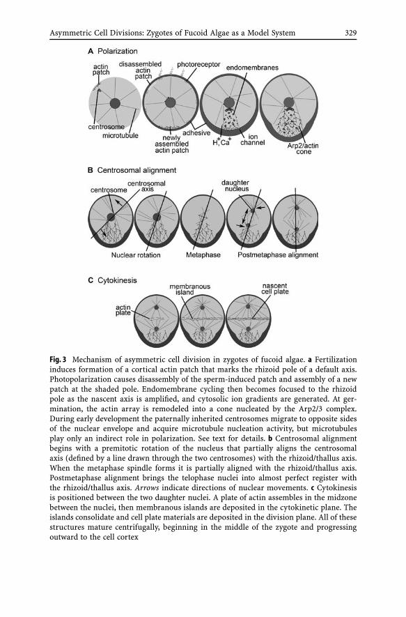
Fig. 3 Mechanism of asymmetric cell division in zygotes of fucoid algae. a Fertilizationinduces formation of a cortical actin patch that marks the rhizoid pole of a default axis.Photopolarization causes disassembly of the sperm-induced patch and assembly of a newpatch at the shaded pole. Endomembrane cycling then becomes focused to the rhizoidpole as the nascent axis is amplified, and cytosolic ion gradients are generated. At ger-mination, the actin array is remodeled into a cone nucleated by the Arp2/3 complex.During early development the paternally inherited centrosomes migrate to opposite sidesof the nuclear envelope and acquire microtubule nucleation activity, but microtubulesplay only an indirect role in polarization. See text for details. b Centrosomal alignmentbegins with a premitotic rotation of the nucleus that partially aligns the centrosomalaxis (defined by a line drawn through the two centrosomes) with the rhizoid/thallus axis.When the metaphase spindle forms it is partially aligned with the rhizoid/thallus axis.Postmetaphase alignment brings the telophase nuclei into almost perfect register withthe rhizoid/thallus axis. Arrows indicate directions of nuclear movements. c Cytokinesisis positioned between the two daughter nuclei. A plate of actin assembles in the midzonebetween the nuclei, then membranous islands are deposited in the cytokinetic plane. Theislands consolidate and cell plate materials are deposited in the division plane. All of thesestructures mature centrifugally, beginning in the middle of the zygote and progressingoutward to the cell cortex

330 S.R. Bisgrove · D.L. Kropf
pronucleus migrates to the egg pronucleus utilizing microtubules (Swope andKropf 1993), and the zygote secretes a cell wall (Quatrano 1982) and an adhe-sive that attaches it firmly to the rock (Hable and Kropf 1998). Once attached,the young zygote monitors its environment for positional information. Per-ceived environmental cues are integrated and used to specify a new growthaxis that is appropriate for the environmental context. Under normal growthconditions the sperm-induced axis is usually overridden by environmentalcues, and it can therefore be considered a default axis to be used only if thezygote fails to perceive positional information.
Unidirectional light is probably the most relevant vector in the intertidalenvironment, and is easy to apply in a laboratory setting. Photopolariza-tion induces a new rhizoid pole on the shaded hemisphere (Fig. 3a), towardthe rocky substratum. Although zygotes can perceive different light quali-ties, blue light is most effective. The photoreceptor is thought to reside at ornear the plasma membrane (Jaffe 1958), and may be a rhodopsin-like protein(Gualtieri and Robinson 2002; Robinson et al. 1998). How light perceptionon one hemisphere of the zygote is transduced into a rhizoid pole on theopposite hemisphere is not well understood, but may involve formation ofcGMP gradients resulting from differential photoreceptor activation (Robin-son and Miller 1997) and/or activation of a plasma membrane redox chain onthe shaded hemisphere (Berger and Brownlee 1994). Pharmacological stud-ies indicate that photopolarization also requires signaling through a tyrosinekinase-like protein (Corellou et al. 2000a). At the downstream end, signaltransduction results in depolymerization of the cortical actin at the sperm-entry site and polymerization of a new branching actin network nucleated bythe Arp2/3 complex at the new rhizoid pole (Alessa and Kropf 1999; Hableet al. 2003; Hable and Kropf 2005). Thus, cortical actin localization is a faithfulmarker of the existing developmental axis.
Beginning about 4 h AF, the existing axis becomes steadily reinforced, oramplified. The essence of axis amplification is targeting of the endomembranesystem and generation of cytosolic ion gradients (Fig. 3a). Both endocytoticand exocytotic limbs of membrane cycling are dispersed throughout the cy-toplasm in young zygotes, but gradually become focused to the rhizoid pole(Hadley et al. 2006). This results in preferential secretion of adhesive at therhizoid and may also establish a cortical domain with unique molecules inthe rhizoid membrane and/or cell wall (Belanger and Quatrano 2000b; Fowlerand Quatrano 1997). Simultaneously, cytosolic gradients of H+ and Ca2+ aregenerated with highest activity at the rhizoid pole (Berger and Brownlee 1993;Kropf et al. 1995; Pu and Robinson 2003). Cytosolic H+ and Ca2+ gradi-ents and endomembrane cycling may comprise a positive feedback loop inwhich local elevation of H+ and Ca2+ activity stimulate secretion and inser-tion of ion transporters at the rhizoid pole, thereby strengthening the iongradients and promoting further secretion. However, it should be noted thatto date there is no direct evidence for transporter accumulation at the rhi-
Asymmetric Cell Divisions: Zygotes of Fucoid Algae as a Model System 331
zoid pole. Surprisingly, the axis remains labile throughout the amplificationperiod; when the direction of the light vector is changed actin, endomem-branes, and ion gradients reposition to the new rhizoid pole.
Just prior to germination, the developmental axis becomes fixed in spaceand insensitive to subsequent environmental cues. Axis fixation is thought toinvolve formation of axis-stabilizing complexes at the rhizoid pole comprisedof transmembrane bridges from the cortical actin to sulfated polysaccharidesin the cell wall (Fowler and Quatrano 1997). Total mRNA accumulates atthe thallus pole during axis fixation (Bouget et al. 1996), and some localizedmRNAs may serve as developmental determinants that are asymmetricallypartitioned when the zygote divides.
Rhizoid outgrowth denotes germination and is driven by an increase intargeted secretion. The branching Arp2/actin network expands dramaticallyat germination forming a continuum that extends from the rhizoid face of thenuclear envelope to the cortical domain in the rhizoid tip (Fig. 3a; Hable andKropf 2005). The very apex is relatively devoid of cytoskeleton and is filledwith secretory vesicles, as has been observed in other tip growing cells in-cluding pollen tubes (Lovy-Wheeler et al. 2005). Germinated zygotes exhibitnegative phototropism, which is preceded by a shift in the actin array andthe vesicle accumulation zone toward the shaded side of rhizoid where newgrowth becomes focused (Hable and Kropf 2005). These and other findings(Brawley and Quatrano 1979) suggest that the extensive actin array trans-ports secretory vesicles from Golgi to the apical growth site. Microtubulesare not required for polarization or germination, but may help organize theactin/endomembrane system. Microtubule depolymerization or stabilizationresults in a more dispersed endomembrane system (Hadley et al. 2006) andfat rhizoids (Kropf et al. 1990).
4Microtubules and Asymmetric Cell Division
Although microtubules are not required for polarization or germination, theyare essential for cell division. They are the major structural component of themitotic spindle, and their organization within the cell determines both theposition of the mitotic apparatus and the placement of the cell plate duringdivision. How, then, are microtubules organized in developing zygotes? Likeanimals, fucoid algae have discrete microtubule organizing centers called cen-trosomes that regulate the distribution and organization of microtubules inthe cell (Fig. 3b; Bisgrove et al. 1997; Motomura and Nagasato 2004; Nagasatoet al. 1999). Hence, the location of the centrosomes during cell division deter-mines both the position of the mitotic apparatus and the subsequent site ofcell plate deposition. Because of their importance, the centrosomes have beenmonitored in zygotes during polarization and cell division.
332 S.R. Bisgrove · D.L. Kropf
4.1Microtubule Organization During Polarization
Unfertilized eggs do not have centrosomes and microtubules emanate fromthe nucleus in an array that is evenly dispersed around the nuclear pe-riphery (Bisgrove et al. 1997; Motomura 1994; Nagasato et al. 1999). Thecentriolar components of the centrosomes are acquired from the flagellarbasal bodies of the sperm at fertilization (Fig. 3a). Since sperm are biflagel-lated, the egg receives two centrioles; they migrate with the sperm pronucleusthrough the cytoplasm and are deposited on the nuclear envelope at karyo-gamy (Bisgrove et al. 1997; Motomura 1994; Motomura and Nagasato 2004;Nagasato et al. 1999; Nagasato 2005; Swope and Kropf 1993). As develop-ment proceeds, the centrosomes slowly separate from each other by migratingaround the nucleus until they reach positions on opposite sides of the nu-clear envelope. At the same time, there is a gradual reorganization of themicrotubules into an array in which microtubules emanate mainly from thetwo perinuclear centrosomes outward into the cortex of the cell. These stepsoccur over several hours and are not completed until shortly before zy-gotes enter mitosis, about 16 h AF. Although centrosomal separation doesoccur concurrently with polarization of the zygote, the two processes appearto proceed independently of each other since treatments that inhibit polar-ization or tip growth do not affect centrosomal separation and vice versa(Bisgrove and Kropf 1998).
Just prior to mitosis, the centrosomes come to rest on opposite sides of thenucleus and microtubules extend from them out into the cortex of the cell.The rhizoid appears to provide a favorable environment for microtubules,since they are more abundant in this part of the zygote. In addition to themicrotubules that emanate from the centrosomes, recent studies in living zy-gotes microinjected with fluorescently labeled tubulin have revealed a corticalarray that is not seen in fixed preparations (Corellou et al. 2005). In young zy-gotes the cortical microtubules are randomly arranged and distributed evenlyaround the cell. However, as zygotes develop, the cortical microtubules lo-calize preferentially to the presumptive rhizoid where they become denser aszygotes germinate and the rhizoid elongates. Although the function of thesecortical microtubules is unknown, it has been postulated that they might beinvolved in shaping the rhizoid as it grows. The microtubules appear to orig-inate in the cell cortex where they form an array that is not contiguous withthe centrosomes. It is, therefore, unlikely that the cortical microtubules areinvolved in positioning the mitotic apparatus or the division site (Corellouet al. 2005). Nonetheless, the abundance of both centrosomal and cortical mi-crotubules suggests that the rhizoid provides an environment conducive tomicrotubule assembly and/or stabilization.
Asymmetric Cell Divisions: Zygotes of Fucoid Algae as a Model System 333
4.2Positioning the Mitotic Apparatus
When zygotes enter mitosis the centrosomes form the poles of the metaphasespindle, and their position determines the placement of the spindle. Initially,the centrosomal axis, defined by a line drawn through the two centrosomes,is not well aligned with the rhizoid/thallus growth axis (Fig. 3b). However,before zygotes enter mitosis there is a nuclear rotation that partially alignsthe centrosomal axis with the growth axis and results in crudely alignedmetaphase spindles (Allen and Kropf 1992; Bisgrove and Kropf 1998, 2001;Corellou et al. 2000b). Alignment of the centrosomes continues as zygotesprogress through mitosis, and by the end of telophase the centrosomal axis isparallel with the growth axis (Bisgrove and Kropf 2001).
Treating zygotes with a battery of inhibitors at different times during cen-trosomal alignment disrupts the premetaphase rotation of the nucleus butdoes not affect alignment during telophase, suggesting that the pre- and post-metaphase alignments are mechanistically different. The existing evidencesupports a model in which premetaphase nuclear rotation is effected by mi-crotubules that extend from the centrosomes out toward the cortex of thezygote (Allen and Kropf 1992; Bisgrove and Kropf 1998). These microtubulesare most likely dynamic, growing out from the centrosomes and disassem-bling back toward them. Microtubules that reach the cell cortex appear to becaptured in the actin-containing bridges that link the plasma membrane tothe cell wall, since treatments that affect actin or the cell wall also disrupt nu-clear rotation (Alessa and Kropf 1999; Bisgrove and Kropf 2001; Henry et al.1996). Actin–cell wall bridges are concentrated in the rhizoid apex (Henryet al. 1996) and so microtubules are preferentially captured there. Motors lo-cated either at the centrosome or the cortex are postulated to exert a pullingforce on the captured microtubules. By chance, one centrosome usually re-sides closer to the rhizoid apex; this centrosome will have more microtubulesin contact with the cortex and will be pulled toward the rhizoid apex. Theother centrosome will move toward the thallus pole, resulting in a rota-tion that partially aligns the centrosomal axis. A similar microtubule-based“search and capture” mechanism is thought to align the mitotic appara-tus in budding yeast and animal cells (reviewed by McCarthy and Goldstein2006). When the metaphase spindle forms, it is crudely aligned with therhizoid/thallus axis. Spindle formation requires the activities of Kinesin-5motors to maintain spindle bipolarity. In addition, Kinesin-5 motors also ap-pear to be involved in maintaining the integrity of spindle poles in fucoidalgae, an activity that has not yet been reported for these motors in other celltypes (Peters and Kropf 2006).
As zygotes exit metaphase, the centrosomal axis continues to align, albeitby a mechanism that appears to be different from the nuclear rotation thatoccurs before mitosis. Although this phase of alignment is not well under-

334 S.R. Bisgrove · D.L. Kropf
stood, it is temporally associated with an elongation of the mitotic apparatusthat occurs during anaphase and telophase (Fig. 3b). One possibility is thatmicrotubule-based centering mechanisms acting on the centrosomes duringspindle elongation could contribute to this phase of alignment (Bisgrove andKropf 2001). Centrosomal centering involves interactions of microtubule endswith stationary objects such as the periphery of the cell. Polymerizing mi-crotubules that impact the cell boundary can exert a force that pushes thecentrosome toward the middle of the cell or, alternatively, cytoplasmic motorsacting on shortening microtubules can pull the centrosome toward the cortex(Howard 2006). In theory, in a cell that is longer than it is wide, centrosomalcentering forces could align the anaphase/telophase mitotic apparatus if thecentrosomes move as a unit. Similar microtubule-based forces appear to beinvolved in centering the nucleus in fission yeast cells (for example see Dagaet al. 2006).
4.3Cytokinesis
By the end of telophase, the centrosomal axis is aligned parallel with therhizoid/thallus axis. Microtubules radiating from the centrosomes on thedaughter nuclei meet and interdigitate in the midzone of the remnant spin-dle. The zone of microtubule overlap extends outward to the cell cortex andpredicts the position of the future division site (Bisgrove et al. 2003). Dur-ing cytokinesis, a plate of actin first appears in the zone where microtubulesmeet, and then membrane is deposited in islands throughout the cytokineticplane (Fig. 3c). The membranous islands fuse into a continuous compartmentinto which cell wall materials are deposited. All of these structures mature ina centrifugal fashion, from the center of the cell outward (Belanger and Qua-trano 2000a; Bisgrove and Kropf 2004). Similar cytoskeletal arrays have beenobserved in other brown algal cells during cytokinesis (Karyophyllis et al.2000; Katsaros et al. 1983, 2006; Katsaros and Galatis 1992; Nagasato and Mo-tomura 2002a,b; Varvarigos et al. 2005). Plant cells also divide centrifugally,but they utilize a unique microtubule-based structure, the phragmoplast, dur-ing cytokinesis (see Jurgens 2005 for a recent review).
How is the division site chosen? In general, there are two ways by whichcells determine a site for cytokinesis:
1. In metazoan, protist, and some plant cells the position of the mitotic ap-paratus during metaphase/anaphase or telophase determines the site ofcytokinesis. In animal cells cytokinesis occurs by furrowing, and spindlemicrotubules appear to deliver signals to the cell cortex that determinethe site of furrow formation (reviewed by Wadsworth 2005). Similarly,during cellularization in endosperm and female gametophytes, radial mi-crotubules define cellular spaces around nuclei and cell plate deposition
Asymmetric Cell Divisions: Zygotes of Fucoid Algae as a Model System 335
occurs at the boundaries (Brown and Lemmon 2001; Otegui and Staehelin2000; Pickett-Heaps et al. 1999).
2. Alternatively, in somatic plant cells, fission yeast, and budding yeast, cellpolarity specifies the site of cytokinesis in accordance with localized corti-cal cues. In these cells the site of cytokinesis is determined before mitosisrather than by the mitotic apparatus during or after the nuclear division(Arkowitz 2001; Hoshino et al. 2003; Marcus et al. 2005; Wasteneys 2002;Wu et al. 2003).
In fucoid algae, the position of the two daughter nuclei at the end of telophasedetermines the division site. This conclusion is based on experiments inwhich the colinearity between the telophase nuclei and the rhizoid/thallusaxis was uncoupled. Cytokinesis always occurred between telophase nucleirather than perpendicular to the rhizoid/thallus axis, indicating that it isnuclear position and not cell polarity that defines the site of cytokinesis (Bis-grove et al. 2003). At the time of cytokinesis, microtubules radiating fromthe centrosomes define domains around the nuclei. Cytokinesis occurs in thezone of microtubule overlap between telophase nuclei, in a manner similar tothe cellularization that occurs in endosperm and female gametophytes.
5Zygotic Cell Division and Cell Fate Decisions
Is proper placement of the zygotic division developmentally important infucoid algae? If so, why? Generally, there are three ways by which asymmet-ric cell divisions influence cell fate decisions: via the intrinsic, extrinsic, andmorphological pathways discussed above. In fucoid algae, there is evidenceindicating that all three pathways may be operational. Zygotic polarity devel-ops in response to positional cues from the environment (extrinsic signals).Sperm entry and environmental vectors, light or ion gradients for instance,determine where the rhizoid will form. When the zygote divides, rhizoid andthallus cells of different shapes are produced, and there is evidence to supportthe idea that these morphological differences are developmentally import-ant. Pulse-treating zygotes with pharmacological agents that perturb eitherthe cytoskeleton or secretion disrupts placement of the division and can af-fect subsequent embryogenesis. In particular, severely misaligned divisionsin which the cell plate bisects the rhizoid tip disrupt the ability of embryosto elongate their rhizoids normally. Rhizoid extension is either blocked ortwo rhizoids are initiated, depending on the pharmacological agent used(Bisgrove and Kropf 1998; Shaw and Quatrano 1996). Finally, there is alsoevidence that suggests developmental determinants may be asymmetricallypartitioned between rhizoid and thallus cells when the zygote divides (in-trinsic signals). Poly(A)+ RNA is preferentially segregated to the thallus in

336 S.R. Bisgrove · D.L. Kropf
germinated zygotes and two-celled embryos, and this asymmetric distribu-tion of mRNA could play a role in determining cell fates (Bouget et al. 1995).Also, in an elegant set of laser microsurgery experiments, Berger et al. (1994)found that thallus cells quickly redifferentiated into rhizoid cells once theycontacted residual cell wall from an ablated rhizoid, suggesting that devel-opmental determinants might be localized in the rhizoid cell wall. Curiously,Bisgrove and Kropf (1998) found that moderate misalignments of the zygoticdivision had little effect on subsequent development. This observation sug-gests that if the division does segregate determinants, they are either tightlylocalized to the apical wall or daughter cell fates do not depend on preciselypartitioning them.
In plants, assessing how the first zygotic division influences developmentis difficult because the relevant cells are buried deep within the maternaltissues of the ovule. Nonetheless, there is reason to believe that intrinsic, ex-trinsic, and morphological pathways may also have roles in plant zygotes andyoung embryos. Plant cells commonly make cell fate decisions in responseto positional information (extrinsic cues), and genetic studies indicate thatgametophytic and sporophytic tissues surrounding the zygote contribute toits development (reviewed by Laux et al. 2004). In addition, analyses of ex-pression patterns have identified transcripts that are expressed in the zygoteand differentially localized to either the apical or the basal cell of the two-celled embryo, suggesting that the first zygotic division differentially parti-tions determinants (Haecker et al. 2004; Laux et al. 2004; Lukowitz et al. 2004;Okamoto et al. 2005; Weterings et al. 2001). Finally, embryos of mutants withmispositioned division planes, such as fass and gnom, have morphologicaldefects, suggesting that division plane alignment is important for morpho-genesis (Busch et al. 1996; Geldner et al. 2003; Mayer et al. 1993; McClintonand Sung 1997; Shevell et al. 1994; Torres-Ruiz and Jurgens 1994).
6Conclusions and Future Directions
Analyses conducted over the last several years have provided us with a ba-sic understanding of how asymmetric cell divisions are regulated in zygotesof fucoid algae. The emerging evidence indicates that there are mechanisticdifferences between asymmetric divisions in brown algal and plant zygotes,a fact that is not surprising given the large phylogenetic distances that separatethe two groups. In plants, the availability of genomic resources and molecu-lar/genetic techniques are facilitating the identification of molecules that mayplay roles in asymmetric divisions and cell fate decisions. The lack of theseresources for any species in the phaeophyte lineage has been perhaps thelargest technical hurdle hindering molecular analyses in the brown algae. Re-cently, a project to sequence the genome of the marine brown alga Ectocarpus

Asymmetric Cell Divisions: Zygotes of Fucoid Algae as a Model System 337
siliculosis was initiated by the French sequencing center GENOSCOPE (Peterset al. 2004). This project will move brown algae forward into the molecu-lar/genomics era and enhance the feasibility of comparative analyses betweenphaeophytes and other eukaryotic lineages at the molecular level.
References
Alessa L, Kropf DL (1999) F-actin marks the rhizoid pole in living Pelvetia compressazygotes. Development 126:201–209
Allen VW, Kropf DL (1992) Nuclear rotation and lineage specification in Pelvetia embryos.Development 115:873–883
Andersen RA (2004) Biology and systematics of heterokont and haptophyte algae. Am JBot 91:1508–1522
Arkowitz RA (2001) Cell polarity: connecting to the cortex. Curr Biol 11:R610–R612Belanger KD, Quatrano RS (2000a) Membrane recycling occurs during asymmetric tip
growth and cell plate formation in Fucus distichus zygotes. Protoplasma 212:24–37Belanger KD, Quatrano RS (2000b) Polarity: the role of localized secretion. Curr Opin
Plant Biol 3:67–72Berger F, Brownlee C (1993) Ratio confocal imaging of free cytoplasmic calcium gradients
in polarising and polarised Fucus zygotes. Zygote 1:9–15Berger F, Brownlee C (1994) Photopolarization of the Fucus sp zygote by blue light in-
volves a plasma membrane redox chain. Plant Physiol 105:519–527Berger F, Taylor A, Brownlee C (1994) Cell fate determination by the cell wall in early
Fucus development. Science 263:1421–1423Betschinger J, Knoblich JA (2004) Dare to be different: asymmetric cell division in
Drosophila, C. elegans, and vertebrates. Curr Biol 14:R674–R685Bisgrove SR, Kropf DL (1998) Alignment of centrosomal and growth axes is a late event
during polarization of Pelvetia compressa zygotes. Dev Biol 194:246–256Bisgrove SR, Kropf DL (2001) Asymmetric cell division in fucoid algae: a role for cortical
adhesions in alignment of the mitotic apparatus. J Cell Sci 114:4319–4328Bisgrove SR, Kropf DL (2004) Cytokinesis in brown algae: studies of asymmetric division
in fucoid zygotes. Protoplasma 223:163–173Bisgrove SR, Nagasato C, Motomura T, Kropf DL (1997) Immunolocalization of centrin in
Fucus distichus and Pelvetia compressa (Fucales, Phaeophyceae). J Phycol 33:823–829Bisgrove SR, Henderson DC, Kropf DL (2003) Asymmetric division in fucoid zygotes is
positioned by telophase nuclei. Plant Cell 15:854–862Bouget F-Y, Gerttula S, Quatrano RS (1995) Spatial redistribution of poly(A)+ RNA dur-
ing polarization of the Fucus zygote is dependent upon microfilaments. Dev Biol171:258–261
Bouget F-Y, Gerttula S, Shaw SL, Quatrano RS (1996) Localization of actin mRNA duringthe establishment of cell polarity and early cell divisions in Fucus embryos. Plant Cell8:189–201
Brawley SH, Quatrano RS (1979) Sulfation of fucoidin in Fucus embryos. IV. Autoradio-graphic investigations of fucoidin sulfation and secretion during differentiation andthe effect of cytochalasin treatment. Dev Biol 73:193–205
Brown RC, Lemmon BE (2001) The cytoskeleton and spatial control of cytokinesis in theplant life cycle. Protoplasma 215:35–49
Brownlee C (2004) From polarity to pattern: early development in fucoid algae. In: Lind-sey K (ed) Annual plant reviews, vol. 12. Blackwell, Oxford, pp 139–155

338 S.R. Bisgrove · D.L. Kropf
Busch M, Mayer U, Jurgens G (1996) Molecular analysis of the Arabidopsis pattern for-mation gene GNOM: gene structure and intragenic complementation. Mol Gen Genet250:681–691
Cheng Q, Pappas V, Hallmann A, Miller SM (2005) Hsp70A and GlsA interact as partnerchaperones to regulate asymmetric division in Volvox. Dev Biol 286:537–548
Corellou F, Potin P, Brownlee C, Kloareg B, Bouget F-Y (2000a) Inhibition of the establish-ment of zygotic polarity by protein tyrosine kinase inhibitors leads to an alteration ofembryo pattern in Fucus. Dev Biol 219:165–182
Corellou FC, Bisgrove SR, Kropf DL, Meijer L, Kloareg B, Bouget F-Y (2000b) A S/M DNAreplication checkpoint prevents nuclear and cytoplasmic events of cell division includ-ing centrosomal axis alignment and inhibits activation of cyclin dependent kinase-likeproteins in fucoid zygotes. Development 127:1651–1660
Corellou F, Coelho SMB, Bouget F-Y, Brownlee C (2005) Spatial re-organisation of corticalmicrotubules in vivo during polarisation and asymmetric division of Fucus zygotes.J Cell Sci 118:2723–2734
Cove D, Benzanilla M, Harries P, Quatrano R (2006) Mosses as model systems for thestudy of metabolism and development. Annu Rev Plant Biol 57:497–520
Cowan CR, Hyman AA (2004) Asymmetric cell division in C. elegans: cortical polarityand spindle positioning. Annu Rev Cell Dev Biol 20:427–453
Daga RR, Yonetani A, Chang F (2006) Asymmetric microtubule pushing forces in nuclearcentering. Curr Biol 16:1544–1550
Drews GN, Yadegari R (2002) Development and function of the angiosperm female game-tophyte. Annu Rev Genet 36:99–124
Fowler JE, Quatrano RS (1997) Plant cell morphogenesis: plasma membrane interactionswith the cytoskeleton and cell wall. Annu Rev Cell Dev Biol 13:697–743
Gallagher K, Smith LG (1997) Asymmetric cell division and cell fate in plants. Curr OpinCell Biol 9:842–848
Geldner N, Anders N, Wolters H, Keicher J, Kornberger W, Muller P, Delbarre A, Ueda T,Nakano A, Jurgens G (2003) The Arabidopsis GNOM ARF-GEF mediates endosomalrecycling, auxin transport, and auxin-dependent plant growth. Cell 112:219–230
Green KJ, Kirk DL (1981) Cleavage patterns, cell lineages, and development of a cytoplas-mic bridge system in Volvox embryos. J Cell Biol 91:743–755
Gualtieri P, Robinson KR (2002) A rhodopsin-like protein in the plasma membrane ofSilvetia compressa eggs. Photochem Photobiol 75:76–78
Hable WE, Kropf DL (1998) Roles of secretion and the cytoskeleton in cell adhesion andpolarity establishment in Pelvetia compressa zygotes. Dev Biol 198:45–56
Hable WE, Kropf DL (2000) Sperm entry induces polarity in fucoid zygotes. Development127:493–501
Hable WE, Kropf DL (2005) The Arp2/3 complex nucleates actin arrays during zygotepolarity establishment and growth. Cell Motil Cytoskel 61:9–20
Hable WE, Miller NR, Kropf DL (2003) Polarity establishment requires dynamic actin infucoid zygotes. Protoplasma 221:193–204
Hadley R, Hable WE, Kropf DL (2006) Polarization of the endomembrane system is anearly event in fucoid zygote development. BMC Plant Biol 6:5
Haecker A, Gross-Hardt R, Geiges B, Sarkar A, Breuninger H, Herrmann M, Laux T (2004)Expression dynamics of WOX genes mark cell fate decisions during early embryonicpatterning in Arabidopsis thaliana. Development 131:657–668
Heidstra R, Welch D, Scheres B (2004) Mosaic analyses using marked activation anddeletion clones dissect Arabidopsis SCARECROW action in asymmetric cell division.Genes Dev 18:1964–1969
Asymmetric Cell Divisions: Zygotes of Fucoid Algae as a Model System 339
Henry CA, Jordan JR, Kropf DL (1996) Localized membrane–wall adhesions in Pelvetiazygotes. Protoplasma 190:39–52
Hoshino H, Yoneda A, Kumagai F, Hasezawa S (2003) Roles of actin-depleted zone andpreprophase band in determining the division site of higher-plant cells, a tobacco BY-2cell line expressing GFP-tubulin. Protoplasma 222:157–165
Howard J (2006) Elastic and damping forces generated by confined arrays of dynamicmicrotubules. Phys Biol 3:54–66
Hurd AM (1920) Effect of unilateral monochromatic light and group orientation of ger-minating Fucus spores. Bot Gaz 70:25–50
Jaffe LF (1958) Tropistic responses of zygotes of the Fucaceae to polarized light. Exp CellRes 15:282–299
Jaffe LF (1969) On the centripetal course of development, the Fucus egg, and self-electrophoresis. Dev Biol Suppl 3:83–111
Jurgens G (2005) Cytokinesis in higher plants. Annu Rev Plant Biol 56:281–299Karyophyllis D, Katsaros C, Dimitriadis I, Galatis B (2000) F-actin organization during the
cell cycle of Sphacelaria rigidula (Phaeophyceae). Eur J Phycol 35:25–33Katsaros C, Galatis B (1992) Immunofluorescence and electron microscopic studies of
microtubule organization during the cell cycle of Dictyota dichotoma (Phaeophyta,Dictyotales). Protoplasma 169:75–84
Katsaros C, Galatis B, Mitrakos K (1983) Fine structural studies on the interphase anddividing apical cells of Sphacelaria tribuloides (Phaeophyta). J Phycol 19:16–30
Katsaros C, Karyophyllis D, Galatis B (2006) Cytoskeleton and morphogenesis in brownalgae. Ann Bot 97:679–693
Kirk DL (2004) Volvox. Curr Biol 14:R599–R600Kirk MM, Ransick A, McRae SE, Kirk DL (1993) The relationship between cell size and
cell fate in Volvox carteri. J Cell Biol 123:191–208Kropf DL, Maddock A, Gard DL (1990) Microtubule distribution and function in early
Pelvetia development. J Cell Sci 97:545–552Kropf DL, Money NP, Gibbon BC (1995) Role of cytosolic pH in axis establishment and
tip growth. Can J Bot 73:S126–S130Kropf DL, Bisgrove SR, Hable WE (1999) Establishing a growth axis in fucoid algae.
Trends Plant Sci 4:490–494Laux T, Wurschum T, Breuninger H (2004) Genetic regulation of embryonic pattern for-
mation. Plant Cell 16:S190–S202Lovy-Wheeler A, Wilsen KL, Baskin TI, Hepler PK (2005) Enhanced fixation reveals the
apical cortical fringe of actin filaments as a consistent feature of the pollen tube.Planta 221:95–104
Lucas JR, Nadeau JA, Sack FD (2006) Microtubule arrays and Arabidopsis stomatal devel-opment. J Exp Bot 57:71–79
Lukowitz W, Roeder A, Parmenter D, Somerville C (2004) A MAPKK kinase gene regu-lates extra-embryonic cell fate in Arabidopsis. Cell 116:109–119
Marcus AI, Dixit R, Cyr RJ (2005) Narrowing of the preprophase microtubule band isnot required for cell division plane determination in cultured plant cells. Protoplasma226:169–174
Mayer U, Buttner G, Jurgens G (1993) Apical–basal pattern formation in the Arabidopsisembryo: studies on the role of the gnom gene. Development 117:149–162
McCarthy EK, Goldstein B (2006) Asymmetric spindle positioning. Curr Opin Cell Biol18:79–85
McClinton RS, Sung R (1997) Organization of cortical microtubules at the plasma mem-brane in Arabidopsis. Planta 201:252–260
340 S.R. Bisgrove · D.L. Kropf
Motomura T (1994) Electron and immunofluorescence microscopy on the fertilization ofFucus distichus (Fucales, Phaeophyceae). Protoplasma 178:97–110
Motomura T, Nagasato C (2004) The first spindle formation in brown algal zygotes. Hy-drobiologia 512:171–176
Murata T, Sugai M (2000) Photoregulation of asymmetric cell division followed by rhizoiddevelopment in the fern Ceratopteris prothalli. Plant Cell Physiol 41:1313–1320
Nadeau JA, Sack FD (2002) Stomatal development in Arabidopsis. In: Somerville CR,Meyerowitz EM (eds) The Arabidopsis book. American Society of Plant Biologists,Rockville, MD, pp 1–28
Nadeau JA, Sack FD (2003) Stomatal development: cross talk puts mouths in place. TrendsPlant Sci 8:294–299
Nagasato C (2005) Behavior and function of paternally inherited centrioles in brown algalzygotes. J Plant Res 118:361–369
Nagasato C, Motomura T (2002a) Influence of the centrosome in cytokinesis of brown al-gae: polyspermic zygotes of Scytosiphon lomentaria (Scytosiphonales, Phaeophyceae).J Cell Sci 115:2541–2548
Nagasato C, Motomura T (2002b) Ultrastructural study on mitosis and cytokinesis in Scy-tosiphon lomentaria zygotes (Scytosiphonales, Phaeophyceae) by freeze-substitution.Protoplasma 219:140–149
Nagasato C, Motomura T, Ichimura T (1999) Influence of centriole behavior on thefirst spindle formation in zygotes of the brown alga Fucus distichus (Fucales, Phaeo-phyceae). Dev Biol 208:200–209
Okamoto T, Scholten S, Lorz H, Kranz E (2005) Identification of genes that are up- ordown-regulated in the apical or basal cell of maize two-celled embryos and monitoringtheir expression during zygote development by a cell manipulation- and PCR-basedapproach. Plant Cell Physiol 46:332–338
Otegui M, Staehelin LA (2000) Syncytial-type cell plates: a novel kind of cell plate in-volved in endosperm cellularization of Arabidopsis. Plant Cell 12:933–947
Park SK, Rahman D, Oh SA, Twell D (2004) Gemini pollen 2, a male and female gameto-phytic cytokinesis defective mutation. Sex Plant Rep 17:63–70
Peters AF, Marie D, Scornet D, Kloareg B, Cock JM (2004) Proposal of Ectocarpus silicu-losus (Ectocarpales, Phaeophyceae) as a model organism for brown algal genetics andgenomics. J Phycol 40:1079–1088
Peters NT, Kropf DL (2006) Kinesin-5 motors are required for organization of spindlemicrotubules in Silvetia compressa zygotes. BMC Plant Biol 6:19
Pickett-Heaps J, Gunning BE, Brown R, Lemmon B, Cleary A (1999) The cytoplast conceptin dividing plant cells: cytoplasmic domains and the evolution of spatially organizedcell. Am J Bot 86:153–172
Pu R, Robinson KR (2003) The involvement of Ca2+ gradients, Ca2+ fluxes, and CaM kinase IIin polarization and germination of Silvetia compressa zygotes. Planta 217:407–416
Quatrano RS (1982) Cell-wall formation in Fucus zygotes: a model system to study the as-sembly and localization of wall polymers. In: Brown RM (ed) Cellulose and other naturalpolymer systems: biogenesis, structure, and degradation. Plenum, New York, pp 45–59
Robinson KR, Miller BJ (1997) The coupling of cyclic GMP and photopolarization ofPelvetia zygotes. Dev Biol 187:125–130
Robinson KR, Lorenzi R, Ceccarelli N, Gualtieri P (1998) Retinal identification in Pelvetiafastigiata. Biochem Biophys Res Commun 243:776–778
Scheres B, Benfey P, Dolan L (2002) Root development. In: Somerville CR, Meyerowitz EM(eds) The Arabidopsis book, DOI:10.1199/tab.0101. American Society of Plant Biolo-gists, Rockville, MD, pp 1–18
Asymmetric Cell Divisions: Zygotes of Fucoid Algae as a Model System 341
Schmitt R (2003) Differentiation of germinal and somatic cells in Volvox carteri. CurrOpin Microbiol 6:608–613
Schumaker KS, Dietrich MA (1998) Hormone-induced signaling during moss develop-ment. Annu Rev Plant Phys Plant Mol Biol 49:501–523
Shaw SL, Quatrano RS (1996) The role of targeted secretion in the establishment ofcell polarity and the orientation of the division plane in Fucus zygotes. Development122:2623–2630
Shevell DE, Leu W-M, Gillmor CS, Xia G, Feldmann KA, Chua N-H (1994) EMB30 is es-sential for normal cell division, cell expansion, and cell adhesion in Arabidopsis andencodes a protein that has similarity to Sec7. Cell 77:1051–1062
Souter M, Lindsey K (2000) Polarity and signalling in plant embryogenesis. J Exp Bot51:971–983
Swope RE, Kropf DL (1993) Pronuclear positioning and migration during fertilization inPelvetia. Dev Biol 157:269–276
Torres-Ruiz RA (2004) Polarity in Arabidopsis embryogenesis. In: Lindsey K (ed) Annualplant reviews, vol 12. Blackwell, Oxford, pp 157–191
Torres-Ruiz R, Jurgens G (1994) Mutations in the FASS gene uncouple pattern formationand morphogenesis in Arabidopsis development. Development 120:2967–2978
Twell D, Park SK, Lalanne E (1998) Asymmetric division and cell-fate determination indeveloping pollen. Trends Plant Sci 3:305–310
Varvarigos V, Galatis B, Katsaros C (2005) A unique pattern of F-actin organizationsupports cytokinesis in vacuolated cells of Macrocystis pyrifera (Phaeophyceae) game-tophytes. Protoplasma 226:241–245
Wadsworth P (2005) Cytokinesis: Rho marks the spot. Curr Biol 15:R871–R874Wasteneys GO (2002) Microtubule organization in the green kingdom: chaos or self-
order? J Cell Sci 115:1345–1354Webb MC, Gunning BE (1991) The microtubular cytoskeleton during development of the
zygote, proembryo and free-nuclear endosperm in Arabidopsis thaliana (L.) Heynh.Planta 184:187–195
Weterings K, Apuya NR, Bi Y, Fischer R, Harada JJ, Goldberg RB (2001) Regional localiza-tion of suspensor mRNAs during early embryo development. Plant Cell 13:2409–2425
Wu J-Q, Kuhn JR, Kovar DR, Pollard TD (2003) Spatial and temporal pathway for as-sembly and constriction of the contractile ring in fission yeast cytokinesis. Dev Cell5:723–734
Zernicka-Goetz M (2004) First cell fate decisions and spatial patterning in the earlymouse embryo. Semin Cell Dev Biol 15:563–572