Algae-associated bacteria in photobioreactors Jie Lian 2020
174
Algae-associated bacteria in photobioreactors Jie Lian
-
Upload
khangminh22 -
Category
Documents
-
view
1 -
download
0
Transcript of Algae-associated bacteria in photobioreactors Jie Lian 2020

Algae-associated bacteria in photobioreactors Jie L
ian 2020
Algae-associated bacteria in photobioreactors
Jie Lian
Algae-associated bacteria in photobioreactors Jie L
ian 2020
Algae-associated bacteria in photobioreactors Jie L
ian 2020
Algae-associated bacteria in photobioreactors
Jie Lian
Invitation
You are cordially invited to attend the public defence
of my PhD thesis entitled:
Algae-associated bacteria in photobioreactors
Thursday 2 July 2020 at 11 a.m. in the Aula ofWageningen University,General Foulkesweg 1a,
Wageningen
Paranymphs:Taojun Wang
Patrick [email protected]


Algae-associated bacteria in photobioreactors
Jie Lian
Thesis committee
Promotors
Prof. Dr Hauke Smidt
Personal chair, Laboratory of Microbiology
Wageningen University & Research
Prof. Dr Rene Wijffels
Professor of Bioprocess Engineering
Wageningen University & Research
Co-promotor
Dr Detmer Sipkema
Associate professor, Laboratory of Microbiology
Wageningen University & Research
Other members
Prof. Dr Eddy Smid, Wageningen University & Research
Dr Dedmer van de Waal, Netherlands Institute of Ecology (NIOO-KNAW), Wageningen
Dr Alette Langenhoff, Wageningen University & Research
Dr David Green, The Scottish Association for Marine Science (SAMS), Oban, UK
This research was conducted under the auspices of the Graduate School VLAG
(Advanced studies in Food Technology, Agrobiotechnology, Nutrition, and Health Sciences)
Algae-associated bacteria in photobioreactors
Jie Lian
Thesis
submitted in fulfilment of the requirement for the degree of doctor
at Wageningen University
by the authority of the Rector Magnificus,
Prof. Dr A.P.J. Mol,
in the presence of the
Thesis Committee appointed by the Academic Board
to be defended in public
on Thursday 2 July 2020
at 11 a.m. in the Aula.

Jie Lian
Algae-associated bacteria in photobioreactors,
164 pages.
PhD thesis, Wageningen University, Wageningen, the Netherlands (2020)
With references, with summary in English
ISBN: 978-94-6395-375-7
DOI: https://doi.org/10.18174/519713
Table of contents
Chapter 1 General introduction and thesis outline 1 Chapter 2 The effect of the algal microbiome on industrial
production of microalgae 13
Chapter 3 Associated bacteria of Botryococcus braunii
(Chlorophyta) 33
Chapter 4 Bacterial diversity in different outdoor pilot
plant photobioreactor types for production of the microalga Nannochloropsis sp. CCAP211/78
55
Chapter 5 Different co-occurring bacteria enhance or
decrease the growth of the microalga Nannochloropsis sp. CCAP211/78
89
Chapter 6 General discussion 115 References 131 Thesis summary 153 Appendices 157


Chapter 1
General introduction and thesis outline

Chapter 1
2
1.1. Symbioses of algae and bacteria
Algae are prominent primary producers in aquatic ecosystems [1, 2]. Approximately 50% of
global photosynthesis is attributed to algae that consequently play pivotal roles in the global
carbon cycle and oxygen flux [3, 4]. They are the ancestors of land plants and can be dated back
to 450~470 million years ago [5]. However, research on plant-microbe interactions has drawn
much more attention than algae-microbe interactions. Plant-microbe interactions have been
investigated with a major focus on plant pathogens, symbiotic nitrogen-fixing rhizobia and
plant-associated arbuscular mycorrhizal fungi [6-8]. In recent years, it has been shown that
multipartite interactions continuously take place in and on plants, and that plant-associated
microorganisms provide a range of benefits for plant growth and health through influencing
nutrient uptake, affecting plant-pathogen interactions and increasing the tolerance to
biotic/abiotic stresses [9-12].
In plant biology, the most intensively studied biome is the rhizosphere. The term rhizosphere
was first introduced in 1904 to describe the narrow region of soil which is subjected to the
influence of plant roots [13, 14]. The microenvironment surrounding algal cells where algae
and bacteria interact is analogous to the plant rhizosphere; thus Bell and Mitchell [15] coined
the term phycosphere to fit an aquatic equivalent.
The long coevolutionary history of algae and bacteria has led to multifaceted and highly
sophisticated interactions [16, 17]. Lucas [18] first pointed out that there are non-predatory
relationships in water between algae and bacteria based on the release of metabolites, ranging
from toxins to vitamins and hormones. Provasoli [19] went a step further to suggest that bacteria
can enhance the growth of algae. Following Provasoli’s review numerous papers provided
ample evidence of algal-bacterial interactions [20-24]. These interactions span a wide range of
ecological relationships from mutualism to commensalism and parasitism [25]. Mutualistic
interactions between algae and bacteria are characterised by bacterial provision of growth-
Introduction and thesis outline
3
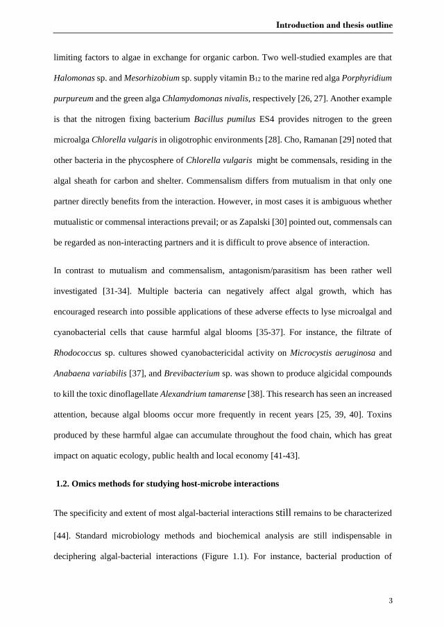
limiting factors to algae in exchange for organic carbon. Two well-studied examples are that
Halomonas sp. and Mesorhizobium sp. supply vitamin B12 to the marine red alga Porphyridium
purpureum and the green alga Chlamydomonas nivalis, respectively [26, 27]. Another example
is that the nitrogen fixing bacterium Bacillus pumilus ES4 provides nitrogen to the green
microalga Chlorella vulgaris in oligotrophic environments [28]. Cho, Ramanan [29] noted that
other bacteria in the phycosphere of Chlorella vulgaris might be commensals, residing in the
algal sheath for carbon and shelter. Commensalism differs from mutualism in that only one
partner directly benefits from the interaction. However, in most cases it is ambiguous whether
mutualistic or commensal interactions prevail; or as Zapalski [30] pointed out, commensals can
be regarded as non-interacting partners and it is difficult to prove absence of interaction.
In contrast to mutualism and commensalism, antagonism/parasitism has been rather well
investigated [31-34]. Multiple bacteria can negatively affect algal growth, which has
encouraged research into possible applications of these adverse effects to lyse microalgal and
cyanobacterial cells that cause harmful algal blooms [35-37]. For instance, the filtrate of
Rhodococcus sp. cultures showed cyanobactericidal activity on Microcystis aeruginosa and
Anabaena variabilis [37], and Brevibacterium sp. was shown to produce algicidal compounds
to kill the toxic dinoflagellate Alexandrium tamarense [38]. This research has seen an increased
attention, because algal blooms occur more frequently in recent years [25, 39, 40]. Toxins
produced by these harmful algae can accumulate throughout the food chain, which has great
impact on aquatic ecology, public health and local economy [41-43].
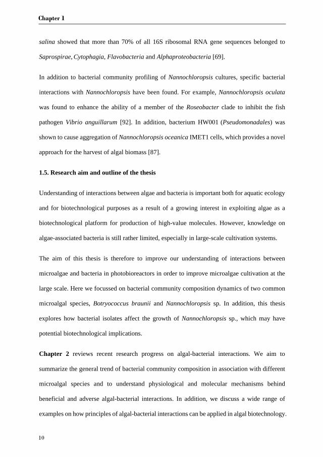
1.2. Omics methods for studying host-microbe interactions
The specificity and extent of most algal-bacterial interactions still remains to be characterized
[44]. Standard microbiology methods and biochemical analysis are still indispensable in
deciphering algal-bacterial interactions (Figure 1.1). For instance, bacterial production of

Chapter 1
4
micronutrients such as vitamin B1 or B12 has been demonstrated in defined co-cultures of
bacteria and algae with standard microbiological approaches [26, 45], and mutualistic/parasitic
relationships between Emiliana huxleyi and Phaeobacter gallaeciensis were studied by detailed
biochemical analysis [46]. More recently, the rapid advances in omics technologies offer
alternative ways to study interactions between algae and bacteria [47].
Figure 1.1. Different approaches used to characterize algal-bacterial interactions, including
examples for their application. Adapted from Cooper and Smith [48].
Combining metagenome, transcriptome and metabolome data is powerful for identifying the
major bacterial groups associated with algae and characterization of the pathways and
molecules involved in trophic exchange or signalling processes (Figure 1.1). Together with
mechanistic examinations of algal physiology and biochemistry [17], these omics-enabled
analyses will allow unravelling how algae sense and cooperate with bacteria, and how
interactions change in response to fluctuating conditions or chemical signals. At the moment,
multi-omics approaches for the study of algae and their associated bacteria are in their initial
stages, but a number of successful cases exist already. For example, a metagenomics approach
was used to elucidate the range of algal-bacterial species engaged in nitrogen-based symbioses

Introduction and thesis outline
5
in the Atlantic Ocean [49]. Furthermore, transcriptomic analysis of a co-culture of the diatom
Pseudonitzschia multiseries and Sulfitobacter sp. SA11 revealed up-regulation of tryptophan
biosynthesis genes in the alga and indole 3-acetic acid (IAA) production in the bacterium. The
produced IAA, an auxin, was found to promote cell division in the diatom [16].
1.3. Microalgae production
Microalgal biomass is a promising source for chemicals, food and feed supplements, and
biofuels [50, 51]. High areal yields, high oil content, low water consumption and the possibility
of production on non-arable land make microalgae more compelling than many other crops [52,
53]. Currently, only algal specialties or high value products, for instance, unsaturated fatty acids
and pigments, are commercially profitable [54, 55]. For commercial production of commodities
from algal biomass the production costs should decrease to less than 1 €/kg dry weight [52, 56],
while with state of the art technology the current cost can be around 3 €/kg as estimated in
techno-economic models for a 100 ha facility [57].
A major parameter affecting microalgal production cost is photosynthetic efficiency [56, 57].
Photosynthetic efficiencies obtained under outdoor conditions (3%) are lower than values
obtained under laboratory conditions (6%) [52]. At least in part this is caused by the
continuously changing weather conditions occurring under outdoor conditions where critical
cultivation parameters such as light intensity cannot be controlled and temperature can only
partly be controlled.
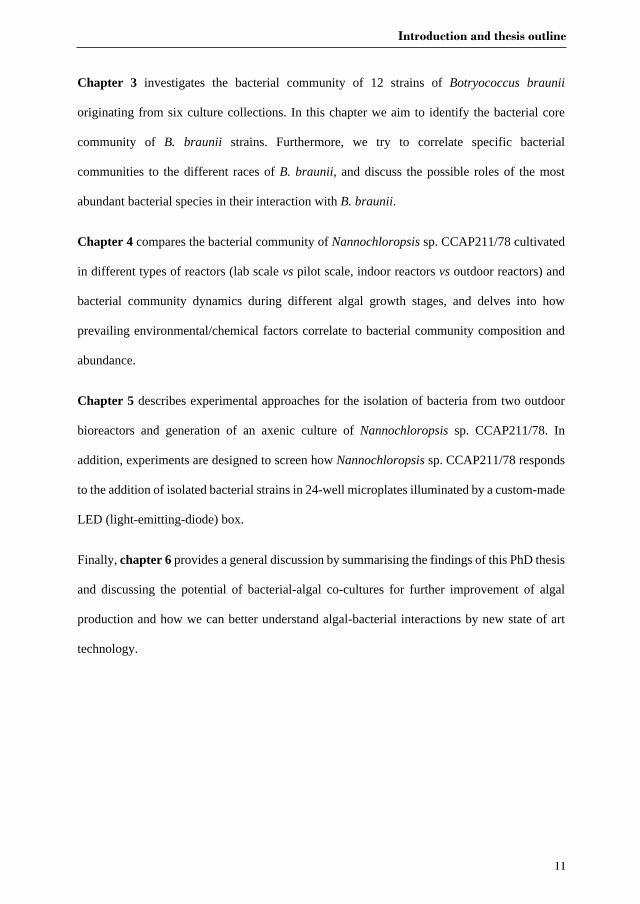
1.3.1. Microalgae production systems
One of the key factors for commercially viable microalgal production is the design of a suitable
reactor system. The open raceway pond, horizontal and vertical tubular photobioreactors, and
flat panel photobioreactors are the most often used designs at a semi-industrial scale [58]. Each
of the reactor designs has its own advantages and disadvantages, which are outlined below.

Chapter 1
6
Raceway pond Horizontal tubular PBR
Flat panel PBR Vertical tubular PBR
Figure 1.2. Overview of cultivation systems installed at the AlgaePARC pilot facility
(Wageningen, the Netherlands). PBR: photobioreactor. Adapted from [59].
1.3.1.1. Open ponds
Open ponds (natural waters and artificial ponds or containers) are the most commonly used
systems to grow algae at large scale [58, 60]. Open ponds are equipped with paddle wheels to
mix algal cultures through long channels. One of the major advantages of open ponds is that
they are cheaper and easier to build and operate than most closed photobioreactors [61, 62].
However, open systems are subject to a range of limitations such as poor light utilization by
algal cells, water evaporation and diffusion of CO2 to the atmosphere.
1.3.1.2. Closed tubular photobioreactors
Introduction and thesis outline
7
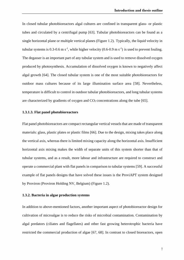
In closed tubular photobioreactors algal cultures are confined in transparent glass- or plastic
tubes and circulated by a centrifugal pump [63]. Tubular photobioreactors can be found as a
single horizontal plane or multiple vertical planes (Figure 1.2). Typically, the liquid velocity in
tubular systems is 0.3-0.6 m s-1, while higher velocity (0.6-0.9 m s-1) is used to prevent fouling.
The degasser is an important part of any tubular system and is used to remove dissolved oxygen
produced by photosynthesis. Accumulation of dissolved oxygen is known to negatively affect
algal growth [64]. The closed tubular system is one of the most suitable photobioreactors for
outdoor mass cultures because of its large illumination surface area [58]. Nevertheless,
temperature is difficult to control in outdoor tubular photobioreactors, and long tubular systems
are characterized by gradients of oxygen and CO2 concentrations along the tube [65].
1.3.1.3. Flat panel photobioreactors
Flat panel photobioreactors are compact rectangular vertical vessels that are made of transparent
materials: glass, plastic plates or plastic films [66]. Due to the design, mixing takes place along
the vertical axis, whereas there is limited mixing capacity along the horizontal axis. Insufficient
horizontal axis mixing makes the width of separate units of this system shorter than that of
tubular systems, and as a result, more labour and infrastructure are required to construct and
operate a commercial plant with flat panels in comparison to tubular systems [59]. A successful
example of flat panels designs that have solved these issues is the ProviAPT system designed
by Proviron (Proviron Holding NV, Belgium) (Figure 1.2).
1.3.2. Bacteria in algae production systems
In addition to above-mentioned factors, another important aspect of photobioreactor design for
cultivation of microalgae is to reduce the risks of microbial contamination. Contamination by
algal predators (ciliates and flagellates) and other fast growing heterotrophic bacteria have
restricted the commercial production of algae [67, 68]. In contrast to closed bioreactors, open

Chapter 1
8
ponds are exposed to their ambient environment, and thus are more prone to contamination in
general [69]. Therefore, only a few of the algal species that can be grown under extreme
conditions (high salinity, low pH, etc.) that hinder successful invasion of ambient
microorganisms are suitable to be grown in open ponds [67]. Although it is a general consensus
that open ponds are more easily contaminated than closed bioreactors, studies that
simultaneously compare bacterial diversity and abundance in open ponds and enclosed
bioreactors are limited in literature. On the other hand, for large-scale production of microalgae
it is neither practical nor economical to completely sterilize the growth media. Therefore, it is
inevitable that all large-scale microalgae production systems contain a number of non-target
organisms [70, 71].
1.4. Targeted algal species and their associated bacteria
1.4.1. Botryococcus braunii
Botryococcus braunii (Chlorophyta) is of industrial interest for its ability to produce significant
amounts of long-chain hydrocarbons (C30-C40) and exopolysaccharides [72-74]. B. braunii can
be subclassified into four races depending on the types of hydrocarbons and exopolysaccharides
produced [75, 76]. Another trait of B. braunii is that it secretes the majority of hydrocarbons
and polysaccharides, facilitating the harvesting of these products. However, wild type strains
of B. braunii are slow growers with a productivity of ~0.1-0.2 g L-1 d-1 [77, 78].
The majority of B. braunii cultures are not axenic [75, 79], but co-exist with various microbes
[80, 81]. Earlier research has revealed the presence of Pseudomonas spp. and Flavobacterium
spp. among other bacteria in B. braunii cultures [82]. Metagenomic profiling showed that B.
braunii-associated bacteria include representatives of Bradyrhizobium and Methylobacterium
(both members of the order Rhizobiales), Dyadobacter, Achromobacter and Asticcacaulis [81].
Furthermore, the addition of selected bacterial species (Rhizobium sp.) to axenic cultures
Introduction and thesis outline
9
resulted in an increase in biomass productivity and hydrocarbon yield of B. braunii [80]. More
recent growth experiments indicated that B. braunii Ba10 has a higher biomass (1.8-fold) and
hydrocarbon (1.5-fold) yield in the presence of “Candidatus Phycosocius bacilliformis” [83].
However, the precise reasons for this increase remain unknown. Contrasting these reports,
Gouveia et al. [84] found that biomass productivity and extracellular carbohydrate production
of B. braunii were significantly enhanced after removal of its associated bacteria with UV-C,
which indicates that bacteria can also be antagonistic to microalgae in this respect.
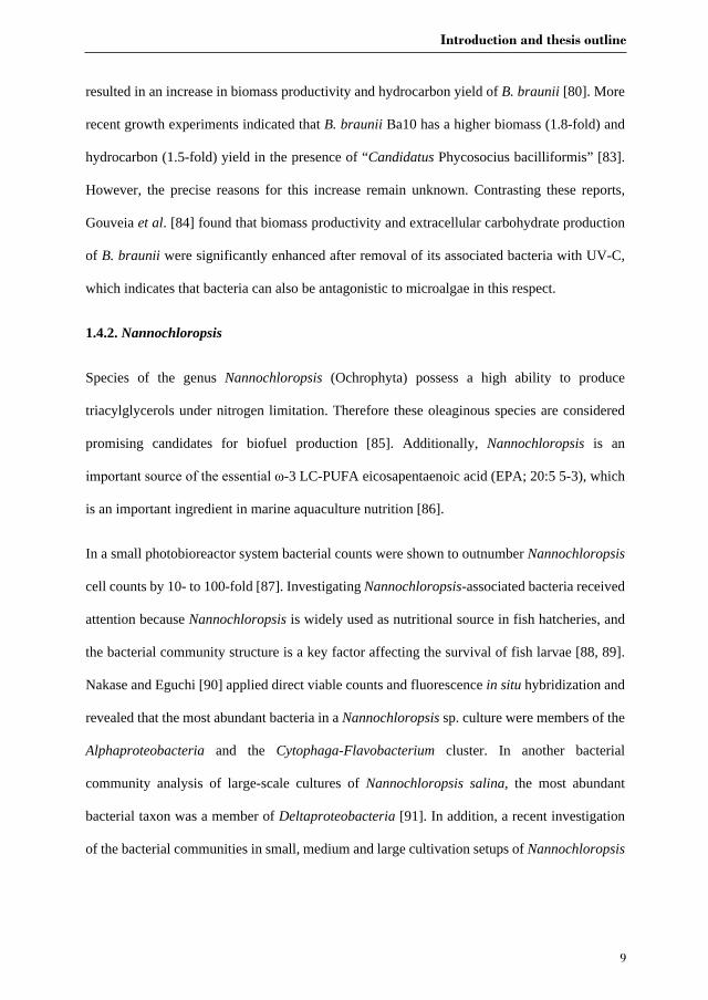
1.4.2. Nannochloropsis
Species of the genus Nannochloropsis (Ochrophyta) possess a high ability to produce
triacylglycerols under nitrogen limitation. Therefore these oleaginous species are considered
promising candidates for biofuel production [85]. Additionally, Nannochloropsis is an
important source of the essential ω-3 LC-PUFA eicosapentaenoic acid (EPA; 20:5 5-3), which
is an important ingredient in marine aquaculture nutrition [86].
In a small photobioreactor system bacterial counts were shown to outnumber Nannochloropsis
cell counts by 10- to 100-fold [87]. Investigating Nannochloropsis-associated bacteria received
attention because Nannochloropsis is widely used as nutritional source in fish hatcheries, and
the bacterial community structure is a key factor affecting the survival of fish larvae [88, 89].
Nakase and Eguchi [90] applied direct viable counts and fluorescence in situ hybridization and
revealed that the most abundant bacteria in a Nannochloropsis sp. culture were members of the
Alphaproteobacteria and the Cytophaga-Flavobacterium cluster. In another bacterial
community analysis of large-scale cultures of Nannochloropsis salina, the most abundant
bacterial taxon was a member of Deltaproteobacteria [91]. In addition, a recent investigation
of the bacterial communities in small, medium and large cultivation setups of Nannochloropsis
Chapter 1
10
salina showed that more than 70% of all 16S ribosomal RNA gene sequences belonged to
Saprospirae, Cytophagia, Flavobacteria and Alphaproteobacteria [69].
In addition to bacterial community profiling of Nannochloropsis cultures, specific bacterial
interactions with Nannochloropsis have been found. For example, Nannochloropsis oculata
was found to enhance the ability of a member of the Roseobacter clade to inhibit the fish
pathogen Vibrio anguillarum [92]. In addition, bacterium HW001 (Pseudomonadales) was
shown to cause aggregation of Nannochloropsis oceanica IMET1 cells, which provides a novel
approach for the harvest of algal biomass [87].
1.5. Research aim and outline of the thesis
Understanding of interactions between algae and bacteria is important both for aquatic ecology
and for biotechnological purposes as a result of a growing interest in exploiting algae as a
biotechnological platform for production of high-value molecules. However, knowledge on
algae-associated bacteria is still rather limited, especially in large-scale cultivation systems.
The aim of this thesis is therefore to improve our understanding of interactions between
microalgae and bacteria in photobioreactors in order to improve microalgae cultivation at the
large scale. Here we focussed on bacterial community composition dynamics of two common
microalgal species, Botryococcus braunii and Nannochloropsis sp. In addition, this thesis
explores how bacterial isolates affect the growth of Nannochloropsis sp., which may have
potential biotechnological implications.
Chapter 2 reviews recent research progress on algal-bacterial interactions. We aim to
summarize the general trend of bacterial community composition in association with different
microalgal species and to understand physiological and molecular mechanisms behind
beneficial and adverse algal-bacterial interactions. In addition, we discuss a wide range of
examples on how principles of algal-bacterial interactions can be applied in algal biotechnology.
Introduction and thesis outline
11
Chapter 3 investigates the bacterial community of 12 strains of Botryococcus braunii
originating from six culture collections. In this chapter we aim to identify the bacterial core
community of B. braunii strains. Furthermore, we try to correlate specific bacterial
communities to the different races of B. braunii, and discuss the possible roles of the most
abundant bacterial species in their interaction with B. braunii.
Chapter 4 compares the bacterial community of Nannochloropsis sp. CCAP211/78 cultivated
in different types of reactors (lab scale vs pilot scale, indoor reactors vs outdoor reactors) and
bacterial community dynamics during different algal growth stages, and delves into how
prevailing environmental/chemical factors correlate to bacterial community composition and
abundance.
Chapter 5 describes experimental approaches for the isolation of bacteria from two outdoor
bioreactors and generation of an axenic culture of Nannochloropsis sp. CCAP211/78. In
addition, experiments are designed to screen how Nannochloropsis sp. CCAP211/78 responds
to the addition of isolated bacterial strains in 24-well microplates illuminated by a custom-made
LED (light-emitting-diode) box.
Finally, chapter 6 provides a general discussion by summarising the findings of this PhD thesis
and discussing the potential of bacterial-algal co-cultures for further improvement of algal
production and how we can better understand algal-bacterial interactions by new state of art
technology.

Chapter 2
The effect of the algal microbiome on industrial production of
microalgae
Jie Lian, Rene H. Wijffels, Hauke Smidt, Detmer Sipkema
Published in Microbial Biotechnology

Chapter 2
14
Abstract
Microbes are ubiquitously distributed and they are also present in algae production systems.
The algal microbiome is a pivotal part of the alga holobiont and has a key role in modulating
algal populations in nature. However, there is a lack of knowledge on the role of bacteria in
artificial systems ranging from laboratory flasks to industrial ponds. Co-existing
microorganisms, and predominantly bacteria, are often regarded as contaminants in algal
research, but recent studies manifested that many algal symbionts not only promote algal
growth but also offer advantages in downstream processing. Because of the high expectations
for microalgae in a bio-based economy, better understanding of benefits and risks of algal-
microbial associations are important for the algae industry. Reducing production cost may be
through applying specific bacteria to enhance algae growth at large scale as well as through
preventing the growth of a broad spectrum of algal pathogens. In this review, we highlight the
latest studies of algae-microbial interactions and their underlying mechanisms, discuss
advantages of large scale algal-bacterial co-cultivation and extend such knowledge to a broad
range of biotechnological applications.
Keywords: microalgae-associated bacteria; algae-bacteria interaction; co-cultivation; algal
biotechnology
Effects of bacteria on production of microalgae
15
2.1. Introduction
During the last forty years efforts have been undertaken to realize the high potential of algal
products for industrial applications. Algae have been widely recognized for their capacity to
produce polysaccharides, lipids, pigments and other valuable compounds in significant amounts
[56]. Algae are used for producing healthy food and food supplements, and as an ingredient in
aquaculture, animal feed and as soil bio-fertilizer [93, 94].
Most algae, if not all, live in symbiosis with multiple associated microorganisms throughout
their lifespan [47]. In many cases, attempts to remove bacteria or fungi from microalgae have
failed. Even in cases where such attempts were successful, microbiota-deprived algae usually
exhibited poorer growth or aberrant phenotypes compared to the original strains, which
indicates that the association between algae and other microorganisms is important for their
existence [95].
Algae are known to release dissolved organic matter or signalling molecules to nurture specific
bacterial communities in the phycosphere [96]. Close interactions in the phycosphere influence
algal evolution and ecology in various ways. First of all, algae such as the diatoms
Phaeodactylum tricornutum and Thalassiasira pseudonana have been shown to have acquired
hundreds of genes predicted to be involved in nitrogen and organic carbon utilization, cell wall
assembly, DNA recombination and the ornithine-urea cycle from co-occurring bacteria during
more than 200 million years [97]. Secondly, bacteria synthesize important compounds for algal
growth stimulation, spore germination, morphogenesis and pathogen resistance [16, 25, 96].
These compounds include micronutrients, siderophores, growth stimulants and antibiotics [46,
98-102]. In addition, symbiotic microorganisms help their algal hosts to cope with changing
environmental conditions [24, 103].
Chapter 2
16
On the other hand, many microbes have been reported to negatively affect algal growth [104,
105] and constitute big constraints for translating laboratory experiments to industrial practice.
Unlike conventional microbial fermentation, large-scale algal cultivation is driven by light and
mostly operated in fully exposed open ponds for microalgae and in open sea for macroalgae.
However, open ponds are more susceptible to biological contaminations, such as viruses,
predators/grazers, and parasites of various sources [106]. Therefore, stable production of algae
in open systems is only possible when contaminants and infections are well studied so that
monitoring and contingency measures can be implemented [107].
Apart from playing a role in enhancing microalgae production, associated bacteria can help the
algae to perform more complex tasks with diverse applications. For instance, algae and bacteria
cooperate in faster and more efficient removal of organic and inorganic waste and hazardous
substances in wastewater treatment [108-110]. In turn, bacterial and viral pathogens are able to
weaken or decompose the algal cell wall, which is a crucial step in algal-based extraction of
chemicals and could also be explored to tackle frequently occurring harmful algae blooms at an
early stage of the bloom [111, 112]. Furthermore, proteins or secondary metabolites of algicidal
bacteria are potential biological agents in algal biomass harvest and cell disruption prior to
biorefinery [113].
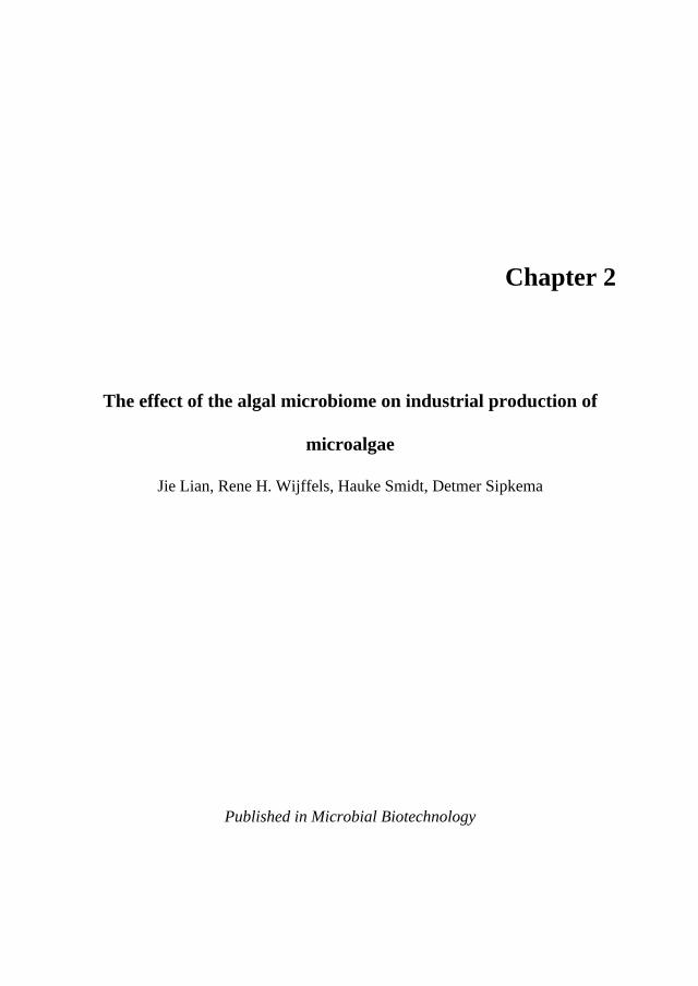
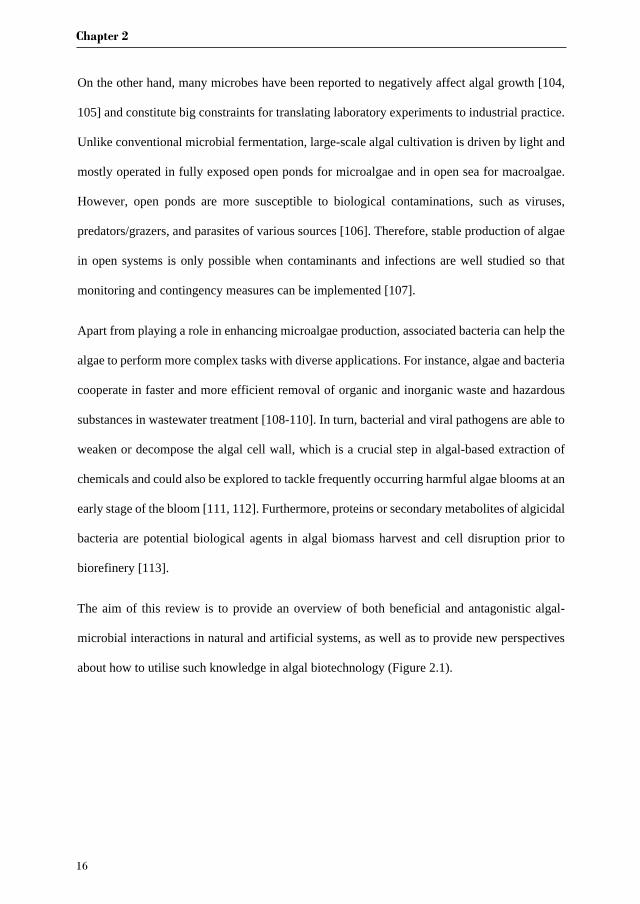
The aim of this review is to provide an overview of both beneficial and antagonistic algal-
microbial interactions in natural and artificial systems, as well as to provide new perspectives
about how to utilise such knowledge in algal biotechnology (Figure 2.1).
Effects of bacteria on production of microalgae
17
Figure 2.1. Potential applications of algal-bacterial interactions in industrial biotechnology
and environmental biotechnology. DOM is dissolved organic matter.
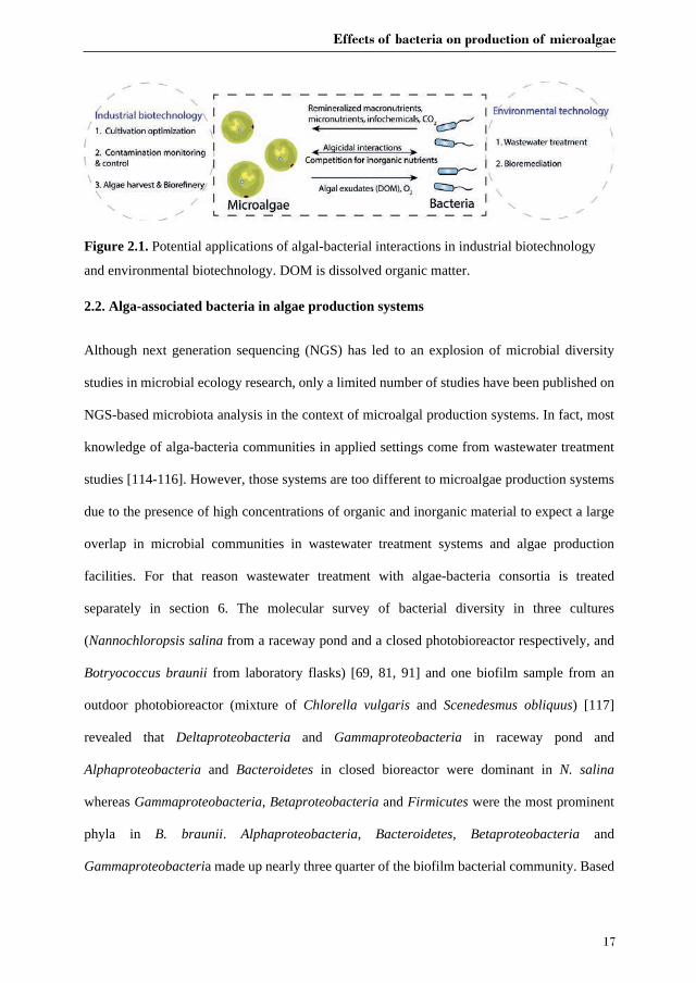
2.2. Alga-associated bacteria in algae production systems
Although next generation sequencing (NGS) has led to an explosion of microbial diversity
studies in microbial ecology research, only a limited number of studies have been published on
NGS-based microbiota analysis in the context of microalgal production systems. In fact, most
knowledge of alga-bacteria communities in applied settings come from wastewater treatment
studies [114-116]. However, those systems are too different to microalgae production systems
due to the presence of high concentrations of organic and inorganic material to expect a large
overlap in microbial communities in wastewater treatment systems and algae production
facilities. For that reason wastewater treatment with algae-bacteria consortia is treated
separately in section 6. The molecular survey of bacterial diversity in three cultures
(Nannochloropsis salina from a raceway pond and a closed photobioreactor respectively, and
Botryococcus braunii from laboratory flasks) [69, 81, 91] and one biofilm sample from an
outdoor photobioreactor (mixture of Chlorella vulgaris and Scenedesmus obliquus) [117]
revealed that Deltaproteobacteria and Gammaproteobacteria in raceway pond and
Alphaproteobacteria and Bacteroidetes in closed bioreactor were dominant in N. salina
whereas Gammaproteobacteria, Betaproteobacteria and Firmicutes were the most prominent
phyla in B. braunii. Alphaproteobacteria, Bacteroidetes, Betaproteobacteria and
Gammaproteobacteria made up nearly three quarter of the biofilm bacterial community. Based
Chapter 2
18
on this limited number of studies, Proteobacteria, and Gammaproteobacteria in particular, are
found associated to cultured microalgae. Cytophagales and Flavobacteriales were the only two
common bacterial orders among four studies. Several other taxa such as Pseudomonadales,
Burkholderiales, Caulobacterales and Rhodobacterales were shared between either two studies.
Our limited knowledge of bacterial communities associated to microalgae that is based on
cultivation-independent studies currently prevents general statements about bacteria that are
frequently found associated to microalgae, but finding correlations between algae and
associated bacteria will be a good starting point for coming up with hypotheses on functional
relationships. Therefore, more studies of bacterial communities found in microalgae bioreactors
are urgently needed to obtain a clearer view on the species and genera that are commonly
associated to algae.
2.3. Beneficial roles of bacteria
Although for most of the bacteria detected in microalgae production systems it is not known
if/how they interact with the microalgae, recent observations have demonstrated that mutualistic
algal-bacterial interactions are prevalent [2]. Multiple bacteria have been tested in co-
cultivation to evaluate the effects on the growth of microalgae [104, 118, 119], or more
specifically looked at the exchange of metabolites and how bacteria may lead to more robust
algal cultures that can better withstand environmental perturbations.
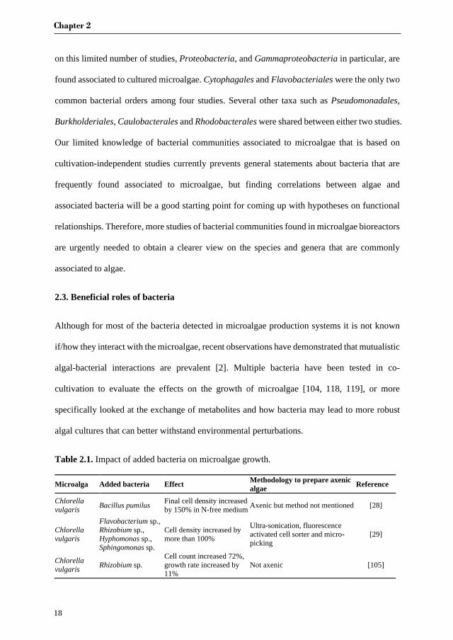
Table 2.1. Impact of added bacteria on microalgae growth.
Microalga Added bacteria Effect Methodology to prepare axenic algae Reference
Chlorella vulgaris Bacillus pumilus Final cell density increased
by 150% in N-free medium Axenic but method not mentioned [28]
Chlorella vulgaris
Flavobacterium sp., Rhizobium sp., Hyphomonas sp., Sphingomonas sp.
Cell density increased by more than 100%
Ultra-sonication, fluorescence activated cell sorter and micro-picking
[29]
Chlorella vulgaris Rhizobium sp.
Cell count increased 72%, growth rate increased by 11%
Not axenic [105]
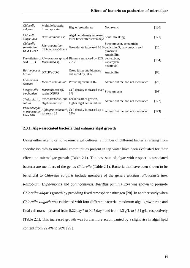
Effects of bacteria on production of microalgae
19
Chlorella vulgaris
Multiple bacteria from tap water Higher growth rate Not axenic [120]
Chlorella ellipsoidea Brevundimonas sp. Algal cell density increased
three times after seven days Serial streaking [121]
Chlorella sorokiniana IAM C-212
Microbacterium trichotecenolyticum Growth rate increased 16 %
Streptomycin, gentamicin, penicillin G, vancomycin and pimaricin
[20]
Dunaliella sp. SAG 19.3
Alteromonas sp. and Muricauda sp.
Biomass enhanced by 22%, 26%
Ampicillin, gentamicin, kanamycin, neomycin
[104]
Botryococcus braunii BOTRYCO-2 Grow faster and biomass
enhanced by 80% Ampicillin [83]
Lobomonas rostrata Mesorhizobium loti Providing vitamin B12 Axenic but method not mentioned [22]
Scrippsiella trochoidea
Marinobacter sp. strain DG879
Cell density increased over 6% Streptomycin [98]
Thalassiosira rotula
Roseobacter sp. and Hyphomonas sp.
Earlier start of growth, higher algal cell numbers Axenic but method not mentioned [122]
Phaeodactylum tricornutum Utex 646
Alphaproteobacteriq sp. strain 29
Cell density increased up to 55% Axenic but method not mentioned [123]
2.3.1. Alga-associated bacteria that enhance algal growth
Using either axenic or non-axenic algal cultures, a number of different bacteria ranging from
specific isolates to microbial communities present in tap water have been evaluated for their
effects on microalgae growth (Table 2.1). The best studied algae with respect to associated
bacteria are members of the genus Chlorella (Table 2.1). Bacteria that have been shown to be
beneficial to Chlorella vulgaris include members of the genera Bacillus, Flavobacterium,
Rhizobium, Hyphomonas and Sphingomonas. Bacillus pumilus ES4 was shown to promote
Chlorella vulgaris growth by providing fixed atmospheric nitrogen [28]. In another study when
Chlorella vulgaris was cultivated with four different bacteria, maximum algal growth rate and
final cell mass increased from 0.22 day-1 to 0.47 day-1 and from 1.3 g/L to 3.31 g/L, respectively
(Table 2.1). This increased growth was furthermore accompanied by a slight rise in algal lipid
content from 22.4% to 28% [29].
Chapter 2
20
Similar to Chlorella, also for other green algae, such as those belonging to the genera Dunaliella,
Botryococcus and Lobomonas beneficial effects were observed when adding specific bacterial
partners to axenic cultures (Table 2.1). Biomass accumulation of Botryococcus braunii was
almost doubled compared with that of axenic cultures [83]. Similarly, biomass production of
Dunaliella sp. SAG 19.3 increased by 22% and 26% when co-cultivated with Alteromonas sp.
or Muricauda sp., respectively [104]. Furthermore, it could be shown that the vitamin B12
synthesizing bacterium Mesorhizobium loti is indispensable for the survival of Lobomonas
rostrata under conditions where the alga is cultivated without exogenous vitamin B12 [22]. Two
diatoms and one dinoflagellate were all observed to benefit from co-existing bacteria (Table
2.1), as indicated by either higher cell numbers or a faster growth rate of the algae. The strongest
stimulation of growth was reported for Phaeodactylum tricornutum in the presence of the
Alphaproteobacterium strain 29, as demonstrated by a 55% rise in cell density [123].
2.3.2. Microbial associated salinity acclimation and thermal tolerance
Salinity is the major environmental factor that determines the distribution and performance of
marine algae [124, 125]. Interestingly, in addition to their more direct ecophysiological roles,
bacteria can also present a gene reservoir for algal evolution towards adaptation to different
environmental conditions via horizontal gene transfer. The green alga Picochlorum sp.
SENEW3 has a wide salt tolerance from at least 0.35% to 10.8% [126]. Compared to its less
halotolerant sisters, the genome of the salt-tolerant strain was found to contain a suite of
additional functional genes, twenty-four of which were derived from bacterial sources and were
functional in response to salt stress [127]. Although not a microalga, it is interesting to note that
the transition of the brown macroalga Ectocarpus sp. strain 371 from seawater to freshwater
medium greatly depended on the associated bacterial community. Strain 371 is a small
filamentous brown alga with broad range salinity tolerance that is mediated by adjusting cell
wall structure and metabolism [128-130]. Cultures deprived of associated microbes were unable

Effects of bacteria on production of microalgae
21
to survive a salinity change, while this capability could be restored by restoring their microbiota
[103].
Temperature is another important factor affecting growth and survival of algae [124]. This is
relevant as industrially grown algal strains in shallow production ponds or flat panel bioreactors
are exposed to considerable temperature fluctuations. The unicellular microalga
Chlamydomanas reinhardtii grows best at a temperature between 20-32 °C [131]. The direct
transfer of C. reinhardtii from an optimum (25°C) to a rather high temperature (45°C) results
in chlorosis and cell death, which are caused by the repression of cobalamin-independent
methionine synthase during heat stress. Through adding exogenous cobalamin or co-cultures of
the alga with a cobalamin-producing bacterium (Sinorhizobium meliloti), cobalamin-dependent
methionine synthase mediated methionine biosynthesis could be re-activated, thereby
preventing death of algal cell [24].
Hence, a better understanding of adaptation and acclimation of both host and microbial
symbionts to environmental changes may provide leads to improve robustness of large-scale
cultivation of algae where environmental conditions cannot be as tightly controlled as in
laboratory-based experiments.
2.3.3. Nutrient provision
Algae mainly need CO2 and inorganic sources of nitrogen and phosphate for growth along with
some micronutrients and cofactors [132]. Since fertilizer-grade nutrient input accounts for a
major proportion of cost in algal cultivation, recycling or provision of these nutrients via
bacteria may eventually make large-scale algal biomass production more economically viable
[133].
2.3.3.1. Macro-nutrients
Chapter 2
22
CO2 is often the limiting substrate in large-scale algal ponds because gas transfer efficiency is
limited from ambient air [134]. The main strategy to boost low CO2 concentrations in algal
cultures is to use CO2-enriched gases, but additional supply of CO2 comes with a significant
cost [133]. Bacterial degradation of organic compounds released by algae contributes an
additional source of CO2 for algal growth, especially during CO2 limiting conditions as this CO2
can be fixed again by algae [135, 136]. This is exemplified with the case of a Chlorella sp.
where carbon limitation was overcome when heterotrophic bacteria from a domestic wastewater
treatment reactor were added to the algae culture and increased productivity of algal biomass
by respectively 4.8 and 3.4 fold in two independent experiments [137].
Nitrogen fixing bacteria reduce atmospheric dinitrogen to ammonium that is the major preferred
nitrogen source for algae growth [132]. For example, Bacillus pumilus ES4 is a plant-growth
promoting bacterium that fixes nitrogen to enhance growth of Chlorella vulgaris [28].
Symbiotic nitrogen fixers are also present in coral holobionts, where they co-occur with
Symbiodinium that is the most commonly coral-associated dinoflagellate genus [138]. Studies
have revealed a strong positive correlation between the cell density of Symbiodinium and the
number of nitrogen fixation gene copies from nitrogen-fixing bacteria, which partly
demonstrate how corals and their dinoflagellate partners could survive in low-nutrient
conditions [139]. The filamentous cyanobacteria Richelia intracellularis and Calothrix
rhizosoleniae are close partners with diatoms living in the oligotrophic open ocean [140].
Higher growth rates were observed for diatoms with cyanobacteria as compared to diatoms
without their nitrogen-fixing cyanobacterial partners. Moreover, using single cell resolution
analyses it was shown that the N2 fixation rates of cyanobacteria increased by 171-420 fold in
symbiotic heterocystous cells associated with the corresponding diatoms as compared to free-
living cyanobacteria [141].
Effects of bacteria on production of microalgae
23
Phosphorus is an essential nutrient for algal growth. In most cases algae can only take up
inorganic phosphorus (Pi) derived from hydrolysis of organic phosphorus (Po) [142]. Bacteria
are the main agents involved in decomposing and mineralizing Po through the secretion of
phosphatases [143], and Po from deteriorating algal cells can then be recycled to optimise algal
yield on phosphate added. This process has been shown to occur with Gordonia sp. txj1302RI
and Burkholderia sp. txj1302Y4, which degraded dissolved Po to provide Microcystis
aeruginosa with Pi needed for its growth in eutrophic lakes with abundant Po but limited Pi [144].
2.3.3.2. Vitamins, phytohormones, iron-siderophore and antibiotics
Bacteria are not only capable of minimizing the requirement for external CO2 and major
essential nutrients (N, P) for algae cultivation through regeneration or fixation [139], but also
provide algal hosts with vitamins [22, 26], phytohormones [16, 96, 145, 146], siderophores [98]
and antibiotics [147]. The heterotrophic bacterium Dinoroseobacter shibae DFL12T has been
demonstrated to provide growth-limiting vitamins B1 and B12 to its dinoflagellate host. Based
on a survey of 326 algal species, it was shown that vitamin B12 is required by more than half of
the algal species [26]. Epiphytic bacteria on seaweed (Bacteroidetes strain YM2-23) produce
the compound thallusin, which is essential for inducing growth, development and
morphogenesis of Monostroma oxyspermum and other Ulva species [148, 149]. Sulfitobacter
sp. SA11 promotes diatom cell division via synthesis of the hormone indole-3-acetic acid [16].
A Marinobacter sp. that lives in close association with Scrippsiella trochoidea is able to
produce an unusual siderophore that promotes algal assimilation of iron [98]. The marine
bacterium Phaeobacter gallaeciensis produces growth hormones (phenylacetic acid) and a broad
spectrum antibiotic (tropodithietic acid) against pathogenic bacteria while the algal host
(Emiliania huxleyi) provides fixed carbon in exchange [46].
Chapter 2
24
Growing a particular strain of microalgae in an appropriate medium or adjusting media recipes
for different algal growth-stages remains a complicated task. In practice, most investigators
tend to use a medium that works for their algae, but might not necessarily be the best one [150].
Understanding the symbiosis between microalgae and bacteria could lead to identification of
missing medium components that could possibly be provided by co-cultivation with bacteria.
2.4. Harmful microbes in algal mass culture
One of the major risks of large-scale intensive algae production is the emergence of viruses,
parasites and bacterial pathogens [151]. Despite current advances in long-term algae cultivation
systems and farm management, it is neither cost-effective nor achievable to completely avoid
undesired contaminants at industrial scale [48]. An increasing number of pathogens and
parasites have been discovered in recent years, and undoubtedly, this number will continue to
grow as investment increases in algal farming [152, 153].
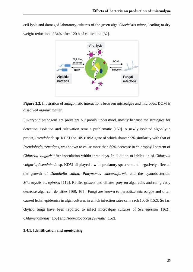
As with terrestrial plants, algae are susceptible to infection by a wide range of viruses, bacteria,
protists and fungi (Figure 2.2)[106]. Oceanic algae are likely living with a multitude of viruses,
however, only few algal viruses have been reported and characterized so far [154]. For example,
the large double-stranded DNA coccolithovirus (EhV, Phycodnaviridae) is able to terminate
Emiliania huxleyi blooms [111, 154, 155]. Algae are also adversely affected by a wide range of
bacteria, however, underlying mechanisms remain underexplored. Algae-associated bacteria
belonging to the families Rhodobacteraceae, Saprospiraceae and Flavobacteriaceae have been
implicated in bleaching of the seaweed Delisea pulchra [156]. Gram negative bacteria such as
members of the genera Alteromonas, Cytophaga, Flavobacterium, Pseudomonas, Saprospira,
Vibrio and Pseudoalteromonas are mainly responsible for rot symptoms [157] and galls on
seaweeds [158]. Furthermore, Microbacterium sp. LB1 was shown to be responsible for algal
Effects of bacteria on production of microalgae
25
cell lysis and damaged laboratory cultures of the green alga Choricistis minor, leading to dry
weight reduction of 34% after 120 h of cultivation [32].
Figure 2.2. Illustration of antagonistic interactions between microalgae and microbes. DOM is
dissolved organic matter.
Eukaryotic pathogens are prevalent but poorly understood, mostly because the strategies for
detection, isolation and cultivation remain problematic [159]. A newly isolated algae-lytic
protist, Pseudobodo sp. KD51 the 18S rRNA gene of which shares 99% similarity with that of
Pseudobodo tremulans, was shown to cause more than 50% decrease in chlorophyll content of
Chlorella vulgaris after inoculation within three days. In addition to inhibition of Chlorella
vulgaris, Pseudobodo sp. KD51 displayed a wide predatory spectrum and negatively affected
the growth of Dunaliella salina, Platymonas subcordiformis and the cyanobacterium
Microcystis aeruginosa [112]. Rotifer grazers and ciliates prey on algal cells and can greatly
decrease algal cell densities [160, 161]. Fungi are known to parasitize microalgae and often
caused lethal epidemics in algal cultures in which infection rates can reach 100% [152]. So far,
chytrid fungi have been reported to infect microalgae cultures of Scenedesmus [162],
Chlamydomonas [163] and Haematococcus pluvialis [152].
2.4.1. Identification and monitoring
Chapter 2
26
Algal biomass losses due to contaminants such as chytrid parasites can be rapid [162].
Therefore, fast and cost-effective methods to identify and control potentially harmful organisms
in algal production systems are necessary. However, microbial community composition in algal
cultures is complex and dynamic. The composition may vary with location, cultivation cycle
stage or method and season [162]. Owing to the development of next generation sequencing
methods, microbial identification can be carried out in a faster and less labour intensive way
[164] and had been shown to effectively identify specific contaminants in algae cultivation
reactors [165] or toxic algal species [166]. When pond or photobioreactor performance is
abnormal, a retrospective analysis of the archived samples could reveal harmful contaminants
and inappropriate operation strategies. Knowledge from long-term operation allows for
identifying the most common and prevalent contaminants and this also gives operators
predictive ability to some extent [106]. Systematic analysis and characterization of
contaminants can be used for the development of specific probes, primers or other biomarkers
for rapid monitoring of algae production systems. For instance, before initiating large-scale
algae production, bacteria in algal inoculation stocks and the surrounding environments (water,
soil, etc.) of the algae farm should be assayed for the presence of biological risks. A specific
microbial pathogen library can be established and molecular tools can then be used to track
harmful organisms of interest, and improving cultivation management.
2.4.2. Contamination and disease control
There is an increasing focus on preventing contamination to decrease major productivity losses
in established systems [167]. Early detection and quantification of contaminants of algal
cultures enables a fast response to infections. To protect algal cells from various contaminants,
conventional methods such as physical filtration [106], applying decreased or elevated pH and
temperatures [168] and chemical agents [169] are neither effective nor economical in algal
industry, and hence new and efficient methods to combat contaminations are urgently needed.

Effects of bacteria on production of microalgae
27
Phaeobacter inhibens reciprocally exchanges beneficial molecules with the microalga
Emiliania huxleyi. Among these molecules is the antibiotic tropodithietic acid thought to kill
other bacteria [170]. In addition, a large screening of microbes indigenous to algae-cultivation
systems has led to the discovery of an anti-fungal protein produced by the bacterium
Streptomyces sp. strain AP77. This protein has been used to cure red rot disease of Porphyra
spp. seaweeds caused by Pythium porphyrae [171]. Hence it is proposed that bacterial
metabolites or bacteria that produce antimicrobial compounds could be supplied to bulk algal
cultures in order to cost-effectively achieve more robust cultures that are less prone to harmful
invaders.
2.5. Downstream processing of algal biomass using symbionts
Traditional mechanical or chemical pre-treatment methods that are used to harvest algal
biomass and disrupt algal cells require a large energy input and are cost intensive [172]. To this
end, algae-associated microbes offer several new alternatives for microalgae harvest and cell
wall disruption.
Harvesting algal biomass is one of most important economic factors in producing compounds
with microalgae [151]. Harvesting algal cells is different from harvesting seeds of oil-bearing
plants, and oil extraction processes based on dry algal biomass are unlikely to be economical
because of the high energy inputs needed to obtain dry algal biomass [151, 173]. Currently, up
to 50% of total cost of biodiesel production is spent on harvesting because of the high energy
input and/or the addition of expensive chemicals. Energy-intensive processes such as
centrifugation are possible for high-value products but are too costly for biofuel applications.
In addition, other methods such as extensive use of chemical flocculants can be applied to aid
in the harvesting process, but could only be cost effective when the required amount is small
Chapter 2
28
[151]. Therefore, development of economic and high efficiency harvesting techniques is
important for alga bulk products, such as biofuels [174].
Bacteria can play an important role in microalgae aggregation [175, 176]. Diatom-attached
bacteria are capable of increasing diatom aggregate formation leading to the settling of
photosynthetically active Thalassiosira weissflogii, while free-living bacteria are not involved
in this process [177]. In another study, mass cultures of Nannochloropsis were observed to form
aggregates that consisted of algal cells, bacteria and debris that together resulted in a complex
structure [178]. Wang et al. isolated a novel bacterium HW001 from Permian groundwater and
demonstrated that this strain is able to stimulate aggregation of both Nannochloropsis oceanica
IMET1 and other potential biofuel-producing green microalgae, diatoms and cyanobacteria [87].
In addition, two potent bioflocculants have been discovered from culture supernatant of
Burkholderia cepacia [179] and Bacillus licheniformis CGMCC 2876 [180]. High flocculation
efficiency of Desmodesmus brasiliensis (>98 %) was achieved at pilot-scale treatment with
poly- -glutamic acid, a bioflocculant produced by Bacillus licheniformis CGMCC 2876 [179].
Besides bacteria, a number of filamentous fungal strains have also been reported to promote
flocculation of microalgae [181-183]. Muradov et al. tested the fungal species (Aspergillus
fumigatus) in co-culture with freshwater and seawater algal species and showed up to 90%
flocculation after 24h of cultivation, while no aggregates were formed in the absence of the
fungus. Furthermore, algal-fungal co-pelletization improved oil extraction efficiency because
fungal secreted hydrolytic enzymes disrupted the thick cell walls of Tetraselmis suecica [184].
The same was seen between Aspergillus lentulus FJ172995 and Chroococcus sp., where algal
and fungal cells formed a pellet, and nearly 100% of biomass settled down within 6 h at an
optimized fungal/algal ratio of 1:3 [185].
2.6. Algae-bacteria based wastewater treatment
Effects of bacteria on production of microalgae
29
High biomass production costs obstruct the economic feasibility and competitiveness of algal
biofuels [186]. The application of a combination of algae cultivation and wastewater treatment
could provide a win-win solution to this problem [151, 187]. Wastewater from municipal
sources, pig production, aquaculture and dairy cattle farming is rich in nutrients such as nitrates,
ammonia and phosphates, which can be used for algae cultivation [132]. Mixed algal-bacterial
populations in wastewater can not only perform more diverse tasks than single strains but are
also better equipped to tolerate environmental fluctuations and pathogen invasions [136].
Moreover, the combination of algae and bacteria improves water treatment efficiency, and
simultaneously the harvested algal biomass as by-product has been considered a promising
source for feeds, biofuels and fertilizer [187, 188].
2.6.1. Carbon, nitrogen and phosphate removal
Algae produce oxygen during photosynthesis that is used by bacteria to mineralize organic
matter [189]. Carbon dioxide released by bacteria during mineralization can in turn be utilised
by algae [190]. Concurrently, abundant compounds in wastewater, such as ammonium and
phosphate are eliminated by algal uptake [191]. Su et al. noted that the synergistic cooperation
between photosynthetic organisms, including algae and cyanobacteria, and activated sludge
bacteria enhanced organic carbon removal efficiencies [108]. More than 91.2% of chemical
oxygen demand was removed, and the highest total nitrogen and phosphorus removal rates were
91.0 ± 7.0% and 93.5 ± 2.5%, respectively. Chlorella sorokiniana [192] and Euglena viridis
[193] were also shown to enhance removal of carbon, nitrogen and phosphorous from piggery
waste water when mixed with bacteria from activated sludge.
2.6.2. Removal of heavy metals and toxic organic compounds
In addition to enhanced removal of excessive nutrients, algal-bacterial consortia were also
shown to be capable of removing heavy metals and toxic organic compounds from wastewater

Chapter 2
30
[190]. Algal cells not only provide stable habitats for the bacteria but also concentrate pollutants
to enhance bioavailability for bacterial degradation [194]. Algal-bacterial consortia
successfully achieved higher biodegradation or removal rates of pollutants than single species
[109].
Heavy metals belong to an important group of contaminants that pose global environmental
risks [195]. Co-cultures of bacteria and algae were capable of removing 80% of the copper and
100% of the cadmium from wastewater in a continuous flow-through column [136]. In addition,
a biofilm with immobilised algae (Ulothrix sp.) and bacteria in a photo-rotating biological
contactor removed 20-50% of a large variety of metals (Cu>Ni>Mn>Zn>Sb>Se>Co>Al)
within a ten-week period [196].
Polycyclic aromatic hydrocarbons are ubiquitous pollutants in various niches that might cast
high risks on human and animal health [197]. A co-culture of the alga Chlorella sorokiniana
and Pseudomonas migulae demonstrated higher phenanthrene degradation rates than most of
the values reported in literature [198]. Luo et al established a consortium consisting of
microalgae (Selenastrum capricornutum) and a bacterium (Mycobacterium sp. strain A1-PYR)
that achieved faster degradation of pyrene than the systems that used algae or bacteria alone
[109]. The same result was obtained by a synthetic consortium combining Synechocystis sp.
and pyrene-degrading bacteria (Pseudomonas sp. and Bacillus sp.). The combination increased
both algal growth and degradation of the polycyclic aromatic hydrocarbon [199].
Given the abovementioned advantages, integration of algae and bacteria has a large potential
for wastewater treatment, especially under aerobic conditions. Oxygen produced by algae in the
system can reduce the aeration demand in conventional activated sludge systems, which
accounts for nearly 50% of the total energy input of the water treatment plants [200]. In addition,
Effects of bacteria on production of microalgae
31
removing nutrients from wastewater with a combination of algae and bacteria can increase the
removal efficiency, system robustness and application potential of the sludge.
2.7. Outlook
Unravelling the complex biological mechanisms of algal-microbial interactions represents a
largely understudied realm to improve production of high-value products and biofuels through
large-scale cultivation of microalgae. Protective bacteria could inhibit growth of bacterial or
fungal contaminants, which cause fouling or negatively affect algal growth. Macro fertilizers
and expensive micronutrients supplied by bacterial metabolism can reduce the need for external
input. Some bacteria are able to enhance synthesis of desired algal metabolites, for instance,
lipids. However, currently our knowledge on algae bacteria interactions is too scattered to
identify generalities with respect to bacterial species that are suitable for co-culture with
microalgae. Alga species-specific knowledge would logically be first developed for industrial
working horse species, such as Arthrospira spp., Chlorella spp., Scenedesmus spp.,
Nannochloropsis spp. and Botryococcus spp.[201]. In addition, the desired microbial
community in algae cultures may depend on the required product specifications (biofuel, feed
and food, fine chemicals) and harvesting methods applied.
Chapter 2
32
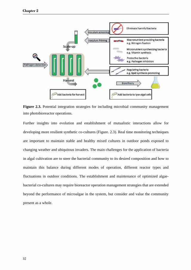
Figure 2.3. Potential integration strategies for including microbial community management
into photobioreactor operations.
Further insights into evolution and establishment of mutualistic interactions allow for
developing more resilient synthetic co-cultures (Figure. 2.3). Real time monitoring techniques
are important to maintain stable and healthy mixed cultures in outdoor ponds exposed to
changing weather and ubiquitous invaders. The main challenges for the application of bacteria
in algal cultivation are to steer the bacterial community to its desired composition and how to
maintain this balance during different modes of operation, different reactor types and
fluctuations in outdoor conditions. The establishment and maintenance of optimized algae-
bacterial co-cultures may require bioreactor operation management strategies that are extended
beyond the performance of microalgae in the system, but consider and value the community
present as a whole.
Chapter 3
Associated bacteria of Botryococcus braunii (Chlorophyta)
Joao D. Gouveia, Jie Lian, Georg Steinert, Hauke Smidt, Detmer Sipkema, Rene
H. Wijffels, Maria J. Barbosa
Published in PeerJ
Chapter 3
34
Abstract
Botryococcus braunii (Chlorophyta) is a green microalga known for producing hydrocarbons
and exopolysaccharides. Improving the biomass productivity of B. braunii and hence, the
productivity of the hydrocarbons and of the exopolysaccharides, will make B. braunii more
attractive for industries. Microalgae usually cohabit with bacteria which leads to the formation
of species-specific communities with environmental and biological advantages. Bacteria have
been found and identified with a few B. braunii strains, but little is known about the bacterial
community across the different strains. A better knowledge of the bacterial community of B.
braunii will help to optimize the biomass productivity, hydrocarbons and exopolysaccharide
accumulation. To better understand the bacterial community diversity of B. braunii, we
screened 12 strains from culture collections. Using 16S rRNA gene analysis by MiSeq we
described the bacterial diversity across twelve B. braunii strains and identified possible shared
communities. We found three bacterial families common to all strains: Rhizobiaceae,
Bradyrhizobiaceae and Comamonadaceae. Additionally, the results also suggest that each
strain has its own specific bacteria that may be the result of long-term isolated culture.
Keywords: Botryococcus braunii; associated bacteria; algal–bacterial interactions; 16S rRNA
sequencing
Associated bacteria of Botryococcus braunii
35
3.1. Introduction
In recent decades many studies have focused on the physiology and cultivation process of
several microalgae with potential for large scale production [202-206]. One microalga of
interest for large scale cultivation is B. braunii because it can produce and secrete long chain
hydrocarbons and exopolysaccharides (EPS) [73, 75, 79]. Hydrocarbons are naturally occurring
compounds consisting entirely of hydrogen and carbon, and are one of the most important
energy resources [207]. B. braunii is differentiated into different races (race A, B, L and S)
depending on the type of hydrocarbons secreted [75, 76]. Race A strains synthesize odd-
numbered alkadienes and trienes (C25 to C31), race B strains synthesize isoprenoid type
compounds termed botryococcenes (C30 to C37), and methylated squalenes (C31 to C34), race L
strains synthesize lycopadiene (C40), and race S strains synthesize C18 epoxy-n-alkanes and C20
saturated n-alkanes [75-77, 79]. EPS can have a range of applications, for example it can be
applied as stabilisers and gelling agents in food products. In addition, it has applications in the
pharmaceutical and cosmeceutical industries [208-210]. B. braunii comprises of a variety of
strains from diverse parts of the world. The strains can differ in the hydrocarbon and
exopolysaccharide content [72, 74, 77, 79, 211-214].
Bacteria can grow in close proximity to the microalgal cells due to the presence of EPS
substances secreted by the microalgae [15]. The presence of bacteria within, or close to this
EPS layer can lead to mutually beneficial interactions as well as interactions that are
antagonistic in nature. Beneficial interactions for microalgae normally provide environmental
advantages, such as nutrient exchange and community resilience to invasion by other species
[215-218]. Antagonistic interactions will usually result in inhibition of the microalgal growth,
either causing cell lysis, or directly competing for nutrients [48, 146, 219]. Studies investigating
interactions of microalgae with bacteria show how important these interactions can be for the
cultivation process [27, 105, 220, 221]. Understanding the interactions of microalgae and
Chapter 3
36
bacteria, and how it can enhance the cultivation for industrial process, could lead to increased
biomass productivity.
So far the bacterial community of B. braunii species is described in only a few studies. The
earliest work is from Chirac and colleagues who described the presence of Pseudomonas sp.
and Flavobacterium sp. in two strains of B. braunii [222]. Rivas and colleagues identified in
the B. braunii UTEX strain the presence of Pseudomonas sp. and Rhizobium sp. [80]. One study
using the B. braunii Ba10 strain showed the presence of rod shaped bacteria in the rim of the
colony aggregations and proposed it is as growth promoting bacteria closely related to
Hyphomonadaceae spp. [83]. One important finding was that B. braunii is a vitamin B12
autotroph, so it does not depend on bacteria for the synthesis of this important metabolite [223].
A more recent study using a B. braunii (race B) strain, revealed the presence of several
Rhizobiales such as Bradyrhizobium, and the presence of Bacteroidetes sp [81]. So far, all
studies have focused on only a few strains making it difficult to have a good overview of what
bacterial community dominates B. braunii.
In this study we looked at twelve strains of B. braunii obtained from several culture collections
to investigate the bacterial community composition that is associated with B. braunii.
3.2. Materials and methods
3.2.1. Strain collections and media preparation
Twelve B. braunii strains were obtained from culture collections (Table 3.1) and transferred to
Erlenmeyer flasks with modified Chu 13 medium [224] without citric acid or vitamins, with the
following composition: 1200 mg L-1 KNO3, 200 mg L-1 MgSO4.2H20, 108 mg L-1 CaCl2.2H2O,
104.8 mg L-1 K2HPO4, 20 mg L-1 Fe-Na2EDTA, 9.4 g L-1 Na2O4Se, 2.86 mg L-1 H3BO3, 1.8
mg L-1 MnSO4.4H2O, 220 g L-1 ZnSO4.7H2O, 90 g L-1 CoSO4.7H2O, 80 g L-1 CuSO4.5H2O,
60 g L-1 Na2MoO4.2H2O, 10 l L-1 H2SO4. The final pH was adjusted to pH 7.2 with NaOH

Associated bacteria of Botryococcus braunii
37
and NaHCO3 was added to a final concentration of 5 mM. The 12 strains were grown in Infors
HT Multriton incubators in 250 mL conical flasks and a volume of 150 mL. The temperature
was set at 23°C, with 2.5 % CO2 enriched air and shaking at 90 rpm. Illumination was provided
by Phillips lamps FL-Tube L 36W/77, with 150 µmol photon m-2 sec-1, and a light:dark
photoperiod of 18:6 h. Flasks were inoculated with B. braunii growing in the active growing
phase, such that the initial absorbance at 680 nm was 0.2. The Erlenmeyer flasks were capped
with aeraseal sterile film (Alphalabs). Samples were taken at day 1, 4, 8 and 11, for 16S rRNA
gene analyses.
Table 3.1. Information of the culture collections providers of Botryococcus braunii strains
and location of origin.
Culture collection Botryococcus braunii Strain (our abbreviation)
Race Location Isolation, date of isolation Reference
Berkeley Showa Race B culturing tanks, Berkley
by unknown, 1980 [225]
Scandinavian Culture Collection of Algae and Protozoa (SCCAP)
K1489 Race A Belgium, Nieuwoort by G. Hansen, 2008 No reference
UTEX Culture Collection of Algae UTEX LB572 (UTEX) Race A Cambridge, England by M. R. Droop,
1950 [77]
Culture Collection of Autotrophic Organisms (CCALA) check
CCALA778 (CCALA) unknown Serra da Estrela (Barragem da Erva da Fome) Portugal
by Santos, 1997 No reference
Culture Collection of Algae and Protozoa (CCAP)
CCAP807/2 (CCAP) Race A Grasmere, Cumbria, England
by Jaworski, 1984 [226]
ALGOBANK-CAEN
AC755 Race A Lingoult-Morvan, France
by Pierre Metzger, 1981 [227]
AC759 Race B Ayame, Ivory Coast by Pierre Metzger, 1984 [212]
AC760 Race B Kossou, Ivory Coast by Pierre Metzger, 1984 [212]
AC761 Race B Paquemar, Martinique, France
by Pierre Metzger, 1983 [227]
AC765 Race L Kossou, Ivory Coast by Pierre Metzger, 1984 [212]
AC767 Race L Songkla Nakarin, Thailand
by Pierre Metzger, 1985 [228]
AC768 Race L Yamoussoukro, Ivory Coast
by Pierre Metzger, 1984 [228]
Chapter 3
38
3.2.2. DNA extraction
On sampling days, 5 mL of fresh culture was harvested with sterilized membrane filters (0.2
m illipore usin a acuum apparatus he filters ere cr opreser e in -80 °C until further
processing. DNA was extracted from the cryopreserved filters that were cut into small pieces
with a sterile scissor. Filter pieces were transferred to a 2 mL sterilized tube with zirconia/silica
beads (Biospecs), and 1 mL S.T.A.R buffer (Roche, USA) was added. Cells were homogenized
for two rounds of 45 seconds, at the speed of 5500 rpm with Precellys (Bertin Technologies).
Then DNA was extracted using the Maxwell 16 Tissue LEV Total RNA purification kit
(Promega, USA) with aid of the Maxwell 16 instrument (Promega, USA). The purity and
quantity of DNA was examined by electrophoresis on a 1% agarose gel and measured with a
Nanodrop (ND1000, Thermo Fisher Scientific Inc., Wilmington). The extracted DNA was
stored at -20 ℃ until further use.
3.2.3. 16S rRNA gene amplification and Miseq sequencing
Amplicons from the V1-V2 region of 16S rRNA genes were generated by a two-step PCR
strategy consisting of a forward primer (27F-DegS = 5’GTTYGATYMTGGCTCAG 3’ where
M = A or C; R = A or G; W = A or T; Y = C or T) and an equimolar mixture of reverse primers
(338R I = 5’GCWGCCTCCCGTAGGAGT 3’ and II = 5’ GCWGCCACCCGTAGGTGT 3’
where M = A or C; R = A or G; W = A or T; Y = C or T). Eighteen bp Universal Tags 1 and 2
(Unitag1 = GAGCCGTAGCCAGTCTGC; Unitag2 = GCCGTGACCGTGACATCG) were
appended at the 5’ end of the forward and reverse primer, respectively [229-231]. The first PCR
mix (50 µL) contained 10 µL 5× HF buffer (Thermo ScientificTM, the Netherlands), 1 µL
dNTP Mix (10 mM; Promega, Leiden, the Netherlands), 1 U of Phusion® Hot Start II High-
Fidelity DNA polymerase (Thermo ScientificTM), 1 µM of 27F-DegS forward primer, 1 µM
of 338R I and II reverse primers, 1 µL template DNA and 32.5 µL nuclease free water.

Associated bacteria of Botryococcus braunii
39
Amplification included an initial denaturation at 98°C for 30 sec; 25 cycles of denaturation at
98°C for 10 sec; annealing at 56°C for 20 sec and elongation at 72°C for 20 sec; and a final
extension at 72°C for 10 min. The PCR product size was examined by 1 % gel electrophoresis.
The second PCR mix (100 µL) contained 62 µL nuclease free water, 5 µL of PCR1 product, 20
µL 5× HF buffer, 2 µL dNTP Mix, 2 U of Phusion® Hot Start II High-Fidelity DNA polymerase,
500 nM of a forward and reverse primer equivalent to the Unitag1 and Unitag2 sequences
respectively, each appended with an 8 nt sample specific barcode. Amplification included an
initial denaturation at 98°C for 30 sec; 5 cycles of denaturation at 98°C for 10 sec, annealing at
52°C for 20 sec and elongation at 72°C for 20 sec; and a final extension at 72°C for 10 min.
The concentration of PCR products was quantified with a Qubit Fluorometer (Life
Technologies, Darmstadt, Germany) in combination with the dsDNA BR Assay kit (Invitrogen,
Carlsbad, CA, USA). Purified products were then pooled in equimolar amounts of 100 ng µL-1
and sequenced on a MiSeq platform (GATC-Biotech, Konstanz, Germany).
3.2.4. Processing MiSeq data
Data was processed using the Quantitative Insights into Microbial Ecology (QIIME) 1.8.0. In
short, paired-end libraries were filtered to contain only read pairs perfectly matching barcodes.
Low quality or ambiguous reads were removed and then chimeric reads were removed and
checked. Sequences with less than 0.1 % were discarded. Remaining filtered sequences were
assigned into Operational Taxonomy Units (OTUs) at 97% threshold using an open reference
method and a customized SILVA 16S rRNA gene reference [232]. Seven samples from day 4
were removed from the results due to contamination during the PCR steps: AC755, AC759,
AC760, AC767, AC768, CCAP and UTEX572. The 16S rRNA gene dataset obtained in this
study is deposited in the Sequence Read Archieve (SRA), NCBI with accession number
SRP102970.
Chapter 3
40
3.2.5. Microbial community analysis
For the interpretation of the microbial community data on family level, the Operational
Taxonomic Unit (OTU) abundance table was converted to relative abundance and visualized as
heatmaps using JColorGrid [233]. Ordination analyses to estimate the relationship of the B.
braunii strains based on dissimilarity of the microbial community compositions among the
individual samples was performed for, a) all strains of B. braunii used in this study, b) all strains
received from ALGOBANK-CAEN culture collection. For both analysis a standardized 97%
OTU table (decostand function, method = hellinger) and the nMDS function metaMDS
(distance = Bray-Curtis) from the vegan package in R was used (R version 3.0.2) [234, 235].
Betadispersion and a permutation test were performed to test homogeneity dispersion within a
group of samples. Adonis from the vegan package in R (v.3.0.2) was used to test significant
differences in bacterial community between strains. Hierarchical clustering analysis was
performed using hclust function in R using method = average.
3.3. Results
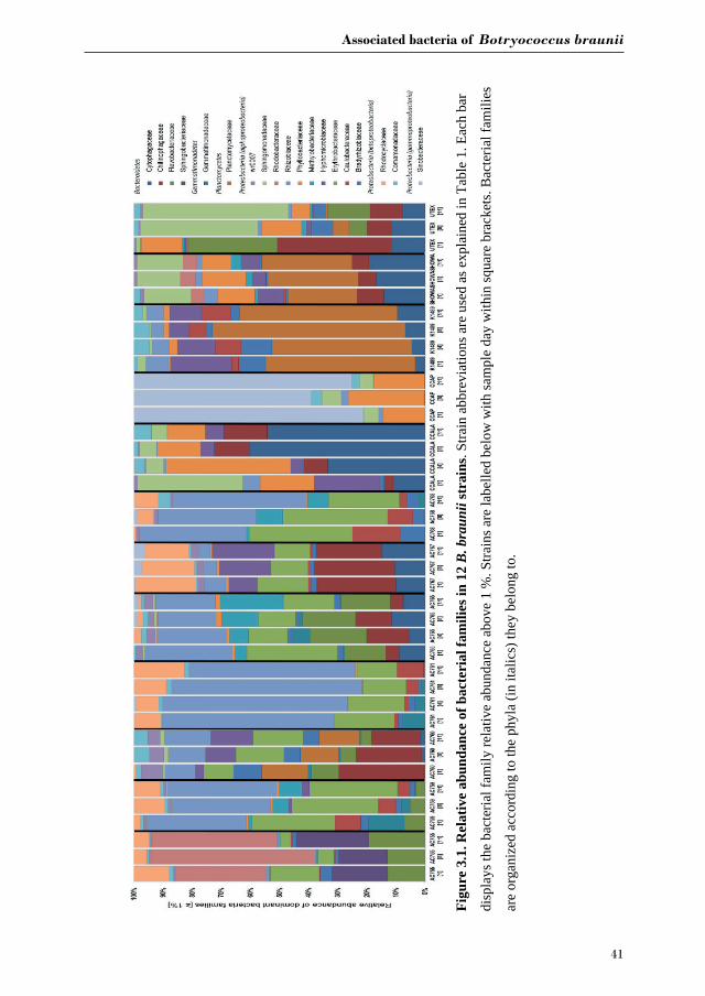
Figure 3.1 shows the bacterial families with a relative abundance above 1 % and a total of four
bacterial phyla associated with B. braunii strains. The four phyla found associated with B.
braunii are the Bacteroidetes, Gemmatimonadetes, Planctomycetes and Proteobacteria.
Proteobacteria is the predominant bacterial phylum and representatives of this taxon are found
in all 12 strains. Bacteroidetes is found in all strains with exception to strains AC761, AC768
and CCAP. Gemmatimonadetes is found only in the CAEN culture (with AC prefix) strains
with exception to AC755. Planctomycetes is found in AC760, CCALA, K1489, Showa and
UTEX strains. Three families are found across all 12 B. braunii strains and all are
Proteobacteria.
Associated bacteria of Botryococcus braunii
41
Figu
re 3
.1. R
elat
ive
abun
danc
e of
bac
teri
al fa
mili
es in
12
B. b
raun
iist
rain
s. St
rain
abb
revi
atio
ns a
re u
sed
as e
xpla
ined
in T
able
1. E
ach
bar
disp
lays
the
bact
eria
l fam
ily re
lativ
e ab
unda
nce
abov
e 1
%. S
train
s are
labe
lled
belo
w w
ith sa
mpl
e da
y w
ithin
squa
re b
rack
ets.
Bac
teria
l fam
ilies
are
orga
nize
d ac
cord
ing
to th
e ph
yla
(in it
alic
s) th
ey b
elon
g to
.
Chapter 3
42
These are the Rhizobiaceae, Bradyrhizobiaceae and Comamonadaceae. Rhizobiaceae is
represented by 1 to 59 % of the bacterial reads. Bradyrhizobiaceae was found within the 1 to
8 % range. Comamonadaceae was found between 1 and 5 %. Two families of bacteria are only
found in the strains obtained from the CAEN culture collection: Erythrobacteraceae with
bacterial reads ranging from 1 to 29 % and Rhodocyclaceae with 1 to 18 %.
Some families of bacteria are particularly dominant in specific strains. Sinobacteraceae is
dominant in CCAP with relative abundances ranging from 59 to 78 %. Planctomycetaceae is
dominant in K1489 strain with relative abundances between 46 and 51 %. Rhizobiaceae is
dominant in AC761 with relative abundances between 55 and 64 %. Other families of bacteria
become dominant as the cultures become older. Rhodobacteraceae is present in AC755 strain
with relative abundances ranging from 28 % at day 1 to 40 % at day 11. Sphingomonadaceae
is present in UTEX with 10 % at day 1 and increases its presence to 47 % at day 11.
Chytophagaceae is dominant in CCALA strain with relative abundance ranging from 10 % at
day 1 to 52 % at day 11.
Because we found three common families across all strains, we wanted to investigate in more
detail the bacterial composition in these selected families and see if we could identify an unique
microorganism present in all strains. Therefore we zoomed in and looked at the Operational
Taxonomy Units (OTUs) distribution belonging to the three families: Rhizobiaceae,
Bradyrhizobiaceae and Comamonadaceae. In addition, we picked the OTUs found only in the
strains obtained from the CAEN culture collection which belong to two families:
Erythrobacteraceae and Rhodocyclaceae. The most abundant OTUs were selected and a total
of 28 OTUs were investigated. From Figure 3.2 it is clear that there is not an OTU that is found
across all strains but rather each family comprises of several different OTUs. The second
important observation is that CCAP strain has no representative OTUs for Bradyrhizobiaceae
and Rhizobiaceae in the most abundant OTUs. The most represented family taxon is
Associated bacteria of Botryococcus braunii
43
Rhizobiaceae with 12 OTUs. From the three families found in the 12 strains, OTU 233 assigned
to the genus Rhizobium has the highest OTU frequency abundance with 10 % and is present in
7 out of 12 strains. The OTUs 143, 88 and 131 assigned to the genus Shinella are present in 9
out of 12 strains. The OTUs 477, 475 and 484 assigned to the genus Bosea cover 11 out of 12
strains. From the two families found only in the cultures originating from the CAEN culture
collection, OTUs 333 and 539 are found in all seven CAEN strains with an assigned genus
Porphyrobacter and Methyloversatilis, respectively.
The most abundant OTUs (as listed in Figure 3.2) were subjected to a Blast search against the
NCBI database to infer their nearest neighbours (Table 3.2). OTUs 88, 115, 143 and 233 are
similar in their nearest neighbours with four different Rhizobium spp. as candidates. Similar
blast results are seen also for OTUs 566 and 567 with the nearest neighbours being
Hydrogenophaga spp. The OTUs 819 and 832 with Dyadobacter spp. as nearest neighbour
dominate CCALA bacterial community. Some OTUs show different species as closest
neighbours such as OTUs 45 and 69 with Frigidibacter albus, Paracoccus sediminis and
Nioella nitratireducens as neighbours. The OTU 415 with high abundance in K1489 belonging
to Planctomycetaceae, has as closest neighbours uncultured bacterium and third closest
neighbour uncultured Planctomyces spp. with the latter showing 87 % identity. The OTU 333
present only in the strains from CAEN culture collection, has 100 % identity with
Sphingomonas as closest two neighbours, and third neighbour, also with 100 %, identity being
Porphyrobacter.

Chapter 3
44
Figu
re 3
.2. H
eatm
ap o
f mos
t abu
ndan
t 16S
rRN
A g
ene O
TU
s bel
ongi
ng to
the f
amili
es R
hizo
biac
eae,
Bra
dyrh
izob
iace
ae, C
omam
onad
acea
e,
Ery
thro
bact
erac
eae
and
Rho
docy
clac
eae.
The
labe
l on
the
right
sho
ws
the
colo
ur c
ode
for t
he re
lativ
e ab
unda
nce.
Fre
quen
cy (a
vera
ge re
lativ
e
abun
danc
e) o
f eac
h O
TU is
sho
wn
in p
erce
ntag
e on
the
right
bet
wee
n br
acke
ts. T
he la
bel o
n th
e le
ft sh
ows
the
fam
ily le
vel a
ndO
TU n
umbe
r
follo
wed
by
genu
s. n.
d m
eans
no
read
s det
ecte
d.
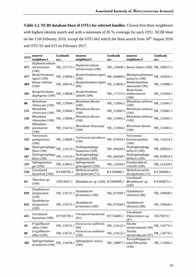
Associated bacteria of Botryococcus braunii
45
Table 3.2. NCBI database blast of OTUs for selected families. Closest first three neighbours
with highest identity match and with a minimum of 85 % coverage for each OTU. NCBI blast
on the 11th February 2016, except the OTU 662 which the blast search from 30th August 2016
and OTU 63 and 415 on February 2017.
OTU nearest neighbour1
Genbank acc.
nearest neighbour2
Genbank acc.
nearest neighbour3
Genbank acc.
475 Hyphomicrobium nitrativorans (100)
NR_121713.1 Hyphomicrobium nitrativorans (100) NR_118448.1 Bosea lathyri (100) NR_108515.1
477 Bradyrhizobium lupini (100) NR_134836.1 Bradyrhizobium lupini
(100) NR_044869.2 Rhodopseudomonas palustris (100) NR_103926.1
484 Bosea robiniae (100) NR_108516.1 Bradyrhizobium lupini
(99) NR_134836.1 Bradyrhizobium ottawaense (99) NR_133988.1
502 Bradyrhizobium daqingense (100) NR_118648.1 Bradyrhizobium
lablabi (100) NR_117513.1 Beijerinckia doebereinerae (100)
NR_116304.1
88 Rhizobium rhizoryzae (100) NR_133844.1 Rhizobium flavum
(100) NR_133843.1 Rhizobium azibense (100) NR_133841.1
115 Rhizobium rhizoryzae (100) NR_133844.1 Rhizobium flavum
(100) NR_133843.1 Rhizobium azibense (100) NR_133841.1
143 Rhizobium rhizoryzae (100) NR_133844.1 Rhizobium flavum
(100) NR_133843.1 Rhizobium azibense (100) NR_133841.1
233 Rhizobium paranaense (100)
NR_134152.1 Rhizobium rhizoryzae (100) NR_133844.1 Rhizobium flavum
(100) NR_133843.1
555 Variovorax guangxiensis (100)
NR_134828.1 Variovorax paradoxus (100) NR_074654.1
Variovorax boronicumulans (100)
NR_114214.1
566 Hydrogenophaga flava (100) NR_114133.1 Hydrogenophaga
bisanensis (100) NR_044268.1 Hydrogenophaga defluvii (100) NR_029024.1
567 Hydrogenophaga flava (100) NR_114133.1 Hydrogenophaga
bisanensis (100) NR_044268.1 Hydrogenophaga defluvii (100) NR_029024.1
333 Sphingomonas gei (100) NR_134812.1 Sphingomonas
ginsengisoli (100) NR_132664.1 Porphyrobacter colymbi (100) NR_114328.1
539 Uncultured bacterium (100) KY606782.1 Methyloversatilis
discipulorum (71) KY284088.1 Methyloversatilis discipulorum (71) KY284080.1
63 Thioclava sp. (100) CP019437.1 Rhodobacter sp. (100) KY608089.1
Uncultured Rhodobacter sp. (100)
KY606875.1
819 Dyadobacter jiangsuensis (100)
NR_134721.1 Dyadobacter fermentans (100) NR_074368.1 Dyadobacter
tibetensis (88) NR_109648.1
832 Dyadobacter jiangsuensis (100)
NR_134721.1 Dyadobacter fermentans (100) NR_074368.1 Dyadobacter
tibetensis (88) NR_109648.1
415 Uncultured bacterium (100) KT769749.1 Uncultured bacterium
(91) KT724695.1 Uncultured Planctomyces sp. (87)
JX576019.1
45 Frigidibacter albus (100) NR_134731.1 Paracoccus sediminis
(96) NR_134122.1 Nioella nitratireducens (94) NR_134776.1
69 Frigidibacter albus (100) NR_134731.1 Paracoccus sediminis
(100) NR_134122.1 Nioella nitratireducens (97) NR_134776.1
302 Sphingorhabdus arenilitoris (100) NR_134184.1 Sphingopyxis italica
(100) NR_108877.1 Parasphingopyxis lamellibrachiae (100)
NR_113006.1
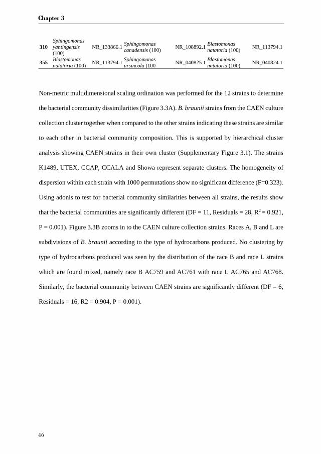
Chapter 3
46
310 Sphingomonas yantingensis (100)
NR_133866.1 Sphingomonas canadensis (100) NR_108892.1 Blastomonas
natatoria (100) NR_113794.1
355 Blastomonas natatoria (100) NR_113794.1 Sphingomonas
ursincola (100 NR_040825.1 Blastomonas natatoria (100) NR_040824.1
Non-metric multidimensional scaling ordination was performed for the 12 strains to determine
the bacterial community dissimilarities (Figure 3.3A). B. braunii strains from the CAEN culture
collection cluster together when compared to the other strains indicating these strains are similar
to each other in bacterial community composition. This is supported by hierarchical cluster
analysis showing CAEN strains in their own cluster (Supplementary Figure 3.1). The strains
K1489, UTEX, CCAP, CCALA and Showa represent separate clusters. The homogeneity of
dispersion within each strain with 1000 permutations show no significant difference (F=0.323).
Using adonis to test for bacterial community similarities between all strains, the results show
that the bacterial communities are significantly different (DF = 11, Residuals = 28, R2 = 0.921,
P = 0.001). Figure 3.3B zooms in to the CAEN culture collection strains. Races A, B and L are
subdivisions of B. braunii according to the type of hydrocarbons produced. No clustering by
type of hydrocarbons produced was seen by the distribution of the race B and race L strains
which are found mixed, namely race B AC759 and AC761 with race L AC765 and AC768.
Similarly, the bacterial community between CAEN strains are significantly different (DF = 6,
Residuals = 16, R2 = 0.904, P = 0.001).
Associated bacteria of Botryococcus braunii
47
Figure 3.3. Non-metric multidimensional scaling (nMDS) ordination (based on Bray-
Curtis distance matrix) of 16S rRNA gene sequences of 12 B. braunii strains. A) ordination
of all strains with CAEN cultures clustering together (within the ellipse dotted line); B)
ordination of the CAEN culture collection strains only. Capital letters in plot B refer to the race
subclassification based on the type of hydrocarbons produced.
3.4. Discussion
It is evident that B. braunii possesses a highly diverse bacterial community as seen by the range
of bacterial phyla and families present in all the strains used in this study (Figure 3.1 (for a more
comprehensive list see Supplementary Figure 3.2)).
From the bacterial community analysis (Figure 3.3A, B), it appears that each B. braunii strain
has a specific bacterial community and no OTU is shared between all strains. The strains from
the CAEN culture collection cluster together while B. braunii strains from other culture
collections appear as separate groups. This implies that the culture collection from which the
strain was obtained could potentially have an effect. With this study we are not able to really
deduce the potential impact of the culture collection on the bacterial community because the
experimental design was not set-up to do so. The presence of weak (within a culture collection)
and strong (between culture collections) migration barriers may explain the bacterial profiles
as obtained in our study and they may be a result of historical contingencies [236] rather than
pointing towards highly specific interactions for a large number of OTUs. OTUs 539 and 333

Chapter 3
48
are only found with the CAEN cultures and contributes towards these strains clustering in close
proximity. OTU 333 is especially high in relative abundance and contributes to the distinctive
clustering of the CAEN culture collection strains. The remaining strains also contain their
specific OTUs that contribute towards their own clustering: OTU 819 and 832 with CCALA,
OTU 310 with UTEX and K1489 with OTU 415. The bacterial community between three race
B and three race L are mixed together (Figure 3.3B). Therefore, no correlation was found
between bacterial community and the type of hydrocarbons produced between the two races.
Similar observations were made in another study using six strains of B. braunii in which the
authors did not find a correlation between the bacteria and type of hydrocarbon produced [237].
Three bacterial families were found to be present with all twelve strains of B. braunii:
Bradyrhizobiaceae, Rhizobiaceae, and Comamonadaceae. Two families were found
abundantly only in the strains from the CAEN culture collection: Erythrobacteraceae and
Rhodocyclaceae. The OTUs 88, 115, 143 and 233 blast hits show these are related to Rhizobium
spp. (Table 3.2). Rhizobium spp. are known to form nodules in the roots of several plants within
the family of legumes and are best known for nitrogen fixation. Nitrogen fixing bacteria were
investigated in association with microalgae and it has been shown that they can enhance
microalgae growth [28]. Rhizobium spp. associated with B. braunii could have a similar role.
Rivas et al. [80] also found a Rhizobium sp. associated with B. braunii in particular UTEX
LB572, and Kim et al. [105] showed the presence of Rhizobium sp. with B. braunii 572.
Sambles et al. [81] identified Rhizobium sp. closely associated with B. braunii after submitting
the cultures through a wash step and antibiotic treatment. Recent studies also shows Rhizobium
spp. present with Chlamydomonas reinhardtii, Chlorella vulgaris and Scenedesmus spp. [105].
Rhizobium spp. seem important to B. braunii strains as it appears in all 12 strains with more
prominence in the CAEN cultures and K1489 with three to four OTUs (Figure 3.2). For the
Associated bacteria of Botryococcus braunii
49
remaining strains CCALA, CCAP, Showa and UTEX, Rhizobium spp. is represented only with
one OTU.
OTU 475 from Bradyrhizobiaceae family shows 100 % similarity with the species
Hyphomicrobium nitrativorans as the two closest neighbours and is present in 10 out of 12 B.
braunii strains. H. nitrativorans is a known denitrifier isolated from a seawater treatment
facility [238]. Denitrification is the process of reducing nitrate into a variety of gaseous
compounds with the final being dinitrogen. Because denitrification mainly occurs in the
absence of oxygen it is unlikely that this is happening within our cultures that are well
oxygenated. The 3rd closest neighbour for OTU 475 is Bosea lathyri and is associated with root
nodules legumes [239].
OTUs 555, 566 and 567 from Comamonadaceae family, appeared in seven out of twelve strains.
The three closest neighbours of OTU 555 were Variovorax spp. and for OTUs 566 and 567
these were Hydrogenophaga spp., Variovorax and Hydrogenophaga spp. are not known for
being symbionts but may be able to support ecosystems by their ability to degrade toxic
compounds and assist in nutrient recycling, therefore potentially producing benefits to other
microorganisms [240, 241]. Comamonadaceae also appeared as one of the main bacteria
families associated with cultivation of microalgae in bioreactors using a mix of fresh water and
municipal water as part of a water treatment strategy [242].
Erythrobacteraceae and Rhodocyclaceae were only found in the strains from CAEN culture
collection. OTU 333 (Erythrobacteraceae) first two closest neighbours are from Sphingomonas
spp., and third closest neighbour is Porphyrobacter spp. isolated from water in a swimming
pool. Most Porphyrobacter spp. isolated originate from aquatic environments [243] and are
associated with fresh water sediments [244]. Porphyrobacter spp. have also been associated
with other microalgae such as Tetraselmis suecica [118]. OTU 539 (Rhodocyclaceae) second
Chapter 3
50
and third closest neighbour is Methyloversatilis discipulorum which is a bacteria found in
biofilms formation in engineered freshwater installations [245]. It is not clear why OTU 333
and 539 are specifically found only in the strains originating from the CAEN culture collection,
but it could be an introduced species during handling. None the less, these two OTUs are present
in high relative abundance (Figure 3.2), and would be interesting to know if they have a positive
or negative influence on the growth of the CAEN strains. It would be interesting to confirm
such statement by attempting the removal of these OTUs and investigate the biomass growth.
Sinobacteraceae is dominant in CCAP (Figure 3.1). This family was proposed in 2008 with the
characterization of a bacteria from a polluted soil in Chi [246]. A recent bacteria related to
hydrocarbon degradation shows similarities with Sinobacteraceae [247]. OTU 63 is highly
abundant in CCAP and could have a negative impact in the cultivation of CCAP strain by
reducing its hydrocarbon content.
The Bactoroidetes family Cytophagaceae dominates the culture CCALA at later stages of
growth (Figure 3.1). Cytophagaceae has also been found present in laboratory scale
photobioreactor cultivation using wastewater for production of microalgae biomass [242]. The
two OTUs that dominate the bacterial community in CCALA are OTU 819 and OTU 832. The
Blast search on NCBI database approximates these two OTUs as Dyadobacter spp. which have
also been found co-habiting with Chlorella spp. [248].
Planctomycetaceae dominates the bacterial community in K1489 strain (Figure 3.1) with one
OTU 415. This family can be found in freshwater biofilms and also strongly associated with
macroalga [249, 250]. Species in this family could possibly be involved in metallic-oxide
formation and be co-players in sulphate-reduction with the latter also involving a sulphur-
reducing bacteria [251].

Associated bacteria of Botryococcus braunii
51
Rhodobacteraceae is present with up to 55 % of bacterial relative abundance in AC755.
Members of this family have been also isolated from other microalgae, namely Chlorella
pyrenoidosa and Scenedesmus obliquus [252]. The OTUs 45 and 69 blast searches in NCBI
database show the closest neighbours to be Frigidibacter albus, Paracoccus sediminis and
Nioella nitratireducens (Table 3.2). All three neighbours were isolated from water
environments [253, 254].
Sphingomonadaceae is mostly found in freshwater and marine sediments [255]. OTUs 302, 310
and 355 from this family were found in 6 out of 12 strains above 1 % relative abundance. OTU
310 is only found in the UTEX strain with Sphingomonas spp. as the two closest neighbours.
Sphingomonas spp. are shown to co-habit with other microalgae such as Chlorella sorokiniana
and C. vulgaris [20, 218]. Sphingomonas spp. have been shown to be able to degrade polycyclic
aromatic hydrocarbons [256] and could possibly be degrading the hydrocarbons secreted by B.
braunii as its carbon source.
Another characteristic of many bacteria is the ability to produce EPS such as species from the
Rhizobiaceae and Bradyrhizobiaceae family [257-259]. This characteristic could play a role on
the colony aggregation of B. brauniii as EPS is known to be essential for biofilm formation
[260]. Therefore it would be interesting in the future to study this possible relationship as B.
braunii is a colony forming organism. Such studies could involve the introduction of bacteria
associated with colony formation such as Terramonas ferruginea as it has been associated with
inducing flocculation in C. vulgaris cultures [261].
With the present high microbial diversity, B. braunii shows qualities in resilience towards
microbial activity, probably due to its colonial morphology and protective phycosphere made
of hydrocarbons and EPS [262]. A number of microbes are potentially beneficial such as
Rhizobium spp. which have been shown to have a positive effect on the biomass productivities
Chapter 3
52
of B. braunii UTEX [80], and Hydrogenophaga with the ability to degrade toxic compounds
[241]. There are also microbes that may cause detrimental effects on hydrocarbon productivities
of B. braunii such as Sphingomonas spp. (OTU 310) with its ability to degrade hydrocarbons
[256]. The removal of such detrimental microbes could enhance cultivation allowing more
nitrogen available for biomass production and increase hydrocarbon accumulation of B. braunii
as well as EPS production at larger industrial scale.
3.5. Conclusion
B. braunii can host a diverse microbial community and it is likely that some form of interaction
is taking place with the members from the Rhizobiaceae, Bradyrhizobiaceae and
Comamonadaceae family, which all belong to the phylum Proteobacteria. There is not a
specific bacterial community correlated to the different types of hydrocarbons produced by race
B and L and mostly likely also not race A. B. braunii has many strains and each seems to have
its own species-specific bacterial community. With a diverse microbial community present, it
is also likely that some bacteria are having antagonistic effects on B. braunii such as
competition with nutrients and degradation of hydrocarbons. Botryococcus is a microalga of
high scientific interest and it is important to understand better the associated bacteria.
Botryococcus-associated bacteria are hard to get rid of (Gouveia, J. unpublished data) and
therefore it is important to start mass cultivation without those bacteria that are most harmful
to the process.
Acknowledgments
This project is carried out with financial support from the European Community under the
seventh framework programme (Project SPLASH, contract nr. 311956). The authors appreciate
the contribution of Iago Teles from Wageningen University, in proofreading the manuscript
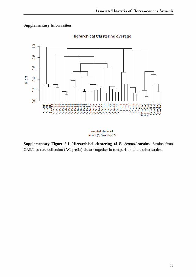
Associated bacteria of Botryococcus braunii
53
Supplementary Information
Supplementary Figure 3.1. Hierarchical clustering of B. braunii strains. Strains from CAEN culture collection (AC prefix) cluster together in comparison to the other strains.
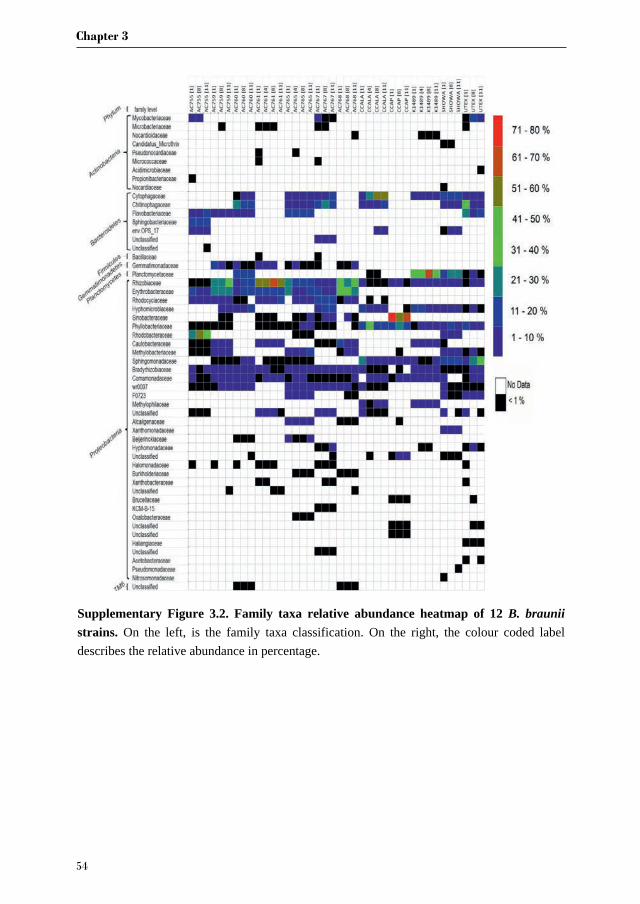
Chapter 3
54
Supplementary Figure 3.2. Family taxa relative abundance heatmap of 12 B. brauniistrains. On the left, is the family taxa classification. On the right, the colour coded label describes the relative abundance in percentage.

Chapter 4
Bacterial diversity in different outdoor pilot plant
photobioreactor types for production of the microalga
Nannochloropsis sp. CCAP211/78
Jie Lian, Georg Steinert, Jeroen de Vree, Sven Meijer, Christa Heryanto,
Rouke Bosma, René H. Wijffels, Maria Barbosa, Hauke Smidt, Detmer Sipkema
Manuscript in preparation

Chapter 4
56
Abstract
A range of microalgae production systems are scaled up towards pilot and demonstration plants.
As large-scale outdoor production cannot be done in complete containment, cultures are (more)
open for bacteria, which may affect the productivity and stability of the production process. We
investigated the bacterial diversity in two indoor reactors and four pilot-scale outdoor reactors
for production of Nannochloropsis sp. CCAP211/78 spanning four months of operation from
July to October. Illumina sequencing of 16S rRNA gene amplicons demonstrated that a wide
variety of bacteria were present in all reactor types, with predominance of Bacteroidetes and
Alphaproteobacteria. Our results showed that bacterial communities were significantly
different in all reactor types (except a horizontal tubular reactor and a vertical tubular reactor)
and also between runs in each reactor. Bacteria common to the majority of samples included
one member each of the Saprospiraceae family and of the NS11-12_marine group (both
Bacteroidetes). Hierarchical clustering analysis revealed two phases during the cultivation
period separated by a major shift in bacterial community composition in the horizontal tubular
reactor, the vertical tubular reactor and the raceway pond. The bacterial classes
Alphaproteobacteria, Sphingobacteriia, Deltaproteobacteria and Flavobacteriia contributed
most to the difference between the two phases, with a stark decrease of the Saprospiraceae and
NS11-12_marine group that initially dominated the bacterial communities. Furthermore, we
observed a less consistent pattern of bacterial taxa appearing in different reactors and runs, most
of which belonging to the classes Deltaproteobacteria and Flavobacteriia. In addition,
canonical correspondence analysis showed that the nitrate concentration in the reactor
significantly correlated with bacterial community composition in most reactor types except the
plastic flat panel reactor, where algal biomass productivity was the key factor associated with
changes in bacterial community structure. This study contributes to our understanding of
bacterial diversity and composition in different types of outdoor reactors exposed to a range of

Bacteria in outdoor photobioreactors
57
dynamic biotic and abiotic factors. Differences in bacterial community composition between
reactor types should be given more attention in order to improve the stability and success rate
for large scale outdoor cultivation of microalgae.
Keywords: Nannochloropsis, outdoor reactors, algae-bacterial interactions, bacterial
community composition
Chapter 4
58
4.1. Introduction
Microalgae are one of the most promising feedstocks for production of food, feed, biofuels and
other valuable chemicals [56, 263]. Algal cultivation does not necessarily compete for arable
land and needs much less water to produce the same amount of biofuel compared to oil crops
[56, 264]. Nevertheless, although algae have many appealing advantages as alternative cell
factories, algal bulk products are still far away from large-scale application in industry due to
high production costs [265].
Scale up of algae cultivation is carried out in different systems, but most commonly in shallow
open ponds or in enclosed plastic tubular photobioreactors [266]. Despite the fact that a good
number of systems has been proposed and tested, the industry is far from settled on a single
approach. The high performance of algal strains in the laboratory can hardly be accomplished
in large-scale outdoor cultivation systems because of varying ambient conditions, including
physicochemical and biological factors [267]. Both open and closed outdoor algae production
systems cannot easily be operated strictly axenically and are thus prone to microbial
contamination. This is a substantial discrepancy compared to laboratory-based studies where
whole reactors can be autoclaved. Therefore, in pilot-scale operation, bacteria present in
photobioreactors cannot be ignored as is often the case for laboratory-based studies. However,
relatively little is known about the bacteria present in algal photobioreactors and about their
effects on algal cultivation [153, 268].
An increasing number of bacteria has been reported to be detrimental to microalgae and can
cause mass algal cell destruction. Harmful impacts may be imposed through direct algal-
bacterial cell contact, such as for the lytic bacteria Saprospira sp. (SS98-5) [269],
Pseudoalteromonas sp. (J18/M01) [31] and Microbacterium sp. LB1 [32]. In addition, the
synthesis of extracellular algicidal compounds may kill the algal host. For instance,
Bacteria in outdoor photobioreactors
59
Streptomyces malaysiensis O4-6 was shown to release compound NIG355 capable of killing
nearly 80% of Phaeocystis globosa in 24 h [33]. Nevertheless, recent studies have revealed
that mutualistic relationships between algae and bacteria may even occur more commonly than
antagonistic interactions [2, 40]. Associated bacteria benefit algal growth in mainly three ways.
First of all, bacteria are key players in decomposing and mineralizing algal waste components,
recycling carbon and phosphorus and making them again available for the algae [137, 144].
Secondly, bacteria can benefit algae through synthesizing a wide range of molecules ranging
from vitamins [22, 26], phytohormones [16, 270], to siderophores [98, 271], which can
stimulate algal growth. Lastly, some bacteria are able to kill algicidal bacteria by secreting
antimicrobial compounds, such as tropodithietic acid in exchange for organic carbon [147].
Knowledge of bacterial communities in outdoor microalgae production systems is currently
nearly non-existent. However, the impact of bacteria already presents in the microalgae
inoculum, as well as temporal variation of bacterial communities due to variation of
environmental parameters that are inevitably occurring in outdoor reactors are likely to be
important for robust operation of these production systems. Therefore, we conducted a
longitudinal study to investigate the composition and dynamics of bacterial communities within
two indoor microalgae inoculum-production systems (i.e., a sterile-operated flat panel
bioreactor and a non-sterile tubular indoor bioreactor) and four outdoor pilot-scale systems (i.e.,
an open raceway pond, a horizontal tubular bioreactor, a vertical tubular bioreactor, and a
plastic flat panel reactor) during the production of the microalga Nannochloropsis sp.
CCAP211/78 (the algal production data have been published by Vree et al. [272]) to assess the
impact of non-sterile outdoor photobioreactor operation. One hundred and twenty-eight
samples were collected from indoor and outdoor bioreactors over a period of four months.
Bacterial 16S rRNA gene fragments were amplified and sequenced to determine the
composition of associated bacterial communities.
Chapter 4
60
4.2. Materials and Methods
4.2.1. Algal cultivation and sampling procedures
Nannochloropsis sp. CCAP211/78 was cultivated in 250 mL Erlenmeyer flasks, followed by
cultivation in a flat panel reactor (FP, 4.5 L) and a horizontal tubular indoor reactor (TI, 280 L)
located in a greenhouse at AlgaePARC (Wageningen, the Netherlands). The biomass harvested
from TI was used to inoculate three pilot-scale outdoor reactors within one week: a horizontal
tubular reactor (HT, 560 L), a vertical tubular reactor (VT, 1060 L) and a raceway pond (RP,
4730 L). An outdoor plastic flat panel reactor (PP, 60 L per panel) was inoculated with biomass
directly harvested from the indoor FP. Samples were taken at AlgaePARC every Monday,
Wednesday and Friday morning from July 3rd - October 16th, 2013. Detailed sample information
can be found in Supplementary Figure S4.1. In addition, details of the production process of
Nannochloropsis sp. CCAP211/78 [272] and a detailed description of outdoor reactors [273]
were published previously. Liquid samples of 5 mL from each of the reactors were filtered
through a sterile polycarbonate filter membrane (0.2 m, Millipore) with a vacuum pump. Filter
membranes were then rolled up, placed in 2 ml Eppendorf tubes and stored at -80 until
further processing. Data generated in this study were derived from two separate reactor runs
performed during the above-mentioned period, with the first and second runs being designated
TI1, HT1, VT1, RP1, PP1 and FP2, TI2, HT2, VT2, RP2, PP2, respectively.
4.2.2. DNA extraction, PCR amplification and sequencing
To isolate total DNA, frozen filters were cut into small pieces with sterile scissors. DNA was
extracted from these pieces using the FastDNA SPIN kit for soil (MP Biomedicals, USA) with
the aid of a Precellys bead beater (Bertin Technologies, France) for two rounds of 45 s at a
speed of 5500 rpm. DNA size and quantity were examined by electrophoresis on a 1% agarose

Bacteria in outdoor photobioreactors
61
gel and measured spectrophotometrically with a Nanodrop (ND1000, Thermo Scientific,
Wilmington, USA). The extracted DNA was stored at -20℃ until further use.
Amplicons from the V1-V2 region of bacterial 16S rRNA genes were generated using a two-
step PCR strategy. Forward primer 27F-DegS (5’-GTTYGATYMTGGCTCAG-3’), and an
equimolar mixture of reverse primers 338R I (5’-GCWGCCTCCCGTAGGAGT-3’) and II (5’-
GCWGCCACCCGTAGGTGT-3’) were appended at the 5 end with 18 bp universal tags
(Unitag1: GAGCCGTAGCCAGTCTGC and Unitag2: GCCGTGACCGTGACATCG for the
forward and reverse primers, respectively). as con ucte in a l reaction olume
containing 1 l template 10 µl 5× HF buffer (Thermo Scientific, The Netherlands), 1 µl
dNTP Mix (10 mM; Promega, Leiden, the Netherlands), 1 of each primer 1 U of Phusion®
Hot Start II High-Fidelity DNA polymerase (Thermo Scientific, The Netherlands) and 32.5 µl
nuclease free water (Qiagen, Germany). The PCR profile included the following steps: Pre-
denaturation at 98°C for 30 s, followed by 25 cycles of denaturation (10 s at 98°C), annealing
(20 s at 56°C), extension (20 s at 72°C), and a final elongation (10 min at 72°C). The PCR
product size was examined by 1% agarose gel electrophoresis. The second PCR was conducted
in a 100 l reaction olume containin µl of the first PCR product, 20 µl 5× HF buffer
(Thermo Scientific), 2 µl dNTP Mix (10 mM; Promega), 500 nM forward and reverse primer
(equivalent to the Unitag1 and Unitag2 sequences, respectively) that were each appended with
an 8 nt sample-specific barcode [231], 2 U of Phusion® Hot Start II High-Fidelity DNA
polymerase (Thermo Scientific) and 62 µl nuclease free water (Qiagen). The PCR conditions
were pre-denaturation at 98°C for 30 s, followed by 5 cycles of denaturation (10 s at 98°C),
annealing (20 s at 52°C), extension (20 s at 72°C), and a final elongation (10 min at 72°C). The
PCR product was examined by gel electrophoresis and purified with the DNA HighPrep kit
(Magbio Genomics, Rockville, MD, USA). The concentration of PCR products was quantified
with a Qubit Fluorometer (Life Technologies, Darmstadt, Germany) in combination with the
Chapter 4
62
dsDNA BR Assay kit (Invitrogen, Carlsbad, CA, USA). Purified products were then pooled in
equimolar amounts (200 ng l-1) and sequenced on a MiSeq platform (GATC-Biotech, currently
part of Eurofins Genomics Germany GmbH, Konstanz, Germany).
4.2.3. Processing of MiSeq data
Illumina sequencing data was processed using the NG-Tax pipeline [274]. In short, paired-end
reads of 2 x 100 nucleotides were combined and filtered to retain only read pairs with perfectly
matching primers and barcodes. Demultiplexing, Operational Taxonomic Unit (OTU) picking,
chimera removal and taxonomic assignment were performed within one single step in NG-Tax.
Filtered sequences were ranked per sample by abundance and unique OTUs (at a 100% identity
level) were added to an initial OTU table for that sample starting from the most abundant
sequence until the abundance was lower than 0.1%. The final OTU table was created by
clustering the reads that were initially discarded to the OTUs from the initial OTU table with a
threshold of 99% similarity. Taxonomic assignment was done using the UCLUST algorithm
[275] and a customized SILVA SSU Ref 111 database [232]. Samples with less than 1000 reads
(Bacterial 16S rRNA gene reads plus chloroplast 16S rRNA gene reads) were removed, and all
chloroplast 16S rRNA reads were removed from the dataset. The number of retained reads for
each sample was calculated again, and samples with less than 100 bacterial 16S rRNA gene
reads were removed as well.
4.2.4. Statistical analysis
All statistical tests were performed in R (v.3.1.2) [235]. First, the OTU table was standardized
by a square root transformation using the decostand function (method = “hellinger”) from the
vegan package [234]. Transformed data was subsequently used to calculate alpha-diversity
indices (Shannon diversity and Richness). Pairwise comparison of alpha-diversity between the
different reactors within each run was calculated using Wilcoxon rank sum test and Benjamini-
Bacteria in outdoor photobioreactors
63
Hochberg p-value adjustment as implemented in the “STATs” package [235, 276]. For further
multivariate analyses the vegdist function from the vegan package was used to create a Bray-
Curtis dissimilarity matrix of the standardised OTU table. Hierarchical clustering of all samples
based on the Bray-Curtis dissimilarity matrix was performed using the “average” method. Then,
a non-metric multidimensional (nMDS) scaling plot was generated using the metaMDS function
based on pairwise Bray-Curtis distances. Overall differences in bacterial communities between
reactors were assessed statistically with PERMANOVA (adonis) from the vegan package.
PERMANOVA was also performed to test whether bacterial communities between reactor
types are significantly different. Detrended Correspondence Analysis (DCA) was performed
and Canonical Correspondence Analysis (CCA) was the best constrained ordination model for
the bacterial communities. Significance of the environmental factors was tested by the envfit
function with 999 Monte Carlo permutations. The overall significance of CCA and of each axis
were tested by analysis of variance (ANOVA) permutation tests. Pearson’s correlation analysis
between each pair of parameters measured in this study was done using rcorr function in
“Hmisc” package. The OTU heatmap was created with the “pheatmap” package.
4.3. Results
4.3.1 Bacterial community profiles
Bacterial community composition dynamics in six different photobioreactors in which
Nannochloropsis sp. CCAP211/78 was grown, was investigated with Illumina MiSeq amplicon
sequencing of the V1-V2 region of bacterial 16S rRNA genes. After removing twenty-one low-
quality samples, we retained 3,574,708 high-quality sequences with an average of 33,408 reads
per sample. These sequences represented 1,217 operational taxonomic units (OTUs). A total of
2,703,376 reads (75.6% of all retained reads, 237 OTUs) were derived from Nannochloropsis
chloroplasts. After removal of chloroplast OTUs from the dataset, 980 bacterial OTUs were
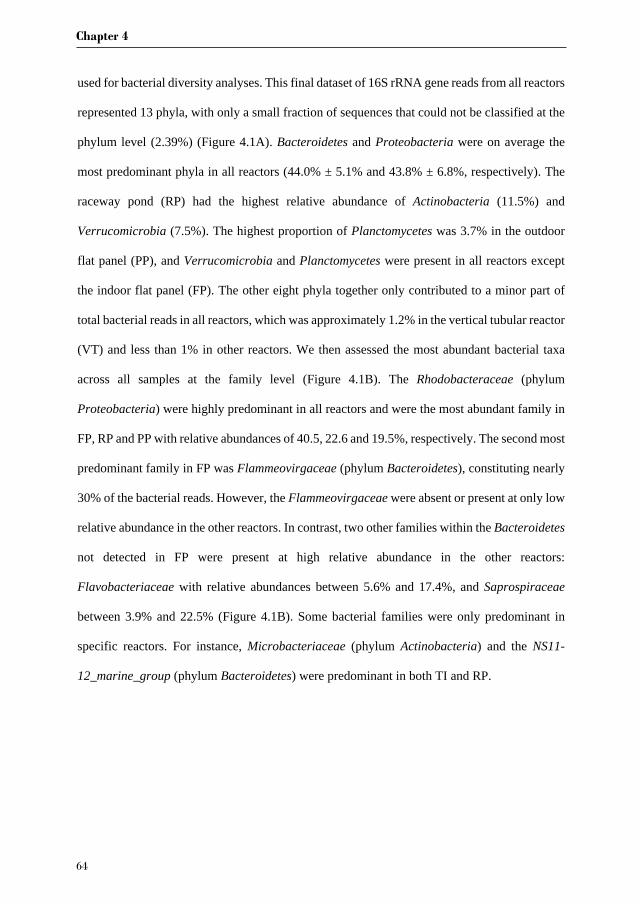
Chapter 4
64
used for bacterial diversity analyses. This final dataset of 16S rRNA gene reads from all reactors
represented 13 phyla, with only a small fraction of sequences that could not be classified at the
phylum level (2.39%) (Figure 4.1A). Bacteroidetes and Proteobacteria were on average the
most predominant phyla in all reactors (44.0% ± 5.1% and 43.8% ± 6.8%, respectively). The
raceway pond (RP) had the highest relative abundance of Actinobacteria (11.5%) and
Verrucomicrobia (7.5%). The highest proportion of Planctomycetes was 3.7% in the outdoor
flat panel (PP), and Verrucomicrobia and Planctomycetes were present in all reactors except
the indoor flat panel (FP). The other eight phyla together only contributed to a minor part of
total bacterial reads in all reactors, which was approximately 1.2% in the vertical tubular reactor
(VT) and less than 1% in other reactors. We then assessed the most abundant bacterial taxa
across all samples at the family level (Figure 4.1B). The Rhodobacteraceae (phylum
Proteobacteria) were highly predominant in all reactors and were the most abundant family in
FP, RP and PP with relative abundances of 40.5, 22.6 and 19.5%, respectively. The second most
predominant family in FP was Flammeovirgaceae (phylum Bacteroidetes), constituting nearly
30% of the bacterial reads. However, the Flammeovirgaceae were absent or present at only low
relative abundance in the other reactors. In contrast, two other families within the Bacteroidetes
not detected in FP were present at high relative abundance in the other reactors:
Flavobacteriaceae with relative abundances between 5.6% and 17.4%, and Saprospiraceae
between 3.9% and 22.5% (Figure 4.1B). Some bacterial families were only predominant in
specific reactors. For instance, Microbacteriaceae (phylum Actinobacteria) and the NS11-
12_marine_group (phylum Bacteroidetes) were predominant in both TI and RP.
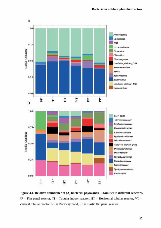
Bacteria in outdoor photobioreactors
65
Figure 4.1. Relative abundance of (A) bacterial phyla and (B) families in different reactors.
FP = Flat panel reactor, TI = Tubular indoor reactor, HT = Horizontal tubular reactor, VT =
Vertical tubular reactor, RP = Raceway pond, PP = Plastic flat panel reactor.
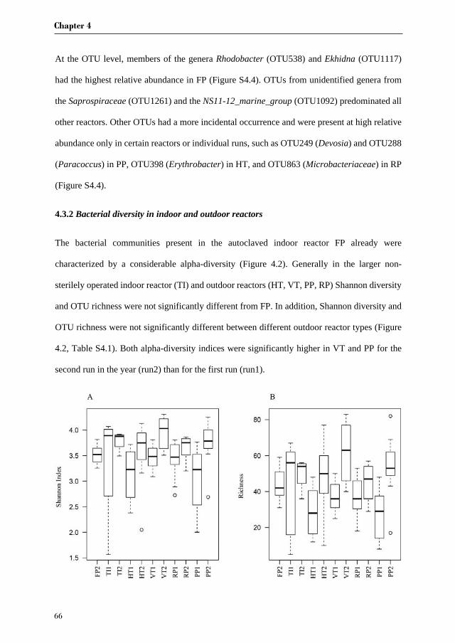
Chapter 4
66
At the OTU level, members of the genera Rhodobacter (OTU538) and Ekhidna (OTU1117)
had the highest relative abundance in FP (Figure S4.4). OTUs from unidentified genera from
the Saprospiraceae (OTU1261) and the NS11-12_marine_group (OTU1092) predominated all
other reactors. Other OTUs had a more incidental occurrence and were present at high relative
abundance only in certain reactors or individual runs, such as OTU249 (Devosia) and OTU288
(Paracoccus) in PP, OTU398 (Erythrobacter) in HT, and OTU863 (Microbacteriaceae) in RP
(Figure S4.4).
4.3.2 Bacterial diversity in indoor and outdoor reactors
The bacterial communities present in the autoclaved indoor reactor FP already were
characterized by a considerable alpha-diversity (Figure 4.2). Generally in the larger non-
sterilely operated indoor reactor (TI) and outdoor reactors (HT, VT, PP, RP) Shannon diversity
and OTU richness were not significantly different from FP. In addition, Shannon diversity and
OTU richness were not significantly different between different outdoor reactor types (Figure
4.2, Table S4.1). Both alpha-diversity indices were significantly higher in VT and PP for the
second run in the year (run2) than for the first run (run1).
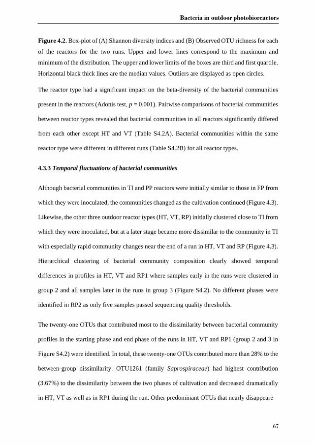
Bacteria in outdoor photobioreactors
67
Figure 4.2. Box-plot of (A) Shannon diversity indices and (B) Observed OTU richness for each
of the reactors for the two runs. Upper and lower lines correspond to the maximum and
minimum of the distribution. The upper and lower limits of the boxes are third and first quartile.
Horizontal black thick lines are the median values. Outliers are displayed as open circles.
The reactor type had a significant impact on the beta-diversity of the bacterial communities
present in the reactors (Adonis test, p = 0.001). Pairwise comparisons of bacterial communities
between reactor types revealed that bacterial communities in all reactors significantly differed
from each other except HT and VT (Table S4.2A). Bacterial communities within the same
reactor type were different in different runs (Table S4.2B) for all reactor types.
4.3.3 Temporal fluctuations of bacterial communities
Although bacterial communities in TI and PP reactors were initially similar to those in FP from
which they were inoculated, the communities changed as the cultivation continued (Figure 4.3).
Likewise, the other three outdoor reactor types (HT, VT, RP) initially clustered close to TI from
which they were inoculated, but at a later stage became more dissimilar to the community in TI
with especially rapid community changes near the end of a run in HT, VT and RP (Figure 4.3).
Hierarchical clustering of bacterial community composition clearly showed temporal
differences in profiles in HT, VT and RP1 where samples early in the runs were clustered in
group 2 and all samples later in the runs in group 3 (Figure S4.2). No different phases were
identified in RP2 as only five samples passed sequencing quality thresholds.
The twenty-one OTUs that contributed most to the dissimilarity between bacterial community
profiles in the starting phase and end phase of the runs in HT, VT and RP1 (group 2 and 3 in
Figure S4.2) were identified. In total, these twenty-one OTUs contributed more than 28% to the
between-group dissimilarity. OTU1261 (family Saprospiraceae) had highest contribution
(3.67%) to the dissimilarity between the two phases of cultivation and decreased dramatically
in HT, VT as well as in RP1 during the run. Other predominant OTUs that nearly disappeare
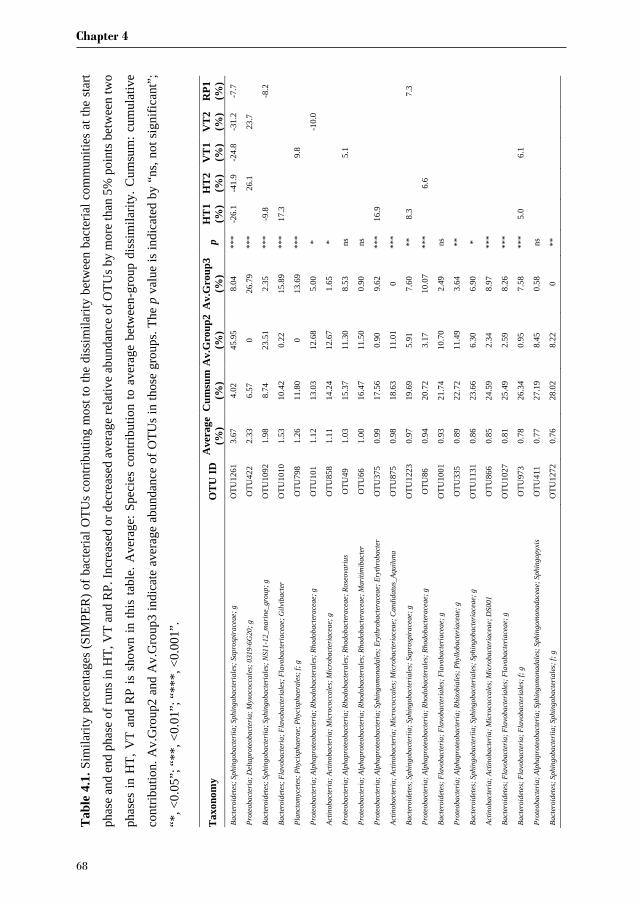
Chapter 4
68
Tab
le 4
.1. S
imila
rity
perc
enta
ges
(SIM
PER
) of b
acte
rial O
TUs
cont
ribut
ing
mos
t to
the
diss
imila
rity
betw
een
bact
eria
l com
mun
ities
at t
he s
tart
phas
e an
d en
d ph
ase
of ru
ns in
HT,
VT
and
RP.
Incr
ease
d or
dec
reas
ed a
vera
ge re
lativ
e ab
unda
nce
of O
TUs b
y m
ore
than
5%
poi
nts b
etw
een
two
phas
es in
HT,
VT
and
RP
is s
how
n in
this
tabl
e. A
vera
ge: S
peci
es c
ontri
butio
n to
ave
rage
bet
wee
n-gr
oup
diss
imila
rity.
Cum
sum
: cum
ulat
ive
cont
ribut
ion.
Av.
Gro
up2
and
Av.
Gro
up3
indi
cate
ave
rage
abu
ndan
ce o
f OTU
s in
thos
e gr
oups
. The
p v
alue
is in
dica
ted
by “
ns, n
ot s
igni
fican
t”;
“*, <
0.05
”; “
**, <
0.01
”; “
***,
<0.
001”
.
Tax
onom
y O
TU
ID
Ave
rage
(%
) C
umsu
m
(%)
Av.
Gro
up2
(%)
Av.
Gro
up3
(%)
p H
T1
(%)
HT
2 (%
) V
T1
(%)
VT
2 (%
) R
P1
(%)
Bac
tero
idet
es; S
phin
goba
cter
iia; S
phin
goba
cter
iale
s; S
apro
spir
acea
e; g
O
TU12
61
3.67
4.
02
45.9
5 8.
04
***
-26.
1 -4
1.9
-24.
8 -3
1.2
-7.7
Pro
teob
acte
ria; D
elta
prot
eoba
cter
ia; M
yxoc
occa
les;
031
9-6G
20; g
O
TU42
2 2.
33
6.57
0
26.7
9 **
*
26.1
23.7
Bac
tero
idet
es; S
phin
goba
cter
iia; S
phin
goba
cter
iale
s; N
S11-
12_m
arin
e_gr
oup;
g
OTU
1092
1.
98
8.74
23
.51
2.35
**
* -9
.8
-8
.2
Bac
tero
idet
es; F
lavo
bact
eria
; Fla
voba
cter
iale
s; F
lavo
bact
eria
ceae
; Gilv
ibac
ter
OTU
1010
1.
53
10.4
2 0.
22
15.8
9 **
* 17
.3
Pla
ncto
myc
etes
; Phy
cisp
haer
ae; P
hyci
spha
eral
es; f
; g
OTU
798
1.26
11
.80
0 13
.69
***
9.8
Pro
teob
acte
ria; A
lpha
prot
eoba
cter
ia; R
hodo
bact
eral
es; R
hodo
bact
erac
eae;
g
OTU
101
1.12
13
.03
12.6
8 5.
00
*
-10.
0
Act
inob
acte
ria; A
ctin
obac
teria
; Mic
roco
ccal
es; M
icro
bact
eria
ceae
; g
OTU
858
1.11
14
.24
12.6
7 1.
65
*
Pro
teob
acte
ria; A
lpha
prot
eoba
cter
ia; R
hodo
bact
eral
es; R
hodo
bact
erac
eae;
Ros
eova
rius
OTU
49
1.03
15
.37
11.3
0 8.
53
ns
5.1
Pro
teob
acte
ria; A
lpha
prot
eoba
cter
ia; R
hodo
bact
eral
es; R
hodo
bact
erac
eae;
Mar
itim
ibac
ter
OTU
66
1.00
16
.47
11.5
0 0.
90
ns
Pro
teob
acte
ria; A
lpha
prot
eoba
cter
ia; S
phin
gom
onad
ales
; Ery
thro
bact
erac
eae;
Ery
thro
bact
er
OTU
375
0.99
17
.56
0.90
9.
62
***
16.9
Act
inob
acte
ria; A
ctin
obac
teria
; Mic
roco
ccal
es; M
icro
bact
eria
ceae
; Can
dida
tus_
Aqui
luna
O
TU87
5 0.
98
18.6
3 11
.01
0 **
*
Bac
tero
idet
es; S
phin
goba
cter
iia; S
phin
goba
cter
iale
s; S
apro
spir
acea
e; g
O
TU12
23
0.97
19
.69
5.91
7.
60
**
8.3
7.
3
Pro
teob
acte
ria; A
lpha
prot
eoba
cter
ia; R
hodo
bact
eral
es; R
hodo
bact
erac
eae;
g
OTU
86
0.94
20
.72
3.17
10
.07
***
6.
6
Bac
tero
idet
es; F
lavo
bact
eria
; Fla
voba
cter
iale
s; F
lavo
bact
eria
ceae
; g
OTU
1001
0.
93
21.7
4 10
.70
2.49
ns
Pro
teob
acte
ria; A
lpha
prot
eoba
cter
ia; R
hizo
bial
es; P
hyllo
bact
eria
ceae
; g
OTU
335
0.89
22
.72
11.4
9 3.
64
**
Bac
tero
idet
es; S
phin
goba
cter
iia; S
phin
goba
cter
iale
s; S
phin
goba
cter
iace
ae; g
O
TU11
31
0.86
23
.66
6.30
6.
90
*
Act
inob
acte
ria; A
ctin
obac
teria
; Mic
roco
ccal
es; M
icro
bact
eria
ceae
; DS0
01
OTU
866
0.85
24
.59
2.34
8.
97
***
Bac
tero
idet
es; F
lavo
bact
eria
; Fla
voba
cter
iale
s; F
lavo
bact
eria
ceae
; g
OTU
1027
0.
81
25.4
9 2.
59
8.26
**
*
Bac
tero
idet
es; F
lavo
bact
eria
; Fla
voba
cter
iale
s; f;
g
OTU
973
0.78
26
.34
0.95
7.
58
***
5.0
6.
1
Pro
teob
acte
ria; A
lpha
prot
eoba
cter
ia; S
phin
gom
onad
ales
; Sph
ingo
mon
adac
eae;
Sph
ingo
pyxi
s O
TU41
1 0.
77
27.1
9 8.
45
0.58
ns
Bac
tero
idet
es; S
phin
goba
cter
iia; S
phin
goba
cter
iale
s; f;
g
OTU
1272
0.
76
28.0
2 8.
22
0 **

Bacteria in outdoor photobioreactors
69
in the end phase were OTU1092 (NS11-12_marine_group) in HT1 and RP1 and OTU101
(Rhodobacteraceae) in VT2. Eight OTUs were strongly increased in the late phase of the runs.
These mostly varied by reactor type and run but many belonged to the classes
Alphaproteobacteria and Flavobacteriia (Table 4.1).
Figure 4.3. Non-metric multidimensional scaling (nMDS) of Bray-Curtis distances based on
normalised relative abundance of OTUs in bacterial communities in six reactor types (different
colours). Each reactor was run twice (except FP), and samples from the first run are indicated
by circles and the second run by squares. Inoculation is indicated by green arrows. First run
samples are sequentially labelled with lower case letters (a-q), and second run samples are
sequentially labelled with upper case letters (A-R). Same letters indicate that samples were
taken at the same day. FP = Flat panel reactor, TI =Tubular indoor reactor, HT = Horizontal
tubular reactor, VT = Vertical tubular reactor, PP = Plastic flat panel reactor, RP = Raceway
pond.
4.3.4 Environmental drivers of Nannochloropsis-associated bacterial community
development
To identify the main driver(s) underlying temporal changes in bacterial community composition
in different reactors as well as differences in bacterial community composition between
Chapter 4
70
different reactor types, the correlation of temperature (Temp), pH, nitrate concentration (NO3-),
photon flux density (PFD), and algal biomass productivity (PRO, defined as volumetric
productivity: g L-1 d-1) with bacterial community structure was investigated. Overall, only
approximately 4.8 % of the compositional variation could be explained by the first axis and
3.5 % by the second axis of the CCA using the parameters evaluated in this study (Figure 4.4A).
The bacterial community in PP correlated best with PRO and Temp, while NO3-, PFD and pH
correlated best with bacterial community composition in all samples of FP and part of the
samples in TI, HT, VT and RP (Figure 4.4A). Another part of the HT and VT samples were
correlated with lower values of Temp, PFD and PRO. From the parameters we measured, Temp,
PFD and PRO were positively correlated with each other (Figure 4.4B). By contrast, NO3- was
negatively correlated with PRO and PFD. This trend also corresponded with the observation
that the first run in the outdoor reactors was characterized by higher PRO than the second run,
which likely resulted from higher Temp and PFD in the first run (Figure S4.5).

Bacteria in outdoor photobioreactors
71
Figure 4.4. (A) Canonical correspondance analysis (CCA) showing correlation between
bacterial communities (response variables) and environmental factors (explanatory variables).
The percentage of variation in the bacterial community explained by each axis is indicated in
parentheses after the axis label. The environmental factors with * significantly contribute to
explaining the observed variation in bacterial community composition (p < 0.05). (B) Pearson
correlation analysis between environmental factors. Only correlation coefficients with p < 0.05
are indicated. The factors included are Temp (temperature), pH, NO3- (nitrate concentration),
PFD (photon flux density), PRO (algal biomass productivity).
Chapter 4
72
4.4. Discussion
Bacteria in association with microalgae have rarely been investigated in large microalgae
culture systems and the studies that did, only assessed one type of outdoor reactor. For instance,
bacterial communities were analysed before in a 300 L outdoor flat panel reactor with
Tetraselmis suecica [118], in a 550 L outdoor tank with Nannochloropsis salina [91], in a 200
L polyethylene flat panel reactor with Nannochloropsis salina [69] and in a 1600 L enclosed
membrane reactor with Desmodesmus and Scenedesmus for treatment of domestic waste water
[162].
4.4.1 Differences between reactors
We compared for the first time the bacterial communities of four pilot-scale outdoor
photobioreactors operated under identical climatological conditions for the production of
Nannochloropsis sp. CCAP211/78. We found bacterial communities were significantly
different between FT, TI and the outdoor reactors (Figure 4.1A and Table S4.2). This result was
in accordance with a previous study that showed that the bacterial community of
Nannochloropsis salina differed between small indoor reactors (Volume: 5 mL-4 L), medium
indoor reactors (Volume: 20-60 L) and a large outdoor reactor (Volume: 200 L) [69]. Fulbright
et al. [69] also reported that OTU richness increased as the size of the reactors increased.
However, neither OTU richness nor Shannon index was significantly different between reactor
types in this study (Figure 4.2, Table S4.2A). This finding reveals that Nannochloropsis cultures
in FP were already colonised by diverse bacteria before being harvested to inoculate other
reactors. We observed a close association between inoculum samples in one reactor and receiver
samples in the other reactor (Figure 4.3), which corroborates that the initial bacterial community
composition in each reactor is largely determined by the bacterial community in precultures
from which the subsequent reactors was inoculated.

Bacteria in outdoor photobioreactors
73
Despite differences in bacterial community composition between reactor types, the most
abundant bacterial phyla in all reactors were similar, with predominance of Proteobacteria
(predominantly Alphaproteobacteria) and Bacteroidetes (Figure 4.1). In previous studies,
Alphaproteobacteria and Bacteroidetes were shown to be the most abundant phyla in
Nannochloropsis cultures [69, 87, 277]. Several bacterial families found in this study are similar
to those found in the cultures of N. salina, which include members of the families
Saprospiraceae, Phyllobacteriaceae, Hyphomonadaceae, Rhodobacteraceae and
Alteromonadaceae [69, 278]. The occurrence of the same taxa associated with Nannochloropsis
species in different environments and locations suggests that these bacteria may have specific
interactions with Nannochloropsis [87, 279]. For example, members of the Phyllobacteriaceae
have been shown to enhance the growth of algae through vitamin supplementation [22] and
nitrogen fixation [105].
At the OTU level, the FP bacterial community was predominated by a few highly abundant
OTUs, whereas more diverse OTUs were found in the larger reactors (Figure S4.4). Three
predominant representatives in FP (Rhodobacter_OTU538, Ekhidna_OTU1117 and
Balneola_OTU835) have previously been found either in cultures of Nannochloropsis oculata
[92], Ectocarpus sp. [103] or Emiliania huxleyi [280]. Yet their roles in algal cultures have not
been characterized. The most abundant taxon (OTU1261) in TI and all outdoor reactors belongs
to the Saprospiraceae (Figure S4.4). The best hit returned by a Blast search against the NCBI
nr/nt database is Phaeodactylibacter xiamenensis (100% identity), which was isolated from a
culture of the marine diatom Phaeodactylum tricornutum [281]. Although two Saprospiraceae-
related OTUs were also observed in the inflowing seawater (Table S4.4), these were different
from the most abundant OTU (OTU1261) and represented only minor fractions of the bacteria
found in the different reactors. In addition, OTU1261 is not closely related to the lytic bacterium
Saprospira sp. (92% identity) that was reported to kill and lyse the cells of the diatom
Chapter 4
74
Chaetoceros ceratosporum [269]. A bacterium belonging to the Saprospiraceae family was
previously found to be most abundant on average (comprising 34.7% ± 14.3% of bacterial
communities) in large-scale cultures of Nannochloropsis salina in a closed polyethylene growth
panel (0.05 m wide × 0.28 m high × 17.3 m long) located outdoor in a water basin [69].
Although no correlation was observed between relative abundance of Saprospiraceae and N.
salina growth performance [69], its ubiquitous predominance in all mass culture systems both
in a previous study and this study suggests that there are important interactions between
members of this family and Nannochloropsis or at least a commensal relationship. Another
OTU common to TI and outdoor reactor samples was classified as a member of the NS11-
12_marine group (Bacteroidetes) that has been mainly detected in marine environments [282].
However, as the unresolved taxonomy indicates, we still know little of their ecological roles.
Some genera with random occurrence in certain reactors or runs, such as Devosia (OTU249),
Paracoccus (OTU288) and Erythrobacter (OTU398) (Figure S4), have been frequently found
to associate with either seaweeds [283-285] or microalgae [286]. Devosia sp. was inferred to
play a role in nitrogen fixation as an epiphytic bacterium associated with the macroalga
Cladophora glomerata [287], and may have a similar interaction with Nannochloropsis.
Paracoccus as well as Erythrobacter were reported to be diatom-associated and found to be
resistant to polyunsaturated aldehydes released by diatom cells upon disruptions by grazers,
suggesting co-evolution of resistance to toxic molecules in diatoms and their associated bacteria
[96].
The longitudinal sampling strategy helped us examine the influence of biotic and abiotic factors
on the structure of Nannochloropsis sp. CCAP211/78-associated bacterial communities.
Temperature, salinity and nutrient concentration (nitrate) are the most important factors
structuring bacterial communities in aquatic environments, such as in estuaries and coastal
seawater [288-290]. Salinity was not measured in this study because the fluctuation in salinity
Bacteria in outdoor photobioreactors
75
is negligible in our experimental setup. To this end, it should be noted that salinity fluctuation
of RP due to evaporation or rain was adjusted by daily addition of fresh water or sodium
chloride. Temperature and nitrate can directly affect bacterial growth, but also influence algal
growth [291, 292], which would in turn affect bacterial growth. Similarly, light intensity and
pH can affect the growth of both algae and bacteria [293, 294]. Correspondence canonical
analysis revealed that nitrate is a primary factor that drives variation in bacterial community
composition in all reactor types except PP. Nitrate is a key chemical that influences microbial
communities through its effects on nutrient utilization and growth [295, 296]. Many bacteria
can utilize nitrate and even compete with algae when nitrate concentration is low [96, 297, 298].
Besides, nitrate was actively consumed by Nannochloropsis, which was demonstrated by the
negative correlation between algae biomass productivity and nitrate concentrations (Figure 4B).
These results indicate that the bacterial community is at least partly structured by the availability
of nitrate but also by the growth of algae. On the other hand, the distinct bacterial community
composition in PP could be explained by the highest biomass productivity of Nannochloropsis.
It is likely that algal physiology and metabolites released by microalgae could substantially
contribute to the distinctness of bacterial communities [272]. It should be noted that the biggest
part of bacterial community variation cannot be explained by the monitored factors included in
the CCA (Figure 4.4A). The omission of some important environmental factors, such as
phosphorus concentration and dissolved organic matter, could be a reason. These environmental
factors were previously shown to affect microbial community composition in marine waters
[299, 300] and should be measured in future studies. In addition, stochastic effects related to
microorganisms entering the reactors from the outside could contribute to the different changes
of bacterial community composition in different systems. For instance, bacteria may enter
reactors through the addition of seawater for the daily dilution of algal biomass, which is

Chapter 4
76
supported by the observation that a range of bacteria are shared between seawater samples and
microalgal cultures (Table S4.3).
4.4.2 Differences between runs
Bacterial community composition was significantly different between runs. Presumably, one
factor governing this difference relates directly to inoculation. Specifically the bacterial
community of the inoculum used for the first run was different from the inoculum for the second
run (Figure 4.3). More importantly, since the first run spanned the period from July till August
and the second run spanned the period from August till October, temperature and light intensity
differed between both runs, which may directly or indirectly change the bacterial community.
These discrepancies between two runs might also be linked to the observation that both alpha-
diversity indices were seemingly higher for the second run than for the first run for all outdoor
reactor types. In marine environments, the maximum OTU richness and evenness were found
in a temperature range from 15 °C to 20 °C, with lower diversity both above and below those
temperatures [301]. All our outdoor reactors were operated at an average temperature >20 °C
(except HT2) and the temperature was at least 2 °C higher during the first run than during the
second run (Figure S4.5). Therefore, the higher temperature in the first run may have led to the
reduction of both alpha-diversity indices. Furthermore, the higher algal biomass productivity
of the first run might have resulted in higher concentrations of extracellular organic compounds,
which favour the growth and dominance of fast growing copiotrophic bacteria and thus
lowering OTU richness and diversity. In addition, this observation can be supported by
independent studies that have found a decrease of OTU richness and/or Shannon diversity
during algal blooms [302-304].
4.4.3 Bacterial community dynamics within runs
Bacteria in outdoor photobioreactors
77
Bacterial community composition also varied within runs in all reactor types from the start of
monitoring to the end (Figure 4.3). The FP reactor showed least variation, whereas this variation
was more apparent in outdoor reactors. Presumably, this variation was caused by the inherently
more variable environmental conditions (temperature, for instance) that were not as well
controlled as in the indoor reactors. We identified a substantial number of the OTUs that
increased pronouncedly in relative abundance near the end of the cultivation in outdoor runs in
HT, VT and RP. These OTUs were annotated as members of the Flavobacteriaceae (2 OTUs)
and Rhodobacteraceae (3 OTUs). These two families were also shown to be dominant in the
stationary phase of batch cultures of Nannochloropsis salina [279], as well as in algal blooms
[305] and in a range of algal production systems in general [117, 162, 306]. Bacteria belonging
to the Flavobacteriaceae are fast-growing specialists observed during algae blooms and
specialize in the degradation of algal-derived complex organic matter [304, 307]. Members of
the Rhodobacteraceae are often most abundant in bacterial communities that are closely
associated with marine algae, including natural phytoplankton blooms and algal cultures [308,
309]. The frequent occurrence of Flavobacteriaceae and Rhodobacteraceae from independent
studies emphasizes the specialized fitness of these taxa for thriving in algal cultures [279].
Three OTUs showed a strong decrease in relative abundance at the end of cultivation including
the most prevalent OTU (Saprospiraceae_OTU1261). It has been shown that the growth phase
and physiological state of algal cultures could serve as selective factors affecting bacterial
composition and governing bacterial community structure [306]. As the growth of
Nannochloropsis sp. CCAP211/78 in outdoor reactors at the end phase of cultivation is often
associated with fouling and contamination as indicated by Vree at al. [272], the observed shift
of the predominant bacterial taxa in relative abundance near the end phase could potentially be
a first indicator of culture instability [310]. Nevertheless, mechanistic insights are needed to
Chapter 4
78
understand the observed correlations of certain bacteria with the growth performance of
Nannochloropsis sp. CCAP211/78.
4.5. Conclusions
16S rRNA gene amplicon sequencing enabled us to gain detailed insights into composition and
dynamics of bacterial communities of Nannochloropsis sp. CCAP211/78 cultures grown under
a range of environmental conditions and different pilot-scale photobioreactor types. We showed
changes in bacterial community composition during the successional scaling up process of algal
cultivation from a small indoor reactor to large outdoor reactors. Each reactor type had a
significantly different bacterial community composition except HT and VT. Bacterial
community composition also significantly differed between runs of each reactor type. The
inoculum source played a critical role in determining the initial bacterial community
composition of each reactor type, whereas the physio-chemical factors affected later
development of bacterial community composition. Nitrate concentration was the main abiotic
factor that could be identified in this study regulating diversity and composition of the bacterial
community in all reactors except PP where algal biomass productivity had a significant impact
on shaping community structure. Although interactions between the bacterial community and
biotic and abiotic factors across different reactors were explored in our study, a large fraction
of the observed variation in community structure could not be explained by the variables we
measured. We also identified a number of bacterial species with large changes in their relative
abundance between the start and end of the cultivation of Nannochloropsis sp. CCAP211/78
and they may serve as a potential indicator of microalgal culture stability

Bacteria in outdoor photobioreactors
79
Supplementary Information

Chapter 4
80
Figure S4.1. (A) Sampling and inoculating information for six reactors of two separate runs.
(B) Overview of cultivation systems in this study. FP = Flat panel reactor, TI = Tubular indoor
reactor, HT = Horizontal tubular reactor, VT = Vertical tubular reactor, RP = Raceway pond,
PP = Plastic flat panel reactor. Pictures were taken at AlgaePARC, Wageningen, the
Netherlands.
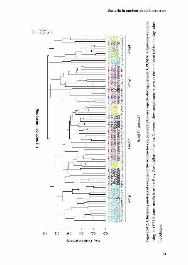
Bacteria in outdoor photobioreactors
81
Figu
re S
4.2.
Clu
ster
ing
anal
ysis
of s
ampl
es o
f the
six
reac
tors
cal
cula
ted
by th
e av
erag
e cl
uste
ring
met
hod
(UPG
MA
). C
lust
erin
g w
as d
one
usin
g th
e O
TU d
ista
nce
mat
rix b
ased
on
Bra
y-C
urtis
(dis
)sim
ilarit
ies.
Num
bers
bel
ow sa
mpl
e na
me
repr
esen
t the
num
ber o
f cul
tivat
ion
days
afte
r
inoc
ulat
ion.
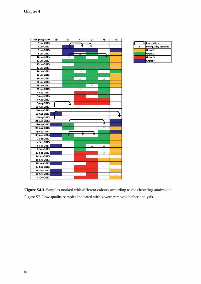
Chapter 4
82
Figure S4.3. Samples marked with different colours according to the clustering analysis in
Figure S2. Low-quality samples indicated with x were removed before analysis.
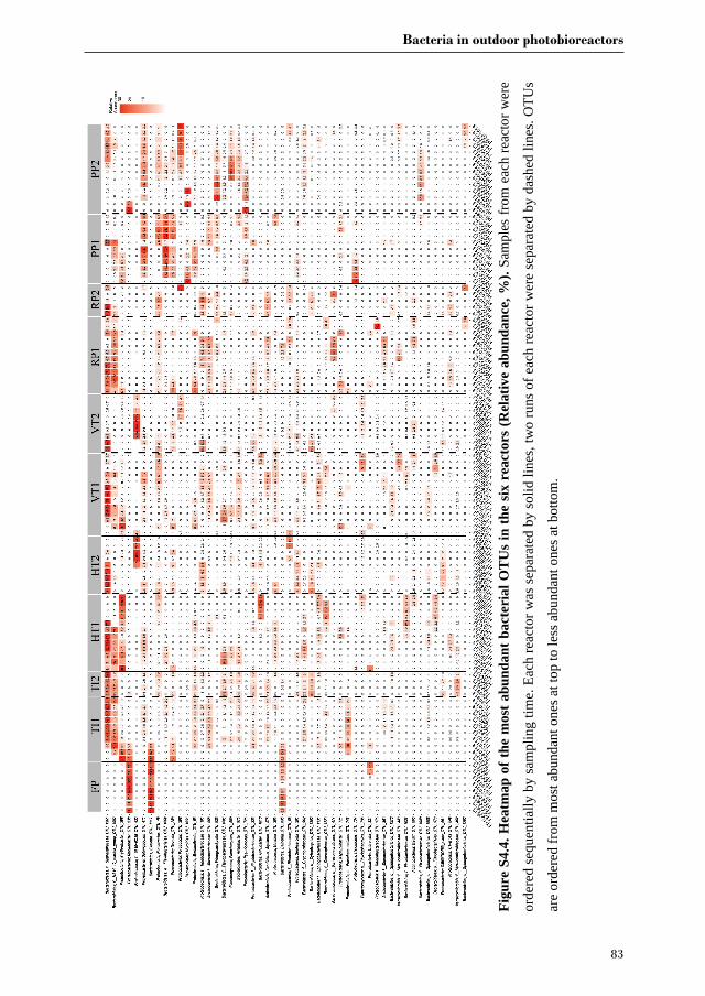
Bacteria in outdoor photobioreactors
83
Fi
gure
S4.
4. H
eatm
ap o
f the
mos
t abu
ndan
t bac
teri
al O
TU
s in
the
six
reac
tors
(Rel
ativ
e ab
unda
nce,
%).
Sam
ples
from
eac
h re
acto
r wer
e
orde
red
sequ
entia
lly b
y sa
mpl
ing
time.
Eac
h re
acto
r was
sepa
rate
d by
solid
line
s, tw
o ru
ns o
f eac
h re
acto
r wer
e se
para
ted
by d
ashe
d lin
es. O
TUs
are
orde
red
from
mos
t abu
ndan
t one
s at t
op to
less
abu
ndan
t one
s at b
otto
m.
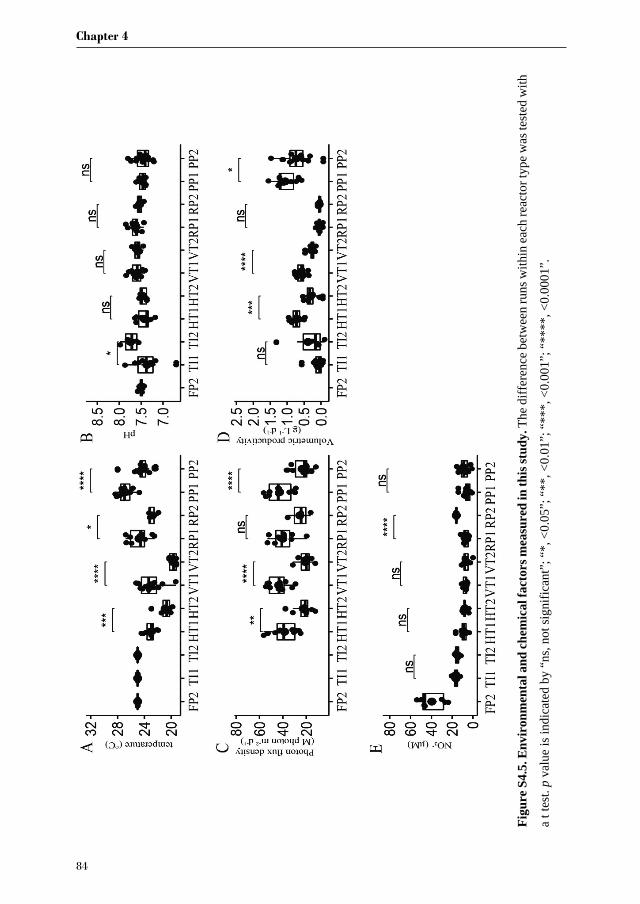
Chapter 4
84
Figu
re S
4.5.
Env
iron
men
tal a
nd ch
emic
al fa
ctor
s mea
sure
d in
this
stud
y. T
he d
iffer
ence
bet
wee
n ru
ns w
ithin
each
reac
tort
ypew
as te
sted
with
at t
est.
p va
lue
is in
dica
ted
by “
ns, n
ot si
gnifi
cant
”; “
*, <
0.05
”; “
**, <
0.01
”; “
***,
<0.
001”
; “**
**, <
0.00
01”.
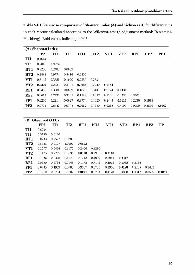
Bacteria in outdoor photobioreactors
85
Table S4.1. Pair-wise comparison of Shannon index (A) and richness (B) for different runs
in each reactor calculated according to the Wilcoxon test (p adjustment method: Benjamini-
Hochberg), Bold values indicate p <0.05.
(A) Shannon Index FP2 TI1 TI2 HT1 HT2 VT1 VT2 RP1 RP2 PP1 TI1 0.4604
TI2 0.2000 0.9774
HT1 0.2230 0.2488 0.0659
HT2 0.3868 0.9774 0.8416 0.0809
VT1 0.8112 0.3681 0.1020 0.2230 0.2331
VT2 0.0379 0.2230 0.3101 0.0084 0.2230 0.0144
RP1 0.8416 0.3681 0.0809 0.1823 0.3101 0.9774 0.0338
RP2 0.4604 0.7426 0.3101 0.1182 0.8447 0.3101 0.2230 0.3101
PP1 0.2230 0.2214 0.0627 0.9774 0.1020 0.2448 0.0110 0.2230 0.1988
PP2 0.0731 0.8442 0.9774 0.0062 0.7640 0.0280 0.4199 0.0659 0.4596 0.0062
(B) Observed OTUs FP2 TI1 TI2 HT1 HT2 VT1 VT2 RP1 RP2 PP1 TI1 0.6734 TI2 0.3790 0.8120 HT1 0.0733 0.2577 0.0785 HT2 0.5545 0.9107 1.0000 0.0822 VT1 0.2577 0.3484 0.1175 0.2466 0.1210 VT2 0.1175 0.2265 0.3196 0.0128 0.2905 0.0180 RP1 0.4326 0.3388 0.1175 0.1712 0.1959 0.8984 0.0317 RP2 0.9590 0.6734 0.7149 0.1175 0.7149 0.2905 0.2095 0.3196 PP1 0.0785 0.1959 0.0785 0.9107 0.0785 0.2916 0.0128 0.2265 0.1403 PP2 0.1210 0.6734 0.9107 0.0091 0.6734 0.0128 0.4838 0.0317 0.3559 0.0091
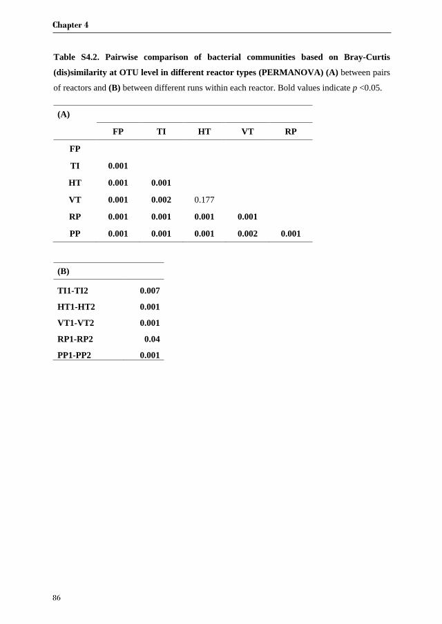
Chapter 4
86
Table S4.2. Pairwise comparison of bacterial communities based on Bray-Curtis
(dis)similarity at OTU level in different reactor types (PERMANOVA) (A) between pairs
of reactors and (B) between different runs within each reactor. Bold values indicate p <0.05.
(A)
FP TI HT VT RP
FP
TI 0.001
HT 0.001 0.001
VT 0.001 0.002 0.177
RP 0.001 0.001 0.001 0.001
PP 0.001 0.001 0.001 0.002 0.001
(B)
TI1-TI2 0.007
HT1-HT2 0.001
VT1-VT2 0.001
RP1-RP2 0.04
PP1-PP2 0.001

Bacteria in outdoor photobioreactors
87
Tab
le S
4.3.
Bac
teri
al O
TU
s sha
red
betw
een
sam
ples
of s
eaw
ater
and
rea
ctor
sam
ples
.
OT
Us
Bac
teri
al ta
xono
my
FP
TI
HD
V
D
RP
PP
889
Actin
obac
teri
a; A
ctin
obac
teri
a; C
oryn
ebac
teri
ales
; Noc
ardi
acea
e; R
hodo
cocc
us
1
880
Actin
obac
teri
a; A
ctin
obac
teri
a; M
icro
cocc
ales
; Mic
roba
cter
iace
ae; S
V1-8
1
10
40
Bact
eroi
dete
s; F
lavo
bact
eria
; Fla
voba
cter
iale
s; F
lavo
bact
eria
ceae
; g
1
933
Bact
eroi
dete
s; F
lavo
bact
eria
; Fla
voba
cter
iale
s; F
lavo
bact
eria
ceae
; NS3
a_m
arin
e_gr
oup
1
936
Bact
eroi
dete
s; F
lavo
bact
eria
; Fla
voba
cter
iale
s; F
lavo
bact
eria
ceae
; NS3
a_m
arin
e_gr
oup
1
1162
Ba
cter
oide
tes;
Fla
voba
cter
ia; F
lavo
bact
eria
les;
Fla
voba
cter
iace
ae; U
lvib
acte
r
1
92
6 Ba
cter
oide
tes;
Fla
voba
cter
ia; F
lavo
bact
eria
les;
Fla
voba
cter
iace
ae; W
inog
rads
kyel
la
1 1
1295
Ba
cter
oide
tes;
Sph
ingo
bact
eriia
; Sph
ingo
bact
eria
les;
Sap
rosp
irac
eae;
g
1
1
1298
Ba
cter
oide
tes;
Sph
ingo
bact
eriia
; Sph
ingo
bact
eria
les;
Sap
rosp
irac
eae;
g
1
1
582
Plan
ctom
ycet
es; P
lanc
tom
ycet
acia
; Pla
ncto
myc
etal
es; f
; g
1
312
Prot
eoba
cter
ia; A
lpha
prot
eoba
cter
ia; C
aulo
bact
eral
es; H
ypho
mon
adac
eae;
g
1
18
Prot
eoba
cter
ia; A
lpha
prot
eoba
cter
ia; D
B1-1
4; f;
g
1
34
Prot
eoba
cter
ia; A
lpha
prot
eoba
cter
ia; O
CS1
16_c
lade
; f; g
1
86
Pr
oteo
bact
eria
; Alp
hapr
oteo
bact
eria
; Rho
doba
cter
ales
; Rho
doba
cter
acea
e; g
1
1 1
1 10
3 Pr
oteo
bact
eria
; Alp
hapr
oteo
bact
eria
; Rho
doba
cter
ales
; Rho
doba
cter
acea
e; g
1
1
1 12
5 Pr
oteo
bact
eria
; Alp
hapr
oteo
bact
eria
; Rho
doba
cter
ales
; Rho
doba
cter
acea
e; g
1 16
2 Pr
oteo
bact
eria
; Alp
hapr
oteo
bact
eria
; Rho
doba
cter
ales
; Rho
doba
cter
acea
e; g
1
1
23
1 Pr
oteo
bact
eria
; Alp
hapr
oteo
bact
eria
; Rho
doba
cter
ales
; Rho
doba
cter
acea
e; L
abre
nzia
1
1 1
1 1
30
9 Pr
oteo
bact
eria
; Alp
hapr
oteo
bact
eria
; Rho
doba
cter
ales
; Rho
doba
cter
acea
e; P
haeo
bact
er
1
1
217
Prot
eoba
cter
ia; A
lpha
prot
eoba
cter
ia; R
hodo
bact
eral
es; R
hodo
bact
erac
eae;
Ros
eoba
cter
_cla
de_D
C5-
80-3
_lin
eage
1
14
5 Pr
oteo
bact
eria
; Alp
hapr
oteo
bact
eria
; Rho
doba
cter
ales
; Rho
doba
cter
acea
e; R
oseo
bact
er_c
lade
_OC
T_lin
eage
1
49
Pr
oteo
bact
eria
; Alp
hapr
oteo
bact
eria
; Rho
doba
cter
ales
; Rho
doba
cter
acea
e; R
oseo
vari
us
1
1 1
1 1
109
Prot
eoba
cter
ia; A
lpha
prot
eoba
cter
ia; R
hodo
bact
eral
es; R
hodo
bact
erac
eae;
Ros
eova
rius
1 1
1
1 10
5 Pr
oteo
bact
eria
; Alp
hapr
oteo
bact
eria
; Rho
doba
cter
ales
; Rho
doba
cter
acea
e; S
ulfit
obac
ter
1
122
Prot
eoba
cter
ia; A
lpha
prot
eoba
cter
ia; R
hodo
bact
eral
es; R
hodo
bact
erac
eae;
Sul
fitob
acte
r
1 14
4 Pr
oteo
bact
eria
; Alp
hapr
oteo
bact
eria
; Rho
doba
cter
ales
; Rho
doba
cter
acea
e; S
ulfit
obac
ter
1 1
1
28
Prot
eoba
cter
ia; A
lpha
prot
eoba
cter
ia; R
hodo
spir
illal
es; f
; g
1
594
Prot
eoba
cter
ia; A
lpha
prot
eoba
cter
ia; R
hodo
spir
illal
es; R
hodo
spir
illac
eae;
Def
luvi
icoc
cus
1
504
Prot
eoba
cter
ia; A
lpha
prot
eoba
cter
ia; S
AR11
_cla
de; S
urfa
ce_1
; g
1
25
Prot
eoba
cter
ia; A
lpha
prot
eoba
cter
ia; S
neat
hiel
lale
s; S
neat
hiel
lace
ae; S
neat
hiel
la
1
1
1 1
375
Prot
eoba
cter
ia; A
lpha
prot
eoba
cter
ia; S
phin
gom
onad
ales
; Ery
thro
bact
erac
eae;
Ery
thro
bact
er
1 1
1
410
Prot
eoba
cter
ia; A
lpha
prot
eoba
cter
ia; S
phin
gom
onad
ales
; Sph
ingo
mon
adac
eae;
Sph
ingo
pyxi
s
1 1
1
41
1 Pr
oteo
bact
eria
; Alp
hapr
oteo
bact
eria
; Sph
ingo
mon
adal
es; S
phin
gom
onad
acea
e; S
phin
gopy
xis
1
1 1
1 1
721
Prot
eoba
cter
ia; G
amm
apro
teob
acte
ria;
Oce
anos
piri
llale
s; O
cean
ospi
rilla
ceae
; Nep
tuni
ibac
ter_
1 1
1 1
1 78
7 Pr
oteo
bact
eria
; Gam
map
rote
obac
teri
a; O
rder
_Inc
erta
e_Se
dis;
Fam
ily_I
ncer
tae_
Sedi
s; g
1
Sum
1
12
13
14
19
14
Pe
rcen
tage
of r
eads
of O
TU
s sha
red
with
seaw
ater
0.
9%
3.3%
8.
9%
5.2%
11
.4%
15
.9%

Chapter 5
Different co-occurring bacteria enhance or decrease the growth of the
microalga Nannochloropsis sp. CCAP211/78
Jie Lian, Patrick Schimmel, Selene Sanchez-Garcia, Rene H. Wijffels,
Hauke Smidt, Detmer Sipkema
Manuscript in preparation
Chapter 5
90
Summary
Marine photosynthetic microalgae are ubiquitously associated with bacteria in nature. However,
the influence of these bacteria on algal cultures in bioreactors is still largely unknown. In this
study, eighteen different bacterial strains were isolated from cultures of Nannochloropsis sp.
CCAP211/78 in two outdoor pilot-scale tubular photobioreactors. The majority of isolates was
affiliated with the classes Alphaproteobacteria and Flavobacteriia. To assess the impact of the
eighteen strains on the growth of Nannochloropsis sp. CCAP211/78, 24-well plates coupled
with custom-made LED boxes were used to simultaneously compare replicate axenic
microalgal cultures with addition of individual bacterial isolates. Co-culturing of
Nannochloropsis sp. CCAP211/78 with these strains demonstrated distinct responses, which
shows that the technique we developed is an efficient method for screening the influence of
harmful/beneficial bacteria. One strain of Maritalea porphyrae (DMSP31) and one strain of
Labrenzia aggregata (YP26) significantly enhanced microalgal growth with a 14% and 12%
increase of the chlorophyll concentration, respectively, whereas flavobacterial strain YP206
greatly inhibited the growth of the microalga with 28% reduction of the chlorophyll
concentration. Our study suggests that algal production systems represent a “natural” source to
isolate and study microorganisms that can either benefit or harm algal cultures. Thus, the
addition of specific bacteria may be used to maximise production of microalgae biomass.
Keywords: Algae-bacteria interaction, algal biotechnology, bacterial isolation, co-cultivation
Bacteria affect the growth of Nannochloropsis sp.
91
5.1. Background
Microalgae show great potential in producing numerous sustainable bioproducts as alternatives
to fossil feedstocks [56, 57, 311]. A long-neglected aspect in algal biomass production is the
role of bacteria that are co-occurring in algae cultivation systems [29, 312]. Algal cultures are
axenic in only a few applications, whereas all microalgae mass production systems inevitably
contain a number of non-target organisms (contaminants), including bacteria [70, 266]. Bacteria
are introduced in algae cultivation systems as algae stocks used as starter cultures are often not
axenic [118, 312, 313]. On the other hand, bacterial contaminants may enter cultivation systems
through multiple operation processes, such as the supplementation of unsterilized medium or
simply as airborne invaders in open algal cultures.
Microalgae-bacteria interactions are prevalent in natural aquatic environments, where
microalgae release exudates into the phycosphere, the region immediately surrounding
individual cells. Chemotaxis drives multiple bacteria to the phycosphere [314], and metabolites
are readily exchanged between algae and bacteria [2]. Although the phycosphere represents
only a tiny area that can be as small as 1 µm surrounding the algal cell, it represents the hotspot
for most of the algal-bacterial interactions that can profoundly affect the productivity and
stability of aquatic ecosystems [2, 96].
Recent research on algal-bacterial interactions has usually been centred around the competitive
or antagonistic aspects, which often involve competition for nutrients [277, 315, 316] or
algicidal activities [2, 317]. For instance, in a microcosm experiment it was found that bacteria
were more efficient than algae in the uptake of phosphorus [315]. The advantage for bacteria is
especially evident under phosphorus-limiting conditions [318]. Apart from competing for
nutrients with algae, some bacteria are known to inhibit algal cell division [319] or cause algal
cell lysis via secretion of algicidal compounds [33, 35, 46].

Chapter 5
92
In contrast to early views that bacteria mostly affect microalgae negatively, it has been
demonstrated that mutualistic relationships between microalgae and bacteria are also prevalent,
or even more common than antagonistic interactions [2, 71]. Proof has been found from
frequent observations that the absence of bacteria in algal cultures negatively affects algal
physiology and growth [221, 320]. In exchange for dissolved organic matter from microalgae,
bacteria fix nitrogen [141, 321] and synthesize a wide range of molecules, including vitamins
[22, 24], the growth-promoting hormone indole-3-acetic acid [16, 270] and the siderophore
vibrioferrin [271, 322]. Such division of labour and close cooperation enable the holobiont to
better adapt to and grow in changing aquatic environments, which has also triggered a growing
interest for applications in industrial settings [323-325].
Contrary to extensive tests of effects of environmental and chemical factors (insolation,
temperature, pH, nutrients, etc.) on algal growth in industrial photobioreactors, only a few
studies have considered the effects of biotic factors such as associated bacteria. In order to
assess the effects of co-occurring bacteria on microalgae in algal cultivation systems, we
isolated and characterized bacteria from two pilot-scale outdoor tubular photobioreactors.
Subsequently, a 24-well plate-based co-cultivation device was used to evaluate algal growth
with addition of the isolated bacterial strains to axenic microalgae. Effects of bacteria on
microalgae were further tested on a double-layer agar plate to verify algal-bacterial interactions.
5.2. Experimental Procedures
5.2.1 Algal cultivation
Nannochloropsis sp. CCAP211/78 cultures used for bacterial isolation were obtained from one
horizontal and one vertical tubular photobioreactor at AlgaePARC, Wageningen, the
Netherlands. Nannochloropsis sp. CCAP 211/78 was cultivated in seawater (Eastern Scheldt,
the Netherlands) enriched with a nutrient stock solution resulting in the following final
Bacteria affect the growth of Nannochloropsis sp.
93
concentrations (in mM); NaNO3, 25; KH2PO4, 1.7; Na2EDTA, 0.56; Fe2SO4·7H2O, 0.11;
MnCl2·2H2O, 0.01; ZnSO4·7H2 3; Co(NO3)2·6H2 3; CuSO4·5H2O, 3;
Na2MoO4·2H2 3. For the cultivation in outdoor photobioreactors, seawater was
chemically sterilized by using sodium hypochlorite. Active chlorite was deactivated by
filtration over activated carbon, followed by filtration across a filter with a pore size of 1 µm.
Subsequently, the nutrient stock solution was added through a sterile filter (0.45 µm). The
detailed description of the cultivation process was given by de Vree, Bosma [265].
For the co-cultivation experiment, the non-axenic pre-cultures of Nannochloropsis sp.
CCAP211/78 (100 mL liquid volume in 250 mL Erlenmeyer flasks) were maintained in
autoclaved seawater supplemented with HEPES (20 mM) and Na2EDTA (5 mM). The nutrient
stock solution with the same final concentrations as above was added to the autoclaved seawater
through a syringe filter m . We refer to this medium as enriched seawater medium (ESW
medium). The Erlenmeyer flask cultures of Nannochloropsis were capped with aeraseal sterile
film (Alphalabs) and placed in an orbital shaker incubator (Sanyo), shaken at 120 rpm,
illuminated with continuous light of 50 µmol photons m 2 s 1 at 25 °C, and the headspace was
enriched with 2 % CO2.
5.2.2 Generation of axenic algal cultures
Axenic cultures of Nannochloropsis sp. CCAP211/78 were prepared using a treatment with an
antibiotics cocktail consisting of Streptomycin ( m ), Gentamycin ( m ),
Ciprofloxacin ( m ), Ampicillin ( m ) and Chloramphenicol ( m ).
Specifically, 2 mL exponentially growing non-axenic Nannochloropsis sp. CCAP211/78 were
taken from a 250 mL flask, washed twice in 2 mL sterile ESW medium and concentrated by
centrifugation at 8000 g for 3 min and transferred into a six-well microplate. After adding the
abovementioned antibiotics cocktail, the plate was incubated at 25 °C and illuminated with a
Chapter 5
94
16/8 h light/dark cycle with a light intensity of 50 µmol photons m 2 s 1. Every two days, the
cultures in the well plate were washed as mentioned before, fresh antibiotics solution was added
and the cultures were incubated as described above. This procedure was repeated four more
times.
Axenicity of Nannochloropsis sp. CCAP211/78 was confirmed via inoculating ESW-YP agar
(sterile ESW supplemented with 1 g/L of yeast extract, 1 g/L of peptone and 15 g/L of agar)
with 100 L of antibiotics-treated algal cultures. Furthermore, algal cultures were incubated for
min ith ml 4’,6-diamidin-2-phenylindol (DAPI, dissolved in phosphate buffer).
Stained samples were inspected with a fluorescence microscope (Olympus). The obtained
axenic Nannochloropsis sp. culture was maintained in 250 mL flasks in the orbital shaker
incubator (Sanyo) according to the same method as described for the non-axenic culture.
5.2.3 DNA isolation and 16S rRNA gene profiling of bacteria
Four samples of 5 mL from two outdoor photobioreactors were vacuum-filtered onto a cellulose
nitrate membrane filter m, Millipore). To isolate the genomic DNA, filters were cut in half
using sterile scissors and DNA was extracted from half a filter using the FastDNA SPIN kit for
soil (MP Biomedicals) with the aid of a Precellys bead beater (Bertin Technologies) with two
rounds of bead beating for 45 s at speed of 5500 rpm.
Amplicons of the 16S ribosomal RNA (rRNA) gene were generated from the extracted DNA
with a two-step PCR reaction carried out in a BIOKÉ SensoQuest Labcycler 48. During the
first step of 16S rRNA gene PCR, a gene amplicon of approximately 311 bp comprising the V1
and V2 regions was generated using degenerate primers 27F-DegS [326] and a mixture of
338R-I and 338R-II [327]. The forward primer was used with Unitag1 attached to the 5’ end of
the primer and Unitag2 was attached to the 5’ end of the reverse primer to facilitate the second
step of the PCR (Supplementary Table S5.1). The first PCR reaction (50 µl) contained 10 µl 5×
Bacteria affect the growth of Nannochloropsis sp.
95
HF buffer (Thermo Scientific, the Netherlands), 1 µl dNTP Mix (10 mM; Promega, Leiden, the
Netherlands), 1 U of Phusion® Hot Start II High-Fidelity DNA polymerase (Thermo Scientific),
500 nM of Unitag1-27F-DegS forward primer, 500 nM of Unitag2-338R I and II reverse primer
and 1 µl template DNA. The PCR was performed using the following conditions: an initial
denaturation at 98°C for 30 s, followed by 25 cycles of denaturation at 98°C for 10 s, annealing
at 56°C for 20 s, elongation at 72°C for 20 s, and a final extension at 72°C for 10 min.
Subsequently, the first PCR product was used as template in a second PCR in order to add
sample-specific barcodes (8 nucleotides). The second PCR reaction (100 µl) contained 20 µl
5× HF buffer, 2 µl dNTP Mix, 2 U of Phusion Hot Start II High-Fidelity DNA polymerase
(Thermo Scientific), 500 nM of a forward and reverse primer equivalent to the Unitag1 and
Unitag2 sequences, respectively, that were each appended with an 8 nt sample specific barcode
(Table S5.1) [328]. The second PCR was performed using the following conditions: an initial
denaturation at 98°C for 30 s, followed by 5 cycles of denaturation at 98°C for 10 s, annealing
at 52°C for 20 s, elongation at 72°C for 20 s, and a final extension at 72°C for 10 min. The
barcoded PCR products from the second PCR were purified using the High-Prep PCR
purification kit (MOBIO) according to the manufacturer’s protocols. The concentration of
purified DNA was measured using a Qubit 2.0 Fluorometer, according to manufacturer’s
instructions for the Qubit dsDNA BR assay (INVITROGEN). Then, the second step PCR
products were pooled in an equimolar concentration and again purified using the High Prep
PCR purification Kit. The purified PCR products (final concentration: 200 ng/µl) were
sequenced at GATC Biotech Europe (Konstanz, Germany, now part of Eurofins Genomics
Germany GmbH) using the Illumina MiSeq Genome Sequencer platform. Nucleotide sequences
of all samples were deposited at NCBI GenBank under Bio Project ID number PRJNA488170
with accession number: SRR7760408.

Chapter 5
96
Illumina sequencing data was processed and analyzed using the NG-Tax pipeline [329] as
previously described by Dat et al. [330]. Briefly, paired-end libraries were combined, and only
read pairs with matching primers and barcodes were retained. Both forward and reverse reads
were trimmed to 100 bp and concatenated to yield sequences of 200 bp that were used for
subsequent sequence data processing. Demultiplexing, OTU picking, chimera removal and
taxonomic assignment were performed within one single step. Reads were ranked per sample
by abundance, and sequences (at a 100% identity level) were added to an initial OTU table
starting from the most abundant sequence until the abundance was lower than 0.1% per sample.
The final OTU table was created by clustering the reads that were initially discarded (as they
represented OTUs <0.1% of the relative abundance) with the OTUs from the initial OTU table
allowing a single mismatch. Taxonomic assignment was done utilizing the UCLUST algorithm
algorithm [331] and the SILVA 111_SSU Ref database [332, 333].
5.2.4 Bacterial isolation and identification
Cryopreserved (15% glycerol) algal cultures from two outdoor reactors (horizontal tubular
bioreactor and vertical tubular bioreactor) at AlgaePARC and stored at -80 °C were used as
inoculum for bacterial isolation. The description of bioreactors and algal cultivation process
was given by de Vree, Bosma [265]. Aliquots of cryopreserved cultures were diluted (104 fold)
and plated on ESW agar (1.5% agar) supplemented with one of the following carbon sources:
2 g/L glucose (ESW-GLU); 2 g/L propionate (ESW-PRO); 2 g/L casamino acids (ESW-CAS);
2 g/L Nannochloropsis extract (ESW-ALG; 2 g freeze-dried Nannochloropsis cells from
AlgaePARC suspended in 10 mL ESW, French-pressed twice at 110 MPa and centrifuged at
8000 g for 5 min. Subsequently, all the resulting supernatant was filter-sterilised (0.2 m) and
added to 1 L of autoclaved ESW medium); 2 g/L succinate (ESW-SUC); 1 g/L yeast extract
and 1 g/L peptone (ESW-YP); 2 g/L palmitate (ESW-PAL) or 0.6 mM
dimethylsulfoniopropionate (ChemCruz, Dallas, TX) (ESW-DMSP). Plates were maintained in
Bacteria affect the growth of Nannochloropsis sp.
97
the dark at room temperature. Single colonies were picked and streaked until pure cultures were
obtained. All the pure bacterial strains were maintained in ESW-YP medium. For bacterial
identification, the 16S rRNA gene was amplified with universal primers 27F and 1492R (Table
S5.1). The PCR reaction (50 µl) contained 10 µl 5× HF buffer (Thermo Scientific), 1 µl dNTP
Mix (10 mM; Promega), 1 U of Phusion® Hot Start II High-Fidelity DNA polymerase (Thermo
Scientific), 1 µM of 27F primer, 1 µM of 1492R primer and 1 µl bacterial culture. The PCR
protocol consisted of a pre-denaturation step (10 min at 98 °C) followed by 30 cycles of
denaturation (30 s at 98 °C), annealing (40 s at 60 °C) and elongation steps (1.5 min at 72 °C)
with a final elongation step at 72°C for 10 min. The purified amplicons (High-Prep PCR
purification kit, MOBIO) were Sanger sequenced with primer 806R (Table S5.1) by GATC
Biotech Europe (Konstanz, Germany). The sequences were clustered into contigs with a cut off
of 99% similarity using ContigExpress (Invitrogen). One representative isolate from each
contig was selected (Table 5.1) and analysed with the BLASTn tool from NCBI
(http://www.ncbi.nlm.nih.gov/) to determine its phylogenetic affiliation. All sequences of
selected isolates were deposited at NCBI GenBank under accession numbers as listed in Table
5.1.
5.2.5 Co-culturing of algae with bacteria in microplates
For co-cultivation in 24-well microplates, both exponentially growing axenic and non-axenic
cultures of Nannochloropsis sp. CCAP211/78 grown in flasks were diluted with ESW medium
supplemented with 5 mM NaHCO3 to a fluorescence intensity of ~5000 (Excitation: 450 nm,
Emission: 685 nm). Single bacterial strains, separately grown in ESW-YP broth, were washed
twice in sterile ESW medium and concentrated by centrifugation (8000 g for 5 min), then re-
suspended in ESW medium and diluted to an OD600 of 0.2. Subsequently, 1 mL of axenic
Nannochloropsis and 50 µL of bacterial strain suspension were inoculated in 24-well plates.
The control cultures (either 1 mL of axenic Nannochloropsis sp. CCAP211/78 or 1 mL of non-
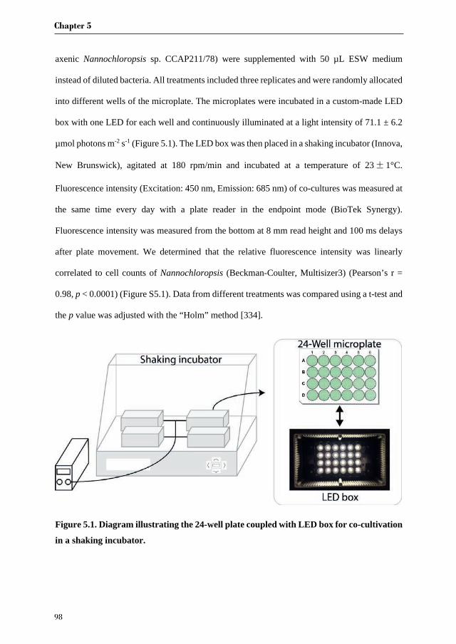
Chapter 5
98
axenic Nannochloropsis sp. CCAP211/78) were supplemented with 50 µL ESW medium
instead of diluted bacteria. All treatments included three replicates and were randomly allocated
into different wells of the microplate. The microplates were incubated in a custom-made LED
box with one LED for each well and continuously illuminated at a light intensity of 71.1 ± 6.2
µmol photons m-2 s-1 (Figure 5.1). The LED box was then placed in a shaking incubator (Innova,
New Brunswick), agitated at 180 rpm/min and incubated at a temperature of 23±1°C.
Fluorescence intensity (Excitation: 450 nm, Emission: 685 nm) of co-cultures was measured at
the same time every day with a plate reader in the endpoint mode (BioTek Synergy).
Fluorescence intensity was measured from the bottom at 8 mm read height and 100 ms delays
after plate movement. We determined that the relative fluorescence intensity was linearly
correlated to cell counts of Nannochloropsis (Beckman-Coulter, Multisizer3) (Pearson’s r =
0.98, p < 0.0001) (Figure S5.1). Data from different treatments was compared using a t-test and
the p value was adjusted with the “Holm” method [334].
Figure 5.1. Diagram illustrating the 24-well plate coupled with LED box for co-cultivation
in a shaking incubator.
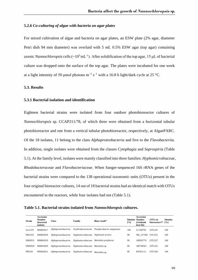
Bacteria affect the growth of Nannochloropsis sp.
99
5.2.6 Co-culturing of algae with bacteria on agar plates
For mixed cultivation of algae and bacteria on agar plates, an ESW plate (2% agar, diameter
Petri dish 94 mm diameter) was overlaid with 5 mL 0.5% ESW agar (top agar) containing
axenic Nannochloropsis cells (~105 mL-1 fter soli ification of the top a ar of bacterial
culture was dropped onto the surface of the top agar. The plates were incubated for one week
at a light intensity of mol photons m 2 s 1 with a 16:8 h light/dark cycle at 25 °C.
5.3. Results
5.3.1 Bacterial isolation and identification
Eighteen bacterial strains were isolated from four outdoor photobioreactor cultures of
Nannochloropsis sp. CCAP211/78, of which three were obtained from a horizontal tubular
photobioreactor and one from a vertical tubular photobioreactor, respectively, at AlgaePARC.
Of the 18 isolates, 11 belong to the class Alphaproteobacteria and five to the Flavobacteriia.
In addition, single isolates were obtained from the classes Cytophagia and Saprospiria (Table
5.1). At the family level, isolates were mainly classified into three families: Hyphomicrobiaceae,
Rhodobacteraceae and Flavobacteriaceae. When Sanger-sequenced 16S rRNA genes of the
bacterial strains were compared to the 138 operational taxonomic units (OTUs) present in the
four original bioreactor cultures, 14 out of 18 bacterial strains had an identical match with OTUs
encountered in the reactors, while four isolates had not (Table 5.1).
Table 5.1. Bacterial strains isolated from Nannochloropsis cultures.
Strain
Accession Number (bacterial isolate)
Class Family Blast result* Identity [%]
Accession Number (Genbank best hit)
OTUs in bioreactors**
Identity [%]
GLU107 MH843917 Alphaproteobacteria Erythrobacteraceae Porphyrobacter sanguineus 100 LC349792 OTU247 100
PRO103 MH843918 Alphaproteobacteria Hyphomicrobiaceae Algimonas arctica 98 NR_137369 OTU321 100
DMSP31 MH843919 Alphaproteobacteria Hyphomicrobiaceae Maritalea porphyrae 99 AB583776 OTU327 100
DMSP20 MH843920 Alphaproteobacteria Hyphomicrobiaceae Maritalea sp. 99 AB758563 OTU331 100
PRO34 MH843921 Alphaproteobacteria Hyphomicrobiaceae Maritalea sp. 96 KP301112 OTU343 100
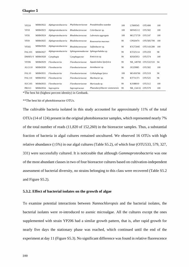
Chapter 5
100
YP210 MH843922 Alphaproteobacteria Phyllobacteriaceae Pseudohoeflea suaedae 100 LT600545 OTU490 100
YP18 MH843923 Alphaproteobacteria Rhodobacteraceae Celeribacter sp. 100 MF045112 OTU582 100
YP26 MH843924 Alphaproteobacteria Rhodobacteraceae Labrenzia aggregata 100 MG273739 OTU247 100
YP29 MH843925 Alphaproteobacteria Rhodobacteraceae Roseovarius mucosus 99 CP020474 OTU585/709 100
YP202 MH843926 Alphaproteobacteria Rhodobacteraceae Sulfitobacter sp. 99 KY272045 OTU143/289 100
PAL103 MH843927 Alphaproteobacteria Sphingomonadaceae Sphingorhabdus sp. 99 KT325114 OTU259 98
DMSP2-Y MH843928 Cytophagia Cytophagaceae Emticicia sp. 99 KP265953 OTU574 100
YP206 MH843929 Flavobacteriia Flavobacteriaceae Aquaticitalea lipolytica 99 NR_149769 OTU532/533 94
ALG110 MH843930 Flavobacteriia Flavobacteriaceae Arenibacter sp. 98 JX529985 OTU582 100
PAL10 MH843931 Flavobacteriia Flavobacteriaceae Cellulophaga lytica 100 MG456766 OTU519 96
PAL110 MH843932 Flavobacteriia Flavobacteriaceae Maribacter sp. 99 KT731371 OTU525 96
SUC105 MH843933 Flavobacteriia Flavobacteriaceae Muricauda sp. 99 KJ188010 OTU512 100
PRO13 MH843934 Saprospiria Saprospiraceae Phaeodactylibacter xiamenensis 99 NR_134132 OTU579 100
*The best hit (highest percent identity) in Genbank.
**The best hit of photobioreactor OTUs.
The cultivable bacteria isolated in this study accounted for approximately 11% of the total
OTUs (14 of 124) present in the original photobioreactor samples, which represented nearly 7%
of the total number of reads (11,820 of 152,260) in the bioreactor samples. Thus, a substantial
fraction of bacteria in algal cultures remained uncultured. We observed 16 OTUs with high
relative abundance ( 5%) in our algal cultures (Table S5.2), of which four (OTU533, 579, 327,
331) were successfully cultured. It is noticeable that although Gammaproteobacteria was one
of the most abundant classes in two of four bioreactor cultures based on cultivation-independent
assessment of bacterial diversity, no strains belonging to this class were recovered (Table S5.2
and Figure S5.2).
5.3.2. Effect of bacterial isolates on the growth of algae
To examine potential interactions between Nannochloropsis and the bacterial isolates, the
bacterial isolates were re-introduced to axenic microalgae. All the cultures except the ones
supplemented with strain YP206 had a similar growth pattern, that is, after rapid growth for
nearly five days the stationary phase was reached, which continued until the end of the
experiment at day 11 (Figure S5.3). No significant difference was found in relative fluorescence
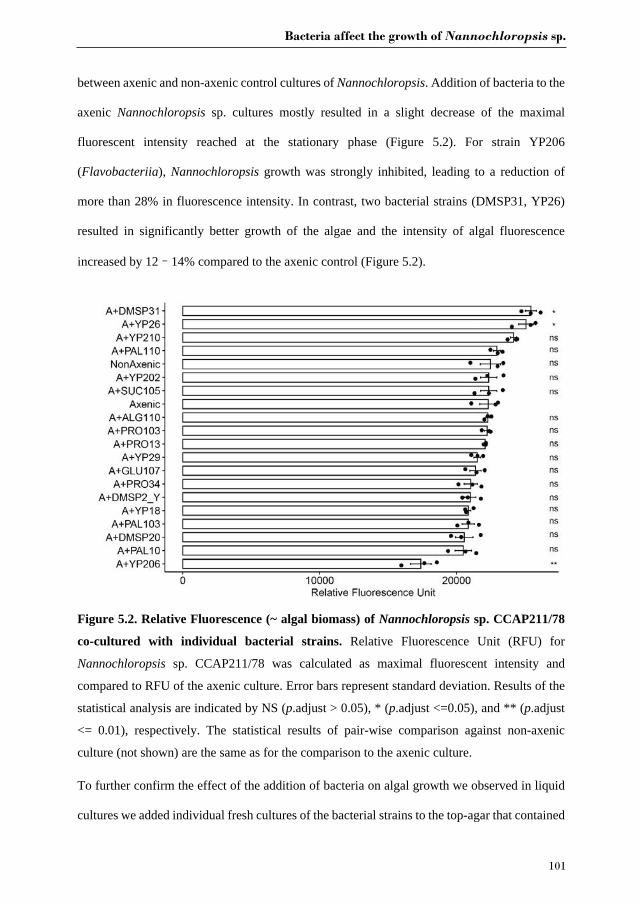
Bacteria affect the growth of Nannochloropsis sp.
101
between axenic and non-axenic control cultures of Nannochloropsis. Addition of bacteria to the
axenic Nannochloropsis sp. cultures mostly resulted in a slight decrease of the maximal
fluorescent intensity reached at the stationary phase (Figure 5.2). For strain YP206
(Flavobacteriia), Nannochloropsis growth was strongly inhibited, leading to a reduction of
more than 28% in fluorescence intensity. In contrast, two bacterial strains (DMSP31, YP26)
resulted in significantly better growth of the algae and the intensity of algal fluorescence
increased by 12–14% compared to the axenic control (Figure 5.2).
Figure 5.2. Relative Fluorescence (~ algal biomass) of Nannochloropsis sp. CCAP211/78
co-cultured with individual bacterial strains. Relative Fluorescence Unit (RFU) for
Nannochloropsis sp. CCAP211/78 was calculated as maximal fluorescent intensity and
compared to RFU of the axenic culture. Error bars represent standard deviation. Results of the
statistical analysis are indicated by NS (p.adjust > 0.05), * (p.adjust <=0.05), and ** (p.adjust
<= 0.01), respectively. The statistical results of pair-wise comparison against non-axenic
culture (not shown) are the same as for the comparison to the axenic culture.
To further confirm the effect of the addition of bacteria on algal growth we observed in liquid
cultures we added individual fresh cultures of the bacterial strains to the top-agar that contained

Chapter 5
102
axenic Nannochloropsis cells on double-layer agar plates. When co-cultured with YP26, growth
of Nannochloropsis was strongly stimulated, indicated by the greenest algal lawns after one-
week of incubation (Figure 5.3). Interestingly, two other bacteria (ALG110, PRO34) slightly
enhanced the algal growth on solid medium (Figure 5.3), whereas this beneficial effect was
absent in liquid culture. Two agar-degrading bacteria (PAL10 and PAL110) formed much
bigger colonies than other strains, and thus promoted the algal growth on a much bigger surface
on the solid medium (Figure 5.3).
Figure 5.3. Co-cultivation of Nannochloropsis sp. CCAP211/78 and bacterial strains on
double-layer agar plates after seven days. ESW (Enriched natural seawater medium) and
ESW-YP (ESW medium with peptone and yeast extract) were used as controls. The labelled
names referred to the added bacteria. YP26 was added on three different plates as replicates
indicated by arrows.
5.4. Discussion
Bacterial isolation was carried out from four samples taken from two outdoor photobioreactors
of AlgaePARC where bacterial strains were present in microalgae cultures that were not
Bacteria affect the growth of Nannochloropsis sp.
103
supplemented with external organic carbon. Therefore, we suspected that heterotrophic co-
occurring bacteria depended on the organic carbon released by microalgae, which suggests at
least a commensal relationship between Nannochloropsis and bacteria. In total, 18 bacterial
strains were isolated from the outdoor photobioreactor samples. These 18 isolates constituted
approximately ten percent of the whole bacterial community, whereas nearly 90% remained
uncultured under the conditions applied in our experiment. Hence it cannot be excluded that
even more potent candidates with respect to an algae growth-promoting effect remain to be
discovered from the yet uncultured fraction. The bacteria in our culture collection were mostly
classified as Alphaproteobacteria and Flavobacteriia (Figure S5.2). It has also been
corroborated by global surveys that phytoplankton-associated bacterial communities are often
restricted to only a few bacterial classes including Alphaproteobacteria (Rhodobacteraceae),
Gammaproteobacteria (Alteromonadaceae) and Flavobacteriia (Flavobacteraceae) [96, 304,
335, 336]. These apparently widespread patterns imply that the lifestyle of some bacteria within
these groups is substantially related to that of algae. Likewise algae can either benefit or suffer
from the bacterial partners [2]. Bacteria reported to be beneficial to microalgae are mostly
Alphaproteobacteria, and algal growth-promoting bacteria belonging to Alphaproteobacteria
are quite diverse, including members of Rhizobium [105], Brevundimonas [121],
Mesorhizobium loti [22], and Hyphomonas [122]. Within Alphaproteobacteria, bacteria from
the family Rhodobacteraceae are frequently associated with algae, of which the most studied
ones are Phaeobacter gallaeciensis [46], Dinoroseobacter shibae [337], Sulfitobacter sp. [16]
and Ruegeria pomeroyi [17].
Labrenzia aggregata (YP26) was the only isolate in our experiment that exhibited significant
growth enhancement both in liquid and solid media (Figure 5.2, Figure 5.3 and Figure S5.3).
Members of the genus Labrenzia have been isolated from a wide range of habitats and found to
be frequently associated with other marine organisms [338]. These organisms include
Chapter 5
104
invertebrates such as molluscs, corals and sponges, and a wide variety of photosynthetic
partners including seaweeds, diatoms, dinoflagellates, green and red algae [339]. Labrenzia
aggregata has also been isolated previously from Nannochloropsis oculata and
Nannochloropsis gaditana [340]. A recent study revealed that Labrenzia sp. increased the
biomass accumulation of the marine microalga Isochrysis galbana by 72% and the growth rate
by 18% [341]. On the other hand, it has been reported that a bacterial isolate (KD531) with 100%
similarity to the partial 16S rRNA gene of our Labrenzia aggregata isolate had an algicidal
effect on Chlorella vulgaris [342]. The addition of bacterial lysate of KD531 to Chlorella
vulgaris cultures caused nearly 20% reduction in biomass dry weight and nearly 60% reduction
in lipid content. The contradiction between these and our observations may be due to strain-
specific differences between isolates of L. aggregata, and/or different interactions of the
bacterium with different algal hosts. Prior research has shown that some bacteria that are
mutualistic to their native algal partner can be parasitic to foreign algae, which hints at co-
adaption and evolution of algae and their associated microbiome [119]. In addition, we added
live bacteria rather than a bacterial lysate, which may lead to a different effect. Nannochloropsis
sp. also appeared to grow faster and denser on a solid growth medium in the presence of
Labrenzia aggregata. To our knowledge, this is the first time that one bacterium has been
shown to strongly promote the growth of microalgae on solid agar. Agar plates have been the
most commonly used method to study algal-bacterial interactions [27, 34]. For example, the
vitamin B12-dependent microalga Lobomonas rostrata could grow on agar plates only when
vitamin B12 or a vitamin B12-synthesizing bacterium (Mesorhizobium loti) was added [27].
Therefore, it is tempting to speculate that the growth promotion observed here for Labrenzia
aggregata could be related to inorganic nutrient exchange or algal acquisition of growth factors
released by bacteria.

Bacteria affect the growth of Nannochloropsis sp.
105
Although the growth increase of Nannochloropsis in the presence of Maritalea porphyrae
(DMSP31) was significant in liquid cultures, this beneficial effect disappeared on the agar plate.
This discrepancy between two screening methods corroborated that algae-bacteria interactions
are complex and may vary under different culture conditions. Therefore, preliminary screening
results should be confirmed by other methods such as flask cultures or bioreactors before claims
regarding beneficial effects of bacteria on large-scale algal growth can be made. It is interesting
to note that Maritalea porphyrae (DMSP31) and Pseudohoeflea suaedae (YP210), the latter of
which caused a numerical, but non-significant increase in algal growth in the plate assay, have
been previously isolated from the thalli of the red alga Pyropia yezoensis [343] and root of the
halophyte Suaeda maritima [344], respectively. However, experimental evidence showed that
these bacteria exhibited no apparent morphogenesis effects on the red alga [345], and therefore
the nature of a symbiotic relationship -if any- with the phototroph remains unknown. The genus
Pseudohoeflea (reclassification of Hoeflea) to which YP210 belongs, has not yet been well
characterized [346], however, it has been speculated that the genus Pseudohoeflea may
resemble some members of the marine Roseobacter clade in phytoplankton colonization by the
production of secondary metabolites that inhibit the growth of competing bacteria (antibiotics)
and promote growth of phytoplankton (auxins) [46, 346].
Three out of five Flavobacteriia strains (ALG110, SUC105, PAL110) showed no significant
effect on the growth of Nannochloropsis cultures, despite the fact that members of the
Flavobacteriia have repeatedly been reported to have antagonistic relationships with algae. For
instance, Kordia algicida was shown to excrete an extracellular protease to lyse algal cells to
acquire their dissolved organic carbon [317], and Croceibacter atlanticus was observed to
release an unidentified molecule to arrest diatom cell division and increase secretion of organic
carbon [319]. On the other hand, the greatest reduction in chlorophyll content in the stationary
phase compared to the controls was observed in the presence of strain YP206 from the

Chapter 5
106
Flavobacteriaceae family (Table 5.1 and Figure 5.2). The closest relative of strain YP206 is
Aquaticitalea lipolytica (99% identity of the 16S rRNA gene) that was isolated from Antarctic
seawater and known to hydrolyse lipids [347]. However, when YP206 was co-cultured on agar
plates with Nannochloropsis, the growth inhibition observed in liquid culture was not observed
(Figure 5.2 and Figure 5.3). Although mechanistic insight requires future research, one can
speculate that the incubation time (seven days) used in the agar plate experiments described
here was too short or that the algal density was still too low on the agar plate for the bacterial
inhibition to take place, as some algicidal bacteria have been shown to only kill senesced algal
cells in the stationary phase or decline phase [46, 337]. This has previously been explained by
competition for limiting nutrients such as nitrogen [348] and phosphorus [315, 349]. However,
that is not likely to be the case for our experiments as nitrogen and phosphorus concentrations
added would support much higher algae concentrations than those present in the stationary
phase, and for nitrogen it was confirmed in the stationary phase that it was not depleted (data
not shown). Alternatively, release of toxic compounds by bacteria could contribute to the
inhibitory effects observed at stationary phase [350, 351]. Many bacteria belonging to the
family Flavobacteriaceae are able to glide on solid surfaces and decompose agar [352]. PAL10
and PAL110 displayed these features and formed larger and concave colonies on the agar
surface (Figure 5.3). Although both strains showed no significant effects on algal growth in
liquid co-culture, they slightly enhanced the growth of Nannochloropsis sp. in the agar-plate
assay (Figure 5.3). A possible explanation for the growth promotion on solid media could be
that Nannochloropsis cells consumed the by-products from the agar degradation by the bacteria.
For instance, Cellulophaga lytica has previously been shown to synthesize different kinds of
agarases [353], and the enzymatic hydrolysis of agar yields monomeric sugars, such as D-
galactose, 3,6-anhydro-L-galactose, and L-galactose-6-sulfate [354]. Research has shown that
Bacteria affect the growth of Nannochloropsis sp.
107
supplementation with galactose increases the growth rate of Nannochloropsis salina by nearly
10 % [355].
PRO13 (Saprospiria) and DMSP2-Y (Cytophagia) were the only two isolates not belonging to
Alphaproteobacteria or Flavobacteriia. Both PRO13 and DMSP2-Y had no significant effect
on the growth of Nannochloropsis sp. A previous study has found that the family
Saprospiraceae was the most prevalent taxon, and also the most abundant one in industrial
cultures of Nannochloropsis salina [69]. The 16S rRNA gene of strain PRO13 was identical to
OTU579 found in the outdoor photobioreactors, particularly in sample HD0105 where this
bacterium made up nearly 25% of the whole bacterial community (Table S5.2). In spite of this
strikingly high relative abundance, co-culturing with strain PRO13 had no significant effect on
the growth of Nannochloropsis sp. neither in liquid co-cultures nor on agar plates (Figure 5.2
and Figure 5.3). Similarly, the study by Fulbright, Robbins-Pianka [69] reported that there was
no correlation between the abundance of Saprospiraceae and growth of N. salina. However,
the prevalence of this bacterium suggests it may have other functions in algal cultures, and the
lytic capability of members of this bacterial family may relate to degrading cell debris for
nutrient recycling [69]. DMSP2-Y is closely related to Emticicia sp., and species from the genus
Emticicia have been recorded to live with Chlorella vulgaris [248] and the macroalga
Cladophora glomerata [287]. Emticicia sp. was found to slightly reduce the growth rate of
axenic Chlorella vulgaris in co-cultivation, but the co-culture revealed prolonged stationary
phase [356].
5.5. Conclusion
In this study, we isolated 18 bacterial strains from two outdoor photobioreactors for cultivation
of microalgae. Two strains assigned to Maritalea porphyrae and Labrenzia aggregata,
respectively, significantly promoted growth of Nannochloropsis sp. CCAP211/78 in liquid
Chapter 5
108
cultures in well plates (14% and 12% increase of chlorophyll in stationary phase compared to
the controls, respectively), and the Labrenzia aggregata strain also notably increased growth
of the alga on agar plates. In addition, one strain most closely related to Aquaticitalea lipolytica
significantly reduced the chlorophyll content with 28% compared to the axenic and non-axenic
controls. Our results suggest that some bacteria from algal production systems may have a
pronounced impact on algal growth under controlled laboratory conditions, an effect that should
be verified for larger-scale algae cultures. Our results indicated that in the practice of improving
the production of microalgae, the bacterial community in algal inocula should be considered. If
harmful bacteria are present, the inoculum should be replaced by an inoculum where these
bacteria are absent to increase the cultivation success. Perhaps even more interesting, beneficial
bacterial strains may be supplemented as a new means to improve algal productivity and culture
stability.
Acknowledgements
Jie Lian would like to thank the China Scholarship Council for financial support (Grant
Number: 201406310023).
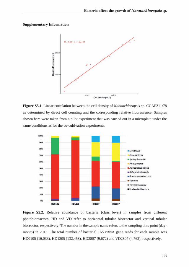
Bacteria affect the growth of Nannochloropsis sp.
109
Supplementary Information
Figure S5.1. Linear correlation between the cell density of Nannochloropsis sp. CCAP211/78
as determined by direct cell counting and the corresponding relative fluorescence. Samples
shown here were taken from a pilot experiment that was carried out in a microplate under the
same conditions as for the co-cultivation experiments.
Figure S5.2. Relative abundance of bacteria (class level) in samples from different
photobioreactors. HD and VD refer to horizontal tubular bioreactor and vertical tubular
bioreactor, respectively. The number in the sample name refers to the sampling time point (day-
month) in 2015. The total number of bacterial 16S rRNA gene reads for each sample was
HD0105 (16,033), HD1205 (132,458), HD2807 (9,672) and VD2807 (4,762), respectively.
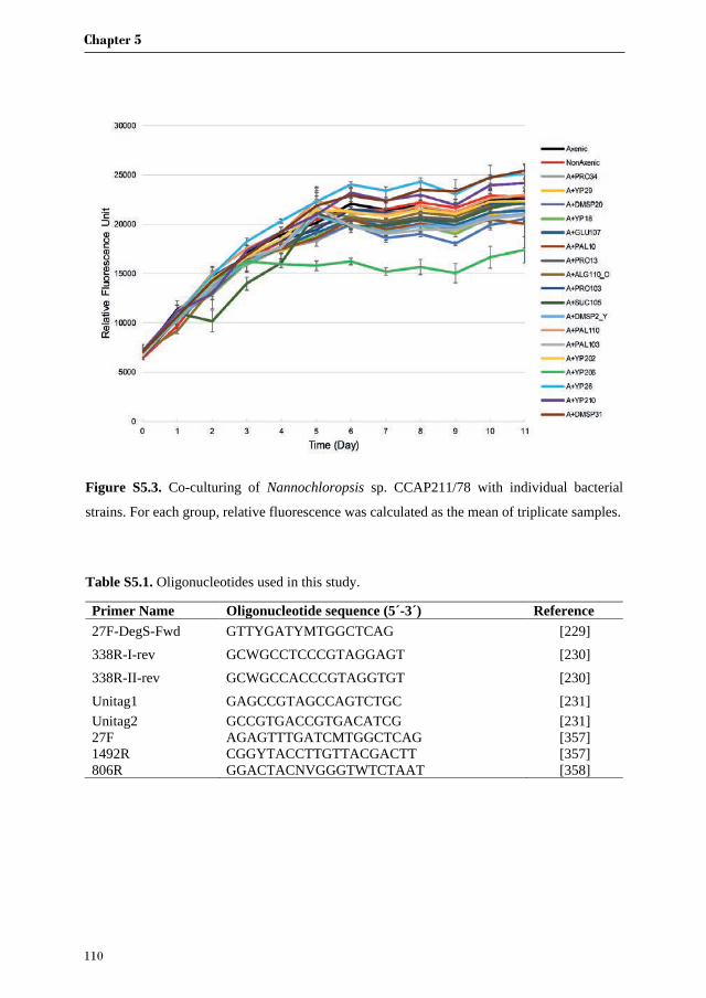
Chapter 5
110
Figure S5.3. Co-culturing of Nannochloropsis sp. CCAP211/78 with individual bacterial
strains. For each group, relative fluorescence was calculated as the mean of triplicate samples.
Table S5.1. Oligonucleotides used in this study.
Primer Name Oligonucleotide sequence (5´-3´) Reference 27F-DegS-Fwd GTTYGATYMTGGCTCAG [229] 338R-I-rev GCWGCCTCCCGTAGGAGT [230] 338R-II-rev GCWGCCACCCGTAGGTGT [230] Unitag1 GAGCCGTAGCCAGTCTGC [231] Unitag2 GCCGTGACCGTGACATCG [231] 27F AGAGTTTGATCMTGGCTCAG [357] 1492R CGGYTACCTTGTTACGACTT [357] 806R GGACTACNVGGGTWTCTAAT [358]
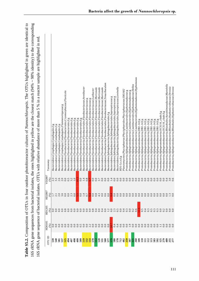
Bacteria affect the growth of Nannochloropsis sp.
111
Tab
le S
5.2.
Com
posi
tion
of O
TUs
in fo
ur o
utdo
or p
hoto
bior
eact
or c
ultu
res
of N
anno
chlo
rops
is. T
he O
TUs
high
light
ed in
gre
en a
re id
entic
al to
16
S rR
NA
gen
e se
quen
ces f
rom
bac
teria
l iso
late
s, th
e on
es h
ighl
ight
ed in
yel
low
are
the
clos
est m
atch
(94%
~ 9
8% id
entit
y) to
the
corr
espo
ndin
g 16
S rR
NA
gen
e se
quen
ce o
f bac
teria
l iso
late
s. O
TUs w
ith re
lativ
e ab
unda
nce
of m
ore
than
5 %
in a
reac
tor s
ampl
e ar
e hi
ghlig
hted
in re
d.
OTU
ID
HD
0105
(%
) H
D12
05
(%)
HD
2807
(%
) V
D28
07
(%)
Taxo
nom
y
539
0.0
0.0
1.7
2.3
Bact
eroi
dete
s;C
ytop
hagi
a;C
ytop
haga
les;
f;g
540
0.0
0.0
2.1
3.3
Bact
eroi
dete
s;C
ytop
hagi
a;C
ytop
haga
les;
f;g
581
0.0
0.0
2.0
2.6
Bact
eroi
dete
s;C
ytop
hagi
a;C
ytop
haga
les;
f;g
537
0.0
0.0
3.6
2.0
Bact
eroi
dete
s;C
ytop
hagi
a;C
ytop
haga
les;
Flam
meo
virg
acea
e;g
525
0.0
0.8
0.0
0.0
Bact
eroi
dete
s;Fl
avob
acte
ria;
Flav
obac
teri
ales
;Cry
omor
phac
eae;
Fluv
iicol
a 49
4 0.
0 0.
0 0.
0 0.
5 Ba
cter
oide
tes;
Flav
obac
teri
a;Fl
avob
acte
rial
es;f;
g 49
7 0.
0 0.
0 2.
3 3.
2 Ba
cter
oide
tes;
Flav
obac
teri
a;Fl
avob
acte
rial
es;f;
g 49
8 0.
0 0.
0 0.
0 1.
0 Ba
cter
oide
tes;
Flav
obac
teri
a;Fl
avob
acte
rial
es;f;
g 49
9 0.
0 0.
0 5.
5 10
.2
Bact
eroi
dete
s;Fl
avob
acte
ria;
Flav
obac
teri
ales
;f;g
509
0.5
0.0
0.0
0.0
Bact
eroi
dete
s;Fl
avob
acte
ria;
Flav
obac
teri
ales
;f;g
519
0.0
0.0
1.4
1.4
Bact
eroi
dete
s;Fl
avob
acte
riia
;Fla
voba
cter
iale
s;Fl
avob
acte
riac
eae;
Aren
ibac
ter
532
0.0
0.0
0.7
0.8
Bact
eroi
dete
s;Fl
avob
acte
riia
;Fla
voba
cter
iale
s;Fl
avob
acte
riac
eae;
g 53
3 2.
2 0.
0 6.
4 6.
7 Ba
cter
oide
tes;
Flav
obac
teri
ia;F
lavo
bact
eria
les;
Flav
obac
teri
acea
e;g
570
0.0
0.8
0.0
0.0
Bact
eroi
dete
s;Fl
avob
acte
riia
;Fla
voba
cter
iale
s;Fl
avob
acte
riac
eae;
Lutib
acte
r 51
2 0.
0 0.
4 0.
8 0.
0 Ba
cter
oide
tes;
Flav
obac
teri
ia;F
lavo
bact
eria
les;
Flav
obac
teri
acea
e;M
uric
auda
52
0 0.
0 0.
0 4.
3 3.
7 Ba
cter
oide
tes;
Flav
obac
teri
ia;F
lavo
bact
eria
les;
Flav
obac
teri
acea
e;M
uric
auda
53
1 0.
0 0.
4 0.
0 0.
0 Ba
cter
oide
tes;
Flav
obac
teri
ia;F
lavo
bact
eria
les;
Flav
obac
teri
acea
e;M
uric
auda
56
0 0.
0 0.
0 0.
6 0.
0 Ba
cter
oide
tes;
Flav
obac
teri
ia;F
lavo
bact
eria
les;
Flav
obac
teri
acea
e;Te
naci
bacu
lum
57
7 0.
0 1.
2 0.
0 0.
0 Ba
cter
oide
tes;
Sphi
ngob
acte
riia
;Sph
ingo
bact
eria
les;
f;g
579
24.7
0.
0 6.
4 1.
4 Ba
cter
oide
tes;
Sphi
ngob
acte
riia
;Sph
ingo
bact
eria
les;S
apro
spir
acea
e;g
580
0.4
2.3
0.0
0.0
Bact
eroi
dete
s;Sp
hing
obac
teri
ia;S
phin
goba
cter
iale
s;Sap
rosp
irac
eae;
g 57
8 0.
4 0.
5 0.
0 0.
0 Ba
cter
oide
tes;
Sphi
ngob
acte
riia
;Sph
ingo
bact
eria
les;S
apro
spir
acea
e;Le
win
ella
71
2 0.
6 0.
0 0.
0 0.
0 B
D1-
5;c;
o;f;g
48
2 0.
0 0.
4 0.
0 0.
0 Pl
anct
omyc
etes
;Phy
cisp
haer
ae;P
hyci
spha
eral
es;P
hyci
spha
erac
eae;
SM1A
02
259
0.0
0.3
0.0
0.0
Prot
eoba
cter
ia;A
lpha
prot
eoba
cter
ia;C
aulo
bact
eral
es;H
ypho
mon
adac
eae;
g 30
7 1.
9 0.
0 0.
0 0.
0 Pr
oteo
bact
eria
;Alp
hapr
oteo
bact
eria
;Cau
loba
cter
ales
;Hyp
hom
onad
acea
e;g
321
0.0
0.2
0.0
0.0
Prot
eoba
cter
ia;A
lpha
prot
eoba
cter
ia;C
aulo
bact
eral
es;H
ypho
mon
adac
eae;
Hel
lea
260
0.0
0.6
0.0
0.0
Prot
eoba
cter
ia;A
lpha
prot
eoba
cter
ia;C
aulo
bact
eral
es;H
ypho
mon
adac
eae;
Hyp
hom
onas
10
9 0.
0 11
.5
0.0
0.0
Prot
eoba
cter
ia;A
lpha
prot
eoba
cter
ia;D
B1-1
4;f;g
11
0 0.
0 0.
5 0.
0 0.
0 Pr
oteo
bact
eria
;Alp
hapr
oteo
bact
eria
;DB1
-14;
f;g
111
0.0
0.4
0.0
0.0
Prot
eoba
cter
ia;A
lpha
prot
eoba
cter
ia;D
B1-1
4;f;g
11
2 0.
0 0.
3 0.
0 0.
0 Pr
oteo
bact
eria
;Alp
hapr
oteo
bact
eria
;DB1
-14;
f;g
113
0.0
0.5
0.0
0.0
Prot
eoba
cter
ia;A
lpha
prot
eoba
cter
ia;D
B1-1
4;f;g
58
3 0.
0 0.
3 0.
0 0.
0 Pr
oteo
bact
eria
;Alp
hapr
oteo
bact
eria
;DB1
-14;
f;g
592
0.0
0.3
0.0
0.0
Prot
eoba
cter
ia;A
lpha
prot
eoba
cter
ia;D
B1-1
4;f;g
12
5 1.
1 0.
0 0.
0 0.
0 Pr
oteo
bact
eria
;Alp
hapr
oteo
bact
eria
;OC
S116
_cla
de;f;
g 27
9 0.
0 0.
2 0.
0 0.
0 Pr
oteo
bact
eria
;Alp
hapr
oteo
bact
eria
;OC
S116
_cla
de;f;
g 48
9 0.
0 0.
0 0.
0 0.
4 Pr
oteo
bact
eria
;Alp
hapr
oteo
bact
eria
;Rhi
zobi
ales
;Aur
antim
onad
acea
e;M
arte
lella
25
5 0.
0 0.
0 1.
3 3.
0 Pr
oteo
bact
eria
;Alp
hapr
oteo
bact
eria
;Rhi
zobi
ales
;Hyp
hom
icro
biac
eae;
Dev
osia
27
7 0.
0 0.
0 0.
0 0.
6 Pr
oteo
bact
eria
;Alp
hapr
oteo
bact
eria
;Rhi
zobi
ales
;Hyp
hom
icro
biac
eae;
Dev
osia

Chapter 5
112
330
1.0
0.0
0.0
0.0
Prot
eoba
cter
ia;A
lpha
prot
eoba
cter
ia;R
hizo
bial
es;H
ypho
mic
robi
acea
e;M
arita
lea
271
0.0
0.4
0.0
0.0
Prot
eoba
cter
ia;A
lpha
prot
eoba
cter
ia;R
hizo
bial
es;P
hyllo
bact
eria
ceae
;g
603
0.0
0.3
0.0
0.0
Prot
eoba
cter
ia;A
lpha
prot
eoba
cter
ia;R
hizo
bial
es;P
hyllo
bact
eria
ceae
;g
265
3.7
7.9
0.0
0.0
Prot
eoba
cter
ia;A
lpha
prot
eoba
cter
ia;R
hizo
bial
es;P
hyllo
bact
eria
ceae
;Hoe
flea
267
0.0
0.7
0.0
0.0
Prot
eoba
cter
ia;A
lpha
prot
eoba
cter
ia;R
hizo
bial
es;P
hyllo
bact
eria
ceae
;Hoe
flea
272
0.0
0.2
0.0
0.0
Prot
eoba
cter
ia;A
lpha
prot
eoba
cter
ia;R
hizo
bial
es;P
hyllo
bact
eria
ceae
;Hoe
flea
273
0.0
0.3
0.0
0.0
Prot
eoba
cter
ia;A
lpha
prot
eoba
cter
ia;R
hizo
bial
es;P
hyllo
bact
eria
ceae
;Hoe
flea
490
0.0
0.0
0.5
0.0
Prot
eoba
cter
ia;A
lpha
prot
eoba
cter
ia;R
hizo
bial
es;P
hyllo
bact
eria
ceae
;Hoe
flea
591
0.0
0.2
0.0
0.0
Prot
eoba
cter
ia;A
lpha
prot
eoba
cter
ia;R
hizo
bial
es;P
hyllo
bact
eria
ceae
;Hoe
flea
299
0.0
0.0
0.7
0.0
Prot
eoba
cter
ia;A
lpha
prot
eoba
cter
ia;R
hizo
bial
es;P
hyllo
bact
eria
ceae
;Nitr
atir
educ
tor
266
0.0
0.5
0.0
0.0
Prot
eoba
cter
ia;A
lpha
prot
eoba
cter
ia;R
hodo
bact
eral
es;f;
g 32
7 6.
5 0.
0 0.
0 0.
0 Pr
oteo
bact
eria
;Alp
hapr
oteo
bact
eria
;Rho
doba
cter
ales
;f;g
331
5.2
0.6
0.0
0.0
Prot
eoba
cter
ia;A
lpha
prot
eoba
cter
ia;R
hodo
bact
eral
es;f;
g 34
3 0.
6 0.
0 0.
0 0.
0 Pr
oteo
bact
eria
;Alp
hapr
oteo
bact
eria
;Rho
doba
cter
ales
;f;g
605
0.0
0.2
0.0
0.0
Prot
eoba
cter
ia;A
lpha
prot
eoba
cter
ia;R
hodo
bact
eral
es;f;
g 60
6 0.
0 0.
2 0.
0 0.
0 Pr
oteo
bact
eria
;Alp
hapr
oteo
bact
eria
;Rho
doba
cter
ales
;f;g
41
0.0
0.6
0.0
0.0
Prot
eoba
cter
ia;A
lpha
prot
eoba
cter
ia;R
hodo
bact
eral
es;R
hodo
bact
erac
eae;
g 65
1.
8 0.
0 0.
0 0.
0 Pr
oteo
bact
eria
;Alp
hapr
oteo
bact
eria
;Rho
doba
cter
ales
;Rho
doba
cter
acea
e;g
81
20.9
0.
0 0.
0 4.
1 Pr
oteo
bact
eria
;Alp
hapr
oteo
bact
eria
;Rho
doba
cter
ales
;Rho
doba
cter
acea
e;g
143
0.0
0.6
0.0
0.0
Prot
eoba
cter
ia;A
lpha
prot
eoba
cter
ia;R
hodo
bact
eral
es;R
hodo
bact
erac
eae;
g 14
4 0.
0 0.
5 0.
0 0.
0 Pr
oteo
bact
eria
;Alp
hapr
oteo
bact
eria
;Rho
doba
cter
ales
;Rho
doba
cter
acea
e;g
145
0.0
4.6
0.0
0.0
Prot
eoba
cter
ia;A
lpha
prot
eoba
cter
ia;R
hodo
bact
eral
es;R
hodo
bact
erac
eae;
g 14
6 0.
0 0.
7 0.
0 0.
0 Pr
oteo
bact
eria
;Alp
hapr
oteo
bact
eria
;Rho
doba
cter
ales
;Rho
doba
cter
acea
e;g
147
2.5
0.0
0.0
0.0
Prot
eoba
cter
ia;A
lpha
prot
eoba
cter
ia;R
hodo
bact
eral
es;R
hodo
bact
erac
eae;
g 15
8 0.
0 0.
4 0.
0 0.
0 Pr
oteo
bact
eria
;Alp
hapr
oteo
bact
eria
;Rho
doba
cter
ales
;Rho
doba
cter
acea
e;g
161
1.5
4.6
0.0
0.0
Prot
eoba
cter
ia;A
lpha
prot
eoba
cter
ia;R
hodo
bact
eral
es;R
hodo
bact
erac
eae;
g 16
2 0.
0 0.
3 0.
0 0.
0 Pr
oteo
bact
eria
;Alp
hapr
oteo
bact
eria
;Rho
doba
cter
ales
;Rho
doba
cter
acea
e;g
168
0.8
0.0
6.4
3.4
Prot
eoba
cter
ia;A
lpha
prot
eoba
cter
ia;R
hodo
bact
eral
es;R
hodo
bact
erac
eae;
g 17
2 0.
0 0.
9 0.
0 0.
0 Pr
oteo
bact
eria
;Alp
hapr
oteo
bact
eria
;Rho
doba
cter
ales
;Rho
doba
cter
acea
e;g
205
0.0
0.5
0.0
0.0
Prot
eoba
cter
ia;A
lpha
prot
eoba
cter
ia;R
hodo
bact
eral
es;R
hodo
bact
erac
eae;
g 20
6 0.
0 0.
7 0.
0 0.
0 Pr
oteo
bact
eria
;Alp
hapr
oteo
bact
eria
;Rho
doba
cter
ales
;Rho
doba
cter
acea
e;g
208
0.0
0.2
0.0
0.0
Prot
eoba
cter
ia;A
lpha
prot
eoba
cter
ia;R
hodo
bact
eral
es;R
hodo
bact
erac
eae;
g 21
1 0.
0 11
.9
0.0
0.0
Prot
eoba
cter
ia;A
lpha
prot
eoba
cter
ia;R
hodo
bact
eral
es;R
hodo
bact
erac
eae;
g 21
2 0.
0 0.
4 0.
0 0.
0 Pr
oteo
bact
eria
;Alp
hapr
oteo
bact
eria
;Rho
doba
cter
ales
;Rho
doba
cter
acea
e;g
213
0.0
0.2
0.0
0.0
Prot
eoba
cter
ia;A
lpha
prot
eoba
cter
ia;R
hodo
bact
eral
es;R
hodo
bact
erac
eae;
g 21
4 0.
0 0.
5 0.
0 0.
0 Pr
oteo
bact
eria
;Alp
hapr
oteo
bact
eria
;Rho
doba
cter
ales
;Rho
doba
cter
acea
e;g
290
0.0
0.0
2.7
12.6
Pr
oteo
bact
eria
;Alp
hapr
oteo
bact
eria
;Rho
doba
cter
ales
;Rho
doba
cter
acea
e;g
582
0.9
0.0
0.0
0.0
Prot
eoba
cter
ia;A
lpha
prot
eoba
cter
ia;R
hodo
bact
eral
es;R
hodo
bact
erac
eae;
g 58
6 0.
0 0.
3 0.
0 0.
0 Pr
oteo
bact
eria
;Alp
hapr
oteo
bact
eria
;Rho
doba
cter
ales
;Rho
doba
cter
acea
e;g
587
0.0
0.2
0.0
0.0
Prot
eoba
cter
ia;A
lpha
prot
eoba
cter
ia;R
hodo
bact
eral
es;R
hodo
bact
erac
eae;
g 70
9 0.
0 0.
0 0.
6 0.
0 Pr
oteo
bact
eria
;Alp
hapr
oteo
bact
eria
;Rho
doba
cter
ales
;Rho
doba
cter
acea
e;g
247
1.6
0.0
0.6
1.8
Prot
eoba
cter
ia;A
lpha
prot
eoba
cter
ia;R
hodo
bact
eral
es;R
hodo
bact
erac
eae;
Labr
enzi
a 51
0.
0 0.
5 0.
0 0.
0 Pr
oteo
bact
eria
;Alp
hapr
oteo
bact
eria
;Rho
doba
cter
ales
;Rho
doba
cter
acea
e;Lo
ktan
ella
14
1 0.
0 0.
0 0.
9 0.
0 Pr
oteo
bact
eria
;Alp
hapr
oteo
bact
eria
;Rho
doba
cter
ales
;Rho
doba
cter
acea
e;M
ariti
mib
acte
r 15
1 2.
5 0.
0 0.
0 0.
0 Pr
oteo
bact
eria
;Alp
hapr
oteo
bact
eria
;Rho
doba
cter
ales
;Rho
doba
cter
acea
e;O
cean
icol
a 28
9 0.
0 0.
6 0.
9 4.
4 Pr
oteo
bact
eria
;Alp
hapr
oteo
bact
eria
;Rho
doba
cter
ales
;Rho
doba
cter
acea
e;Ph
aeob
acte
r 28
2 0.
0 0.
0 0.
9 0.
0 Pr
oteo
bact
eria
;Alp
hapr
oteo
bact
eria
;Rho
doba
cter
ales
;Rho
doba
cter
acea
e;Rh
odob
acte
r 40
0.
0 0.
4 0.
0 0.
0 Pr
oteo
bact
eria
;Alp
hapr
oteo
bact
eria
;Rho
doba
cter
ales
;Rho
doba
cter
acea
e;Ro
seob
acte
r_cl
ade_
AS-2
1_lin
eage
43
0.
0 0.
6 0.
0 0.
0 Pr
oteo
bact
eria
;Alp
hapr
oteo
bact
eria
;Rho
doba
cter
ales
;Rho
doba
cter
acea
e;Ro
seob
acte
r_cl
ade_
AS-2
1_lin
eage
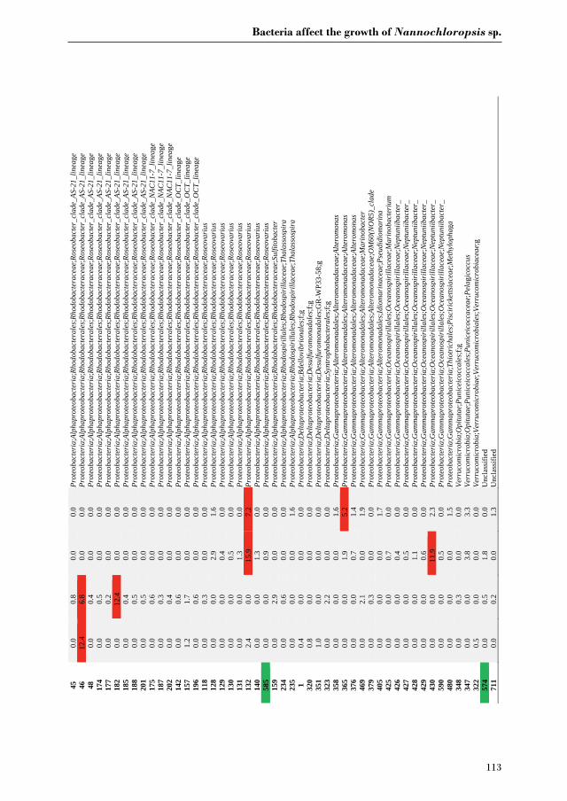
Bacteria affect the growth of Nannochloropsis sp.
113
45
0.0
0.8
0.0
0.0
Prot
eoba
cter
ia;A
lpha
prot
eoba
cter
ia;R
hodo
bact
eral
es;R
hodo
bact
erac
eae;
Rose
obac
ter_
clad
e_AS
-21_
linea
ge
46
12.4
6.
8 0.
0 0.
0 Pr
oteo
bact
eria
;Alp
hapr
oteo
bact
eria
;Rho
doba
cter
ales
;Rho
doba
cter
acea
e;Ro
seob
acte
r_cl
ade_
AS-2
1_lin
eage
48
0.
0 0.
4 0.
0 0.
0 Pr
oteo
bact
eria
;Alp
hapr
oteo
bact
eria
;Rho
doba
cter
ales
;Rho
doba
cter
acea
e;Ro
seob
acte
r_cl
ade_
AS-2
1_lin
eage
17
4 0.
0 0.
5 0.
0 0.
0 Pr
oteo
bact
eria
;Alp
hapr
oteo
bact
eria
;Rho
doba
cter
ales
;Rho
doba
cter
acea
e;Ro
seob
acte
r_cl
ade_
AS-2
1_lin
eage
17
7 0.
0 0.
2 0.
0 0.
0 Pr
oteo
bact
eria
;Alp
hapr
oteo
bact
eria
;Rho
doba
cter
ales
;Rho
doba
cter
acea
e;Ro
seob
acte
r_cl
ade_
AS-2
1_lin
eage
18
2 0.
0 12
.4
0.0
0.0
Prot
eoba
cter
ia;A
lpha
prot
eoba
cter
ia;R
hodo
bact
eral
es;R
hodo
bact
erac
eae;
Rose
obac
ter_
clad
e_AS
-21_
linea
ge
185
0.0
0.4
0.0
0.0
Prot
eoba
cter
ia;A
lpha
prot
eoba
cter
ia;R
hodo
bact
eral
es;R
hodo
bact
erac
eae;
Rose
obac
ter_
clad
e_AS
-21_
linea
ge
188
0.0
0.5
0.0
0.0
Prot
eoba
cter
ia;A
lpha
prot
eoba
cter
ia;R
hodo
bact
eral
es;R
hodo
bact
erac
eae;
Rose
obac
ter_
clad
e_AS
-21_
linea
ge
201
0.0
0.5
0.0
0.0
Prot
eoba
cter
ia;A
lpha
prot
eoba
cter
ia;R
hodo
bact
eral
es;R
hodo
bact
erac
eae;
Rose
obac
ter_
clad
e_AS
-21_
linea
ge
175
0.0
0.6
0.0
0.0
Prot
eoba
cter
ia;A
lpha
prot
eoba
cter
ia;R
hodo
bact
eral
es;R
hodo
bact
erac
eae;
Rose
obac
ter_
clad
e_N
AC11
-7_l
inea
ge
187
0.0
0.3
0.0
0.0
Prot
eoba
cter
ia;A
lpha
prot
eoba
cter
ia;R
hodo
bact
eral
es;R
hodo
bact
erac
eae;
Rose
obac
ter_
clad
e_N
AC11
-7_l
inea
ge
202
0.0
0.4
0.0
0.0
Prot
eoba
cter
ia;A
lpha
prot
eoba
cter
ia;R
hodo
bact
eral
es;R
hodo
bact
erac
eae;
Rose
obac
ter_
clad
e_N
AC11
-7_l
inea
ge
142
0.0
0.6
0.0
0.0
Prot
eoba
cter
ia;A
lpha
prot
eoba
cter
ia;R
hodo
bact
eral
es;R
hodo
bact
erac
eae;
Rose
obac
ter_
clad
e_O
CT_
linea
ge
157
1.2
1.7
0.0
0.0
Prot
eoba
cter
ia;A
lpha
prot
eoba
cter
ia;R
hodo
bact
eral
es;R
hodo
bact
erac
eae;
Rose
obac
ter_
clad
e_O
CT_
linea
ge
196
0.0
0.6
0.0
0.0
Prot
eoba
cter
ia;A
lpha
prot
eoba
cter
ia;R
hodo
bact
eral
es;R
hodo
bact
erac
eae;
Rose
obac
ter_
clad
e_O
CT_
linea
ge
118
0.0
0.3
0.0
0.0
Prot
eoba
cter
ia;A
lpha
prot
eoba
cter
ia;R
hodo
bact
eral
es;R
hodo
bact
erac
eae;
Rose
ovar
ius
128
0.0
0.0
2.9
1.6
Prot
eoba
cter
ia;A
lpha
prot
eoba
cter
ia;R
hodo
bact
eral
es;R
hodo
bact
erac
eae;
Rose
ovar
ius
129
0.0
0.0
0.4
0.0
Prot
eoba
cter
ia;A
lpha
prot
eoba
cter
ia;R
hodo
bact
eral
es;R
hodo
bact
erac
eae;
Rose
ovar
ius
130
0.0
0.0
0.5
0.0
Prot
eoba
cter
ia;A
lpha
prot
eoba
cter
ia;R
hodo
bact
eral
es;R
hodo
bact
erac
eae;
Rose
ovar
ius
131
0.0
0.0
1.3
0.0
Prot
eoba
cter
ia;A
lpha
prot
eoba
cter
ia;R
hodo
bact
eral
es;R
hodo
bact
erac
eae;
Rose
ovar
ius
132
2.4
0.0
15.9
7.
2 Pr
oteo
bact
eria
;Alp
hapr
oteo
bact
eria
;Rho
doba
cter
ales
;Rho
doba
cter
acea
e;Ro
seov
ariu
s 14
0 0.
0 0.
0 1.
3 0.
0 Pr
oteo
bact
eria
;Alp
hapr
oteo
bact
eria
;Rho
doba
cter
ales
;Rho
doba
cter
acea
e;Ro
seov
ariu
s 58
5 0.
0 0.
0 0.
9 0.
0 Pr
oteo
bact
eria
;Alp
hapr
oteo
bact
eria
;Rho
doba
cter
ales
;Rho
doba
cter
acea
e;Ro
seov
ariu
s 15
9 0.
0 2.
9 0.
0 0.
0 Pr
oteo
bact
eria
;Alp
hapr
oteo
bact
eria
;Rho
doba
cter
ales
;Rho
doba
cter
acea
e;Su
lfito
bact
er
234
0.0
0.6
0.0
0.0
Prot
eoba
cter
ia;A
lpha
prot
eoba
cter
ia;R
hodo
spir
illal
es;R
hodo
spir
illac
eae;
Thal
asso
spir
a 23
5 0.
0 0.
0 0.
0 1.
6 Pr
oteo
bact
eria
;Alp
hapr
oteo
bact
eria
;Rho
dosp
irill
ales
;Rho
dosp
irill
acea
e;Th
alas
sosp
ira
1 0.
4 0.
0 0.
0 0.
0 Pr
oteo
bact
eria
;Del
tapr
oteo
bact
eria
;Bde
llovi
brio
nale
s;f;g
32
0 0.
8 0.
0 0.
0 0.
0 Pr
oteo
bact
eria
;Del
tapr
oteo
bact
eria
;Des
ulfu
rom
onad
ales
;f;g
351
1.0
0.0
0.0
0.0
Prot
eoba
cter
ia;D
elta
prot
eoba
cter
ia;D
esul
furo
mon
adal
es;G
R-W
P33-
58;g
32
3 0.
0 2.
2 0.
0 0.
0 Pr
oteo
bact
eria
;Del
tapr
oteo
bact
eria
;Syn
trop
hoba
cter
ales
;f;g
358
0.0
0.0
0.0
1.6
Prot
eoba
cter
ia;G
amm
apro
teob
acte
ria;A
ltero
mon
adal
es;A
ltero
mon
adac
eae;
Alte
rom
onas
36
5 0.
0 0.
0 1.
9 5.
2 Pr
oteo
bact
eria
;Gam
map
rote
obac
teria
;Alte
rom
onad
ales
;Alte
rom
onad
acea
e;Al
tero
mon
as
376
0.0
0.0
0.7
1.4
Prot
eoba
cter
ia;G
amm
apro
teob
acte
ria;A
ltero
mon
adal
es;A
ltero
mon
adac
eae;
Alte
rom
onas
46
9 0.
0 2.
1 0.
0 1.
9 Pr
oteo
bact
eria
;Gam
map
rote
obac
teria
;Alte
rom
onad
ales
;Alte
rom
onad
acea
e;M
arin
obac
ter
379
0.0
0.3
0.0
0.0
Prot
eoba
cter
ia;G
amm
apro
teob
acte
ria;A
ltero
mon
adal
es;A
ltero
mon
adac
eae;
OM
60(N
OR5
)_cl
ade
405
0.0
0.0
0.0
1.7
Prot
eoba
cter
ia;G
amm
apro
teob
acte
ria;A
ltero
mon
adal
es;Id
iom
arin
acea
e;Ps
eudi
diom
arin
a 42
5 0.
0 0.
0 0.
7 0.
0 Pr
oteo
bact
eria
;Gam
map
rote
obac
teria
;Oce
anos
piri
llale
s;O
cean
ospi
rilla
ceae
;Mar
inob
acte
rium
42
6 0.
0 0.
0 0.
4 0.
0 Pr
oteo
bact
eria
;Gam
map
rote
obac
teria
;Oce
anos
piri
llale
s;O
cean
ospi
rilla
ceae
;Nep
tuni
ibac
ter_
42
7 0.
0 0.
0 0.
5 0.
0 Pr
oteo
bact
eria
;Gam
map
rote
obac
teria
;Oce
anos
piri
llale
s;O
cean
ospi
rilla
ceae
;Nep
tuni
ibac
ter_
42
8 0.
0 0.
0 1.
1 0.
0 Pr
oteo
bact
eria
;Gam
map
rote
obac
teria
;Oce
anos
piri
llale
s;O
cean
ospi
rilla
ceae
;Nep
tuni
ibac
ter_
42
9 0.
0 0.
0 0.
6 0.
0 Pr
oteo
bact
eria
;Gam
map
rote
obac
teria
;Oce
anos
piri
llale
s;O
cean
ospi
rilla
ceae
;Nep
tuni
ibac
ter_
43
0 0.
0 0.
0 11
.9
2.3
Prot
eoba
cter
ia;G
amm
apro
teob
acte
ria;O
cean
ospi
rilla
les;
Oce
anos
piri
llace
ae;N
eptu
niib
acte
r_
590
0.0
0.0
0.5
0.0
Prot
eoba
cter
ia;G
amm
apro
teob
acte
ria;O
cean
ospi
rilla
les;
Oce
anos
piri
llace
ae;N
eptu
niib
acte
r_
480
0.0
0.0
0.0
1.5
Prot
eoba
cter
ia;G
amm
apro
teob
acte
ria;T
hiot
rich
ales
;Pisc
iric
ketts
iace
ae;M
ethy
loph
aga
348
0.0
0.3
0.0
0.0
Verr
ucom
icro
bia;
Opi
tuta
e;Pu
nice
icoc
cale
s;f;g
34
7 0.
0 0.
0 3.
8 3.
3 Ve
rruc
omic
robi
a;O
pitu
tae;
Puni
ceic
occa
les;
Puni
ceic
occa
ceae
;Pel
agic
occu
s 32
2 0.
5 0.
0 0.
0 0.
0 Ve
rruc
omic
robi
a;Ve
rruc
omic
robi
ae;V
erru
com
icro
bial
es;V
erru
com
icro
biac
eae;
g 57
4 0.
0 0.
5 1.
8 0.
0 U
ncla
ssifi
ed
711
0.0
0.2
0.0
1.3
Unc
lass
ified


Chapter 6
General discussion
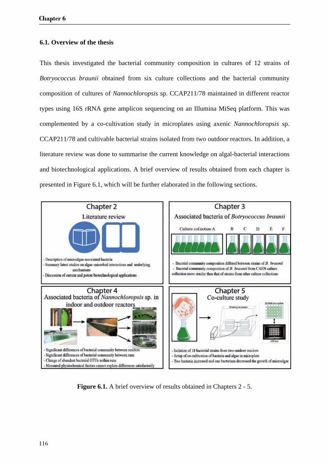
Chapter 6
116
6.1. Overview of the thesis
This thesis investigated the bacterial community composition in cultures of 12 strains of
Botryococcus braunii obtained from six culture collections and the bacterial community
composition of cultures of Nannochloropsis sp. CCAP211/78 maintained in different reactor
types using 16S rRNA gene amplicon sequencing on an Illumina MiSeq platform. This was
complemented by a co-cultivation study in microplates using axenic Nannochloropsis sp.
CCAP211/78 and cultivable bacterial strains isolated from two outdoor reactors. In addition, a
literature review was done to summarise the current knowledge on algal-bacterial interactions
and biotechnological applications. A brief overview of results obtained from each chapter is
presented in Figure 6.1, which will be further elaborated in the following sections.
Figure 6.1. A brief overview of results obtained in Chapters 2 - 5.
General discussion
117
6.2. Microalgae-associated bacteria
Numerous studies over the past 100 years have shown that bacteria are involved in complex
interactions with microalgae [96]. Particularly the rapid and ongoing developments in next-
generation sequencing technologies have led to an increase of research on the bacterial
communities associated with microalgae [71]. However, as microalgae are an extremely diverse
group of eukaryotic organisms and comprise of tens of thousands of species, only a few model
algal species or algae of biotechnological or ecological interest have been surveyed for their
associated bacterial communities to date [69, 118, 252, 359, 360].
Among all the microalgae, diatoms are the best studied taxa with respect to their associated
bacteria [306, 361]. A molecular survey of bacterial diversity from cultures of six diatom genera
(Ditylum, Thalassiosira, Asterionella, Chaetoceros, Leptocylindrus, and Coscinodiscus)
revealed distinct bacterial phylotypes associated with each species. However,
Alphaproteobacteria (including the genera Sulfitobacter, Roseobacter, Ruegeria, and
Erythrobacter), Bacteroidetes and to a lesser extent Betaproteobacteria were among the most
prominent taxa across all diatoms examined [361]. In a number of other studies it was shown
that with respect to the bacterial communities of diatom cultures the Proteobacteria and
Bacteroidetes are the main bacterial phyla associated with diatoms. At the genus level,
Sulfitobacter, Roseobacter, Alteromonas and Flavobacterium have been repeatedly found to be
associated with diatoms [16, 306, 359, 360, 362, 363].
For this thesis, we observed that even at the strain level B. braunii cultures were associated with
distinct bacterial communities. Interestingly, seven B. braunii strains obtained from the CAEN
culture collection harbored more similar bacterial communities as compared to strains from
other culture collections (Chapter 3, Figure 3.3A), suggesting that observed differences are at
least in part due to different maintenance approaches exerted by those culture collections. Our
Chapter 6
118
results are consistent with a previous study that showed that the bacterial communities of
different strains of the diatom species Thalassiosira rotula were significantly different [359].
Sapp et al. [363] also demonstrated that diatom cultures exhibited differences in bacterial
community composition over time after isolation, suggesting that changes in environmental
conditions may be important drivers of bacterial community composition.
In addition, we examined similarities and differences in bacterial community composition
between Botryococcus braunii (Chapter 3) and Nannochloropsis sp. CCAP211/78 (Chapter
4). To this end, we compared the cultures of 12 strains of B. braunii with Nannochloropsis sp.
CCAP211/78 cultured in a flat panel reactor (FP), as only for this reactor, strain CCAP211/78
was grown in a small volume with sterilized medium, similar to conditions of B. braunii
cultures. Nevertheless, it should be noted that the growth medium for both microalgae was
different – a fact that is ignored here, but which should receive more attention in future studies.
At the phylum level, Proteobacteria and Bacteroidetes were the most predominant bacterial
taxa with both Nannochloropsis sp. CCAP211/78 and B. braunii. However, at family level,
distinct predominant families were found to be associated with Nannochloropsis sp.
CCAP211/78 and B. braunii. Rhizobiaceae, Erythrobacteraceae and Cytophagaceae were the
most abundant families in cultures of B. braunii (Chapter 3, Figure 3.1), whereas
Rhodobacteraceae, Flammeovirgaceae and Oceanospirillaceae predominated the bacterial
community of Nannochloropsis sp. CCAP211/78 cultures in FP (Chapter 4, Figure S4.4).
There is an ongoing debate ongoing about whether the associations of bacteria with algae are
species-specific. Jasti et al. [364] observed that Alexandrium strains isolated from the Gulf of
Maine shared many bacterial phylotypes with other Alexandrium cultures, as based on
denaturing gradient gel electrophoresis of PCR-amplified 16S rRNA gene fragments, regardless
of geographic origin, whereas bacteria associated with Alexandrium were different from those
found associated with other phytoplankton species isolated from the same habitat. Likewise,
General discussion
119
another study demonstrated that two axenic marine diatoms (Thalassiosira rotula and
Skeletonema costatum) incubated with the same natural bacterial assemblages exhibited very
different bacterial community composition and were dominated by distinct phylogenetic groups
[306]. Our results generally reinforce the notion that different microalgae harbour specific
bacterial communities as we observed highly different bacterial communities associated with
Nannochloropsis sp. (mainly Rhodobacteraceae, Flammeovirgaceae and Oceanospirillaceae),
and B. braunii (mainly Rhizobiaceae, Erythrobacteraceae and Cytophagaceae), which also
differed from bacterial communities previously reported to be associated with different diatom
species (mainly Rhodobacteraceae, Flavobacteraceae and Alteromonadaceae). Some bacterial
taxa, such as the family Rhodobacteraceae are found together with Nannochloropsis sp.,
diatoms [96, 306, 319, 359] and many surveyed phytoplankton samples [96, 304, 336, 365], but
conservation is in general only observed at the family level. Overall, it seems that the
composition of the bacterial communities is species-specific for microalgae.
One hypothesis to explain the existence of species-specific interactions between algae and
bacteria is the niche hypothesis: if one algal species provides a defined and stable niche for a
long period of time, the same well-adapted bacterial species will outcompete others when
present [366]. Species-specific association of bacteria with algae may depend on various factors
such as algal exudates [361, 367], surface structure and cellular components of algae [364, 368],
cellular storage products and antibiotic production by algae [364]. Among these factors, algal
exudates may play the most important role in structuring the bacterial community [359]. This
assumption is supported by studies that several microalgal cultures secrete distinct exudates
especially with regard to polysaccharides [369, 370]. Therefore, microalgae that share
similarities in the composition of exudates might also be associated with similar bacterial
communities [359]. In this context it should be noted that in our experiments we did not find
correlations between bacterial community composition and the type of hydrocarbons produced
Chapter 6
120
by different strains of, although B. braunii can be subclassified to different races (races A, B,
and L) based on the type of hydrocarbons produced (Chapter 3). A future study to further
investigate the effects of other microalgal exudates, e.g. carbohydrates and amino acids, on the
composition and function of algal-associated bacteria would be of interest.
6.3. Reactor types and bacterial community composition
We surveyed bacterial community composition of Nannochloropsis sp. taken from different
reactor types, ranging from a small laboratory reactor to large-scale outdoor reactors (Chapter
4). At the phylum level, generally a similar bacterial community composition was observed
between each reactor type. However, at the OTU level we observed that each reactor type
contained significantly different bacterial communities except a horizontal tubular reactor (HT)
and a vertical tubular reactor (VT).
Reactor types varied in a number of physicochemical factors, which may be responsible for the
observed differences in bacterial community composition between reactors. Our assumption is
supported by a previous study showing that bacterial community composition of
Nannochloropsis salina was significantly different between small indoor reactors (Volume: 5
mL-4 L), medium indoor reactors (Volume: 20-60 L) and a large outdoor reactor (Volume: 200
L) [69]. Differences in numerous environmental factors and aspects of reactor configuration
(for instance, temperature, the ratio of surface area to volume and light intensity) during
cultivation at small, medium, and large scales might affect bacterial populations and cause
distinct bacterial communities to dominate different reactor systems [69]. HT and VT were not
different in bacterial community composition, which may be due to their comparable
configuration, which was indeed reflected by the small variation of physicochemical factors
between these two reactors (Chapter 4). However, none of the physicochemical factors
measured in our experiment alone could satisfactorily explain the shifts in the associated
General discussion
121
bacterial communities in different reactors (Chapter 4, Figure 4.4A). The distinct bacterial
communities in different reactor types are probably determined by the combination of
environmental variables and reactor configuration/operation, which makes it more difficult to
exactly pinpoint the factors responsible for bacterial community structuring. Furthermore, in
principle we only have data from a handful of reactors and two reactor runs, which means that
the number of independent experimental units is very small. The latter may lead to a rather big
impact of stochastic effects during the reactor operation that may affect the bacterial community
development. Altogether, at this point it is challenging to discern in a statistically sound way
the impact of environmental factors. Nevertheless, additional research that includes more
controlled experiments where environmental conditions are varied needs to be done, and
multiple reactors from different locations over multiple runs can be monitored and compared.
6.4. Applying defined co-cultures to understand algal-bacterial interactions
Instead of ignoring them or treating bacteria as contaminants as a whole, investigating their
interactions with microalgae could enable exploration of the concept of “synthetic ecology” as
a microalgae (co-)cultivation technique [371, 372].
Co-cultivation is not a novel idea in biotechnology [372-374], although much of the focus has
been on bacterial interactions [375]. In traditional microbially-based processes such as
anaerobic digestion, fermentation, and bioremediation, the most efficient microbial
assemblages are often selected and subsequently carefully maintained. With the exception of
wastewater treatment with microalgae and bacteria, this approach is not common in algal
biotechnology, however, the awareness of harnessing algae-bacteria interactions to enhance
productivity and increase the financial and environmental benefits of cultivating algae has been
accepted by the research community in recent years [372]. A number of examples have been
reviewed in chapter 2. In chapter 5, by applying a co-culture method we showed that two

Chapter 6
122
bacteria could significantly enhance the growth of a microalga. However, as the bacterial strains
tested in co-culture only accounted for 10% of the bacterial community in terms of relative
abundance in the algal cultures, many more potent bacterial candidates remain to be isolated
and tested for their potential to affect the growth of microalgae.
In addition to co-culturing microalgae with one bacterium, a synthetic community approach
could be used to select and test different defined microbial assemblages for their potential to
increase productivity as well as resistance to contamination. To my knowledge, there are no
studies related to this concept. To this end, random combinations of isolated bacterial strains
could be grown with algae in the microplate system we developed, and the co-cultures with
highest productivity could be selected and tested for large-scale cultivation. However, it can be
anticipated that this approach will also face a range of challenges such as how much complexity
of such engineered consortia is required and how the stability of synthetic communities can be
maintained. Considering the highly complex and dynamic nature of bacterial communities
observed in algal cultures, these natural communities may well harbour functional redundancy,
and experiments will have to show to what extent such redundancy can be reduced to simplify
communities, or whether functional redundancy is required to ensure culture stability when
facing dynamic environmental conditions in large scale outdoor production systems.
6.5. New opportunities for the deployment of algal-bacterial interactions
As mentioned in Chapter 1 and Chapter 2, competition and antagonistic interactions are
prevalent relationships between phytoplankton and bacteria [2, 96, 376]. However, our aim was
to explore mutualistic interactions between algae and bacteria for applications in algal
biotechnology. Thus, in this chapter we focus more on the beneficial effects of bacteria on algae.
One of the most widely studied mutualistic interactions is the obligate relationship between
vitamin-synthesizing bacteria and algal species that cannot synthesize several of the vitamins

General discussion
123
that are essential for their growth [2]. For example, ~50% of 326 algal species examined in a
study required exogenous supply of vitamins B1, B7 or B12 [26], whereas bacteria can synthesize
these vitamins to sustain algal growth in exchange for organic carbon [17, 22, 26, 27]. Another
common obligate mutualism involves nitrogen-fixing cyanobacteria and microalgae, where the
cyanobacteria provide fixed nitrogen to the algae in exchange for amino acids and organic
carbon [141, 321, 377]. Many marine bacteria, such as Marinobacter species, can alleviate iron
limitation of diatoms, dinoflagellates and coccolithophores by excreting siderophores that have
exceptionally high iron affinity [378]. In turn, algae release dissolved organic carbon to sustain
bacterial growth [98]. In another example, Sulfitobacter sp. SA11 was shown to use diatom-
derived tryptophan to produce the hormone indole-3-acetic acid that promotes the cell division
of the diatom Pseudo-nitzschia multiseries [16]. Interestingly, the molecular exchanges in a
range of algal-bacterial interactions bear resemblance to interactions that dominate the
rhizosphere of plants [2]. Therefore, the methods and knowledge on plant-microbe interactions
might guide and accelerate the research on algal-bacterial interactions.
Although some bacterial isolates have been shown to exert a positive effect on the growth of
microalgae in laboratory Erlenmeyer flasks [83, 105, 120, 123, 270], it does not mean that under
conditions in a large-scale bioreactor these bacteria continue to exert the same effect. Therefore,
bacteria that are beneficial to microalgae in flasks must be tested in large-scale cultures. For
instance, once we have gained mechanistic insight into the abovementioned synthetic
communities, the co-cultures with higher productivity could be transferred to larger-scale
reactors for further validation. If such co-cultures of higher productivity were to be employed
successfully at large scale, a lower production cost of algal biomass would be achieved [71,
372]. Nevertheless, scientists should further seek potential applications for algae-bacteria co-
culturing technology. For instance, would co-cultures of microalgae and bacteria have the
possibility to produce high added value molecules more cost-effective than other conventional
Chapter 6
124
production methods? A case in point is that an algae-bacterial co-culture was found more
efficient in H2 production than algae and bacteria separately [379]. Or would such co-culture
allow production of molecules that could otherwise not be produced? Such a molecule could be
the potent broad-spectrum antibiotic tropodithietic acid that can only be produced by co-
cultivation of Emiliania huxleyi and Phaeobacter gallaeciensis [46].
On the other hand, advantages of algal-bacterial interactions through co-cultivation could be
decreased contamination. When microalgae are co-cultured with symbiotic bacteria, the
invasion by other bacteria may be decreased because invading bacteria would be less likely to
establish in an already occupied niche [27]. Furthermore, studies of antagonistic or algicidal
bacteria are also useful because of the potential application in mitigating harmful algal blooms,
and more relevantly, in preventing the growth of algicidal bacteria when algae are grown at
large scale for aquaculture or other valuable products [380]. Related to the latter, Ganuza et al.
[381] investigated the possibility to alleviate bacterial infection of the green alga Chlorella by
the parasitic bacterium Vampirovibrio through decreasing pH of the medium for 15 min in the
presence of acetate. The treatment was successful in prolonging algal cultivation, and the
algicidal bacterium appeared to be unable to build up immunity to this treatment. Bagwell et al.
[382] also investigated the possibility of preventing Vampirovibrio infection of Chlorella
cultures. In this case, the infection was prevented by inducing the production of bioactive small
peptides and glycosides by Chlorella under iron limitation.
6.6. Novel methodologies to study algae-associated bacteria and algae-bacterial
interactions
6.6.1. New primer set to characterize algae-associated bacteria
A common approach to characterize the composition of algae-associated bacterial communities
comprises PCR amplification and sequencing of 16S rRNA genes using a universal bacterial
General discussion
125
primer set [383]. Routine sampling strategies for studying algal microbiomes do not often
separate algal and bacterial cells, which results in the combination of bacterial and algal DNA
in the samples. However, as algal plastid genomes encode 16S rRNA closely related to that of
cyanobacteria, sequencing data sets frequently comprise more than 75% plastid sequences
(Chapter 4). As a result, bacterial diversity may be largely underrepresented due to the
decreased sequencing depth.
Thomas et al. [384] evaluated several primer sets to amplify a 450 bp fragment spanning the
V3-V4 region while minimizing the amplification of plastid sequences. The new primer set
NOCHL was shown to have lower coverage for plastids and cover a significantly higher
bacterial richness than other tested primer sets. This primer set is a potent alternative to study
bacterial communities in environments where plastid contamination can be an issue such as
terrestrial plants and microalgae.
6.6.2. Mass spectrometry imaging
Information obtained from (meta)genomic/transcriptomic and proteomic approaches is a
prerequisite for a systems-level understanding of symbiont physiology [385], but is not
sufficient to provide direct evidence of potential metabolic signatures because microbial
interactions often involve a dynamic metabolic crosstalk and chemical communication [386,
387]. Therefore, a metabolomics approach has been developed as a powerful tool to unravel the
means and consequences of microbial interaction at the metabolic level. Recent advances in
mass spectrometry imaging (MSI) technologies allow the localization of specific metabolites
of various dynamic processes and biotic interactions at the microscale level and, consequently,
monitoring of metabolic changes at high spatiotemporal resolution [388]. MSI has been widely
used to study the metabolic footprints of host-associated bacteria [385], bacterial population
dynamics [389] and to detect pathogens [390]. Likewise, MSI is applicable to study algal-
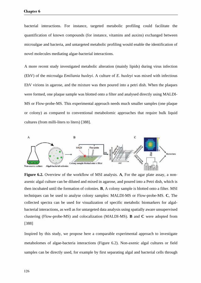
Chapter 6
126
bacterial interactions. For instance, targeted metabolic profiling could facilitate the
quantification of known compounds (for instance, vitamins and auxins) exchanged between
microalgae and bacteria, and untargeted metabolic profiling would enable the identification of
novel molecules mediating algae-bacterial interactions.
A more recent study investigated metabolic alteration (mainly lipids) during virus infection
(EhV) of the microalga Emiliania huxleyi. A culture of E. huxleyi was mixed with infectious
EhV virions in agarose, and the mixture was then poured into a petri dish. When the plaques
were formed, one plaque sample was blotted onto a filter and analysed directly using MALDI-
MS or Flow-probe-MS. This experimental approach needs much smaller samples (one plaque
or colony) as compared to conventional metabolomic approaches that require bulk liquid
cultures (from milli-liters to liters) [388].
Figure 6.2. Overview of the workflow of MSI analysis. A, For the agar plate assay, a non-
axenic algal culture can be diluted and mixed in agarose, and poured into a Petri dish, which is
then incubated until the formation of colonies. B, A colony sample is blotted onto a filter. MSI
techniques can be used to analyse colony samples: MALDI-MS or Flow-probe-MS. C, The
collected spectra can be used for visualization of specific metabolic biomarkers for algal-
bacterial interactions, as well as for untargeted data analysis using spatially aware unsupervised
clustering (Flow-probe-MS) and colocalization (MALDI-MS). B and C were adopted from
[388]
Inspired by this study, we propose here a comparable experimental approach to investigate
metabolomes of algae-bacteria interactions (Figure 6.2). Non-axenic algal cultures or field
samples can be directly used, for example by first separating algal and bacterial cells through
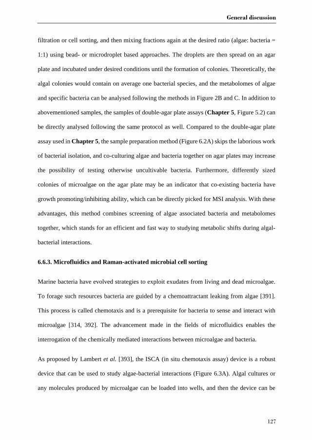
General discussion
127
filtration or cell sorting, and then mixing fractions again at the desired ratio (algae: bacteria =
1:1) using bead- or microdroplet based approaches. The droplets are then spread on an agar
plate and incubated under desired conditions until the formation of colonies. Theoretically, the
algal colonies would contain on average one bacterial species, and the metabolomes of algae
and specific bacteria can be analysed following the methods in Figure 2B and C. In addition to
abovementioned samples, the samples of double-agar plate assays (Chapter 5, Figure 5.2) can
be directly analysed following the same protocol as well. Compared to the double-agar plate
assay used in Chapter 5, the sample preparation method (Figure 6.2A) skips the laborious work
of bacterial isolation, and co-culturing algae and bacteria together on agar plates may increase
the possibility of testing otherwise uncultivable bacteria. Furthermore, differently sized
colonies of microalgae on the agar plate may be an indicator that co-existing bacteria have
growth promoting/inhibiting ability, which can be directly picked for MSI analysis. With these
advantages, this method combines screening of algae associated bacteria and metabolomes
together, which stands for an efficient and fast way to studying metabolic shifts during algal-
bacterial interactions.
6.6.3. Microfluidics and Raman-activated microbial cell sorting
Marine bacteria have evolved strategies to exploit exudates from living and dead microalgae.
To forage such resources bacteria are guided by a chemoattractant leaking from algae [391].
This process is called chemotaxis and is a prerequisite for bacteria to sense and interact with
microalgae [314, 392]. The advancement made in the fields of microfluidics enables the
interrogation of the chemically mediated interactions between microalgae and bacteria.
As proposed by Lambert et al. [393], the ISCA (in situ chemotaxis assay) device is a robust
device that can be used to study algae-bacterial interactions (Figure 6.3A). Algal cultures or
any molecules produced by microalgae can be loaded into wells, and then the device can be
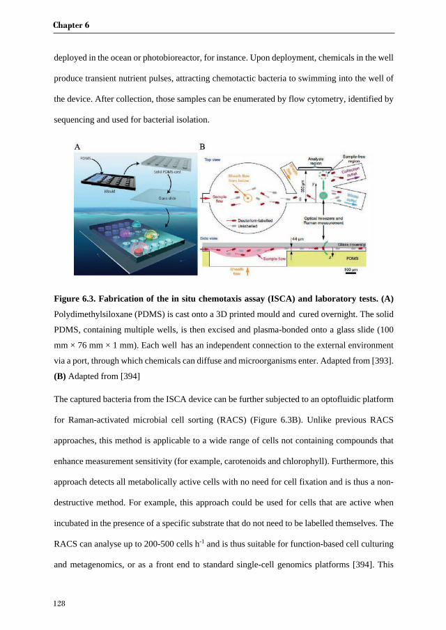
Chapter 6
128
deployed in the ocean or photobioreactor, for instance. Upon deployment, chemicals in the well
produce transient nutrient pulses, attracting chemotactic bacteria to swimming into the well of
the device. After collection, those samples can be enumerated by flow cytometry, identified by
sequencing and used for bacterial isolation.
Figure 6.3. Fabrication of the in situ chemotaxis assay (ISCA) and laboratory tests. (A)
Polydimethylsiloxane (PDMS) is cast onto a 3D printed mould and cured overnight. The solid
PDMS, containing multiple wells, is then excised and plasma-bonded onto a glass slide (100
mm × 76 mm × 1 mm). Each well has an independent connection to the external environment
via a port, through which chemicals can diffuse and microorganisms enter. Adapted from [393].
(B) Adapted from [394]
The captured bacteria from the ISCA device can be further subjected to an optofluidic platform
for Raman-activated microbial cell sorting (RACS) (Figure 6.3B). Unlike previous RACS
approaches, this method is applicable to a wide range of cells not containing compounds that
enhance measurement sensitivity (for example, carotenoids and chlorophyll). Furthermore, this
approach detects all metabolically active cells with no need for cell fixation and is thus a non-
destructive method. For example, this approach could be used for cells that are active when
incubated in the presence of a specific substrate that do not need to be labelled themselves. The
RACS can analyse up to 200-500 cells h-1 and is thus suitable for function-based cell culturing
and metagenomics, or as a front end to standard single-cell genomics platforms [394]. This
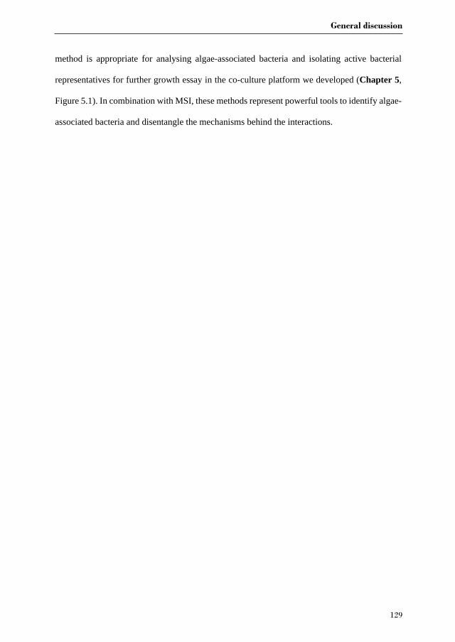
General discussion
129
method is appropriate for analysing algae-associated bacteria and isolating active bacterial
representatives for further growth essay in the co-culture platform we developed (Chapter 5,
Figure 5.1). In combination with MSI, these methods represent powerful tools to identify algae-
associated bacteria and disentangle the mechanisms behind the interactions.


References

References
132
References
1. Armbrust, E.V., et al., The genome of the diatom Thalassiosira pseudonana: ecology, evolution, and metabolism. Science, 2004. 306(5693): p. 79-86.
2. Seymour, J.R., et al., Zooming in on the phycosphere: the ecological interface for phytoplankton–bacteria relationships. Nature microbiology, 2017. 2(7): p. 17065.
3. Falkowski, P.G., The role of phytoplankton photosynthesis in global biogeochemical cycles. Photosynthesis research, 1994. 39(3): p. 235-258.
4. Field, C.B., et al., Primary production of the biosphere: integrating terrestrial and oceanic components. science, 1998. 281(5374): p. 237-240.
5. Palmer, J.D., D.E. Soltis, and M.W. Chase, The plant tree of life: an overview and some points of view. American Journal of Botany, 2004. 91(10): p. 1437-1445.
6. Müller, D.B., et al., The plant microbiota: systems-level insights and perspectives. Annual review of genetics, 2016. 50: p. 211-234.
7. Parniske, M., Arbuscular mycorrhiza: the mother of plant root endosymbioses. Nature Reviews Microbiology, 2008. 6(10): p. 763.
8. Udvardi, M. and P.S. Poole, Transport and metabolism in legume-rhizobia symbioses. Annual review of plant biology, 2013. 64: p. 781-805.
9. Berendsen, R.L., C.M. Pieterse, and P.A. Bakker, The rhizosphere microbiome and plant health. Trends in plant science, 2012. 17(8): p. 478-486.
10. Bulgarelli, D., et al., Structure and functions of the bacterial microbiota of plants. Annual review of plant biology, 2013. 64: p. 807-838.
11. Mendes, R., P. Garbeva, and J.M. Raaijmakers, The rhizosphere microbiome: significance of plant beneficial, plant pathogenic, and human pathogenic microorganisms. FEMS microbiology reviews, 2013. 37(5): p. 634-663.
12. Vorholt, J.A., Microbial life in the phyllosphere. Nature Reviews Microbiology, 2012. 10(12): p. 828.
13. Hiltner, L., Uber nevere Erfahrungen und Probleme auf dem Gebiet der Boden Bakteriologie und unter besonderer Beurchsichtigung der Grundungung und Broche. Arbeit. Deut. Landw. Ges. Berlin, 1904. 98: p. 59-78.
14. Hartmann, A., M. Rothballer, and M. Schmid, Lorenz Hiltner, a pioneer in rhizosphere microbial ecology and soil bacteriology research. Plant Soil, 2008. 312(1-2): p. 7-14.
15. Bell, W. and R. Mitchell, Chemotactic and growth responses of marine bacteria to algal extracellular products. The Biological Bulletin, 1972. 143(2): p. 265-277.
16. Amin, S., et al., Interaction and signalling between a cosmopolitan phytoplankton and associated bacteria. Nature, 2015. 522(7554): p. 98-101.
17. Durham, B.P., et al., Cryptic carbon and sulfur cycling between surface ocean plankton. Proceedings of the National Academy of Sciences, 2015. 112(2): p. 453-457.
18. Lucas, C., The ecological effects of external metabolites. Biological Reviews, 1947. 22(3): p. 270-295.
19. Provasoli, L., Nutrition and ecology of protozoa and algae. Annual review of microbiology, 1958. 12(1): p. 279-308.
20. Watanabe, K., et al., Symbiotic association in Chlorella culture. FEMS microbiology ecology, 2005. 51(2): p. 187-196.
21. Krohn-Molt, I., et al., Insights into Microalga and Bacteria Interactions of Selected Phycosphere Biofilms Using Metagenomic, Transcriptomic, and Proteomic Approaches. Frontiers in Microbiology, 2017. 8: p. 1941.
22. Grant, M.A., et al., Direct exchange of vitamin B12 is demonstrated by modelling the growth dynamics of algal-bacterial cocultures. ISME J, 2014. 8(7): p. 1418-27.
References
133
23. Landa, M., et al., Shifts in bacterial community composition associated with increased carbon cycling in a mosaic of phytoplankton blooms. ISME J, 2016. 10(1): p. 39-50.
24. Xie, B., et al., Chlamydomonas reinhardtii thermal tolerance enhancement mediated by a mutualistic interaction with vitamin B12-producing bacteria. ISME J, 2013. 7(8): p. 1544-55.
25. Ramanan, R., et al., Algae–bacteria interactions: evolution, ecology and emerging applications. Biotechnology advances, 2016. 34(1): p. 14-29.
26. Croft, M.T., et al., Algae acquire vitamin B12 through a symbiotic relationship with bacteria. Nature, 2005. 438(7064): p. 90-3.
27. Kazamia, E., et al., Mutualistic interactions between vitamin B12‐dependent algae and heterotrophic bacteria exhibit regulation. Environmental microbiology, 2012. 14(6): p. 1466-1476.
28. Hernandez, J.-P., et al., Growth promotion of the freshwater microalga Chlorella vulgaris by the nitrogen-fixing, plant growth-promoting bacterium Bacillus pumilus from arid zone soils. European Journal of Soil Biology, 2009. 45(1): p. 88-93.
29. Cho, D.H., et al., Enhancing microalgal biomass productivity by engineering a microalgal-bacterial community. Bioresour Technol, 2014. 175c: p. 578-585.
30. Zapalski, M.K., Is absence of proof a proof of absence? Comments on commensalism. Palaeogeography, Palaeoclimatology, Palaeoecology, 2011. 302(3-4): p. 484-488.
31. Mai-Prochnow, A., et al., Biofilm development and cell death in the marine bacterium Pseudoalteromonas tunicata. Applied and environmental microbiology, 2004. 70(6): p. 3232-3238.
32. Ivanova, J., G. Stoyancheva, and I. Pouneva, Lysis of Antarctic algal strains by bacterial pathogen. Antonie Van Leeuwenhoek, 2014. 105(6): p. 997-1005.
33. Zheng, X., et al., A marine algicidal actinomycete and its active substance against the harmful algal bloom species Phaeocystis globosa. Applied microbiology and biotechnology, 2013. 97(20): p. 9207-9215.
34. Hertweck, C., et al., Antagonistic bacteria disrupt calcium homeostasis and immobilize algal cells. Nature communications, 2017. 8: p. 1756.
35. Wang, B., et al., A marine bacterium producing protein with algicidal activity against Alexandrium tamarense. Harmful Algae, 2012. 13: p. 83-88.
36. Kim, M.-J., S.-Y. Jeong, and S.-J. Lee, Isolation, identification, and algicidal activity of marine bacteria against Cochlodinium polykrikoides. Journal of Applied Phycology, 2008. 20(6): p. 1069-1078.
37. Lee, Y.-K., et al., Cyanobactericidal effect of Rhodococcus sp. isolated from eutrophic lake on Microcystis sp. Biotechnology letters, 2010. 32(11): p. 1673-1678.
38. Bai, S.J., et al., Algicidal effects of a novel marine actinomycete on the toxic dinoflagellate Alexandrium tamarense. Current Microbiology, 2011. 62(6): p. 1774-1781.
39. Anderson, D.M., A.D. Cembella, and G.M. Hallegraeff, Progress in understanding harmful algal blooms: paradigm shifts and new technologies for research, monitoring, and management. Annual review of marine science, 2012. 4: p. 143-176.
40. Buchan, A., et al., Master recyclers: features and functions of bacteria associated with phytoplankton blooms. Nature Reviews Microbiology, 2014.
41. Heisler, J., et al., Eutrophication and harmful algal blooms: a scientific consensus. Harmful algae, 2008. 8(1): p. 3-13.
42. Landsberg, J.H., The effects of harmful algal blooms on aquatic organisms. Reviews in Fisheries Science, 2002. 10(2): p. 113-390.
43. Grattan, L.M., S. Holobaugh, and J.G. Morris Jr, Harmful algal blooms and public health. Harmful algae, 2016. 57: p. 2-8.

References
134
44. Brodie, J., et al., Biotic interactions as drivers of algal origin and evolution. New Phytologist, 2017. 216(3): p. 670-681.
45. Wagner-Döbler, I., et al., The complete genome sequence of the algal symbiont Dinoroseobacter shibae: a hitchhiker's guide to life in the sea. The ISME journal, 2010. 4(1): p. 61-77.
46. Seyedsayamdost, M.R., et al., The Jekyll-and-Hyde chemistry of Phaeobacter gallaeciensis. Nature chemistry, 2011. 3(4): p. 331-335.
47. Dittami, S.M., D. Eveillard, and T. Tonon, A metabolic approach to study algal–bacterial interactions in changing environments. Molecular ecology, 2014. 23(7): p. 1656-1660.
48. Cooper, M.B. and A.G. Smith, Exploring mutualistic interactions between microalgae and bacteria in the omics age. Current opinion in plant biology, 2015. 26: p. 147-153.
49. Krupke, A., et al., Distribution of a consortium between unicellular algae and the N2 fixing cyanobacterium UCYN ‐ A in the North Atlantic Ocean. Environmental microbiology, 2014. 16(10): p. 3153-3167.
50. Wijffels, R.H., M.J. Barbosa, and M.H. Eppink, Microalgae for the production of bulk chemicals and biofuels. Biofuels, Bioproducts and Biorefining, 2010. 4(3): p. 287-295.
51. Draaisma, R.B., et al., Food commodities from microalgae. Current opinion in biotechnology, 2013. 24(2): p. 169-177.
52. Norsker, N.-H., et al., Microalgal production—a close look at the economics. Biotechnology advances, 2011. 29(1): p. 24-27.
53. Mascarelli, A.L., Gold rush for algae. 2009, Nature Publishing Group. 54. Chew, K.W., et al., Microalgae biorefinery: high value products perspectives.
Bioresource technology, 2017. 229: p. 53-62. 55. Eppink, M.H., et al., From current algae products to future biorefinery practices: a
review, in Biorefineries. 2017, Springer. p. 99-123. 56. Wijffels, R.H. and M.J. Barbosa, An outlook on microalgal biofuels. Science, 2010.
329(5993): p. 796-799. 57. Ruiz, J., et al., Towards industrial products from microalgae. Energy & Environmental
Science, 2016. 9(10): p. 3036-3043. 58. Ugwu, C., H. Aoyagi, and H. Uchiyama, Photobioreactors for mass cultivation of algae.
Bioresource technology, 2008. 99(10): p. 4021-4028. 59. de Vree, J.H., Outdoor production of microalgae. 2016: Wageningen University. 60. Kumar, K., et al., Recent trends in the mass cultivation of algae in raceway ponds.
Renewable and Sustainable Energy Reviews, 2015. 51: p. 875-885. 61. Grobbelaar, J.U., Factors governing algal growth in photobioreactors: the “open”
versus “closed” debate. Journal of applied phycology, 2009. 21(5): p. 489. 62. Slade, R. and A. Bauen, Micro-algae cultivation for biofuels: cost, energy balance,
environmental impacts and future prospects. Biomass and bioenergy, 2013. 53: p. 29-38.
63. Norsker, N.H., et al., Microalgal production — A close look at the economics. 2011. 29(1): p. 24-27.
64. Sousa, C., et al., Growth of the microalgae Neochloris oleoabundans at high partial oxygen pressures and sub-saturating light intensity. Bioresource technology, 2012. 104: p. 565-570.
65. Ugwu, C., J. Ogbonna, and H. Tanaka, Design of static mixers for inclined tubular photobioreactors. Journal of Applied Phycology, 2003. 15(2-3): p. 217-223.
66. Wolf, J., et al., Multifactorial comparison of photobioreactor geometries in parallel microalgae cultivations. Algal research, 2016. 15: p. 187-201.
References
135
67. Xu, L., et al., Microalgal bioreactors: challenges and opportunities. Engineering in Life Sciences, 2009. 9(3): p. 178-189.
68. Gupta, P.L., S.-M. Lee, and H.-J. Choi, A mini review: photobioreactors for large scale algal cultivation. World Journal of Microbiology and Biotechnology, 2015. 31(9): p. 1409-1417.
69. Fulbright, S.P., et al., Bacterial community changes in an industrial algae production system. Algal Research, 2018. 31: p. 147-156.
70. Newby, D.T., et al., Assessing the potential of polyculture to accelerate algal biofuel production. Algal research, 2016. 19: p. 264-277.
71. Lian, J., et al., The effect of the algal microbiome on industrial production of microalgae. Microbial Biotechnology, 2018. 11(5): p. 806-818.
72. Wolf, F.R., Botryococcus braunii an unusual hydrocarbon-producing alga. Applied Biochemistry and Biotechnology Advances, 1983. 8(3): p. 249.
73. Fernandes, H., et al., Biosynthesis of high concentrations of an exopolysaccharide during the cultivation of the microalgaBotryococcus braunii. Biotechnology letters, 1989. 11(6): p. 433-436.
74. Gouveia, J.D., et al., Botryococcus braunii strains compared for biomass productivity, hydrocarbon and carbohydrate content. Journal of applied phycology, 2017. 248: p. 77-86.
75. Metzger, P. and C. Largeau, Botryococcus braunii: a rich source for hydrocarbons and related ether lipids. Applied microbiology biotechnology, 2005. 66(5): p. 486-496.
76. Kawachi, M., et al., Relationship between hydrocarbons and molecular phylogeny of Botryococcus braunii. Algal Research, 2012. 1(2): p. 114-119.
77. Eroglu, E., S. Okada, and A. Melis, Hydrocarbon productivities in different Botryococcus strains: comparative methods in product quantification. Journal of applied phycology, 2011. 23(4): p. 763-775.
78. Cabanelas, I.T.D., et al., Botryococcus, what to do with it? Effect of nutrient concentration on biorefinery potential. Algal Research, 2015. 11: p. 43-49.
79. Dayananda, C., et al., Autotrophic cultivation of Botryococcus braunii for the production of hydrocarbons and exopolysaccharides in various media. Biomass and Bioenergy, 2007. 31(1): p. 87-93.
80. Rivas, M.O., P. Vargas, and C.E. Riquelme, Interactions of Botryococcus braunii cultures with bacterial biofilms. Microbial ecology, 2010. 60(3): p. 628-635.
81. Sambles, C., et al., Metagenomic analysis of the complex microbial consortium associated with cultures of the oil-rich alga Botryococcus braunii. MicrobiologyOpen, 2017. 6(4): p. 6196-6206.
82. Chirac, C., et al., Bacterial influence upon growth and hydrocarbon production of the green alga Botryococcus braunii 1. Journal of Phycology, 1985. 21(3): p. 380-387.
83. Tanabe, Y., et al., A novel alphaproteobacterial ectosymbiont promotes the growth of the hydrocarbon-rich green alga Botryococcus braunii. Scientific Reports, 2015. 5: p. 10467.
84. Gouveia, J.D., et al., Effect of removal of bacteria on the biomass and extracellular carbohydrate productivity of Botryococcus braunii. Journal of Applied Phycology, 2019: p. 1-11.
85. Rodolfi, L., et al., Microalgae for Oil: Strain Selection, Induction of Lipid Synthesis and Outdoor Mass Cultivation in a Low-Cost Photobioreactor. Biotechnology Bioengineering, 2010. 102(1): p. 100-112.
86. Dipasmita, P., et al., The effect of light, salinity, and nitrogen availability on lipid production by Nannochloropsis sp. Applied Microbiology Biotechnology, 2011. 90(4): p. 1429.

References
136
87. Wang, H., et al., Novel bacterial isolate from Permian groundwater, capable of aggregating potential biofuel-producing microalga Nannochloropsis oceanica IMET1. Appl Environ Microbiol, 2012. 78(5): p. 1445-53.
88. Skjermo, J. and O. Vadstein, Techniques for microbial control in the intensive rearing of marine larvae. Aquaculture, 1999. 177(1-4): p. 333-343.
89. Salvesen, I., J. Skjermo, and O. Vadstein, Growth of turbot (Scophthalmus maximus L.) during first feeding in relation to the proportion of r/K-strategists in the bacterial community of the rearing water. Aquaculture, 1999. 175(3-4): p. 337-350.
90. Nakase, G. and M. Eguchi, Analysis of bacterial communities in Nannochloropsis sp. cultures used for larval fish production. Fisheries science, 2007. 73(3): p. 543-549.
91. Carney, L.T., et al., Pond Crash Forensics: Presumptive identification of pond crash agents by next generation sequencing in replicate raceway mass cultures of Nannochloropsis salina. Algal Research, 2016. 17: p. 341-347.
92. Sharifah, E.N. and M. Eguchi, The phytoplankton Nannochloropsis oculata enhances the ability of Roseobacter clade bacteria to inhibit the growth of fish pathogen Vibrio anguillarum. PLoS One, 2011. 6(10): p. e26756.
93. Sharma, N.K., et al., Sustainability and cyanobacteria (blue-green algae): facts and challenges. Journal of applied phycology, 2011. 23(6): p. 1059-1081.
94. Shields, R.J. and I. Lupatsch, Algae for aquaculture and animal feeds. J Anim Sci, 2012. 21: p. 23-37.
95. Hom, E.F., et al., A chemical perspective on microalgal–microbial interactions. Trends in plant science, 2015. 20(11): p. 689-693.
96. Amin, S.A., M.S. Parker, and E.V. Armbrust, Interactions between diatoms and bacteria. Microbiol Mol Biol Rev, 2012. 76(3): p. 667-84.
97. Bowler, C., et al., The Phaeodactylum genome reveals the evolutionary history of diatom genomes. Nature, 2008. 456(7219): p. 239-244.
98. Amin, S.A., et al., Photolysis of iron–siderophore chelates promotes bacterial–algal mutualism. Proceedings of the National Academy of Sciences, USA, 2009. 106(40): p. 17071-17076.
99. Bruhn, J.B., L. Gram, and R. Belas, Production of antibacterial compounds and biofilm formation by Roseobacter species are influenced by culture conditions. Applied and environmental microbiology, 2007. 73(2): p. 442-450.
100. Wahl, M., et al., The second skin: ecological role of epibiotic biofilms on marine organisms. Frontiers in microbiology, 2012. 3: p. 292.
101. Natrah, F.M.I., et al., Significance of microalgal-bacterial interactions for aquaculture. Reviews in Aquaculture, 2013. 6(1): p. 48-61.
102. Danchin, A. and S. Braham, Coenzyme B12 synthesis as a baseline to study metabolite contribution of animal microbiota. Microbial biotechnology, 2017. 10(4): p. 688-701.
103. Dittami, S.M., et al., Host–microbe interactions as a driver of acclimation to salinity gradients in brown algal cultures. The ISME journal, 2016. 10(1): p. 51-63.
104. Le Chevanton, M., et al., Screening and selection of growth-promoting bacteria for Dunaliella cultures. Algal Research, 2013. 2(3): p. 212-222.
105. Kim, B.-H., et al., Role of Rhizobium, a plant growth promoting bacterium, in enhancing algal biomass through mutualistic interaction. Biomass and Bioenergy, 2014. 69(0): p. 95-105.
106. Carney, L.T. and T.W. Lane, Parasites in algae mass culture. Frontiers in microbiology, 2014. 5: p. 278.
107. Mendes, L.B.B. and A.B. Vermelho, Allelopathy as a potential strategy to improve microalgae cultivation. Biotechnology for biofuels, 2013. 6(1): p. 1.
References
137
108. Su, Y., A. Mennerich, and B. Urban, Synergistic cooperation between wastewater-born algae and activated sludge for wastewater treatment: influence of algae and sludge inoculation ratios. Bioresource technology, 2012. 105: p. 67-73.
109. Luo, S., et al., Pyrene degradation accelerated by constructed consortium of bacterium and microalga: effects of degradation products on the microalgal growth. Environ Sci Technol, 2014. 48(23): p. 13917-24.
110. Cavaliere, M., et al., Cooperation in microbial communities and their biotechnological applications. Environmental microbiology, 2017. 19(8): p. 2949-2963.
111. Wilson, W.H., G. Tarran, and M.V. Zubkov, Virus dynamics in a coccolithophore-dominated bloom in the North Sea. Deep Sea Research Part II: Topical Studies in Oceanography, 2002. 49(15): p. 2951-2963.
112. Chen, Z., et al., First report of Pseudobodo sp, a new pathogen for a potential energy-producing algae: Chlorella vulgaris cultures. PLoS One, 2014. 9(3): p. e89571.
113. Lenneman, E.M., P. Wang, and B.M. Barney, Potential application of algicidal bacteria for improved lipid recovery with specific algae. FEMS Microbiol Lett, 2014. 354(2): p. 102-10.
114. Sun, L., et al., A novel membrane bioreactor inoculated with symbiotic sludge bacteria and algae: Performance and microbial community analysis. Bioresource technology, 2018. 251: p. 311-319.
115. Yang, J., et al., Potential of wastewater treatment using a concentrated and suspended algal-bacterial consortium in a photo membrane bioreactor. Chemical Engineering Journal, 2018. 335: p. 154-160.
116. García, D., et al., Comparative evaluation of piggery wastewater treatment in algal-bacterial photobioreactors under indoor and outdoor conditions. Bioresource technology, 2017. 245: p. 483-490.
117. Krohn-Molt, I., et al., Metagenome survey of a multispecies and alga-associated biofilm revealed key elements of bacterial-algal interactions in photobioreactors. Applied and environmental microbiology, 2013. 79(20): p. 6196-6206.
118. Biondi, N., et al., The bacterial community associated with Tetraselmis suecica outdoor mass cultures. Journal of Applied Phycology, 2017. 29(1): p. 67-78.
119. Sison-Mangus, M.P., et al., Host-specific adaptation governs the interaction of the marine diatom, Pseudo-nitzschia and their microbiota. The ISME journal, 2014. 8(1): p. 63-76.
120. Lakaniemi, A.M., et al., Growth of Chlorella vulgaris and associated bacteria in photobioreactors. Microb Biotechnol, 2012. 5(1): p. 69-78.
121. Park, Y., et al., Growth promotion of Chlorella ellipsoidea by co-inoculation with Brevundimonas sp. isolated from the microalga. Hydrobiologia, 2007. 598(1): p. 219-228.
122. Grossart, H.-P. and M. Simon, Interactions of planktonic algae and bacteria: effects on algal growth and organic matter dynamics. Aquatic Microbial Ecology, 2007. 47(2): p. 163.
123. Bruckner, C.G., et al., Growth and release of extracellular organic compounds by benthic diatoms depend on interactions with bacteria. Environmental microbiology, 2011. 13(4): p. 1052-1063.
124. Ras, M., J.-P. Steyer, and O. Bernard, Temperature effect on microalgae: a crucial factor for outdoor production. Reviews in Environmental Science and Bio/Technology, 2013. 12(2): p. 153-164.
125. Olsenz, J.L., Stress ecology in Fucus: abiotic, biotic and genetic interactions. Advances in marine biology, 2011. 59(57): p. 37.
References
138
126. Wang, S., et al., Microalgal assemblages in a poikilohaline pond. Journal of Phycology, 2014. 50(2): p. 303-309.
127. Foflonker, F., et al., Genome of the halotolerant green alga Picochlorum sp. reveals strategies for thriving under fluctuating environmental conditions. Environmental microbiology, 2015. 17(2): p. 412-426.
128. Ritter, A., et al., Copper stress proteomics highlights local adaptation of two strains of the model brown alga Ectocarpus siliculosus. Proteomics, 2010. 10(11): p. 2074-2088.
129. Tonon, T., et al., Toward systems biology in brown algae to explore acclimation and adaptation to the shore environment. Omics: a journal of integrative biology, 2011. 15(12): p. 883-892.
130. Charrier, B., et al., Development and physiology of the brown alga Ectocarpus siliculosus: two centuries of research. New Phytologist, 2008. 177(2): p. 319-332.
131. Schroda, M., The Chlamydomonas genome reveals its secrets: chaperone genes and the potential roles of their gene products in the chloroplast. Photosynthesis research, 2004. 82(3): p. 221-240.
132. Singh, M. and K. Das, Low Cost Nutrients for Algae Cultivation, in Algal Biorefineries. 2014, Springer. p. 69-82.
133. Clarens, A.F., et al., Environmental life cycle comparison of algae to other bioenergy feedstocks. Environmental science & technology, 2010. 44(5): p. 1813-1819.
134. Putt, R., et al., An efficient system for carbonation of high-rate algae pond water to enhance CO 2 mass transfer. Bioresource technology, 2011. 102(3): p. 3240-3245.
135. Mouget, J.L., et al., Algal growth enhancement by bacteria: is consumption of photosynthetic oxygen involved? FEMS microbiology ecology, 1995. 18(1): p. 35-43.
136. Subashchandrabose, S.R., et al., Consortia of cyanobacteria/microalgae and bacteria: biotechnological potential. Biotechnology advances, 2011. 29(6): p. 896-907.
137. Bai, X., P. Lant, and S. Pratt, The contribution of bacteria to algal growth by carbon cycling. Biotechnology and bioengineering, 2015. 112(4): p. 688-695.
138. Silverstein, R.N., A.M. Correa, and A.C. Baker, Specificity is rarely absolute in coral–algal symbiosis: implications for coral response to climate change. Proceedings of the Royal Society of London B: Biological Sciences, 2012. 279(1738): p. 2609-2618.
139. Reshef, L., et al., The coral probiotic hypothesis. Environmental Microbiology, 2006. 8(12): p. 2068-2073.
140. Fiore, C.L., et al., Nitrogen fixation and nitrogen transformations in marine symbioses. Trends in microbiology, 2010. 18(10): p. 455-463.
141. Foster, R.A., et al., Nitrogen fixation and transfer in open ocean diatom–cyanobacterial symbioses. The ISME journal, 2011. 5(9): p. 1484-1493.
142. Zhu, Y., et al., Characterization of organic phosphorus in lake sediments by sequential fractionation and enzymatic hydrolysis. Environmental science & technology, 2013. 47(14): p. 7679-7687.
143. Kononova, S. and M. Nesmeyanova, Phosphonates and their degradation by microorganisms. Biochemistry (Moscow), 2002. 67(2): p. 184-195.
144. Zhao, G., et al., The importance of bacteria in promoting algal growth in eutrophic lakes with limited available phosphorus. Ecological Engineering, 2012. 42: p. 107-111.
145. Sule, P. and R. Belas, A novel inducer of Roseobacter motility is also a disruptor of algal symbiosis. Journal of bacteriology, 2013. 195(4): p. 637-646.
146. Segev, E., et al., Dynamic metabolic exchange governs a marine algal-bacterial interaction. eLife, 2016. 5: p. e17473.
147. Seyedsayamdost, M.R., et al., Hybrid Biosynthesis of Roseobacticides from Algal and Bacterial Precursor Molecules. Journal of the American Chemical Society, 2014. 136(43): p. 15150-15153.

References
139
148. Twigg, M.S., et al., Interference with the germination and growth of Ulva zoospores by quorum‐sensing molecules from Ulva‐associated epiphytic bacteria. Environmental microbiology, 2014. 16(2): p. 445-453.
149. Matsuo, Y., et al., Isolation of an Algal Morphogenesis Inducer from a Marine Bacterium. Science, 2005. 307(5715): p. 1598-1598.
150. Andersen, R.A., Algal culturing techniques. 2005: Academic press. 151. Pienkos, P.T. and A. Darzins, The promise and challenges of microalgal-derived
biofuels. Biofuels, Bioproducts and Biorefining, 2009. 3(4): p. 431-440. 152. Hoffman, Y., et al., Isolation and characterization of a novel chytrid species (phylum
Blastocladiomycota), parasitic on the green alga Haematococcus. mycological research, 2008. 112(1): p. 70-81.
153. Georgianna, D.R. and S.P. Mayfield, Exploiting diversity and synthetic biology for the production of algal biofuels. Nature, 2012. 488(7411): p. 329-335.
154. Brussaard, C.P. and J.M. Martinez, Algal bloom viruses. Plant Viruses, 2008. 2(1): p. 1-13.
155. Schatz, D., et al., Hijacking of an autophagy‐like process is critical for the life cycle of a DNA virus infecting oceanic algal blooms. New Phytologist, 2014. 204(4): p. 854-863.
156. Zozaya‐Valdés, E., et al., Microbial community function in the bleaching disease of the marine macroalgae Delisea pulchra. Environmental microbiology, 2017. 19(8): p. 3012-3024.
157. Ashen, J.B. and L.J. Goff, Molecular and ecological evidence for species specificity and coevolution in a group of marine algal-bacterial symbioses. Applied and environmental microbiology, 2000. 66(7): p. 3024-3030.
158. Wang, G., et al., Phylogenetic analysis of epiphytic marine bacteria on Hole-Rotten diseased sporophytes of Laminaria japonica. Journal of Applied Phycology, 2008. 20(4): p. 403-409.
159. Gachon, C.M., et al., Algal diseases: spotlight on a black box. Trends in plant science, 2010. 15(11): p. 633-640.
160. Sarma, S., P.S.L. Jurado, and S. Nandini, Effect of three food types on the population growth of Brachionus calyciflorus and Brachionus patulus<(Rotifera: Brachionidae). International Journal of Tropical Biology and Conservation, 2001. 49(1): p. 77-84.
161. Moreno-Garrido, I. and J. Canavate, Assessing chemical compounds for controlling predator ciliates in outdoor mass cultures of the green algae Dunaliella salina. Aquacultural engineering, 2001. 24(2): p. 107-114.
162. Carney, L.T., et al., Microbiome analysis of a microalgal mass culture growing in municipal wastewater in a prototype OMEGA photobioreactor. Algal Research, 2014. 4: p. 52-61.
163. Shin, W., S.M. Boo, and J.E. Longcore, Entophlyctis apiculata, a chytrid parasite of Chlamydomonas sp.(Chlorophyceae). Canadian journal of botany, 2001. 79(9): p. 1083-1089.
164. Graham, L.E., L.W. Wilcox, and J.J. Knack, Why we need more algal metagenomes1. Journal of Phycology, 2015. 51(6): p. 1029-1036.
165. Wichers, J.H., D. Sipkema, and L. Sijtsma, Rapid detection of eukaryotic contaminants in microalgae cultures. 2016.
166. Edvardsen, B., et al., Molecular probes and microarrays for the detection of toxic algae in the genera Dinophysis and Phalacroma (Dinophyta). Environmental Science and Pollution Research, 2013. 20(10): p. 6733-6750.
167. Stephens, E., et al., An economic and technical evaluation of microalgal biofuels. Nature biotechnology, 2010. 28(2): p. 126-128.

References
140
168. Borowitzka, M.A., Commercial production of microalgae: ponds, tanks, tubes and fermenters. Journal of biotechnology, 1999. 70(1): p. 313-321.
169. Lee, Y.-K., Microalgal mass culture systems and methods: their limitation and potential. Journal of applied phycology, 2001. 13(4): p. 307-315.
170. Wang, R., É. Gallant, and M.R. Seyedsayamdost, Investigation of the Genetics and Biochemistry of Roseobacticide Production in the Roseobacter Clade Bacterium Phaeobacter inhibens. mBio, 2016. 7(2): p. e02118-15.
171. Woo, J.-H. and Y. Kamei, Antifungal mechanism of an anti-Pythium protein (SAP) from the marine bacterium Streptomyces sp. strain AP77 is specific for Pythium porphyrae, a causative agent of red rot disease in Porphyra spp. Applied Microbiology and Biotechnology, 2003. 62(4): p. 407-413.
172. Prajapati, S.K., et al., Pretreatment of algal biomass using fungal crude enzymes. Algal Research, 2015. 8: p. 8-14.
173. Ghasemi Naghdi, F., et al., Progress on lipid extraction from wet algal biomass for biodiesel production. Microbial biotechnology, 2016. 9(6): p. 718-726.
174. Tanzi, C.D., M.A. Vian, and F. Chemat, New procedure for extraction of algal lipids from wet biomass: A green clean and scalable process. Bioresource technology, 2013. 134: p. 271-275.
175. Grossart, H., et al., Interactions between marine snow and heterotrophic bacteria: aggregate formation and microbial dynamics. Aquatic microbial ecology, 2006. 42: p. 19-26.
176. Grossart, H.P., G. Czub, and M. Simon, Algae–bacteria interactions and their effects on aggregation and organic matter flux in the sea. Environmental Microbiology, 2006. 8(6): p. 1074-1084.
177. Gärdes, A., et al., Diatom-associated bacteria are required for aggregation of Thalassiosira weissflogii. The ISME journal, 2011. 5(3): p. 436-445.
178. Rodolfi, L., et al., Growth medium recycling in Nannochloropsis sp. mass cultivation. Biomolecular engineering, 2003. 20(4): p. 243-248.
179. Manheim, D. and Y. Nelson, Settling and bioflocculation of two species of algae used in wastewater treatment and algae biomass production. Environmental Progress & Sustainable Energy, 2013. 32(4): p. 946-954.
180. Ndikubwimana, T., et al., Harvesting of freshwater microalgae with microbial bioflocculant: a pilot-scale study. Biotechnology for biofuels, 2016. 9(1): p. 1.
181. Zhang, J. and B. Hu, A novel method to harvest microalgae via co-culture of filamentous fungi to form cell pellets. Bioresource technology, 2012. 114: p. 529-535.
182. Xie, S., et al., Efficient coagulation of microalgae in cultures with filamentous fungi. Algal Research, 2013. 2(1): p. 28-33.
183. Wrede, D., et al., Co-cultivation of fungal and microalgal cells as an efficient system for harvesting microalgal cells, lipid production and wastewater treatment. PloS one, 2014. 9(11): p. e113497.
184. Muradov, N., et al., Fungal-assisted algal flocculation: application in wastewater treatment and biofuel production. Biotechnology for biofuels, 2015. 8(24): p. 1-23.
185. Prajapati, S.K., et al., A method for simultaneous bioflocculation and pretreatment of algal biomass targeting improved methane production. Green Chem., 2016. 18(19): p. 5230-5238.
186. Olguín, E.J., Dual purpose microalgae–bacteria-based systems that treat wastewater and produce biodiesel and chemical products within a Biorefinery. Biotechnology advances, 2012. 30(5): p. 1031-1046.
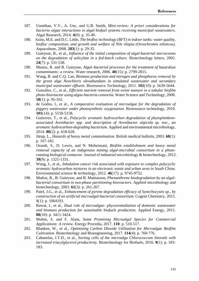
References
141
187. Unnithan, V.V., A. Unc, and G.B. Smith, Mini-review: A priori considerations for bacteria–algae interactions in algal biofuel systems receiving municipal wastewaters. Algal Research, 2014. 4(0): p. 35-40.
188. Azim, M.E. and D.C. Little, The biofloc technology (BFT) in indoor tanks: water quality, biofloc composition, and growth and welfare of Nile tilapia (Oreochromis niloticus). Aquaculture, 2008. 283(1): p. 29-35.
189. Guieysse, B., et al., Influence of the initial composition of algal-bacterial microcosms on the degradation of salicylate in a fed-batch culture. Biotechnology letters, 2002. 24(7): p. 531-538.
190. Munoz, R. and B. Guieysse, Algal–bacterial processes for the treatment of hazardous contaminants: a review. Water research, 2006. 40(15): p. 2799-2815.
191. Wang, B. and C.Q. Lan, Biomass production and nitrogen and phosphorus removal by the green alga Neochloris oleoabundans in simulated wastewater and secondary municipal wastewater effluent. Bioresource Technology, 2011. 102(10): p. 5639-5644.
192. González, C., et al., Efficient nutrient removal from swine manure in a tubular biofilm photo-bioreactor using algae-bacteria consortia. Water Science and Technology, 2008. 58(1): p. 95-102.
193. de Godos, I., et al., A comparative evaluation of microalgae for the degradation of piggery wastewater under photosynthetic oxygenation. Bioresource technology, 2010. 101(14): p. 5150-5158.
194. Gutierrez, T., et al., Polycyclic aromatic hydrocarbon degradation of phytoplankton-associated Arenibacter spp. and description of Arenibacter algicola sp. nov., an aromatic hydrocarbon-degrading bacterium. Applied and environmental microbiology, 2014. 80(2): p. 618-628.
195. Järup, L., Hazards of heavy metal contamination. British medical bulletin, 2003. 68(1): p. 167-182.
196. Orandi, S., D. Lewis, and N. Moheimani, Biofilm establishment and heavy metal removal capacity of an indigenous mining algal-microbial consortium in a photo-rotating biological contactor. Journal of industrial microbiology & biotechnology, 2012. 39(9): p. 1321-1331.
197. Wang, J., et al., Inhalation cancer risk associated with exposure to complex polycyclic aromatic hydrocarbon mixtures in an electronic waste and urban area in South China. Environmental science & technology, 2012. 46(17): p. 9745-9752.
198. Muñoz, R., B. Guieysse, and B. Mattiasson, Phenanthrene biodegradation by an algal-bacterial consortium in two-phase partitioning bioreactors. Applied microbiology and biotechnology, 2003. 61(3): p. 261-267.
199. Patel, J.G., et al., Enhancement of pyrene degradation efficacy of Synechocystis sp., by construction of an artificial microalgal-bacterial consortium. Cogent Chemistry, 2015. 1(1): p. 1064193.
200. Rawat, I., et al., Dual role of microalgae: phycoremediation of domestic wastewater and biomass production for sustainable biofuels production. Applied Energy, 2011. 88(10): p. 3411-3424.
201. Mobin, S. and F. Alam, Some Promising Microalgal Species for Commercial Applications: A review. Energy Procedia, 2017. 110: p. 510-517.
202. Blanken, W., et al., Optimizing Carbon Dioxide Utilization for Microalgae Biofilm Cultivation. Biotechnology and Bioengineering, 2017. 114(4): p. 769-776.
203. Cabanelas, I.T.D., et al., Sorting cells of the microalga Chlorococcum littorale with increased triacylglycerol productivity. Biotechnology for Biofuels, 2016. 9(1): p. 183-183.
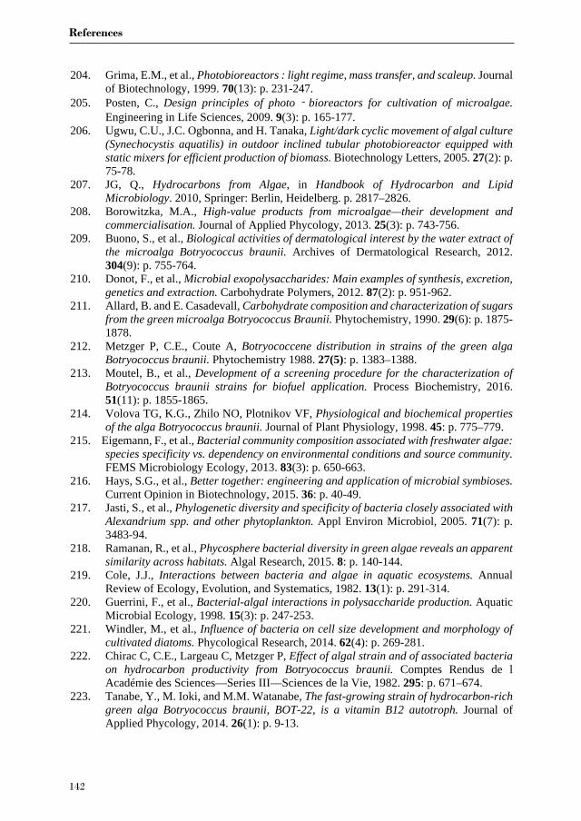
References
142
204. Grima, E.M., et al., Photobioreactors : light regime, mass transfer, and scaleup. Journal of Biotechnology, 1999. 70(13): p. 231-247.
205. Posten, C., Design principles of photo‐bioreactors for cultivation of microalgae. Engineering in Life Sciences, 2009. 9(3): p. 165-177.
206. Ugwu, C.U., J.C. Ogbonna, and H. Tanaka, Light/dark cyclic movement of algal culture (Synechocystis aquatilis) in outdoor inclined tubular photobioreactor equipped with static mixers for efficient production of biomass. Biotechnology Letters, 2005. 27(2): p. 75-78.
207. JG, Q., Hydrocarbons from Algae, in Handbook of Hydrocarbon and Lipid Microbiology. 2010, Springer: Berlin, Heidelberg. p. 2817–2826.
208. Borowitzka, M.A., High-value products from microalgae—their development and commercialisation. Journal of Applied Phycology, 2013. 25(3): p. 743-756.
209. Buono, S., et al., Biological activities of dermatological interest by the water extract of the microalga Botryococcus braunii. Archives of Dermatological Research, 2012. 304(9): p. 755-764.
210. Donot, F., et al., Microbial exopolysaccharides: Main examples of synthesis, excretion, genetics and extraction. Carbohydrate Polymers, 2012. 87(2): p. 951-962.
211. Allard, B. and E. Casadevall, Carbohydrate composition and characterization of sugars from the green microalga Botryococcus Braunii. Phytochemistry, 1990. 29(6): p. 1875-1878.
212. Metzger P, C.E., Coute A, Botryococcene distribution in strains of the green alga Botryococcus braunii. Phytochemistry 1988. 27(5): p. 1383–1388.
213. Moutel, B., et al., Development of a screening procedure for the characterization of Botryococcus braunii strains for biofuel application. Process Biochemistry, 2016. 51(11): p. 1855-1865.
214. Volova TG, K.G., Zhilo NO, Plotnikov VF, Physiological and biochemical properties of the alga Botryococcus braunii. Journal of Plant Physiology, 1998. 45: p. 775–779.
215. Eigemann, F., et al., Bacterial community composition associated with freshwater algae: species specificity vs. dependency on environmental conditions and source community. FEMS Microbiology Ecology, 2013. 83(3): p. 650-663.
216. Hays, S.G., et al., Better together: engineering and application of microbial symbioses. Current Opinion in Biotechnology, 2015. 36: p. 40-49.
217. Jasti, S., et al., Phylogenetic diversity and specificity of bacteria closely associated with Alexandrium spp. and other phytoplankton. Appl Environ Microbiol, 2005. 71(7): p. 3483-94.
218. Ramanan, R., et al., Phycosphere bacterial diversity in green algae reveals an apparent similarity across habitats. Algal Research, 2015. 8: p. 140-144.
219. Cole, J.J., Interactions between bacteria and algae in aquatic ecosystems. Annual Review of Ecology, Evolution, and Systematics, 1982. 13(1): p. 291-314.
220. Guerrini, F., et al., Bacterial-algal interactions in polysaccharide production. Aquatic Microbial Ecology, 1998. 15(3): p. 247-253.
221. Windler, M., et al., Influence of bacteria on cell size development and morphology of cultivated diatoms. Phycological Research, 2014. 62(4): p. 269-281.
222. Chirac C, C.E., Largeau C, Metzger P, Effect of algal strain and of associated bacteria on hydrocarbon productivity from Botryococcus braunii. Comptes Rendus de l Académie des Sciences—Series III—Sciences de la Vie, 1982. 295: p. 671–674.
223. Tanabe, Y., M. Ioki, and M.M. Watanabe, The fast-growing strain of hydrocarbon-rich green alga Botryococcus braunii, BOT-22, is a vitamin B12 autotroph. Journal of Applied Phycology, 2014. 26(1): p. 9-13.
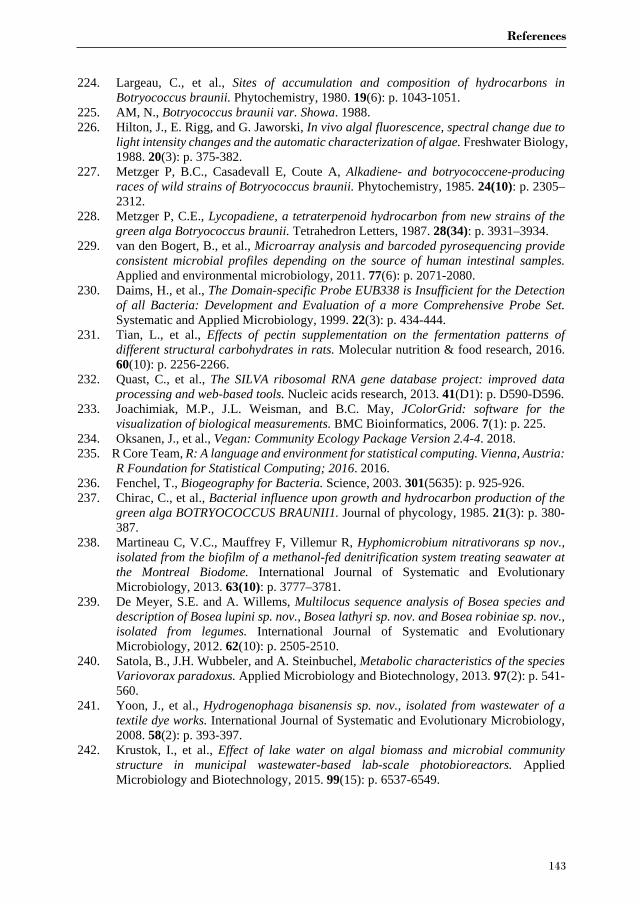
References
143
224. Largeau, C., et al., Sites of accumulation and composition of hydrocarbons in Botryococcus braunii. Phytochemistry, 1980. 19(6): p. 1043-1051.
225. AM, N., Botryococcus braunii var. Showa. 1988. 226. Hilton, J., E. Rigg, and G. Jaworski, In vivo algal fluorescence, spectral change due to
light intensity changes and the automatic characterization of algae. Freshwater Biology, 1988. 20(3): p. 375-382.
227. Metzger P, B.C., Casadevall E, Coute A, Alkadiene- and botryococcene-producing races of wild strains of Botryococcus braunii. Phytochemistry, 1985. 24(10): p. 2305–2312.
228. Metzger P, C.E., Lycopadiene, a tetraterpenoid hydrocarbon from new strains of the green alga Botryococcus braunii. Tetrahedron Letters, 1987. 28(34): p. 3931–3934.
229. van den Bogert, B., et al., Microarray analysis and barcoded pyrosequencing provide consistent microbial profiles depending on the source of human intestinal samples. Applied and environmental microbiology, 2011. 77(6): p. 2071-2080.
230. Daims, H., et al., The Domain-specific Probe EUB338 is Insufficient for the Detection of all Bacteria: Development and Evaluation of a more Comprehensive Probe Set. Systematic and Applied Microbiology, 1999. 22(3): p. 434-444.
231. Tian, L., et al., Effects of pectin supplementation on the fermentation patterns of different structural carbohydrates in rats. Molecular nutrition & food research, 2016. 60(10): p. 2256-2266.
232. Quast, C., et al., The SILVA ribosomal RNA gene database project: improved data processing and web-based tools. Nucleic acids research, 2013. 41(D1): p. D590-D596.
233. Joachimiak, M.P., J.L. Weisman, and B.C. May, JColorGrid: software for the visualization of biological measurements. BMC Bioinformatics, 2006. 7(1): p. 225.
234. Oksanen, J., et al., Vegan: Community Ecology Package Version 2.4-4. 2018. 235. R Core Team, R: A language and environment for statistical computing. Vienna, Austria:
R Foundation for Statistical Computing; 2016. 2016. 236. Fenchel, T., Biogeography for Bacteria. Science, 2003. 301(5635): p. 925-926. 237. Chirac, C., et al., Bacterial influence upon growth and hydrocarbon production of the
green alga BOTRYOCOCCUS BRAUNII1. Journal of phycology, 1985. 21(3): p. 380-387.
238. Martineau C, V.C., Mauffrey F, Villemur R, Hyphomicrobium nitrativorans sp nov., isolated from the biofilm of a methanol-fed denitrification system treating seawater at the Montreal Biodome. International Journal of Systematic and Evolutionary Microbiology, 2013. 63(10): p. 3777–3781.
239. De Meyer, S.E. and A. Willems, Multilocus sequence analysis of Bosea species and description of Bosea lupini sp. nov., Bosea lathyri sp. nov. and Bosea robiniae sp. nov., isolated from legumes. International Journal of Systematic and Evolutionary Microbiology, 2012. 62(10): p. 2505-2510.
240. Satola, B., J.H. Wubbeler, and A. Steinbuchel, Metabolic characteristics of the species Variovorax paradoxus. Applied Microbiology and Biotechnology, 2013. 97(2): p. 541-560.
241. Yoon, J., et al., Hydrogenophaga bisanensis sp. nov., isolated from wastewater of a textile dye works. International Journal of Systematic and Evolutionary Microbiology, 2008. 58(2): p. 393-397.
242. Krustok, I., et al., Effect of lake water on algal biomass and microbial community structure in municipal wastewater-based lab-scale photobioreactors. Applied Microbiology and Biotechnology, 2015. 99(15): p. 6537-6549.
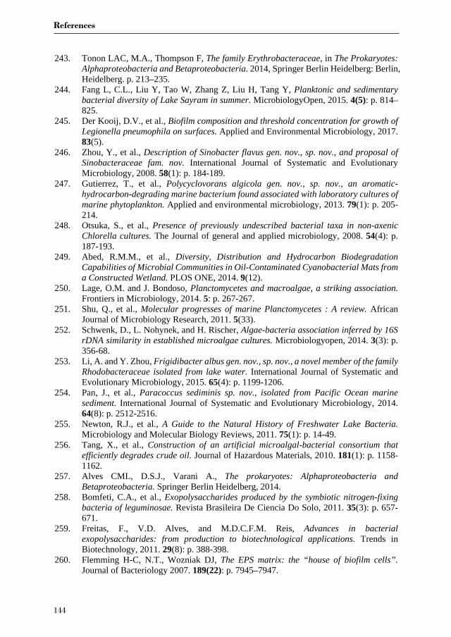
References
144
243. Tonon LAC, M.A., Thompson F, The family Erythrobacteraceae, in The Prokaryotes: Alphaproteobacteria and Betaproteobacteria. 2014, Springer Berlin Heidelberg: Berlin, Heidelberg. p. 213–235.
244. Fang L, C.L., Liu Y, Tao W, Zhang Z, Liu H, Tang Y, Planktonic and sedimentary bacterial diversity of Lake Sayram in summer. MicrobiologyOpen, 2015. 4(5): p. 814–825.
245. Der Kooij, D.V., et al., Biofilm composition and threshold concentration for growth of Legionella pneumophila on surfaces. Applied and Environmental Microbiology, 2017. 83(5).
246. Zhou, Y., et al., Description of Sinobacter flavus gen. nov., sp. nov., and proposal of Sinobacteraceae fam. nov. International Journal of Systematic and Evolutionary Microbiology, 2008. 58(1): p. 184-189.
247. Gutierrez, T., et al., Polycyclovorans algicola gen. nov., sp. nov., an aromatic-hydrocarbon-degrading marine bacterium found associated with laboratory cultures of marine phytoplankton. Applied and environmental microbiology, 2013. 79(1): p. 205-214.
248. Otsuka, S., et al., Presence of previously undescribed bacterial taxa in non-axenic Chlorella cultures. The Journal of general and applied microbiology, 2008. 54(4): p. 187-193.
249. Abed, R.M.M., et al., Diversity, Distribution and Hydrocarbon Biodegradation Capabilities of Microbial Communities in Oil-Contaminated Cyanobacterial Mats from a Constructed Wetland. PLOS ONE, 2014. 9(12).
250. Lage, O.M. and J. Bondoso, Planctomycetes and macroalgae, a striking association. Frontiers in Microbiology, 2014. 5: p. 267-267.
251. Shu, Q., et al., Molecular progresses of marine Planctomycetes : A review. African Journal of Microbiology Research, 2011. 5(33).
252. Schwenk, D., L. Nohynek, and H. Rischer, Algae-bacteria association inferred by 16S rDNA similarity in established microalgae cultures. Microbiologyopen, 2014. 3(3): p. 356-68.
253. Li, A. and Y. Zhou, Frigidibacter albus gen. nov., sp. nov., a novel member of the family Rhodobacteraceae isolated from lake water. International Journal of Systematic and Evolutionary Microbiology, 2015. 65(4): p. 1199-1206.
254. Pan, J., et al., Paracoccus sediminis sp. nov., isolated from Pacific Ocean marine sediment. International Journal of Systematic and Evolutionary Microbiology, 2014. 64(8): p. 2512-2516.
255. Newton, R.J., et al., A Guide to the Natural History of Freshwater Lake Bacteria. Microbiology and Molecular Biology Reviews, 2011. 75(1): p. 14-49.
256. Tang, X., et al., Construction of an artificial microalgal-bacterial consortium that efficiently degrades crude oil. Journal of Hazardous Materials, 2010. 181(1): p. 1158-1162.
257. Alves CML, D.S.J., Varani A., The prokaryotes: Alphaproteobacteria and Betaproteobacteria. Springer Berlin Heidelberg, 2014.
258. Bomfeti, C.A., et al., Exopolysaccharides produced by the symbiotic nitrogen-fixing bacteria of leguminosae. Revista Brasileira De Ciencia Do Solo, 2011. 35(3): p. 657-671.
259. Freitas, F., V.D. Alves, and M.D.C.F.M. Reis, Advances in bacterial exopolysaccharides: from production to biotechnological applications. Trends in Biotechnology, 2011. 29(8): p. 388-398.
260. Flemming H-C, N.T., Wozniak DJ, The EPS matrix: the “house of biofilm cells”. Journal of Bacteriology 2007. 189(22): p. 7945–7947.
References
145
261. Lee, J., et al., Microalgae-associated bacteria play a key role in the flocculation of Chlorella vulgaris. Bioresource Technology, 2013. 131: p. 195-201.
262. Weiss, T.L., et al., Colony Organization in the Green Alga Botryococcus braunii (Race B) Is Specified by a Complex Extracellular Matrix. Eukaryotic Cell, 2012. 11(12): p. 1424-1440.
263. Stephens, E., et al., Future prospects of microalgal biofuel production systems. Trends in plant science, 2010. 15(10): p. 554-564.
264. Mata, T.M., A.A. Martins, and N.S. Caetano, Microalgae for biodiesel production and other applications: A review. Renewable and Sustainable Energy Reviews, 2010. 14(1): p. 217-232.
265. de Vree, J.H., et al., Turbidostat operation of outdoor pilot-scale photobioreactors. Algal Research, 2016. 18: p. 198-208.
266. Zittelli, G.C., et al., Photobioreactors for mass production of microalgae. Handbook of Microalgal Culture: Applied Phycology and Biotechnology, Second Edition, 2013: p. 225-266.
267. Wen, X., et al., Effective cultivation of microalgae for biofuel production: a pilot-scale evaluation of a novel oleaginous microalga Graesiella sp. WBG-1. Biotechnology for biofuels, 2016. 9(1): p. 123.
268. Scott, S.A., et al., Biodiesel from algae: challenges and prospects. Current opinion in biotechnology, 2010. 21(3): p. 277-286.
269. Furusawa, G., et al., Algicidal activity and gliding motility of Saprospira sp. SS98-5. Canadian journal of microbiology, 2003. 49(2): p. 92-100.
270. Dao, G.-H., et al., Enhanced microalgae growth through stimulated secretion of indole acetic acid by symbiotic bacteria. Algal research, 2018. 33: p. 345-351.
271. Lupette, J., et al., Marinobacter dominates the bacterial community of the Ostreococcus tauri phycosphere in culture. Frontiers in microbiology, 2016. 7: p. 1414.
272. Vree, J.H., et al., Comparison of four outdoor pilot-scale photobioreactors. Biotechnology for biofuels, 2015. 8(1): p. 215.
273. Bosma, R., et al., Design and construction of the microalgal pilot facility AlgaePARC. Algal Research, 2014. 6, Part B: p. 160-169.
274. Ramiro-Garcia, J., et al., NG-Tax, a highly accurate and validated pipeline for analysis of 16S rRNA amplicons from complex biomes [version 1; referees: 2 approved with reservations, 1 not approved]. Vol. 5. 2016.
275. Edgar, R.C., Search and clustering orders of magnitude faster than BLAST. Bioinformatics, 2010. 26(19): p. 2460-2461.
276. Benjamini, Y. and Y. Hochberg, Controlling the false discovery rate: a practical and powerful approach to multiple testing. Journal of the royal statistical society. Series B (Methodological), 1995: p. 289-300.
277. Wang, H., et al., Effects of bacterial communities on biofuel-producing microalgae: stimulation, inhibition and harvesting. Crit Rev Biotechnol, 2014: p. 1-12.
278. Kimbrel, J.A., et al., Host selection and stochastic effects influence bacterial community assembly on the microalgal phycosphere. Algal Research, 2019. 40: p. 101489.
279. Geng, H., et al., Longitudinal analysis of microbiota in microalga Nannochloropsis salina cultures. Microbial ecology, 2016. 72(1): p. 14-24.
280. Rosana, A.R.R., et al., Draft genome sequences of seven bacterial strains isolated from a polymicrobial culture of coccolith-bearing (C-Type) Emiliania huxleyi M217. Genome Announc., 2016. 4(4): p. e00673-16.
281. Chen, Z., et al., Phaeodactylibacter xiamenensis gen. nov., sp. nov., a member of the family Saprospiraceae isolated from the marine alga Phaeodactylum tricornutum.
References
146
International journal of systematic and evolutionary microbiology, 2014. 64(10): p. 3496-3502.
282. Meziti, A., et al., Spatially uniform but temporally variable bacterioplankton in a semi-enclosed coastal area. Systematic and Applied Microbiology, 2015. 38(5): p. 358-367.
283. Marshall, K., et al., Effect of Marine Bacterial Isolates on the Growth and Morphology of Axenic Plantlets of the Green Alga Ulva linza. Microbial Ecology, 2006. 52(2): p. 302-310.
284. Burke, C., et al., Composition, uniqueness and variability of the epiphytic bacterial community of the green alga Ulva australis. The ISME journal, 2011. 5(4): p. 590-600.
285. Ismail, A., et al., Antimicrobial Activities of Bacteria Associated with the Brown Alga Padina pavonica. Frontiers in Microbiology, 2016. 7: p. 1072.
286. Kim, B.-H., et al., Role of Rhizobium, a plant growth promoting bacterium, in enhancing algal biomass through mutualistic interaction. biomass and bioenergy, 2014. 69: p. 95-105.
287. Zulkifly, S., et al., The epiphytic microbiota of the globally widespread macroalga Cladophora glomerata (Chlorophyta, Cladophorales). American journal of botany, 2012. 99(9): p. 1541-1552.
288. Wang, Y., et al., Seasonal dynamics of bacterial communities in the surface seawater around subtropical Xiamen Island, China, as determined by 16S rRNA gene profiling. Marine Pollution Bulletin, 2019. 142: p. 135-144.
289. Kirchman, D.L., et al., Biogeography of major bacterial groups in the Delaware Estuary. 2005. 50(5): p. 1697-1706.
290. Campbell, B.J. and D.L. Kirchman, Bacterial diversity, community structure and potential growth rates along an estuarine salinity gradient. The ISME journal, 2013. 7(1): p. 210-220.
291. Converti, A., et al., Effect of temperature and nitrogen concentration on the growth and lipid content of Nannochloropsis oculata and Chlorella vulgaris for biodiesel production. Chemical Engineering and Processing: Process Intensification, 2009. 48(6): p. 1146-1151.
292. Pal, D., et al., The effect of light, salinity, and nitrogen availability on lipid production by Nannochloropsis sp. Applied microbiology and biotechnology, 2011. 90(4): p. 1429-1441.
293. Alonso-Sáez, L., et al., Effect of natural sunlight on bacterial activity and differential sensitivity of natural bacterioplankton groups in northwestern Mediterranean coastal waters. Appl. Environ. Microbiol., 2006. 72(9): p. 5806-5813.
294. Bartley, M.L., et al., pH effects on growth and lipid accumulation of the biofuel microalgae Nannochloropsis salina and invading organisms. Journal of applied phycology, 2014. 26(3): p. 1431-1437.
295. Garcia, N.S., J.A. Bonachela, and A.C. Martiny, Interactions between growth-dependent changes in cell size, nutrient supply and cellular elemental stoichiometry of marine Synechococcus. The ISME journal, 2016. 10(11): p. 2715-2724.
296. Zhou, J., et al., Microbial community structure and associations during a marine dinoflagellate bloom. Frontiers in microbiology, 2018. 9: p. 1201.
297. Jiang, X., H. Dang, and N. Jiao, Ubiquity and diversity of heterotrophic bacterial nasA genes in diverse marine environments. PloS one, 2015. 10(2).
298. Diner, R.E., et al., Genetic manipulation of competition for nitrate between heterotrophic bacteria and diatoms. Frontiers in microbiology, 2016. 7: p. 880.
299. Zorz, J.K., et al., Drivers of regional bacterial community structure and diversity over depth and time in the Northwest Atlantic Ocean. Frontiers in microbiology, 2019. 10: p. 281.
References
147
300. Hou, D., et al., Environmental factors shape water microbial community structure and function in shrimp cultural enclosure ecosystems. Frontiers in microbiology, 2017. 8: p. 2359.
301. Milici, M., et al., Low diversity of planktonic bacteria in the tropical ocean. Scientific reports, 2016. 6(1): p. 1-9.
302. Wemheuer, B., et al., Impact of a phytoplankton bloom on the diversity of the active bacterial community in the southern North Sea as revealed by metatranscriptomic approaches. FEMS Microbiology Ecology, 2014. 87(2): p. 378-389.
303. Yang, C., et al., Illumina sequencing-based analysis of free-living bacterial community dynamics during an Akashiwo sanguine bloom in Xiamen sea, China. Scientific reports, 2015. 5: p. 8476.
304. Teeling, H., et al., Substrate-controlled succession of marine bacterioplankton populations induced by a phytoplankton bloom. Science, 2012. 336(6081): p. 608-611.
305. Fuhrman, J.A., et al., Annually reoccurring bacterial communities are predictable from ocean conditions. Proceedings of the National Academy of Sciences, 2006. 103(35): p. 13104-13109.
306. Grossart, H.P., et al., Marine diatom species harbour distinct bacterial communities. Environmental Microbiology, 2005. 7(6): p. 860-873.
307. Williams, T.J., et al., The role of planktonic F lavobacteria in processing algal organic matter in coastal E ast A ntarctica revealed using metagenomics and metaproteomics. Environmental microbiology, 2013. 15(5): p. 1302-1317.
308. Buchan, A., J.M. González, and M.A. Moran, Overview of the marine Roseobacter lineage. Appl. Environ. Microbiol., 2005. 71(10): p. 5665-5677.
309. Simon, M., et al., Phylogenomics of Rhodobacteraceae reveals evolutionary adaptation to marine and non-marine habitats. The ISME journal, 2017. 11(6): p. 1483-1499.
310. Mancuso, F.P., et al., Diversity and Temporal Dynamics of the Epiphytic Bacterial Communities Associated with the Canopy-Forming Seaweed Cystoseira compressa (Esper) Gerloff and Nizamuddin. Front Microbiol, 2016. 7: p. 476.
311. Berthold, D.E., et al., Enhancing algal biomass and lipid production through bacterial co-culture. Biomass and bioenergy, 2019. 122: p. 280-289.
312. Biondi, N., et al., Tetraselmis suecica F&M‐M33 growth is influenced by its associated bacteria. Microbial biotechnology, 2018. 11(1): p. 211-223.
313. Gouveia, J.D., et al., Associated bacteria of Botryococcus braunii (Chlorophyta). PeerJ, 2019. 7: p. e6610.
314. Smriga, S., et al., Chemotaxis toward phytoplankton drives organic matter partitioning among marine bacteria. Proceedings of the National Academy of Sciences, 2016. 113(6): p. 1576-1581.
315. Liu, H., et al., Shifting nutrient-mediated interactions between algae and bacteria in a microcosm: evidence from alkaline phosphatase assay. Microbiological research, 2012. 167(5): p. 292-298.
316. Le Chevanton, M., et al., Effects of nitrogen limitation on Dunaliella sp.–Alteromonas sp. interactions: from mutualistic to competitive relationships. Frontiers in Marine Science, 2016. 3: p. 123.
317. Paul, C. and G. Pohnert, Interactions of the algicidal bacterium Kordia algicida with diatoms: regulated protease excretion for specific algal lysis. PloS one, 2011. 6(6): p. e21032.
318. Zubkov, M.V., et al., Microbial control of phosphate in the nutrient‐depleted North Atlantic subtropical gyre. Environmental Microbiology, 2007. 9(8): p. 2079-2089.
319. van Tol, H.M., S.A. Amin, and E.V. Armbrust, Ubiquitous marine bacterium inhibits diatom cell division. The ISME journal, 2017. 11(1): p. 31.

References
148
320. Bolch, C.J., T.A. Subramanian, and D.H. Green, The toxic dinoflagellate Gymnodinium catenatum (Dinophyceae) requires marine bacteria for growth. Journal of phycology, 2011. 47(5): p. 1009-1022.
321. Thompson, A.W., et al., Unicellular cyanobacterium symbiotic with a single-celled eukaryotic alga. Science, 2012. 337(6101): p. 1546-50.
322. Amin, S.A., et al., Boron binding by a siderophore isolated from marine bacteria associated with the toxic dinoflagellate Gymnodinium catenatum. Journal of the American Chemical Society, 2007. 129(3): p. 478-479.
323. Hom, E.F.Y., et al., A Chemical Perspective on Microalgal–Microbial Interactions. Trends in Plant Science, 2015. 20(11): p. 689-693.
324. Yao, S., et al., Microalgae–bacteria symbiosis in microalgal growth and biofuel production: a review. Journal of applied microbiology, 2019. 126(2): p. 359-368.
325. Lutzu, G.A. and N. Turgut Dunford, Interactions of microalgae and other microorganisms for enhanced production of high-value compounds. Front Biosci, 2018. 23: p. 1487-1504.
326. van den Bogert, B., et al., Microarray analysis and barcoded pyrosequencing provide consistent microbial profiles depending on the source of human intestinal samples. Appl Environ Microbiol, 2011. 77(6): p. 2071-80.
327. Daims, H., et al., The Domain-specific Probe EUB338 is Insufficient for the Detection of all Bacteria: Development and Evaluation of a more Comprehensive Probe Set. Systematic and Applied Microbiology, 1999. 22(3): p. 434-444.
328. Tian, L., et al., Effects of pectin supplementation on the fermentation patterns of different structural carbohydrates in rats. Mol Nutr Food Res, 2016. 60(10): p. 2256-2266.
329. Ramiro-Garcia, J., et al., NG-Tax, a highly accurate and validated pipeline for analysis of 16S rRNA amplicons from complex biomes. F1000Research, 2016. 5: p. 1791.
330. Dat, T.T.H., et al., Archaeal and bacterial diversity and community composition from 18 phylogenetically divergent sponge species in Vietnam. PeerJ, 2018. 6.
331. Edgar, R.C., Search and clustering orders of magnitude faster than BLAST. Bioinformatics, 2010. 26(19): p. 2460-1.
332. Quast, C., et al., The SILVA ribosomal RNA gene database project: improved data processing and web-based tools. Nucleic Acids Res, 2013. 41(Database issue): p. D590-6.
333. Yilmaz, P., et al., The SILVA and "All-species Living Tree Project (LTP)" taxonomic frameworks. Nucleic Acids Res, 2014. 42(Database issue): p. D643-8.
334. Holm, S., A Simple Sequentially Rejective Multiple Test Procedure. Scandinavian Journal of Statistics, 1979. 6(2): p. 65-70.
335. van Tol, H.M., S.A. Amin, and E.V. Armbrust, Ubiquitous marine bacterium inhibits diatom cell division. ISME J, 2016.
336. Goecke, F., et al., Algae as an important environment for bacteria–phylogenetic relationships among new bacterial species isolated from algae. Phycologia, 2013. 52(1): p. 14-24.
337. Wang, H., et al., Identification of genetic modules mediating the Jekyll and Hyde interaction of Dinoroseobacter shibae with the dinoflagellate Prorocentrum minimum. Frontiers in Microbiology, 2015. 6.
338. Weber, C.F. and G.M. King, Physiological, ecological, and phylogenetic characterization of Stappia, a marine CO-oxidizing bacterial genus. Applied and environmental microbiology, 2007. 73(4): p. 1266-1276.
339. Coates, C.J. and M. Wyman, A denitrifying community associated with a major, marine nitrogen fixer. Environmental microbiology, 2017. 19(12): p. 4978-4992.
References
149
340. Han, J., et al., Co-culturing bacteria and microalgae in organic carbon containing medium. Journal of Biological Research-Thessaloniki, 2016. 23(1): p. 8.
341. Sandhya, S. and K. Vijayan, Symbiotic association among marine microalgae and bacterial flora: a study with special reference to commercially important Isochrysis galbana culture. Journal of Applied Phycology, 2019: p. 1-8.
342. Chen, Z., et al., Influence of plaque-forming bacterium, Rhodobacteraceae sp. on the growth of Chlorella vulgaris. Bioresource Technology, 2014. 169: p. 784-788.
343. Fukui, Y., et al., Maritalea porphyrae sp. nov., isolated from a red alga (Porphyra yezoensis), and transfer of Zhangella mobilis to Maritalea mobilis comb. nov. International journal of systematic and evolutionary microbiology, 2012. 62(1): p. 43-48.
344. Chung, E.J., et al., Hoeflea suaedae sp. nov., an endophytic bacterium isolated from the root of the halophyte Suaeda maritima. International journal of systematic and evolutionary microbiology, 2013. 63(6): p. 2277-2281.
345. Fukui, Y., et al., Isolation of Hyphomonas strains that induce normal morphogenesis in protoplasts of the marine red alga Pyropia yezoensis. Microbial ecology, 2014. 68(3): p. 556-566.
346. Bentzon-Tilia, M., L. Riemann, and L. Gram, Draft genome sequence of Hoeflea sp. strain BAL378, a potential producer of bioactive compounds. Genome announcements, 2014. 2(6): p. e01213-14.
347. Xamxidin, M., et al., Aquaticitalea lipolytica gen. nov., sp. nov., isolated from Antarctic seawater. International journal of systematic and evolutionary microbiology, 2016. 66(7): p. 2657-2663.
348. Meseck, S., et al., Nutrient interactions between phytoplankton and bacterioplankton under different carbon dioxide regimes. Journal of Applied Phycology, 2006(3): p. 229-237.
349. Danger, M., et al., Control of phytoplankton–bacteria interactions by stoichiometric constraints. 2007. 116(7): p. 1079-1086.
350. Fukami, K., T. Nishijima, and Y. Ishida, Stimulative and inhibitory effects of bacteria on the growth of microalgae. Hydrobiologia, 1997. 358(1): p. 185-191.
351. Mitsutani, A., et al., Analysis of algicidal proteins of a diatom-lytic marine bacterium Pseudoalteromonas sp. strain A25 by two-dimensional electrophoresis. Phycologia 2001(3): p. 286-291.
352. Nedashkovskaya, O.I., et al., Maribacter gen. nov., a new member of the family Flavobacteriaceae, isolated from marine habitats, containing the species Maribacter sedimenticola sp. nov., Maribacter aquivivus sp. nov., Maribacter orientalis sp. nov. and Maribacter ulvicola sp. nov. International journal of systematic and evolutionary microbiology, 2004. 54(4): p. 1017-1023.
353. Lee, Y.-S. and Y.-L. Choi, Complete genome sequence and analysis of three kinds of β-agarase of Cellulophaga lytica DAU203 isolated from marine sediment. Marine genomics, 2017. 35: p. 43-46.
354. Chi, W.-J., Y.-K. Chang, and S.-K. Hong, Agar degradation by microorganisms and agar-degrading enzymes. Applied microbiology and biotechnology, 2012. 94(4): p. 917-930.
355. Velu, P., M.J. Peter, and E. Sanniyasi, Effect of various carbon sources on biochemical production in marine microalgae Nannochloropsis salina (Eustigmatophyceae), Dunaliella tertiolecta (Chlorophyceae) and Tetraselmis suecica (Chlorodendrophyceae). Int. J. Curr. Microbiol. App. Sci, 2015. 4(3): p. 207-215.
References
150
356. Vu, H.T., et al., Cocultivated bacteria can increase or decrease the culture lifetime of Chlorella vulgaris. The Journal of general and applied microbiology, 2010. 56(5): p. 413-418.
357. Turner, S., et al., Investigating deep phylogenetic relationships among cyanobacteria and plastids by small subunit rRNA sequence analysis 1. Journal of Eukaryotic Microbiology, 1999. 46(4): p. 327-338.
358. Apprill, A., et al., Minor revision to V4 region SSU rRNA 806R gene primer greatly increases detection of SAR11 bacterioplankton. Aquatic Microbial Ecology, 2015. 75(2): p. 129-137.
359. Sapp, M., et al., Species-specific bacterial communities in the phycosphere of microalgae? Microbial ecology, 2007. 53(4): p. 683-699.
360. Behringer, G., et al., Bacterial Communities of Diatoms Display Strong Conservation Across Strains and Time. Frontiers in microbiology, 2018. 9: p. 659.
361. Schäfer, H., et al., Genetic diversity of ‘satellite’bacteria present in cultures of marine diatoms. FEMS Microbiology Ecology, 2002. 42(1): p. 25-35.
362. Sapp, M., et al., Bacterial community dynamics during the winter–spring transition in the North Sea. FEMS Microbiology Ecology, 2007. 59(3): p. 622-637.
363. Sapp, M., A. Wichels, and G. Gerdts, Impacts of cultivation of marine diatoms on the associated bacterial community. Applied and environmental microbiology, 2007. 73(9): p. 3117-3120.
364. Jasti, S., et al., Phylogenetic diversity and specificity of bacteria closely associated with Alexandrium spp. and other phytoplankton. Appl. Environ. Microbiol., 2005. 71(7): p. 3483-3494.
365. Green, D.H., et al., Bacterial Diversity Associated with the Coccolithophorid Algae Emiliania huxleyi and Coccolithus pelagicus f. braarudii. BioMed Research International, 2015. 2015: p. 194540-194540.
366. Hardin, G., The competitive exclusion principle. science, 1960. 131(3409): p. 1292-1297.
367. Šimek, K., et al., Alga-derived substrates select for distinct betaproteobacterial lineages and contribute to niche separation in Limnohabitans strains. Appl. Environ. Microbiol., 2011. 77(20): p. 7307-7315.
368. Dang, H. and C.R. Lovell, Bacterial primary colonization and early succession on surfaces in marine waters as determined by amplified rRNA gene restriction analysis and sequence analysis of 16S rRNA genes. Appl. Environ. Microbiol., 2000. 66(2): p. 467-475.
369. Myklestad, S., Release of extracellular products by phytoplankton with special emphasis on polysaccharides. Science of The Total Environment, 1995. 165: p. 155-164.
370. Villacorte, L.O., et al., Characterisation of algal organic matter produced by bloom-forming marine and freshwater algae. Water Research, 2015. 73: p. 216-230.
371. Kazamia, E., D.C. Aldridge, and A.G. Smith, Synthetic ecology – A way forward for sustainable algal biofuel production? Journal of Biotechnology, 2012. 162(1): p. 163-169.
372. Kazamia, E., et al., An engineered community approach for industrial cultivation of microalgae. Industrial Biotechnology, 2014. 10(3): p. 184-190.
373. Bader, J., et al., Relevance of microbial coculture fermentations in biotechnology. Journal of applied microbiology, 2010. 109(2): p. 371-387.
374. Sabra, W., et al., Biosystems analysis and engineering of microbial consortia for industrial biotechnology. Engineering in Life Sciences, 2010. 10(5): p. 407-421.
References
151
375. Goers, L., P. Freemont, and K.M. Polizzi, Co-culture systems and technologies: taking synthetic biology to the next level. Journal of the Royal Society Interface, 2014. 11(96): p. 20140065.
376. Mayali, X. and F. Azam, Algicidal Bacteria in the Sea and their Impact on Algal Blooms. Journal of Eukaryotic Microbiology, 2004. 51(2): p. 139-144.
377. Hilton, J.A., et al., Genomic deletions disrupt nitrogen metabolism pathways of a cyanobacterial diatom symbiont. Nature Communications, 2013. 4(1): p. 1767-1767.
378. Vraspir, J.M. and A. Butler, Chemistry of marine ligands and siderophores. Annual Review of Marine Science, 2009. 1(1): p. 43-63.
379. Fakhimi, N., et al., Acetic acid is key for synergetic hydrogen production in Chlamydomonas-bacteria co-cultures. Bioresource Technology, 2019. 289: p. 121648.
380. Mayali, X., Metabolic Interactions Between Bacteria and Phytoplankton. Frontiers in microbiology, 2018. 9: p. 727.
381. Ganuza, E., et al., A novel treatment protects Chlorella at commercial scale from the predatory bacterium Vampirovibrio chlorellavorus. Frontiers in microbiology, 2016. 7: p. 848.
382. Bagwell, C.E., et al., Discovery of bioactive metabolites in biofuel microalgae that offer protection against predatory bacteria. Frontiers in microbiology, 2016. 7: p. 516.
383. Klindworth, A., et al., Evaluation of general 16S ribosomal RNA gene PCR primers for classical and next-generation sequencing-based diversity studies. Nucleic acids research, 2013. 41(1): p. e1-e1.
384. Thomas, F., et al., Evaluation of a new primer combination to minimize plastid contamination in 16S rDNA metabarcoding analyses of alga‐associated bacterial communities. Environmental microbiology reports, 2019.
385. Ryffel, F., et al., Metabolic footprint of epiphytic bacteria on Arabidopsis thaliana leaves. ISME J, 2016. 10(3): p. 632-43.
386. Olive, A.J. and C.M. Sassetti, Metabolic crosstalk between host and pathogen: sensing, adapting and competing. Nature reviews Microbiology, 2016. 14(4): p. 221.
387. Magnúsdóttir, S. and I. Thiele, Modeling metabolism of the human gut microbiome. Current opinion in biotechnology, 2018. 51: p. 90-96.
388. Schleyer, G., et al., In plaque-mass spectrometry imaging of a bloom-forming alga during viral infection reveals a metabolic shift towards odd-chain fatty acid lipids. Nature Microbiology, 2019. 4(3): p. 527-538.
389. Watrous, J.D., et al., Microbial metabolic exchange in 3D. The ISME journal, 2013. 7(4): p. 770.
390. Lasch, P., et al., Identification of highly pathogenic microorganisms by matrix-assisted laser desorption ionization–time of flight mass spectrometry: results of an interlaboratory ring trial. Journal of clinical microbiology, 2015. 53(8): p. 2632-2640.
391. Seymour, J.R., et al., Chemoattraction to dimethylsulfoniopropionate throughout the marine microbial food web. Science, 2010. 329(5989): p. 342-345.
392. Sonnenschein, E.C., et al., Chemotaxis of Marinobacter adhaerens and its impact on attachment to the diatom Thalassiosira weissflogii. Appl. Environ. Microbiol, 2012. 78(19): p. 6900-6907.
393. Lambert, B.S., et al., A microfluidics-based in situ chemotaxis assay to study the behaviour of aquatic microbial communities. Nature microbiology, 2017. 2(10): p. 1344.
394. Lee, K.S., et al., An automated Raman-based platform for the sorting of live cells by functional properties. Nature microbiology, 2019. 4(6): p. 1035.


Thesis summary

Summary
154
Summary
Research described in this thesis aimed to investigate the microalgal microbial ecology by
integrating both cultivation-dependent and cultivation-independent approaches. In addition, we
examined effects of bacteria on growth of microalgae by developing a coculture system.
Understanding the bacterial community composition and functional associations between
microalgae and bacteria will provide important insights for both aquatic ecology and algal
biotechnology.
Microalgae are living with diverse microbial communities and interactions between microalgae
and bacteria are prevalent and strongly influence carbon and nutrient cycling in aquatic
ecosystems. A brief overview of the current knowledge of algae-bacteria interactions and
current research methodologies was given in chapter 1. This chapter furthermore introduced
the microalgae production facilities at AlgaePARC, and highlighted two research objects
(Nannochloropsis sp. and Botryococcus braunii), and current knowledge on their associated
bacteria.
In chapter 2 we comprehensively reviewed the recent research progress on algae-bacteria
interactions and summarized the current knowledge on functional aspects of algae-associated
microbiomes. In addition, we discussed the applications of knowledge on algae-bacteria
interactions in algal biomass production and various other related biotechnological innovations.
Finally, we concluded that principles of algae-bacteria interactions can be integrated into many
aspects of the algal production chain, which may help push the limit of the algal industry.
In chapter 3, we studied the bacterial community associated with 12 strains of B. braunii. The
changes of bacterial community composition were monitored over a period of 12 days. It was
clear from this study that B. braunii hosts a wide variety of bacterial species, among which the
bacterial families Rhizobiaceae, Bradyrhizobiaceae and Comamonadaceae were found in all
Summary
155
12 strains. These families that all belong to the phylum Proteobacteria could have important
interactions with B. braunii. Additionally, although each strain displayed a different bacterial
community composition, all the strains from the CAEN culture collection had more similar
bacterial communities, suggesting that the algae culture collection could have an influence on
the bacterial community composition. Several bacterial genera were shown to be exclusively
abundant in CAEN strains including Rhizobium spp. and Porphyrobacter spp.
In chapter 4, we compared bacterial community composition in cultures of Nannochloropsis
sp. CCAP211/78 grown simultaneously in four outdoor large-scale bioreactors. We observed
that the bacterial community composition significantly differed between all reactor types except
for horizontal and vertical tubular bioreactors. Although the bacterial communities varied
within each bioreactor at different cultivation stages, we found that a member of the family
Saprospiraceae was the most abundant taxon (OUT_1261) in all large non-sterilized
bioreactors. This indicates that Saprospiraceae may play important roles in Nannochloropsis
cultures.
In chapter 5, we isolated bacteria from one horizontal tubular bioreactor and one vertical
tubular bioreactor. Among all the isolates eighteen bacteria were phylogenetically classified as
different species/strains based on their 16S rRNA gene sequence. Axenic cultures of
Nannochloropsis sp. CCAP211/78 were successfully obtained by treatment with antibiotics. A
microplate-based assay system coupled with a custom-made LED box was developed to assess
the growth of axenic Nannochloropsis sp. CCAP211/78 with addition of isolated bacterial
strains. Out of 18 strains, one Maritalea porphyrae strain (DMSP31) and one Labrenzia
aggregata strain (YP26) were highlighted as having a significantly positive effect on microalga
growth, whereas one flavobacterial strain (YP206) was shown to reduce this growth. YP26 was
also found to substantially enhance the growth of Nannochloropsis sp CCAP211/78 using the
Summary
156
double-agar plate assay, which indicates that YP26 has strong positive impacts on
Nannochloropsis sp CCAP211/78.
Finally, chapter 6 elaborated on the implications of findings from the research described in this
thesis. Bacterial community composition of Nannochloropsis sp CCAP211/7 and B. braunii
was compared, and the results generally supported that bacterial communities are algal species
specific. Additionally, I suggested that the combination of environmental variables and reactor
configuration/operation (in addition to stochastic effects) may be responsible for the differences
in bacterial community composition observed between different reactor types and between runs.
Further exploration and exploitation of algae-bacteria interactions is needed. Therefore, I
proposed several new techniques, including using a new primer set to better characterize
bacteria living with microalgae, using advanced mass spectrometry to tap into the metabolic
changes of microalgae when they interact with bacteria, and using microfluidics coupled with
Raman-based cell sorting to isolate bacteria of interest. Future research should consider these
approaches in combination with coculture experiments in order to gain better understanding on
bacterial community composition and functional interactions between microalgae and bacteria.

Appendices

Appendices
158
Acknowledgements
After spending more than five years in Wageningen, it is time to say goodbye to everyone I met
here. Before doing that, I would like to acknowledge many people for their guidance and
support during my PhD adventure.
Dear Hauke, thanks for giving me the opportunity to join the MIB family. I will never forget
every discussion we had, you always gave me suggestions on a new perspective and made me
think out of box. You are so critical and passionate on science, I could not imagine how my
PhD journey would end up if you were not involved in my project.
Detmer, you are the best supervisor ever! I am so lucky to join the marine group and work with
you. Every time when I encountered troubles, you were always patient to explain me everything
in detail. Your friendliness and humour often led me to think you are not a supervisor, but a
close and knowledgeable friend. I was so encouraged by your attitude towards science, your
dedication and persistence. Being supervised by you is by all means the best adventure of my
life I could imagine.
Dear René, thank you for participating in my project. I always appreciate your comprehensive
knowledge on microalgae and all the indispensable inputs to my thesis you provided.
I was really honoured to be part of the MIB with so many excellent professors and talent
colleagues and students. Thanks Willem, you are the captain of MIB. Your leadership and
vision are truly inspiring. Many thanks to the supportive technician team of MIB: Ineke, Steven,
Philippe, Merlijn, Tom, Ton, Sjon and Wim. Anja, thanks for helping me arrange all
bureaucratic things.
MolEco group is a warm international family. I enjoyed a lot of all the meetings, discussions
and group activities (MolEco cakes, Christmas dinner, lab trips). The PhD trip in 2017 to
Germany, Denmark and Sweden was so much fun. Many thanks to the committee for
organisation. I also want to thank my team members during this trip: Daan, Sudarshan, Alex
and Ioannis. I would also like to thank all friends and colleagues from Bacgen, MicFys, and
SSB for your kindness and friendship. Javier, Jasper and Bart, thanks for your assistance on
bioinformatics.
I would also like to thank people from BPE, Jeroen, thank you for nice collaboration on chapter
4. Joao, you are always energetic and passionate, it is nice to work with you on chapter 3.

Acknowledgements
159
Snezana, Wendy, Rouke and Rick, thank all of you for help when I was working in BPE and
AlgaePARC. Xiao & Yimin, I often feel so late to meet this great couple, thanks for the good
food and wine you provided. Yimin, many thanks for organising tea tasting events. Fengzheng
and Chunzhe, thanks for your help and fun talks we had in Radix.
It was great pleasure to work with Marine Group members: Dennis, Johanna, Indra, Tika,
Dat, Georg, Menia, Catarina, Leyre and Grant. Indra, I will remember all the happy chats
with you and the delicious Indonesian food you cooked while you were still in Asserpark. Your
constantly positive attitude impressed me so much, thank you for all the help and advice. I am
sure that I will visit you and Titi at Bali soon. Dennis, I will miss the “movie night” and “office
chat” with you and Indra. Georg, you were kind of another supervisor to me, thanks for helping
me so much on data analysis and all. Grant, you are such a nice guy, it is so great to meet you
and thanks for hosting me in Naples. Johanna, you are the source of happiness, your laughing
will be always missed. Dat and Tika, it was happy to teach the master course with you, I wish
both of you all the best and would get PhD diploma soon. Catarina, thanks for introducing me
into bouldering, I wish to do more sports with you. Menia and Leyre, thank you for all the
discussions and help.
I was happy to supervise Patrick and Selene for your master theses. Thanks for your hard work
that contributed significantly to this thesis. I am so glad that both of you are doing PhD now.
Best wishes to you.
I would like to thank all my officemates during the past years: Alexzandr, Mark, Indra, Peng,
Dennis, Dat, Sudarshan, LooWee, Johanna, Yue, Atsushi, Leyre, Menia and Ouyang. You
guys are all so nice and we had a great time together.
Leiden would be one of the Dutch cities I visited most frequently, thanks Xiao & Chao for
hosting me so many times with delicious food and all the anecdotes we exchanged. I wish you
all the best of luck.
I would especially like to thank Chinese community in MIB: Ye, Yue, Yifan, Ying, Yuan,
Peng, Ran, Caifang, Chen & Ying, Taojun, Ouyang, Zhuang, Zhifei and Ying. Thanks for
your support and help, you are all my family members. I would give my special thanks to Yifan.
You are the master chef, thanks for inviting me to join the dinner with you so many times. I
will always remember the wine tasting nights you organised, and we travelled together to
explore many cities in the Netherlands and Europe.

Appendices
160
During living in Wageningen for more than five years, I met a large number of Chinese friends.
Yinxing, Xu, Juncai, Yin, Xing, Wei, Ningjing, Peiyu, Hao, Jingbin, Lei, Man, Ya, Peng,
Yuxing, Jie, Yale. Thank you for all your care, advice and friendship. I enjoyed every minute
spent with you all, which made my life in Wageningen so joyful.
It was great memories to meet many Chinese friends from Plant Breeding Group: Huayi, I
cannot thank you enough for your kindness and warm heart. Thanks again for being my close
friend and having holidays with me in Italy, Morocco and Croatia. Xinfang and Min, I shared
many sweet memories with you, thanks for your friendship and support. Hongmei, Lina and
Ning, I consider myself super lucky to meet you in Wageningen, your friendship is very
precious to me. Thanks for your care and support.
I lived in Asserpark44, 8B5 for more than 5 years since I moved to Wageningen. I never had
the idea to move out thanks to all easy-going corridor mates. I would give my special thanks to
Yujue, ZhiWei, He, Sibong and Yangyang. All the memorable moments with you guys will
be missed.
Last but not least, I would like to thank all my family members. My dearest parents, you gave
the power and freedom to pursue my dream. My love to you will never goes end. My lovely
sister, thanks for everything you have done to support me. Other family members from both my
father’s side and my mom’s side must be acknowledged as well, thanks for all your love and
care.
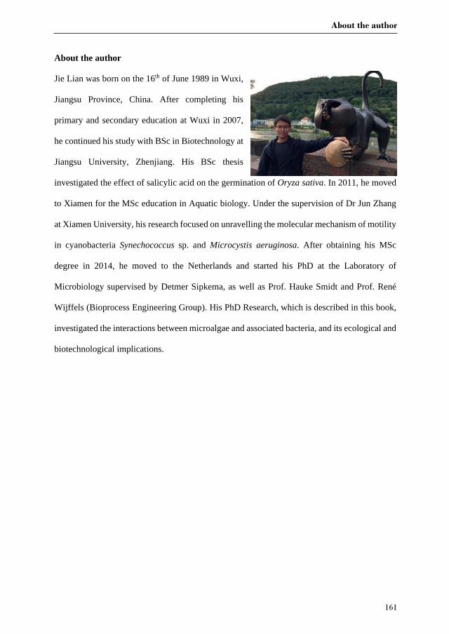
About the author
161
About the author
Jie Lian was born on the 16th of June 1989 in Wuxi,
Jiangsu Province, China. After completing his
primary and secondary education at Wuxi in 2007,
he continued his study with BSc in Biotechnology at
Jiangsu University, Zhenjiang. His BSc thesis
investigated the effect of salicylic acid on the germination of Oryza sativa. In 2011, he moved
to Xiamen for the MSc education in Aquatic biology. Under the supervision of Dr Jun Zhang
at Xiamen University, his research focused on unravelling the molecular mechanism of motility
in cyanobacteria Synechococcus sp. and Microcystis aeruginosa. After obtaining his MSc
degree in 2014, he moved to the Netherlands and started his PhD at the Laboratory of
Microbiology supervised by Detmer Sipkema, as well as Prof. Hauke Smidt and Prof. René
Wijffels (Bioprocess Engineering Group). His PhD Research, which is described in this book,
investigated the interactions between microalgae and associated bacteria, and its ecological and
biotechnological implications.
Appendices
162
List of publications
Lian, J., Wijffels, R.H., Smidt, H., and Sipkema, D. (2018) The effect of the algal microbiome
on industrial production of microalgae, Microbial biotechnology 11: 806-818.
Gouveia, J.D.*, Lian, J.*, Steinert, G., Smidt, H., Sipkema, D., Wijffels, R.H., Barbosa, M.J.
2019. Associated bacteria of Botryococcus braunii (Chlorophyta) PeerJ 7:e6610
https://doi.org/10.7717/peerj.6610
Lian, J., Steinert, G, de Vree, J.H, Meijer S., Heryanto, C., Bosma, R., Wijffels, R.H., Barbosa,
M.J., Smidt, H., and Sipkema, D. Bacterial diversity in outdoor pilot plant production of the
microalga Nannochloropsis sp. CCAP211/78. Manuscript in preparation.
Lian, J., Schimmel, P., Sanchez-Garcia, S., Wijffels, R.H., Smidt, H., Sipkema, D. Enhanced
or decreased growth of Nannochloropsis sp. CCAP211/78 cultivated with diverse bacteria.
Manuscript submitted.
Lian, J., Nijsse, B., Koehorst, J.J., Wijffels, R.H., Smidt, H., Sipkema, D. Dual RNA-Seq
unveils interactions between Nannochloropsis sp. CCAP211/78 and two associated bacteria.
Manuscript in preparation.
Other publications:
Lian, J. *, Zheng, X. *, Chen, Y., Zhuo, X., He, C, Shi, Q., Zheng, Q., Lin, TH., Sun, J., Guo,
WD., Cai, R., Jiao, N. Microbial transformation of distinct exogenous substrates into
analogous refractory compounds. Manuscript Submitted.
* These authors contributed equally.
Overview of completed training activities
163
Overview of completed training activities
Discipline specific activities
Courses
SIAM Metagenomics Course, Nijmegen, NL 2016
BluePharmTrain Summer school: Statistic analysis using R, Wageningen, NL 2016
Big data course, Wageningen, NL 2017
Advance course in Bioprocess design, Wageningen, NL 2018
Meetings
KNVM Spring Meeting, Arnhem, NL 2017
New Approaches and Concepts in Microbiology, Heidelberg, DE 2017
SAME15, Zagreb, HR 2017
Prospects and challenges for the development of algal biotechnology, Bielefeld, DE 2017
Microbiology centennial conference, Wageningen, NL 2017
KNVM Microbial Ecology symposium, Wageningen, NL 2017
Young algaeneers symposium, Oban, UK 2018
Wageningen meets Ghent symposium, Ghent, BE 2018
XMAS, Xiamen, CN 2019
General courses
VLAG PhD week, Baarlo, NL 2015
Project and Time management, Wageningen, NL 2015
Scientific Writing, Wageningen, NL 2016
Reviewing a scientific paper, Wageningen, NL 2017
Adobe Illustrator for scientists, Wageningen, NL 2018
Writing grant proposal, Wageningen, NL 2018
Optional
Preparing project proposal 2014
Molecular Ecology group meeting and Marine Microbiology group meeting 2014-2018
PhD meeting in Microbiology 2014-2018
Microbiology PhD trip 2017
The research described in this thesis was financially supported by the Laboratory of
Microbiology. The scholarship for the author was provided by the China Scholarship Council.
Financial support from Wageningen University for printing this thesis is gratefully
acknowledged.
Cover design by: Jie Lian & Qiuhui Pang
Layout design by: Jie Lian & Digiforce
Printed by: Digiforce

Algae-associated bacteria in photobioreactors Jie L
ian 2020
Algae-associated bacteria in photobioreactors
Jie Lian
Algae-associated bacteria in photobioreactors Jie L
ian 2020
Algae-associated bacteria in photobioreactors Jie L
ian 2020
Algae-associated bacteria in photobioreactors
Jie Lian
Invitation
You are cordially invited to attend the public defence
of my PhD thesis entitled:
Algae-associated bacteria in photobioreactors
Thursday 2 July 2020 at 11 a.m. in the Aula ofWageningen University,General Foulkesweg 1a,
Wageningen
Paranymphs:Taojun Wang
Patrick [email protected]