Mapping hippocampal and ventricular change in Alzheimer disease
Upload
independentCategory
view
3download
0
Free Radical Biology & Medicine 44 (2008) 2051–2057
Contents lists available at ScienceDirect
Free Radical Biology & Medicine
j ourna l homepage: www.e lsev ie r.com/ locate / f reeradb iomed
Original Contribution
Brain oxidative stress in a triple-transgenic mouse model of Alzheimer disease
Rosa Resende a,b, Paula Isabel Moreira b,c, Teresa Proença d, Atul Deshpande e, Jorge Busciglio e,Cláudia Pereira a,b,⁎, Catarina Resende Oliveira a,b
a Institute of Biochemistry, Faculty of Medicine, University of Coimbra, 3004-504 Coimbra, Portugalb Center for Neuroscience and Cell Biology, University of Coimbra, 3004-504 Coimbra, Portugalc Institute of Physiology, Faculty of Medicine, University of Coimbra, 3004-504 Coimbra, Portugald Department of Neurology, Coimbra University Hospitals, University of Coimbra, 3004-504 Coimbra, Portugale Department of Neurobiology and Behavior, University of California at Irvine, Irvine, CA 92697, USA
Abbreviations: AD, Alzheimer disease; Aβ, amyloid-droperoxide; GPx, glutathione peroxidase; GRd, glutatglutathione; GSSG, oxidized glutathione; HPLC, high-pgraphy; MDA, malondialdehyde; NBT, nitroblue tetrtangle; OPT, ortho-phetaldialdehyde; PHF, paired helicdismutase; 3×Tg-AD, triple-transgenic model of Alzheimacid; TBARS, thiobarbituric acid-reactive substances.⁎ Corresponding author. Institute of Biochemistry, Fac
Coimbra, 3004-504 Coimbra, Portugal. Fax: +351 23982E-mail address: [email protected] (C. Pereira).
0891-5849/$ – see front matter © 2008 Elsevier Inc. Aldoi:10.1016/j.freeradbiomed.2008.03.012
A B S T R A C T
A R T I C L E I N F OArticle history:
Alzheimer disease (AD) is a Received 8 December 2007Revised 29 February 2008Accepted 18 March 2008Available online 28 March 2008Keywords:Alzheimer disease3×Tg-AD mouseOxidative stressLipid peroxidationAntioxidantsFree radicals
neurodegenerative disease which is characterized by the presence of extracellularsenile plaques mainly composed of amyloid-β peptide (Aβ), intracellular neurofibrillary tangles, and selectivesynaptic and neuronal loss. AD brains revealed elevated levels of oxidative stress markers which have beenimplicated in Aβ-induced toxicity. In the present work we addressed the hypothesis that oxidative stressoccurs early in the development of AD and evaluated the extension of the oxidative stress and the levels ofantioxidants in an in vivo model of AD, the triple-transgenic mouse, which develops plaques, tangles, andcognitive impairments and thus mimics AD progression in humans. We have shown that in this model, levelsof antioxidants, namely, reduced glutathione and vitamin E, are decreased and the extent of lipidperoxidation is increased. We have also observed increased activity of the antioxidant enzymes glutathioneperoxidase and superoxide dismutase. These alterations are evident during the Aβ oligomerization period,before the appearance of Aβ plaques and neurofibrillary tangles, supporting the view that oxidative stressoccurs early in the development of the disease.
© 2008 Elsevier Inc. All rights reserved.
Alzheimer disease (AD) is a neurodegenerative disease character-ized by the presence of senile plaques mainly composed of fibrillaramyloid-β peptide (Aβ) [1] and neurofibrillary tangles (NFTs)composed of paired helical filaments (PHF) of hyperphosphorylatedtau [2,3]. Plaques and tangles are present mainly in brain regionsinvolved in learning and memory such as cortex and hippocampus.These affected regions typically exhibit synaptic and neuronal loss,with cholinergic and glutamatergic neurons being the most affected[4].
Whereas the majority of AD patients suffer from the sporadic formof the disease, there is an inherited familial form caused by raremutations in the APP or PS1 gene [5]. However, the neuropathologicalfeatures are shared by both sporadic and familiar forms. Aβ can
β peptide; t-BHP, tert-butylhy-hione redutase; GSH, reducederformance liquid chromato-azolium; NFT, neurofibrillaryal filament; SOD, superoxideer disease; TBA, thiobarbituric
ulty of Medicine, University of2776.
l rights reserved.
accumulate through overproduction or decreased clearance, leadingto neurotoxicity and cell death. Although the mechanisms throughwhich Aβ exerts its toxicity remain unclear, it seems that oxidativestress plays an important role [6,7]. Aβ and oxidative stress are linkedto each other because Aβ produces oxidative stress [8–11], and pro-oxidants, in turn, increase Aβ production [12,13]. Moreover, severalantioxidants, namely vitamin E and melatonin, were shown to protectneurons from Aβ-induced toxicity [8–11]. Aβ-mediated oxidativestress can be due to either an increase in reactive oxygen species (ROS)production or a decrease in the endogenous antioxidants, namely, inthe activity of antioxidant enzymes such as superoxide dismutase(SOD) and glutathione peroxidase (GPx) and of nonenzymatic anti-oxidants such as vitamin E and GSH [14]. Elevated levels of oxidativestress markers, namely protein carbonyls, thiobarbituric acid-reactivesubstances (TBARS), 4-hydroxy-2-trans-nonenal (HNE), 8-hydroxy-2-deoxyguanine (8-OHdG), and 8-hydroxyguanine (8-OHG), have beenfound in AD brains [15,16] and it has been suggested that oxidativestress is an early event that contributes to AD pathology before theappearance of amyloid plaques [17,18].
Recently, Oddo and colleagues developed a new AD mouse modelthat harbors PS1 M146 V, APPSwe, and tauP301L mutations [19] and thatprogressively develops extracellular senile plaques, intracellular NFTs,and cognitive impairments [19–21]. Furthermore, this model is crucialto study the relationship between Aβ and tau pathologies. In fact, it

2052 R. Resende et al. / Free Radical Biology & Medicine 44 (2008) 2051–2057
has been demonstrated that genetically augmenting tau levels andhyperphosphorylation in the 3×Tg-AD mouse has no effect on theonset and progression of Aβ pathology, suggesting that the link be-tween Aβ and tau is predominantly if not exclusively unidirectional[22]. Moreover, Aβ immunotherapy reduces soluble tau and amelio-rates behavioral deficits in old transgenic mice [23,24]. In addition,Billings and colleagues [25] have demonstrated that spatial trainingreduces Aβ and tau pathologies and amielorates the spatial memorydecline.
Given the critical role that oxidative stress plays in the pathogenesisof AD, the present work was aimed at evaluating the extension ofoxidative stress and the levels of antioxidant defenses in the 3×Tg-ADmouse that closely mimics AD progression in humans [19,20]. We haveobserved increased levels of oxidative stress, in particular, enhancedlipid peroxidation and decreased levels/activity of both enzymatic andnonenzymatic antioxidants, in these transgenic mice before the appear-ance of Aβ plaques and neurofibrillary tangles, supporting the view thatoxidative stress occurs early in the development of the disease.
Experimental procedures
Materials
Reduced (GSH) and oxidized glutathione (GSSG), nitroblue tetra-zolium (NBT), GPx and glutathione reductase (GRd), SOD, xanthineoxidase, hypoxanthine, ortho-phetaldialdehyde (OPT), N-ethylmalei-mide (NEM), β-nicotinamide adenine dinucleotide phosphate reducedform (β-NADPH), and tert-butylhydroperoxide (t-BHP) were obtainedfrom Sigma Chemical Co. (St. Louis, MO, USA). The Oxiselect HNE–HisAdduct ELISA Kit was purchased from Cell Biolabs (San Diego, CA,USA). All the other chemicals were obtained from Sigma Chemical Co.or from Merck kgaA (Damstadt, Germany).
Transgenic mice and brain homogenate preparation
The derivation and characterization of triple-transgenic (3×Tg-AD)mice have been described previously [19,20]. Briefly, humanAPP cDNAharboring the Swedish mutation (KM670/671NL) and human four-repeat tau harboring the P301L mutation were comicroinjected intosingle-cell embryos of homozygous PS1 M146V knock-in mice. The PS1mice were originally generated on a hybrid 129/C57BL6 background[26]. Age-and gender-matched nontransgenic and PS1 knock-in micewere used as controls. Non-Tg, PS1, and 3×Tg-AD mice were obtainedfrom Dr. Frank LaFerla's laboratory at the Department of Neurobiologyand Behaviour and Institute for Brain Aging and Dementia, Universityof California at Irvine. Brain cortices isolated from 3-to 5-month-oldfemaleswere frozen and stored at –80 °C before being homogenized in0.32M sucrose,1mMEDTA,10mMTris, pH7.4. Protein content of brainhomogenates was determined by using the Bio-Rad protein dye assayreagent.
Measurement of lipid peroxidation
The extent of lipid peroxidation in brain homogenates was deter-mined bymeasuring TBARS andmalondialdehyde (MDA). TBARS levelswere quantified using the TBA assay [27]. Brain homogenates werediluted two times with 15% trichloroacetic acid, 0.375% TBA, 0.25 MHCl, and 0.015% 2,6-di-tert-butyl-4-methylphenol and boiled for15 min. The samples were chilled on ice and centrifuged for 10 minat 95.5 g in an Eppendorf 5810R centrifuge. The absorbance of thecollected supernatants was then measured at 530 nm using a micro-plate reader (SpectraMax Plus 384;Molecular Devices). The amount ofTBARS formed was calculated using a molar extinction coefficient of1.56×105M-1 cm-1 and expressed as nanomoles TBARS produced permilligram of protein. The MDA levels were determined by high-performance liquid chromatography (HPLC) [28], using a Gilson HPLC
apparatus with a reverse-phase column (RP18 Spherisorb, S5 OD2).The samples were eluted at a flow rate of 1 ml/min and detection wasperformedat 532nm. TheMDAcontentwas calculated froma standardcurve prepared using the thiobarbituric acid–MDA complex and wasexpressed as nanomoles permilligram of protein. As ameasure of lipidperoxidation, the levels of hydroxynonenal–histidine (HNE–His)protein adducts were also quantified by using the Oxiselect HNE–HisAdduct ELISA Kit (Cell Biolabs, Inc.). The quantity of HNE–His proteinadduct in brain homogenates was determined using a standard curvecontaining known amounts of HNE–BSA (0-10 μg/ml).
Measurement of superoxide dismutase activity
The activity of SODwas evaluated using a spectrophotometric assaydescribed by Flohé and Ötting [29]. After 2 min incubation of 100 μg ofprotein in 1.4 ml of phosphate buffer (50 mM K2HPO4 and 100 μMEDTA, pH 7.8) containing 200 μl 0.025mMhypoxanthine, 66.7 μl TritonX-100, and 66.7 μl 0.1 mM NBT, the reaction was initiated by theaddition of 2 μl 0.025 U/ml xanthine oxidase. The reduction of NBTwasmeasured at 550 nm (V560 UV/Vis spectrophotometer) for 3 min, at25°C against a blank prepared in the absence of hypoxanthine. Theactivity of SOD was calculated using a standard curve containingknown amounts of SOD (0.25-2 U).
Measurement of glutathione peroxidase and glutathionereductase activities
GPx and GRd activities were determined spectrophotometrically at340 nm by the analysis of NADPH oxidation [30,31]. The activity of GPxwas measured after a 5-min incubation, in the dark, of 10 μl of eachsample with 100 μl phosphate buffer (0.25 M KH2PO4, 0.25 M K2HPO4,0.5 mM EDTA, pH 7.0), 100 μl 10 mMGSH, 100 μl 1 unit GRd, and 480 μlH2O. Then, 100 μl 2.5 mM NADPH and 100 μl 12 mM t-BHP were addedand the absorbance was measured at 340 nm (Jasco V560 UV/Visspectrophotometer) for 5 min, with continuous stirring, against blanksprepared in the absence of NADPH.
For the determination of the activity of the GRd, 200 μl of eachsample was incubated for 30 s with 1 ml phosphate buffer (0.2 MKH2PO4, 2mMEDTA, pH 7.0),100 μl 2 mMNADPH, and 700 μl H2O. Thereaction was initiated by the addition of 20 mM GSSG. After 3 min at30°C with continuous stirring, the absorbance was measured at340 nm (Jasco V560 UV/Vis spectrophotometer), against blanks in theabsence of GSSG. Results were normalized for the amount of proteinper sample.
Measurement of glutathione content
Brain levels of reduced and oxidized glutathione were measuredusing a fluorimetric assay, according to Hissin and Hilf [32]. Briefly,1 mg of protein from the brain homogenates was rapidly centrifugedat 100,000 g (Beckman, TL-100 ultracentrifuge) for 30 minwith 1.5 mlphosphate buffer (100 mM NaH2PO4, 5 mM EDTA, pH 8.0) and 0.5 ml2.5% H3PO4 (v/v). GSH levels were measured after the additionof 100 μl of OPT (1 mg/ml in methanol) to 100 μl of the samplesupernatant and 1.8 ml phosphate buffer and incubation at roomtemperature for 15 min. For GSSG determination, 250 μl of the super-natant was added to 100 μl of NEM (5 mg/ml in methanol) andincubated at room temperature for 30min. Then,140 μl of this mixturewas incubated for 15 min with 100 μl OPT in 1.76 ml NaOH (100 mM).Finally, the fluorescence was measured at 420-and 350-nm emissionand excitation wavelengths, respectively. The measurements wereperformed in a Perkin–Elmer Luminescence Spectrometer LS 50B. TheGSH and GSSG levels were determined by comparison with linearstandard curves containing known concentrations of GSH or GSSG(0-1 μg) and results were normalized for the amount of protein persample.

Fig. 1. Lipid peroxidation occurs in the brain of 3×Tg-AD mice. Brain homogenates wereprepared from cerebral cortex of 3-to 5-month-old 3×Tg-ADmice and also age-matchedPS1 and non-Tg mice and the extent of lipid peroxidationwas evaluated by determiningthe production of (A) MDA, (B) HNE–His protein adduct, and (C) TBARS. Data, expressedas nanomoles per milligram of protein or microgram per milliliter, are means±SEM ofthe values from at least 10 animals. ⁎pb0.5, ⁎⁎pb0.01, statistically significant comparedwith the control mice. #p b0.5, statistically significant compared with PS1 mice.
Fig. 2. The enzymatic activity of antioxidant enzymes is affected in the 3×Tg-ADmice. Inbrain homogenates prepared from 3-to 5-month-old 3×Tg-AD, PS1, and non-Tgmice theactivity of (A) SOD and also of the enzymes of the glutathione redox cycle, (B) GPxand (C) GRd, was determined spectrophotometrically. Data, expressed as units permilligram of protein, are the means±SEM of the values from at least 10 animals. ⁎pb0.5,⁎⁎⁎pb0.001, statistically significant compared with the control mice. #pb0.5,statistically significant compared with PS1 mice.
2053R. Resende et al. / Free Radical Biology & Medicine 44 (2008) 2051–2057
Extraction and quantification of vitamin E
Extraction and separation of vitamin E (α-tocopherol) from brainhomogenates were performed by following a previously describedprotocol by Vatassery and Younoszai [33]. Briefly,1.5ml sodiumdodecylsulfate (10mM)was added to 0.5mgbrain homogenate, followed by theaddition of 2 ml ethanol. Then, 2 ml hexane and 50 μl of 3 M KCl wereadded, and the mixture was vortexed for about 3 min. The extract wascentrifuged at 2000 rpm (Sorvall RT6000 refrigerated centrifuge) and1 ml of the upper phase, containing n-hexane (n-hexane layer), wasrecovered and evaporated to dryness under a stream of N2 and kept at-80°C. The extractwasdissolved inn-hexane, and vitamin E contentwasanalyzed by reverse-phase HPLC. A Spherisorb S10w column(4.6×200 nm) was eluted with n-hexane modified with 0.9% methanol,at a flow rate of 1.5 ml/min. Detection was performed by a UV detectorat 287 nm. The levels of vitamin E were calculated as nanomoles permilligram of protein.
Statistical analysis
Data were expressed as the means±SEM of the indicated numberof experiments. Statistical significance was determined by using one-way ANOVA followed by Tukey post hoc tests. The differences wereconsidered significant for p values b0.05.
Results
Lipid peroxidation is enhanced in the 3×Tg-AD mice
The extent of lipid peroxidation was evaluated by measuring thelevels of MDA and TBARS, which are products of the oxidativemodification of lipids [16,34]. HNE, another lipid peroxidation marker[16], structurally modifies proteins, forming stable adducts termedadvanced lipid peroxidation end products. Because His residues aremajor targets [35], we have also quantified the levels of HNE–Hisprotein adducts. Brain homogenates were prepared from the cerebralcortex of 3-to 5-month-old 3×Tg-AD mice and the levels of MDA,TBARS, and HNE–His were compared with those determined in age-matched PS1 mice and also in non-Tg animals. At this age the 3×Tg-ADmousehas not yet developedneither amyloid plaques norNFTs [19,20].The PS1 mice express mutant PS1 protein at normal physiologicallevels in the absence of endogenous wild-type mouse PS1 [26]. Asshown in Fig.1A an increase inMDA levels occurred in the brains of the3×Tg-ADmice, but not in the PS1mice. Similarly, the levels of HNE–Hisprotein adduct are higher in 3×Tg-AD mice than in non-Tg and PS1mice (Fig. 1B). A significant increase in the levels of TBARS wasmeasured in both 3×Tg-AD and PS1 mice (Fig. 1C) compared with thatdetermined in non-Tg littermates.
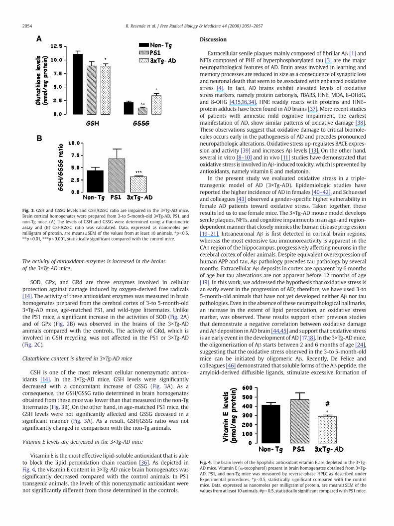
Fig. 3. GSH and GSSG levels and GSH/GSSG ratio are impaired in the 3×Tg-AD mice.Brain cortical homogenates were prepared from 3-to 5-month-old 3×Tg-AD, PS1, andnon-Tg mice. (A) The levels of GSH and GSSG were determined using a fluorimetricassay and (B) GSH/GSSG ratio was calculated. Data, expressed as nanomoles permilligram of protein, are means±SEM of the values from at least 10 animals. ⁎pb0.5,⁎⁎pb0.01, ⁎⁎⁎pb0.001, statistically significant compared with the control mice.
Fig. 4. The brain levels of the lipophilic antioxidant vitamin E are depleted in the 3×Tg-AD mice. Vitamin E (α-tocopherol) present in brain homogenates obtained from 3×Tg-AD, PS1, and non-Tg mice was measured by reverse-phase HPLC as described underExperimental procedures. ⁎pb0.5, statistically significant compared with the controlmice. Data, expressed as nanomoles per milligram of protein, are means±SEM of thevalues fromat least 10 animals. #pb0.5, statistically significant comparedwith PS1mice.
2054 R. Resende et al. / Free Radical Biology & Medicine 44 (2008) 2051–2057
The activity of antioxidant enzymes is increased in the brainsof the 3×Tg-AD mice
SOD, GPx, and GRd are three enzymes involved in cellularprotection against damage induced by oxygen-derived free radicals[14]. The activity of these antioxidant enzymes was measured in brainhomogenates prepared from the cerebral cortex of 3-to 5-month-old3×Tg-AD mice, age-matched PS1, and wild-type littermates. Unlikethe PS1 mice, a significant increase in the activities of SOD (Fig. 2A)and of GPx (Fig. 2B) was observed in the brains of the 3×Tg-ADanimals compared with the controls. The activity of GRd, which isinvolved in GSH recycling, was not affected in the PS1 or 3×Tg-AD(Fig. 2C).
Glutathione content is altered in 3×Tg-AD mice
GSH is one of the most relevant cellular nonenzymatic antiox-idants [14]. In the 3×Tg-AD mice, GSH levels were significantlydecreased with a concomitant increase of GSSG (Fig. 3A). As aconsequence, the GSH/GSSG ratio determined in brain homogenatesobtained from these micewas lower than that measured in the non-Tglittermates (Fig. 3B). On the other hand, in age-matched PS1 mice, theGSH levels were not significantly affected and GSSG deceased in asignificant manner (Fig. 3A). As a result, GSH/GSSG ratio was notsignificantly changed in comparison with the non-Tg animals.
Vitamin E levels are decreased in the 3×Tg-AD mice
Vitamin E is themost effective lipid-soluble antioxidant that is ableto block the lipid peroxidation chain reaction [36]. As depicted inFig. 4, the vitamin E content in 3×Tg-AD mice brain homogenates wassignificantly decreased compared with the control animals. In PS1transgenic animals, the levels of this nonenzymatic antioxidant werenot significantly different from those determined in the controls.
Discussion
Extracellular senile plaques mainly composed of fibrillar Aβ [1] andNFTs composed of PHF of hyperphosphorylated tau [3] are the majorneuropathological features of AD. Brain areas involved in learning andmemory processes are reduced in size as a consequence of synaptic lossand neuronal death that seem to be associatedwith enhanced oxidativestress [4]. In fact, AD brains exhibit elevated levels of oxidativestress markers, namely protein carbonyls, TBARS, HNE, MDA, 8-OHdG,and 8-OHG [4,15,16,34]. HNE readily reacts with proteins and HNE–protein adducts have been found in AD brains [37]. More recent studiesof patients with amnestic mild cognitive impairment, the earliestmanifestation of AD, show similar patterns of oxidative damage [38].These observations suggest that oxidative damage to critical biomole-cules occurs early in the pathogenesis of AD and precedes pronouncedneuropathologic alterations. Oxidative stress up-regulates BACE expres-sion and activity [39] and increases Aβ levels [13]. On the other hand,several in vitro [8–10] and in vivo [11] studies have demonstrated thatoxidative stress is involved inAβ-induced toxicity,which is preventedbyantioxidants, namely vitamin E and melatonin.
In the present study we evaluated oxidative stress in a triple-transgenic model of AD (3×Tg-AD). Epidemiologic studies havereported the higher incidence of AD in females [40–42], and Schuesseland colleagues [43] observed a gender-specific higher vulnerability infemale AD patients toward oxidative stress. Taken together, theseresults led us to use female mice. The 3×Tg-ADmouse model developssenile plaques, NFTs, and cognitive impairments in an age-and region-dependentmanner that closelymimics the humandisease progression[19–21]. Intraneuronal Aβ is first detected in cortical brain regions,whereas the most extensive tau immunoreactivity is apparent in theCA1 region of the hippocampus, progressively affecting neurons in thecerebral cortex of older animals. Despite equivalent overexpression ofhuman APP and tau, Aβ pathology precedes tau pathology by severalmonths. Extracellular Aβ deposits in cortex are apparent by 6 monthsof age but tau alterations are not apparent before 12 months of age[19]. In this work, we addressed the hypothesis that oxidative stress isan early event in the progression of AD; therefore, we have used 3-to5-month-old animals that have not yet developed neither Aβ nor taupathologies. Even in the absence of these neuropathological hallmarks,an increase in the extent of lipid peroxidation, an oxidative stressmarker, was observed. These results support other previous studiesthat demonstrate a negative correlation between oxidative damageandAβ deposition in AD brain [44,45] and support that oxidative stressis an early event in the development of AD [17,18]. In the 3×Tg-ADmice,the oligomerization of Aβ starts between 2 and 6 months of age [24],suggesting that the oxidative stress observed in the 3-to 5-month-oldmice can be initiated by oligomeric Aβ. Recently, De Felice andcolleagues [46] demonstrated that soluble forms of the Aβ peptide, theamyloid-derived diffusible ligands, stimulate excessive formation of
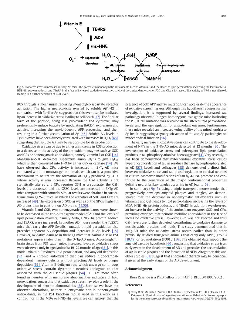
Fig. 5. Oxidative stress is increased in 3×Tg-ADmice. The decrease in nonenzymatic antioxidants such as vitamin E and GSH leads to lipid peroxidation, increasing the levels of MDA,HNE–His protein adducts, and TBARS. In the face of increased oxidative stress the activity of the antioxidant enzymes SOD and GPx is increased. The activity of GRd is not affected,leading to a further depletion of GSH levels.
2055R. Resende et al. / Free Radical Biology & Medicine 44 (2008) 2051–2057
ROS through a mechanism requiring N-methyl-D-aspartate receptoractivation. The higher neurotoxicity exerted by soluble Aβ1-42 incomparison with fibrillar Aβ suggests that this event can be mediatedby an increase in oxidative stress leading to cell death [47]. The fibrillarform of the peptide, being less pro-oxidant and cytotoxic, maypreferentially induce toxicity by modulating BACE-1 expression andactivity, increasing the amyloidogenic APP processing, and thenresulting in a further accumulation of Aβ [48]. Soluble Aβ levels inTg2576mice have been directly correlated with increases in H2O2 [48],suggesting that soluble Aβ may be responsible for its production.
Oxidative stress can be due to either an increase in ROS productionor a decrease in the activity of the antioxidant enzymes such as SODand GPx or nonenzymatic antioxidants, namely, vitamin E or GSH [14].Manganese-SOD detoxifies superoxide anion (O2
U−) to give H2O2,which is then converted into H2O by either GPx or catalase [14]. Wehave observed that GPx activity is increased in 3×Tg-AD micecompared with the nontransgenic animals, which can be a protectivemechanism to neutralize the formation of H2O2 produced by SOD,whose activity is also increased. Because the GRd activity is notstatistically altered and GPx requires GSH as a substrate, the GSHlevels are decreased and the GSSG levels are increased in 3×Tg-ADmice compared with controls. Similar results were obtained in corticaltissue from Tg2576 mice, in which the activities of SOD and GPx areincreased [49]. The expression of SOD as well as of the GPx is higher inAD brains than in control non-AD brains [15,50].
Vitamin E and GSH, two nonenzymatic antioxidants, were shownto be decreased in the triple-transgenic model of AD and the levels oflipid peroxidation markers, namely MDA, HNE–His protein adduct,and TBARS, were increased. In another AD mouse model, the Tg2576mice that carry the APP Swedish mutation, lipid peroxidation alsoprecedes apparent Aβ deposition and increases in Aβ levels [18].However, oxidative damage in these Tg mice that harbor APP or PS1mutations appears later than in the 3×Tg-AD mice. Accordingly, inbrain tissue from PS1 M146 L mice, increased levels of oxidative stresswere observed only in aged animals (19–22months of age) [51]. In thismodel, vitamin E reduces lipid peroxidation, and amyloid deposition[52] and a chronic antioxidant diet can reduce hippocampal-dependent memory deficits without affecting Aβ levels or plaquedeposition [53]. Vitamin E-deficient rats, which undergo continuousoxidative stress, contain dystrophic neuritis analogous to thatassociated with the AD senile plaques [54]. PHF are more oftenfound in neurites with membrane abnormalities indicative of lipidperoxidation, suggesting that oxidative stress may play a role in thedevelopment of neuritic abnormalities [55]. Because we have notobserved alterations, neither in enzymatic nor in nonenzymaticantioxidants, in the PS1 knock-in mouse used in this work as acontrol, nor in the MDA or HNE–His levels, we can suggest that the
presence of both APP and taumutations can accelerate the appearanceof oxidative stress markers. Although this hypothesis requires furtherinvestigation, it is supported by several findings. Increased taupathology observed in aged homozygous transgenic mice harboringthe P301L tau mutation was revealed in the altered lipid peroxidationlevels and the up-regulation of antioxidant enzymes. Furthermore,these mice revealed an increased vulnerability of the mitochondria toAβ insult, suggesting a synergistic action of tau and Aβ pathologies onmitochondrial function [56].
The early increase in oxidative stress can contribute to the develop-ment of NFTs in the 3×Tg-AD mice, detected at 12 months [20]. Theinvolvement of oxidative stress and subsequent lipid peroxidationproducts in tau phosphorylation has been suggested [4]. Very recently, ithas been demonstrated that mitochondrial oxidative stress causeshyperphosphorylation of tau in residues that are hyperphosphorylatedin AD [57]. Lovell and colleagues [58] demonstrated a direct linkbetween oxidative stress and tau phosphorylation in cortical neuronsin culture. Moreover, modifications of tau by 4-HNE promote and con-tribute to the generation of the major conformational propertiesdefining neurofibrillary tangles occurring in AD brains [59].
In summary (Fig. 5), using a triple-transgenic mouse model thatprogressively develops amyloid plaques and tangles, we demon-strated that the decrease in nonenzymatic antioxidants such asvitamin E and GSH leads to lipid peroxidation, increasing the levels ofMDA, HNE–His protein adducts, and TBARS. In addition, we observedan increase in the activity of the antioxidant enzymes SOD and GPx,providing evidence that neurons mobilize antioxidants in the face ofincreased oxidative stress. However, GRd was not affected and thusGSH levels are further depleted, contributing to oxidative damage tonucleic acids, proteins, and lipids. This study demonstrated that in3×Tg-AD mice the oxidative stress occurs earlier than in otherpreviously studied transgenic animals that carry only APP (Tg2576)[18,49] or tau mutations (P301L) [56]. The obtained data support theamyloid cascade hypothesis [60], suggesting that oxidative stress is anearly event in the development of AD and precedes the accumulationof Aβ in senile plaques and the formation of NFTs. Altogether, this andother studies [61] suggest that antioxidant therapy may be beneficialif given at the early stages of the AD development.
Acknowledgment
Rosa Resende is a Ph.D. fellow from FCT (SFRH/BD/11005/2002).
References
[1] Terry, R. D.;Masliah, E.; Salmon, D. P.; Butters, N.; DeTeresa, R.; Hill, R.; Hansen, L. A.;Katzman, R. Physical basis of cognitive alterations in Alzheimer's disease: synapticloss is the major correlate of cognitive impairment. Ann. Neurol. 30:572–580; 1991.
2056 R. Resende et al. / Free Radical Biology & Medicine 44 (2008) 2051–2057
[2] Price, D. L.; Sisodia, S. S.; Borchelt, D. R. Genetic neurodegenerative diseases: thehuman illness and transgenic models. Science 282:1079–1083; 1998.
[3] Avila, J. Tau protein, the main component of paired helical filaments. J. AlzheimersDis. 9:171–175; 2006.
[4] Mattson, M. P. Pathways towards and away from Alzheimer's disease. Nature430:631–639; 2004.
[5] Tanzi, R. E.; Kovacs, D. M.; Kim, T. W.; Moir, R. D.; Guenette, S. Y.; Wasco, W. Thegene defects responsible for familial Alzheimer's disease. Neurobiol. Dis.3:159–168; 1996.
[6] Butterfield, D. A.; Griffin, S.; Munch, G.; Pasinetti, G. M. Amyloid beta-peptide andamyloid pathology are central to the oxidative stress and inflammatory cascadesunder which Alzheimer's disease brain exists. J. Alzheimers Dis. 4:193–201; 2002.
[7] Haass, C.; Selkoe, D. J. Soluble protein oligomers in neurodegeneration: lessonsfrom the Alzheimer's amyloid beta-peptide. Nat. Rev., Mol. Cell Biol. 8:101–112;2007.
[8] Cardoso, S. M.; Oliveira, C. R. Glutathione cycle impairment mediates A beta-induced cell toxicity. Free Radic. Res. 37:241–250; 2003.
[9] Melo, J. B.; Agostinho, P.; Oliveira, C. R. Involvement of oxidative stress in theenhancement of acetylcholinesterase activity induced by amyloid beta-peptide.Neurosci. Res. 45:117–127; 2003.
[10] Cutler, R. G.; Kelly, J.; Storie, K.; Pedersen, W. A.; Tammara, A.; Hatanpaa, K.;Troncoso, J. C.; Mattson, M. P. Involvement of oxidative stress-induced abnorm-alities in ceramide and cholesterol metabolism in brain aging and Alzheimer'sdisease. Proc. Natl. Acad. Sci. U. S. A. 101:2070–2075; 2004.
[11] Jhoo, J. H.; Kim, H. C.; Nabeshima, T.; Yamada, K.; Shin, E. J.; Jhoo, W. K.; Kim, W.;Kang, K. S.; Jo, S. A.; Woo, J. I. Beta-amyloid (1-42)-induced learning and memorydeficits in mice: involvement of oxidative burdens in the hippocampus andcerebral cortex. Behav. Brain Res. 155:185–196; 2004.
[12] Paola, D.; Domenicotti, C.; Nitti, M.; Vitali, A.; Borghi, R.; Cottalasso, D.; Zaccheo,D.; Odetti, P.; Strocchi, P.; Marinari, U. M.; Tabaton, M.; Pronzato, M. A. Oxida-tive stress induces increase in intracellular amyloid β-protein productionand selective activation of βI and βII PKCs in NT2 cells. Biochem. Biophys. Res.Commun. 268:642–646; 2000.
[13] Tamagno, E.; Parola, M.; Bardini, P.; Piccini, A.; Borghi, R.; Guglielmotto, M.;Santoro, G.; Davit, A.; Danni, O.; Smith, M. A.; Perry, G.; Tabaton, M. Beta-site APPcleaving enzyme up-regulation induced by 4-hydroxynonenal is mediated bystress-activated protein kinases pathways. J. Neurochem. 92:628–636; 2005.
[14] Chauhan, V.; Chauhan, A. Oxidative stress in Alzheimer's disease. Pathophysiology13:195–208; 2006.
[15] Lovell, M. A.; Ehmann, W. D.; Butler, S. M.; Markesbery, W. R. Elevatedthiobarbituric acid-reactive substances and antioxidant enzyme activity in thebrain in Alzheimer's disease. Neurology 45:1594–1601; 1995.
[16] Butterfield, D. A.; Reed, T.; Newman, S. F.; Sultana, R. Roles of amyloid β-peptide-associated oxidative stress and brain protein modifications in the pathogenesis ofAlzheimer's disease and mild cognitive impairment. Free Radic. Biol. Med.43:658–677; 2007.
[17] Nunomura, A.; Perry, G.; Aliev, G.; Hirai, K.; Takeda, A.; Balraj, E. K.; Jones, P. K.;Ghanbari, H.; Wataya, T.; Shimohama, S.; Chiba, S.; Atwood, C. S.; Petersen, R. B.;Smith, M. A. Oxidative damage is the earliest event in Alzheimer disease.J. Neuropathol. Exp. Neurol. 60:759–767; 2001.
[18] Praticò, D.; Uryu, K.; Leight, S.; Trojanoswki, J. Q.; Lee, V. M. Increased lipidperoxidation precedes amyloid plaque formation in an animal model of Alzheimeramyloidosis. J. Neurosci. 21:4183–4187; 2001.
[19] Oddo, S.; Caccamo, A.; Shepherd, J. D.; Murphy, M. P.; Golde, T. E.; Kayed, R.;Metherate, R.; Mattson, M. P.; Akbari, Y.; LaFerla, F. M. Triple-transgenic model ofAlzheimer's disease with plaques and tangles: intracellular Ab and synapticdysfunction. Neuron 39:409–421; 2003.
[20] Oddo, S.; Caccamo, A.; Kitazawa, M.; Tseng, B. P.; LaFerla, F. M. Amyloid depositionprecedes tangle formation in a triple transgenic model of Alzheimer's disease.Neurobiol. Aging 24:1063–1070; 2003.
[21] Billings, L. M.; Oddo, S.; Green, K. N.; McGaugh, J. L.; LaFerla, F. M. IntraneuronalAbeta causes the onset of early Alzheimer's disease-related cognitive deficits intransgenic mice. Neuron 45:675–688; 2005.
[22] Oddo, S.; Caccamo, A.; Cheng, D.; Jouleh, B.; Torp, R.; LaFerla, F. M. Geneticallyaugmenting tau levels does not modulate the onset or progression of Abetapathology in transgenic mice. J. Neurochem. 102:1053–1063; 2007.
[23] Oddo, S.; Billings, L.; Kesslak, J. P.; Cribbs, D. H.; LaFerla, F. M. Abeta immunotherapyleads to clearance of early, but not late, hyperphosphorylated tau aggregates viathe proteasome. Neuron 43:321–332; 2004.
[24] Oddo, S.; Caccamo, A.; Tran, L.; Lambert, M. P.; Glabe, C. G.; Klein,W. L.; LaFerla, F.M.Temporal profile of amyloid-beta (Abeta) oligomerization in an in vivo modelof Alzheimer disease: a link between Abeta and tau pathology. J. Biol. Chem.28:1599–15604; 2006.
[25] Billings, L. M.; Green, K. N.; McGaugh, J. L.; LaFerla, F. M. Learning decreases Abeta⁎56 and tau pathology and ameliorates behavioral decline in 3×Tg-AD mice.J. Neurosci 27:751–761; 2007.
[26] Guo, Q.; Fu,W.; Sopher, B. L.; Miller, M.W.;Ware, C. B.; Martin, G. M.; Mattson, M. P.Increased vulnerability of hippocampal neurons to excitotoxic necrosis inpresenilin-1 mutant knock-in mice. Nat. Med. 5:101–106; 1999.
[27] Buege, J. A.; Aust, S. D. Microsomal lipid peroxidation. In: Colowick, S.P., Kaplan, N.O. (Eds.), Methods in Enzymology, vol. 52. Academic Press, New York,pp. 302–310.pp.; 1967.
[28] Wong, S. H.; Knight, J. A.; Hopfer, S. M.; Zaharia, O.; Leach Jr., C. N.; SundermanJr., F. W. Lipoperoxides in plasma as measured by liquid-chromatographic sepa-ration of malondialdehyde–thiobarbituric acid adduct. Clin. Chem. 33:214–220;1987.
[29] Flohe, L.; Otting, F. Superoxide dismutase assays. Methods Enzymol. 105:93–104;1984.
[30] Paglia, D. E.; Valentine, W. N. Studies on the quantitative and qualitative char-acterization of erythrocyte glutathione peroxidase. J. Lab. Clin. Med. 70:158–169;1967.
[31] Goldberg, D. M.; Richard, S. J. Methods enzymatic analysis. Academic Press, NewYork,pp. 258–265; 1983.
[32] Hissin, P. J.; Hilf, R. A fluorometric method for determination of oxidized andreduced glutathione in tissues. Anal. Biochem. 74:214–226; 1976.
[33] Vatassery, G. T.; Younoszai, R. Alpha tocopherol levels in various regions of thecentral nervous systems of the rat and guinea pig. Lipids 13:828–831; 1978.
[34] Petersen, R. B.; Nunomura, A.; Lee, H. G.; Casadesus, G.; Perry, G.; Smith, M. A.; Zhu,X. Signal transduction cascades associated with oxidative stress in Alzheimer'sdisease. J. Alzheimers Dis. 1:143–152; 2007.
[35] Uchida, K.; Stadtman, E. R. Modification of histidine residues in proteins byreaction with 4-hydroxynonenal. Proc. Natl. Acad. Sci. U. S. A. 89:4544–4548; 1992.
[36] Machlin, L. J.; Bendich, A. Free radical tissue damage: protective role of antioxidantnutrients. FASEB J. 1:441–445; 1987.
[37] Sayre, L. M.; Zelasko, D. A.; Harris, P. L.; Perry, G.; Salomon, R. G.; Smith, M. A. 4-Hydroxynonenal-derived advanced lipid peroxidation end products are increasedin Alzheimer's disease. J. Neurochem. 68:2092–2097; 1997.
[38] Lovell, M. A.; Markesbery, W. R. Oxidative DNA damage in mild cognitive impair-ment and late-stage Alzheimer's disease. Nucleic Acids Res. 35:7:497–7504;2007.
[39] Tamagno, E.; Bardini, P.; Obbili, A.; Vitali, A.; Borghi, R.; Zaccheo, D.; Pronzato,M. A.;Danni, O.; Smith, M. A.; Perry, G.; Tabaton,M. Oxidative stress increases expressionand activity of BACE in NT2 neurons. Neurobiol. Dis. 10:279–288; 2002.
[40] Andersen, K.; Launer, L. J.; Dewey, M. E.; Letenneur, L.; Ott, A.; Copeland, J. R.;Dartigues, J. F.; Kragh-Sorensen, P.; Baldereschi, M.; Brayne, C.; Lobo, A.; Martinez-Lage, J. M.; Stijnen, T.; Hofman, A. Gender differences in the incidence of AD andvascular dementia: the EURODEM studies. EURODEM Incidence Research Group.Neurology 53:1992–1997; 1999.
[41] Brayne, C.; Gill, C.; Huppert, F. A.; Barkley, C.; Gehlhaar, E.; Girling, D. M.; O'Connor,D. W.; Paykel, E. S. Incidence of clinically diagnosed subtypes of dementia in anelderly population. Cambridge Project for Later Life. Br. J. Psychiatry 167:255–262;1995.
[42] Fratiglioni, L.; Viitanen, M.; von Strauss, E.; Tontodonati, V.; Herlitz, A.; Winblad, B.Very old women at highest risk of dementia and Alzheimer's disease: incidencedata from the Kungsholmen Project, Stockholm. Neurology 48:132–138; 1997.
[43] Schuessel, K.; Leutner, S.; Cairns, N. J.; Müller, W. E.; Eckert, A. Impact of gender onupregulation of antioxidant defence mechanisms in Alzheimer's disease brain.J. Neural Transm. 111:1167–1182; 2004.
[44] Mclean, C. A.; Cherny, R. A.; Fraser, F. W.; Fuller, S. J.; Smith, M. J.; Beyreuther, K.;Bush, A. I.; Masters, C. L. Soluble pool of Ab amyloid as a determinant of severity ofneurodegeneration in Alzheimer's disease. Ann. Neurol. 46:860–866; 1999.
[45] Wang, J.; Dickson, D. W.; Trojanowski, J. Q.; Lee, V. M. The levels of soluble versusinsoluble brain Aβ distinguish Alzheimer's disease from normal and pathologicaging. Exp. Neurol. 158:328–337; 1999.
[46] De Felice, F. G.; Velasco, P. T.; Lambert, M. P.; Viola, K.; Fernandez, S. J.; Ferreira,S. T.; Klein, W. L. Abeta oligomers induce neuronal oxidative stress through anN-methyl-D-aspartate receptor-dependent mechanism that is blocked by theAlzheimer drug memantine. J. Biol. Chem. 282:11590–11601; 2007.
[47] Tamagno, E.; Bardini, P.; Guglielmotto, M.; Danni, O.; Tabaton, M. The variousaggregation states of β-amyloid 1-42 mediate different effects on oxidative stress,neurodegeneration, and BACE-1 expression. Free Radic. Biol. Med. 41:202–212;2006.
[48] Manczak, M.; Anekonda, T. S.; Henson, E.; Park, B. S.; Quinn, J.; Reddy, P. H.Mitochondria are a direct site of A beta accumulation in Alzheimer's diseaseneurons: implications for free radical generation and oxidative damage in diseaseprogression. Hum. Mol. Genet. 15:1437–1449; 2006.
[49] Apelt, J.; Bigl, M.; Wunderlich, P.; Schliebs, R. Aging-related increase in oxidativestress correlates with developmental pattern of beta-secretase activity and beta-amyloid plaque formation in transgenic Tg2576 mice with Alzheimer-likepathology. Int. J. Dev. Neurosci. 22:475–484; 2004.
[50] De Leo, M. E.; Borrello, S.; Passantino, M.; Palazzotti, B.; Mordente, A.; Daniele, A.;Filippini, V.; Galeotti, T.; Masullo, C. Oxidative stress and overexpression ofmanganese superoxide dismutase in patients with Alzheimer's disease. NeurosciLett. 250:173–176; 1998.
[51] Schuessel, K.; Frey, C.; Jourdan, C.; Keil, U.; Weber, C. C.; Müller-Spahn, F.; Müller,W. E.; Eckert, A. Aging sensitizes toward ROS formation and lipid peroxidation inPS1M146L transgenic mice. Free Radic. Biol. Med. 40:850–862; 2006.
[52] Sung, S.; Yao, Y.; Uryu, K.; Yang, H.; Lee, V. M.; Trojanowski, J. Q.; Praticò, D. Earlyvitamin E supplementation in young but not aged mice reduces Abeta levelsand amyloid deposition in a transgenic model of Alzheimer's disease. FASEB J.18:323–325; 2004.
[53] Quinn, J. F.; Bussiere, J. R.; Hammond, R. S.; Montine, T. J.; Henson, E.; Jones, R. E.;Stackman Jr., R. W. Chronic dietary alpha-lipoic acid reduces deficits inhippocampal memory of aged Tg2576 mice. Neurobiol. Aging 28:213–225; 2007.
[54] Heslop, K. E.; Goss-Sampson, M. A.; Muller, D. P.; Curzon, G. Serotonin metabolismand release in frontal cortex of rats on a vitamin E-deficient diet. J. Neurochem66:860–864; 1996.
[55] Praprotnik, D.; Smith, M. A.; Richey, P. L.; Vinters, H. V.; Perry, G. Plasmamembranefragility in dystrophic neurites in senile plaques of Alzheimer's disease: an index ofoxidative stress. Acta Neuropathol. 91:1–5; 1996.
[56] David, D. C.; Hauptmann, S.; Scherping, I.; Schuessel, K.; Keil, U.; Rizzu, P.; Ravid, R.;Dröse, S.; Brandt, U.; Müller, W. E.; Eckert, A.; Götz, J. Proteomic and functional

2057R. Resende et al. / Free Radical Biology & Medicine 44 (2008) 2051–2057
analyses reveal a mitochondrial dysfunction in P301L tau transgenic mice. J. Biol.Chem. 280:23802–23814; 2005.
[57] Melov, S.; Adlard, P. A.; Morten, K.; Johnson, F.; Golden, T. R.; Hinerfeld, D.;Schilling, B.; Mavros, C.; Masters, C. L.; Volitakis, I.; Li, Q. X.; Laughton, K.; Hubbard,A.; Cherny, R. A.; Gibson, B.; Bush, A. I. Mitochondrial oxidative stress causeshyperphosphorylation of tau. PLoS ONE 2:e536; 2007.
[58] Lovell, M. A.; Xiong, S.; Xie, C.; Davies, P.; Markesbery, W. R. Induction of hyper-phosphorylated tau in primary rat cortical neuron cultures mediated by oxidativestress and glycogen synthase kinase-3. J. Alzheimers Dis. 6:659–671; 2004.
[59] Liu, Q.; Smith, M. A.; Avilá, J.; DeBernardis, J.; Kansal, M.; Takeda, A.; Zhu, X.;Nunomura, A.; Honda, K.; Moreira, P. I.; Oliveira, C. R.; Santos, M. S.; Shimohama,S.; Aliev, G.; de la Torre, J.; Ghanbari, H. A.; Siedlak, S. L.; Harris, P. L.; Sayre, L. M.;Perry, G. Alzheimer-specific epitopes of tau represent lipid peroxidation-inducedconformations. Free Radic. Biol. Med. 38:746–754; 2005.
[60] Hardy, J.; Selkoe, D. J. The amyloid hypothesis of Alzheimer's disease: progress andproblems on the road to therapeutics. Science 297:353–356; 2002.
[61] Smith, D. G.; Cappai, R.; Barnham, K. J. The redox chemistry of the Alzheimer'sdisease amyloid beta peptide. Biochim. Biophys. Acta 1768:1976–1990; 2007.
Copyright © 2022 FDOKUMEN