
Brain networks of bottom-up triggered and top-down controlled shifting of auditory attention
10
Research Report Brain networks of bottom-up triggered and top-down controlled shifting of auditory attention Juha Salmi a,b, ⁎ , Teemu Rinne a , Sonja Koistinen a , Oili Salonen c , Kimmo Alho a a Department of Psychology, PO Box 9, FI-00014 University of Helsinki, Finland b Advanced Magnetic Imaging Centre, Helsinki University of Technology, Finland c Helsinki Medical Imaging Center, Helsinki University Central Hospital, Finland ARTICLE INFO ABSTRACT Article history: Accepted 25 June 2009 Available online 3 July 2009 During functional magnetic resonance imaging (fMRI), our participants selectively attended to tone streams at the left or right, and occasionally shifted their attention from one stream to another as guided by a centrally presented visual cue. Duration changes in the to-be-attended stream served as targets. Loudness deviating tones (LDTs) occurred infrequently in both streams to catch attention in a bottom-up manner, as indicated by their effects on reaction times to targets. LDTs activated the right temporo-parietal junction (TPJ), posterior parts of the left inferior/middle frontal gyrus (IFG/MFG), ventromedial parts of the superior parietal lobule (SPL), and left frontal eye field/premotor cortex (FEF/PMC). In addition, LDTs in the to- be-ignored sound stream were associated with enhanced activity in the ventromedial prefrontal cortex (VMPFC) possibly related to evaluation of the distracting event. Top-down controlled cue-guided attention shifts (CASs) activated bilateral areas in the SPL, intraparietal sulcus (IPS), FEF/PMC, TPJ, IFG/MFG, and cingulate/medial frontal gyrus, and crus I/II of the cerebellum. Thus, our results suggest that in audition top-down controlled and bottom-up triggered shifting of attention activate largely overlapping temporo-parietal, superior parietal and frontal areas. As the IPS, superior parts of the SPL, and crus I/II were activated specifically by top-down controlled attention shifts, and the VMPFC was specifically activated by bottom- up triggered attention shifts, our results also suggest some differences between auditory top- down controlled and bottom-up triggered shifting of attention. © 2009 Elsevier B.V. All rights reserved. Keywords: Attention Auditory Dorsal/ventral attention system fMRI Human brain 1. Introduction In everyday life, attention is dynamically allocated depend- ing on the events in the external environment and current behavioral goals. A sudden sound in the environment, for example, a cough heard during a conversation, may trigger in a bottom-up manner an involuntary attention shift from the currently attended speaker to the person who coughs. In this case, the event triggering bottom-up attention shift may be evaluated as irrelevant and one may continue following the conversation. However, if attention is triggered by a relevant sound (e.g., a cell phone ringing in your pocket; Roye et al., 2007), it is likely that the original task is replaced with a new one (answering the phone or thinking who might be calling you). Attention may also be shifted voluntarily in top-down control, for example, from one speaker to another during a conversation, according to the current behavioral goals. BRAIN RESEARCH 1286 (2009) 155 – 164 ⁎ Corresponding author. Department of Psychology, PO Box 9, FI-00014 University of Helsinki, Finland. Fax: +358 9 191 29450. E-mail address: [email protected] (J. Salmi). 0006-8993/$ – see front matter © 2009 Elsevier B.V. All rights reserved. doi:10.1016/j.brainres.2009.06.083 available at www.sciencedirect.com www.elsevier.com/locate/brainres
-
Upload
independent -
Category
Documents
-
view
6 -
download
0
Transcript of Brain networks of bottom-up triggered and top-down controlled shifting of auditory attention
B R A I N R E S E A R C H 1 2 8 6 ( 2 0 0 9 ) 1 5 5 – 1 6 4
ava i l ab l e a t www.sc i enced i rec t . com
www.e l sev i e r. com/ l oca te /b ra in res
Research Report
Brain networks of bottom-up triggered and top-downcontrolled shifting of auditory attention
Juha Salmia,b,⁎, Teemu Rinnea, Sonja Koistinena, Oili Salonenc, Kimmo Alhoa
aDepartment of Psychology, PO Box 9, FI-00014 University of Helsinki, FinlandbAdvanced Magnetic Imaging Centre, Helsinki University of Technology, FinlandcHelsinki Medical Imaging Center, Helsinki University Central Hospital, Finland
A R T I C L E I N F O
⁎ Corresponding author. Department of PsychE-mail address: [email protected] (J.
0006-8993/$ – see front matter © 2009 Elsevidoi:10.1016/j.brainres.2009.06.083
A B S T R A C T
Article history:Accepted 25 June 2009Available online 3 July 2009
During functional magnetic resonance imaging (fMRI), our participants selectively attendedto tone streamsat the left or right, andoccasionally shifted their attention fromone streamtoanother as guidedbya centrally presentedvisual cue.Duration changes in the to-be-attendedstream served as targets. Loudness deviating tones (LDTs) occurred infrequently in bothstreams to catch attention in a bottom-up manner, as indicated by their effects on reactiontimes to targets. LDTs activated the right temporo-parietal junction (TPJ), posterior parts ofthe left inferior/middle frontal gyrus (IFG/MFG), ventromedial parts of the superior parietallobule (SPL), and left frontal eye field/premotor cortex (FEF/PMC). In addition, LDTs in the to-be-ignored sound stream were associated with enhanced activity in the ventromedialprefrontal cortex (VMPFC) possibly related to evaluation of the distracting event. Top-downcontrolled cue-guidedattentionshifts (CASs) activated bilateral areas in theSPL, intraparietalsulcus (IPS), FEF/PMC, TPJ, IFG/MFG, and cingulate/medial frontal gyrus, and crus I/II of thecerebellum. Thus, our results suggest that in audition top-down controlled and bottom-uptriggeredshiftingof attentionactivate largely overlapping temporo-parietal, superiorparietaland frontal areas. As the IPS, superior parts of the SPL, and crus I/II were activated specificallyby top-downcontrolledattention shifts, and theVMPFCwas specifically activatedbybottom-up triggered attention shifts, our results also suggest somedifferences between auditory top-down controlled and bottom-up triggered shifting of attention.
© 2009 Elsevier B.V. All rights reserved.
Keywords:AttentionAuditoryDorsal/ventral attention systemfMRIHuman brain
1. Introduction
In everyday life, attention is dynamically allocated depend-ing on the events in the external environment and currentbehavioral goals. A sudden sound in the environment, forexample, a cough heard during a conversation, may triggerin a bottom-up manner an involuntary attention shift fromthe currently attended speaker to the person who coughs. Inthis case, the event triggering bottom-up attention shift may
ology, PO Box 9, FI-00014Salmi).
er B.V. All rights reserved
be evaluated as irrelevant and one may continue followingthe conversation. However, if attention is triggered by arelevant sound (e.g., a cell phone ringing in your pocket;Roye et al., 2007), it is likely that the original task is replacedwith a new one (answering the phone or thinking who mightbe calling you). Attention may also be shifted voluntarily intop-down control, for example, from one speaker to anotherduring a conversation, according to the current behavioralgoals.
University of Helsinki, Finland. Fax: +358 9 191 29450.
.
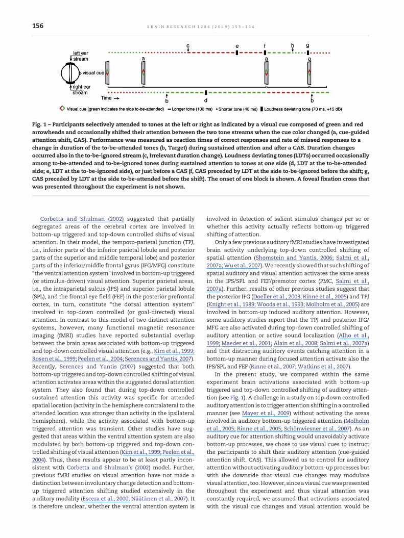
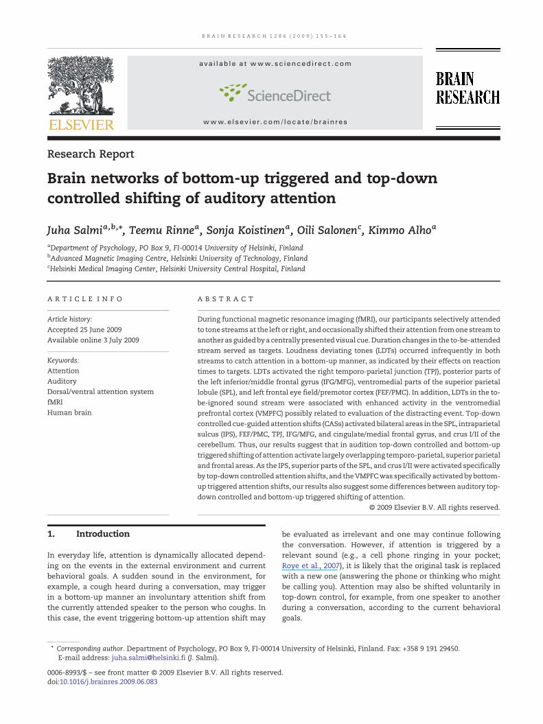
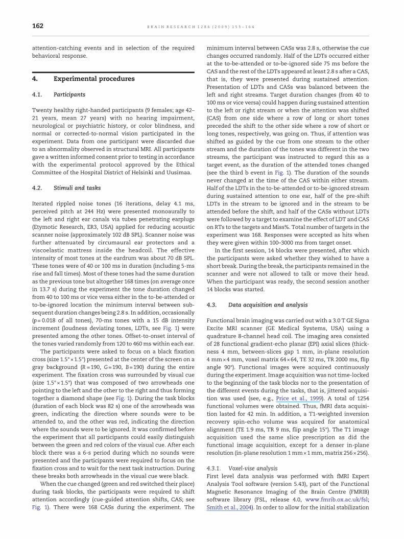
Fig. 1 – Participants selectively attended to tones at the left or right as indicated by a visual cue composed of green and redarrowheads and occasionally shifted their attention between the two tone streams when the cue color changed (a, cue-guidedattention shift, CAS). Performance was measured as reaction times of correct responses and rate of missed responses to achange in duration of the to-be-attended tones (b, Target) during sustained attention and after a CAS. Duration changesoccurred also in the to-be-ignored stream (c, Irrelevant duration change). Loudness deviating tones (LDTs) occurred occasionallyamong to-be-attended and to-be-ignored tones during sustained attention to tones at one side (d, LDT at the to-be-attendedside; e, LDT at the to-be-ignored side), or just before a CAS (f, CAS preceded by LDT at the side to-be-ignored before the shift; g,CAS preceded by LDT at the side to-be-attended before the shift). The onset of one block is shown. A foveal fixation cross thatwas presented throughout the experiment is not shown.
156 B R A I N R E S E A R C H 1 2 8 6 ( 2 0 0 9 ) 1 5 5 – 1 6 4
Corbetta and Shulman (2002) suggested that partiallysegregated areas of the cerebral cortex are involved inbottom-up triggered and top-down controlled shifts of visualattention. In their model, the temporo-parietal junction (TPJ,i.e., inferior parts of the inferior parietal lobule and posteriorparts of the superior and middle temporal lobe) and posteriorparts of the inferior/middle frontal gyrus (IFG/MFG) constitute“the ventral attention system” involved in bottom-up triggered(or stimulus-driven) visual attention. Superior parietal areas,i.e., the intraparietal sulcus (IPS) and superior parietal lobule(SPL), and the frontal eye field (FEF) in the posterior prefrontalcortex, in turn, constitute “the dorsal attention system”involved in top-down controlled (or goal-directed) visualattention. In contrast to this model of two distinct attentionsystems, however, many functional magnetic resonanceimaging (fMRI) studies have reported substantial overlapbetween the brain areas associated with bottom-up triggeredand top-down controlled visual attention (e.g., Kim et al., 1999;Rosenet al., 1999; Peelenet al., 2004; SerencesandYantis, 2007).Recently, Serences and Yantis (2007) suggested that bothbottom-up triggeredand top-downcontrolled shifting of visualattention activates areaswithin the suggested dorsal attentionsystem. They also found that during top-down controlledsustained attention this activity was specific for attendedspatial location (activity in the hemisphere contralateral to theattended location was stronger than activity in the ipsilateralhemisphere), while the activity associated with bottom-uptriggered attention was transient. Other studies have sug-gested that areas within the ventral attention system are alsomodulated by both bottom-up triggered and top-down con-trolled shifting of visual attention (Kimet al., 1999; Peelen et al.,2004). Thus, these results appear to be at least partly incon-sistent with Corbetta and Shulman's (2002) model. Further,previous fMRI studies on visual attention have not made adistinctionbetween involuntary changedetectionandbottom-up triggered attention shifting studied extensively in theauditory modality (Escera et al., 2000; Näätänen et al., 2007). Itis therefore unclear, whether the ventral attention system is
involved in detection of salient stimulus changes per se orwhether this activity actually reflects bottom-up triggeredshifting of attention.
Only a fewprevious auditory fMRI studieshave investigatedbrain activity underlying top-down controlled shifting ofspatial attention (Shomstein and Yantis, 2006; Salmi et al.,2007a;Wuetal., 2007).Werecentlyshowedthatsuchshiftingofspatial auditory and visual attention activates the same areasin the IPS/SPL and FEF/premotor cortex (PMC, Salmi et al.,2007a). Further, results of other previous studies suggest thatthe posterior IFG (Doeller et al., 2003; Rinne et al., 2005) and TPJ(Knight et al., 1989;Woods et al., 1993; Molholm et al., 2005) areinvolved in bottom-up induced auditory attention. However,some auditory studies report that the TPJ and posterior IFG/MFG are also activated during top-down controlled shifting ofauditory attention or active sound localization (Alho et al.,1999; Maeder et al., 2001; Alain et al., 2008; Salmi et al., 2007a)and that distracting auditory events catching attention in abottom-up manner during focused attention activate also theIPS/SPL and FEF (Rinne et al., 2007; Watkins et al., 2007).
In the present study, we compared within the sameexperiment brain activations associated with bottom-uptriggered and top-down controlled shifting of auditory atten-tion (see Fig. 1). A challenge in a study on top-down controlledauditory attention is to trigger attention shifting in a controlledmanner (see Mayer et al., 2009) without activating the areasinvolved in auditory bottom-up triggered attention (Molholmet al., 2005; Rinne et al., 2005; Schönwiesner et al., 2007). As anauditory cue for attention shifting would unavoidably activatebottom-up processes, we chose to use visual cues to instructthe participants to shift their auditory attention (cue-guidedattention shift, CAS). This allowed us to control for auditoryattentionwithout activating auditory bottom-upprocesses butwith the downside that visual cue changes may modulatevisualattention, too.However, sinceavisual cuewaspresentedthroughout the experiment and thus visual attention wasconstantly required, we assumed that activations associatedwith the visual cue changes and visual attention would be
Fig. 2 – RTs and Miss%s for target tones during sustained attention (left) and following immediately a CAS (right) when theywere not preceded by an LDT, when theywere preceded by an LDT in the to-be-attended stream, andwhen theywere precededby an LDT in the to-be-ignored stream (p<0.0001, ****; p<0.01, **).
157B R A I N R E S E A R C H 1 2 8 6 ( 2 0 0 9 ) 1 5 5 – 1 6 4
negligible in comparison with modulations of brain activityrelated to auditory attention in the present paradigm. Bottom-up triggered auditory attention shifts were initiated by task-irrelevantphysicallydeviant sounds (loudnessdeviating tones,LDTs) occurring both in the to-be-ignored and to-be-attendedlocations. LDTs occurred either independent of the CASs orimmediately before the CASs. Throughout the experiment, theparticipantswere required to fixate on a cross at the center of ascreen, direct their auditoryattentionas indicatedby thevisualcue around the fixation cross, and to respond to target durationchanges occurring among the attended sounds.
Based on earlier fMRI studies that investigated separatelyeither bottom-up triggered (Rinne et al., 2007; Watkins et al.,2007) or top-down controlled attention (Shomstein and Yantis2006; Salmi et al., 2007a; Wu et al., 2007), we hypothesized thatbottom-up triggered shifting of auditory attention caused byLDTs and top-down controlled shifting of auditory attentionassociated with CASs would activate overlapping brain net-works intheparietal and frontal areas. LDTs inthe to-be-ignoredlocation were expected to be more distracting than the LDTs inthe to-be-attended location, as LDTs in the to-be-ignoredlocation would probably trigger spatial attention shifting.Further,weexpectedthatbycomparingbrainactivityassociatedwith LDTs in the to-be-ignored and to-be-attended location, wecould reveal the brain network of bottom-up triggered shifts ofauditory spatial attention. Finally,wehypothesized that LDTs ina to-be-ignored location would facilitate subsequent top-downcontrolled CASs to that location, while LDTs in the to-be-attendedstreamof sounds rightbeforeaCASmightdistract top-down controlled shifting of attention to the other stream. Theseeffects of LDTs on top-down controlled attention were alsoexpected to modulate brain activity associated with CASs.
2. Results
2.1. Behavioral results
Behavioral results are shown in Fig. 2. During sustained-attention (left panel, black bar), the mean reaction time (RT)to targets not preceded by an LDT was 1161 ms (s.e.m. 47 ms)and percentage ofmissed responses to the targets (Miss%) was
5.1% (s.e.m. 1.6%). The relatively long RTs and occurrence ofresponse errors indicates that the present task was difficulteven for targets not preceded by an LDT or a CAS. This was alsoour intention,becausesuchdemandingtask forcesparticipantsto focus their attention strongly on the to-be-attended input.
As expected, during sustained attention, RTs becamelonger (paired samples t-test, t(18)=2.9, p <0.01) when a targetwas preceded by an LDT in the to-be-ignored stream (leftpanel, white bar), as comparedwith targets not preceded by anLDT. LDTs in the to-be-ignored stream also increased Miss%(t(18)=2.9, p <0.01) for the subsequent targets as comparedwith targets not preceded by an LDT during sustainedattention. These results indicate that the LDTs indeed caughtparticipants' attention in a bottom-up manner. However,somewhat unexpectedly, during sustained attention, RTs tothe targets became shorter (t(18)=6.8, p <0.0001) when a targetwas preceded by an LDTs in the same, to-be-attended tonestream (left panel, grey bar). This suggests that bottom-uptriggered auditory attention may facilitate performance if thetriggering event occurs among to-be-attended sounds.
Also CASs affected participants' performance. For targetsnot precededby an LDTbut by a CAS, themeanRTwas 1297ms(s.e.m. 57 ms; right panel, black bar), which was significantlylonger (t(18)=3.9, p <0.001) than the RT for targets duringsustained attention. As expected, LDTs catching attention in abottom-up manner facilitated performance after a CAS if theyoccurred in a streamthatwas tobe attendedafter a subsequentCAS: RTs became significantly shorter (t(18)=3.7, p <0.01) fortargets precededbyaCAS, if theCASwasprecededby anLDT inthe stream that was to be ignored before the CAS (right panel,white bar), as compared with targets following a CAS notpreceded by an LDT. However, unlike we expected, LDTs in theto-be-attended streampreceding a CAS to the other streamdidnot have a significant effect on target detection following suchCAS (right panel, gray bar) as compared with target detectionafter a CAS not preceded by an LDT in either stream.
2.2. fMRI results
2.2.1. Brain activations induced by LDTs and CASsComparison of brain activity associated with LDT events withbrain activity associated with sustained attention without

Fig. 3 – Top: Bottom-up influences on brain activity caused by LDTs during auditory sustained attention (blue, Z>3.0, p<0.01).Middle: Top-down controlled CAS contrastedwith auditory sustained attention (red, Z>5.0, p<0.01, and yellow, Z>3.0, p<0.01).Bottom: Activation time-series in the right SPL, right TPJ and posterior cerebellar (Post. Cb) ROIs during different events. Notethat figure at the top middle shows activity at the posterior temporal cortex. However, below the brain surface this activitycluster reached TPJ.
158 B R A I N R E S E A R C H 1 2 8 6 ( 2 0 0 9 ) 1 5 5 – 1 6 4
LDTs revealed activity (Z >3.0, cluster-corrected p <0.01)related to processing of LDTs and bottom-up attention tothem in the right TPJ, ventromedial parts of the bilateral SPL,posterior parts of the left IFG/MFG, and left FEF/premotorcortex (PMC; Fig. 3, top). Top-down controlled attention shifts,in turn, activated (Z >3.0, cluster-corrected p <0.01) the IPS/SPL, FEF/PMC, TPJ, IFG/MFG, cingulate/medial frontal gyrus(CG/medFG), lateral and ventromedial occipital cortex (OC),and crus I/II of the posterior cerebellum (Post. Cb; middle),bilaterally, as indicated by comparison of brain activityfollowing CASs with activity during sustained attention.Thus, bottom-up triggered shifting of attention associatedwith LDTs and top-down controlled shifting of attentionassociated with CASs activated overlapping areas in theventromedial SPL, left IFG/MFG, left FEF/PMC, and right TPJ.
Effects of LDTs on CAS-related brain activity were studiedin regions of interest (ROIs) defined based on the activityclusters revealed by the comparison of CASs and sustainedattention (see, previous paragraph). A repeated-measuresANOVA for activation time-series (Fig. 3, bottom) withfactors Event Type (CAS with no LDT and CAS preceded byan LDT in the to-be-attended stream) and Time from EventOnset (4–6, 6–8, 8–10 s) suggested that activity in the rightSPL (F(1,18)=8.6, p <0.01) and posterior cerebellum (F(1,18)=6.9, p <0.05) ROI was lower when a CAS was preceded by anLDT in the stream that was to be attended before the CAS(Fig. 3, bottom, green line) as compared with CASs with nopreceding LDT (white line). In other brain regions, LDTs didnot cause significant modulations on activity associatedwith CASs.
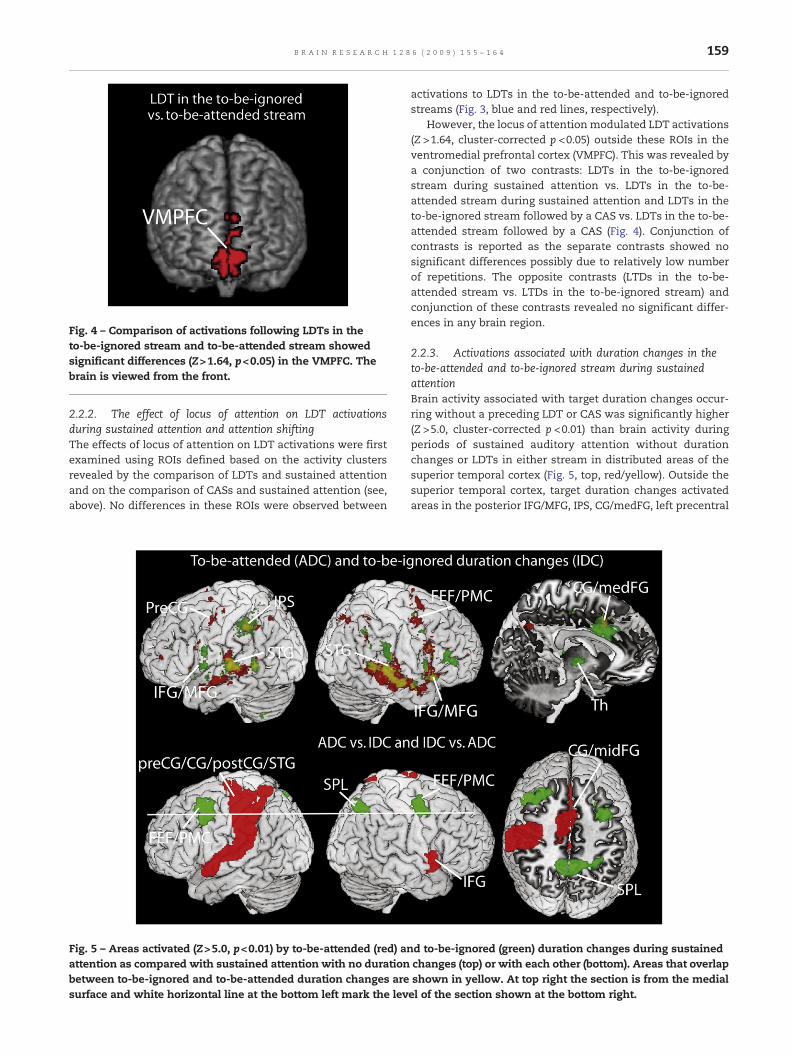
Fig. 4 – Comparison of activations following LDTs in theto-be-ignored stream and to-be-attended stream showedsignificant differences (Z>1.64, p<0.05) in the VMPFC. Thebrain is viewed from the front.
159B R A I N R E S E A R C H 1 2 8 6 ( 2 0 0 9 ) 1 5 5 – 1 6 4
2.2.2. The effect of locus of attention on LDT activationsduring sustained attention and attention shiftingThe effects of locus of attention on LDT activations were firstexamined using ROIs defined based on the activity clustersrevealed by the comparison of LDTs and sustained attentionand on the comparison of CASs and sustained attention (see,above). No differences in these ROIs were observed between
Fig. 5 – Areas activated (Z>5.0, p<0.01) by to-be-attended (red) anattention as compared with sustained attention with no durationbetween to-be-ignored and to-be-attended duration changes aresurface and white horizontal line at the bottom left mark the lev
activations to LDTs in the to-be-attended and to-be-ignoredstreams (Fig. 3, blue and red lines, respectively).
However, the locus of attentionmodulated LDT activations(Z >1.64, cluster-corrected p <0.05) outside these ROIs in theventromedial prefrontal cortex (VMPFC). This was revealed bya conjunction of two contrasts: LDTs in the to-be-ignoredstream during sustained attention vs. LDTs in the to-be-attended stream during sustained attention and LDTs in theto-be-ignored stream followed by a CAS vs. LDTs in the to-be-attended stream followed by a CAS (Fig. 4). Conjunction ofcontrasts is reported as the separate contrasts showed nosignificant differences possibly due to relatively low numberof repetitions. The opposite contrasts (LTDs in the to-be-attended stream vs. LTDs in the to-be-ignored stream) andconjunction of these contrasts revealed no significant differ-ences in any brain region.
2.2.3. Activations associated with duration changes in theto-be-attended and to-be-ignored stream during sustainedattentionBrain activity associated with target duration changes occur-ring without a preceding LDT or CAS was significantly higher(Z >5.0, cluster-corrected p <0.01) than brain activity duringperiods of sustained auditory attention without durationchanges or LDTs in either stream in distributed areas of thesuperior temporal cortex (Fig. 5, top, red/yellow). Outside thesuperior temporal cortex, target duration changes activatedareas in the posterior IFG/MFG, IPS, CG/medFG, left precentral
d to-be-ignored (green) duration changes during sustainedchanges (top) or with each other (bottom). Areas that overlapshown in yellow. At top right the section is from the medialel of the section shown at the bottom right.

160 B R A I N R E S E A R C H 1 2 8 6 ( 2 0 0 9 ) 1 5 5 – 1 6 4
gyrus, and FEF/PMC, bilaterally (top, red/yellow). Activity in theleft precentral gyrus, central sulcus, and postcentral gyrus(contralateral to the hand of response), right IFG, and CG/medFG was observed when target duration changes werecompared with duration changes in the to-be-ignored stream(Z >3.0, cluster-corrected p <0.01, bottom, red). Durationchanges in the to-be-ignored stream, in turn, enhanced brainactivity significantly (Z >5.0, cluster-corrected p <0.01) in theSTG, IPS, CG/midFG, TPJ, posterior IFG/MFG, and thalamus incomparison with brain activity during sustained-attentionperiods without duration changes or LDTs in either stream(Fig. 5, top, green/yellow). Contrasting brain activity associatedwith to-be-ignoredduration changes to activity associatedwithtarget duration changes revealed that to-be-ignored durationchanges elicited activity (Z >3.0, cluster-corrected p <0.01) inthe SPL and FEF/PMC bilaterally (Fig. 5, bottom, green).
3. Discussion
The present auditory task was made difficult intentionally toensure strongly focused auditory attention. Therefore, asexpected, even RTs to targets without a preceding CAS orLDT were relatively long and the participants also omittedsome responses. The effects of LDTs on RTs to subsequenttargets suggested that LDTs captured attention in a bottom-upmanner: LDTs in the to-be-ignored stream most likely causedspatial attention shift to the to-be-ignored stream (i.e., to thewrong direction) as RTs to subsequent targets were prolongedduring sustained attention (Doeller et al., 2003; Grimm et al.,2008) and shortened when a target was preceded by a CAS tothe stream where the LTD had just occurred. Thus, an LDT inthe stream that was to be ignored before a CAS and attendedafter it served as an additional exogenous cue for attentionshifting. Moreover, LDTs in the to-be-attended stream duringsustained attention shortened RTs to subsequent targets. Thiseffect was probably caused by facilitation of the sensory ormotor processes, or both, needed in target processing due toenhanced alertness for events in the attended stream (Hackleyand Valle-Inclán, 2003). Finally, RTs to targets preceded by aCASs were longer than RTs to targets during sustainedattention probably because of the cost associated withattention shifting right before the target (see Salmi et al.,2007b).
In keeping with the previous studies that have separatelyexamined auditory bottom-up triggered (Doeller et al., 2003;Rinne et al., 2005; Molholm et al., 2005; Rinne et al., 2007;Watkins et al., 2007) or top-down controlled shifts of attention(Shomstein and Yantis 2006; Salmi et al., 2007a; Wu et al.,2007), we observed activity in the STG/MTG, TPJ, SPL, IFG/MFGand FEF/PMC for bottom-up triggered attention shifts, andactivity in the IPS/SPL, FEF/PMC, STG/MTG, TPJ, IFG/MFG, CG/medFG, and crus I/II of the posterior cerebellum for the top-down controlled attention shifts. In contrast to previousstudies of auditory top-down controlled attention, we foundactivity also in the lateral and ventromedial OC for top-downcontrolled attention shifts. This OC activity was probablyrelated to processing of the visual cue.
In contradiction with our hypothesis, LDTs in the to-be-attended location preceding a CAS did not distract this CAS
according to the lack of significant difference in RTs andresponse accuracy between targets following such CASs andthose following CASs not preceded by an LDT (Fig. 2). However,CAS-related activity in the right SPL and posterior cerebellumROI was lower when a CAS was preceded by an LDT in thestream that was to be attended before the CAS to the otherstream (Fig. 3). Thus, these LDTs indeed appeared to suppresssubsequent top-down controlled CASs in a bottom-upmanneralthough this suppression was not observable in taskperformance.
3.1. Activity in the areas corresponding to the ventralattention system
Based on studies of visual attention, Corbetta and Shulman(2002) suggested that the ventral attention system isspecialized in detection of salient or unexpected stimuli.Consistently, in the present study, the posterior IFG/MFGand TPJ were activated by LDTs. Activity in the right TPJ andposterior IFG/MFG was observed also for the CASs in thepresent study, even when they were not preceded by anLDT. This result is in line with the findings of some previousstudies reporting activity in the TPJ and posterior IFG/MFGduring top-down controlled auditory attention (e.g., Alho etal., 1999; Salmi et al., 2007a). However, these findings arediscrepant with the visual-attention model of Corbetta andShulman suggesting that the ventral attention system isspecific for bottom-up triggered attention. The discrepancymight be due to differences between auditory and visualattention or between the auditory and visual experimentaldesigns, or both. Still, as described in Introduction, severalprevious studies using trial-by-trial cueing have also sug-gested an overlap between the brain networks of top-downcontrolled and bottom-up triggered attention in the visualmodality (Rosen et al., 1999; Peelen et al., 2004), implyingthat the posterior IFG/MFG and TPJ are not specialized inbottom-up triggered attention, but are activated also by top-down controlled attention. The present results providefurther support to the view that the areas within thesuggested ventral (visual) attention system are involved inboth bottom-up triggered and top-down controlled attentionin audition.
In addition to the LDTs and CASs, the posterior IFG/MFGwas activated by duration changes in the to-be-attended andto-be-ignored streams during sustained attention. This isconsistent with previous studies investigating brain activa-tions to infrequent duration, pitch and location changes intones (Rinne et al., 2000; Opitz et al., 2002; Molholm et al., 2005;Rinne et al., 2005; Tse et al., 2006; Deouell, 2007; Schönwiesneret al., 2007). The present IFG/MFG activity to to-be-ignoredduration changes was probably associated with auditorychange detection and bottom-up triggered attention shifting(Rinne et al., 2000; Deouell, 2007).
3.2. Activity in the areas corresponding to the dorsalattention system
In keeping with the model of visual attention by Corbettaand Shulman (2002) and recent studies of top-down con-trolled auditory attention shifting (Shomstein and Yantis

161B R A I N R E S E A R C H 1 2 8 6 ( 2 0 0 9 ) 1 5 5 – 1 6 4
2006; Salmi et al., 2007a; Wu et al., 2007), the presentactivations in the IPS/SPL and FEF support the involvementof these areas in top-down controlled attention shifting. Inthe parietal cortex, especially in the IPS and dorsal parts ofthe SPL enhanced activity was observed for the CASs, butnot for the LDTs. Moreover, our results show that activity inthe dorsal attention system, especially in the right SPL, ismodulated by events that trigger bottom-up attention (see,Fig. 3, bottom). The activity in the IPS and FEF/PMC observedin a comparison of brain activity elicited by to-be-ignoredduration changes during sustained attention with activityassociated with target duration changes (Fig. 5, bottom)might also be related to interaction of dorsal and ventralattention systems, as the to-be-ignored duration changesmay have been followed by active suppression of attentionshifting to the to-be-ignored stream and/or suppression ofmotor responses that the participants were prepared tomake to duration changes.
However, some of our results concerning the dorsalattention system argue against full segregation of the brainnetworks of bottom-up triggered and top-down controlledattention: (1) the ventromedial parts of SPL (see, Fig. 3) showedactivity also when trials associated with LDTs and to-be-ignored duration changes were compared with sustainedattention with no duration changes or LDTs, (2) enhancedbilateral FEF/PMC activity was observed when to-be-ignoredduration changes were compared with to-be-attended dura-tion changes, and (3) enhanced left FEF/PMC activity wasobservedwhen LDTswere comparedwith sustained attention.The results showing that the ventromedial SPL was activatedby both the CASs and LDTs, while the superior parts of SPLwere activated specifically by the CASs, suggest that differentregions of the SPL might serve different functions in auditoryattention. The activity in the ventromedial SPL and FEF/PMCassociated with LDTs and to-be-ignored duration changesmight be related to the involvement of these areas in bottom-up triggered attention (Serences and Yantis, 2007; Watkins etal., 2007), such as initiation of involuntary attention shifts. Theeffects of top-down control in processing of the LDTs and to-be-ignored duration changes cannot be ruled out, however, asvoluntary reorienting of attention back to the task after aninvoluntary attention shift to the LDTs or to-be-ignoredduration changes, or active suppression of bottom-up proces-sing of the LTDs and to-be-ignored duration changes might berequired.
3.3. Activity in the CG/midFG and cerebellum
As many previous studies of visual attention (Kim et al.,1999; Rosen et al., 1999; Peelen et al., 2004; Kincade et al.,2005; Salmi et al., 2007a) and auditory attention (Mayer et al.,2006; Salmi et al., 2007a; Watkins et al., 2007), we observedactivity also in the CG/midFG and posterior cerebellum. TheCG/midFG is suggested to be involved in conflict processing,preparation of movements, and monitoring of performance(see, e.g., Isomura and Takada, 2004). In the present study,CG/midFG activity associated with target processing mayhave been involved in preparation of movements ormonitoring of performance, or both. The posterior cerebel-lum, in turn, is suggested to be involved in voluntary shifting
of attention (Allen et al., 1997; Le et al., 1998; Townsend etal., 1999; Salmi et al. 2007a). In accordance with this, thepresent results showed activity in the crus I/II of thecerebellum associated with CASs. The crus I/II was activatedalso in our other study on voluntary control of auditoryattention (Salmi et al., 2007a). However, the present resultsalso showed that posterior cerebellar activity following LDTsdecreased as compared with sustained attention with noLDTs or duration changes (Fig. 3, bottom right). This mightbe caused, for example, by gating of signals from the parietaland frontal cortex to the ponto-cerebellar system (Schmah-mann and Pandya, 1997), possibly due to LDT-inducedprocesses interfering with processes involved in top-downcontrolled goal-directed behavior. The present results do not,however, allow conclusions on the exact role of decreasedcerebellar activity associated with LDTs.
3.4. Activity in the ventromedial prefrontal cortex
LDTs in the to-be-ignored stream were associated withenhanced activity in the VMPFC when they were comparedwith LDTs in the to-be-attended stream. In parallel with this,RTs to the targets preceded by an LDT during sustainedattention were longer when the LDT occurred in the to-be-ignored stream than when it occurred in the to-be-attendedsteam. As LDTs in the to-be-ignored stream were irrelevantdistractors, this activity may be related to the suggested roleof the VMPFC in inhibition of irrelevant stimuli (Rule et al.,2002). However, VMPFC activity was also observed when LDTin the to-be-ignored stream was followed by a CAS, that is,when the LDT served as an exogenous cue and facilitatedthe RT to a target in the same stream where the LDT hadjust occurred and where attention was shifted as guided bya CAS between the LDT and target. Rule et al. (2002), as wellas others (Bechara et al., 2000; Rolls 2000), have suggestedthat the VMPFC is not only involved in inhibition ofirrelevant stimuli, but also in the decision of when torelease this inhibition in order to facilitate attention shifting.The present VMPFC activations may be related to suchevaluation.
3.5. Conclusions
The present results suggest that bottom-up triggered andtop-down controlled auditory attention shifting are asso-ciated with enhanced activity in brain areas belonging to thedorsal and ventral attention systems in vision (Corbetta andShulman, 2002), and also in the CG/midFG, crus I/II of theposterior cerebellum, and VMPFC. Our findings suggest thatdifferent areas of the SPL might have different roles inauditory attention, since the IPS and superior parts of theSPL are activated specifically by top-down controlled atten-tion shifting, while the ventromedial SPL is activated also byLDTs and to-be-ignored duration changes. Moreover, ourresults suggest that in audition, the so-called ventralattention system is not involved only in bottom-up proces-sing of changes in the environment and attention shiftstriggered by them, but also in top-down controlled attentionshifts. Finally, the present results suggest that the VMPFCmay have an important role in evaluating the significance of
162 B R A I N R E S E A R C H 1 2 8 6 ( 2 0 0 9 ) 1 5 5 – 1 6 4
attention-catching events and in selection of the requiredbehavioral response.
4. Experimental procedures
4.1. Participants
Twenty healthy right-handed participants (9 females; age 42–21 years, mean 27 years) with no hearing impairment,neurological or psychiatric history, or color blindness, andnormal or corrected-to-normal vision participated in theexperiment. Data from one participant were discarded dueto an abnormality observed in structural MRI. All participantsgave a written informed consent prior to testing in accordancewith the experimental protocol approved by the EthicalCommittee of the Hospital District of Helsinki and Uusimaa.
4.2. Stimuli and tasks
Iterated rippled noise tones (16 iterations, delay 4.1 ms,perceived pitch at 244 Hz) were presented monoaurally tothe left and right ear canals via tubes penetrating earplugs(Etymotic Research, ER3, USA) applied for reducing acousticscanner noise (approximately 102 dB SPL). Scanner noise wasfurther attenuated by circumaural ear protectors and aviscoelastic mattress inside the headcoil. The effectiveintensity of most tones at the eardrum was about 70 dB SPL.These tones were of 40 or 100 ms in duration (including 5-msrise and fall times). Most of these tones had the same durationas the previous tone but altogether 168 times (on average oncein 13.7 s) during the experiment the tone duration changedfrom 40 to 100 ms or vice versa either in the to-be-attended orto-be-ignored location the minimum interval between sub-sequent duration changes being 2.8 s. In addition, occasionally(p= 0.018 of all tones), 70-ms tones with a 15 dB intensityincrement (loudness deviating tones, LDTs, see Fig. 1) werepresented among the other tones. Offset-to-onset interval ofthe tones varied randomly from 120 to 460mswithin each ear.
The participants were asked to focus on a black fixationcross (size 1.5°×1.5°) presented at the center of the screen on agray background (R =190, G=190, B=190) during the entireexperiment. The fixation cross was surrounded by visual cue(size 1.5°×1.5°) that was composed of two arrowheads onepointing to the left and the other to the right and thus formingtogether a diamond shape (see Fig. 1). During the task blocks(duration of each block was 82 s) one of the arrowheads wasgreen, indicating the direction where sounds were to beattended to, and the other was red, indicating the directionwhere the sounds were to be ignored. It was confirmed beforethe experiment that all participants could easily distinguishbetween the green and red colors of the visual cue. After eachblock there was a 6-s period during which no sounds werepresented and the participants were required to focus on thefixation cross and to wait for the next task instruction. Duringthese breaks both arrowheads in the visual cue were black.
When the cue changed (green and red switched their place)during task blocks, the participants were required to shiftattention accordingly (cue-guided attention shifts, CAS; seeFig. 1). There were 168 CASs during the experiment. The
minimum interval between CASs was 2.8 s, otherwise the cuechanges occurred randomly. Half of the LDTs occurred eitherat the to-be-attended or to-be-ignored side 75 ms before theCAS and the rest of the LDTs appeared at least 2.8 s after a CAS,that is, they were presented during sustained attention.Presentation of LDTs and CASs was balanced between theleft and right streams. Target duration changes (from 40 to100ms or vice versa) could happen during sustained attentionto the left or right stream or when the attention was shifted(CAS) from one side where a row of long or short tonespreceded the shift to the other side where a row of short orlong tones, respectively, was going on. Thus, if attention wasshifted as guided by the cue from one stream to the otherstream and the duration of the tones was different in the twostreams, the participant was instructed to regard this as atarget event, as the duration of the attended tones changed(see the third b event in Fig. 1). The duration of the soundsnever changed at the time of the CAS within either stream.Half of the LDTs in the to-be-attended or to-be-ignored streamduring sustained attention to one ear, half of the pre-shiftLDTs in the stream to be ignored and in the stream to beattended before the shift, and half of the CASs without LDTswere followed by a target to examine the effect of LDT and CASon RTs to the targets andMiss%. Total number of targets in theexperiment was 168. Responses were accepted as hits whenthey were given within 100–3000 ms from target onset.
In the first session, 14 blocks were presented, after whichthe participants were asked whether they wished to have ashort break. During the break, the participants remained in thescanner and were not allowed to talk or move their head.When the participant was ready, the second session another14 blocks was started.
4.3. Data acquisition and analysis
Functional brain imaging was carried out with a 3.0 T GE SignaExcite MRI scanner (GE Medical Systems, USA) using aquadrature 8-channel head coil. The imaging area consistedof 28 functional gradient-echo planar (EPI) axial slices (thick-ness 4 mm, between-slices gap 1 mm, in-plane resolution4 mm×4 mm, voxel matrix 64×64, TE 32 ms, TR 2000 ms, flipangle 90°). Functional images were acquired continuouslyduring the experiment. Image acquisitionwas not time-lockedto the beginning of the task blocks nor to the presentation ofthe different events during the tasks, that is, jittered acquisi-tion was used (see, e.g., Price et al., 1999). A total of 1254functional volumes were obtained. Thus, fMRI data acquisi-tion lasted for 42 min. In addition, a T1-weighted inversionrecovery spin-echo volume was acquired for anatomicalalignment (TE 1.9 ms, TR 9 ms, flip angle 15°). The T1 imageacquisition used the same slice prescription as did thefunctional image acquisition, except for a denser in-planeresolution (in-plane resolution 1mm×1mm,matrix 256×256).
4.3.1. Voxel-vise analysisFirst level data analysis was performed with fMRI ExpertAnalysis Tool software (version 5.43), part of the FunctionalMagnetic Resonance Imaging of the Brain Centre (FMRIB)software library (FSL, release 4.0, www.fmrib.ox.ac.uk/fsl;Smith et al., 2004). In order to allow for the initial stabilization
163B R A I N R E S E A R C H 1 2 8 6 ( 2 0 0 9 ) 1 5 5 – 1 6 4
of the fMRI signal, the first 6 volumes of the session wereexcluded from analysis (during this time no task was pre-sented). The data were motion corrected and spatiallysmoothed with a Gaussian kernel of 7 mm (FWHM), and high-pass filtered (cutoff 150 s). Statistical analysis was performedusing the FMRIB Improved Linear Model (FILM). Explanatoryvariables were derived from timing (onset of each event) ofdifferent events (LDTs in the to-be-attended and to-be-ignoredstream, CAS, LDTs in the to-be-attended and to-be-attendedstream with CAS, target duration change during sustainedattention, to-be-ignored duration change during sustainedattention) and rest periods. The hemodynamic response toCASs, LDTs, and duration changes during sustained attentionwasmodeled using a double-gamma-function and its temporalderivative and rest periods were modeled using a gammafunction. The high-pass filtering applied to the model was thesame as that applied to the data. Sustained-attention periodswhen no targets or LDTswere presented served as a baseline inthemodel. Contrasts were specified to create Z-statistic imagestesting for the effects of sustained attention, attention shiftingand duration changes. The exact comparisons are carefullyreported in the Results section. The effects of target processingon LDT and CAS-related activity (see Fig. 3) was reduced bycontrasting these events with same amount of targets thatoccurred independent of LDTs and CASs.
The two halves of the fMRI experiment were analyzedseparately and then combined in a second level analysis (fixedeffects). For group (third level) analysis, Z-statistic images ofeach participant were transformed into a standard space(MNI152; Montreal Neurological Institute). The group analysiswas performed using FMRIB's Local Analysis of Mixed Effects(FLAME). Statistical inference was carried out using Gaussianrandom field theory and cluster-based thresholding, using acluster-formingZ threshold (1.64–6.5 depending on thecontrast)and a (corrected) cluster significance threshold (P <0.01–0.05).
4.3.2. Region of interest analysisSeveral regions of interest (ROIs) were defined based on thegroup activation clusters to extract the activation time-series. Each group activation cluster (contrasts: LDTs vssustained attention and CASs vs. sustained attention) wasincluded as a ROI in the analysis but only statisticallysignificant results are reported. For the ROI analysis, thedata were first motion corrected and high-pass filtered(cutoff 60 s) using FSL. Then the ROI data were transferredto percent signal change values relative to the mean ROIsignal across all volumes. After this the time points(volumes) were sorted in time relatively to the onset of theblock, the ROI time-series was linearly interpolated, andtemporally smoothed using a low-pass Butterworth filter.Finally, the baseline of the ROI time-series was set to themean of time window of 5–0 s before block onset.
Acknowledgments
This research was supported by the Academy of Finland(grants #210587, 210186 and 209709), Nordic Council for theestablishment of a Nordic Center of Excellence in CognitiveControl (NCOE grant #40043), Finnish Cultural Foundation,
Finnish Graduate School of Psychology, and by the Funds ofthe University of Helsinki. We thank Ms. Marita Kattelus forher help in collecting the fMRI data and Dr. AlexanderDegerman for his helpful comments in interpreting ourresults.
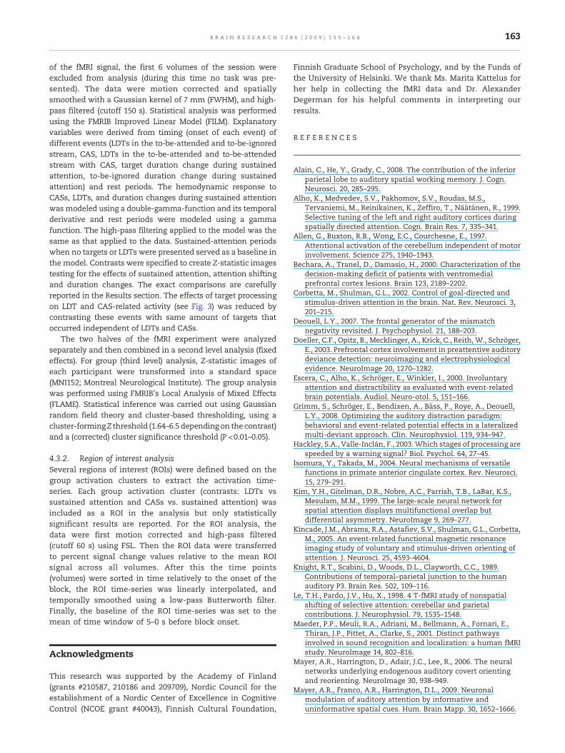
R E F E R E N C E S
Alain, C., He, Y., Grady, C., 2008. The contribution of the inferiorparietal lobe to auditory spatial working memory. J. Cogn.Neurosci. 20, 285–295.
Alho, K., Medvedev, S.V., Pakhomov, S.V., Roudas, M.S.,Tervaniemi, M., Reinikainen, K., Zeffiro, T., Näätänen, R., 1999.Selective tuning of the left and right auditory cortices duringspatially directed attention. Cogn. Brain Res. 7, 335–341.
Allen, G., Buxton, R.B., Wong, E.C., Courchesne, E., 1997.Attentional activation of the cerebellum independent of motorinvolvement. Science 275, 1940–1943.
Bechara, A., Tranel, D., Damasio, H., 2000. Characterization of thedecision-making deficit of patients with ventromedialprefrontal cortex lesions. Brain 123, 2189–2202.
Corbetta, M., Shulman, G.L., 2002. Control of goal-directed andstimulus-driven attention in the brain. Nat. Rev. Neurosci. 3,201–215.
Deouell, L.Y., 2007. The frontal generator of the mismatchnegativity revisited. J. Psychophysiol. 21, 188–203.
Doeller, C.F., Opitz, B., Mecklinger, A., Krick, C., Reith, W., Schröger,E., 2003. Prefrontal cortex involvement in preattentive auditorydeviance detection: neuroimaging and electrophysiologicalevidence. NeuroImage 20, 1270–1282.
Escera, C., Alho, K., Schröger, E., Winkler, I., 2000. Involuntaryattention and distractibility as evaluated with event-relatedbrain potentials. Audiol. Neuro-otol. 5, 151–166.
Grimm, S., Schröger, E., Bendixen, A., Bäss, P., Roye, A., Deouell,L.Y., 2008. Optimizing the auditory distraction paradigm:behavioral and event-related potential effects in a lateralizedmulti-deviant approach. Clin. Neurophysiol. 119, 934–947.
Hackley, S.A., Valle-Inclán, F., 2003.Which stages of processing arespeeded by a warning signal? Biol. Psychol. 64, 27–45.
Isomura, Y., Takada, M., 2004. Neural mechanisms of versatilefunctions in primate anterior cingulate cortex. Rev. Neurosci.15, 279–291.
Kim, Y.H., Gitelman, D.R., Nobre, A.C., Parrish, T.B., LaBar, K.S.,Mesulam, M.M., 1999. The large-scale neural network forspatial attention displays multifunctional overlap butdifferential asymmetry. NeuroImage 9, 269–277.
Kincade, J.M., Abrams, R.A., Astafiev, S.V., Shulman, G.L., Corbetta,M., 2005. An event-related functional magnetic resonanceimaging study of voluntary and stimulus-driven orienting ofattention. J. Neurosci. 25, 4593–4604.
Knight, R.T., Scabini, D., Woods, D.L., Clayworth, C.C., 1989.Contributions of temporal–parietal junction to the humanauditory P3. Brain Res. 502, 109–116.
Le, T.H., Pardo, J.V., Hu, X., 1998. 4 T-fMRI study of nonspatialshifting of selective attention: cerebellar and parietalcontributions. J. Neurophysiol. 79, 1535–1548.
Maeder, P.P., Meuli, R.A., Adriani, M., Bellmann, A., Fornari, E.,Thiran, J.P., Pittet, A., Clarke, S., 2001. Distinct pathwaysinvolved in sound recognition and localization: a human fMRIstudy. NeuroImage 14, 802–816.
Mayer, A.R., Harrington, D., Adair, J.C., Lee, R., 2006. The neuralnetworks underlying endogenous auditory covert orientingand reorienting. NeuroImage 30, 938–949.
Mayer, A.R., Franco, A.R., Harrington, D.L., 2009. Neuronalmodulation of auditory attention by informative anduninformative spatial cues. Hum. Brain Mapp. 30, 1652–1666.
164 B R A I N R E S E A R C H 1 2 8 6 ( 2 0 0 9 ) 1 5 5 – 1 6 4
Molholm, S., Martinez, A., Ritter,W., Javitt, D.C., Foxe, J.J., 2005. Theneural circuitry of pre-attentive auditory change-detection: anfMRI study of pitch and duration mismatch negativitygenerators. Cereb. Cortex 15, 545–551.
Näätänen, R., Paavilainen, P., Rinne, T., Alho, K., 2007. Themismatch negativity (MMN) in basic research of centralauditory processing: a review. Clin. Neurophysiol. 118,2544–2590.
Opitz, B., Rinne, T., Mecklinger, A., von Cramon, D.Y., Schröger, E.,2002. Differential contribution of frontal and temporal corticesto auditory change detection: fMRI and ERP results.NeuroImage 15, 167–174.
Peelen, M.V., Heslenfeld, D.J., Theeuwes, J., 2004. Endogenous andexogenous attention shifts are mediated by the samelarge-scale neural network. NeuroImage 22, 822–830.
Price, C.J., Veltman, D.J., Ashburner, J., Josephs, O., Friston, K.J.,1999. The critical relationship between the timing of stimuluspresentation and data acquisition in blocked designs withfMRI. NeuroImage 10, 36–44.
Rinne, T., Alho, K., Ilmoniemi, R.J., Virtanen, J., Näätänen, R., 2000.Separate time behaviors of the temporal and frontal mismatchnegativity sources. NeuroImage 12, 14–19.
Rinne, T., Degerman, A., Alho, K., 2005. Superior temporal andinferior frontal cortices are activated by infrequent soundduration decrements: an fMRI study. NeuroImage 26, 66–72.
Rinne, T., Kirjavainen, S., Salonen, O., Degerman, A., Kang, X.,Woods, D.L., Alho, K., 2007. Distributed cortical networks forfocused auditory attention and distraction. Neurosci. Lett. 416,247–251.
Rolls, E.T., 2000. The orbitofrontal cortex and reward. Cereb. Cortex10, 284–294.
Rosen, A.C., Rao, S.M., Caffarra, P., Scaglioni, A., Bobholz, J.A.,Woodley, S.J., Hammeke, T.A., Cunningham, J.M., Prieto, T.E.,Binder, J.R., 1999. Neural basis of endogenous and exogenousspatial orienting. A functional MRI study. J. Cogn. Neurosci. 11,135–152.
Roye, A., Jacobsen, T., Schröger, E., 2007. Personal significanceis encoded automatically by the human brain: an event-related potential study with ringtones. Eur. J. Neurosci. 26,784–790.
Rule, R.R., Shimamura, A.P., Knight, R.T., 2002. Orbitofrontal cortexand dynamic filtering of emotional stimuli. Cogn. Affect.Behav. Neurosci. 2, 264–270.
Salmi, J., Rinne, T., Degerman, A., Salonen, O., Alho, K., 2007a.Orienting andmaintenance of spatial attention in audition and
vision: multimodal and modality-specific brain activations.Brain Struct. Funct. 212, 181–194.
Salmi, J., Rinne, T., Degerman, A., Alho, K., 2007b. Orienting andmaintenance of spatial attention in audition and vision: anevent-related brain potential study. Eur. J. Neurosci. 25,3725–3733.
Schmahmann, J.D., Pandya, D.N., 1997. Anatomic organization ofthe basilar pontine projections from prefrontal cortices inrhesus monkey. J. Neurosci. 17, 438–458.
Schönwiesner, M., Novitski, N., Pakarinen, S., Carlson, S.,Tervaniemi, M., Näätänen, R., 2007. Heschl's gyrus, posteriorsuperior temporal gyrus, and mid-ventrolateral prefrontalcortex have different roles in the detection of acoustic changes.J. Neurophysiol. 97, 2075–2082.
Serences, J.T., Yantis, S., 2007. Spatially selective representationsof voluntary and stimulus-driven attentional priority inhuman occipital, parietal, and frontal cortex. Cereb. Cortex 17,284–293.
Shomstein, S., Yantis, S., 2006. Parietal cortex mediates voluntarycontrol of spatial and nonspatial auditory attention.J. Neurosci. 26, 435–439.
Smith, S.M., Jenkinson, M., Woolrich, M.W., Beckmann, C.F.,Behrens, T.E., Johansen-Berg, H., Bannister, P.R., De Luca, M.,Drobnjak, I., Flitney, D.E., Niazy, R.K., Saunders, J., Vickers, J.,Zhang, Y., De Stefano, N., Brady, J.M., Matthews, P.M., 2004.Advances in functional and structural MR image analysis andimplementation as FSL. NeuroImage 23, 208–219.
Townsend, J., Courchesne, E., Covington, J., Westerfield, M., Harris,N.S., Lyden, P., Lowry, T.P., Press, G.A., 1999. Spatial attentiondeficits in patients with acquired or developmental cerebellarabnormality. J. Neurosci. 19, 5632–5643.
Tse, C.Y., Tien, K.R., Penney, T.B., 2006. Event-related opticalimaging reveals the temporal dynamics of right temporal andfrontal cortex activation in pre-attentive change detection.NeuroImage 29, 314–320.
Watkins, S., Dalton, P., Lavie, N., Rees, G., 2007. Brain mechanismsmediating auditory attentional capture in humans. Cereb.Cortex 17, 1694–1700.
Woods, D.L., Knight, R.T., Scabini, D., 1993. Anatomical substratesof auditory selective attention: behavioral andelectrophysiological effects of posterior association cortexlesions. Cogn. Brain Res. 1, 227–240.
Wu, C.T., Weissman, D.H., Roberts, K.C., Woldorff, M.G., 2007. Theneural circuitry underlying the executive control of auditoryspatial attention. Brain Res. 1134, 187–198.