Brain-derived neurotrophic factor enhances GABA release probability and nonuniform distribution of...
11
Cellular/Molecular Brain-Derived Neurotrophic Factor Enhances GABA Release Probability and Nonuniform Distribution of N- and P/Q-Type Channels on Release Sites of Hippocampal Inhibitory Synapses Pietro Baldelli, 1 Jesus-Miguel Hernandez-Guijo, 2 Valentina Carabelli, 2 and Emilio Carbone 1,2 1 Istituto Nazionale di Fisica della Materia Research Unit and 2 Department of Neuroscience, Nanostructured Interfaces and Surfaces Center, I-10125 Turin, Italy Long-lasting exposures to brain-derived neurotrophic factor (BDNF) accelerate the functional maturation of GABAergic transmission in embryonic hippocampal neurons, but the molecular bases of this phenomenon are still debated. Evidence in favor of a postsynaptic site of action has been accumulated, but most of the data support a presynaptic site effect. A crucial issue is whether the enhancement of evoked IPSCs (eIPSCs) induced by BDNF is attributable to an increase in any of the elementary parameters controlling neurosecretion, namely the probability of release, the number of release sites, the readily releasable pool (RRP), and the quantal size. Here, using peak-scaled variance analysis of miniature IPSCs, multiple probability fluctuation analysis, and cumulative amplitude analysis of action potential-evoked postsynaptic currents, we show that BDNF increases release probability and vesicle replenishment with little or no effect on the quantal size, the number of release sites, the RRP, and the Ca 2 dependence of eIPSCs. BDNF treatment changes markedly the distribution of Ca 2 channels controlling neurotransmitter release. It enhances markedly the contribution of N- and P/Q-type channels, which summed to 100% (“supra-additivity”), and deletes the contribution of R-type channels. BDNF acceler- ates the switch of presynaptic Ca 2 channel distribution from “segregated” to “nonuniform” distribution. This maturation effect was accompanied by an uncovered increased control of N-type channels on paired-pulse depression, otherwise dominated by P/Q-type channels in untreated neurons. Nevertheless, BDNF preserved the fast recovery from depression associated with N-type channels. These novel presynaptic BDNF actions derive mostly from an enhanced overlapping and better colocalization of N- and P/Q-type channels to vesicle release sites. Key words: neurotrophins; multiple probability fluctuation analysis; peak-scaled variance analysis; inhibitory synaptic transmission; N- and P/Q-type channels; RRP Introduction During neuronal development, brain-derived neurotrophic fac- tor (BDNF) promotes the formation, maturation, and stabiliza- tion of both glutamatergic and GABAergic synapses in the CNS, regulating the balance between excitatory and inhibitory trans- mission. This is a fundamental step for neural circuit formation (Poo, 2001; Lessmann et al., 2003). To date, one of the most unclear aspects of the action of BDNF on the CNS concerns the molecular mechanism underlying the chronic effect on synaptic functions. In particular, it is not known how long-term exposures to BDNF can induce strong potentiation and increased connec- tivity of inhibitory synapses in various brain regions (Vicario- Abejon et al., 2002). The available data on inhibitory synapses indicate that BDNF causes net increases in the frequency of min- iature IPSCs (mIPSCs) and in the size of spontaneous and evoked IPSCs (eIPSCs) (Rutherford et al., 1997; Vicario-Abejon et al., 1998; Huang et al., 1999; Bolton et al., 2000; Baldelli et al., 2002; Yamada et al., 2002). Some of the above effects could be ascribed to an increased number of inhibitory neurons and to an en- hanced arborization of dendritic and axonal processes (Vicario- Abejon et al., 1998; Kohara et al., 2003). This response, however, was clearly observed in hippocampal and cerebellar slices (Marty et al., 2000; Seil and Drake-Baumann, 2000) and hippocampal cultures of embryonic day 16 (E16) rat embryos (Vicario-Abejon et al., 1998) but was much less evident in E18 embryos (Sherwood and Lo, 1999; Bolton et al., 2000; Baldelli et al., 2002). Most of the published data support a presynaptic site of action for BDNF that results in an increased size and intensity of GAD immunopositive terminals (Huang et al. 1999; Bolton et al., 2000; Yamada et al., 2002), an enhanced frequency of mIPSCs (Vicario- Abejon et al., 1998; Bolton et al., 2000; Baldelli et al., 2002), and a Received Oct. 12, 2004; revised Feb. 16, 2005; accepted Feb. 16, 2005. This project was supported by the Cavalieri-Ottolenghi foundation, by Italian Ministero dell’Istruzione, dell’Universita ` e della Ricerca Grant 2003/249, and by a Marie Curie Fellowship to J.M.H.-G. (contract HPMF-CT- 2000-00899). We thank Fabio Benfenati and Egidio D’Angelo for helpful discussions. We thank also Claudio Franchino and Brunella Tedesco for helping with cell cultures and preparation of solutions. Correspondence should be addressed to Pietro Baldelli, Department of Experimental Medicine, Section of Human Physiology, University of Genoa, Viale Benedetto XV, 3, I-16132 Genova, Italy. E-mail: [email protected]. DOI:10.1523/JNEUROSCI.4227-04.2005 Copyright © 2005 Society for Neuroscience 0270-6474/05/253358-11$15.00/0 3358 • The Journal of Neuroscience, March 30, 2005 • 25(13):3358 –3368
-
Upload
independent -
Category
Documents
-
view
3 -
download
0
Transcript of Brain-derived neurotrophic factor enhances GABA release probability and nonuniform distribution of...

Cellular/Molecular
Brain-Derived Neurotrophic Factor Enhances GABARelease Probability and Nonuniform Distribution of N- andP/Q-Type Channels on Release Sites of HippocampalInhibitory Synapses
Pietro Baldelli,1 Jesus-Miguel Hernandez-Guijo,2 Valentina Carabelli,2 and Emilio Carbone1,2
1Istituto Nazionale di Fisica della Materia Research Unit and 2Department of Neuroscience, Nanostructured Interfaces and Surfaces Center, I-10125 Turin,Italy
Long-lasting exposures to brain-derived neurotrophic factor (BDNF) accelerate the functional maturation of GABAergic transmission inembryonic hippocampal neurons, but the molecular bases of this phenomenon are still debated. Evidence in favor of a postsynaptic siteof action has been accumulated, but most of the data support a presynaptic site effect. A crucial issue is whether the enhancement ofevoked IPSCs (eIPSCs) induced by BDNF is attributable to an increase in any of the elementary parameters controlling neurosecretion,namely the probability of release, the number of release sites, the readily releasable pool (RRP), and the quantal size.
Here, using peak-scaled variance analysis of miniature IPSCs, multiple probability fluctuation analysis, and cumulative amplitudeanalysis of action potential-evoked postsynaptic currents, we show that BDNF increases release probability and vesicle replenishmentwith little or no effect on the quantal size, the number of release sites, the RRP, and the Ca 2� dependence of eIPSCs. BDNF treatmentchanges markedly the distribution of Ca 2� channels controlling neurotransmitter release. It enhances markedly the contribution of N-and P/Q-type channels, which summed to �100% (“supra-additivity”), and deletes the contribution of R-type channels. BDNF acceler-ates the switch of presynaptic Ca 2� channel distribution from “segregated” to “nonuniform” distribution. This maturation effect wasaccompanied by an uncovered increased control of N-type channels on paired-pulse depression, otherwise dominated by P/Q-typechannels in untreated neurons. Nevertheless, BDNF preserved the fast recovery from depression associated with N-type channels. Thesenovel presynaptic BDNF actions derive mostly from an enhanced overlapping and better colocalization of N- and P/Q-type channels tovesicle release sites.
Key words: neurotrophins; multiple probability fluctuation analysis; peak-scaled variance analysis; inhibitory synaptic transmission; N-and P/Q-type channels; RRP
IntroductionDuring neuronal development, brain-derived neurotrophic fac-tor (BDNF) promotes the formation, maturation, and stabiliza-tion of both glutamatergic and GABAergic synapses in the CNS,regulating the balance between excitatory and inhibitory trans-mission. This is a fundamental step for neural circuit formation(Poo, 2001; Lessmann et al., 2003). To date, one of the mostunclear aspects of the action of BDNF on the CNS concerns themolecular mechanism underlying the chronic effect on synapticfunctions. In particular, it is not known how long-term exposuresto BDNF can induce strong potentiation and increased connec-
tivity of inhibitory synapses in various brain regions (Vicario-Abejon et al., 2002). The available data on inhibitory synapsesindicate that BDNF causes net increases in the frequency of min-iature IPSCs (mIPSCs) and in the size of spontaneous and evokedIPSCs (eIPSCs) (Rutherford et al., 1997; Vicario-Abejon et al.,1998; Huang et al., 1999; Bolton et al., 2000; Baldelli et al., 2002;Yamada et al., 2002). Some of the above effects could be ascribedto an increased number of inhibitory neurons and to an en-hanced arborization of dendritic and axonal processes (Vicario-Abejon et al., 1998; Kohara et al., 2003). This response, however,was clearly observed in hippocampal and cerebellar slices (Martyet al., 2000; Seil and Drake-Baumann, 2000) and hippocampalcultures of embryonic day 16 (E16) rat embryos (Vicario-Abejonet al., 1998) but was much less evident in E18 embryos (Sherwoodand Lo, 1999; Bolton et al., 2000; Baldelli et al., 2002).
Most of the published data support a presynaptic site of actionfor BDNF that results in an increased size and intensity of GADimmunopositive terminals (Huang et al. 1999; Bolton et al., 2000;Yamada et al., 2002), an enhanced frequency of mIPSCs (Vicario-Abejon et al., 1998; Bolton et al., 2000; Baldelli et al., 2002), and a
Received Oct. 12, 2004; revised Feb. 16, 2005; accepted Feb. 16, 2005.This project was supported by the Cavalieri-Ottolenghi foundation, by Italian Ministero dell’Istruzione,
dell’Universita e della Ricerca Grant 2003/249, and by a Marie Curie Fellowship to J.M.H.-G. (contract HPMF-CT-2000-00899). We thank Fabio Benfenati and Egidio D’Angelo for helpful discussions. We thank also ClaudioFranchino and Brunella Tedesco for helping with cell cultures and preparation of solutions.
Correspondence should be addressed to Pietro Baldelli, Department of Experimental Medicine, Section of HumanPhysiology, University of Genoa, Viale Benedetto XV, 3, I-16132 Genova, Italy. E-mail: [email protected].
DOI:10.1523/JNEUROSCI.4227-04.2005Copyright © 2005 Society for Neuroscience 0270-6474/05/253358-11$15.00/0
3358 • The Journal of Neuroscience, March 30, 2005 • 25(13):3358 –3368
higher paired-pulse depression (PPD) reflecting increased vesicledepletion (Baldelli et al., 2002). However, an increase in GABAA
receptor density has been also observed, suggesting the involve-ment of postsynaptic mechanisms (Yamada et al., 2002). Despitethese well established findings, there is an impressive lack of in-formation on how BDNF affects the elementary parameters con-trolling neurotransmitter release: probability of release [releaseprobability (Pr) and vesicle release probability (Pves)], GABAA
receptor conductance, number of release sites, number of vesiclesforming the readily releasable pool (RRP), and the distributionand role of presynaptic Ca 2� channels. This is a critical issue thatwould help to clarify the basic mechanisms of action of BDNF ondeveloping neurons.
Here, using the variance analysis of mIPSCs (Traynelis andJaramillo, 1998), the fluctuation analysis, and the cumulative am-plitude profile of eIPSCs (Schneggenburger et al., 1999; Clementsand Silver, 2000), we show that in inhibitory GABAergic syn-apses, BDNF primarily produces an increased Pr and Pves, withlittle change in the number of release sites, the RRP, quantalcontent, and unitary conductance of GABAA receptors. In linewith previous reports indicating a BDNF-induced increase inCa 2� channel synthesis and KCl-evoked synaptic activity(Baldelli et al., 2000, 2002), we bring here new evidence in favor ofa BDNF-induced increase in Pr and Pves associated with an en-hanced contribution and “nonuniform” distribution of N- andP/Q-type channels to action potential-evoked IPSCs togetherwith a new role of N-type channels in PPD. This represents anovel mechanism of the action of BDNF on the maturation ofinhibitory synapses, which may result from a better coupling ofpresynaptic Ca 2� channels with vesicle release sites.
Materials and MethodsCell culture and BDNF treatment. All experiments were performed inaccordance with the guidelines established by the National Council onAnimal Care and approved by the local Animal Care Committee of TurinUniversity. Pregnant Sprague Dawley rats were killed by inhalation ofCO2, and E18 rats were removed immediately by cesarean section. Re-moval and dissection of the hippocampus, isolation of neurons, andculturing procedures were as described previously (Baldelli et al., 2000).The isolated hippocampal neurons were plated at low density (200 cells/mm 2) and allowed to establish for 4 d, after which 15 ng/ml BDNF(Sigma, St. Louis, MO) was added every 3 d for the 3– 4 weeks of theculture. When required, the cultured neurons were exposed to the ty-rosine kinase B (TrkB)-specific inhibitor anti-TrkB IgG1 (10 �g/ml)(clone 47; Transduction Laboratories, Lexington, KY), which effectivelyinhibits BDNF binding to TrkB receptors (Balkowiec and Katz, 2000). Allof the experiments were recorded in 14 –21 d in vitro (DIV) neurons.
Current recordings, data acquisition, and analysis. Patch electrodes, fab-ricated from thick borosilicate glasses (Hilgenberg, Mansfield, Ger-many), were pulled and fire-polished to a final resistance of 2– 4 M�.Patch-clamp recordings were performed in whole-cell configuration us-ing an EPC-9 amplifier (HEKA Electronic, Lambrecht, Germany). mIP-SCs and eIPSCs were acquired at 1–10 kHz sample frequency and filteredat one-half the acquisition rate with an eight-pole low-pass Bessel filter.Recordings with leak currents of �100 pA or a series resistance of �20M� were discarded. Data acquisition was performed using Pulse pro-grams (HEKA Elektronic). All of the experiments were performed atroom temperature (22–24°C). Data are expressed as mean � SEM fornumber of cells (n). Unpaired Student’s t tests were used, and p � 0.05was considered significant.
Solutions and drugs. mIPSCs and eIPSCs were recorded by superfusingthe whole-cell-clamped postsynaptic neuron with Tyrode’s solution con-taining (in mM): 2 CaCl2, 150 NaCl, 1 MgCl2, 10 HEPES, 4 KCl, and 10glucose, pH 7.4. When required, solutions with higher Ca 2� concentra-tions (5–10 mM) were obtained by lowering the NaCl content. The uns-elective glutamate receptor antagonist kynurenic acid (1 mM) (Sigma)
was added to the Tyrode’s solution to block excitatory transmission.Tetrodotoxin (0.3 �M) was added to block spontaneous action potentialpropagation. When recording eIPSCs, the external solution was supple-mented with the selective GABAB inhibitor (2S)-3-[[(1S)-1(3,4-dichlorophenyl) ethyl] amino-2-hydroxypropyl] (phenylmethyl) phos-phinic acid (5 �M). The neuron was constantly superfused through agravity system, as described previously (Baldelli et al., 2002). The perfu-sion solution could be changed rapidly (50 – 60 ms) and applied for con-trolled periods. The tip of the perfusion pipette (100 –200 �m) wasplaced 40 – 80 �m to the soma. The standard internal solution was (inmM): 90 CsCl, 20 TEA-Cl, 10 EGTA, 10 glucose, 1 MgCl2, 4 ATP, 0.5GTP, and 15 phosphocreatine, pH 7.4. K � was substituted for Cs � andTEA � in the pipette solution to block outward K � currents. N-(2,6-dimethylphenylcarbamoylmethyl) triethylammonium bromide (10 mM)was added to block Na � currents activated during an eIPSC. Solutionscontaining nifedipine, �-conotoxin-GVIA (�-CTx-GVIA), and�-agatoxin-IVA (�-Aga-IVA) were prepared as indicated previously(Magnelli et al., 1998). BDNF (Sigma) was dissolved in distilled watercontaining 1 mg/ml BSA and maintained in stock solutions at �80°C.
Peak-scaled variance analysis. For the reasons discussed by Traynelis etal. (1993), analysis of mIPSCs was preferred to the analysis of eIPSCs todetermine the conductance of postsynaptic receptor channels. Briefly,when averaged mIPSCs and eIPSCs are normalized and superimposed, itis immediately apparent that the rise time of evoked currents could alsobe twofold slower than miniature synaptic currents. Two possible expla-nations for this are (1) that inhibitory fiber stimulation activates severaldiscrete release sites or (2) that there is multiple vesicle release at individ-ual sites, even when evoked release is apparently synchronous. In eithercase, asynchronous transmitter release will increase the deviation of in-dividual synaptic currents from the mean IPSC time course. Such currentdeviation could potentially introduce errors in the evaluation of postsyn-aptic single-channel conductance. On the contrary, the mIPSCs that re-sult from the release of one or more transmitter packets presumably arisefrom nearly synchronous release of transmitter and receptor activationand are ideal for nonstationary fluctuation analysis
The variance analysis of mIPSCs was performed as described byTraynelis et al. (1993) using MiniAnalysis programs (version 6.0.1, Syn-aptosoft, Leonia, NJ). The requirements for such analysis include thestability of the current decay time course throughout the recording andthe absence of any correlation between decay time course and peak am-plitude. For each recording, we used a number of events ranging between40 and 120 events, eliminating all of the mIPSCs with the decay timedistorted by double peaks or anomalous noise. Four to 20 min of contin-uous stationary activity was recorded in control and BDNF-treatedneurons.
To isolate current fluctuations associated with the random channel-gating properties from those arising from variation in neurotransmitterrelease and number of postsynaptic receptors, the mean waveform ob-tained from the average of 40 –120 traces was scaled to the peak of eachindividual mIPSC and subtracted (Traynelis et al., 1993, Nusser et al.,1997). The current–variance relationship for the decay of mIPSCs wascalculated by dividing the decay phase (10 � �decay) into 10 –30 sectionsbased on equal fractional reductions in the amplitude. This resulted insections that contained on average the same number of channel closures.Baseline variance (�B
2) was measured from postevent current (5.3 � 3.3pA 2) and subtracted from the variance associated with each section. Thesingle-channel current was calculated by fitting the relationship betweenpeak-scaled variance � 2(t) and the mean amplitude I(t) with the follow-ing equation: � 2(t) � iI(t) � (I 2(t)/Nch) � �B
2, where i is the weightedmean unitary current and Nch is the number of channels open at the peakof mIPSCs. I(t) is defined as follows: I(t) � iPoNch, with Po indicating theprobability of postsynaptic channel opening. Scaling the average to thepeak of mIPSCs provides a better estimate of the unitary postsynapticcurrent. However, this method is based on the assumption that the in-trinsic stochastic variance is null at the peak of individual mIPSCs [i.e.,that all postsynaptic channels participating to a given synaptic current areopen at the peak of the current (Po � 1)]. As discussed by Traynelis et al.(1993) and De Koninck and Mody (1994), several factors make the vari-ance �0 at the peak of the synaptic current. In this case, the current–
Baldelli et al. • BDNF Potentiation of eIPSCs in the Hippocampus J. Neurosci., March 30, 2005 • 25(13):3358 –3368 • 3359

variance relationship is skewed, and the only parameter that can be reli-ably estimated is the unitary current i (initial slope of the parabola).
eIPSCs and PPD. Monosynaptic GABAergic eIPSCs were investigatedin pairs of 14 –21 DIV cultured neurons. Presynaptic stimuli were deliv-ered through a glass pipette of 1 �m tip diameter filled with Tyrode’ssolution placed in contact with the soma of the GABAergic interneuronin a loose-seal configuration (Baldelli et al., 2002). Current pulses of 0.1ms and variable amplitude (6 –24 �A) delivered by an isolated pulsestimulator (model 2100; A-M-System, Carlsburg, WA) were required toinduce monosynaptic eIPSCs with short latency (2– 4 ms). To ensure thatonly the synaptic contacts of the selected presynaptic neuron were stim-ulated by the extracellular stimulating pipette, for the analysis we usedonly those eIPSCs that were completely lost after a few micrometer dis-placements from the soma of the presynaptic neuron and preserved anall-or-none response to stimuli of graded intensity. The evoked currentsremained stable for stimulation intensities two times the threshold. Stim-ulation intensity was set at 1.5 times the threshold for all experiments.The postsynaptic neuron was voltage-clamped and maintained at a hold-ing potential of �70 mV. The current artifact produced by the presyn-aptic extracellular stimulation was subtracted in all of the eIPSCs shown.During long-lasting experiments in which the extracellular solution waschanged several times, such as during dose–responses with different con-centrations of extracellular calcium ([Ca 2�]o), a correction factor of5–14% was used to compensate for the slight amplitude rundown ofeIPSCs.
Synaptic depression and recovery from depression induced by paired-pulse stimulation were studied by recording the eIPSCs associated withtwo consecutive stimuli separated by 20 – 800 ms interpulse intervals.The current traces used for the analysis were averages of eight consecutiveresponses repeated every 8 s. eIPSCs were inspected visually and rejectedif spontaneous activity altered the recordings. PPD was calculated asfollows: PPD � 1 � (A2/A1), with A1 and A2 indicating the amplitude ofthe first and second eIPSCs. According to the activity-dependent deple-tion model of available release sites (Betz, 1970), A1 is equal to the prod-uct of the number of release sites ( N) multiplied by Pr and the quantumsize (q): A1 � NqPr. Immediately after the first pulse, the number ofrelease sites that remain effective is N2 � N � NPr, and the amplitude ofthe second eIPSCs is A2 � N2qPr � NqPr(1 � Pr), if no facilitation ispresent and Pr remains unchanged during the two eIPSCs. Althoughthese assumptions are often not valid, it follows that the PPD at briefinterpulses furnishes an estimate of Pr: PPD � 1 � (A2/A1) � Pr.
We made no systematic measurements to verify these hypotheses inour experimental conditions. On the contrary, we had evidence that PPDat 20 –50 ms significantly overestimated the Pr values calculated from themultiple probability fluctuation analysis illustrated below. Nevertheless,we found that variations in PPD within the same cell followed ratherclosely the variations in Pr. For instance, by changing extracellular Ca 2�
from 2 to 5 mM, we obtained Pr changes of 28 and 29% in control andBDNF treated-neurons, respectively (see Fig. 3D), and changes in meanPPD of 24 and 25% in corresponding groups of neurons (see Fig. 6 B). Inaddition to this, we never observed paired-pulse facilitation but ratherhomogeneous depressions to paired-pulse stimulations under condi-tions of high probability of release. Thus, although PPD could not betaken as an absolute indicator of Pr, changes in PPD appeared to besufficiently well correlated to changes in Pr within the same cell. This is inline with recent observations on excitatory synapses for which averagePPD is not found to be a reliable indicator of Pr in a cell population butrather to be linearly related to Pr within the same cell (Oleskevich et al.,2000).
Multiple probability fluctuation analysis. Analysis of the variance–mean(V–M) plots was used to estimate three main parameters describing thesynaptic function (Clements and Silver, 2000): the average amplitude ofthe postsynaptic response to a vesicle of transmitter (Qav), the averageprobability of vesicle release from a release site (Prav), and the number ofindependent release sites ( N). The three parameters were derived fromthe parabolic relationship between eIPSC variance (� 2) and meanpostsynaptic current amplitude (Iav): � 2 � AIav � BIav
2, recorded underdifferent release probability conditions by assuming that Iav � NQavPrav
(Silver et al., 1998; Reid and Clements, 1999). A and B (the initial slope
and the curvature of the parabola, respectively) are free parameters thatare adjusted to optimally fit the V–M plots and used to calculate aweighted mean of Prav and Qav and a lower limit for the number ofindependent release sites, Nmin:
Nmin � 1/BQav � A/(1 � CVi
2)Prav � Iav (B/A) (1 � CVi
2).CVi is the coefficient of variation of mIPSC amplitudes at an individual
release site. In our case, CVi is 0.41 � 0.04 (n � 13) in controls and 0.43 �0.04 (n � 12) in BDNF-treated cells (see Fig. 1 D), indicating that thecorrection factor (1 � CVi
2) appears as a minor adjustment in bothconditions (17–18%). The adjustment, however, introduces a small dis-crepancy between Qav and mean mIPSC amplitudes (19.3 vs 25.2 pA incontrols) (see Figs. 1 E, 3E), which does not limit the conclusions of ouranalysis. The estimated lower values of Qav in control and BDNF-treatedcells are likely associated with the asynchronous release of unitary events,which produces eIPSCs of lower peak amplitudes than the linear sum ofsynchronized elementary events. In our case, BDNF did not specificallyaffect the mean latency associated with asynchronous release, whichcould introduce either delayed activation or prolonged decay phases ofeIPSCs. In fact, BDNF had no effect on both the rising and the decayingphase of eIPSCs (see Results) and preserved the kinetic properties ofmIPSCs (Baldelli et al., 2002). This implies that there is no serious need tocorrect the discrepancy between Qav and mean mIPSCs when comparingeffects in control and BDNF-treated neurons. Another possibility for thelower value of Qav is that the mIPSC amplitudes are overestimated be-cause of the occasional coincidence of two release events. Attempts tofilter out double-peak events will not abolish all such paired events.
Pr was varied by changing [Ca 2�]o (2 and 5 mM) or adding Cd 2� to theexternal solution (0.5, 2, and 6 �M). Iav and � 2 were calculated over astable epoch of 30 –150 events after the wash-in of each extracellularsolution. Presynaptic stimulation continued during the wash-in at 0.1Hz. After the solution exchange, the eIPSC amplitude remained stablethroughout the subsequent analysis epoch. The variance attributable tothe recording noise was estimated in the region before the test pulse andsubtracted from the eIPSC variance. A zero point was included in eachV–M plot to indicate that the noise variance was subtracted. In 10% ofepochs, the synaptic response decreased during the recording period, andthe variance was calculated after subtracting a fitted regression line. Thisrundown correction was usually required under conditions in which Pr
was high (Oleskevich et al., 2000). Data were rejected if the decrease was�20%.
Cumulative eIPSC amplitude analysis during trains. To complete the setof parameters characterizing the synaptic function, we also estimated theRRP of synchronous release (RRPsyn) and the probability that any givenvesicle in the readily releasable pool will be released. We indicated thisquantity as Pves to distinguish it from Prav (Schneggenburger et al., 2002).RRPsyn was determined by summing up peak IPSC amplitudes during 15repetitive stimuli applied at a frequency of 10 Hz. This analysis assumesthat depression during the steady-state phase is limited by a constantreplenishment of vesicles and that equilibrium occurs between releaseand vesicle replenishment (Schneggenburger et al., 1999). The number ofdata points to include in the linear fit of the steady-state phase was eval-uated by adopting an objective criterion. Starting from the last data point(15th stimulus), we calculated the best linear fit including the maximalnumber of data points. We observed in all control (n � 8) and treated(n � 8) neurons that the cumulative amplitude profile showed the bestlinearity, after the first eight stimuli, in the range of 800 –1400 ms. Toestimate the cumulative eIPSC amplitudes, the last seven data pointswere then fitted by linear regression and back-extrapolated to time 0 (seeFig. 4 B). The intercept with the y-axis gave RRPsyn, and the ratio betweenthe first eIPSC amplitude (I1) and RRPsyn furnished Pves. DividingRRPsyn by the mean amplitude of mIPSCs, we could also determine thetotal number of vesicles ready for release (Nsyn).
ResultsRat embryo hippocampal neurons maintained in culture until 3weeks of age were used to evaluate the effect of long-term expo-sure to BDNF on miniature and action potential-evoked IPSCs
3360 • J. Neurosci., March 30, 2005 • 25(13):3358 –3368 Baldelli et al. • BDNF Potentiation of eIPSCs in the Hippocampus
(mIPSCs and eIPSCs). As reported previously (Baldelli et al.,2002), BDNF treatment (15 ng/ml added 4 d after plating andreadded every 3 d for the 3– 4 weeks of culture) enhanced thespontaneous and KCl-evoked inhibitory transmission dependingon the DIV of neurons. Here, we focused on 14 –21 DIV neuronsbecause of the high reproducibility and low fatigability of thesynaptic response to action potential stimulation. This is an es-sential requisite for long-lasting electrophysiological protocolsand for correctly determining the elementary parameters oftransmitter release.
GABAergic synaptic activity recorded from most of the 14 –21DIV neurons (�97%) was characterized by action potential-evoked IPSCs with fast activation (�activ � 2.8 � 0.36 ms) andslow decay (�decay � 61.3 � 0.8 ms). These eIPSCs closely resem-bled those generated by hippocampal CA1 interneurons, the ac-tivity of which is controlled by N- and P/Q-type presynapticCa 2� channels, and are classified as slow GABAA inhibitory re-sponses (Wilson et al., 2001). Occasionally, we also observed fasteIPSCs with decay times in the order of 20 ms that were systemati-cally discarded from the analysis. This, together with the observationthat the GABAergic transmission in 14–21 DIV neurons was con-trolled by mixtures of N- and P/Q-type channels (see below andOhno-Shosaku et al., 1994), suggested that our recordings derivedfrom a rather homogeneous population of GABAergic interneuronscharacterized by slow-decaying inhibitory responses.
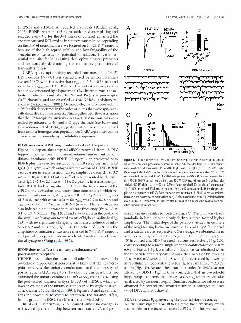
BDNF increases eIPSC amplitude and mIPSC frequencyFigure 1A depicts three typical eIPSCs recorded from 18 DIVhippocampal neurons that were maintained under control con-ditions, incubated with BDNF (15 ng/ml), or pretreated withBDNF plus the selective antibody for TrkB receptors, anti-TrkBIgG1 (10 �g/ml), which antagonizes the action of BDNF. BDNFcaused a net increase in mean eIPSC amplitude (from 2.1 to 3.7nA; n � 18; p � 0.01) that was effectively prevented by the anti-TrkB IgG1 (2.3 vs 2.1 nA; n � 18). Despite the increase in ampli-tude, BDNF had no significant effect on the time course of theeIPSCs, the activation and decay time constants of which re-mained nearly unchanged: �activ was 2.8 � 0.36 pA and �decay was61.3 � 8.4 ms with controls (n � 6); �activ was 2.9 � 0.38 pA and�decay was 57.6 � 7.3 ms with BDNF (n � 6). The neurotrophinalso induced a net increase in miniature frequency (from 1.0 �0.1 to 1.5 � 0.2 Hz) (Fig. 1B,C) and a weak shift in the profile ofthe amplitude histogram toward events of higher amplitude (Fig.1D), with no significant changes to the mean amplitude of mIP-SCs (25.2 and 27.3 pA) (Fig. 1E). The action of BDNF on theamplitude of miniatures was more marked in 7–14 DIV neuronsand probably depended on an accelerated maturation of func-tional synapses (Wang et al., 1995).
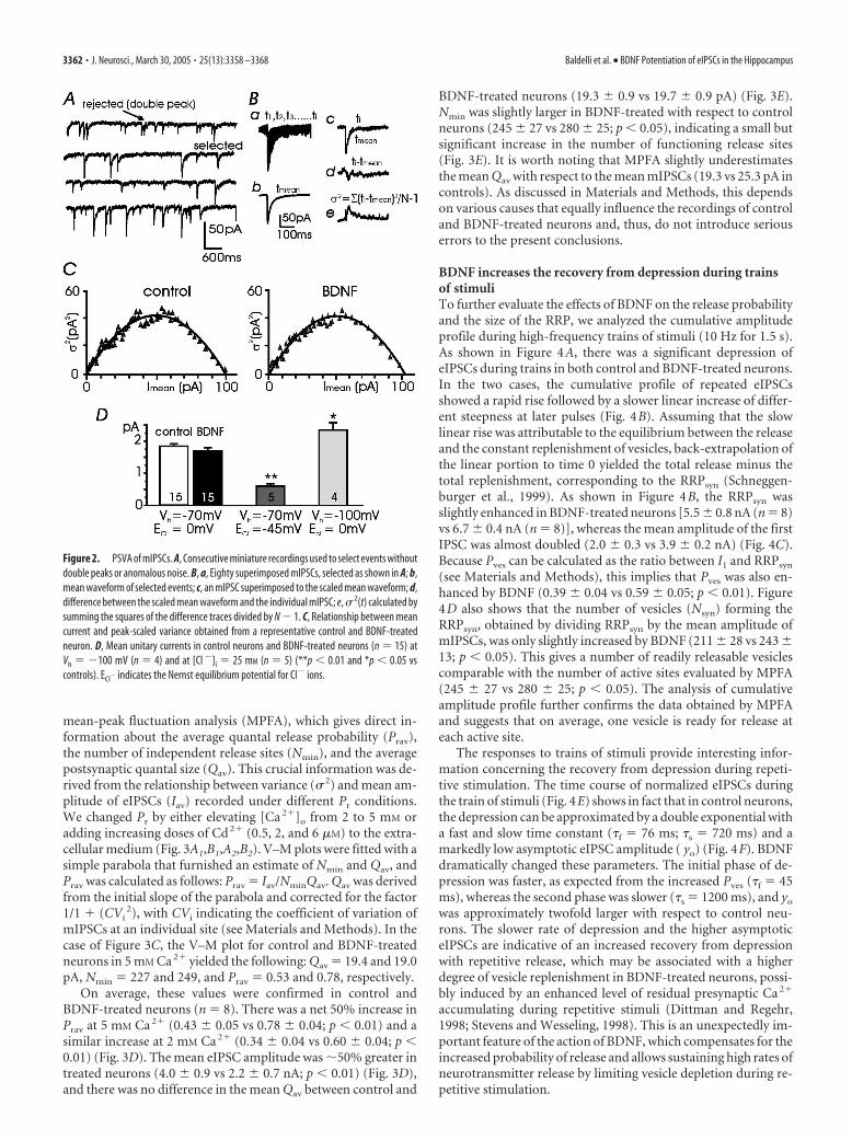
BDNF does not affect the unitary conductance ofpostsynaptic receptorsIf BDNF does not alter the mean amplitude of miniature events in3-week-old hippocampal neurons, it is likely that the neurotro-phin preserves the unitary conductance and the density ofpostsynaptic GABAA receptors. To examine this possibility, weestimated the unitary conductance of GABAA channels by usingthe peak-scaled variance analysis (PSVA) of mIPSCs, which al-lows an estimate of the unitary current carried by single postsyn-aptic channels (Traynelis et al., 1993). Figure 2, A and B, summa-rizes the procedure followed to determine the variance, � 2(t),from a group of mIPSCs (see Materials and Methods).
In 14 –21 DIV neurons, BDNF caused almost no changes in� 2(t), yielding a relationship between mean current, I, and peak-
scaled variance similar to controls (Fig. 2C). The plot was clearlyparabolic in both cases and only slightly skewed toward higheramplitudes. The initial slope of the parabola yielded an estimateof the weighted single channel current: 1.8 and 1.7 pA for controland treated neurons, respectively. On average, we obtained meanunitary currents, i, of 1.8 � 0.1 pA (n � 15) and 1.7 � 0.1 pA (n �15) in control and BDNF-treated neurons, respectively (Fig. 2D),corresponding to a mean single-channel conductance of 26.0 �0.9 and 24.6 � 1.3 pS. A similar conductance was obtained whenthe amplitude of unitary current was either increased by loweringVh to �100 mV (24.8 � 1.2 pS; n � 4) or decreased by loweringintracellular Cl� concentration ([Cl�]i) to 25 mM (23.0 � 2.0 pS;n � 5) (Fig. 2D). Because the mean amplitude of mIPSCs was notaltered by BDNF (Fig. 1E), we concluded that in 3-week-oldhippocampal neurons, the density of GABAA receptors was alsounaffected by the neurotrophin. Similar conductance values wereobtained for control and treated neurons in younger cultures(7–14 DIV; data not shown).
BDNF increases Pr , preserving the quantal size of vesiclesWe then investigated how BDNF altered the elementary eventsresponsible for the increased size of eIPSCs. For this, we used the
Figure 1. Effects of BDNF on eIPSC and mIPSC GABAergic currents recorded at the soma ofwhole-cell-clamped hippocampal neurons. A, Left, eIPSCs recorded from 14 –21 DIV neuronsunder control conditions, with BDNF and BDNF plus anti-TrkB IgG1 (Vh � �70 mV). Right,Mean amplitude of eIPSCs in the conditions and number of neurons indicated; **p � 0.01versus controls and anti-TrkB IgG1 plus BDNF using one-way ANOVA. B, Consecutive recordingsof mIPSCs in 20 DIV control neurons (left) and 20 DIV BDNF-treated neurons (4 d without plus16 d with BDNF) (right); Vh ��70 mV. C, Mean frequency of mIPSCs calculated from groups of14 –21 DIV control and BDNF-treated neurons. *p � 0.02 versus controls. D, Overlapped am-plitude distributions of mIPSCs from the same two neurons in B. BDNF causes a consistentincrease in the occurrence of events (filled bars). E, Mean amplitudes of mIPSCs calculated fromgroups of 14 –21 DIV control and BDNF-treated neurons (the number of neurons for each con-dition is indicated in each bar).
Baldelli et al. • BDNF Potentiation of eIPSCs in the Hippocampus J. Neurosci., March 30, 2005 • 25(13):3358 –3368 • 3361
mean-peak fluctuation analysis (MPFA), which gives direct in-formation about the average quantal release probability (Prav),the number of independent release sites (Nmin), and the averagepostsynaptic quantal size (Qav). This crucial information was de-rived from the relationship between variance (� 2) and mean am-plitude of eIPSCs (Iav) recorded under different Pr conditions.We changed Pr by either elevating [Ca 2�]o from 2 to 5 mM oradding increasing doses of Cd 2� (0.5, 2, and 6 �M) to the extra-cellular medium (Fig. 3A1,B1,A2,B2). V–M plots were fitted with asimple parabola that furnished an estimate of Nmin and Qav, andPrav was calculated as follows: Prav � Iav/NminQav. Qav was derivedfrom the initial slope of the parabola and corrected for the factor1/1 � (CVi
2), with CVi indicating the coefficient of variation ofmIPSCs at an individual site (see Materials and Methods). In thecase of Figure 3C, the V–M plot for control and BDNF-treatedneurons in 5 mM Ca 2� yielded the following: Qav � 19.4 and 19.0pA, Nmin � 227 and 249, and Prav � 0.53 and 0.78, respectively.
On average, these values were confirmed in control andBDNF-treated neurons (n � 8). There was a net 50% increase inPrav at 5 mM Ca 2� (0.43 � 0.05 vs 0.78 � 0.04; p � 0.01) and asimilar increase at 2 mM Ca 2� (0.34 � 0.04 vs 0.60 � 0.04; p �0.01) (Fig. 3D). The mean eIPSC amplitude was 50% greater intreated neurons (4.0 � 0.9 vs 2.2 � 0.7 nA; p � 0.01) (Fig. 3D),and there was no difference in the mean Qav between control and
BDNF-treated neurons (19.3 � 0.9 vs 19.7 � 0.9 pA) (Fig. 3E).Nmin was slightly larger in BDNF-treated with respect to controlneurons (245 � 27 vs 280 � 25; p � 0.05), indicating a small butsignificant increase in the number of functioning release sites(Fig. 3E). It is worth noting that MPFA slightly underestimatesthe mean Qav with respect to the mean mIPSCs (19.3 vs 25.3 pA incontrols). As discussed in Materials and Methods, this dependson various causes that equally influence the recordings of controland BDNF-treated neurons and, thus, do not introduce seriouserrors to the present conclusions.
BDNF increases the recovery from depression during trainsof stimuliTo further evaluate the effects of BDNF on the release probabilityand the size of the RRP, we analyzed the cumulative amplitudeprofile during high-frequency trains of stimuli (10 Hz for 1.5 s).As shown in Figure 4A, there was a significant depression ofeIPSCs during trains in both control and BDNF-treated neurons.In the two cases, the cumulative profile of repeated eIPSCsshowed a rapid rise followed by a slower linear increase of differ-ent steepness at later pulses (Fig. 4B). Assuming that the slowlinear rise was attributable to the equilibrium between the releaseand the constant replenishment of vesicles, back-extrapolation ofthe linear portion to time 0 yielded the total release minus thetotal replenishment, corresponding to the RRPsyn (Schneggen-burger et al., 1999). As shown in Figure 4B, the RRPsyn wasslightly enhanced in BDNF-treated neurons [5.5 � 0.8 nA (n � 8)vs 6.7 � 0.4 nA (n � 8)], whereas the mean amplitude of the firstIPSC was almost doubled (2.0 � 0.3 vs 3.9 � 0.2 nA) (Fig. 4C).Because Pves can be calculated as the ratio between I1 and RRPsyn
(see Materials and Methods), this implies that Pves was also en-hanced by BDNF (0.39 � 0.04 vs 0.59 � 0.05; p � 0.01). Figure4D also shows that the number of vesicles (Nsyn) forming theRRPsyn, obtained by dividing RRPsyn by the mean amplitude ofmIPSCs, was only slightly increased by BDNF (211 � 28 vs 243 �13; p � 0.05). This gives a number of readily releasable vesiclescomparable with the number of active sites evaluated by MPFA(245 � 27 vs 280 � 25; p � 0.05). The analysis of cumulativeamplitude profile further confirms the data obtained by MPFAand suggests that on average, one vesicle is ready for release ateach active site.
The responses to trains of stimuli provide interesting infor-mation concerning the recovery from depression during repeti-tive stimulation. The time course of normalized eIPSCs duringthe train of stimuli (Fig. 4E) shows in fact that in control neurons,the depression can be approximated by a double exponential witha fast and slow time constant (�f � 76 ms; �s � 720 ms) and amarkedly low asymptotic eIPSC amplitude ( yo) (Fig. 4F). BDNFdramatically changed these parameters. The initial phase of de-pression was faster, as expected from the increased Pves (�f � 45ms), whereas the second phase was slower (�s � 1200 ms), and yo
was approximately twofold larger with respect to control neu-rons. The slower rate of depression and the higher asymptoticeIPSCs are indicative of an increased recovery from depressionwith repetitive release, which may be associated with a higherdegree of vesicle replenishment in BDNF-treated neurons, possi-bly induced by an enhanced level of residual presynaptic Ca 2�
accumulating during repetitive stimuli (Dittman and Regehr,1998; Stevens and Wesseling, 1998). This is an unexpectedly im-portant feature of the action of BDNF, which compensates for theincreased probability of release and allows sustaining high rates ofneurotransmitter release by limiting vesicle depletion during re-petitive stimulation.
Figure 2. PSVA of mIPSCs. A, Consecutive miniature recordings used to select events withoutdouble peaks or anomalous noise. B, a, Eighty superimposed mIPSCs, selected as shown in A; b,mean waveform of selected events; c, an mIPSC superimposed to the scaled mean waveform; d,difference between the scaled mean waveform and the individual mIPSC; e, � 2(t) calculated bysumming the squares of the difference traces divided by N � 1. C, Relationship between meancurrent and peak-scaled variance obtained from a representative control and BDNF-treatedneuron. D, Mean unitary currents in control neurons and BDNF-treated neurons (n � 15) atVh � �100 mV (n � 4) and at [Cl �]i � 25 mM (n � 5) (**p � 0.01 and *p � 0.05 vscontrols). ECl� indicates the Nernst equilibrium potential for Cl� ions.
3362 • J. Neurosci., March 30, 2005 • 25(13):3358 –3368 Baldelli et al. • BDNF Potentiation of eIPSCs in the Hippocampus

BDNF preserves the Ca 2� dependence ofGABAergic transmissionAn increased Pr after BDNF exposure could underlie differentactions on presynaptic mechanisms. A possibility is that BDNFincreases the Ca 2� dependence of synaptic transmission by in-creasing the efficacy by which Ca 2� flowing through presynapticCa 2� channels stimulates vesicles fusion. To examine this, westudied the dose–response relationship of eIPSC amplitude ver-sus [Ca 2�]o in control and BDNF-treated neurons. In both cases,the eIPSCs increased steeply with [Ca 2�]o and saturated at �5mM (Fig. 5A1). BDNF mainly increased the size of eIPSCs withoutaltering the shape of the dose–response curve. In other words, theslope coefficient (Ca 2� cooperativity) and the dissociation con-stant (KD) remained unchanged (n � 3.3 vs 3.2; KD � 1.03 vs1.07 mM). As a consequence, the percentage increase in eIPSCsafter BDNF treatment remained constant at different levels of[Ca 2�]o, suggesting that potentiation of eIPSCs occurred re-gardless of the levels of [Ca 2�]o (Fig. 5A2).
Having shown that BDNF does not affect the Ca 2� depen-dence of eIPSCs, we then examined whether BDNF could affectthe Ca 2� dependence of PPD at different levels of [Ca 2�]o (seeMaterials and Methods). Under control conditions, with 2 mM
external Ca 2�, the PPD at a 50 ms interpulse interval was 0.49and decreased to 25 and 2% when [Ca 2�]o was lowered to 1and 0.5 mM, respectively (Fig. 5B1,B2). At higher levels of[Ca 2�]o, PPD increased moderately. As a result, the PPD of in-hibitory synapses in cultured hippocampal neurons was steeply
Ca 2� dependent and remarkably high at 2mM [Ca 2�]o, a hallmark of central syn-apses operating near maximal Pr underphysiological conditions. With respect tocontrols, BDNF-treated neurons dis-played higher PPDs at all [Ca 2�]o levelsbut similar Ca 2� dependence (Fig. 5B3).
Increased dominance of N- and P/Q-type channels on eIPSCs inBDNF-treated neuronsThe increase in Pr observed regardless ofthe levels of extracellular Ca 2� in BDNF-treated neurons may have different ori-gins. As already shown at the somatic level(Baldelli et al., 2000), it is possible thatBDNF acts by selectively increasing the ex-pression or changing the proportions ofpresynaptic Ca 2� channels coupled withexocytosis. To assay this possibility, we ex-amined how selective Ca 2� channel an-tagonists affect action potential-evokedIPSCs and PPD in control and BDNF-treated neurons. As shown below, previ-ous information on Ca 2� channel distri-bution derived from KCl-evoked IPSCs(Baldelli et al., 2002) furnishes a ratherdistorted view of presynaptic Ca 2� signal-ing involved in GABA release and thusshould be taken as mainly qualitative ofthe action of BDNF.
An initial series of experiments wasperformed by sequentially applying nifed-ipine (3 �M), �-Ctx-GVIA (1 �M), and�-Aga-IVA (0.5 �M) on the same neuron,to block L-, N-, and P/Q-type channels.
Figure 6A shows the responses of two representative neurons,maintained under control conditions (left) or exposed to BDNF(right). As shown, nifedipine had reversible weak blocking effectson the eIPSCs in both control and treated neurons (�3%),whereas �-Ctx-GVIA and �-Aga-IVA had irreversible blockingeffects that either preserved 15% of the eIPSCs under controlconditions or fully blocked the response in BDNF-treated neu-rons. Accounting for the small contribution of L-type channels,these data indicate that R-type channels contribute partially tosynaptic transmission in control neurons and are absent inBDNF-treated neurons. Interestingly, by inverting the applica-tion of the two toxins in control neurons, we obtained compara-ble blocks of the eIPSCs (total block between 80 and 85%; seebelow), suggesting that N- and P/Q-type channels control dis-tinct release sites in BDNF-untreated neurons and also that pre-synaptic Ca 2� channels are distributed in a “segregated” manner.
A second series of experiments was performed to evaluateprecisely the contribution of each Ca 2� channel type to synaptictransmission by applying separately the Ca 2� channel antago-nists on control and BDNF-treated neurons. As shown in Figure6B, under control conditions (open bars), nifedipine induced aweak inhibition (2.7%; n � 12), whereas �-Ctx-GVIA and�-Aga-IVA blocked 34.7% (n � 10) and 46.8% (n � 12) of theinitial synaptic current, respectively. A mean contribution of10.9% was estimated for the R-channel. In BDNF-treated neu-rons, the action of nifedipine remained nearly unchanged (2.8%;n � 11), whereas the average inhibition by �-Ctx-GVIA and
Figure 3. Multiprobability variance analysis used to estimate quantal parameters. A1, A2, Superimposed eIPSCs (8 traces foreach condition) recorded in two representative control and BDNF-treated neurons at 0.1 Hz. Changing [Ca 2�] (2–5 mM) orincreasing Cd 2� (0.5, 2, and 6 �M) altered Pr. B1, B2, eIPSC amplitude versus time from the two representative neurons. C1, C2, Thevariance of the eIPSC amplitude is plotted against the mean for each epoch and fitted with a parabola to estimate Qav and Nmin. D,Mean IPSC and mean Prav in control and BDNF-treated neurons (**p � 0.01 vs controls; n � 8). Prav was calculated at 2 and 5 mM
[Ca 2�]o. E, Average quantal size (Qav) and number of release sites (Nmin) in control and BDNF-treated neurons (*p � 0.05 vscontrols; n � 8). Qav was obtained from the initial slope of the parabola corrected for 1/1 � (CVi
2) (see Materials and Methods).
Baldelli et al. • BDNF Potentiation of eIPSCs in the Hippocampus J. Neurosci., March 30, 2005 • 25(13):3358 –3368 • 3363

�-Aga-IVA increased to 49.4% (n � 10;p � 0.01) and 66.5% (n � 11; p � 0.01),respectively, with a net increase of 42% inboth cases. The total block of N- and P/Q-type channels increased from 81.5 to116.9%, and R-type channels no longercontributed to the eIPSCs. We concludethat in BDNF-treated neurons, N- andP/Q-type channels assumed full control ofsynaptic inhibitory transmission and thattogether they control �100% the size ofeIPSCs (supra-additivity). This is strongevidence that in BDNF-treated neurons, apercentage of release sites are controlledby mixed populations of N- and P/Q-typechannels that are distributed in a nonuni-form manner. Notice that supra-additivityand the switching from segregated to non-uniform distribution of presynaptic Ca2�
channels with BDNF were totally over-looked in a previous work using KCl-evokedIPSCs (Baldelli et al., 2002). In this case, the1 s KCl stimulation overestimated the con-tribution of distal R-type channels and di-minished that of N- and P/Q-type channelsmore proximal to the release sites. In partic-ular, P/Q-type channels appeared to con-tribute less than R- and N-type channels un-der control conditions, whereas with actionpotential stimulation, their contribution isalways maximal with and without BDNF(Fig. 6B).
BDNF increases the control of PPDassociated with N-type channelsTo gain a deeper insight into the action ofBDNF, we next examined how N- andP/Q-type channels specifically affect PPDand recovery from depression. We foundthat in control neurons, PPD had large ini-tial values (65.7% at t � 20 ms) (Fig.7A,C), and recovery from depression waswell approximated by a single exponentialwith the time constant �rec � 85.6 ms (Fig.7A,E, �). PPD and recovery from depres-sion remained almost unaltered whenP/Q-type channels were mainly responsi-ble for the eIPSCs (�-Ctx-GVIA applied).PPD was 59.2% at t � 20 ms (Fig. 7A,C)and �rec � 98.9 ms (Fig. 7A,F, ‚). On thecontrary, the PPD markedly decreased(33.3% at t � 20 ms) (Fig. 7A,C) and the�rec was shortened (�rec � 49.5 ms) (Fig.7A,G) when N-type channels were mainlyresponsible of neurotransmission (�-Aga-IVA applied). This suggests distinctroles of N- and P/Q-type channels in con-trolling GABA release in control neurons. P/Q-type channelsdominate the control of PPD and determine the slow recovery ofdepression, which is related to vesicle replenishment and dependson residual Ca 2� at the presynaptic terminals (Dittman and Re-gehr, 1998). N-type channels control PPD less effectively but have
marked effects on the recovery of PPD, as proven by the fasterrate of recovery supported by these channels.
BDNF had a marked effect on the contribution of N- andP/Q-type channels to PPD. PPD markedly increased with BDNF(85.9% at t � 20 ms) (Fig. 7B,D). Recovery from depression
Figure 4. Estimate of the RRPsyn and Pves through cumulative amplitude analysis. A, eIPSCs recorded during a train of repetitivestimuli of 10 Hz in a control (left) and a BDNF-treated (right) neuron. B, Cumulative eIPSC amplitude during 10 Hz trains in eightcontrol and eight treated neurons. Data points in the range of 0.8 –1.4 s were fitted by linear regression and back-extrapolated totime 0 to estimate the cumulative eIPSC amplitudes before steady-state depression (RRPsyn). C, The mean amplitude of the firsteIPSC (I1) is nearly doubled in treated neurons (filled bars; n � 8) versus control neurons (open bars; n � 8) (**p � 0.01 vscontrols), whereas the mean RRPsyn is only slightly increased (*p � 0.05 vs controls). D, Mean Pves (**p � 0.01 vs controls) andmean number of vesicles forming the RRPsyn (*p � 0.05 vs controls). E, Plot of eIPSC amplitude versus time during repetitivestimulation fitted with a double-exponential function: I(t) � yo � Af e(�t/�f) � As e(�t/�s). F, Mean values of the parametersobtained by the double-exponential fit of E (n � 8; ** p � 0.01 vs controls).
Figure 5. Ca 2� dependence of GABAergic transmission in control and treated neurons. A1, Mean eIPSC versus [Ca 2�]o fittedwith a Hill equation: I � Imax [Ca 2�]n/([Ca 2�]n � KD ). BDNF mainly increased the size of eIPSCs (Imax ) without altering the slopecoefficient ( n) and KD (n � 3.3 vs 3.2; KD � 1.03 vs 1.07 mM; Imax � 2.6 vs 4.4 nA) (6 � n � 32). A2, Percentage increase in eIPSCsafter BDNF treatment remained constant at different [Ca 2�]o levels. Data were obtained from A1. B1, Response to paired-pulsestimulation (50 ms time interval) at increasing [Ca 2�]o levels in control and treated neurons. Each trace represents the average ofsix consecutive recordings (stimulation frequency, 0.066 Hz). B2, Mean PPD versus [Ca 2�]o in control and treated neurons (6 �n � 10). B3, Percentage increase in PPD in the presence of BDNF did not change between 2 and 10 mM [Ca 2�]o. Data wereobtained from B2.
3364 • J. Neurosci., March 30, 2005 • 25(13):3358 –3368 Baldelli et al. • BDNF Potentiation of eIPSCs in the Hippocampus
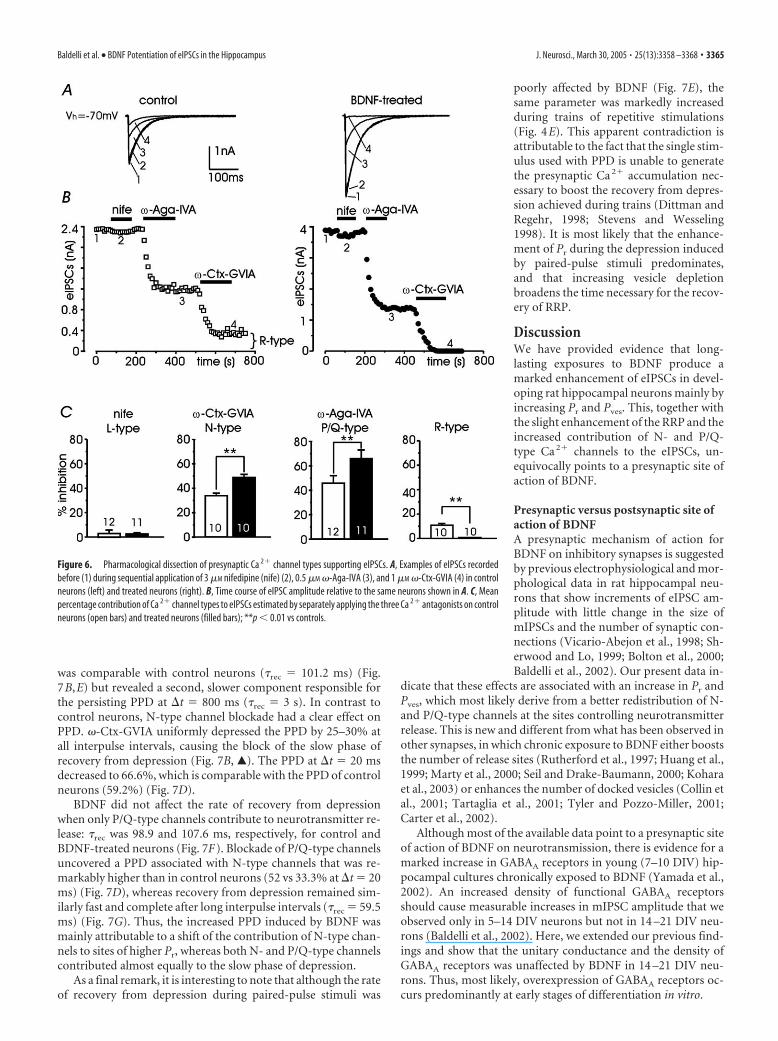
was comparable with control neurons (�rec � 101.2 ms) (Fig.7B,E) but revealed a second, slower component responsible forthe persisting PPD at t � 800 ms (�rec � 3 s). In contrast tocontrol neurons, N-type channel blockade had a clear effect onPPD. �-Ctx-GVIA uniformly depressed the PPD by 25–30% atall interpulse intervals, causing the block of the slow phase ofrecovery from depression (Fig. 7B, Œ). The PPD at t � 20 msdecreased to 66.6%, which is comparable with the PPD of controlneurons (59.2%) (Fig. 7D).
BDNF did not affect the rate of recovery from depressionwhen only P/Q-type channels contribute to neurotransmitter re-lease: �rec was 98.9 and 107.6 ms, respectively, for control andBDNF-treated neurons (Fig. 7F). Blockade of P/Q-type channelsuncovered a PPD associated with N-type channels that was re-markably higher than in control neurons (52 vs 33.3% at t � 20ms) (Fig. 7D), whereas recovery from depression remained sim-ilarly fast and complete after long interpulse intervals (�rec � 59.5ms) (Fig. 7G). Thus, the increased PPD induced by BDNF wasmainly attributable to a shift of the contribution of N-type chan-nels to sites of higher Pr, whereas both N- and P/Q-type channelscontributed almost equally to the slow phase of depression.
As a final remark, it is interesting to note that although the rateof recovery from depression during paired-pulse stimuli was
poorly affected by BDNF (Fig. 7E), thesame parameter was markedly increasedduring trains of repetitive stimulations(Fig. 4E). This apparent contradiction isattributable to the fact that the single stim-ulus used with PPD is unable to generatethe presynaptic Ca 2� accumulation nec-essary to boost the recovery from depres-sion achieved during trains (Dittman andRegehr, 1998; Stevens and Wesseling1998). It is most likely that the enhance-ment of Pr during the depression inducedby paired-pulse stimuli predominates,and that increasing vesicle depletionbroadens the time necessary for the recov-ery of RRP.
DiscussionWe have provided evidence that long-lasting exposures to BDNF produce amarked enhancement of eIPSCs in devel-oping rat hippocampal neurons mainly byincreasing Pr and Pves. This, together withthe slight enhancement of the RRP and theincreased contribution of N- and P/Q-type Ca 2� channels to the eIPSCs, un-equivocally points to a presynaptic site ofaction of BDNF.
Presynaptic versus postsynaptic site ofaction of BDNFA presynaptic mechanism of action forBDNF on inhibitory synapses is suggestedby previous electrophysiological and mor-phological data in rat hippocampal neu-rons that show increments of eIPSC am-plitude with little change in the size ofmIPSCs and the number of synaptic con-nections (Vicario-Abejon et al., 1998; Sh-erwood and Lo, 1999; Bolton et al., 2000;Baldelli et al., 2002). Our present data in-
dicate that these effects are associated with an increase in Pr andPves, which most likely derive from a better redistribution of N-and P/Q-type channels at the sites controlling neurotransmitterrelease. This is new and different from what has been observed inother synapses, in which chronic exposure to BDNF either booststhe number of release sites (Rutherford et al., 1997; Huang et al.,1999; Marty et al., 2000; Seil and Drake-Baumann, 2000; Koharaet al., 2003) or enhances the number of docked vesicles (Collin etal., 2001; Tartaglia et al., 2001; Tyler and Pozzo-Miller, 2001;Carter et al., 2002).
Although most of the available data point to a presynaptic siteof action of BDNF on neurotransmission, there is evidence for amarked increase in GABAA receptors in young (7–10 DIV) hip-pocampal cultures chronically exposed to BDNF (Yamada et al.,2002). An increased density of functional GABAA receptorsshould cause measurable increases in mIPSC amplitude that weobserved only in 5–14 DIV neurons but not in 14 –21 DIV neu-rons (Baldelli et al., 2002). Here, we extended our previous find-ings and show that the unitary conductance and the density ofGABAA receptors was unaffected by BDNF in 14 –21 DIV neu-rons. Thus, most likely, overexpression of GABAA receptors oc-curs predominantly at early stages of differentiation in vitro.
Figure 6. Pharmacological dissection of presynaptic Ca 2� channel types supporting eIPSCs. A, Examples of eIPSCs recordedbefore (1) during sequential application of 3 �M nifedipine (nife) (2), 0.5 �M �-Aga-IVA (3), and 1 �M �-Ctx-GVIA (4) in controlneurons (left) and treated neurons (right). B, Time course of eIPSC amplitude relative to the same neurons shown in A. C, Meanpercentage contribution of Ca 2� channel types to eIPSCs estimated by separately applying the three Ca 2� antagonists on controlneurons (open bars) and treated neurons (filled bars); **p � 0.01 vs controls.
Baldelli et al. • BDNF Potentiation of eIPSCs in the Hippocampus J. Neurosci., March 30, 2005 • 25(13):3358 –3368 • 3365

Higher Pr and recovery fromdepression: a strategy to enhancepresynaptic efficiencyBDNF induces a marked increase in Pves,but the size of the RRP is only slightly aug-mented (Fig. 4). This conclusion is inagreement with the observation thatBDNF does not affect the size of the recy-cling pool (Yamada et al., 2002) and pos-sibly of the RRP as well (Murthy andStevens, 1999). An action on Pves preserv-ing the RRP is the opposite of what occursin excitatory synapses, in which long-termexposures to BDNF enhance presynapticactivity by increasing the RRP (Collin etal., 2001; Tartaglia et al., 2001; Tyler andPozzo-Miller, 2001; Carter et al., 2002).Thus, BDNF follows different strategies toenhance the rate of vesicle release in exci-tatory and inhibitory synapses, increasingeither the RRP or Pves.
An increased Pr (or Pves) might haveserious drawbacks during high-frequencystimulation in synapses that function at ahigh rate of release and saturating Ca 2�
conditions, as in our case. Control eIPSCsundergo rapid depression during trains ofstimuli (Fig. 4E), which would be wors-ened after a 50% increase in Pr. However,BDNF is able to slow down the rate of de-pression, maintaining high eIPSC ampli-tudes during trains. The twofold lowerrate of depression observed with BDNF is possibly associatedwith an increased rate of vesicle replenishment attributable to anenhanced level of residual Ca 2� accumulating presynapticallyduring repetitive stimuli (Dittman and Regehr, 1998; Stevens andWesseling, 1998). This new unexpected finding reinforces thepresynaptic nature of the action of BDNF and can be associatedwith the BDNF-induced supra-additivity of presynaptic Ca 2�
channels in controlling eIPSCs. BDNF may increase the amountof presynaptic terminals in which N- and P/Q-type channels arecoexpressed and cooperate to control vesicle release, enhancingthe possibility of intraterminal Ca 2� accumulation during high-frequency stimulation. Indeed, other presynaptic mechanismscould account for the higher rate of vesicle recycling induced bythe neurotrophin. BDNF activation of TrkB receptors and in-creased intraterminal Ca 2� concentration activate mitogen-activated protein (MAP)-kinase and calmodulin kinase II, whichphosphorylate synapsins at distinct sites (Benfenati et al., 1992;Jovanovic et al., 1996) and increase vesicle mobilization from thereserve to the RRP (Greengard et al., 1993). During high-frequency stimulation, the level of synapsin phosphorylationstrictly correlates with synapsin dispersion and with the amountof vesicles sustaining repetitive release (Chi et al., 2001). Thus,BDNF could increase the rate of vesicle replenishment throughdirect (TrkB/MAP-kinase pathway) or indirect (Ca 2�/calmodu-lin kinase II pathway) synapsin phosphorylation, promoting ves-icle mobilization from the reserve pool.
Presynaptic N- and P/Q-type channels as targets of the actionof BDNFOur data show clearly that BDNF changes both the distributionand contribution of N- and P/Q-type channels controlling the
eIPSCs. The two channels control distinct release sites in BDNF-untreated neurons (segregated distribution), and their contribu-tion is limited to 82% of the total eIPSCs. With BDNF, this per-centage increases to 117%, increasing the percentage of releasesites containing mixtures of the two channels (nonuniform dis-tribution). This could represent a new form of synapse matura-tion in developing neurons induced by BDNF.
An appropriate colocalization of presynaptic Ca 2� channelsat the vesicle release sites, rather than a generalized increase inCa 2� channel density as the cause of the higher Pr with BDNF, isalso supported by two other findings. First, neurotransmission isnear saturation under control conditions, and Pr increases verylittle at �2 mM Ca 2. Because saturation of Ca 2� fluxes throughopen Ca 2� channels occurs at �50 mM Ca 2� (Hess et al., 1986),it is evident that local [Ca 2�] at the nanodomains formed byCa 2� channels and docked vesicles (Augustine, 2001) is alreadymaximal during control eIPSCs in 2 mM Ca 2�. Additional eleva-tions of [Ca 2�]o or increased densities of distantly located Ca 2�
channels would produce only minor changes. It is worth noting thatsaturation of eIPSCs occurs at values significantly smaller than thesize of the RRPsyn (2.6 vs 5.5 nA in controls and 4.4 vs 6.7 nA withBDNF) and, thus, the number of available vesicles and Ca2� entryare not limiting factors for the increase in Pr with BDNF. Most likely,the main limitation to Pr is the quantity of Ca2� ions effectivelyentering the terminal through presynaptic Ca2� channels, whichmay derive from a better colocalization of Ca2� channels to therelease active zones (Spafford and Zamponi, 2003). Second, BDNFenhances the expression of syntaxin 1 and synaptotagmin 1 (Wanget al., 1995; Vicario-Abejon et al. 1998; Tartaglia et al. 2001; Yamadaet al. 2002). The two proteins bind selectively to the “synprint” re-gion of N- and P/Q-type channels but not to R-type channels, which
Figure 7. Pharmacological dissection of presynaptic Ca 2� channel types supporting PPD and recovery from depression. A,Percentage of PPD vs time in control neurons in the absence of toxins and in the presence of 1 �M �-Ctx-GVIA and 0.5 �M
�-Aga-IVA. Interevent intervals ranged between 20 and 800 ms. Solid curves are monoexponential fits, with initial amplitude and�rec given in Results (for each point, 9 � n � 16). B, Same as in A but in the presence of BDNF (9 � n � 15). C, Mean percentageof PPD calculated at t � 20 ms in control neurons under the indicated condition (**p � 0.01; 9 � n � 16). D, Same as in C butin the presence of BDNF (filled bars; 9 � n � 15). The open bars are taken from C, and statistical comparisons (**p � 0.01) are asindicated. E–G, Monoexponential fit of the percentage of PPD versus time taken from A and B for control neurons (solid lines) andtreated neurons (dashed lines) in the absence of toxins (E) and in the presence of �-Ctx-GVIA (F ) and �-Aga-IVA (G).
3366 • J. Neurosci., March 30, 2005 • 25(13):3358 –3368 Baldelli et al. • BDNF Potentiation of eIPSCs in the Hippocampus

lack the binding sequence (Catterall, 2000). Thus, an increased ex-pression of vesicle-binding proteins would favor the colocalizationof N- and P/Q-type channels rather than R-type channels, increasingthe Pr.
A final consideration concerns the role that N- and P/Q-typechannels play in the control of PPD and recovery from depres-sion. As for other neurons (Mintz et al., 1995; Reid et al., 1997,2003; Wilson et al., 2001), we observed nonuniform reduction ofPr after blockade of either N- or P/Q-type channels. In our case,P/Q-type channels appear to be strongly coupled with the releasesites, and BDNF does not change the already high PPD associatedwith these channels (Fig. 7F). The increased contribution of P/Q-type channels to the size of eIPSCs may simply derive from anincreased nonuniform distribution of these channels in terminalspurely controlled by the N-type.
A relevant issue of the present study is that BDNF seems par-ticularly effective in increasing the contribution of N-type chan-nels to PPD, which is rather low in control neurons. The apparentcontradiction that N-type channels contribute to the eIPSCs(35% block) and little to the PPD is attributable to the fact thatPPD does not simply depend on vesicle depletion (Pr) but also onvesicle replenishment, which is Ca 2� dependent and determinesthe rate of recovery from depression (Dittman and Regehr, 1998).The inability of �-Ctx-GVIA to affect PPD suggests that vesicledepletion induced by Ca 2� influx through N-type channels iswell counterbalanced by an effective Ca 2�-dependent recoveryfrom depression by the Ca 2� ions flowing through the samechannel types. Notice that recovery from depression is particu-larly fast when N-types are the only channels supporting GABArelease and BDNF does not affect this feature (Fig. 7G). This isline with recent observations on two species of mature inhibitorysynapses possessing distinct populations of presynaptic Ca 2�
channels (Poncer et al., 2000). The striatum oriens interneuronsexpressing only P/Q-type channels show marked PPD at all in-terpulse intervals, whereas the striatum radiatum interneurons,possessing only N-type channels, display facilitation at short in-tervals and weak PPD at longer intervals.
In conclusion, we have brought new evidence in favor of anovel action of BDNF on presynaptic Ca 2� channels that leads toa sharp increase in Pr and accounts for most of the increase ininhibitory postsynaptic signaling in developing hippocampalneurons. Maturation and stabilization of GABAergic synapsesare expected to play a key role in balancing the activity of excita-tory synapses during neuronal network formation (Vicario-Abejon et al., 2002). Our data suggest the possibility that a bettercolocalization of presynaptic Ca 2� channels with the vesicle fu-sion machinery may be a common mechanism adopted by inhib-itory synapses to increase their strength in the hippocampus andother brain regions with chronic exposure to BDNF.
ReferencesAugustine GJ (2001) How does calcium trigger neurotransmitter release?
Curr Opin Neurobiol 11:320 –326.Baldelli P, Forni PE, Carbone E (2000) BDNF, NT-3 and NGF induce dis-
tinct new Ca 2� channel synthesis in developing hippocampal neurons.Eur J Neurosci 12:4017– 4032.
Baldelli P, Novara M, Carabelli V, Hernandez-Guijo JM, Carbone E (2002)BDNF up-regulates evoked GABAergic transmission in developing hip-pocampal neurons by potentiating presynaptic N- and P/Q-type Ca 2�
channel signaling. Eur J Neurosci 16:2297–2310.Balkowiec A, Katz DM (2000) Activity-dependent release of endogenous
brain-derived neurotrophic factor from primary sensory neurons de-tected by ELISA in situ. J Neurosci 20:7417–7423.
Benfenati F, Valtorta F, Rubenstein JL, Gorelick FS, Greengard P, Czernik AJ
(1992) Synaptic vesicle-associated Ca 2�/calmodulin-dependent proteinkinase II is a binding protein for synapsin I. Nature 359:417– 420.
Betz WJ (1970) Depression of transmitter release at the neuromuscularjunction of the frog. J Physiol (Lond) 206:629 – 644.
Bolton MM, Pittman AJ, Lo DC (2000) Brain-derived neurotrophic factordifferentially regulates excitatory and inhibitory synaptic transmission inhippocampal cultures. J Neurosci 20:3221–3232.
Carter AR, Chen C, Schwartz PM, Segal RA (2002) Brain-derived neurotro-phic factor modulates cerebellar plasticity and synaptic ultrastructure.J Neurosci 22:1316 –1327.
Catterall WA (2000) Structure and regulation of voltage-gated Ca2� chan-nels. Annu Rev Cell Dev Biol 16:521–555.
Chi P, Greengard P, Ryan TA (2001) Synapsin dispersion and reclusteringduring synaptic activity. Nat Neurosci 4:1187–1193.
Clements JD, Silver RA (2000) Unveiling synaptic plasticity: a new graphicaland analytical approach. Trends Neurosci 23:105–113.
Collin C, Vicario-Abejon C, Rubio ME, Wenthold RJ, McKay RD, Segal M(2001) Neurotrophins act at presynaptic terminals to activate synapsesamong cultured hippocampal neurons. Eur J Neurosci 13:1273–1282.
De Koninck Y, Mody I (1994) Noise analysis of miniature IPSCs in adult ratbrain slices: properties and modulation of synaptic GABAA receptorchannels. J Neurophysiol 71:1318 –1335.
Dittman JS, Regehr WG (1998) Calcium dependence and recovery kineticsof presynaptic depression at the climbing fiber to Purkinje cell synapse.J Neurosci 18:6147– 6162.
Greengard P, Valtorta F, Czernik AJ, Benfenati F (1993) Synaptic vesiclephosphoproteins and regulation of synaptic function. Science259:780 –785.
Hess P, Lansman JB, Tsien RW (1986) Calcium channel selectivity for diva-lent and monovalent cations: voltage and concentration dependence ofsingle channel current in ventricular heart cells. J Gen Physiol 88:293–319.
Huang ZJ, Kirkwood A, Pizzorusso T, Porciatti V, Morales B, Bear MF, MaffeiL, Tonegawa S (1999) BDNF regulates the maturation of inhibition andthe critical period of plasticity in mouse visual cortex. Cell 98:739 –755.
Jovanovic JN, Benfenati F, Siow YL, Sihra TS, Sanghera JS, Pelech SL, Green-gard P, Czernik AJ (1996) Neurotrophins stimulate phosphorylation ofsynapsin I by MAP kinase and regulate synapsin I–actin interactions. ProcNatl Acad Sci USA 93:3679 –3683.
Kohara K, Kitamura A, Adachi N, Nishida M, Itami C, Nakamura S, TsumotoT (2003) Inhibitory but not excitatory cortical neurons require presyn-aptic brain-derived neurotrophic factor for dendritic development, asrevealed by chimera cell culture. J Neurosci 23:6123– 6131.
Lessmann V, Gottmann K, Malcangio M (2003) Neurotrophin secretion:current facts and future prospects. Prog Neurobiol 69:341–374.
Magnelli V, Baldelli P, Carbone E (1998) Antagonist-resistant calcium cur-rents in rat embryo motoneurons. Eur J Neurosci 10:1810 –1825.
Marty S, Wehrle R, Sotelo C (2000) Neuronal activity and brain-derivedneurotrophic factor regulate the density of inhibitory synapses in organo-typic slice cultures of postnatal hippocampus. J Neurosci 20:8087– 8095.
Mintz IM, Sabatini BL, Regehr WG (1995) Calcium control of transmitterrelease at a cerebellar synapse. Neuron 15:675– 688.
Murthy VN, Stevens CF (1999) Reversal of synaptic vesicle docking at cen-tral synapses. Nat Neurosci 2:503–507.
Nusser Z, Cull-Candy S, Farrant M (1997) Differences in synaptic GABAA
receptor number underlie variation in GABA mini amplitude. Neuron19:697–709.
Ohno-Shosaku T, Kazunari H, Sawada A, Yamamoto C (1994) Contribu-tions of multiple calcium channel types to GABAergic transmission in ratcultured hippocampal neurons. Neurosci Lett 181:145–148.
Oleskevich S, Clements J, Walmsley B (2000) Release probability modulatesshort-term plasticity at a rat giant terminal. J Physiol (Lond) 524:513–523.
Poncer JC, McKinney RA, Gahwiler BH, Thompson SM (2000) Differentialcontrol of GABA release at synapses from distinct interneurons in rathippocampus. J Physiol (Lond) 528:123–130.
Poo MM (2001) Neurotrophins as synaptic modulators. Nat Rev Neurosci2:24 –32.
Reid CA, Clements JD (1999) Postsynaptic expression of long-term poten-tiation in the rat dentate gyrus demonstrated by variance-mean analysis.J Physiol (Lond) 518:121–130.
Reid CA, Clements JD, Bekkers JM (1997) Nonuniform distribution ofCa 2� channel subtypes on presynaptic terminals of excitatory synapses inhippocampus cultures. J Neurosci 17:2738 –2745.
Baldelli et al. • BDNF Potentiation of eIPSCs in the Hippocampus J. Neurosci., March 30, 2005 • 25(13):3358 –3368 • 3367
Reid CA, Bekkers JM, Clements JD (2003) Presynaptic Ca 2� channels: afunctional patchwork. Trends Neurosci 26:683– 687.
Rutherford LC, DeWan A, Lauer HM, Turrigiano GG (1997) Brain-derivedneurotrophic factor mediates the activity-dependent regulation of inhi-bition in neocortical cultures. J Neurosci 17:4527– 4535.
Schneggenburger R, Meyer AC, Neher E (1999) Released fraction and totalsize of a pool of immediately available transmitter quanta at a calyx syn-apse. Neuron 23:399 – 409.
Schneggenburger R, Sakaba T, Neher E (2002) Vesicle pools and short-termsynaptic depression: lessons from a large synapse. Trends Neurosci25:206 –212.
Seil FJ, Drake-Baumann R (2000) TrkB receptor ligands promote activity-dependent inhibitory synaptogenesis. J Neurosci 20:5367–5373.
Sherwood NT, Lo DC (1999) Long-term enhancement of central synaptictransmission by chronic brain-derived neurotrophic factor treatment.J Neurosci 19:7025–7036.
Silver RA, Momiyama A, Cull-Candy SG (1998) Locus of frequency-dependent depression identified with multiple-probability fluctuationanalysis at rat climbing fibre–Purkinje cell synapses. J Physiol (Lond)510:881–902.
Spafford JD, Zamponi GW (2003) Functional interactions between presyn-aptic calcium channels and the neurotransmitter release machinery. CurrOpin Neurobiol 13:308 –314.
Stevens CF, Wesseling JF (1998) Activity-dependent modulation of the rateat which synaptic vesicles become available to undergo exocytosis. J Neu-rosci 21:415– 424.
Tartaglia N, Du J, Tyler WJ, Neale E, Pozzo-Miller L, Lu B (2001) Proteinsynthesis-dependent and -independent regulation of hippocampal syn-apses by brain-derived neurotrophic factor. J Biol Chem276:37585–37593.
Traynelis SF, Jaramillo F (1998) Getting the most out of noise in the centralnervous system. Trends Neurosci 21:137–145.
Traynelis SF, Silver RA, Cull-Candy SG (1993) Estimated conductance ofglutamate receptor channels activated during EPSCs at the cerebellarmossy fiber– granule cell synapse. Neuron 11:279 –289.
Tyler WJ, Pozzo-Miller LD (2001) BDNF enhances quantal neurotransmit-ter release and increases the number of docked vesicles at the active zonesof hippocampal excitatory synapses. J Neurosci 21:4249 – 4258.
Vicario-Abejon C, Collin C, McKay RD, Segal M (1998) Neurotrophins in-duce formation of functional excitatory and inhibitory synapses betweencultured hippocampal neurons. J Neurosci 18:7256 –7271.
Vicario-Abejon C, Owens D, McKay R, Segal M (2002) Role of neurotro-phins in central synapse formation and stabilization. Nat Rev Neurosci3:965–974.
Wang T, Xie K, Lu B (1995) Neurotrophins promote maturation of devel-oping neuromuscular synapses. J Neurosci 15:4796 – 4805.
Wilson RI, Kunos G, Nicoll RA (2001) Presynaptic specificity of endocan-nabinoid signaling in the hippocampus. Neuron 31:453– 462.
Yamada MK, Nakanishi K, Ohba S, Nakamura T, Ikegaya Y, Nishiyama N,Matsuki N (2002) Brain-derived neurotrophic factor promotes the mat-uration of GABAergic mechanisms in cultured hippocampal neurons.J Neurosci 22:7580 –7585.
3368 • J. Neurosci., March 30, 2005 • 25(13):3358 –3368 Baldelli et al. • BDNF Potentiation of eIPSCs in the Hippocampus