Creatine as a compatible osmolyte in muscle cells exposed to hypertonic stress

http://asn.sagepub.com/ASN Neuro
http://asn.sagepub.com/content/6/6/1759091414554945The online version of this article can be found at:
DOI: 10.1177/1759091414554945
2014 6: ASN NeuroMarcelo Farina, Ana Lúcia S. Rodrigues and Manuela G. López
Mauricio Peña Cunha, Maria D. Martín-de-Saavedra, Alejandro Romero, Javier Egea, Fabiana K. Ludka, Carla I. Tasca, Parkinson's ModelIn Vitro
Both Creatine and Its Product Phosphocreatine Reduce Oxidative Stress and Afford Neuroprotection in an
Published by:
http://www.sagepublications.com
On behalf of:
American Society for Neurochemistry
can be found at:ASN NeuroAdditional services and information for
http://asn.sagepub.com/cgi/alertsEmail Alerts:
http://asn.sagepub.com/subscriptionsSubscriptions:
http://www.sagepub.com/journalsReprints.navReprints:
http://www.sagepub.com/journalsPermissions.navPermissions:
What is This?
- Nov 24, 2014Version of Record >>
by guest on November 30, 2014asn.sagepub.comDownloaded from by guest on November 30, 2014asn.sagepub.comDownloaded from

XML Template (2014) [16.10.2014–6:13pm] [1–16]//blrnas3.glyph.com/cenpro/ApplicationFiles/Journals/SAGE/3B2/ASNJ/Vol00000/140011/APPFile/SG-ASNJ140011.3d (ASN) [PREPRINTER stage]
Original Article
Both Creatine and Its ProductPhosphocreatine Reduce OxidativeStress and Afford Neuroprotectionin an In Vitro Parkinson’s Model
Mauricio Pena Cunha1,2,3, Maria D. Martın-de-Saavedra1,2,4,Alejandro Romero1,2,5, Javier Egea1,2,6, Fabiana K. Ludka3,7,Carla I. Tasca1, Marcelo Farina3, Ana Lucia S. Rodrigues3, andManuela G. Lopez1,2
Abstract
Creatine is the substrate for creatine kinase in the synthesis of phosphocreatine (PCr). This energetic system is endowed of
antioxidant and neuroprotective properties and plays a pivotal role in brain energy homeostasis. The purpose of this study
was to investigate the neuroprotective effect of creatine and PCr against 6-hydroxydopamine (6-OHDA)-induced mitochon-
drial dysfunction and cell death in rat striatal slices, used as an in vitro Parkinson’s model. The possible involvement of the
signaling pathway mediated by phosphatidylinositol-3 kinase (PI3K), protein kinase B (Akt), and glycogen synthase kinase-3b(GSK3b) was also evaluated. Exposure of striatal slices to 6-OHDA caused a significant disruption of the cellular homeostasis
measured as 3-(4,5 dimethylthiazol-2-yl)-2,5-diphenyl-tetrazolium bromide reduction, lactate dehydrogenase release, and
tyrosine hydroxylase levels. 6-OHDA exposure increased the levels of reactive oxygen species and thiobarbituric acid
reactive substances production and decreased mitochondrial membrane potential in rat striatal slices. Furthermore,
6-OHDA decreased the phosphorylation of Akt (Serine473) and GSK3b (Serine9). Coincubation with 6-OHDA and creatine
or PCr reduced the effects of 6-OHDA toxicity. The protective effect afforded by creatine or PCr against 6-OHDA-induced
toxicity was reversed by the PI3K inhibitor LY294002. In conclusion, creatine and PCr minimize oxidative stress in striatum
to afford neuroprotection of dopaminergic neurons.
Keywords
6-OHDA, creatine, phosphocreatine, PI3K, neuroprotective, oxidative stress
Introduction
Parkinson’s disease (PD) is the second most commonneurodegenerative disease associated to aging (LoBianco et al., 2004). Animal and cell models used tostudy the pathological mechanisms underlying neurode-generation in PD often involve the administration oftoxins that selectively destroy or interrupt the activityof nigrostriatal dopaminergic neurons. These modelsinclude the treatment with reserpine, 6-hydroxydopamine(6-OHDA), or 1-methyl-4-phenyl-1,2,3,6-tetrahydropyri-dine (MPTP), which have contributed to the understand-ing of neurodegeneration in PD (Carlsson et al., 1957;Ungerstedt, 1971; Langston et al., 1984).
6-OHDAwas first isolated in the 1950s by Adachi et al.(1964), and Ungerstedt (1971) was the first to use this
1Facultad de Medicina, Instituto Teofilo Hernando, Universidad Autonoma
de Madrid, Spain2Departamento de Farmacologıa y Terapeutica, Facultad de Medicina,
Universidad Autonoma de Madrid, Spain3Departamento de Bioquımica, Universidade Federal de Santa Catarina,
Centro de Ciencias Biologicas, Florianopolis, SC, Brazil4Department of Physiology, Northwestern University, Feinberg School of
Medicine, Chicago, IL, USA5Departamento de Toxicologıa y Farmacologıa, Facultad de Veterinaria,
Universidad Complutense de Madrid, Spain6Instituto de Investigacion Sanitaria Hospital de la Princesa, Madrid, Spain7Department of Pharmacy, Universidade do Contestado, Canoinhas, SC,
Brazil
Corresponding Author:
Mauricio Pena Cunha, Universidade Federal de Santa Catarina, Centro de
Ciencias Biologicas, Campus Universitario, Trindade, Florianopolis
88040900, SC, Brazil.
Email: [email protected]
ASN Neuro
October-December 2014: 1–16
! The Author(s) 2014
Reprints and permissions:
sagepub.co.uk/journalsPermissions.nav
DOI: 10.1177/1759091414554945
asn.sagepub.com
Creative Commons CC-BY: This article is distributed under the terms of the Creative Commons Attribution 3.0 License
(http://www.creativecommons.org/licenses/by/3.0/) which permits any use, reproduction and distribution of the work without further permission
provided the original work is attributed as specified on the SAGE and Open Access pages (http://www.uk.sagepub.com/aboutus/openaccess.htm).
by guest on November 30, 2014asn.sagepub.comDownloaded from
XML Template (2014) [16.10.2014–6:13pm] [1–16]//blrnas3.glyph.com/cenpro/ApplicationFiles/Journals/SAGE/3B2/ASNJ/Vol00000/140011/APPFile/SG-ASNJ140011.3d (ASN) [PREPRINTER stage]
compound to cause injury in the nigrostriatal dopamin-ergic pathway in rats nearly 50 years ago. Nowadays,6-OHDA is widely employed for both in vitro and in vivoPD research (Valette et al., 1972; Roeling et al., 1995;Annett et al., 1997). This neurotoxin shows high affinityfor the dopamine transporter (Lehmensiek et al., 2006),and once inside the neuron, it accumulates and undergoesnonenzymatic auto-oxidation, promoting reactive oxygenspecies (ROS) formation (Blandini et al., 2008) and select-ive damage of dopaminergic/catecholaminergic neurons.Because 6-OHDA induces ATP depletion (Tirmensteinet al., 2005), it has been hypothesized that mitochondrialdysfunction is related to cell death induced by 6-OHDA(Tobon-Velasco et al., 2013). Based on this evidence, onemay propose that agents that improve cellular bioener-getics could reverse this neurodegenerative process.
The creatine kinase/phosphocreatine (PCr) system hasbeen reported to play a complex and multifaceted role inbrain energy homeostasis by maintaining high ADP levelsat the site ATP is generated in the mitochondria and lowlevels at the site of ATP utilization (Bessman andCarpenter, 1985; Wyss et al., 1992). In brain cells, ATPlevels are regulated by the cytosolic brain-specific isoformof creatine kinase (BB-CK) along with the mitochondrialisoform (ubiquitous mitochondrial creatine kinase, uMT-CK) and their substrates creatine and PCr, respectively(Hemmer and Wallimann, 1993). In the caudate, puta-men, and midbrain of PD patients, a bilateral reductionof high-energy phosphates such as ATP and PCr isreported, suggesting that compounds that increase thehigh-energy phosphates could reverse neurodegenerationin PD (Hattingen et al., 2009). For example, oral creatinesupplementation attenuates L-DOPA-induced dyskinesiain 6-OHDA-lesioned rats (Valastro et al., 2009).
Creatine has shown neuroprotective properties in dif-ferent preclinal models (Matthews et al., 1999; Andreset al., 2005a, 2005b; Hosamani et al., 2010; Yong-Keeet al., 2011; Cunha et al., 2013); there are studies thatascribed this effect to its capacity to buffer cellular ATPlevels coupled with mitochondrial targeted antioxidantproperties in cell and mammalian models (Lensmanet al., 2006; Sestili et al., 2006). Although the reversibleconversion of creatine and ATP into PCr and ADP bymitochondrial creatine kinase to generate highly diffus-ible PCr energy reserves is an important element in cel-lular homeostasis, creatine also presents pleiotropiceffects, as it modulates mitochondrial dysfunction, pos-sesses antioxidant properties, and can inhibit the openingof the mitochondrial permeability transition pore (Lawleret al., 2002; Dedeoglu et al., 2003; Rambo et al., 2013). Itremains to be established if these other mechanisms ofactions can contribute to its neuroprotective effect against6-OHDA-induced cell death.
As a product of the creatine kinase reaction, PCr accu-mulates within the cell in high concentrations. Several
studies have reported the enigmatic protective cardiaceffect of PCr in cardioplegic solutions (Sharov et al.,1986, 1987; Ruda et al., 1988), as PCr is unlikely tocross plasma membranes. However, a recent studyshows that PCr can directly bind to phospholipid-con-taining membranes with low affinity, alters structuraland conformational parameters of phospholipid lipo-somes, and protects phospholipid liposomes and erythro-cytes from permeabilization induced by melittin,doxorubicin, hypoosmotic stress, or saponin (Tokarska-Schlattner et al., 2012). These results suggest that theinteraction between PCr and membrane phospholipidsmay not only protect cellular membranes against variousinsults but could also have implications for many physio-logical membrane-related functions that are relevant forhealth and disease. For example, rats pretreated with PCrand exposed to focal cerebral ischemia injury had betterneurologic scores, less infarction volume, fewer ultra-structural histopathologic changes, lower thiobarbituricacid reactive substances (TBARS) levels, and reducedapoptosis as compared with control group (Rauchovaet al., 2002; Li et al., 2011).
The positive results obtained with creatine in experi-mental studies prompted its use in clinical trials in PDpatients. In a pilot trial, creatine supplementationimproved mood and led to a smaller dose increase ofdopaminergic therapy in PD patients (Bender et al.,2006). Furthermore, PD rate progression was slowed byalmost 50% at 1 year in the creatine-treated patients(Investigators NN-P, 2006). Although PCr has shownantiarrhythmic effects in patients with acute myocardialinfarction (Ruda et al., 1988), there is scarce data regard-ing its potential effect in neurodegenerative diseases.
In this context, this study was designed to determinethe possible neuroprotective and antioxidant actions ofcreatine and its product PCr in rat striatal slices exposedto the neurotoxin 6-OHDA, as an in vitro model of PD.The main advantages of using striatal slices, instead ofcell cultures, is that they can be obtained from adultrodent brain, the pattern of synaptic connections withinthe slice is minimally altered, neuron-astrocyte-microgliainteractions are preserved, and they contain the molecu-lar machinery that allows them to maintain dopaminehomeostasis and intact architecture of the dopamine ter-minal fields for several hours (Somjen et al., 1987). Ourresults indicate that both creatine and PCr are capable ofproviding neuroprotection of striatal slices exposed to thedopaminergic toxin 6-OHDA.
Materials and Methods
Materials
The following drugs and reagents were used: creatinemonohydrate, dimethyl sulfoxide (DMSO), LY23390,
2 ASN Neuro
by guest on November 30, 2014asn.sagepub.comDownloaded from

XML Template (2014) [16.10.2014–6:13pm] [1–16]//blrnas3.glyph.com/cenpro/ApplicationFiles/Journals/SAGE/3B2/ASNJ/Vol00000/140011/APPFile/SG-ASNJ140011.3d (ASN) [PREPRINTER stage]
N-Acetylcysteine (NAC), PCr (all from Sigma ChemicalCompany, St Louis, MO, USA), and Dulbecco’s modi-fied Eagle’s medium (DMEM, Gibco).
Animals
All experiments were performed using adult maleSprague-Dawley rats (275–325 g) from a colony of ouranimal quarters. The experiments were performed afterapproval of the protocol by the institutional EthicsCommittee, in accordance with the European guidelinesfor the use and care of animals for research in accordancewith the European Union Directive of September 22,2010 (2010/63/UE) and with the Spanish Royal Decreeof February 1, 2013 (53/2013). All efforts were made tominimize animal suffering and to reduce the number ofanimals used in the experiments.
Striatal Slices Preparation
Rats were decapitated under sodium pentobarbital anes-thesia (60mg/kg, ip), forebrains were rapidly dissectedand placed into ice-cold Krebs bicarbonate dissectionbuffer (pH 7.4), containing (in mM) NaCl 120, KCl 2,CaCl2 0.5, NaHCO3 26, MgSO4 10, KH2PO4 1.18, glu-cose 11, and sucrose 200. The chamber solutions wereprebubbled with 95% O2/5% CO2 gas mixture for atleast 45min before slice immersion, to ensure O2 satur-ation. Thereafter, corpus of striatum was dissected andsectioned in transverse slices of 350 mm (for MTT assay)or 200 mm (for fluorescence and lactate dehydrogenase[LDH] assays) thick using a McIlwain Tissue Chopper(The Mickle Laboratory Engineering Co. Ltd.,Gomshall, England). After an initial stabilization periodof 30 min, slices were incubated in Krebs bicarbonatebuffer (KRB) with the following composition (in mM):NaCl 120, KCl 2, CaCl2 2, NaHCO3 26, MgSO4 1.19,KH2PO4 1.18, and glucose 11 gassed with 95% O2/5%CO2. After preincubation in KRB, striatal slices weremaintained in a nutritive incubation medium composedof 50% of KRB, 50% DMEM, 20mM HEPES, and100 mg/ml gentamicin, at 37�C in a CO2 atmosphere, asused in organotypic slices cultures (Gahwiler, 1987;Gahwiler et al., 1997) and previously adapted and vali-dated to tissue slices by Molz et al. (2008).
Slices Treatment
To investigate the concentration-dependent effects of 6-OHDA on cell viability, slices were exposed for 4 h to6-OHDA (50–300 mM) diluted in the incubationmedium described earlier.
To determine the potential neuroprotective effect of cre-atine or PCr on the toxicity induced by 6-OHDA, thesecompounds were coincubated with 6-OHDA for 4 h.
Moreover, aiming at investigating the involvement ofphosphatidylinositol-3 kinase (PI3K) activation in the pro-tective effect of creatine or PCr, parallel experiments wereperformed in the presence of 30 mMLY-294002, which wasadded to the medium 30min before and throughoutcotreatment with 6-OHDA plus creatine or PCr.
Evaluation of Striatal Slices Viability (MTT Assay)
Striatal slice homeostasis (metabolic function) was deter-mined through the ability of the cells to reduce MTT(Mosmann, 1983). After treatments, striatal slices wereincubated with MTT (0.5mg/ml) in KRB solution for30min at 37�C. The tetrazolium ring of MTT can becleaved by active dehydrogenases to produce a precipi-tated formazan salt. The formazan was solubilized byadding DMSO which gave a colored compound whoseoptical density was measured at 540 nm.
Evaluation of Cell Membrane Damage as LDHRelease
After treatments, plasma membrane integrity in striatumslices was monitored by measuring LDH released into theincubation media (Sobrado et al., 2004) using a commer-cial kit (Cytotoxicity Detection Kit, Roche) and follow-ing the manufacturer’s indications.
Measurement of ROS Production in Striatal Slices
The molecular probe 20,70-dichlorodihydrofluoresceindiacetate (H2DCFDA) that diffuses through the cellmembrane and is hydrolyzed by intracellular esterasesto the nonfluorescent form 20,70-dichlorofluorescein(DCFH) was used. DCFH reacts with intracellularROS (such as H2O2) to form dichlorofluorescein(DCF), a green fluorescent dye. DCF fluorescence inten-sity is proportional to the amount of ROS. The measure-ment of ROS formation was based on the protocolsstandardized by Ali et al. (1992), with few modifications(Colle et al., 2012). After treatments, striatal slices werehomogenized, and an aliquot of 20 mL of the homogenatewas used for protein determination. H2DCFDA (5 mM)was added to supernatants, and fluorescence was readafter 30min using excitation and emission wavelengthsof 480 and 525 nm, respectively. ROS levels, expressedas nmol of oxidized DCF per mg protein, were calculatedby interpolation in a standard curve of oxidized DCF(constructed in parallel), corrected by the content of pro-tein per sample and expressed as percentage of DCF oxi-dized formed versus the control values. Data from fiveexperiments per group were collected and analyzed.
Additionally, the estimation of reactive ROS forma-tion in the putamen and globus pallidus was carriedout, as described by Egea et al. (2012). Immediately
Cunha et al. 3
by guest on November 30, 2014asn.sagepub.comDownloaded from

XML Template (2014) [16.10.2014–6:13pm] [1–16]//blrnas3.glyph.com/cenpro/ApplicationFiles/Journals/SAGE/3B2/ASNJ/Vol00000/140011/APPFile/SG-ASNJ140011.3d (ASN) [PREPRINTER stage]
after chopper sectioning, 200 -mm-thick striatal slices wereloaded with 80 mM H2DCFDA for 45min in Krebs solu-tion (120mM NaCl, 2mM KCl, 2mM CaCl2, 26mMNaHCO3, 1.19mM MgSO4, 1.18mM KH2PO4, and11mM glucose). Subsequently, the slices were washedtwice with Krebs solution, and the protocol was started.Fluorescence was measured in a fluorescence-invertedNIKON eclipse T2000-U microscope (NikonInstruments Europe, Badhoevedorp, the Netherlands).Wavelengths of excitation and emission were 485 and520 nm, respectively. Images were taken at globus palli-dus and putamen at magnifications of 100�.Fluorescence analysis was performed using theMetamorph Program version 7.0. Fluorescence in basalconditions was taken as 100%, and experimental vari-ables were normalized with respect to this value.
Lipid Peroxidation Assay
Lipid peroxidation was assessed in homogenates obtainedfrom the striatal slices (four slices per test) by the assay ofTBARS formation, according to previous reports (Riosand Santamaria, 1991; Colle et al., 2012). Immediately,after the last incubation, slices were homogenized in500 mL of ultrapurified water and an aliquot of 20 mL ofthe homogenate was separated for protein determination.The remaining homogenates were mixed with 1mL of theTBA reagent (containing 15% of trichloroacetic acid,0.375% of thiobarbituric acid, and 2.5%, v/v of HCl)to be reincubated in a boiling water bath (95�C) for30min. Samples were then centrifuged at 3,000 � g for15min. The optical density of supernatants was estimatedat 540 nm. The concentrations of MDA (expressed asnmol of MDA per mg protein) were calculated by inter-polation in a standard curve of MDA (constructed inparallel), corrected by the content of protein per sampleand expressed as percent of MDA formed versus the con-trol values. Data from five experiments per group werecollected and analyzed.
Studies on 6-OHDA Autoxidation
The autoxidation of 6-OHDA was followed spectro-photometrically by monitoring the formation of p-quin-one at 490 nm (Sachs et al., 1975; Soto-Otero et al., 2000).For each assay, 300 ml of nutritive culture medium wasincubated in a microplate at 37�C. Then, the autoxidationwas initiated with addition of 6-OHDA at a final concen-tration of 0.5mM. The monitoring of kinetics was imme-diately initiated and maintained for the subsequent 5min.
Measurement of Mitochondrial Membrane Potential
Slices were loaded with the mitochondrial selectiveFuorescent dye, tetramethylrhodamine ethyl ester
(TMRE, 3 mM) for 15min at 37�C (Egea et al., 2007;Dal-Cim et al., 2013). Then, they were washed threetimes with KRB. Finally, Fuorescence was measuredin a Fuorescence-inverted NIKON eclipse T2000-Umicroscope. Wavelengths of excitation and emissionfor TMRE (mitochondrial membrane potential) were550 and 590 nm, respectively. Images were taken atthe putamen and globus pallidus at magniEcations of100�. Fluorescence analysis was performed using theMetamorph program version 7.0 (Molecular Devices,LLC, Sunnyvale, CA, USA). Fluorescence was mea-sured in three different areas within the same striatumregion to obtain the average value. AverageFuorescence in basal conditions was taken as 100%,and experimental variables were normalized withrespect to this.
Western Blot Analysis
Striatal slices were lysed in 100 ml ice-cold lysis buffer(1% Nonidet P-40, 10% glycerol, 137mM NaCl,20mM Tris–HCl, pH 7.5, 1 mg/ml leupeptin, 1mM phe-nylmethylsulfonyl fluoride, 20mM NaF, 1mM sodiumpyrophosphate, and 1mM Na3VO4). A tablet of prote-ase inhibitor cocktail (complete Mini, Roche) was addedfor each 10ml of buffer. Protein concentrations weremeasured according to the method described by Lowryet al. (1951), using bovine serum albumin as a standard.Equivalent amounts of protein were electrophoresed in10% sodium dodecyl sulfate denaturing polyacrylamideslab gels. After transfer to an Immobilon-P TransferMembrane (Millipore, Bedford, MA, USA) at roomtemperature, membranes were blocked in Tris-bufferedsaline with 0.05% Tween 20 (TBST) containing 5% ofalbumin, and incubated for 2 h at room temperaturewith the primary antibody against p-Akt, proteinkinase B (Akt), p-GSK3b, glycogen synthase kinase-3b(GSK3b), tyrosine hydroxylase (TH), or b-actin(1:1,000; Santa Cruz Biotechnology Inc., Santa Cruz,CA, USA) and incubated for 1 h with secondary anti-bodies conjugated with peroxidase (1:10,000). Theimmunoblots were developed using enhanced chemilu-minescence reagents (Amersham Biosciences). Opticaldensity was quantified using the program ScionImage� Alpha 4.0.3.2. Control conditions were takenas 100% and experimental variables were normalizedwith respect to this value.
Data Analysis
Data are represented as meansþSEM. Comparisonsamong experimental and control groups were performedby one-way ANOVA followed by the Newman-Keulspost hoc test. Statistical difference was accepted whenp� .05.
4 ASN Neuro
by guest on November 30, 2014asn.sagepub.comDownloaded from

XML Template (2014) [16.10.2014–6:13pm] [1–16]//blrnas3.glyph.com/cenpro/ApplicationFiles/Journals/SAGE/3B2/ASNJ/Vol00000/140011/APPFile/SG-ASNJ140011.3d (ASN) [PREPRINTER stage]
Results
6-OHDA Induces Cell Death in the Striatal Slices
6-OHDA is one of the most common neurotoxins used inin vivo and in vitro neurodegenerative models of centralcatecholaminergic projections, including the nigrostriatalsystem (Ungerstedt, 1968, 1971, 1976; Sachs et al., 1975;Blum et al., 2001). In the present study, rat striatal slicesexposed to 6-OHDA, in the range of 50 to 300 mM, for 4 hsignificantly decreased cell viability in a concentration-dependent manner; the survival rate for MTT analyseswas 78.6% at 50 mM and 68.8% at 300 mM 6-OHDA(Figure 1(a)). Furthermore, this toxic compound signifi-cantly increased cell membrane damage; LDH releaserates were 116.6% at 100 mM and 129.0% at 300 mM(Figure 1(b)). 6-OHDA, at the concentration of300 mM, also induced a significant cell dyshomeostasisin striatal slices represented as a significant reduction ofTH levels (60%; Figure 1(c)). Based on these results, weselected 300 mM 6-OHDA for the following experiments.
Creatine and PCr Afford Neuroprotection Against6-OHDA-Induced Cell Death in Striatal Slices
Concentrations of 2.5 to 10mM of creatine or PCr werechosen in this study based on previous studies thatdemonstrated neuroprotective actions of creatine, at themilimolar range, against MPPþ or rotenone cytotoxicity(Andres et al., 2005b; Yong-Kee et al., 2011). Cells
cotreated with various concentrations of creatine orPCr (2.5–10mM) plus 6-OHDA (300mM) for 4 h pre-sented a significant increase on survival rate, measuredas MTT metabolism, in relation to the 6-OHDA groupalone. Significant protection of creatine and PCr wasachieved at 2.5mM (12.2% and 17.8% increase in cellsurvival, as compared with the 6-OHDA group), andmaximum protection was achieved at a concentration of5mM (24.6% and 26.4% increase in cell survival; Figure2(a) and (c)). Furthermore, creatine and PCr at concen-trations of 2.5 to 10mM attenuated 6-OHDA-inducedincrease in LDH in striatal slices; significant protectionof creatine and PCr was achieved at 2.5mM (12.7% and22.4% decrease LDH release, respectively, as comparedwith the 6-OHDA group). Maximum protection wasobtained at a concentration of 5mM (20.8% and26.6% increase in cell survival; Figure 2(b) and (d)).Considering that the concentration of 5mM of creatineand PCr exerted maximum protection against the toxicinsult, we selected this concentration to evaluate theirmechanism of action.
Creatine or PCr Do Not Inhibit 6-OHDAAuto-Oxidation
6-OHDA toxicity has been shown to be directly corre-lated to its auto-oxidation rate (Graham et al., 1978;Soto-Otero et al., 2000). Therefore, we spectrophotomet-rically monitored the autoxidation of 6-OHDA in a nutri-tive incubation medium to investigate the potential
Figure 1. 6-OHDA induces cell dyshomeostasis in striatal slices. Cell viability measured as MTT reduction (a), lactate dehydrogenase
release (b), or TH immunocontent (c) in rat striatal slices after 4 h exposure to 6-OHDA (50–300 mM). Panel (d) shows a representative
immunoblotting for TH and b-actin. Each column represents the meanþ SEM of five to seven experiments. **p< .01, ***p< .001
compared with vehicle-treated slices.
Cunha et al. 5
by guest on November 30, 2014asn.sagepub.comDownloaded from

XML Template (2014) [16.10.2014–6:13pm] [1–16]//blrnas3.glyph.com/cenpro/ApplicationFiles/Journals/SAGE/3B2/ASNJ/Vol00000/140011/APPFile/SG-ASNJ140011.3d (ASN) [PREPRINTER stage]
scavenger effects of creatine or PCr. As shown inFigure 3, there was a rapid increase in the absorbanceat 490 nm following rectangular hyperbolic kinetics, indi-cating dopamine autoxidation. The initial rate for thisprocess was estimated at 75.53� 10�3� 4.50� 10�3
�A/min. The corresponding solution manifested a rapidformation of red chromophores which after 24 h changedto an insoluble black pigment. The presence of 10mMNAC (a positive control) significantly inhibited theincrease in the absorbance at 350 nm, and the solutionremained colorless for 14 days. Creatine or PCr (2.5–10mM) did not change the rate of 6-OHDA autoxidationat 490 nm following a rectangular hyperbolic kinetics,with slopes of 76.61� 10� 3
� 4.71� 10�3, 80.69� 10�3
� 8.14� 10�3, and 84.04� 10�3� 9.92� 10�3 (cre-atine); and 78.31� 10�3� 5.03, 83.39� 10�3� 8.03, and74.2� 10�3� 5.51 (PCr) �A/min, respectively.
Creatine and PCr Inhibit 6-OHDA-Induced Elevationof Intracellular ROS in Striatal Slices
Oxidative stress contributes to the cascade leading todopamine cell degeneration in PD (Jenner, 2003).6-OHDA induced ROS formation in the homogenate ofstriatal slices and also in putamen and globus pallidus. Ina first set of experiments, striatal slices cotreated withcreatine (5mM) or PCr (5mM) plus 6-OHDA (300 mM)for 4 h presented a decreased ROS formation, in compari-son to the 6-OHDA group (Figure 4(i)). To analyze spe-cific nuclei of striatum, the striatal slices were subjected tosame experimental neurotoxicity/neuroprotection para-digm, and ROS generation was analyzed by fluorescencemicroscopy, as shown in the photomicrographs of theputamen (Figure 4(a) to (d)) and globus pallidus(Figure 4(e) to (h)). Fluorescence analysis indicated thatcreatine (5mM) or PCr (5mM) prevented the increase in
Figure 2. Creatine and PCr afford neuroprotection in striatal slices exposed to 6-OHDA. MTT reduction (a and c, respectively) or LDH
activity (b and d, respectively) was analyzed 4 h after 6-OHDA addition. Coincubation (4 h during 6-OHDA incubation) of the rat striatal
slices with creatine (2.5–10 mM) or PCr (2.5–10 mM) increased cell viability (a and c, respectively) and decreased the LDH release (b and d,
respectively) as compared with the group incubated with 6-OHDA alone. Each column represents the meanþ SEM of six to eight
experiments. ***p< .001 compared with the vehicle-treated basal. #p< .05, ##p< .01, ###p< .001 compared with 300 mM 6-OHDA group.
6 ASN Neuro
by guest on November 30, 2014asn.sagepub.comDownloaded from

XML Template (2014) [16.10.2014–6:13pm] [1–16]//blrnas3.glyph.com/cenpro/ApplicationFiles/Journals/SAGE/3B2/ASNJ/Vol00000/140011/APPFile/SG-ASNJ140011.3d (ASN) [PREPRINTER stage]
ROS formation caused by 6-OHDA (300mM) in putamen(Figure 4(j)) and globus pallidus (Figure 4(k)).
Creatine and PCr Abolished 6-OHDA-InducedMitochondrial Membrane Potential Changes inputamen and globus pallidus in Rat Striatal Slices
There is accumulating evidence from in vitro and in vivostudies suggesting that mitochondrial abnormalities are acommon event in PD (Langston et al., 1984; Bindoffet al., 1989; Parker et al., 1989; Abou-Sleiman et al.,2006). Considering that mitochondrial membrane poten-tial directly affects mitochondrial activity, we monitoredthis parameter in our experimental paradigm. As shownin Figure 5, 6-OHDA increased TMRE fluorescence inputamen (Figure 5(b) and (i)) and globus pallidus (Figure5(f) and (j)) as an indication of mitochondrial depolariza-tion. Under these experimental conditions, creatine orPCr (5mM) mitigated mitochondrial depolarizationcaused by 6-OHDA in the putamen (Figure 5(c), (d)and (i)) and globus pallidus (Figure 5(g), (h) and (j)).
Involvement of PI3K/Akt/GSK-3� in theNeuroprotective and Antioxidant Effectof Creatine and PCr
Significant attention has been given to the potential roleof defective PI3K/Akt signaling in PD neurodegenerationand to the possibility that activation of Akt may provideneuroprotection in this neurodegenerative disease(Timmons et al., 2009). We therefore investigatedwhether PI3K/Akt/GSK3b signaling is involved in the
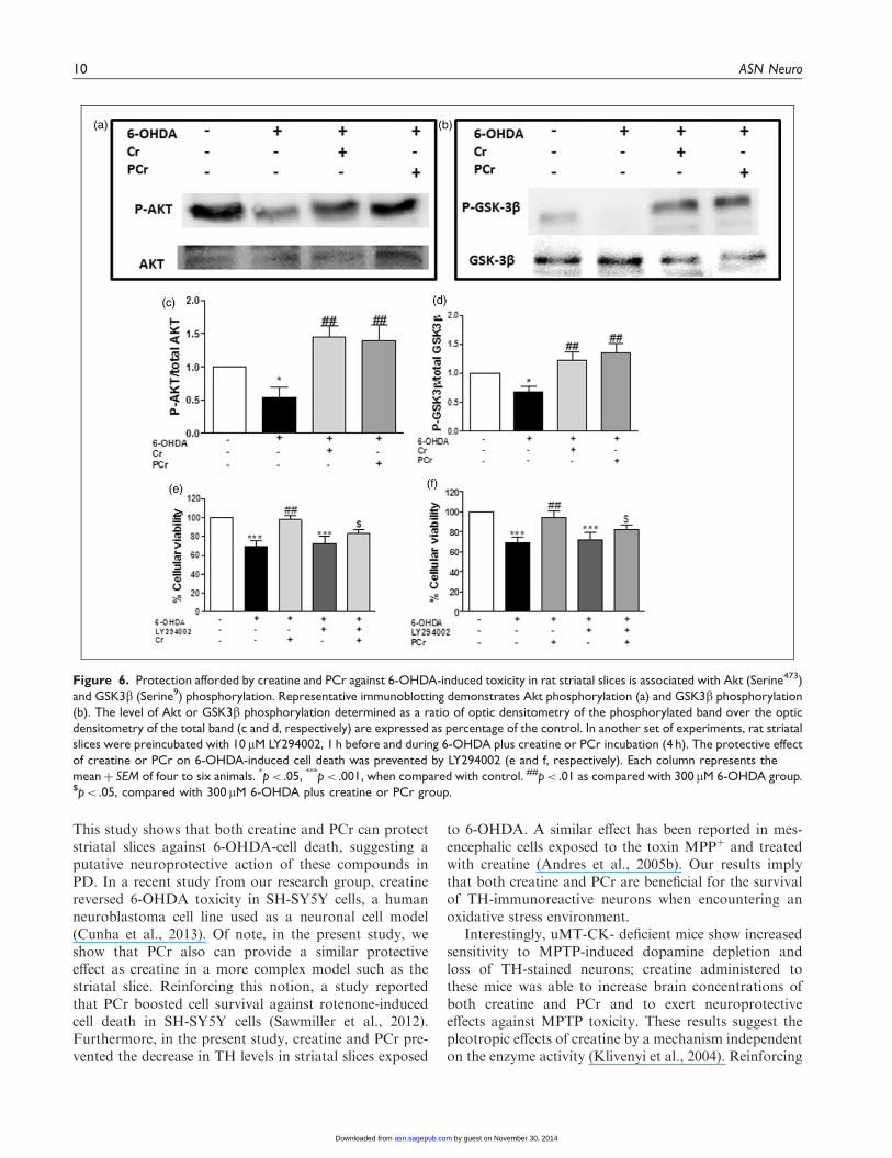
neuroprotective and antioxidant effect found for creatineand PCr against 6-OHDA-induced neurotoxicity in stri-atal slices. We first found that 6-OHDA induced a sig-nificant decreased in Akt and GSK3b phosphorylationand that creatine and PCr suppressed 6-OHDA-inducedphosphorylation dysfunctions (Figure 6(c) and (d),respectively).
The participation of the PI3K/Akt pathway was fur-ther demonstrated when we found that the protectiveactions of creatine and PCr were abolished in the pres-ence of the PI3K inhibitor LY294002 (Figure 6(e)and (f)).
Data from literature show that total Akt and p-Akt(Serine473) is found at high levels in TH immunopositivedopaminergic neurons in human brain, and selective lossof these neurons is accompanied by a marked decrease oftotal Akt and p-Akt (Serine473) levels in the PD brain(Timmons et al., 2009). We therefore considered of inter-est to investigate how LY294002 could modify the abilityof creatine and PCr to restore TH levels in striatal slicesexposed to 6-OHDA. As shown in Figure 7(b) and (c),the presence of LY294002 (30mM) abolished the capacityof creatine and PCr to restore TH levels in striatal slicesexposed to 6-OHDA.
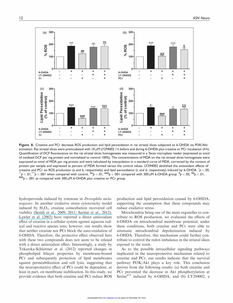
Because antioxidant compounds activate Akt andexert a protective effect (Toth et al., 2003; Latha et al.,2013; Sui et al., 2014), we investigated whether PI3K/Aktsignaling was involved in the effect of creatine or PCragainst 6-OHDA-induced oxidative stress in striatalslices. Creatine and PCr suppressed 6-OHDA-inducedincrease in ROS formation (Figure 8(a) and (b), respect-ively) and TBARS levels (Figure 8(c) and (d),
Figure 3. Creatine or PCr do not inhibit 6-OHDA auto-oxidation. The autoxidation of 6-OHDA in a nutritive incubation medium was
monitored during 3 min. Neither creatine (5 mM) nor PCr (5 mM) blocked 6-OHDA auto-oxidation. The positive control NAC (10 mM)
prevented the 6-OHDA auto-oxidation. Data are represented as mean of three experimental determinations.
Cunha et al. 7
by guest on November 30, 2014asn.sagepub.comDownloaded from

XML Template (2014) [16.10.2014–6:13pm] [1–16]//blrnas3.glyph.com/cenpro/ApplicationFiles/Journals/SAGE/3B2/ASNJ/Vol00000/140011/APPFile/SG-ASNJ140011.3d (ASN) [PREPRINTER stage]
respectively). Under these experimental conditions,LY394002 blocked the antioxidant effect of creatineand PCr.
Taken together, the above results clearly show the par-ticipation of the PI3K/Akt pathway in the protectiveactions of creatine and PCr in striatal slices exposed tothe neurotoxin 6-OHDA.
Discussion
The main finding of this study is that the guanidine com-pounds creatine and PCr can afford neuroprotection inan in vitro model of PD by a mechanism that implicatesmodulation of oxidative stress through the activation ofthe PI3K/Akt/GSK3b intracellular pathway.
6-OHDA can induce dopaminergic cell death by threemain mechanisms: (a) ROS generation by intra or extra-cellular auto-oxidation; (b) hydrogen peroxide (H2O2)
formation induced by monoamine oxidase activity, and(c) direct inhibition of the mitochondrial respiratorychain. These events lead to oxidative stress, decrease incellular ATP availability (Tirmenstein et al., 2005), andthereby, to cell death (Blum et al., 2001). Likewise, in ourexperimental protocol, 6-OHDA caused increased levelsof ROS and lipid peroxidation, mitochondrial depolar-ization, and toxicity of rat striatal slices.
Brain metabolism impairment has been identified as animportant mechanism underlying the pathophysiology ofPD (Shoffner et al., 1991; Berndt et al., 2013). The creat-ine kinase/PCr system controls brain energy homeostasisand has been implicated in several neurodegenerativeconditions (Adhihetty and Beal, 2008); for example, inPD patients, total creatine kinase activity decreases andcytotoxic ROS increases (Janetzky et al., 1994; Aksenovaet al., 1999). Also, in brains exposed to agents that pro-mote free radical production, creatine kinase activity
Figure 4. Creatine and PCr inhibit oxidative stress induced by 6-OHDA in rat striatal slices. ROS generation was estimated with the
fluorescent probe, 20,70-dichlorofluorescein diacetate (H2DCFDA). DCF fluorescence images in the putamen (a–d) and globus pallidus
(e–h) under the different experimental conditions indicated in each panel taken at 100�. Quantification of DCF fluorescence on rat striatal
slices homogenates was measured in a Tecan microplate reader and expressed as nmol of oxidized DCF per mg protein and normalized to
control 100%. The quantification of DCF fluorescence in the putamen (j) and globus pallidus (k) was measured in an epifluorescence
NIKON eclipse T2000-U microscope. Quantification of the mean fluorescence obtained under each experimental condition in putamen
and globus pallidus is normalized respect to control (100%). Each column represents the meanþ SEM of six to nine experiments. **p< .01
compared with the basal group. ###p< .01 as compared with 300mM 6-OHDA group.
8 ASN Neuro
by guest on November 30, 2014asn.sagepub.comDownloaded from

XML Template (2014) [16.10.2014–6:13pm] [1–16]//blrnas3.glyph.com/cenpro/ApplicationFiles/Journals/SAGE/3B2/ASNJ/Vol00000/140011/APPFile/SG-ASNJ140011.3d (ASN) [PREPRINTER stage]
decreases (Burmistrov et al., 1992), suggesting that com-pounds that block creatine kinase inhibition induced byoxidative stress could be neuroprotective in diseases likePD. Interestingly, a recent study demonstrates that cre-atine levels are increased in the right striatum following6-OHDA striatal lesions in rats, which is interpreted as anendogenous compensation mechanism to restore energysupply in response to 6-OHDA-induced injury. Thisobservation suggests a key role of this guanidine-likecompound against 6-OHDA toxicity (Gao et al., 2013)and has led to the hypothesis that agents that modulatethe bioenergetic status, mainly the creatine-containingcompounds, could reverse the neurodegenerative processinduced by 6-OHDA.
Creatine has been widely used as an ergogenic aid toimprove exercise performance in humans and also toameliorate oxidative stress-mediated diseases (Perskyand Brazeau, 2001; Wyss and Schulze, 2002; Andreset al., 2008). Creatine supplementation can protect neu-rons against neurotoxins in vitro (Brewer and Wallimann,2000) and it can slow down the progression of certainneurodegenerative conditions, as assessed in experimentalanimal models (Andreassen et al., 2001; Hosamani et al.,2010; Cunha et al., 2012). Furthermore, there are clinicaltrials that indicate that creatine supplementation could bebeneficial in diseases like Huntington’s, amyotrophic lat-eral sclerosis, depression, or PD (Mazzini et al., 2001;Roitman et al., 2007; Beal, 2011; Gualano et al., 2012).
Basal 6-OHDA 300μM 6-OHDA + Cr 5mM 6-OHDA + PCr 5mM
Basal 6-OHDA 300μM 6-OHDA + Cr 5mM 6-OHDA + PCr 5mM
(e) (f) (g) (h)
0
50
100
150
200
**# #
6-OHDACrPCr
---
+--
++-
+-+
% M
itoc
hond
rial
Mem
bran
e P
oten
tial
0
50
100
150
200
** # #
6-OHDACrPCr
---
+--
++-
+-+
% M
itoc
hond
rial
Mem
bran
e P
oten
tial
(j)
(a) (b) (c) (d)
(i)
Figure 5. Creatine or PCr prevent loss of mitochondrial membrane potential in the rat striatal slices subjected to 6-OHDA. TMRE
fluorescence in the putamen (a–d) and globus pallidus (e–h) was analyzed 4 h after coincubation with 6-OHDA plus creatine or PCr, as
detailed in the Materials and Methods section. The quantification of TMRE fluorescence was measured in an epifluorescence NIKON
eclipse T2000-U microscope (i, j). The top part of the figure illustrates representative photomicrographs of putamen and globus pallidus at
100�. Quantification of the mean fluorescence obtained under each experimental condition in putamen and globus pallidus is normalized
respect to control (100%). Each column represents the meansþ SEM of six to eight experiments carried out in triplicates. **p< .01
compared with the basal group. #p< .05 compared with 300 mM 6-OHDA group.
Cunha et al. 9
by guest on November 30, 2014asn.sagepub.comDownloaded from
XML Template (2014) [16.10.2014–6:13pm] [1–16]//blrnas3.glyph.com/cenpro/ApplicationFiles/Journals/SAGE/3B2/ASNJ/Vol00000/140011/APPFile/SG-ASNJ140011.3d (ASN) [PREPRINTER stage]
This study shows that both creatine and PCr can protectstriatal slices against 6-OHDA-cell death, suggesting aputative neuroprotective action of these compounds inPD. In a recent study from our research group, creatinereversed 6-OHDA toxicity in SH-SY5Y cells, a humanneuroblastoma cell line used as a neuronal cell model(Cunha et al., 2013). Of note, in the present study, weshow that PCr also can provide a similar protectiveeffect as creatine in a more complex model such as thestriatal slice. Reinforcing this notion, a study reportedthat PCr boosted cell survival against rotenone-inducedcell death in SH-SY5Y cells (Sawmiller et al., 2012).Furthermore, in the present study, creatine and PCr pre-vented the decrease in TH levels in striatal slices exposed
to 6-OHDA. A similar effect has been reported in mes-encephalic cells exposed to the toxin MPPþ and treatedwith creatine (Andres et al., 2005b). Our results implythat both creatine and PCr are beneficial for the survivalof TH-immunoreactive neurons when encountering anoxidative stress environment.
Interestingly, uMT-CK- deficient mice show increasedsensitivity to MPTP-induced dopamine depletion andloss of TH-stained neurons; creatine administered tothese mice was able to increase brain concentrations ofboth creatine and PCr and to exert neuroprotectiveeffects against MPTP toxicity. These results suggest thepleotropic effects of creatine by a mechanism independenton the enzyme activity (Klivenyi et al., 2004). Reinforcing
Figure 6. Protection afforded by creatine and PCr against 6-OHDA-induced toxicity in rat striatal slices is associated with Akt (Serine473)
and GSK3b (Serine9) phosphorylation. Representative immunoblotting demonstrates Akt phosphorylation (a) and GSK3b phosphorylation
(b). The level of Akt or GSK3b phosphorylation determined as a ratio of optic densitometry of the phosphorylated band over the optic
densitometry of the total band (c and d, respectively) are expressed as percentage of the control. In another set of experiments, rat striatal
slices were preincubated with 10mM LY294002, 1 h before and during 6-OHDA plus creatine or PCr incubation (4 h). The protective effect
of creatine or PCr on 6-OHDA-induced cell death was prevented by LY294002 (e and f, respectively). Each column represents the
meanþ SEM of four to six animals. *p< .05, ***p< .001, when compared with control. ##p< .01 as compared with 300 mM 6-OHDA group.$p< .05, compared with 300mM 6-OHDA plus creatine or PCr group.
10 ASN Neuro
by guest on November 30, 2014asn.sagepub.comDownloaded from

XML Template (2014) [16.10.2014–6:13pm] [1–16]//blrnas3.glyph.com/cenpro/ApplicationFiles/Journals/SAGE/3B2/ASNJ/Vol00000/140011/APPFile/SG-ASNJ140011.3d (ASN) [PREPRINTER stage]
this notion, in a Drosophila melanogaster model, it wasshown that creatine can protect from oxidative stress,although insects, expressing arginine kinase instead orcreatine kinase, are not able to synthesize PCr(Hosamani et al., 2010). The pleiotropic effects of creatinecould comprise antioxidant effects and activation of sig-naling pathways, including Akt/PKB.
The antioxidant activity of creatine or PCr emerges asmechanism that is likely to play a supportive role in thecreatine or PCr cytoprotection paradigm (Sestili et al.,2011). Several studies, most with creatine, corroboratethis action. For example, Hosamani et al. (2010)showed that creatine reduced ROS production andother oxidative markers such as malondialdehyde and
Figure 7. Involvement of PI3K/Akt in the protective effect of creatine and PCr on TH levels. Rat striatal slices were preincubated with
10 mM LY294002 1 h before and during 6-OHDA plus creatine or PCr incubation (4 h). (a) Representative immunoblotting of tyrosine
hydroxylase is shown. The TH immunocontent determined as a ratio of optic densitometry of the TH band over the optic densitometry of
the b-actin band are expressed as percentage of the control. Creatine (5 mM) or PCr (5 mM) prevented the decrease of TH levels induced
by 6-OHDA in the rat striatal slices, and these effects are significantly abolished by LY294002 (b and c, respectively). Each column
represents the meanþ SEM of four to six animals. ***p< .001, when compared with control. ##p< .01 compared with 300 mM 6-OHDA
group. $p< .05 compared with 300mM 6-OHDA plus creatine or PCr group.
Cunha et al. 11
by guest on November 30, 2014asn.sagepub.comDownloaded from
XML Template (2014) [16.10.2014–6:13pm] [1–16]//blrnas3.glyph.com/cenpro/ApplicationFiles/Journals/SAGE/3B2/ASNJ/Vol00000/140011/APPFile/SG-ASNJ140011.3d (ASN) [PREPRINTER stage]
hydroperoxide induced by rotenone in Drosophila mela-nogaster. In another oxidative stress cytotoxicity modelinduced by H2O2, creatine coincubation recovered cellviability (Sestili et al., 2009, 2011; Sartini et al., 2012).Lawler et al. (2002) have reported a direct antioxidanteffect of creatine in a cellular system against aqueous rad-ical and reactive species ions; however, our results showthat neither creatine nor PCr block the auto-oxidation of6-OHDA. Therefore, the protective effect observed herewith these two compounds does not seem to be relatedwith a direct antioxidant effect. Interestingly, a study byTokarska-Schlattner et al. (2012) reported changes inphospholipid bilayer properties by membrane-boundPCr and subsequently protection of lipid membranesagainst permeabilization and cell lysis, suggesting thatthe neuroprotective effect of PCr could be dependent, atleast in part, on membrane stabilization. In this study, weprovide evidence that both creatine and PCr reduce ROS
production and lipid peroxidation caused by 6-OHDA,supporting the assumption that these compounds mayreduce oxidative stress.
Mitochondria being one of the main organelles to con-tribute to ROS production, we evaluated the effects of6-OHDA on mitochondrial membrane potential; underthese conditions, both creatine and PCr were able toattenuate mitochondrial depolarization induced by6-OHDA. Therefore, this mechanism could further con-tribute to control the redox imbalance in the striatal slicesexposed to the toxin.
As to the possible intracellular signaling pathwaysimplicated in the neuroprotective mechanism related tocreatine and PCr, our results indicate that the survivalpathway PI3K/Akt plays a key role. This conclusionderives from the following results: (a) both creatine andPCr prevented the decrease in Akt phosphorylation atSerine473 induced by 6-OHDA, and (b) LY294002, a
Figure 8. Creatine and PCr decrease ROS production and lipid peroxidation in rat striatal slices subjected to 6-OHDA via PI3K/Akt
activation. Rat striatal slices were preincubated with 10 mM LY294002 1 h before and during 6-OHDA plus creatine or PCr incubation (4 h).
Quantification of DCF fluorescence on the rat striatal slices homogenates was measured in a Tecan microplate reader (expressed as nmol
of oxidized DCF per mg protein and normalized to control 100%). The concentrations of MDA on the rat striatal slices homogenate were
expressed as nmol of MDA per mg protein and were calculated by interpolation in a standard curve of MDA, corrected by the content of
protein per sample and expressed as percent of MDA formed versus the control values. LY294002 abolished the antioxidant effects of
creatine and PCr on ROS production (a and b, respectively) and lipid peroxidation (c and d, respectively) induced by 6-OHDA. *p< .05,**p< .01,***p< .001 when compared with control. ##p< .01, ###p< .001 compared with 300 mM 6-OHDA group. $p< .05, $$p< .01,$$$p< .001 as compared with 300 mM 6-OHDA plus creatine or PCr group.
12 ASN Neuro
by guest on November 30, 2014asn.sagepub.comDownloaded from
XML Template (2014) [16.10.2014–6:13pm] [1–16]//blrnas3.glyph.com/cenpro/ApplicationFiles/Journals/SAGE/3B2/ASNJ/Vol00000/140011/APPFile/SG-ASNJ140011.3d (ASN) [PREPRINTER stage]
PI3K/Akt inhibitor, blocked the protective, the antioxi-dant, and recovery of TH levels afforded by creatine andPCr in slices exposed to 6-OHDA. In line with theseresults, a previous study showed that creatine increasedAkt activity in C2C12 cells, a mouse myoblast cell line(Deldicque et al., 2007), and afforded neuroprotection viaPI3K/Akt in the human neuroblastoma SH-SY5Y cellline (Cunha et al., 2013).
We have also investigated the effect of creatine andPCr on GSK3b; this serine/threonine kinase was origin-ally identified as a regulator of glycogen metabolism butis now recognized as an important modulator of apop-tosis. The inactivation of GSK3 can be induced by phos-phorylation at one of its N-terminal serine residues:Serine21 for GSK3a and Serine9 for GSK3b (Plyteet al., 1992). The administration of agents that causephosphorylation and consequent inactivation of GSK-3b is considered an interesting pharmacologic neuropro-tective strategy. Phosphorylation of GSK3 can bemediated by several kinases, including Akt (Rommelet al., 1999; Jope and Roh, 2006). Literature data havereported that pretreatment with TDZD-8, lithium, orL803-mts (GSK3 inhibitors) reduces 6-OHDA-inducedcell death (Chen et al., 2004), and knockdown ofGSK3b attenuates 6-OHDA-induced apoptosis in SH-SY5Y cells (Li et al., 2011). In the present study, wehave observed that 6-OHDA decreases GSK3b Serine9
phosphorylation, and both creatine and PCr were ableto reverse this effect.
In conclusion, our findings identify creatine and PCras potent natural protective factors for dopaminergic cellsurvival; their neuroprotective mechanism does not seemto be related to a direct scavenger effect but to mitochon-drial membrane stabilization, activation of the survivalsignaling pathway PI3K/Akt/GSK3b, and control ofthe redox balance.
Declaration of Conflicting Interests
The authors declared no potential conflicts of interest with respect
to the research, authorship, and/or publication of this article.
Funding
The authors disclosed receipt of the following financial support for
the research, authorship, and/or publication of this article: This work
was supported by grants from the Ministerio de Economıa y
Competitividad (Ref. SAF2012-32223) and the Spanish Ministry
of Health (Instituto de Salud Carlos III) RENEVAS-RETICS-
RD06/0026 to M. G. L.; and the Conselho Nacional de
Desenvolvimento Cientıfico e Tecnologico (CNPq; 307687/2009-
0), Coordenacao de Aperfeicoamento de Pessoal de Ensino
Superior (CAPES), Fundacao de Apoio a Pesquisa Cientıfica
e Tecnologica do Estado de Santa Catarina (FAPESC; 1347/
2010-1), Rede Instituto Brasileiro de Neurociencia (IBN-Net/
CNPq), and NENASC project (PRONEX Program
CNPq/FAPESC) to A. L. S. R, C.I.T. and MF.
References
Abou-Sleiman, P. M., Muqit, M. M., & Wood, N. W. (2006).
Expanding insights of mitochondrial dysfunction in
Parkinson’s disease. Nature Reviews Neuroscience 7: 207–219.
Adachi, K., Takeda, Y., Senoh, S., & Kita, H. (1964). Metabolism
of P-hydroxyphenylacetic acid in Pseudomonas ovalis.
Biochimica et Biophysica Acta 93: 483–493.
Adhihetty, P. J., & Beal, M. F. (2008). Creatine and its potential
therapeutic value for targeting cellular energy impairment in
neurodegenerative diseases. Neuromolecular Medicine 10:
275–290.
Aksenova, M. V., Aksenov, M. Y., Payne, R. M., Trojanowski,
J. Q., Schmidt, M. L., Carney, J. M., . . . Markesbery, W. R.
(1999). Oxidation of cytosolic proteins and expression of cre-
atine kinase BB in frontal lobe in different neurodegenerative
disorders. Dementia and Geriatric Cognitive Disorders 10:
158–165.
Ali, S. F., LeBel, C. P., & Bondy, S. C. (1992). Reactive oxygen
species formation as a biomarker of methylmercury and tri-
methyltin neurotoxicity. Neurotoxicology 13: 637–648.
Andreassen, O. A., Dedeoglu, A., Ferrante, R. J., Jenkins, B. G.,
Ferrante, K. L., Thomas, M., . . . Beal, M. F. (2001). Creatine
increase survival and delays motor symptoms in a transgenic
animal model of Huntington’s disease. Neurobiology of Disease
8: 479–491.
Andres, R. H., Ducray, A. D., Perez-Bouza, A., Schlattner, U.,
Huber, A. W., Krebs, S. H., . . . Widmer, H. R. (2005a).
Creatine supplementation improves dopaminergic cell survival
and protects against MPPþ toxicity in an organotypic tissue
culture system. Cell Transplantation 14: 537–550.
Andres, R. H., Ducray, A. D., Schlattner, U., Wallimann, T., &
Widmer, H. R. (2008). Functions and effects of creatine in the
central nervous system. Brain Research Bulletin 76: 329–343.
Andres, R. H., Huber, A. W., Schlattner, U., Perez-Bouza, A.,
Krebs, S. H., Seiler, R. W., . . . Widmer, H. R. (2005b). Effects
of creatine treatment on the survival of dopaminergic neurons in
cultured fetal ventral mesencephalic tissue. Neuroscience 133:
701–713.
Annett, L. E., Torres, E. M., Clarke, D. J., Ishida, Y., Barker, R. A.,
Ridley, R. M., . . . Dunnett, S. B. (1997). Survival of nigral grafts
within the striatum of marmosets with 6-OHDA lesions depends
critically on donor embryo age. Cell Transplantation 6:
557–569.
Beal, E. (2011). Parkinson disease: Protein-based stem cells gener-
ate healthy dopamine neurons. Nature Reviews Neurology 7:
357.
Bender, A., Koch, W., Elstner, M., Schombacher, Y., Bender, J.,
Moeschl, M., . . . Klopstock, T. (2006). Creatine supplementa-
tion in Parkinson disease: A placebo-controlled randomized
pilot trial. Neurology 67: 1262–1264.
Berndt, N., Holzhutter, H. G., & Bulik, S. (2013). Implications of
enzyme deficiencies on mitochondrial energy metabolism and
reactive oxygen species formation of neurons involved in rote-
none-induced Parkinson’s disease: A model-based analysis. The
FEBS Journal 280: 5080–5093.
Bessman, S. P., & Carpenter, C. L. (1985). The creatine-creatine
phosphate energy shuttle. Annual Review of Biochemistry 54:
831–862.
Cunha et al. 13
by guest on November 30, 2014asn.sagepub.comDownloaded from
XML Template (2014) [16.10.2014–6:13pm] [1–16]//blrnas3.glyph.com/cenpro/ApplicationFiles/Journals/SAGE/3B2/ASNJ/Vol00000/140011/APPFile/SG-ASNJ140011.3d (ASN) [PREPRINTER stage]
Bindoff, L. A., Birch-Machin, M., Cartlidge, N. E., Parker, W. D.
Jr., & Turnbull, D. M. (1989). Mitochondrial function in
Parkinson’s disease. Lancet 2: 49.
Blandini, F., Armentero, M. T., & Martignoni, E. (2008). The 6-
hydroxydopamine model: News from the past. Parkinsonism &
Related Disorders 14(Suppl. 2): S124–S129.
Blum, D., Torch, S., Lambeng, N., Nissou, M., Benabid, A. L.,
Sadoul, R., . . . Verna, J. M. (2001). Molecular pathways
involved in the neurotoxicity of 6-OHDA, dopamine and
MPTP: Contribution to the apoptotic theory in Parkinson’s dis-
ease. Progress in Neurobiology 65: 135–172.
Brewer, G. J., & Wallimann, T. W. (2000). Protective effect of the
energy precursor creatine against toxicity of glutamate and beta-
amyloid in rat hippocampal neurons. Journal of Neurochemistry
74: 1968–1978.
Burmistrov, S. O., Mashek, O. P., & Kotin, A. M. (1992). The
action of acute alcoholic intoxication on the antioxidant
system and creatine kinase activity in the brain of rat embryos.
Eksperimental’naia i klinicheskaia farmakologiia 55: 54–56.
Carlsson, A., Lindqvist, M., & Magnusson, T. (1957). 3,4-
Dihydroxyphenylalanine and 5-hydroxytryptophan as reserpine
antagonists. Nature 180: 1200.
Chen, G., Bower, K. A., Ma, C., Fang, S., Thiele, C. J., & Luo, J.
(2004). Glycogen synthase kinase 3beta (GSK3beta) mediates
6-hydroxydopamine-induced neuronal death. FASEB Journal:
Official Publication of the Federation of American Societies
for Experimental Biology 18: 1162–1164.
Colle, D., Hartwig, J. M., Soares, F. A., & Farina, M. (2012).
Probucol modulates oxidative stress and excitotoxicity in
Huntington’s disease models in vitro. Brain Research Bulletin
87: 397–405.
Cunha, M. P., Machado, D. G., Capra, J. C., Jacinto, J., Bettio, L.
E., & Rodrigues, A. L. (2012). Antidepressant-like effect of
creatine in mice involves dopaminergic activation. Journal of
Psychopharmacology 26: 1489–1501.
Cunha, M. P., Martin-de-Saavedra, M. D., Romero, A., Parada, E.,
Egea, J., Del Barrio, L., . . . Lopez, M. G. (2013). Protective
effect of creatine against 6-hydroxydopamine-induced cell
death in human neuroblastoma SH-SY5Y cells: Involvement
of intracellular signaling pathways. Neuroscience 238: 185–194.
Dal-Cim, T., Ludka, F. K., Martins, W. C., Reginato, C., Parada, E.,
Egea, J., . . . Tasca, C. I. (2013). Guanosine controls inflamma-
tory pathways to afford neuroprotection of hippocampal slices
under oxygen and glucose deprivation conditions. Journal of
Neurochemistry 126: 437–450.
Dedeoglu, A., Kubilus, J. K., Yang, L., Ferrante, K. L., Hersch, S.
M., Beal, M. F., . . . Ferrante, R. J. (2003). Creatine therapy pro-
vides neuroprotection after onset of clinical symptoms in
Huntington’s disease transgenic mice. Journal of
Neurochemistry 85: 1359–1367.
Deldicque, L., Theisen, D., Bertrand, L., Hespel, P., Hue, L., &
Francaux, M. (2007). Creatine enhances differentiation of myo-
genic C2C12 cells by activating both p38 and Akt/PKB path-
ways. American Journal of Physiology Cell Physiology 293:
C1263–C1271.
Egea, J., Martin-de-Saavedra, M. D., Parada, E., Romero, A., Del
Barrio, L., Rosa, A. O., . . . Lopez, M. G. (2012). Galantamine
elicits neuroprotection by inhibiting iNOS, NADPH oxidase and
ROS in hippocampal slices stressed with anoxia/reoxygenation.
Neuropharmacology 62: 1082–1090.
Egea, J., Rosa, A. O., Sobrado, M., Gandia, L., Lopez, M. G., &
Garcia, A. G. (2007). Neuroprotection afforded by nicotine
against oxygen and glucose deprivation in hippocampal slices
is lost in alpha7 nicotinic receptor knockout mice. Neuroscience
145: 866–872.
Gahwiler, B. H. (1987). Organotypic slice cultures: A model for
interdisciplinary studies. Progress in Clinical and Biological
Research 253: 13–18.
Gahwiler, B. H., Capogna, M., Debanne, D., McKinney, R. A., &
Thompson, S. M. (1997). Organotypic slice cultures: A tech-
nique has come of age. Trends in Neurosciences 20: 471–477.
Gao, H. C., Zhu, H., Song, C. Y., Lin, L., Xiang, Y., Yan, Z.
H., . . . Li, X. K. (2013). Metabolic changes detected by ex
vivo high resolution 1H NMR spectroscopy in the striatum of
6-OHDA-induced Parkinson’s rat. Molecular Neurobiology 47:
123–130.
Graham, D. G., Tiffany, S. M., Bell, W. R. Jr., & Gutknecht, W. F.
(1978). Autoxidation versus covalent binding of quinones as the
mechanism of toxicity of dopamine, 6-hydroxydopamine, and
related compounds toward C1300 neuroblastoma cells in vitro.
Molecular Pharmacology 14: 644–653.
Gualano, B., Roschel, H., Lancha, A. H. Jr., Brightbill, C. E., &
Rawson, E. S. (2012). In sickness and in health: The widespread
application of creatine supplementation. Amino Acids 43:
519–529.
Ha, H. C., Woster, P. M., Yager, J. D., & Casero, R. A. Jr. (1997).
The role of polyamine catabolism in polyamine analogue-
induced programmed cell death. Proceedings of the National
Academy of Sciences of the United States of America 94:
11557–11562.
Hattingen, E., Magerkurth, J., Pilatus, U., Mozer, A., Seifried, C.,
Steinmetz, H., . . . Hilker, R. (2009). Phosphorus and proton
magnetic resonance spectroscopy demonstrates mitochondrial
dysfunction in early and advanced Parkinson’s disease. Brain:
A Journal of Neurology 132: 3285–3297.
Hemmer, W., & Wallimann, T. (1993). Functional aspects of cre-
atine kinase in brain. Developmental Neuroscience 15: 249–260.
Hosamani, R., Ramesh, S. R., & Muralidhara (2010). Attenuation
of rotenone-induced mitochondrial oxidative damage and neu-
rotoxicty in Drosophila melanogaster supplemented with creat-
ine. Neurochemical Research 35: 1402–1412.
Investigators NN-P. A randomized, double-blind, futility clinical
trial of creatine and minocycline in early Parkinson disease.
Neurology 66: 664–671.
Janetzky, B., Hauck, S., Youdim, M. B., Riederer, P., Jellinger, K.,
Pantucek, F., . . . Reichmann, H. (1994). Unaltered aconitase
activity, but decreased complex I activity in substantia nigra
pars compacta of patients with Parkinson’s disease.
Neuroscience Letters 169: 126–128.
Jenner, P. (2003). Oxidative stress in Parkinson’s disease. Annals of
Neurology 53(Suppl. 3): S26–S36.
Jope, R. S., & Roh, M. S. (2006). Glycogen synthase kinase-3
(GSK3) in psychiatric diseases and therapeutic interventions.
Current Drug Targets 7: 1421–1434.
Klivenyi, P., Calingasan, N. Y., Starkov, A., Stavrovskaya, I. G.,
Kristal, B. S., Yang, L., . . . Beal, M. F. (2004). Neuroprotective
mechanisms of creatine occur in the absence of mitochondrial
creatine kinase. Neurobiology of Disease 15: 610–617.
Langston, J. W., Langston, E. B., & Irwin, I. (1984). MPTP-
induced parkinsonism in human and non-human
14 ASN Neuro
by guest on November 30, 2014asn.sagepub.comDownloaded from

XML Template (2014) [16.10.2014–6:13pm] [1–16]//blrnas3.glyph.com/cenpro/ApplicationFiles/Journals/SAGE/3B2/ASNJ/Vol00000/140011/APPFile/SG-ASNJ140011.3d (ASN) [PREPRINTER stage]
primates—Clinical and experimental aspects. Acta Neurologica
Scandinavica Supplement 100: 49–54.
Latha, R., Shanthi, P., & Sachdanandam, P. (2013). Kalpaamruthaa
modulates oxidative stress in cardiovascular complication asso-
ciated with type 2 diabetes mellitus through PKC-beta/Akt sig-
naling. Canadian Journal of Physiology and Pharmacology 91:
901–912.
Lawler, J. M., Barnes, W. S., Wu, G., Song, W., & Demaree, S.
(2002). Direct antioxidant properties of creatine. Biochemical
and Biophysical Research Communications 290: 47–52.
Lehmensiek, V., Tan, E. M., Liebau, S., Lenk, T., Zettlmeisl, H.,
Schwarz, J., . . . Storch, A. (2006). Dopamine transporter-
mediated cytotoxicity of 6-hydroxydopamine in vitro depends
on expression of mutant alpha-synucleins related to Parkinson’s
disease. Neurochemistry International 48: 329–340.
Lensman, M., Korzhevskii, D. E., Mourovets, V. O., Kostkin, V.
B., Izvarina, N., Perasso, L., . . . Balestrino, M. (2006).
Intracerebroventricular administration of creatine protects
against damage by global cerebral ischemia in rat. Brain
Research 1114: 187–194.
Li, Y., Luo, F., Wei, L., Liu, Z., & Xu, P. (2011). Knockdown of
glycogen synthase kinase 3 beta attenuates 6-hydroxydopamine-
induced apoptosis in SH-SY5Y cells. Neuroscience Letters 487:
41–46.
Lo Bianco, C., Schneider, B. L., Bauer, M., Sajadi, A., Brice, A.,
Iwatsubo, T., . . . Aebischer, P. (2004). Lentiviral vector delivery
of parkin prevents dopaminergic degeneration in an alpha-synu-
clein rat model of Parkinson’s disease. Proceedings of the
National Academy of Sciences of the United States of America
101: 17510–17515.
Lowry, O. H., Rosebrough, N. J., Farr, A. L., & Randall, R. J.
(1951). Protein measurement with the Folin phenol reagent.
The Journal of Biological Chemistry 193: 265–275.
Matthews, R. T., Ferrante, R. J., Klivenyi, P., Yang, L., Klein, A.
M., Mueller, G., . . . Beal, M. F. (1999). Creatine and cyclocrea-
tine attenuate MPTP neurotoxicity. Experimental Neurology
157: 142–149.
Mazzini, L., Balzarini, C., Colombo, R., Mora, G., Pastore, I., De
Ambrogio, R., . . . Caligari, M. (2001). Effects of creatine sup-
plementation on exercise performance and muscular strength in
amyotrophic lateral sclerosis: Preliminary results. The Journal
of the Neurological Sciences 191: 139–144.
Molz, S., Decker, H., Dal-Cim, T., Cremonez, C., Cordova, F. M.,
Leal, R. B., . . . Tasca, C. I. (2008). Glutamate-induced toxicity
in hippocampal slices involves apoptotic features and p38
MAPK signaling. Neurochemical Research 33: 27–36.
Mosmann, T. (1983). Rapid colorimetric assay for cellular
growth and survival: Application to proliferation and
cytotoxicity assays. The Journal of Immunological Methods
65: 55–63.
Parker, W. D. Jr., Boyson, S. J., & Parks, J. K. (1989).
Abnormalities of the electron transport chain in idiopathic
Parkinson’s disease. Annals of Neurology 26: 719–723.
Persky, A. M., & Brazeau, G. A. (2001). Clinical pharmacology of
the dietary supplement creatine monohydrate. Pharmacological
Reviews 53: 161–176.
Plyte, S. E., Hughes, K., Nikolakaki, E., Pulverer, B. J., &
Woodgett, J. R. (1992). Glycogen synthase kinase-3:
Functions in oncogenesis and development. Biochimica et
Biophysica Acta 1114: 147–162.
Rambo, L. M., Ribeiro, L. R., Della-Pace, I. D., Stamm, D. N.,
da Rosa Gerbatin, R., Prigol, M., . . . Royes, L. F. (2013). Acute
creatine administration improves mitochondrial membrane
potential and protects against pentylenetetrazol-induced seiz-
ures. Amino Acids 44: 857–868.
Rauchova, H., Koudelova, J., Drahota, Z., & Mourek, J. (2002).
Hypoxia-induced lipid peroxidation in rat brain and protective
effect of carnitine and phosphocreatine. Neurochemical
Research 27: 899–904.
Rios, C., & Santamaria, A. (1991). Quinolinic acid is a potent lipid
peroxidant in rat brain homogenates. Neurochemical Research
16: 1139–1143.
Roeling, T. A., Docter, G. J., Voorn, P., Melchers, B. P., Wolters,
E. C., & Groenewegen, H. J. (1995). Effects of unilateral 6-
hydroxydopamine lesions on neuropeptide immunoreactivity
in the basal ganglia of the common marmoset, Callithrix jac-
chus, a quantitative immunohistochemical analysis. The Journal
of Chemical Neuroanatomy 9: 155–164.
Roitman, S., Green, T., Osher, Y., Karni, N., & Levine, J. (2007).
Creatine monohydrate in resistant depression: A preliminary
study. Bipolar Disorder 9: 754–758.
Rommel, C., Clarke, B. A., Zimmermann, S., Nunez, L., Rossman,
R., Reid, K., . . . Glass, D. J. (1999). Differentiation stage-speci-
fic inhibition of the Raf-MEK-ERK pathway by Akt. Science
286: 1738–1741.
Ruda, M., Samarenko, M. B., Afonskaya, N. I., & Saks, V. A.
(1988). Reduction of ventricular arrhythmias by phosphocrea-
tine (Neoton) in patients with acute myocardial infarction. The
American Heart Journal 116: 393–397.
Sachs, C., Jonsson, G., Heikkila, R., & Cohen, G. (1975). Control
of the neurotoxicity of 6-hydroxydopamine by intraneuronal
noradrenaline in rat iris. Acta Physiologica Scandinavica 93:
345–351.
Sartini, S., Sestili, P., Colombo, E., Martinelli, C., Bartolini, F.,
Ciuffoli, S., . . . Cuppini, R. (2012). Creatine affects in vitro
electrophysiological maturation of neuroblasts and protects
them from oxidative stress. Journal of Neuroscience Research
90: 435–446.
Sawmiller, D. R., Nguyen, H. T., Markov, O., & Chen, M. (2012).
High-energy compounds promote physiological processing of
Alzheimer’s amyloid-beta precursor protein and boost cell sur-
vival in culture. Journal of Neurochemistry 123: 525–531.
Sestili, P., Barbieri, E., Martinelli, C., Battistelli, M., Guescini, M.,
Vallorani, L., . . . Stocchi, V. (2009). Creatine supplementation
prevents the inhibition of myogenic differentiation in oxida-
tively injured C2C12 murine myoblasts. Molecular Nutrition
& Food Research 53: 1187–1204.
Sestili, P., Martinelli, C., Bravi, G., Piccoli, G., Curci, R.,
Battistelli, M., . . . Stocchi, V. (2006). Creatine supplementation
affords cytoprotection in oxidatively injured cultured mamma-
lian cells via direct antioxidant activity. Free Radical Biology
and Medicine 40: 837–849.
Sestili, P., Martinelli, C., Colombo, E., Barbieri, E., Potenza, L.,
Sartini, S., . . . Fimognari, C. (2011). Creatine as an antioxidant.
Amino Acids 40: 1385–1396.
Sharov, V. G., Afonskaya, N. I., Ruda, M. Y., Cherpachenko, N.
M., Pozin, E., Markosyan, R. A., . . . Saks, V. A. (1986).
Protection of ischemic myocardium by exogenous phosphocrea-
tine (neoton): Pharmacokinetics of phosphocreatine, reduction
of infarct size, stabilization of sarcolemma of ischemic
Cunha et al. 15
by guest on November 30, 2014asn.sagepub.comDownloaded from
XML Template (2014) [16.10.2014–6:13pm] [1–16]//blrnas3.glyph.com/cenpro/ApplicationFiles/Journals/SAGE/3B2/ASNJ/Vol00000/140011/APPFile/SG-ASNJ140011.3d (ASN) [PREPRINTER stage]
cardiomyocytes, and antithrombotic action. Biochemical
Medicine and Metabolic Biology 35: 101–114.
Sharov, V. G., Saks, V. A., Kupriyanov, V. V., Lakomkin, V. L.,
Kapelko, V. I., Steinschneider, A., . . . Javadov, S. A. (1987).
Protection of ischemic myocardium by exogenous phosphocrea-
tine. I. Morphologic and phosphorus 31-nuclear magnetic res-
onance studies. The Journal of Thoracic and Cardiovascular
Surgery 94: 749–761.
Shoffner, J. M., Watts, R. L., Juncos, J. L., Torroni, A., & Wallace,
D. C. (1991). Mitochondrial oxidative phosphorylation defects
in Parkinson’s disease. Annals of Neurology 30: 332–339.
Sobrado, M., Roda, J. M., Lopez, M. G., Egea, J., & Garcia, A. G.
(2004). Galantamine and memantine produce different degrees
of neuroprotection in rat hippocampal slices subjected to
oxygen-glucose deprivation. Neuroscience Letters 365:
132–136.
Somjen, G. G., Aitken, P. G., Balestrino, M., & Schiff, S. J. (1987).
Uses and abuses of in vitro systems in the study of the patho-
physiology of the central nervous system. In: A. Schurr, T.
J. Teyler, & M. T. Tseng (Eds.). Brain slices in the study of
brain damage. Basel, Switzerland: Karger, pp. 89–104.
Soto-Otero, R., Mendez-Alvarez, E., Hermida-Ameijeiras, A.,
Munoz-Patino, A. M., & Labandeira-Garcia, J. L. (2000).
Autoxidation and neurotoxicity of 6-hydroxydopamine in the
presence of some antioxidants: Potential implication in relation
to the pathogenesis of Parkinson’s disease. Journal of
Neurochemistry 74: 1605–1612.
Sui, X. Q., Xu, Z. M., Xie, M. B., & Pei, D. A. (2014). Resveratrol
inhibits hydrogen peroxide-induced apoptosis in endothelial
cells via the activation of PI3K/Akt by miR-126. Journal of
Atherosclerosis and Thrombosis 21: 108–118.
Timmons, S., Coakley, M. F., Moloney, A. M., & O’Neill, C.
(2009). Akt signal transduction dysfunction in Parkinson’s dis-
ease. Neuroscience Letters 467: 30–35.
Tirmenstein, M. A., Hu, C. X., Scicchitano, M. S., Narayanan, P.
K., McFarland, D. C., Thomas, H. C., . . . Schwartz, L. W.
(2005). Effects of 6-hydroxydopamine on mitochondrial func-
tion and glutathione status in SH-SY5Y human neuroblastoma
cells. Toxicology in Vitro 19: 471–479.
Tobon-Velasco, J. C., Limon-Pacheco, J. H., Orozco-Ibarra, M.,
Macias-Silva, M., Vazquez-Victorio, G., Cuevas, E., . . .
Santamaria, A. (2013). 6-OHDA-induced apoptosis and
mitochondrial dysfunction are mediated by early modulation
of intracellular signals and interaction of Nrf2 and NF-kappaB
factors. Toxicology 304: 109–119.
Tokarska-Schlattner, M., Epand, R. F., Meiler, F., Zandomeneghi,
G., Neumann, D., Widmer, H. R., . . . Schlattner, U. (2012).
Phosphocreatine interacts with phospholipids, affects membrane
properties and exerts membrane-protective effects. PLoS One 7:
e43178.
Toth, A., Halmosi, R., Kovacs, K., Deres, P., Kalai, T., Hideg,
K., . . . Sumegi, B. (2003). Akt activation induced by an antioxi-
dant compound during ischemia-reperfusion. Free Radical
Biology and Medicine 35: 1051–1063.
Ungerstedt, U. (1968). 6-Hydroxy-dopamine induced degeneration
of central monoamine neurons. European Journal of
Pharmacology 5: 107–110.
Ungerstedt, U. (1971). Adipsia and aphagia after 6-hydroxydopa-
mine induced degeneration of the nigro-striatal dopamine
system. Acta Physiologica Scandinavica Supplement 367:
95–122.
Ungerstedt, U. (1976). 6-hydroxydopamine-induced degeneration
of the nigrostriatal dopamine pathway: The turning syndrome.
Pharmacology & Therapeutics. Part B 2: 37–40.
Valastro, B., Dekundy, A., Danysz, W., & Quack, G. (2009). Oral
creatine supplementation attenuates L-DOPA-induced dyskin-
esia in 6-hydroxydopamine-lesioned rats. Behavioural Brain
Research 197: 90–96.
Valette, G., Rainova, L., & Caro, J. O. (1972). Changes caused by
hydroxy-6 dopamine and alpha methyl p-tyrosine in the tachy-
phylactic effect of indirect sympathomimetics (ephedrine and
tyramine) on perfused and isolated organs of rats. Comptes
Rendus Hebdomadaires des Seances de l Academie des
Sciences D 275: 1823–1826.
Wyss, M., & Schulze, A. (2002). Health implications of creatine:
Can oral creatine supplementation protect against neurological
and atherosclerotic disease? Neuroscience 112: 243–260.
Wyss, M., Smeitink, J., Wevers, R. A., & Wallimann, T. (1992).
Mitochondrial creatine kinase: A key enzyme of aerobic energy
metabolism. Biochimica et Biophysica Acta 1102: 119–166.
Yong-Kee, C. J., Salomonczyk, D., & Nash, J. E. (2011).
Development and validation of a screening assay for the evalu-
ation of putative neuroprotective agents in the treatment of
Parkinson’s disease. Neurotoxicity Research 19: 519–526.
16 ASN Neuro
by guest on November 30, 2014asn.sagepub.comDownloaded from
Copyright © 2022 FDOKUMEN