Bones of The Lower Limbs - KSUMSC
14
Bones of The Lower Limbs Anatomy Team 434 Color Index: ▪ Important Points ▪ Helping notes ▪ Explanation If you have any complaint or suggestion please don’t hesitate to contact us on: [email protected]
-
Upload
khangminh22 -
Category
Documents
-
view
0 -
download
0
Transcript of Bones of The Lower Limbs - KSUMSC
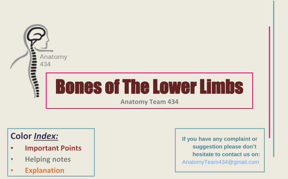
Bones of The Lower LimbsAnatomy Team 434
Color Index:▪ Important Points
▪ Helping notes
▪ Explanation
If you have any complaint or
suggestion please don’t
hesitate to contact us on:

● At the end of the lecture the students should be able to:
● Classify the bones of the three regions of the lower limb (thigh, leg and foot).
● Memorize the main features of the
–Bones of the thigh (femur & patella)
–Bones of the leg (tibia & Fibula).
–Bones of the foot (tarsals, metatarsals and phalanges)
● Recognize the sides of the bone
OBJECTIVES

General Term Meaning
Processes that
helps to form
joints
Condyle Large, rounded articular
Facet Smooth, flat surface
Head Enlarged portion at an end of a bone
Ramus Branch or extension of a bone
Processes that
provide for the
attachment of
muscles and
ligaments
Crest Narrow ridge
Epicondyle Linea
(line)Process on or above a condyle Narrow ridge (less prominent than a crest)
Spine Sharp or pointed process (spinous process)
TrochanterLarge, irregularly shaped process (found only on the femur) ( for attachment of other structures
(ligaments))
Tubercle Small, knoblike process (trabecular : site of muscle attachment)
Tuberosity Large, knoblike process
New Terms
REMEMBER:lower and upper ends of bones are important for articulations.
.
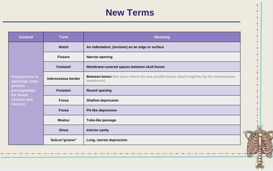
General Term Meaning
Depressions or
openings (may
provide
passageways
for blood
vessels and
nerves)
Notch An indentation, (incision) on an edge or surface
Fissure Narrow opening
Fontanel Membrane-covered spaces between skull bones
Interosseous borderBetween bones (the place where the two parallel bones attach together by the interosseous
membrane)
Foramen Round opening
Fossa Shallow depression
Fovea Pit-like depression
Meatus Tube-like passage
Sinus Interior cavity
Sulcus"groove" Long, narrow depression
New Terms
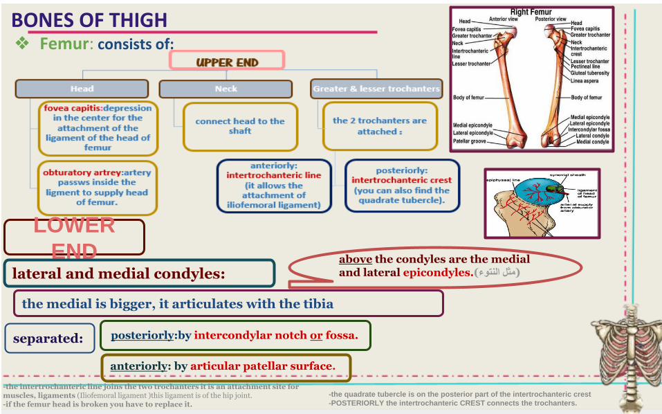
BONES OF THIGH ❖ Femur: consists of:
-the intertrochanteric line joins the two trochanters it is an attachment site for muscles, ligaments (Iliofemoral ligament )this ligament is of the hip joint.-if the femur head is broken you have to replace it.
LOWER
ENDlateral and medial condyles:
the medial is bigger, it articulates with the tibia
separated:
anteriorly: by articular patellar surface.
posteriorly:by intercondylar notch or fossa.
above the condyles are the medial and lateral epicondyles.( النتوءمثل )
-the quadrate tubercle is on the posterior part of the intertrochanteric crest
-POSTERIORLY the intertrochanteric CREST connects the trochanters.
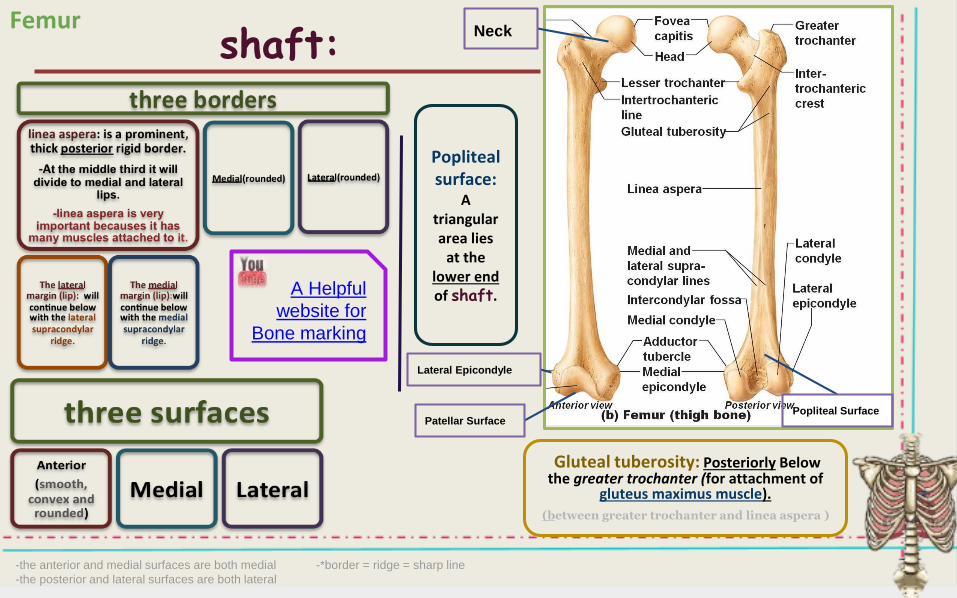
shaft:
Gluteal tuberosity: Posteriorly Belowthe greater trochanter (for attachment of
gluteus maximus muscle).
(between greater trochanter and linea aspera )
Popliteal surface:
A triangular area lies
at the lower endof shaft.
Neck
Lateral Epicondyle
Patellar Surface Popliteal Surface
A Helpful
website for
Bone marking
Femur
-the anterior and medial surfaces are both medial -*border = ridge = sharp line
-the posterior and lateral surfaces are both lateral
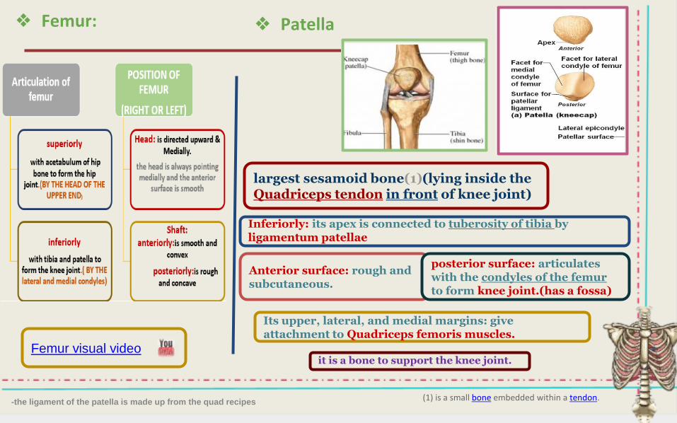
❖ Femur:
Femur visual video
❖ Patella
-the ligament of the patella is made up from the quad recipes(1) is a small bone embedded within a tendon.
largest sesamoid bone(1)(lying inside the Quadriceps tendon in front of knee joint)
Inferiorly: its apex is connected to tuberosity of tibia byligamentum patellae
Anterior surface: rough and subcutaneous.
posterior surface: articulates with the condyles of the femur to form knee joint.(has a fossa)
Its upper, lateral, and medial margins: give attachment to Quadriceps femoris muscles.
it is a bone to support the knee joint.

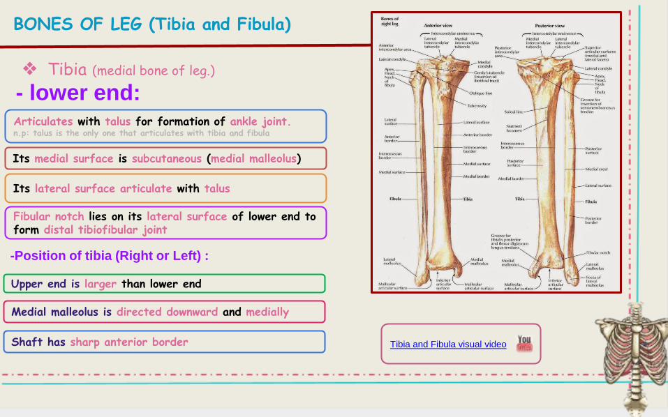
BONES OF LEG (Tibia and Fibula)
❖ Tibia (medial bone of leg.)
-tibia’s upper end is larger than lower end

# Tibial tuberosity:
# THREE borders:
# THREE surfaces
-Interosseous membrane is between lateral border of tibia and medial border of fibula.-the surface between the anterior border and medial border is medial ( the sharp subcutaneous shaft of the tibia)
BONES OF LEG (Tibia and Fibula)
❖ Tibia (medial bone of leg.)
-Shaft :
-Its upper smooth part gives attachment to ligamentum patellae.-Its lower rough part is subcutaneous
- Anterior border is sharp and subcutaneous(the tuberosity on the superior part of it)
- Medial border- Lateral border also called “interosseous border”. n.p:medial and lateral borders are rough.
- Medial : subcutaneous. n.p:it is between anterior and medial border
- Lateral- Posterior has oblique line, soleal line for attachment of soleus muscle
❖ Tibia (medial bone of leg.)
- lower end:Articulates with talus for formation of ankle joint. n.p: talus is the only one that articulates with tibia and fibula
Its medial surface is subcutaneous (medial malleolus)
Its lateral surface articulate with talus
Fibular notch lies on its lateral surface of lower end to form distal tibiofibular joint
Upper end is larger than lower end
Medial malleolus is directed downward and medially
Shaft has sharp anterior border
-Position of tibia (Right or Left) :
Tibia and Fibula visual video
BONES OF LEG (Tibia and Fibula)
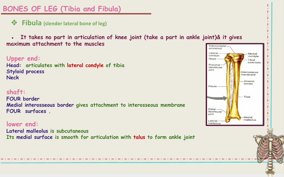
● It takes no part in articulation of knee joint (take a part in ankle joint)& it gives maximum attachment to the muscles
Upper end:Head: articulates with lateral condyle of tibiaStyloid processNeck
shaft:FOUR borderMedial interosseous border gives attachment to interosseous membraneFOUR surfaces .
lower end:Lateral malleolus is subcutaneousIts medial surface is smooth for articulation with talus to form ankle joint
❖ Fibula (slender lateral bone of leg)
BONES OF LEG (Tibia and Fibula)
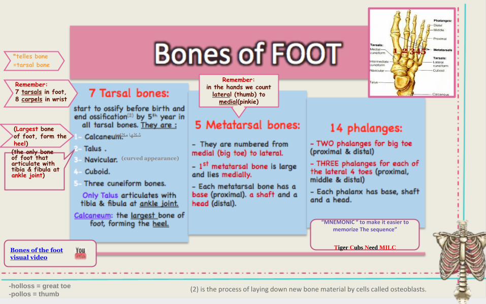
Remember:7 tarsals in foot,8 carpels in wrist
(2) is the process of laying down new bone material by cells called osteoblasts.
Remember:in the hands we count
lateral (thumb) tomedial(pinkie)
مكلكعهشكلها
(curved appearance)
*telles bone
=tarsal bone
(Largest bone of foot, form the heel)
(the only bone of foot that articulate with tibia & fibula atankle joint)
“MNEMONIC “ to make it easier to memorize The sequence”
Tiger Cubs Need MILCBones of the foot visual video
-holloss = great toe
-pollos = thumb
MCQ’s
1-The gluteus maximus muscle is attached to the greater
trochanter:a)True b)False
2-The patella is the largest sesamoid bone in the body:
a)True
b)False
3-The ligamentum patellae of the patella is connected to
tuberosity of tibia:
a)True
b)False
4-Calcaneum and talus are the only bones that articulate with
tibia and fibula at ankle joint:
a)True
b)False
5-A thick posterior border of the shaft of femur is called:
a)Fovea capitis
b)Intertrochanteric crest
c)Linea aspera
d)Gluteal tuberosity
6-Which one of the following tarsal bones forms the heel:
a)Talus
b)Calcaneum
c)Navicular
d)Cuboid
Answers:
1-b
2-a
3-a
4-b
5-c
6-b
7-d
8-d
9-c
10-a
Extra questions (not all are included):
Lower Limb Anatomy MCQs
7-Which one of the following is not a surface for FEMUR:
a)Anterior
b)Medial
c)Lateral
d)Posterior
8-Which of the following is not a border for TIBIA:
a)Anterior
b)Medial
c)Lateral
d)Posterior
9-The Triangular area that lies at the lower end
of the femur shaft is called:
a)Lateral condyle
b)Patellar groove
c)Popliteal surface
d)Medial condyle
10-The area between the lateral and medial condyle
in the posterior of the FEMUR:
a)Intercondylar fossa
b)Patellar surface
c)Linea aspera
d)Popliteal surface