Biodegradable polymer coated 45S5 Bioglassderived glass-ceramic scaffolds for bone tissue...
8
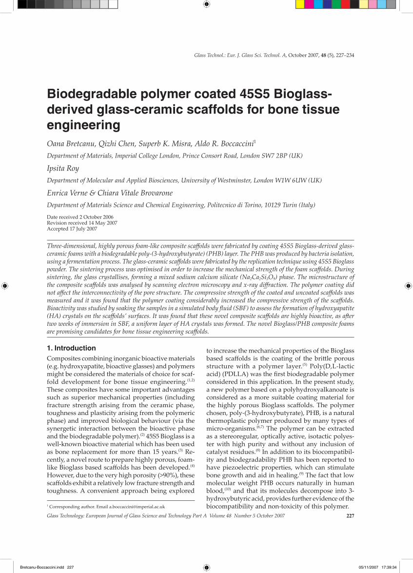
Glass Technology: European Journal of Glass Science and Technology Part A Volume 48 Number 5 October 2007 227 1. Introduction Composites combining inorganic bioactive materials (e.g. hydroxyapatite, bioactive glasses) and polymers might be considered the materials of choice for scaf- fold development for bone tissue engineering. (1,2) These composites have some important advantages such as superior mechanical properties (including fracture strength arising from the ceramic phase, toughness and plasticity arising from the polymeric phase) and improved biological behaviour (via the synergetic interaction between the bioactive phase and the biodegradable polymer). (2) 45S5 Bioglass is a well-known bioactive material which has been used as bone replacement for more than 15 years. (3) Re- cently, a novel route to prepare highly porous, foam- like Bioglass based scaffolds has been developed. (4) However, due to the very high porosity (>90%), these scaffolds exhibit a relatively low fracture strength and toughness. A convenient approach being explored to increase the mechanical properties of the Bioglass based scaffolds is the coating of the brile porous structure with a polymer layer. (5) Poly(D,L-lactic acid) (PDLLA) was the first biodegradable polymer considered in this application. In the present study, a new polymer based on a polyhydroxyalkanoate is considered as a more suitable coating material for the highly porous Bioglass scaffolds. The polymer chosen, poly-(3-hydroxybutyrate), PHB, is a natural thermoplastic polymer produced by many types of micro-organisms. (6,7) The polymer can be extracted as a stereoregular, optically active, isotactic polyes- ter with high purity and without any inclusion of catalyst residues. (8) In addition to its biocompatibil- ity and biodegradability PHB has been reported to have piezoelectric properties, which can stimulate bone growth and aid in healing. (9) The fact that low molecular weight PHB occurs naturally in human blood, (10) and that its molecules decompose into 3- hydroxybutyric acid, provides further evidence of the biocompatibility and non-toxicity of this polymer. Biodegradable polymer coated 45S5 Bioglass- derived glass-ceramic scaffolds for bone tissue engineering Oana Bretcanu, Qizhi Chen, Superb K. Misra, Aldo R. Boccaccini 1 Department of Materials, Imperial College London, Prince Consort Road, London SW7 2BP (UK) Ipsita Roy Department of Molecular and Applied Biosciences, University of Westminster, London W1W 6UW (UK) Enrica Verne & Chiara Vitale Brovarone Department of Materials Science and Chemical Engineering, Politecnico di Torino, 10129 Turin (Italy) Date received 2 October 2006 Revision received 14 May 2007 Accepted 17 July 2007 Three-dimensional, highly porous foam-like composite scaffolds were fabricated by coating 45S5 Bioglass-derived glass- ceramic foams with a biodegradable poly-(3-hydroxybutyrate) (PHB) layer. The PHB was produced by bacteria isolation, using a fermentation process. The glass-ceramic scaffolds were fabricated by the replication technique using 45S5 Bioglass powder. The sintering process was optimised in order to increase the mechanical strength of the foam scaffolds. During sintering, the glass crystallises, forming a mixed sodium calcium silicate (Na 2 Ca 2 Si 3 O 9 ) phase. The microstructure of the composite scaffolds was analysed by scanning electron microscopy and x-ray diffraction. The polymer coating did not affect the interconnectivity of the pore structure. The compressive strength of the coated and uncoated scaffolds was measured and it was found that the polymer coating considerably increased the compressive strength of the scaffolds. Bioactivity was studied by soaking the samples in a simulated body fluid (SBF) to assess the formation of hydroxyapatite (HA) crystals on the scaffolds’ surfaces. It was found that these novel composite scaffolds are highly bioactive, as aſter two weeks of immersion in SBF, a uniform layer of HA crystals was formed. The novel Bioglass/PHB composite foams are promising candidates for bone tissue engineering scaffolds. 1 Corresponding author. Email [email protected] Glass Technol.: Eur. J. Glass Sci. Technol. A, October 2007, 48 (5), 227–234 Bretcanu-Boccaccini.indd 227 05/11/2007 17:39:34
Transcript of Biodegradable polymer coated 45S5 Bioglassderived glass-ceramic scaffolds for bone tissue...
Glass Technology: European Journal of Glass Science and Technology Part A Volume 48 Number 5 October 2007 227
1. IntroductionComposites combining inorganic bioactive materials (e.g. hydroxyapatite, bioactive glasses) and polymers might be considered the materials of choice for scaf-fold development for bone tissue engineering.(1,2) These composites have some important advantages such as superior mechanical properties (including fracture strength arising from the ceramic phase, toughness and plasticity arising from the polymeric phase) and improved biological behaviour (via the synergetic interaction between the bioactive phase and the biodegradable polymer).(2) 45S5 Bioglass is a well-known bioactive material which has been used as bone replacement for more than 15 years.(3) Re-cently, a novel route to prepare highly porous, foam-like Bioglass based scaffolds has been developed.(4) However, due to the very high porosity (>90%), these scaffolds exhibit a relatively low fracture strength and toughness. A convenient approach being explored
to increase the mechanical properties of the Bioglass based scaffolds is the coating of the brittle porous structure with a polymer layer.(5) Poly(D,L-lactic acid) (PDLLA) was the first biodegradable polymer considered in this application. In the present study, a new polymer based on a polyhydroxyalkanoate is considered as a more suitable coating material for the highly porous Bioglass scaffolds. The polymer chosen, poly-(3-hydroxybutyrate), PHB, is a natural thermoplastic polymer produced by many types of micro-organisms.(6,7) The polymer can be extracted as a stereoregular, optically active, isotactic polyes-ter with high purity and without any inclusion of catalyst residues.(8) In addition to its biocompatibil-ity and biodegradability PHB has been reported to have piezoelectric properties, which can stimulate bone growth and aid in healing.(9) The fact that low molecular weight PHB occurs naturally in human blood,(10) and that its molecules decompose into 3-hydroxybutyric acid, provides further evidence of the biocompatibility and non-toxicity of this polymer.
Biodegradable polymer coated 45S5 Bioglass-derived glass-ceramic scaffolds for bone tissue engineeringOana Bretcanu, Qizhi Chen, Superb K. Misra, Aldo R. Boccaccini1
Department of Materials, Imperial College London, Prince Consort Road, London SW7 2BP (UK)
Ipsita RoyDepartment of Molecular and Applied Biosciences, University of Westminster, London W1W 6UW (UK)
Enrica Verne & Chiara Vitale BrovaroneDepartment of Materials Science and Chemical Engineering, Politecnico di Torino, 10129 Turin (Italy)
Date received 2 October 2006Revision received 14 May 2007Accepted 17 July 2007
Three-dimensional, highly porous foam-like composite scaffolds were fabricated by coating 45S5 Bioglass-derived glass-ceramic foams with a biodegradable poly-(3-hydroxybutyrate) (PHB) layer. The PHB was produced by bacteria isolation, using a fermentation process. The glass-ceramic scaffolds were fabricated by the replication technique using 45S5 Bioglass powder. The sintering process was optimised in order to increase the mechanical strength of the foam scaffolds. During sintering, the glass crystallises, forming a mixed sodium calcium silicate (Na2Ca2Si3O9) phase. The microstructure of the composite scaffolds was analysed by scanning electron microscopy and x-ray diffraction. The polymer coating did not affect the interconnectivity of the pore structure. The compressive strength of the coated and uncoated scaffolds was measured and it was found that the polymer coating considerably increased the compressive strength of the scaffolds. Bioactivity was studied by soaking the samples in a simulated body fluid (SBF) to assess the formation of hydroxyapatite (HA) crystals on the scaffolds’ surfaces. It was found that these novel composite scaffolds are highly bioactive, as after two weeks of immersion in SBF, a uniform layer of HA crystals was formed. The novel Bioglass/PHB composite foams are promising candidates for bone tissue engineering scaffolds.
1 Corresponding author. Email [email protected]
Glass Technol.: Eur. J. Glass Sci. Technol. A, October 2007, 48 (5), 227–234
Bretcanu-Boccaccini.indd 227 05/11/2007 17:39:34

228 Glass Technology: European Journal of Glass Science and Technology Part A Volume 48 Number 5 October 2007
In the present investigation bacteria-derived PHB has for the first time been combined with 45S5 Bioglass in composite scaffolds for bone tissue engineering. These scaffolds are intended for appli-cations in cancellous bone substitution after trauma incidents. The pore morphology and macrostructure of the scaffolds before and after coating with PHB as well as the coating homogeneity were investigated. A preliminary characterisation of the mechanical properties of the scaffolds is presented.
2. Materials and methods
2.1. Fabrication of Bioglass-derived glass-ceramic scaffoldsThe 45S5 Bioglass-derived glass-ceramic scaffolds were fabricated by the replication technique.(4) The starting materials were fully reticulated polyurethane (PU) foams (Recticel, UK, 60 and 45 ppi) and 45S5 Bioglass powder (NovaMin USA, particle size <5 μm). The PU foam was used as a frame for the replication method. It was impregnated with a Bioglass slurry and heated up to 1100°C. During heating, the foam burnt out and the glass sintered, yielding a 3D porous Bioglass-derived glass-ceramic scaffold.
The slurry was prepared dissolving 3 wt% of poly(D,L-lactic acid) (PDLLA) (Purac biochem, Gorinchem, Holland) in dimethylcarbonate (DMC) (Sigma Aldrich), whilst stirring. When PDLLA was completely dissolved, 40 wt% 45S5 Bioglass powder was added. The resulting suspension was stirred for 1 h, using a magnetic stirrer. Small pieces of polyurethane foam (25×15×15 mm3) were immersed in the slurry and manually rotated to ensure homo-geneous slurry infiltration. The foams were then extracted from the suspension and squeezed to remove excess slurry. The resulting samples (green bodies) were then dried at room temperature for 1 day and subsequently heat treated in a chamber furnace, at a temperature of 1100°C for 1 h. During the heat treatment, the organic phases (PDLLA, DMC and the polyurethane foam) were burnt out, leaving a foam-like Bioglass structure. At the same time, the glass phase crystallised, giving a glass-ceramic structure with enhanced mechanical properties, as discussed elsewhere.(4)
2.2. Polymer production
PHB samples were isolated from a newly charac-terised Gram positive bacteria Bacillus cereus SPV obtained from the culture collection of University of Westminster (UK). The bacteria were grown in batch cultures in a 20 l laboratory fermentor. The fermentor was sterilised whilst it contained only Kannan and Rehacek medium salts.(11) More details of the fermentation process are given elsewhere.(6)
The cells were harvested by centrifuging at 5000 g and then lyophilised. The cell mass was then lysed in 16% sodium hypochlorite (pH 11) at 37°C for 1 h, centrifuged at 4500 rpm, and the residue was washed twice with water, acetone, ethanol and diethyl ether.(12) Finally, the residue was dried and subjected to Soxhlet extraction for 24 h using chloroform as the solvent. The chloroform solution containing PHB was then concentrated by a vacuum rotary evaporator and the PHB was precipitated from chloroform using ten volumes of ice cold methanol. The precipitate obtained was centrifuged and air dried and stored for further use.
2.3. Polymer coating of scaffolds
The PHB solution was prepared by dissolving 3 wt% PHB in chloroform, at temperatures between 60 and 80°C for 2 h, using a reverse flow apparatus (Soxhlet). The Bioglass-derived glass-ceramic scaf-folds were then coated with the PHB solution by a dip coating process. The samples were completely immersed in the PHB solution for 5 min, in a sealed glass container. After that, they were taken out and dried in air at room temperature for at least 4 h, in a fume cupboard.
2.4. Characterisation
Microstructural characterisation of the Bioglass-derived glass-ceramic scaffolds before and after coating was undertaken using x-ray diffraction (XRD) and scanning electron microscopy (SEM). A digital camera was used for documenting the macroscopic structure of the samples.
X-ray diffraction patterns were obtained using a Philips diffractometer with Cu Kα radiation, a step size of 0·04° (2θ) and a fixed counting time of 2 secs per step. The XRD patterns were collected on powder samples over 10–70° 2θ, at 40 kV and 40 mA. Crystalline phase identification was undertaken using the ‘X'Pert HighScore’ program, applying the PCPDFWIN database.
SEM was carried out on a JEOL JSM 840A instru-ment coupled with energy dispersive spectroscopy (EDS). Samples were gold coated and observed at an accelerating voltage of 15 kV.
The compression strength of foams was measured by a Zwick/Roell Z010 instrument, using a cell load of 1 kN and a crosshead speed of 0·5 mm/min. The load was applied until a compression strain of 70% was reached.
A bioactivity test was carried out using the stand-ard in vitro procedure described by Kokubo et al.(13) Small pieces of Bioglass based glass-ceramic scaffolds approximately 5×5×5 mm3 in size were immersed in 10 ml of acellular simulated body fluid (SBF) in coni-
Proc. Eighth Eur. Soc. glaSS Sci. tEchnol. conf., SundErland, uK, 10–14 SEPtEmbEr 2006
Bretcanu-Boccaccini.indd 228 05/11/2007 17:39:34
Glass Technology: European Journal of Glass Science and Technology Part A Volume 48 Number 5 October 2007 229
cal flasks. The pH of the SBF solution was buffered at 7·25 at 37°C before soaking the samples. The conical flasks were placed in an incubator at a controlled temperature of 37°C. The SBF solution was refreshed twice a week, to simulate body fluid circulation inside a human body. The samples were removed from SBF solution after 3, 7, 14, 21 and 28 days. After extraction from the SBF, the samples were gently rinsed with deionised water, and left to dry at room temperature.
The formation of hydroxyapatite layers on the surface of the samples was examined by SEM and XRD.
3. Results and discussion
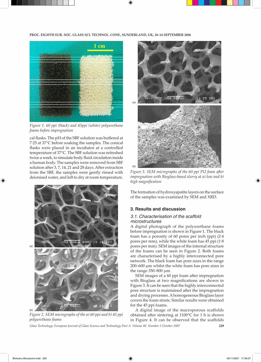
3.1. Characterisation of the scaffold microstructuresA digital photograph of the polyurethane foams before impregnation is shown in Figure 1. The black foam has a porosity of 60 pores per inch (ppi) (2·4 pores per mm), while the white foam has 45 ppi (1·8 pores per mm). SEM images of the internal structure of the foams can be seen in Figure 2. Both foams are characterised by a highly interconnected pore network. The black foam has pore sizes in the range 200–600 μm whilst the white foam has pore sizes in the range 350–800 μm.
SEM images of a 60 ppi foam after impregnation with Bioglass at two magnifications are shown in Figure 3. It can be seen that the highly interconnected pore structure is maintained after the impregnation and drying processes. A homogeneous Bioglass layer covers the foam struts. Similar results were obtained for the 45 ppi foams.
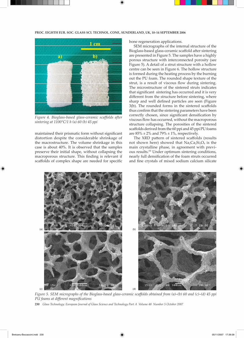
A digital image of the macroporous scaffolds obtained after sintering at 1100°C for 1 h is shown in Figure 4. It can be observed that the scaffolds
Figure 1. 60 ppi (black) and 45ppi (white) polyurethane foams before impregnation
1 cm
Figure 2. SEM micrographs of the a) 60 ppi and b) 45 ppi polyurethane foams
(a)
(b)
Figure 3. SEM micrographs of the 60 ppi PU foam after impregnation with Bioglass-based slurry at a) low and b) high magnification
(a)
(b)
Proc. Eighth Eur. Soc. glaSS Sci. tEchnol. conf., SundErland, uK, 10–14 SEPtEmbEr 2006
Bretcanu-Boccaccini.indd 229 05/11/2007 17:39:37
230 Glass Technology: European Journal of Glass Science and Technology Part A Volume 48 Number 5 October 2007
maintained their prismatic form without significant distortion despite the considerable shrinkage of the macrostructure. The volume shrinkage in this case is about 40%. It is observed that the samples preserve their initial shape, without collapsing the macroporous structure. This finding is relevant if scaffolds of complex shape are needed for specific
bone regeneration applications.SEM micrographs of the internal structure of the
Bioglass-based glass-ceramic scaffold after sintering are presented in Figure 5. The samples have a highly porous structure with interconnected porosity (see Figure 5). A detail of a strut structure with a hollow centre can be seen in Figure 6. The hollow structure is formed during the heating process by the burning out the PU foam. The rounded shape texture of the strut, is a result of viscous flow during sintering. The microstructure of the sintered struts indicates that significant sintering has occurred and it is very different from the structure before sintering, where sharp and well defined particles are seen (Figure 3(b). The rounded forms in the sintered scaffolds thus confirm that the sintering parameters have been correctly chosen, since significant densification by viscous flow has occurred, without the macroporous structure collapsing. The porosities of the sintered scaffolds derived from the 60 ppi and 45 ppi PU foams are 85% ± 2% and 79% ± 1%, respectively.
The XRD pattern of sintered scaffolds (results not shown here) showed that Na2Ca2Si3O9 is the main crystalline phase, in agreement with previ-ous results.(4) Under optimum sintering conditions, nearly full densification of the foam struts occurred and fine crystals of mixed sodium calcium silicate
Figure 4. Bioglass-based glass-ceramic scaffolds after sintering at 1100°C/1 h (a) 60 (b) 45 ppi
1 cm
a) b)
1 cm
a) b)
Figure 5. SEM micrographs of the Bioglass-based glass-ceramic scaffolds obtained from (a)–(b) 60 and (c)–(d) 45 ppi PU foams at different magnifications
(a)
(c)
(b)
(d)
Proc. Eighth Eur. Soc. glaSS Sci. tEchnol. conf., SundErland, uK, 10–14 SEPtEmbEr 2006
Bretcanu-Boccaccini.indd 230 05/11/2007 17:39:39

Glass Technology: European Journal of Glass Science and Technology Part A Volume 48 Number 5 October 2007 231
were formed.SEM micrographs of the Bioglass-based glass-
ceramic/polymer composite are shown in Figure 7. It can be seen that the composite scaffolds maintained the open pore interconnected structure after coating with PHB. However, as seen in the higher magnifica-tion images (Figures 7(b) and (d)), the coating is not homogeneous. The thickness of the polymer coating is <5 μm. Further work is in progress to improve the quality of the PHB coating, for example by increasing
the coating time (from 5 to 15 mins) or by introduc-ing a double or triple coating procedure involving successive dipping cycles.
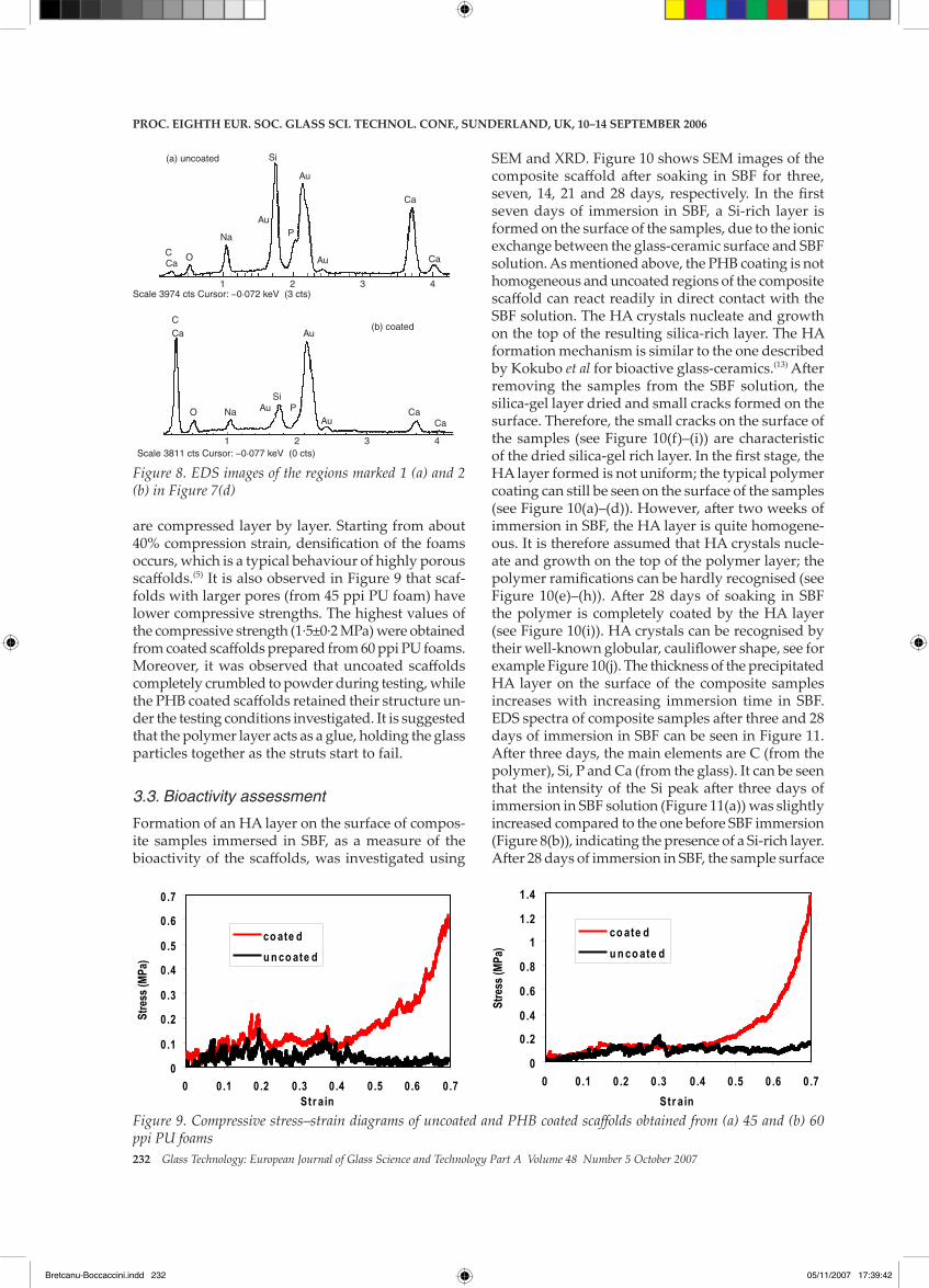
The EDS spectra of the regions marked 1 and 2 in Figure 7(d) are shown in Figure 8. Region 1 is an uncoated area, corresponding to the Bioglass-based glass-ceramic surface. Region 2 is a PHB coated area. Therefore, the first spectrum contains the main ele-ments of Bioglass (Si, Ca, Na, P). The highest peak in this case is the Si peak, due to the large amount of this element in the glass composition. The intensities of the Si, Ca, Na, and P peaks in the spectra of the coated region are lower due to the polymer coating.
3.2. Mechanical behaviour
Typical compressive stress–strain curves of uncoated and coated scaffolds are shown in Figure 9 for scaf-folds obtained from 45 and 60 ppi PU foams. The plot-ted curves are average values based on five samples. As can be seen, the compressive strength and work of fracture of the scaffolds was considerably increased by coating with PHB. The polymer layer covers and fills the microcracks situated on the strut surfaces, improving the mechanical stability of the scaffold. The reduction of microcracking on the strut surfaces led to fewer irregularities in the stress–strain curves. During loading, with increasing stress, the scaffolds
Figure 6. SEM micrograph of a strut structure in a Bi-oglass-based glass-ceramic scaffold obtained from 45ppi PU foam
Figure 7. SEM micrographs of Bioglass-derived glass-ceramic/polymer composite scaffolds obtained from (a)–(b) 45 and (c)–(d) 60 ppi foam at different magnifications
1
2
(a)
(c)
(b)
(d)
Proc. Eighth Eur. Soc. glaSS Sci. tEchnol. conf., SundErland, uK, 10–14 SEPtEmbEr 2006
Bretcanu-Boccaccini.indd 231 05/11/2007 17:39:39
232 Glass Technology: European Journal of Glass Science and Technology Part A Volume 48 Number 5 October 2007
are compressed layer by layer. Starting from about 40% compression strain, densification of the foams occurs, which is a typical behaviour of highly porous scaffolds.(5) It is also observed in Figure 9 that scaf-folds with larger pores (from 45 ppi PU foam) have lower compressive strengths. The highest values of the compressive strength (1·5±0·2 MPa) were obtained from coated scaffolds prepared from 60 ppi PU foams. Moreover, it was observed that uncoated scaffolds completely crumbled to powder during testing, while the PHB coated scaffolds retained their structure un-der the testing conditions investigated. It is suggested that the polymer layer acts as a glue, holding the glass particles together as the struts start to fail.
3.3. Bioactivity assessment
Formation of an HA layer on the surface of compos-ite samples immersed in SBF, as a measure of the bioactivity of the scaffolds, was investigated using
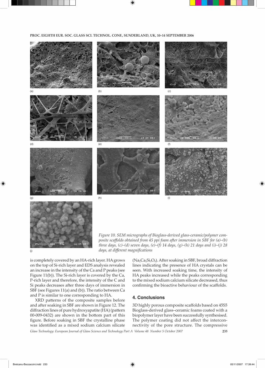
SEM and XRD. Figure 10 shows SEM images of the composite scaffold after soaking in SBF for three, seven, 14, 21 and 28 days, respectively. In the first seven days of immersion in SBF, a Si-rich layer is formed on the surface of the samples, due to the ionic exchange between the glass-ceramic surface and SBF solution. As mentioned above, the PHB coating is not homogeneous and uncoated regions of the composite scaffold can react readily in direct contact with the SBF solution. The HA crystals nucleate and growth on the top of the resulting silica-rich layer. The HA formation mechanism is similar to the one described by Kokubo et al for bioactive glass-ceramics.(13) After removing the samples from the SBF solution, the silica-gel layer dried and small cracks formed on the surface. Therefore, the small cracks on the surface of the samples (see Figure 10(f)–(i)) are characteristic of the dried silica-gel rich layer. In the first stage, the HA layer formed is not uniform; the typical polymer coating can still be seen on the surface of the samples (see Figure 10(a)–(d)). However, after two weeks of immersion in SBF, the HA layer is quite homogene-ous. It is therefore assumed that HA crystals nucle-ate and growth on the top of the polymer layer; the polymer ramifications can be hardly recognised (see Figure 10(e)–(h)). After 28 days of soaking in SBF the polymer is completely coated by the HA layer (see Figure 10(i)). HA crystals can be recognised by their well-known globular, cauliflower shape, see for example Figure 10(j). The thickness of the precipitated HA layer on the surface of the composite samples increases with increasing immersion time in SBF. EDS spectra of composite samples after three and 28 days of immersion in SBF can be seen in Figure 11. After three days, the main elements are C (from the polymer), Si, P and Ca (from the glass). It can be seen that the intensity of the Si peak after three days of immersion in SBF solution (Figure 11(a)) was slightly increased compared to the one before SBF immersion (Figure 8(b)), indicating the presence of a Si-rich layer. After 28 days of immersion in SBF, the sample surface
Figure 9. Compressive stress–strain diagrams of uncoated and PHB coated scaffolds obtained from (a) 45 and (b) 60 ppi PU foams
0
0.1
0.2
0.3
0.4
0.5
0.6
0.7
0 0.1 0.2 0.3 0.4 0.5 0.6 0.7Str ain
Stre
ss (M
Pa)
co ate du n co ate d
0
0.2
0.4
0.6
0.8
1
1.2
1.4
0 0.1 0.2 0.3 0.4 0.5 0.6 0.7Str ain
Stre
ss (M
Pa)
co ate du n co ate d
Figure 8. EDS images of the regions marked 1 (a) and 2 (b) in Figure 7(d)
Scale 3974 cts Cursor: −0·072 keV (3 cts) 1 2 3 4
CCa
O
Na
Au
Si
P
Ca
Au
Au
Ca
(a) uncoated
1 2 3 4Scale 3811 cts Cursor: −0·077 keV (0 cts)
Au CaCa
Au
PSi
AuNaO
CaC
(b) coated
Proc. Eighth Eur. Soc. glaSS Sci. tEchnol. conf., SundErland, uK, 10–14 SEPtEmbEr 2006
Bretcanu-Boccaccini.indd 232 05/11/2007 17:39:42
Glass Technology: European Journal of Glass Science and Technology Part A Volume 48 Number 5 October 2007 233
Figure 10. SEM micrographs of Bioglass-derived glass-ceramic/polymer com-posite scaffolds obtained from 45 ppi foam after immersion in SBF for (a)–(b) three days, (c)–(d) seven days, (e)–(f) 14 days, (g)–(h) 21 days and (i)–(j) 28 days, at different magnifications
(a) (b) (c)
(d) (e) (f)
(g) (h) (i)
(j)
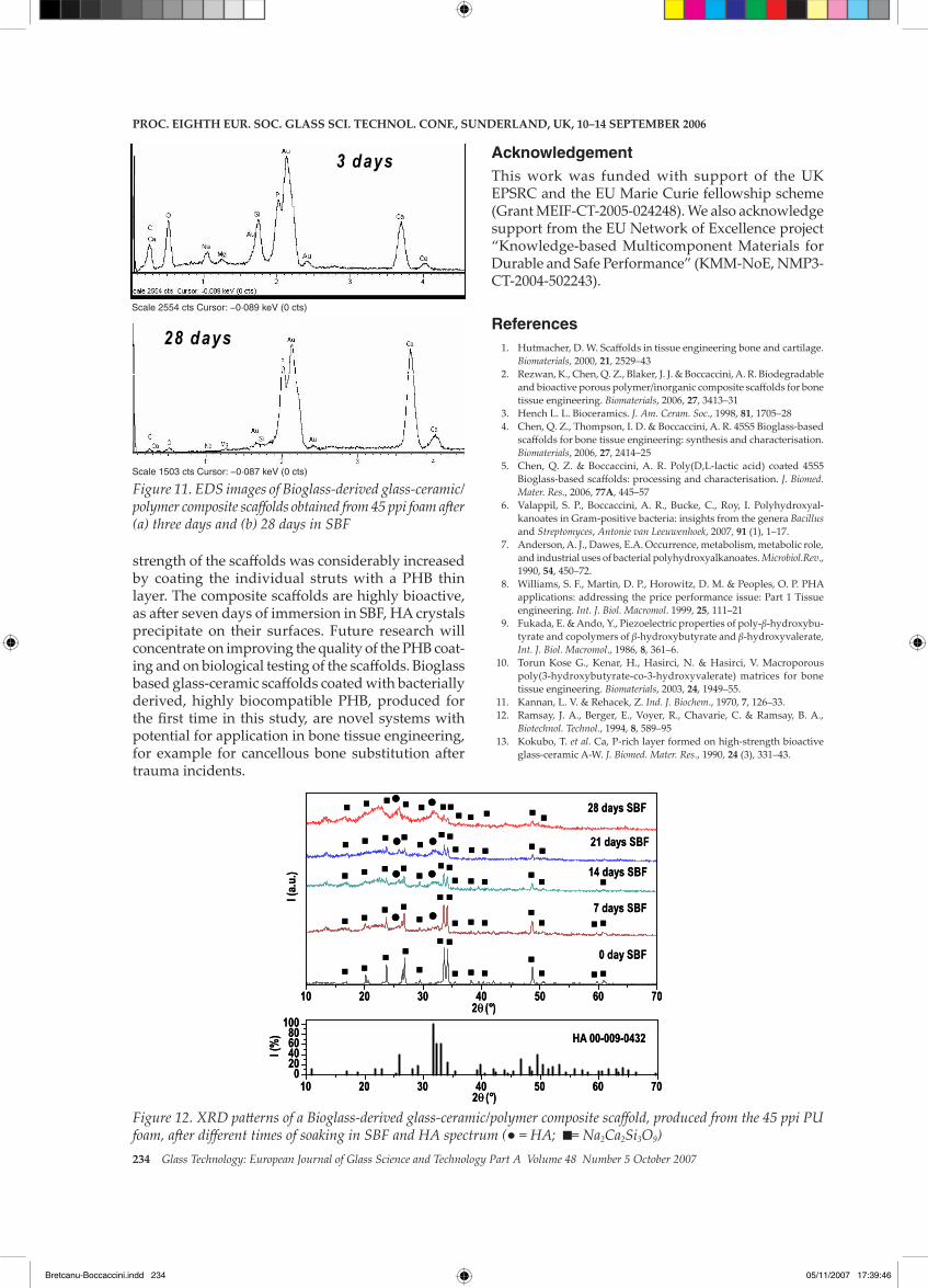
is completely covered by an HA-rich layer. HA grows on the top of Si-rich layer and EDS analysis revealed an increase in the intensity of the Ca and P peaks (see Figure 11(b)). The Si-rich layer is covered by the Ca, P-rich layer and therefore, the intensity of the C and Si peaks decreases after three days of immersion in SBF (see Figures 11(a) and (b)). The ratio between Ca and P is similar to one corresponding to HA.
XRD patterns of the composite samples before and after soaking in SBF are shown in Figure 12. The diffraction lines of pure hydroxyapatite (HA) (pattern 00-009-0432) are shown in the bottom part of this figure. Before soaking in SBF the crystalline phase was identified as a mixed sodium calcium silicate
(Na2Ca2Si3O9). After soaking in SBF, broad diffraction lines indicating the presence of HA crystals can be seen. With increased soaking time, the intensity of HA peaks increased while the peaks corresponding to the mixed sodium calcium silicate decreased, thus confirming the bioactive behaviour of the scaffolds.
4. Conclusions
3D highly porous composite scaffolds based on 45S5 Bioglass-derived glass–ceramic foams coated with a biopolymer layer have been successfully synthesised. The polymer coating did not affect the intercon-nectivity of the pore structure. The compressive
Proc. Eighth Eur. Soc. glaSS Sci. tEchnol. conf., SundErland, uK, 10–14 SEPtEmbEr 2006
Bretcanu-Boccaccini.indd 233 05/11/2007 17:39:44
234 Glass Technology: European Journal of Glass Science and Technology Part A Volume 48 Number 5 October 2007
Figure 11. EDS images of Bioglass-derived glass-ceramic/polymer composite scaffolds obtained from 45 ppi foam after (a) three days and (b) 28 days in SBF
3 days 3 days 3 days
Scale 2554 cts Cursor: −0·089 keV (0 cts)
28 days 28 days 28 days
Scale 1503 cts Cursor: −0·087 keV (0 cts)
Figure 12. XRD patterns of a Bioglass-derived glass-ceramic/polymer composite scaffold, produced from the 45 ppi PU foam, after different times of soaking in SBF and HA spectrum (● = HA; = Na2Ca2Si3O9)
10 20 30 40 50 60 70
HA 00-009-0432
28 days SBF
21 days SBF
7 days SBF
0 day SBF
I (a.
u.) 14 days SBF
2θ (°)
10 20 30 40 50 60 700
20406080
100
2θ (°)
I (%
)
10 20 30 40 50 60 70
HA 00-009-0432
28 days SBF
21 days SBF
7 days SBF
0 day SBF
I (a.
u.) 14 days SBF
2θ (°)
10 20 30 40 50 60 700
20406080
100
2θ (°)
I (%
)
strength of the scaffolds was considerably increased by coating the individual struts with a PHB thin layer. The composite scaffolds are highly bioactive, as after seven days of immersion in SBF, HA crystals precipitate on their surfaces. Future research will concentrate on improving the quality of the PHB coat-ing and on biological testing of the scaffolds. Bioglass based glass-ceramic scaffolds coated with bacterially derived, highly biocompatible PHB, produced for the first time in this study, are novel systems with potential for application in bone tissue engineering, for example for cancellous bone substitution after trauma incidents.
AcknowledgementThis work was funded with support of the UK EPSRC and the EU Marie Curie fellowship scheme (Grant MEIF-CT-2005-024248). We also acknowledge support from the EU Network of Excellence project “Knowledge-based Multicomponent Materials for Durable and Safe Performance” (KMM-NoE, NMP3-CT-2004-502243).
References 1. Hutmacher, D. W. Scaffolds in tissue engineering bone and cartilage.
Biomaterials, 2000, 21, 2529–43 2. Rezwan, K., Chen, Q. Z., Blaker, J. J. & Boccaccini, A. R. Biodegradable
and bioactive porous polymer/inorganic composite scaffolds for bone tissue engineering. Biomaterials, 2006, 27, 3413–31
3. Hench L. L. Bioceramics. J. Am. Ceram. Soc., 1998, 81, 1705–28 4. Chen, Q. Z., Thompson, I. D. & Boccaccini, A. R. 45S5 Bioglass-based
scaffolds for bone tissue engineering: synthesis and characterisation. Biomaterials, 2006, 27, 2414–25
5. Chen, Q. Z. & Boccaccini, A. R. Poly(D,L-lactic acid) coated 45S5 Bioglass-based scaffolds: processing and characterisation. J. Biomed. Mater. Res., 2006, 77a, 445–57
6. Valappil, S. P., Boccaccini, A. R., Bucke, C., Roy, I. Polyhydroxyal-kanoates in Gram-positive bacteria: insights from the genera Bacillus and Streptomyces, Antonie van Leeuwenhoek, 2007, 91 (1), 1–17.
7. Anderson, A. J., Dawes, E.A. Occurrence, metabolism, metabolic role, and industrial uses of bacterial polyhydroxyalkanoates. Microbiol.Rev., 1990, 54, 450–72.
8. Williams, S. F., Martin, D. P., Horowitz, D. M. & Peoples, O. P. PHA applications: addressing the price performance issue: Part 1 Tissue engineering. Int. J. Biol. Macromol. 1999, 25, 111–21
9. Fukada, E. & Ando, Y., Piezoelectric properties of poly-β-hydroxybu-tyrate and copolymers of β-hydroxybutyrate and β-hydroxyvalerate, Int. J. Biol. Macromol., 1986, 8, 361–6.
10. Torun Kose G., Kenar, H., Hasirci, N. & Hasirci, V. Macroporous poly(3-hydroxybutyrate-co-3-hydroxyvalerate) matrices for bone tissue engineering. Biomaterials, 2003, 24, 1949–55.
11. Kannan, L. V. & Rehacek, Z. Ind. J. Biochem., 1970, 7, 126–33. 12. Ramsay, J. A., Berger, E., Voyer, R., Chavarie, C. & Ramsay, B. A.,
Biotechnol. Technol., 1994, 8, 589–95 13. Kokubo, T. et al. Ca, P-rich layer formed on high-strength bioactive
glass-ceramic A-W. J. Biomed. Mater. Res., 1990, 24 (3), 331–43.
Proc. Eighth Eur. Soc. glaSS Sci. tEchnol. conf., SundErland, uK, 10–14 SEPtEmbEr 2006
Bretcanu-Boccaccini.indd 234 05/11/2007 17:39:46