Beyond Watson and Crick: DNA methylation and molecular enzymology of DNA methyltransferases
20
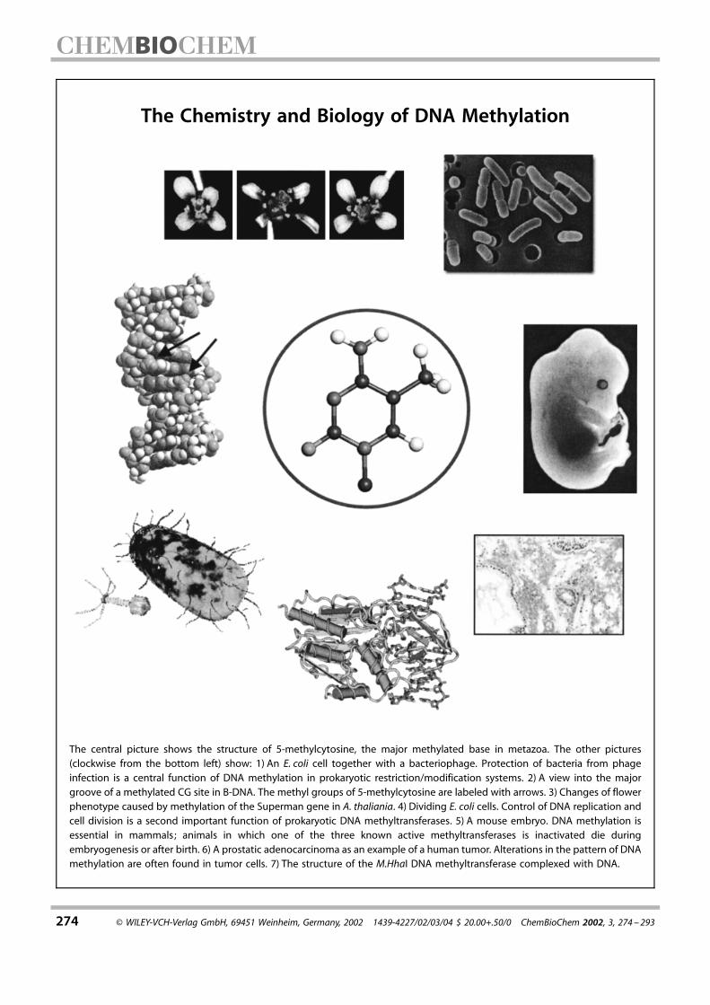
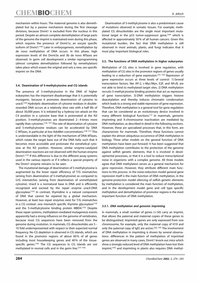
274 ¹ WILEY-VCH-Verlag GmbH, 69451 Weinheim, Germany, 2002 1439-4227/02/03/04 $ 20.00+.50/0 ChemBioChem 2002, 3, 274 ± 293 The central picture shows the structure of 5-methylcytosine, the major methylated base in metazoa. The other pictures (clockwise from the bottom left) show: 1) An E. coli cell together with a bacteriophage. Protection of bacteria from phage infection is a central function of DNA methylation in prokaryotic restriction/modification systems. 2) A view into the major groove of a methylated CG site in B-DNA. The methyl groups of 5-methylcytosine are labeled with arrows. 3) Changes of flower phenotype caused by methylation of the Superman gene in A. thaliania. 4) Dividing E. coli cells. Control of DNA replication and cell division is a second important function of prokaryotic DNA methyltransferases. 5) A mouse embryo. DNA methylation is essential in mammals; animals in which one of the three known active methyltransferases is inactivated die during embryogenesis or after birth. 6) A prostatic adenocarcinoma as an example of a human tumor. Alterations in the pattern of DNA methylation are often found in tumor cells. 7) The structure of the M.HhaI DNA methyltransferase complexed with DNA. The Chemistry and Biology of DNA Methylation
-
Upload
independent -
Category
Documents
-
view
0 -
download
0
Transcript of Beyond Watson and Crick: DNA methylation and molecular enzymology of DNA methyltransferases
274 ! WILEY-VCH-Verlag GmbH, 69451 Weinheim, Germany, 2002 1439-4227/02/03/04 $ 20.00+.50/0 ChemBioChem 2002, 3, 274 ±293
The central picture shows the structure of 5-methylcytosine, the major methylated base in metazoa. The other pictures(clockwise from the bottom left) show: 1) An E. coli cell together with a bacteriophage. Protection of bacteria from phageinfection is a central function of DNA methylation in prokaryotic restriction/modification systems. 2) A view into the majorgroove of a methylated CG site in B-DNA. The methyl groups of 5-methylcytosine are labeled with arrows. 3) Changes of flowerphenotype caused by methylation of the Superman gene in A. thaliania. 4) Dividing E. coli cells. Control of DNA replication andcell division is a second important function of prokaryotic DNA methyltransferases. 5) A mouse embryo. DNA methylation isessential in mammals; animals in which one of the three known active methyltransferases is inactivated die duringembryogenesis or after birth. 6) A prostatic adenocarcinoma as an example of a human tumor. Alterations in the pattern of DNAmethylation are often found in tumor cells. 7) The structure of the M.HhaI DNA methyltransferase complexed with DNA.
The Chemistry and Biology of DNA Methylation

ChemBioChem 2002, 3, 274 ± 293 ! WILEY-VCH-Verlag GmbH, 69451 Weinheim, Germany, 2002 1439-4227/02/03/04 $ 20.00+.50/0 275
Beyond Watson and Crick:DNA Methylation and Molecular Enzymologyof DNA MethyltransferasesAlbert Jeltsch*[a]
DNA methyltransferases catalyze the transfer of a methyl groupfrom S-adenosyl-L-methionine to cytosine or adenine bases in DNA.These enzymes challenge the Watson/Crick dogma in twoinstances: 1) They attach inheritable information to the DNA thatis not encoded in the nucleotide sequence. This so-called epigeneticinformation has many important biological functions. In prokar-yotes, DNA methylation is used to coordinate DNA replication andthe cell cycle, to direct postreplicative mismatch repair, and todistinguish self and nonself DNA. In eukaryotes, DNA methylationcontributes to the control of gene expression, the protection of thegenome against selfish DNA, maintenance of genome integrity,parental imprinting, X-chromosome inactivation in mammals, andregulation of development. 2) The enzymatic mechanism of DNA
methyltransferases is unusual, because these enzymes flip theirtarget base out of the DNA helix and, thereby, locally disrupt theB-DNA helix. This review describes the biological functions of DNAmethylation in bacteria, fungi, plants, and mammals. In addition,the structures and mechanisms of the DNA methyltransferases,which enable them to specifically recognize their DNA targets andto induce such large conformational changes of the DNA, arediscussed.
KEYWORDS:
DNA methylation ¥ enzyme catalysis ¥ epigenetics ¥gene expression ¥ protein ±DNA interactions
1. Introduction: DNA Methylation as anExtension of the Genetic Code
It has been known for decades that DNA from various sourcescontains the methylated bases N6-methyladenine, 5-methylcy-tosine, and N4-methylcytosine in addition to the four standardnucleobases (Scheme 1). It should be noted that these methy-lated bases are natural components of DNA; this distinguishesthem from a large variety of chemically modified bases that canbe formed by alkylation or oxidative damage of the DNA. DNAmethylation is introduced enzymatically by DNA methyltransfer-ases (MTases) after DNA replication. These enzymes use S-adenosyl-L-methionine (AdoMet) as the donor of an activatedmethyl group and modify the DNA in a sequence-specificmanner, usually at palindromic sites (Table 1). The methylationdoes not interfere with the Watson/Crick pairing properties ofadenine and cytosine but the methyl group is positioned in the
Scheme 1. Structures of methylated bases occurring in DNA.
major groove of the DNA, where it can easily be detected byproteins interacting with the DNA. Thereby, methylation addsextra information to the DNA that is not encoded in thesequence, and the methylated bases can be considered the 5th,6th, and 7th letters of the genetic alphabet.
The process of DNA methylation is intimately interwoven withDNA replication, which inherently destroys DNA methylationbecause the newly synthesized DNA strand does not carry anymethylation. Since it is usually palindromic sequences that aremodified, a methylation mark is present on both strands of theDNA (for example: GmATC/GmATC! ™fully methylated"). Duringsemiconservative DNA replication, these sites are converted intohemimethylated ones (GmATC/GATC), which are re-transformedby a DNA methyltransferase into the fully methylated state.
The focus of this review is the chemistry, enzymology, andbiological function of DNA methylation. There exist several otherexcellent reviews on DNA methylation to whom the reader isreferred for additional details on structures and mechanisms ofDNA MTases,[1±5] DNA methylation in prokaryotes,[6±8] andeukaryotic DNA methylation.[9±15]
[a] Priv.-Doz. Dr. A. JeltschInstitut f¸r Biochemie, FB8Justus-Liebig-Universit‰tHeinrich-Buff-Ring 5835392 Giessen (Germany)Fax: ("49)641-99-35409E-mail : [email protected]

A. Jeltsch
276 ChemBioChem 2002, 3, 274 ±293
2. The Role of DNA Methylation in Prokaryotes
In prokaryotes, all three types of DNA methylation describedabove are observed. Here, DNA methylation has three majorbiological roles: 1) distinction of self and nonself DNA, 2) direc-tion of postreplicative mismatch repair, and 3) control of DNAreplication and cell cycle. The first of these issues is associatedwith restriction/modification systems (RM systems)[16, 17] whichfunction as a defense against infection of bacteria by bacter-iophages and are the source of the overwhelming majority ofDNA methyltransferases found in prokaryotes. In addition to amethyltransferase, RM systems also contain a restriction endo-nuclease that specifically cleaves DNA invading the cell atdefined recognition sites.[18±20] The cellular DNA is protectedagainst this attack by methylation within the recognition site. If aphage DNA escapes from cleavage, the phage can infect the celland its progeny carries the same methylation pattern as the host
cell. Then, other bacteria containing the same RM system are nolonger protected against infection. Therefore, it is advantageousthat different bacteria carry RM systems with different recog-nition sequences and, as a result, a large diversity of RM systemswith different recognition sequences has been developedthrough the billions of years of bacteria/bacteriophage coevo-lution. So far, over 2000 different RM systems are known, 700different DNA MTases have been sequenced which recognizeand methylate almost 300 different DNA sequences (see: http://www.neb.com/rebase[21] ). In these systems the sequence con-text of the DNA methylation carries the information that allowsthe cell to discriminate between self and nonself DNA.
The role of DNA methylation in DNA repair and control of DNAreplication is best understood in the Escherichia coli dam system,where DNA is modified at adenine residues in GATC sequen-ces.[22, 23] Similar systems are present in other !-proteobacteria. Inthese bacteria the methylation status of GATC sites cyclesbetween hemimethylation (GATC/GmATC) immediately afterDNA replication and full methylation (GmATC/GmATC) afterremethylation by the dam MTase; at most sites this occurs 2 ±4 s after replication.[24] During the short time span between DNAreplication and dam methylation, a directed repair of replicationerrors is possible, because the methylation mark allows theunmethylated daughter strand which must be repaired and themethylated original template strand whose nucleotide sequenceis correct to be distinguished. Moreover, dam methylation isused to couple the bacterial cell cycle to DNA replication,because a number of gene promotors are induced in thehemimethylated state.[23, 25] The only DNA region that remainshemimethylated for a longer period (#20 min) is the origin ofDNA replication,[26, 27] because the SeqA protein binds to it and,thereby, prevents dam methylation. This is used for the controlof DNA replication, because hemimethylated origins of repli-cation are not active. Taken together, hemimethylation of damsites is used to encode two kinds of information: 1) it indicatesthat a DNA molecule already has been replicated, and 2) it labelsthe parental strand after DNA replication. Moreover, dam
Albert Jeltsch was born in Kassel (Ger-many) in 1966. He studied Biochemistryat the University of Hannover andreceived his doctoral degree in 1994 inthe group of Alfred Pingoud for workdealing with the molecular enzymologyof restriction endonucleases. He finish-ed his Habilitation on the molecularenzymology of DNA methyltransferasesin 1999. Today, he is assistant professorat the Institute for Biochemistry of theJustus-Liebig University Giessen, wherehe heads a group working on DNA methylation. He received theGerhard-Hess award of the Deutsche Forschungsgemeinschaft in1999 and the BioFuture award of the Bundesministerium f¸rBildung und Forschung in 2001.
Table 1. Properties of typical DNA MTases.
Recognition sequence Size[a] Classification Comment
prokaryotic enzymes:M.HhaI GCGC 327 cytosine-C5 part of an RM systemM.HaeIII GGCC 330 cytosine-C5 part of an RM systemM.EcoRV GATAC 298 adenine-N6 (" type) part of an RM systemE. coli dam GATC 278 adenine-N6 (" type)M.PvuII CAGCTG 336 cytosine-N4 (# type) part of an RM systemM.TaqI TCGA 421 adenine-N6 (! type) part of an RM system
eukaryotic enzymes:Dnmt1 (mouse) CG 1620 cytosine-C5 high preference for hemimethylated CG/mCG sites[228]
Dnmt2 (mouse) ? 415 ? catalytic activity has not yet been shownDnmt3a (mouse) CG 908 cytosine-C5 also methylates non-CG sites with high activity[136, 206]
Dnmt3b (mouse) CG 859 cytosine-C5 specificity has not yet been investigated in detailMet1 (A. thaliana) CG 1537 cytosine-C5 Dnmt1-typeCMT3 (A. thaliana) CNG 839 cytosine-C5 chromomethylaseDRM2 (A. thaliana) ? 626 cytosine-C5 ? Dnmt3-typeMasc1 (A. immersus) ? 537 cytosine-C5 de novo DNA MTase in vivo, inactive in vitroMasc2 (A. immersus) ? 1356 cytosine-C5 active in vitro, function in vivo unknown
[a] Size is given as the number of amino acid residues.
DNA Methylation
ChemBioChem 2002, 3, 274 ± 293 277
methylation plays a role in the segregation of the E. colichromosome. Despite these important functions, dammethylation is not essential in E. coli, but it is involved inthe pathogenicity of different bacteria like Bordetellapertussis,[28] E. coli,[29] Salmonella thyphimurium,[30±33] andNeisseria meningitidis,[34] a fact that suggests thatinhibitors of dam methylation might be effective asantibacterial drugs.
A similar system of DNA methylation is observed inCaulobacter cresentus and other "-proteobacteria.[35] TheCcrM MTase modifies adenine residues within GANTCsequences. Its biology is less clear understood than thatof the dam system, but it is known that the expression ofthe CcrM MTase is correlated with the cell cycle ofC. cresentus ; this suggests that DNA methylation isinvolved in cell cycle control in these organisms. Incontrast to the dam enzyme, the CcrM MTase is anessential protein in "-proteobacteria.[36] Since the groupof "-proteobacteria contains important human patho-gens and adenine-MTases appear not to exist in highereukaryotes, the CcrM MTases are attractive drug tar-gets–promising inhibitors that are specific for adenineMTases have just been described.[37]
2.1. The structure of prokaryotic DNAmethyltransferases
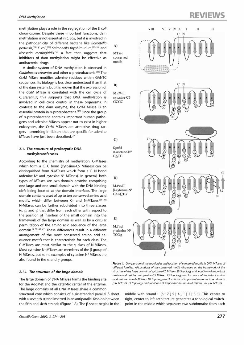
According to the chemistry of methylation, C-MTaseswhich form a C$C bond (cytosine-C5 MTases) can bedistinguished from N-MTases which form a C$N bond(adenine-N6 and cytosine-N4 MTases). In general, bothtypes of MTases are two-domain proteins comprisingone large and one small domain with the DNA bindingcleft being located at the domain interface. The largedomain contains a set of up to ten conserved amino acidmotifs, which differ between C- and N-MTases.[38±40]
N-MTases can be further subdivided into three classes(", #, and !) that differ from each other with respect tothe position of insertion of the small domain into theframework of the large domain as well as by a circularpermutation of the amino acid sequence of the largedomain.[4, 38, 40, 41] These differences result in a differentarrangement of the most conserved amino acid se-quence motifs that is characteristic for each class. TheC-MTases are most similar to the ! class of N-MTases.Most cytosine-N4 MTases are members of the # group ofN-MTases, but some examples of cytosine-N4 MTases arealso found in the " and ! groups.
2.1.1. The structure of the large domain
The large domain of DNA MTases forms the binding sitefor the AdoMet and the catalytic center of the enzyme.The large domains of all DNA MTases share a commonstructural core which consists of a six-stranded parallel # sheetwith a seventh strand inserted in an antiparallel fashion betweenthe fifth and sixth strands (Figure 1A). The # sheet begins in the
middle with strand 1 (6 % 7& 5% 4 & 1 % 2 % 3 % ). This center toright, center to left architecture generates a topological switch-point in the middle which separates two subdomains from each
Figure 1. Comparison of the topologies and location of conserved motifs in DNA MTases ofdifferent families. A) Locations of the conserved motifs displayed on the framework of thestructure of the large domain of cytosine-C5 MTases. B) Topology and locations of importantamino acid residues in cytosine-C5 MTases. C) Topology and locations of important aminoacid residues in !-N MTases. D) Topology and locations of important amino acid residues in"-N MTases. E) Topology and locations of important amino acid residues in #-N MTases.

A. Jeltsch
278 ChemBioChem 2002, 3, 274 ±293
other. The structure of both the subdomains is based on a #
sheet that is flanked by 1 ± 2 " helices on either side with adoubly wound open "/#/" sandwich as the core structure. Theright subdomain (#1 ±#3) creates the AdoMet binding site, theleft one (#4±#7) the binding site for the extrahelical target base(see below). Both binding sites are hydrophobic pockets that arelocated at equivalent positions within the subdomains, a factwhich suggests that the catalytic domain mighthave arisen by a gene duplication.[40] This type ofstructure is also observed in other AdoMet-dependent MTases like RNA MTases, proteinMTases, and small-molecule MTases.[4, 5] The largesubdomains of C- and N-MTases contain up toten characteristic amino acid motifs, where themotifs I (DXFXGXG), IV (GFPCQ), and VI (ENV) aremost conserved in C-MTases[42] and the motifs I(DXFXGXG) and IV ((D/N/S)PP(Y/F)) in N-MT-ases.[40] Although only motif I is similar betweenboth classes of enzymes, it has been found thatcorresponding motifs are located at structurallyequivalent positions in the proteins and formsimilar interactions to the cofactor and otherparts of the protein[4, 5]–a clear example ofproteins with almost no similarity at the aminoacid sequence level that share a remarkablyconserved structure. For example, in both types of enzymesresidues located within the motifs IV and VI interact with theflipped target base and play the most important roles in catalysis(see below).
2.1.2. The structure of the small domain
The small domains of different DNA MTases are dissimilar inamino acid sequence, size, and structure. So far structures of twobacterial C-MTases (M.HhaI[43] and M.HaeIII[44] ) are known.Although the catalytic domains of both proteins can be easilysuperimposed on each other, the structures of the small domainsare dissimilar. The small domain of M.HhaI comprises 81 aminoacid residues and consists of 7 # strands whereas that of M.HaeIII(92 amino acid residues) has no extensive secondary structure.The heterogeneity is even larger in the family of N-MTases,where, so far, four structures are known: M.TaqI,[45] M.PvuII,[46]
DpnM,[47] and M.RsrI.[48] The size of the ™small" domain of theM.TaqI MTase is 177 amino acid residues. It mainly consists of #strands with three " helices forming a three helical bundle onone end. In contrast the small domain of M.PvuII just comprisesone " helix with the two adjacent loops (26 amino acid residuesin total). In DpnM the small domain (91 amino acid residues) iscomposed of an "-helical cluster comprising four " helices, andin M.RsrI approximately 40 amino acid residues are folded into ashort 310-helix and two " helices arranged in a helical bundle.This divergence in structure goes in parallel with a difference infunction, because the small domains of DNA MTases form manybut not all of the sequence-specific contacts between the DNAand the MTase (see Section 2.2.5). These contacts mediate therecognition of the DNA sequence of the target site that ischaracteristic for each enzyme.
2.1.3. Target-base flipping
The most interesting structural and mechanistic feature of DNAMTases became apparent with the first crystal structure of a DNAMTase in complex with substrate DNA, because these enzymescompletely flip the target base out of the DNA helix (Figure 2)and bind it into a hydrophobic pocket in the large domain of the
enzyme that creates the active site for methyl group trans-fer.[44, 49, 50] The target-base binding pocket is formed by residuesfrom motifs IV, VI, and VIII which are located in the large domainof the MTase at the ends of #4 and #5 and at the beginning of #7,respectively. The reason for this unusual mechanism might be inthe catalytic mechanism of C- and N-MTases (see below) whichrequires an intimate contact of the enzymes to the aromatic ringsystem of the target base; this contact would not be possible ifthe base were located inside the DNA double helix. Later, otherDNA-interacting enzymes were identified that also make use ofbase flipping, perhaps because they also require a close contactto the bases of the DNA or they must open up space for thecatalytic machinery working at the backbone of the DNA. Theseinclude many DNA repair enzymes like uracil-DNA glycosylaseand T4 endonuclease V.[51±54]
So far, structures of three different DNA MTases in complexwith target DNA are available. Base flipping is observed in all ofthem, however, the structural adaptations of the DNA after baseflipping are dissimilar in all three cases. In M.HhaI (GCGC, themodified base is underlined), the DNA retains an almost B-DNA-like structure with the exception of the flipped target base. TheN1, N2, and O6 positions of the orphan guanine base arecontacted by a glutamine residue from the small domain of theenzyme that is inserted into the DNA and occupies the space ofthe flipped cytosine.[49] In the M.HaeIII ±DNA structure, the basesof the recognition sequence (GGCC) undergo extensive rear-rangements. The orphan guanosine is tilted and pairs with thecytosine in the 3! direction from the target cytosine. Thereby, theguanine usually paired to this residue is left orphan and a largecleft is created in the DNA that is filled in part with solvent.[44]
Finally, in the M.TaqI ±DNA structure (TCGA), the orphanthymidine residue is shifted towards the center of the double
Figure 2. Structure of the M.HhaI ± DNA complex. On the right-hand side, the DNA is displayed aloneto allow the target base, rotated out of the DNA helix, to be easily seen.

DNA Methylation
ChemBioChem 2002, 3, 274 ± 293 279
helix in a manner leads to a compression of the DNA backbone.In its new position, the thymidine in part occupies the space ofthe rotated adenine.[50] Thus, the structural adaptations of theenzyme±DNA complex after base flipping are dependent on theprotein and the sequence of the DNA and are different in eachcase. For example, in the M.HaeIII ±DNA complex the orphanguanine can only ™steal" a cytosine for base pairing, because therecognition sequence of M.HaeIII is GGCC.
2.1.4. The AdoMet binding site
The AdoMet binding site is remarkable conserved in all DNA (andalso non-DNA) MTases. It is created by residues from themotifs I ± III and X, which form conserved contacts to almostevery hydrogen-bond donor and acceptor of the AdoMet and, inaddition, several hydrophobic interactions to the cofactor(Scheme 2). (A detailed compilation of the atomic contactsbetween MTases and AdoMet as seen in different MTase/AdoMetco-crystal structures can be found in ref. [47] .) The roles of manyof these residues have been confirmed by mutagenesis experi-ments (see refs. [7, 55] , and references cited therein). There is justone exception to this general similarity : the Phe residue in motif I(DXFXGXG) is not present in !-type N-MTases, like M.TaqI. Inthese enzymes, a highly conserved Phe in motif V closely
approaches the position of the missing Phe residue of motif Iand functionally replaces it. Many DNA MTases bind so stronglyto AdoMet that it is copurified over several chromatographysteps. In M.TaqI it has been shown that the product of themethylation reaction S-adenosyl-L-homocysteine (AdoHcy),which does not carry a positive charge at the sulfur center,binds to the MTase in a different conformation than AdoMet,such that its homocysteine moiety interacts with the active siteresidues located in motif IV.[56] A similar result was obtained withsinefungin (adenosyl-L-ornithine), a natural inhibitor of DNAMTases, which has an inverted charge configuration at the'CH$NH"
3 center as compared with the 'S"$CH3 center ofAdoMet, but otherwise is isoelectronic to AdoMet. These resultsexplain why most MTases show a pronounced product inhibitionby AdoHcy and a strong inhibition by sinefungin. Also AdoMetcan bind to the binding pockets of MTases in two differentmodes (see ref. [57] , and references cited therein). The functionalrelevance of this observation is still not known.
More than one AdoMet binding site has been observed insome DNA MTases.[46, 58±61] It has been shown in several instancesthat binding of a second AdoMet can allosterically influence theactive site of the MTase. However it is not known if the secondAdoMet has mechanistic relevance, for example, if it can it bechanneled into the active site. Since the cofactor binding site of
MTases is embedded in the enzyme±DNAcomplex, there has to be a channel for thecofactor to diffuse through if cofactor exchangeis possible when the DNA is bound. Theefficiency of such exchange processes wouldbe greatly enhanced if these channels hadbinding sites for the AdoMet that have a loweraffinity than the active-site binding pocket. Onecould speculate that such sites are observed inthe above-mentioned cases, which is in agree-ment with the finding that the affinities of theallosteric sites for AdoMet are usually signifi-cantly lower than that of the catalytic bindingsite.
2.2. The mechanism of DNAmethyltransferases
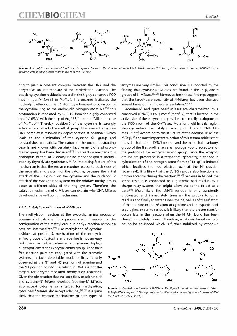
2.2.1. Catalytic mechanismof cytosine-C5 MTases
Although AdoMet is a very effective donor formethyl groups, methylation of cytosine residuesat position 5 is not a trivial reaction, becausecytosine is an electron-poor heterocyclic aro-matic ring system and position 5 of cytosine isnot capable of making a nucleophilic attack onthe methyl group of AdoMet. Therefore, thereaction catalyzed by the enzyme follows thereaction pathway of a Michael addition(Scheme 3).[62, 63] Initially a cysteine SH groupfrom the active site of the enzyme makes anucleophilic attack at position 6 of the cytosine
Scheme 2. Structure of the cofactor of the DNA MTases, AdoMet, and its potent competitor sinefungin.In the lower panel the AdoMet binding site of the DpnMMTase is shown schematically.[47] The positions ofatoms (N!main-chain nitrogen atom; OD, OE, NE!delta (D) and epsilon (E) side-chain oxygen andnitrogen atoms of Asp, Glu, and Trp, respectively) and numbers of amino acid residues in contact with theAdoMet are indicated, the numbers of the motifs are given in parenthesis. Hydrogen bonds are indicatedby dotted lines and hydrophobic contacts by dashed wedges.
A. Jeltsch
280 ChemBioChem 2002, 3, 274 ±293
ring to yield a covalent complex between the DNA and theenzyme as an intermediate of the methylation reaction. Theattacking cysteine residue is located in the highly conserved PCQmotif (motif IV; Cys81 in M.HhaI). The enzyme facilitates thenucleolytic attack on the C6 atom by a transient protonation ofthe cytosine ring at the endocyclic nitrogen atom N3;[64] thisprotonation is mediated by Glu119 from the highly conservedmotif VI (ENV) with the help of Arg165 from motif VIII in the caseof M.HhaI.[65] Thereby, position 5 of the cytosine is stronglyactivated and attacks the methyl group. The covalent enzyme±DNA complex is resolved by deprotonation at position 5 whichleads to the elimination of the cysteine SH group andreestablishes aromaticity. The nature of the proton abstractingbase is not known with certainty, involvement of a phospho-diester group has been discussed.[65] This reaction mechanism isanalogous to that of 2!-deoxyuridine monophosphate methyl-ation by thymidylate synthetase.[66] An interesting feature of thismechanism is that the enzyme requires access to both sides ofthe aromatic ring system of the cytosine, because the initialattack of the SH group on the cytosine and the nucleophilicattack of the cytosine ring system on the AdoMet methyl groupoccur at different sides of the ring system. Therefore, thecatalytic mechanism of C-MTases can explain why DNA MTasesdeveloped a base-flipping mechanism.
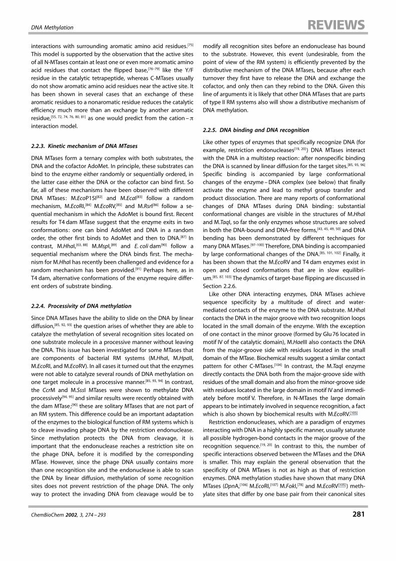
2.2.2. Catalytic mechanism of N-MTases
The methylation reaction at the exocyclic amino groups ofadenine and cytosine rings proceeds with inversion of theconfiguration of the methyl group in an SN2 reaction without acovalent intermediate.[67] Like methylation of cytosineresidues at position 5, methylation of the exocyclicamino groups of cytosine and adenine is not an easytask, because neither adenine nor cytosine displaysnucleophilicity at the exocyclic amino group, since theirfree electron pairs are conjugated with the aromaticsystems. In fact, detectable nucleophilicity is onlyobserved at the N1 and N3 positions of adenine andthe N3 position of cytosine, which in DNA are not thetargets for enzyme-mediated methylation reactions.Given the observation that the specificity of adenine-N6
and cytosine-N4 MTases overlaps (adenine-N6 MTasesalso accept cytosine as a target for methylation,cytosine-N4 MTases also accept adenine),[68, 69] it is quitelikely that the reaction mechanisms of both types of
enzymes are very similar. This conclusion is supported by thefinding that cytosine-N4 MTases are found in the ", #, and !
groups of N-MTases.[40, 70] Moreover, both these findings suggestthat the target-base specificity of N-MTases has been changedseveral times during molecular evolution.[68, 70]
Adenine-N6 and cytosine-N4 MTases are characterized by aconserved (D/N/S)PP(Y/F) motif (motif IV), that is located in theactive site of the enzyme at a position structurally analogous tothe PCQ motif of the C-MTases. Mutations within this regionstrongly reduce the catalytic activity of different DNA MT-ases.[55, 71±74] According to the structure of the adenine-N6 MTaseM.TaqI,[50] the most important function of this tetrapeptide is thatthe side chain of the D/N/S residue and the main-chain carbonylgroup of the first proline serve as hydrogen-bond acceptors forthe protons of the exocyclic amino group. Since the acceptorgroups are presented in a tetrahedral geometry, a change inhybridization of the nitrogen atom from sp2 to sp3 is inducedwhich localizes the free electron pair at the N6 position(Scheme 4). It is likely that the D/N/S residue also functions asproton acceptor during the reaction,[46, 48] because in M.PvuII theserine residue is connected to a glutamic acid residue by acharge relay system, that might allow the serine to act as abase.[46] Most likely, the D/N/S residue is only transientlyprotonated and immediately transfers the proton to otherresidues and finally to water. Given the pKa values of the N6 atomof the adenine or the N4 atom of cytosine and an aspartic acid,asparagine, or serine residue, it is likely that the proton transferoccurs late in the reaction when the N$CH3 bond has beenalmost completely formed. Therefore, a cationic transition statehas to be envisaged which is further stabilized by cation ±$
Scheme 3. Catalytic mechanism of C-MTases. The figure is based on the structure of the M.HhaI ± DNA complex.[49, 65] The cysteine residue is from motif IV (PCQ), theglutamic acid residue is from motif VI (ENV) of the C-MTase.
Scheme 4. Catalytic mechanism of N-MTases. The figure is based on the structure of theM.TaqI ± DNA complex.[50] The aspartate and proline residues in the figure are from motif IV ofthe N-MTase (D/N/S)PP(Y/F).
DNA Methylation
ChemBioChem 2002, 3, 274 ± 293 281
interactions with surrounding aromatic amino acid residues.[75]
This model is supported by the observation that the active sitesof all N-MTases contain at least one or evenmore aromatic aminoacid residues that contact the flipped base,[76±79] like the Y/Fresidue in the catalytic tetrapeptide, whereas C-MTases usuallydo not show aromatic amino acid residues near the active site. Ithas been shown in several cases that an exchange of thesearomatic residues to a nonaromatic residue reduces the catalyticefficiency much more than an exchange by another aromaticresidue,[55, 72, 74, 76, 80, 81] as one would predict from the cation ±$interaction model.
2.2.3. Kinetic mechanism of DNA MTases
DNA MTases form a ternary complex with both substrates, theDNA and the cofactor AdoMet. In principle, these substrates canbind to the enzyme either randomly or sequentially ordered, inthe latter case either the DNA or the cofactor can bind first. Sofar, all of these mechanisms have been observed with differentDNA MTases: M.EcoP15I[82] and M.EcaI[83] follow a randommechanism, M.EcoRI,[84] M.EcoRV,[85] and M.RsrI[86] follow a se-quential mechanism in which the AdoMet is bound first. Recentresults for T4 dam MTase suggest that the enzyme exits in twoconformations: one can bind AdoMet and DNA in a randomorder, the other first binds to AdoMet and then to DNA.[87] Incontrast, M.HhaI,[63, 88] M.MspI,[89] and E. coli dam[90] follow asequential mechanism where the DNA binds first. The mecha-nism for M.HhaI has recently been challenged and evidence for arandom mechanism has been provided.[91] Perhaps here, as inT4 dam, alternative conformations of the enzyme require differ-ent orders of substrate binding.
2.2.4. Processivity of DNA methylation
Since DNA MTases have the ability to slide on the DNA by lineardiffusion,[85, 92, 93] the question arises of whether they are able tocatalyze the methylation of several recognition sites located onone substrate molecule in a processive manner without leavingthe DNA. This issue has been investigated for some MTases thatare components of bacterial RM systems (M.HhaI, M.HpaII,M.EcoRI, and M.EcoRV). In all cases it turned out that the enzymeswere not able to catalyze several rounds of DNA methylation onone target molecule in a processive manner.[85, 93, 94] In contrast,the CcrM and M.SssI MTases were shown to methylate DNAprocessively[94, 95] and similar results were recently obtained withthe dam MTase;[90] these are solitary MTases that are not part ofan RM system. This difference could be an important adaptationof the enzymes to the biological function of RM systems which isto cleave invading phage DNA by the restriction endonuclease.Since methylation protects the DNA from cleavage, it isimportant that the endonuclease reaches a restriction site onthe phage DNA, before it is modified by the correspondingMTase. However, since the phage DNA usually contains morethan one recognition site and the endonuclease is able to scanthe DNA by linear diffusion, methylation of some recognitionsites does not prevent restriction of the phage DNA. The onlyway to protect the invading DNA from cleavage would be to
modify all recognition sites before an endonuclease has boundto the substrate. However, this event (undesirable, from thepoint of view of the RM system) is efficiently prevented by thedistributive mechanism of the DNA MTases, because after eachturnover they first have to release the DNA and exchange thecofactor, and only then can they rebind to the DNA. Given thisline of arguments it is likely that other DNA MTases that are partsof type II RM systems also will show a distributive mechanism ofDNA methylation.
2.2.5. DNA binding and DNA recognition
Like other types of enzymes that specifically recognize DNA (forexample, restriction endonucleases[19, 20] ) DNA MTases interactwith the DNA in a multistep reaction: after nonspecific bindingthe DNA is scanned by linear diffusion for the target sites.[85, 93, 96]
Specific binding is accompanied by large conformationalchanges of the enzyme±DNA complex (see below) that finallyactivate the enzyme and lead to methyl group transfer andproduct dissociation. There are many reports of conformationalchanges of DNA MTases during DNA binding: substantialconformational changes are visible in the structures of M.HhaIand M.TaqI, so far the only enzymes whose structures are solvedin both the DNA-bound and DNA-free forms,[43, 45, 49, 50] and DNAbending has been demonstrated by different techniques formany DNA MTases.[97±100] Therefore, DNA binding is accompaniedby large conformational changes of the DNA.[85, 101, 102] Finally, ithas been shown that the M.EcoRV and T4 dam enzymes exist inopen and closed conformations that are in slow equilibri-um.[85, 87, 103] The dynamics of target-base flipping are discussed inSection 2.2.6.
Like other DNA interacting enzymes, DNA MTases achievesequence specificity by a multitude of direct and water-mediated contacts of the enzyme to the DNA substrate. M.HhaIcontacts the DNA in the major groove with two recognition loopslocated in the small domain of the enzyme. With the exceptionof one contact in the minor groove (formed by Glu76 located inmotif IV of the catalytic domain), M.HaeIII also contacts the DNAfrom the major-groove side with residues located in the smalldomain of the MTase. Biochemical results suggest a similar contactpattern for other C-MTases.[104] In contrast, the M.TaqI enzymedirectly contacts the DNA both from the major-groove side withresidues of the small domain and also from the minor-groove sidewith residues located in the large domain in motif IV and immedi-ately before motif V. Therefore, in N-MTases the large domainappears to be intimately involved in sequence recognition, a factwhich is also shown by biochemical results with M.EcoRV.[105]
Restriction endonucleases, which are a paradigm of enzymesinteracting with DNA in a highly specific manner, usually saturateall possible hydrogen-bond contacts in the major groove of therecognition sequence.[19, 20] In contrast to this, the number ofspecific interactions observed between the MTases and the DNAis smaller. This may explain the general observation that thespecificity of DNA MTases is not as high as that of restrictionenzymes. DNA methylation studies have shown that many DNAMTases (DpnA,[106] M.EcoRI,[107] M.FokI,[78] and M.EcoRV[105] ) meth-ylate sites that differ by one base pair from their canonical sites
A. Jeltsch
282 ChemBioChem 2002, 3, 274 ±293
with rates only reduced by factors of 5 ± 10. Such sites usually arecleaved at least 100 ±1000 times more slowly by restrictionenzymes. Moreover, even sites that differ in more than one basepair from the specific site are modified with rates only reducedby 1 ±2 orders of magnitude (EcoRV,[105] M.FokI[78] )–similarresults were never obtained with a restriction enzyme.
2.2.6. Base flipping
Apart from the three crystal structure analyses already men-tioned there is now biochemical evidence confirming that DNAMTases in general make use of a base-flipping mechanism. Thisincludes the demonstration of very high cross-linking yields ofreactive base analogues located at the target position,[77, 78] highaccessibility of the target base towards chemical probes likepermanganate,[108] the observation of an increase in fluorescenceof 2-aminopurine when it is located at the position of the targetbase,[61, 86, 102, 109, 110] and the observation that many DNA MTasesbind more strongly to substrates in which the target base hasbeen replaced by a base analogue or another base such that abase mismatch is created.[78, 86, 101, 111±114] Although these studieswere carried out with different enzymes and none of them finallyproves that a base rotating mechanism is operative, when takentogether the available evidence strongly suggests that all DNAMTases make use of base flipping as an integral part of theirreaction mechanism.
The thermodynamics of base flipping is illustrated by thepreference of several MTases for binding to substrates whichcontain a base mismatch at the target position. The moststraightforward explanation for this observation is that baseflipping requires disruption of the Watson/Crick hydrogen bondsof the target base, which makes flipping more favorable if thetarget base is not engaged in a regular base pair. On the otherhand the rotated target base, as well as the partner base in theDNA helix, could be contacted by the MTase, a process thatshould provide interaction energy and specificity, because suchcontacts are diminished in cases where the nature of one ofthese bases is altered. The general observation that the balanceof these energetic contributions is almost always favorable withmismatched substrates demonstrates that no strong, specificinteractions are formed between the enzyme and the targetbase pair. The absence of a specific interaction with the targetbase is also demonstrated by the following two observations:1) Structures of M.HhaI with oligonucleotides containing mis-matches at the target position show that, in addition to cytosine,adenine and uracil are also flipped by the enzyme and bound inthe same hydrophobic pocket.[115] 2) Several adenine-N6 andcytosine-N4 MTases can modify adenine and cytosine targetbases[68, 69] and cytosine-C5 MTases can also methylate uracil,albeit at low efficiency.[112, 113]
The dynamics of base flipping have been most intensivelystudied with the M.HhaI cytosine-C5 MTase and the M.EcoRIadenine-N6 MTase. For M.HhaI it has been shown that baseflipping comprises at least two steps: first, the base is rotated outof the DNA helix, but not tightly bound by the enzyme and thenit is inserted into the hydrophobic binding pocket of theenzyme.[116] This result is in good agreement to the general
finding that base flipping is not very specific (see above). Also, inM.EcoRI, base flipping is a two-step process[111] with very fast rateconstants, of the order of 100 s$1, for the first step.[117]
The mechanism of base flipping is particularly enlightened bythe structure of a complex of the M.HhaI MTase with a substratecontaining an abasic nucleotide at the target position.[118] Even inthis case the sugar moiety is flipped out of its normal positionand located outside of the DNA helix, in a very similar manner tothat observed if a base is present. In the light of this result,™nucleotide flipping" would be a more appropriate designationfor the phenomenon. It shows that no interaction between theflipped base and the enzyme is required for base flipping. Sincethe conformational adaptations that lead to a stabilization of theorphan base in the DNA are different in all the MTase structures,the ™relaxation" of the complex structure after nucleotiderotation is also not likely to play a vital role in the process. Thispresumption has been verified in part for M.HhaI, where it hasbeen shown that Gln237, the amino acid residue that is insertedinto the DNA to fill the place of the flipped cytosine and thatforms all contacts to the partner guanine, can be replaced byother residues with moderate loss of activity.[119] So, it is mostlikely that contacts of the enzyme to the phosphodiester groupson either side of the flipped nucleotide are involved in baseflipping.[52] During base flipping, the phosphodiester groups arerotated and the distance between them must be transientlyincreased. Both of these processes could be induced orsupported by contacts of the enzyme to the nonbridgingoxygen atoms which do not necessarily have to persist in thefinal conformation where ™back-rotation" is prevented by thestructural adaptations of the complex. Interaction of the enzymewith the phosphodiester groups flanking the flipped base couldalso serve to synchronize nucleotide rotation and conforma-tional changes of the protein.
3. The Role of DNA Methylation in Eukaryotes
3.1. DNA methylation in higher eukaryotes
In higher eukaryotes DNA methylation is the only knowncovalent modification of the DNA. It has a plethora of importantdifferent functions and many biological processes includinghuman diseases are influenced by DNA methylation. So far, inmetazoa only cytosine-C5 methylation has been found in DNA;this methylation mainly occurs at CG sequences, about 60 ±90%of which are modified in mammals (corresponding to 3 ± 8% ofall cytosine residues). Thereby, a pattern of modified andunmodified CG sites is created. Thus, unlike the case inprokaryotes where the DNA sequence alone determines thesite of methylation, in eukaryotes a pattern of modified andnonmodified recognition sites exists (Figure 3). Since methyla-tion takes place in both DNA strands at palindromic sites, DNAreplication transforms the pattern of unmodified and fullymethylated sites into a pattern comprising unmodified andhemimethylated CG sites. Therefore, after DNA replication theinformation encoded in the pattern of DNA methylation is stillavailable and the initial pattern of methylation can be reestab-lished by a maintenance MTase that specifically modifies hemi-

DNA Methylation
ChemBioChem 2002, 3, 274 ± 293 283
Figure 3. Dynamics of DNA methylation in mammals. Unmethylated CG sitesare indicated by gray boxes. Fully methylated CG sites are colored black, with themethyl groups are represented by two small, black rectangles. HemimethylatedCG sites are colored gray and black and carry only one black rectangle.
methylated but not unmethylated target sites. Therefore, thepattern of DNA methylation is stable over cell divisions andsomatically inherited (occasionally even through the germ line).Nevertheless, it can be edited, either by de novo methylation orby demethylation, which makes DNA methylation a unique wayto encode information in a stable but reversible manner. Giventhese properties, DNA methylation is ideally suited to controlprocesses like cellular differentiation or development.
Certainly, DNA methylation is a central mechanism in epige-netic inheritance (that is, transmission of inheritable informationnot encoded in the DNA sequence). Epigenetics is a rapidlydeveloping field with a great impact on gene regulation,development, and important therapeutic perspectives[120] . Otherepigenetic mechanisms include covalent modifications of thehistone proteins (like acetylation, methylation, and phosphor-ylation) which influence the accessibility of the chromatin andgene expression[121] and the expression of noncoding RNAmolecules that lead to specific gene silencing.[122] Although thedetailed relation of DNA methylation, silencing by noncodingRNAs, and histone modification is not entirely clear, evidence isaccumulating that these processes are interdependent and actin a synergistic fashion.[123±128] However, DNA methylation, so far,is the only epigenetic process whose mechanism of inheritance(maintenance methylation) is understood at a molecular level.
Currently, it is unknown how the pattern of DNA methylationis generated and edited by a combination of specific de novomethylation and demethylation. In principle, there are fouralternative mechanisms which do not exclude each other: 1) TheMTases and demethylases could have an intrinsic specificity forcertain target regions, as is the rule for prokaryotic DNA MTases.However, so far there is no evidence in favor of such amechanism being available. 2) The MTases and demethylasescould be directed by other proteins to the sites of methylationand demethylation. This model is supported by the observationthat all active DNA MTases known in mammals have largeN-terminal regions that interact with other proteins and areinvolved in targeting the enzymes to certain cellular locations(see below). 3) Binding of other proteins could protect regions ofthe DNA from de novo methylation or demethylation, a modelthat is also supported by convincing experimental evi-
dence.[129±132] 4) The accessibility of the chromatin might controlwhich regions of the DNA are subject to methylation ordemethylation. This model is supported by the general relationbetween DNA methylation and chromatin remodeling. More-over, chromatin boundary elements which restrict methylationand demethylation to certain domains of the DNA have recentlybeen identified.[133]
Unlike in mammals, DNA methylation in plants is alsoobserved at CNG sites (where N is any base), a fact that suggeststhe presence of two independent codes of DNA methylation inthis kingdom of life.[134] It has been shown recently, that CNGmethylation, like CG methylation (see below), serves to repressgene expression.[135] Methylation of non-CG (and non-CNG)sequences has also been observed in mammals, in particular inearly phases of development (see ref. [136], and references citedtherein). The biological relevance of this type of methylation,which does not have a straightforward mechanism of main-tenance as it does not occur at palindromic sites, is not knownyet. Finally, very recently evidence for methylation of CCWGGsites (where W is A or T) in human DNA has been reported (seeref. [137] , and references cited therein).
3.2. DNA demethylation
In mammals the pattern of DNA methylation is set not only byDNA MTases but also by DNA demethylation.[138±140] In thisprocess 5-methylcytosine is converted back into cytosine.Demethylation occurs if a second round of DNA replicationtakes place before maintenance methylation has been completed(passive demethylation). In this case one of the daughter strandsis hemimethylated and one is unmethylated. Therefore, passivedemethylation is a rather slow process (5 replication cycles arerequired to reduce the methylation level to !5%). In contrast tothis, in active demethylation the removal of 5-methylcytosine isinitiated by a demethylase enzyme. So far, only one activemechanism of DNA demethylation has been established bio-chemically. In this mechanism the methylated base is excised bya glycosidase and the resulting lesion is repaired by the cellularrepair machinery. However, the observation of a genome-widedemethylation within hours that is not accompanied by largeamounts of DNA replication and repair[141] suggests that it is alsopossible to convert methylcytosine directly into cytosine,although breaking a C$C bond is energetically difficult. Thisprocess could follow a mechanism that resembles the methyl-ation reaction itself and would be initiated by a nucleophilicattack of the enzyme on the C6 position of the 5-methylcytosineand formation of a covalent enzyme±DNA intermediate.[142]
Alternatively, an oxidative mechanism appears possible, thatwould finally release the methyl group as carbon dioxide.
3.3. Dynamics of the DNA methylation pattern in mammals
During the development of mammals DNA methylation anddemethylation are orchestrated to achieve major changes in themethylation pattern.[143] The DNA is methylated in oocytes andsperms. After fertilization, before the onset of the first cleavagedivisions, the paternal DNA is rapidly demethylated by an active
A. Jeltsch
284 ChemBioChem 2002, 3, 274 ±293
mechanism within hours. The maternal genome is also demeth-ylated but by a passive mechanism during the first cleavagedivisions, because Dnmt1 is excluded from the nucleus in thisperiod. Despite an almost complete demethylation of large partsof the genome the imprint is maintained even during this phase,which requires the presence of Dnmt1o, an oocyte specificisoform of Dnmt1.[144] Later in embryogenesis, remethylation byde novo methylation of DNA occurs. In this phase, highexpression levels of the Dnmt3a and 3b de novo MTases areobserved. In germ cell development a similar reprogramming(almost complete demethylation followed by remethylation)takes place which erases the original and sets a new, sex-specificimprint on the DNA.
3.4. Deamination of 5-methylcytosine and CG islands
The presence of 5-methylcytosine in the DNA of highereukaryotes has the important disadvantage that it is potentiallymutagenic, because it promotes deamination of cytosine touracil.[145] Hydrolytic deamination of cytosine residues in double-stranded DNA occurs at a relatively slow rate with a half life ofabout 30,000 years. It is initiated by a hydroxy ion attack on theC4 position in a cytosine base that is protonated at the N3position. 5-methylcytosines are deaminated 2 ±4 times morerapidly than cytosines.[146, 147] The rate of deamination of cytosineand 5-methylcytosine residues can be accelerated by DNAC-MTases, in particular at low AdoMet concentrations.[148±151] Thisis understandable in the light of the mechanism of DNA MTases,which rotate the target base out of the DNA helix such that itbecomes more accessible and protonate the extrahelical cyto-sine at the N3 position. However, similar enzyme-catalyzeddeamination reactions are not observed with the human enzymeDnmt1.[152] If this difference is due to the different assay systemsused in the various reports or if it reflects a special property ofthe Dnmt1 enzyme remains to be seen.
The mutational damage of deamination of 5-methylcytosine isaugmented by the lower repair efficiency of T/G mismatches(arising from deamination of 5-methylcytosine) as compared toU/G mismatches (arising from deamination of unmethylatedcytosine). Uracil is a nonnatural base in DNA and is efficientlyrecognized and excised by the repair enzyme uracil-DNAglycosylase.[153] In contrast, thymidine is a natural componentof DNA that cannot be repaired by a global mechanism.However, at least two repair enzymes exist for T/G mismatchesin a CG context: one mismatch specific thymine glycosylase[154]
and the 5-methylcytosine binding protein MBD4.[155] Despitethese repair systems, methylation-mediated mutagenesis eventsapparently had a strong influence on the genome of vertebrates,because most CG sequences have been removed from thegenome during evolution. In mammals, CG dinucleotides are 5 ±10 fold underrepresented with respect to their expected normalfrequency. No CG depletion is observed in CG islands, which arefound in the promotor regions of about 60% of all genesincluding most housekeeping genes and 40% of the tissue-specific genes.[156] The CG sequences in CG islands are notmethylated in normal cells and in the germ line.[157±159]
Deamination of 5-methylcytosine is also a predominant causeof mutations observed in somatic tissues. For example, meth-ylated CG dinucleotides are the single most important muta-tional target in the p53 tumor-suppressor gene,[160] which isaffected in approximately 50% of all human cancers. Given thismutational burden, the fact that DNA methylation is stillobserved in most animals, plants, and fungi indicates that itmust play important biological roles.
3.5. The functions of DNA methylation in higher eukaryotes
Methylation of CG sites is involved in gene regulation, withmethylation of CG sites in the promotor regions of genes usuallyleading to a reduction of gene expression.[161, 162] Repression ofgene expression occurs at three levels of control : 1) Severaltranscription factors, like AP-2, c-Myc/Myn, E2F, and NF"B, arenot able to bind to methylated target sites. 2) DNA methylationrecruits 5-methylcytosine binding proteins that act as repressorsof gene transcription. 3) DNA methylation triggers histonedeacetylation and thereby induces chromatin condensationwhich leads to a strong and stable repression of gene expression.Therefore, DNA methylation is a general tool for gene regulationthat can be considered as an evolutionary device involved inmany different biological functions.[11] In mammals, genomicimprinting and X-chromosome inactivation are mediated byDNAmethylation, as described in detail in the following sections.However, these are specialized processes that, in this form, arecharacteristic for mammals. Therefore, these functions cannotexplain the almost ubiquitous occurrence of DNA methylation inbiology. Three other models on the general function of DNAmethylation have been put forward: It has been suggested thatDNA methylation contributes to the protection of the genomeagainst selfish genetic elements, that it is involved in devel-opmental processes, or that it is a tool to reduce transcriptionalnoise in organisms with a complex genome. All three modelsagree that DNA methylation serves as a general mechanism forgene repression. However, they attribute different main func-tions to this process: in the noise-reduction model general generepression itself is the main function of DNA methylation, in thegenome-protection model silencing of selfish genetic elementsby methylation is considered the main function of methylation,and in the development model gene and cell type specificmethylation and demethylation of promotor regions is the mostimportant function of DNA methylation.
3.5.1. DNA methylation and genomic imprinting
In mammals, a small number of genes (#50) carry an imprint,that allows the paternal and maternal copies of these genes tobe distinguished. Imprinted genes are only expressed from onechromosome, for example, only the maternal copy of H19 andonly the paternal copy of Igf2 are active.[163, 164] The involvementof DNA methylation in imprinting is shown by several observa-tions: differences in the pattern of methylation of imprintedgenes are observed in many cases, Dnmt1 knock-out mice whichshow a strongly reduced level of DNAmethylation have lost theirimprint,[165] and imprinting in plants also requires DNA methyl-

DNA Methylation
ChemBioChem 2002, 3, 274 ± 293 285
ation.[166] The mechanism of imprinting of the H19/Igf2 pair hasrecently been determined: it involves one enhancer whose effectis directed to the nearby H19 in the maternal genome. Itsinfluence on the Igf2 gene is prevented by binding of the CTCFprotein between the enhancer and the gene; CTCF functions as achromatin boundary element. In the paternal genome, the H19promotor sequence and the CTCF binding site are methylatedand thereby inactivated. Under these conditions, CTCF does notbind and the enhancer acts on the Ifg2 gene.[167±169] The imprint istransmitted as a certain pattern of methylation of imprintedgenes which is set in the gonads during spermatogenesis andoogenesis. After fertilization the imprint persists in the somaticcells for the whole life of the individual. In contrast, in the germcells the imprint must be erased and reset, because the newimprint depends on the sex of the individual. So, the paternaland maternal copies of imprinted genes will obtain a femaleimprint in oocytes and a male imprint in sperms. Therefore, thisprocess depends on both characteristic features of DNAmethylation: stable and inheritable silencing of genes and thepossibility to alter the encoded information if required.
3.5.2. DNA methylation and X-chromosome inactivation
In mammals, females carry two X chromosomes while maleshave only one. Therefore, dose compensation is required for thegenes encoded on the X chromosome. To this end, one Xchromosome is inactivated in a process that involves specificexpression of the Xist RNA from the inactivated chromosome, aswell as dense methylation and histone deacetylation of theinactivated chromosome.[170, 171] All these processes act syner-gistically to ensure the extreme stability of the inactive state,[124]
but it is known that DNA methylation is required for X-chromo-some inactivation[172, 173] and it has been shown that mouseembryos deficient in Dnmt1 do not show stable maintenance ofX-chromosome inactivation.[174] The initial choice of which Xchromosome is inactivated is made in early embryogenesis. Inembryonic tissues one chromosome is selected for inactivationin a random fashion whereas in extraembryogenic tissues thepaternal chromosome is always chosen for inactivation. There-fore, X-chromosome inactivation is also an example of parentalimprinting. Interestingly, X-chromosome inactivation, like im-printing, depends on the CTCF repressor protein that detects themethylation state of the DNA.[268, 269]
3.5.3. DNA methylation and protection from selfish geneticelements
The human genome is challenged by different types of selfishgenetic elements, like transposons, retrotransposons, and virus-es. Surprisingly "40% of our genome is made of such selfishDNA.[175] Since a random integration of transposable elementsinto the genome is an important source for mutations,prevention of transposon mobility by transcriptional silencingof these sequences is crucial for life. It has been suggested thatDNA methylation is involved in protection of the genomeagainst genetic parasites.[176] Today, a role of DNA methylation ingenome protection is beyond reasonable doubt: transposons
and other repetitive DNA sequences are usually relatively rich inCG sequences and heavily methylated[175] and an increase intranscription of transposons is observed in Dnmt1 knock-outembryonic stem cells[177] and cell lines.[178] These cells also showan elevated rate of mutations involving gene rearrange-ments.[179] Similar observations were made in plants wherereduction of DNA methylation leads to the expression andmobilization of transposons.[180] Moreover, the function of DNAmethylation in the fungus Ascobulus immersus is also to protectthe genome against foreign DNA in a process called MIP(methylation induced premitotically).[181]
However, other mechanisms also serve to protect the genomeagainst parasitic DNA. One such mechanism is RNA interfer-ence.[122, 182] At least in plants, RNA interference appears to beconnected to DNA methylation,[183±185] a fact suggesting thepresence of a programmable DNA methyltransferase which sofar has not been identified. However, RNA interference is alsoobserved in Saccharomyces cerevisiae and Caenorhabditis ele-gans, which do not methylate DNA. Thus, at least in thesespecies, methods for genome protection exist that are inde-pendent of DNA methylation. In other species, genome protec-tion is unlikely to be a function of DNA methylation becauseinvertebrates, insects, and many fungi do not show the dense,genome-wide methylation that is required for genome protec-tion, and it has been shown that repetitive DNA is notmethylated in Chiona intestinalis, an invertebrate chordate.[186]
Thus, whereas it is beyond doubt that DNA methylationcontributes to genome protection in many species includingmammals and plants, it is unlikely that this is the only function ofDNA methylation.
3.5.4. DNA methylation and the reduction of transcriptionalnoise
It has been proposed that DNA methylation serves to reducetranscriptional noise in organisms with complex genomes.[187]
This model is supported by the findings that in mammals mostCG sequences are methylated and therefore the defaultexpression status of most genes is ™off". In apparent agreementto this model, in Dnmt1 (and p53) knock-out cell lines theexpression of 4 ± 10% of the detectable genes is induced, whileonly 1 ± 2% are repressed;[178] this, of course, does not rule out aspecific role of DNA methylation and demethylation duringdevelopment.
However the noise-reduction model is based on olderestimates of the number of genes in different species, whichrecently have been revised considerably. According to the resultsof the genome-sequencing projects, the number of genes inC. elegans (19000) and Drosophila melanogaster (13000) is notdramatically lower than in Arabidopsis thaliana (20000) andhumans (35000 ± 40000). Nevertheless, in humans and A. thali-ana most of the CG sequences are methylated (in A. thaliana,CNG sequences are also modified) but C. elegans does not haveDNA methylation and D. melanogaster does not carry genome-wide methylation, although some methylcytosine is pres-ent.[188, 189] Although the best way to measure the ™complexity"of a genome may be disputable, the question arises of whether a

A. Jeltsch
286 ChemBioChem 2002, 3, 274 ±293
two± threefold increase in the total number of genes whencomparing C. elegans and humans makes an additional level ofgene control–with all its disadvantages (see above)–necessary.
3.5.5. DNA methylation and development
It was suggested more than 25 years ago that DNA methylationas an inheritable but flexible epigenic mark is involved indevelopment.[190, 191] Although this is still under debate,[12] therenow is firm evidence from in vivo and knock-out studies indifferent species that DNA methylation is involved in genesilencing during development. Dnmt1 and Dnmt3b knock-outmice die during early embryogenesis, Dnmt3a knock-out miceare runted and die shortly after birth.[192, 193] It is interesting thatDnmt1 knock-out embryonic stem cells are viable despite areduced level of methylation but die after induction of differ-entiation,[178, 192] a fact which also supports a role of DNAmethylation in development. Xenopus laevis eggs depleted inDnmt1 show disregulation of gene expression and prematureactivation of genes.[194] In zebra fish DNA methylation is requiredduring gastrulation and somite patterning.[195]
Recently, evidence has been provided that protein bindingprotects the DNA from de novo methylation.[131, 132] This resultsuggests that DNA methylation could be a tool to freeze thetranscriptional state of a cell for the future and transmit it tofollowing cell generations. In agreement with this model, it hasbeen shown that demethylation of DNA is involved in theactivation of the M-lysozyme gene during differentiation ofmurine macrophage cells[196, 197] and the liver specific tyrosineaminotransferase (Tat) gene.[198] The Tat gene is selectivelyactivated in response to the release of glucocorticoid hormonesbefore birth. After the glucocorticoid stimulus, a rapid chromatinremodeling is observed within minutes and this is followed bydemethylation after 2 ± 3 days. In contrast to the chromatinremodeling, the demethylation is stable after hormone with-drawal. Furthermore, demethylation recruits additional tran-scription factors to the DNA. This result shows that demeth-ylation serves to memorize a regulatory event during develop-ment and that it contributes to the fine tuning of geneexpression. An example of the involvement of DNA methylationin tissue-specific gene expression is the endothelial nitric oxidesynthase. The exclusive expression of this enzyme in theendothel is accompanied by a selective demethylation of itspromoter region only in endothelial cells (P. A. Marsden, personalcommunication). It is likely that many more examples of tissue-specific alterations of the DNA-methylation pattern will be foundduring the genome-wide analyses of the methylation status ofgene promotor regions in different tissues that are currently inprogress.
3.6. DNA methylation and disease
Alterations in the pattern of DNA methylation are frequentlyobserved in cancerous tissues.[199, 200] Most often a generalhypomethylation of the DNA is accompanied by a hypermeth-ylation at specific loci. Both of these processes can have cancer-promoting effects. Hypomethylation leads to genomic instability
often observed in cancer cells and may lead to an activation ofretrotransposons. In addition, the expression of oncogenes maybecome stimulated. Hypermethylation is often found in thepromotor regions of tumor-suppressor genes and there it hasthe same effect as a mutation in the gene itself. Depending onthe tumor type, epigenetic inactivation of tumor-suppressorgenes can be the predominant method of functional gene loss intumor cells. It should be noticed that methylation defects incancer cells, at least in principle, are reversible, which makes DNAmethylation a promising target for a new generation ofanticancer drugs. A genome-wide demethylation is also ob-served during aging and may contribute to the loss of generegulation in aging cells.[201]
Two genetic diseases are due to mutations in proteins relatedto DNA methylation.[14] ICF is a rare autosomal, recessive diseasethat is associated with immunodeficiency, centromere instability,and facial abnormalities. It has been shown that mutations in thednmt3b gene are responsible for this syndrome. In ICF patients,the DNA is hypomethylated mainly at the satellite regions 2 and3 of chromosomes 1, 9, and 16.[193, 202, 203] Moreover, mutations inthe MeCP2 protein cause Rett syndrome, a severe neurologicalregression starting 6 ±18 months after birth.[204] MeCP2 specifi-cally recognizes 5-methylcytosine and thereby is one of theproteins that read the methylation code and translate it intobiological effects.[205] Rett syndrome is the second most commongenetic disorder among Caucasian females, occurring onceevery 10000 ±15000 live births. It is X-chromosome linked anddominant, such that patients have one mutated and one wild-type allele. Males having an affected X-chromosome are mostlikely not viable.
4. DNA Methyltransferases in HigherEukaryotes
The distribution of DNA MTases in different species may alsoshed light on the function of DNA methylation. While theunicellular fungus S. cerevisiae does not have DNA methylation,other multicellular fungi do have it (Neurospora crassa, Ascobulusimmersus). DNA methylation is found in some insects, but theamount of methylation and the number of enzymes involved inmethylation is quite low. (D. melanogaster contains only oneputatitve cytosine-C5 MTase and the amount of 5-methylcyto-sine is about tenfold lower than in mammals.) Evidence for DNAmethylation also exists for several other insects (see ref. [188] ,and references cited therein) and at least one annelid.[207] Higherlevels of CG methylation and homologues of the Dnmt1maintenance MTase are found in the deuterostomia branch ofCoelomatae, including Paracentrotus lividus (sea urchin), Daniorerio (zebra fish), Xenopus laevis (claw frog), and mammals. Invertebrates, a genome-wide methylation is observed, whereas ininvertebrates the genomes are predominantly unmodified witha minor fraction of methylated DNA.[208] In contrast to all theseexamples, the nematode C. elegans does not have any DNAmethylation. So far, this is the only case of a multicellularorganism clearly devoid of DNA methylation. Interestingly,nematodes have a strictly deterministic way of developmentwhere the fate of each cell is genetically determined. In contrast

DNA Methylation
ChemBioChem 2002, 3, 274 ± 293 287
most animals follow a flexible way of ontogenesis that ischaracterized by a position-dependent development of cells andcapabilities of regeneration. It is noticable that nematodes showthis unusual way of development and that they have renouncedDNA methylation, which is an important tool for development inother species. Perhaps, DNA methylation is required for flexibledevelopment as an epigenetic device for cell programming butit is dispensable for a deterministic development.
4.1. Mammalian DNA MTases
So far, three active DNA MTases (Dnmt1, Dnmt3a, and Dnmt3b)and one candidate protein (Dnmt2) have been identified inmammals (Table 1, Figure 4). The properties of these enzymes arereviewed in the next sections.
4.1.1. Dnmt1
Dnmt1 is a very large protein which comprises 1620 amino acidresidues (Table 1, Figure 4). In vivo, alternative start codons aswell as different splicing isoforms have been described.[209±212]
The Dnmt1b isoform carries 48 additional amino acid residues inthe very N-terminal part of the protein. Its methylation activity issimilar to that of the previously described form of Dnmt1 invitro.[211, 212] An oocyte-specific form of Dnmt1 (Dnmt1o) lacksabout 150 amino acid residues at the N terminus. It is also activein vivo and in vitro.[209, 210] This isoform has been implicated in themaintenance of the parental imprint during the first cleavagedivisions, where a global demethylation occurs.[144]
The C-terminal part of Dnmt1 forms the catalytic domain andcontains all the amino acid sequence motifs characteristic forprokaryotic DNA cytosine-C5 MTases (Figure 4).[213, 214] The N-ter-minal part of the enzyme contains a nuclear localization signal[1]
and a region that directs the enzyme to replication foci.[215]
Within this region a major phosphorylation site of Dnmt1 hasbeen identified.[216] Furthermore, the N-terminal part of Dnmt1contains a zinc-binding domain[217, 218] of the CXXC zinc-fingertype. This region contains eight conserved cysteine residues and
binds to zinc.[218] Similar sequences are observed in otherproteins that bind to 5-methylcytosine and HRX-related tran-scription factors. The N-terminal part of Dnmt1 also contains aregion homologous to the polybromo-1 protein from chicken[1]
which is built up of two BAH domains. It might be involved inprotein ±protein interactions. In Dnmt1, it is also implicated indirecting the enzyme to replication foci.[219] The N-terminal partof Dnmt1 has been shown to interact with several other proteins,like PCNA,[220] the transcriptional co-repressor DMAP1,[221] thehistone deacetylases HDAC1[222, 223] and HDAC2,[221] and thetranscription factor E2F1,[223] as well as the Rb tumor-suppressorprotein.[223] Thus, it appears that the N-terminal part of Dnmt1serves as a platform for assembly of various proteins involved inchromatin condensation and gene regulation. However, itshould be noticed that so far it has not been shown in any ofthese cases that these interactions have a role in vivo what-soever.
Dnmt1 shows a significant preference for hemimethylatedDNA,[224±228] a fact suggesting a role for the enzyme inmaintenance methylation in vivo (Figure 5). Dnmt1 knock-outembryonic stem cells and mice show a strongly reduced level of
Figure 5. Specificity of Dnmt1. Catalytic activities are determined with duplexoligonucleotide substrates containing one hemimethylated CG site, one un-methylated CG site, or no CG sites at all (data were taken from ref. [228]).
DNA methylation but cells still display de novo MTase activity.[192]
The Dnmt1 knock-out animals show a recessive, lethal pheno-type; embryos were stunted, delayed in development, and didnot survive past mid-gestation. In contrast, Dnmt1 knock-out
Figure 4. Schematic drawing of the primary structures and domain arrangement of Dnmt1, Dnmt2, Dnmt3a, and Dnmt3b. The scale bar (bottom right) indicates thelength corresponding to 200 amino acid residues.

A. Jeltsch
288 ChemBioChem 2002, 3, 274 ±293
embryonic stem cells lines are viable, despite the reduced levelof DNA methylation. Several observations indicate that Dnmt1also participates in cell cycle control in mammals. It has beenobserved that Dnmt1 is involved in the Jun/Fos signal trans-duction pathway as a downstream effector.[229] Moreover,depletion of cells from Dnmt1 leads to p53-driven apopto-sis[178, 230] and to inhibition of DNA replication.[231] If these effectsare due to demethylation of the DNA or if Dnmt1 has additionalfunctions in the cell that do not depend on its catalytic activity isnot yet known.
The catalytic domain of Dnmt1 is under tight allosteric controlby the N-terminal part of the enzyme, as shown by theobservations that the isolated catalytic domain does notmethylate DNA in vivo or in vitro.[228, 232, 233] Interestingly, Dnmt1is allosterically activated to methylate unmodified target sites bybinding to methylated DNA.[228, 234, 235] Binding of methylatedDNA occurs within the N-terminal part of the enzyme, most likelyto the zinc domain, which forms a direct protein ±proteincontact to the catalytic domain of the enzyme.[228] After allostericstimulation, Dnmt1 has a similar activity on unmethylated andhemimethylated DNA, which suggests that this enzyme couldalso have a role in de novo methylation of DNA (Figure 6). Theallosteric activation mechanism of Dnmt1 makes DNA methyl-ation behave in an all-or-none fashion, because some methyl-ation will always attract more methylation. This explains theobservation that methylation tends to spread from heavilymethylated regions of the DNA into neighboring unmethylatedregions (spreading of methylation[236] ). After extensive spread-ing, only completely unmethylated and fully methylated regionsof the DNA that are separated by chromatin boundary elementscoexist.[133] This all-or-none behavior might increase the efficien-cy of switching on and off gene expression by DNA methylation.
4.1.2. Dnmt2
Dnmt2 belongs to a large family of proteins conserved fromSchizosaccharomyces pombe to man.[237, 238] The Dnmt2 enzymesare relatively small (the mouse enzyme comprises 391 aminoacid residues) and resemble prokaryotic MTases in that they donot have a large non-MTase N-terminal domain (Table 1, Fig-ure 4). These proteins contain all the sequence motifs character-istic for cytosine-C5 MTases. However, catalytic activity has notyet been shown for any protein belonging to this family[238, 239]
and a knock-out of Dnmt2 in embryonic stem cells does notshow a phenotype.[240] In accordance with the sequencehomology of Dnmt2 to prokaryotic MTases, the structure ofthis protein in complex with AdoHcy strongly resembles thatof the M.HhaI enzyme (both structures can be superimposedwith a root mean square deviation of !1 ä for the C"
positions).[239] Interestingly, Dnmt2 was found to interact withDNA in a denaturant-resistant, most likely covalent manner;Dnmt2 ±DNA complexes could not be disrupted in sodiumdodecylsulfate polyacrylamide gel electrophoresis (SDS-PAGE).[239] However, it is not known if irreversible binding toDNA also occurs in vivo or if it only takes place in vitro, becausethe enzyme is not able to complete a catalytic cycle of DNAmethylation such that the covalent intermediate is trapped. Arole of Dnmt2 in one of the methylation dependent processes inthe cell can only be excluded by generation of a Dnmt2 knock-out mouse strain and careful investigation of the animals overseveral generations. In any case, Dnmt2 is the first MTase-likeprotein from higher eukaryotes whose structure has beensolved.
S. pombe contains a gene (pmt1) for a Dnmt2 homologue that,like the other Dnmt2 proteins, is inactive in vitro.[241] However,the Pmt1 protein could be transformed into an active DNAMTase that modified DNA at CCWGG sequences in vitro by justone mutation.[242] This result, which certainly needs furthervalidation, suggests that other Dnmt2 proteins also mightrecognize CCWGG sequences.
4.1.3. Dnmt3a and Dnmt3b
The Dnmt3 MTases were discovered in 1998.[243] Dnmt3a and 3bare heavily expressed in embryonic tissues, whereas only lowexpression is observed in differentiated cells.[243, 244] The geneproducts from mice comprise 908 and 859 amino acid residues,respectively, and share 36% amino acid sequence identity witheach other ("80% in the C-terminal part of the proteins; Table 1,Figure 4). There are several alternative splice variants ofDnmt3b,[243] two of which are active, one is not.[245] The Dnmt3aand 3b enzymes contain a cysteine-rich region that is similar tothe ATRX zinc-finger. Moreover, they contain a PWWP domainwhich occurs in proteins that play a role in cell growth anddifferentiation. The structure of the PWWP domain from Dnmt3bhas been solved and it has been shown that this domain
interacts with DNA.[270] The ATRX domain of Dnmt3aassociates with the histone deacetylase HDAC1.[246]
Moreover, Dnmt3a interacts with RP58, a DNA-bindingtranscriptional repressor protein found at transcription-ally silent heterochromatin.[246] Mutations in humanDnmt3b cause ICF, a severe, hereditary dis-ease.[193, 202, 203] In ICF patients, hypomethylation of theDNA is observed at the classical satellite regions 2 and 3of chromosomes 1, 9 and, 16, a fact suggesting thatDnmt3b is involved in the methylation of theseregions.[193, 202, 203] Transgenic mice lacking Dnmt3a andDnmt3b, singly and in combination, are hypomethylatedand die in the embryonic stages (Dnmt3a$/$/Dnmt3b$/$,Dnmt3b$/$) or shortly after birth (Dnmt3a$/$) ; this
Figure 6. Model of functional cooperation of Dnmt3a and Dnmt1 in de novo methylation ofDNA. This model is based on two central observations: 1) Dnmt3a does not modify DNA in aprocessive reaction, which makes a fast methylation of one domain of the DNA by this enzymedifficult, and 2) Dnmt1 is stimulated by binding to methylated DNA. M!methyl group.

DNA Methylation
ChemBioChem 2002, 3, 274 ± 293 289
indicates the critical role of these enzymes during develop-ment.[193] The observation that the phenotype of a double knock-out is more severe than any of the single knock-outs suggeststhat Dnmt3a and 3b have partially overlapping functions.However, both proteins are essential, neither of them cancompletely replace the other. Therefore, there must be alsodistinctive roles for both enzymes. Since in ICF patients andDnmt3b knock-out mice demethylation is mainly observed atsatellite 2 and 3 DNA, one role of Dnmt3b might be methylationof these repetitive parts of the human genome.
Baculovirus-expressed Dnmt3a and Dnmt3b proteins, as wellas E. coli-expressed Dnmt3a, methylate CG dinucleotides withoutsignificant preference for hemimethylated DNA,[206, 243] whichsuggests a role of these enzymes in de novo methylation. Denovo methylation of DNA by Dnmt3a and 3b was alsodemonstrated in vivo after expression in human cell lines[247]
and by expression of Dnmt3a in transgenic D. melanogaster.[248]
Dnmt3a and 3b have a lower preference for CG sites than Dnmt1and also methylate DNA at asymmetric non-CG sites (CA"CC, CT).[136, 206, 245] The biological relevance of this non-CGmethylation is not known so far.
Dnmt3a expressed in E. coli catalyzes the methylation of DNAin a distributive manner.[206] This result, together with theobservation that Dnmt1 is allosterically activated by binding tomethylated DNA, suggests the interesting model that Dnmt3aand Dnmt1 might functionally cooperate in de novo methylationof DNA. In this model, Dnmt3a would be directed toward atarget region of the DNA and introduce one or a few methylgroups. Then, Dnmt1 might be recruited and spread themethylation over the whole domain of the DNA (Figure 6).[206, 228]
4.2. Plant DNA MTases
DNA methylation in plants also occurs at CNG sites, leading tothe modification of up to 40% of all cytosine residues (6% inArabidopsis thaliana).[134, 249, 250] A much higher number of DNAMTases are known in plants than in animals (compare, forexample, 11 in A. thaliana with 4 in mice;[134] Table 1). InA. thaliana four enzymes of the Dnmt1 family, one enzyme ofthe Dnmt3 family, one member of the Dnmt2 family, threemembers of the family of chromomethylases, and one memberof the masc1 family, an MTase family first identified in the fungusAscobulus immersus, are known. Interestingly, the Dnmt3homologue in A. thaliana contains the conserved motifs ofcytosine-C5 MTases in a different order (a domain-rearrangedmethyltransferase or DRM),[251] which suggests a circular permu-tation of the protein, similar to that observed in prokaryoticN-MTases.[40, 41] Chromomethylases are only present in plants.They are characterized by the insertion of a chromodomainbetween the conserved motifs II and IV of the DNA MTase.[252]
Chromodomains were first identified in Drosophila proteinswhere they direct proteins to the heterochromatin. Chromo-methylases have been shown to be responsible for methylationat CNG sites, which only occurs in plants and contributes toepigenetic gene silencing.[135, 253, 254]
The abundance of DNA MTases in plants, as well as thepresence of at least two independent methylation codes (CG and
CNG), might be related to the fact that plants as sessileorganisms cannot escape from unfavorable environmentalconditions. Therefore, they require a fine adaptation of theirgenetic program to the microenvironment at each habitat. It isclearly favorable if this adaptation can be inherited since mostoffspring will seed nearby. However, different adaptations arerequired at different locations, such that any change should bereversible–conditions that are ideally met by epigeneticchanges mediated by DNA methylation. In fact, it is welldocumented that DNA methylation is involved in geneticallystable epimutations, that is, inheritable but also reversiblephenotypic changes in plants. For example, methylated alleles ofthe A. thaliana SUPERMAN locus in genetic crosses behave likeunstable mutant alleles, with a frequency of spontaneousreversion of "1%.[255] Different epimutations affect othermorphological traits like leaf structure, time of flowering, orflower structure.[249, 256, 257]
4.3. DNA MTases in fungi
Fungi typically have low levels of DNA methylation, approx-imately 1.5% of all cytosine residues are modified in Neurosporacrassa[258] and 0.25% in Aspergillus flavus.[259] In fungi methylationis restricted mostly to transposons and other repeats.[250] In somespecies like S. cerevisiae and S. pombe, DNA methylation is notdetectable at all. Nevertheless, S. pombe contains a gene for aDnmt2 homologue (Pmt1) that could be activated by a singlemutation (see above).[242] This result suggests that the endog-enous protein also might have some catalytic activity thatescaped detection in vitro. Then, S. pombe might contain some5-methylcytosine, perhaps only at certain developmental stages.In Neurospora crassa, DNA methylation is involved in a processcalled repeat induced point mutation (RIP) that serves to silenceforeign DNA. In this organism a high de novo methylationactivity is detectable.[260] Methylation in N. crassa is due to onlyone MTase, the dim-2 enzyme.[261] The enzymology of fungalDNA MTases is also well investigated in A. immersus, the firstorganism in which the presence of a bona fide de novo DNAMTase has been shown, the Masc1 enzyme.[262] Masc1 is a shortprotein, that lacks the large N-terminal extension typical formammalian DNA MTase. It is involved in a methylation-depend-ent inactivation of repetitive DNA, MIP (methylation inducedpremitotically). The enzyme turned out to be catalyticallyinactive in vitro and disruption of Masc1 had no effect onviability of A. immersus and its maintenance methylation but itdid prevent de novo methylation of DNA repeats. Knock-outcells are arrested at early stages of sexual reproduction, whichindicates that the Masc1 protein is required for this develop-mental process, a result that again demonstrates that DNAmethylation is involved in development. A second DNA MTase ofA. immersus, Masc2, is a large protein comprising 1356 aminoacid residues. This enzyme is catalytically active in vitro, but aknock-out did not reveal any phenotype in vivo.[263, 264] Therefore,an additional DNA MTase responsible for maintenance methyl-ation must be present in A. immersus.

A. Jeltsch
290 ChemBioChem 2002, 3, 274 ±293
5. Future Perspectives
5.1. Prokaryotic MTases
There are numerous interesting open questions regarding theenzymology and biology of prokaryotic DNA MTases:! Definitely more structures are needed. Structures of related
MTases with different recognition sites will help to understandDNA recognition by these enzymes. Structures of MTases incomplex with nonspecific DNA and with specific DNA in anunflipped state would shed light on the conformationalchanges of the enzyme±DNA complex during enzymaticturnover.
! The physicochemical mechanism of base flipping is not wellunderstood. Transient interactions, like interactions of theMTases with the phosphate groups flanking the target, haveto be defined and investigated by experiments.
! Prokaryotic DNA MTases provide a unique tool to study themolecular evolution of DNA recognition. However, theavailable sequence information needs to be complementedby enzyme±DNA structures of related enzymes with differentrecognition sites and by biochemical studies to elucidate thefunctional relevance of the contacts between the enzyme andthe DNA.
! Medical applications: Adenine methylation has been shownto be important for bacteria and in certain cases involved inpathogenicity. Since, as far as we know, adenine MTases arenot present in mammals, these enzymes could be aninteresting drug target in the future.
5.2. Eukaryotic MTases
Many aspects of the biology of DNA methylation in highereukaryotes are still mysterious. In addition, we are only justbeginning to collect information on the enzymology of theeukaryotic MTases (apart from Dnmt1 which was cloned over10 years ago and has been well investigated); this is certainly aprerequisite for a detailed understanding of the whole process ofDNA methylation. Future issues include:! The mechanism of DNA demethylation in eukaryotes and the
enzymes involved need to be better understood.! The generation of the pattern of DNA methylation during
embryogenesis is not understood at a molecular level. Also,we are only just beginning to understand the mechanisms ofdirected changes of the pattern of methylation duringdevelopment by de novo methylation and demethylation.
! It is still not clear if Dnmt1 has a role in de novo methylation ofDNA in vivo although some evidence suggests this. Moreover,it has been discussed that it might have roles independent ofits methylation activity. Many interaction partners of Dnmt1have been found, but so far these interactions have not beenshown to be important for the in vivo function of the enzyme.Also, the molecular enzymology of the Dnmt3a and 3benzymes has to be investigated in much more detail.
! DNA methylation at nonpalindromic sites (non-CG and non-CNG) is observed in mammals and plants. In mammals,Dnmt3a is involved in this process. However, the biologicalfunction of this process is still completely unknown. Does it
contribute to gene silencing? Is there a mechanism tomaintain asymmetric methylation?
! Do CG and CNG (and perhaps also CCWGG) representindependent methylation codes with distinctive biologicalfunctions or are they functionally connected?
! Aberrant DNA methylation is often involved in carcinogenesisand is also implicated in aging. Since DNA methylation, inprinciple, is reversible, the possibility exists to correct errors inthe methylation pattern and thereby revert a pathologicalphenotype. In fact, it is possible to inhibit carcinogenesis inmice and to revert a transformed phenotype in a cell cultureby inhibition of Dnmt1 and reduction of DNA methyla-tion.[265, 266]
! Understanding the biology of DNA methylation is veryimportant for cloning experiments, because if mammals arecloned from a differentiated cell, the methylation patternmust be reset. It is likely, that methylation errors are causativeto the low yields of cloning of mammals and developmentalabnormalities of cloned embryos.[143, 267]
Work in the authors' laboratory has been supported by theDeutsche Forschungsgemeinschaft (JE 252/2-3, JE 252/1-2), theBMBF (BioFuture program), the European Union (BIO4CT980328),and the Justus-Liebig Universit‰t. It is a pleasure to thank A.Pingoud for his advice and generous support. Comments andsuggestions on the manuscript by C. Beck, H. Gowher, and A.Hermann are gratefully acknowledged.
[1] T. H. Bestor, G. L. Verdine, Curr. Opin. Cell Biol. 1994, 6, 380.[2] X. Cheng, Curr. Opin. Struct. Biol. 1995, 5, 4.[3] X. Cheng, Annu. Rev. Biophys. Biomol. Struct. 1995, 24, 293.[4] E. B. Faumann, R. M. Blumenthal, X. Cheng in S-adenosylmethionine-
dependent methyltransferases: Structures and functions (Eds. : X. Cheng,R. M. Blumenthal), World Scientific Publishing, Singapore, 1999, p. 1.
[5] X. Cheng, R. J. Roberts, Nucleic Acids Res. 2001, 29, 3784.[6] M. Noyer-Weidner, T. A. Trautner in DNA Methylation: Molecular Biology
and Biological Significance (Eds. : J. P. Jost, H. P. Saluz), Birkhauser, Basel,1993, p. 40.
[7] D. T. F. Dryden, in S-Adenosylmethionine-dependent Methyltransferases:Structures and Functions (Eds. : X. Cheng, R. M. Blumenthal), WorldScientific Publishing, Singapore, 1999, p. 283.
[8] T. A. Bickle, D. H. Kruger, Microbiol. Rev. 1993, 57, 434.[9] R. L. P. Adams, Bioessays 1995, 17, 139.
[10] P. M. Vertino in S-Adenosylmethionine-dependent Methyltransferases:Structures and Functions (Eds. : X. Cheng, R. M. Blumenthal), WorldScientific Publishing, Singapore, 1999, p. 341.
[11] V. Colot, J. L. Rossignol, Bioessays 1999, 21, 402.[12] T. H. Bestor, Hum. Mol. Genet. 2000, 9, 2395.[13] K. D. Robertson, A. P. Wolffe, Nat. Rev. Genet. 2000, 1, 11.[14] B. Hendrich, Curr. Biol. 2000, 10, R60.[15] P. A. Jones, D. Takai, Science 2001, 293, 1068.[16] G. G. Wilson, N. E. Murray, Annu. Rev. Genet. 1991, 25, 585.[17] J. Heitman in Genetic Engineering (Ed. : J. K. Setlow), Plenum Press, New
York, 1993, p. 57.[18] R. J. Roberts, S. E. Halford in Nucleases (Eds. : S. M. Linn, R. S. Lloyd, R. J.
Roberts), Cold Spring Harbor Laboratory Press, Cold Spring Harbor,1993, p. 35.
[19] A. Pingoud, A. Jeltsch, Eur. J. Biochem. 1997, 246, 1.[20] A. Pingoud, A. Jeltsch, Nucleic Acids Res. 2001, 29, 3705.[21] R. J. Roberts, D. Macelis, Nucleic Acids Res. 2001, 29, 268.[22] W. Messer, M. Noyer-Weidner, Cell 1988, 54, 735.[23] F. Barras, M. G. Marinus, Trends Genet. 1989, 5, 139.[24] I. Stancheva, T. Koller, J. M. Sogo, EMBO J. 1999, 18, 6542.

DNA Methylation
ChemBioChem 2002, 3, 274 ± 293 291
[25] M. G. Marinus, Annu. Rev. Genet. 1987, 21, 113.[26] J. L. Campbell, N. Kleckner, Cell 1990, 62, 967.[27] M. Lu, J. L. Campbell, E. Boye, N. Kleckner, Cell 1994, 77, 413.[28] S. Goldman, Y. Navon, F. Fish, Microb Pathog. 1987, 2, 327.[29] W. B. Hale, M. W. van der Woude, B. A. Braaten, D. A. Low, Mol. Genet.
Metab. 1998, 65, 191.[30] D. M. Heithoff, R. L. Sinsheimer, D. A. Low, M. J. Mahan, Science 1999, 284,
967.[31] M. Enserink, Science 1999, 284, 883.[32] F. Garcia-Del Portillo, M. G. Pucciarelli, J. Casadesus, Proc. Natl. Acad. Sci.
USA 1999, 96, 11578.[33] D. M. Heithoff, R. L. Sinsheimer, D. A. Low, M. J. Mahan, Philos. Trans. R.
Soc. Lond. Ser. B 2000, 355, 633.[34] C. Bucci, A. Lavitola, P. Salvatore, L. Del Giudice, D. R. Massardo, C. B.
Bruni, P. Alifano, Mol. Cell 1999, 3, 435.[35] A. Reisenauer, L. S. Kahng, S. McCollum, L. Shapiro, J. Bacteriol. 1999, 181,
5135.[36] C. Stephens, A. Reisenauer, R. Wright, L. Shapiro, Proc. Natl. Acad. Sci. USA
1996, 93, 1210.[37] D. C. Wahnon, V. K. Shier, S. J. Benkovic, J. Am. Chem. Soc. 2001, 123, 976.[38] G. G. Wilson, Methods Enzymol. 1992, 216, 259.[39] S. Kumar, W. S. Johnson, M. Tomasz, Biochemistry 1993, 32, 1364.[40] T. Malone, R. M. Blumenthal, X. Cheng, J. Mol. Biol. 1995, 253, 618.[41] A. Jeltsch, J. Mol. Evol. 1999, 49, 161.[42] S. Kumar, X. Cheng, S. Klimasauskas, M. Sha, J. Posfai, R. J. Roberts, G. G.
Wilson, Nucleic Acids Res. 1994, 22, 1.[43] X. Cheng, S. Kumar, J. Posfai, J. W. Pflugrath, R. J. Roberts, Cell 1993, 74,
299.[44] K. M. Reinisch, L. Chen, G. L. Verdine, W. N. Lipscomb, Cell 1995, 82, 143.[45] J. Labahn, J. Granzin, G. Schluckebier, D. P. Robinson, W. E. Jack, I.
Schildkraut, W. Saenger, Proc. Natl. Acad. Sci. USA 1994, 91, 10957.[46] W. Gong, M. O'Gara, R. M. Blumenthal, X. Cheng, Nucleic Acids Res. 1997,
25, 2702.[47] P. H. Tran, Z. R. Korszun, S. Cerritelli, S. S. Springhorn, S. A. Lacks, Structure
1998, 6, 1563.[48] R. D. Scavetta, C. B. Thomas, M. A. Walsh, S. Szegedi, A. Joachimiak, R. I.
Gumport, M. E. A. Churchill, Nucleic Acids Res. 2000, 28, 3950.[49] S. Klimasauskas, S. Kumar, R. J. Roberts, X. Cheng, Cell 1994, 76,
357.[50] K. Goedecke, M. Pignot, R. S. Goody, A. J. Scheidig, E. Weinhold, Nat.
Struct. Biol. 2001, 8, 121.[51] R. J. Roberts, Cell 1995, 82, 9.[52] X. Cheng, R. M. Blumenthal, Structure 1996, 4, 639.[53] R. S. Lloyd, X. Cheng, Biopolymers 1997, 44, 139.[54] R. J. Roberts, X. Cheng, Annu. Rev. Biochem. 1998, 67, 181.[55] M. Roth, S. Helm-Kruse, T. Friedrich, A. Jeltsch, J. Biol. Chem. 1998, 273,
17333.[56] G. Schluckebier, M. Kozak, N. Bleimling, E. Weinhold, W. Saenger, J. Mol.
Biol. 1997, 265, 56.[57] M. O'Gara, X. Zhang, R. J. Roberts, X. Cheng, J. Mol. Biol. 1999, 287, 201.[58] G. M. Adams, R. M. Blumenthal, Biochemistry 1997, 36, 8284.[59] A. Bergerat, W. Guschlbauer, Nucleic Acids Res. 1990, 18, 4369.[60] A. Bergerat, W. Guschlbauer, G. V. Fazakerley, Proc. Natl. Acad. Sci. USA
1991, 88, 6394.[61] E. G. Malygin, A. A. Evdokimov, V. V. Zinoviev, L. G. Ovechkina, W. M.
Lindstrom, N. O. Reich, S. L. Schlagman, S. Hattman, Nucleic Acids Res.2001, 29, 2361.
[62] D. V. Santi, A. Norment, C. E. Garrett, Proc. Natl. Acad. Sci. USA 1984, 81,6993.
[63] J. C. Wu, D. V. Santi, J. Biol. Chem. 1987, 262, 4778.[64] L. Chen, A. M. MacMillan, G. L. Verdine, J. Am. Chem. Soc. 1993, 115, 5318.[65] M. O'Gara, S. Klimasauskas, R. J. Roberts, X. Cheng, J. Mol. Biol. 1996, 261,
634.[66] J. C. Wu, D. V. Santi in Biochemistry and Biology of DNA Methylation (Eds. :
G. L. Cantoni, A. Razin, A. R. Liss), Wiley-Liss, New York, 1986, p. 119.[67] A. L. Pogolotti, A. Ono, R. Subramaniam, D. V. Santi, J. Biol. Chem. 1988,
263, 7461.[68] A. Jeltsch, F. Christ, M. Fatemi, M. Roth, J. Biol. Chem. 1999, 274, 19538.[69] A. Jeltsch, Biol. Chem. 2001, 382, 707.[70] J. M. Bujnicki, M. Radlinska, Nucleic Acids Res. 1999, 27, 4501.[71] H. Sugisaki, K. Yamamoto, M. Takanami, J. Biol. Chem. 1991, 266, 13952.
[72] D. F. Willcock, D. T. F. Dryden, N. E. Murray, EMBO J. 1994, 13, 3902.[73] J. B. Guyot, J. Grassi, U. Hahn, W. Guschlbauer, Nucleic Acids Res. 1993, 21,
3183.[74] H. Kong, C. L. Smith, Nucleic Acids Res. 1997, 25, 3687.[75] G. Schluckebier, J. Labahn, J. Granzin, W. Saenger, Biol. Chem. 1998, 379,
389.[76] T. Friedrich, M. Roth, S. Helm-Kruse, A. Jeltsch, Biol. Chem. 1998, 379, 475.[77] B. Holz, N. Dank, J. E. Eickhoff, G. Lipps, G. Krauss, E. Weinhold, J. Biol.
Chem. 1999, 274, 15066.[78] A. Jeltsch, M. Roth, T. Friedrich, J. Mol. Biol. 1999, 285, 1121.[79] D. L. Wong, N. O. Reich, Biochemistry 2000, 39, 15410.[80] H. Pues, N. Bleimling, B. Holz, J. Wolcke, E. Weinhold, Biochemistry 1999,
38, 1426.[81] M. Roth, A. Jeltsch, Nucleic Acids Res. 2001, 29, 3137.[82] D. N. Rao, M. G. P. Page, T. A. Bickle, J. Mol. Biol. 1989, 209, 599.[83] L. Szilak, A. Der, F. Deak, P. Venetianer, Eur. J. Biochem. 1993, 218, 727.[84] N. O. Reich, N. Mashhoon, Biochemistry 1991, 30, 2933.[85] H. Gowher, A. Jeltsch, J. Mol. Biol. 2000, 303, 93.[86] S. S. Szegedi, N. O. Reich, R. I. Gumport, Nucleic Acids Res. 2000, 28, 3962.[87] A. A. Evdokimov, V. V. Zinoviev, E. G. Malygin, S. L. Schlagman, S.
Hattman, J. Biol. Chem. 2001, 276, 30.[88] W. M. Lindstrom, J. Flynn, N. O. Reich, J. Biol. Chem. 2000, 275, 4912.[89] S. K. Bhattacharya, A. K. Dubey, J. Biol. Chem. 1999, 274, 14743.[90] S. Urig, H. Gowher, A. Hermann, C. Beck, A. Humeny, A. Jeltsch, 2002,
submitted.[91] G. Vilkaitis, E. Merkiene, S. Serva, E. Weinhold, S. Klimasauskas, J. Biol.
Chem. 2001, 276, 20924.[92] G. Nardone, J. George, J. G. Chirikjian, J. Biol. Chem. 1986, 261, 12128.[93] M. A. Surby, N. O. Reich, Biochemistry 1996, 35, 2201.[94] P. Renbaum, A. Razin, FEBS Lett. 1992, 313, 243.[95] A. J. Berdis, I. Lee, J. K. Coward, C. Stephens, R. Wright, L. Shapiro, S. J.
Benkovic, Proc. Natl. Acad. Sci. USA 1998, 95, 2874.[96] M. A. Surby, N. O. Reich, Biochemistry 1996, 35, 2209.[97] S. Cal, B. A. Connolly, J. Biol. Chem. 1996, 271, 1008.[98] R. A. Garcia, C. J. Bustamante, N. O. Reich, Proc. Natl. Acad. Sci. USA 1996,
93, 7618.[99] A. K. Dubey, S. K. Bhattacharya, Nucleic Acids Res. 1997, 25, 2025.
[100] T. Rasko, C. Finta, A. Kiss, Nucleic Acids Res. 2000, 28, 3083.[101] S. Cal, B. A. Connolly, J. Biol. Chem. 1997, 272, 490.[102] Y. V. Reddy, D. N. Rao, J. Mol. Biol. 2000, 298, 597.[103] A. Jeltsch, T. Friedrich, M. Roth, J. Mol. Biol. 1998, 275, 747.[104] A. Kiss, G. Posfai, G. Zsurka, T. Rasko, P. Venetianer, Nucleic Acids Res.
2001, 29, 3188.[105] C. Beck, S. Cranz, M. Solmaz, M. Roth, A. Jeltsch, Biochemistry 2001, 40,
10956.[106] S. Cerritelli, S. S. Springhorn, S. A. Lacks, Proc. Natl. Acad. Sci. USA 1989,
86, 9223.[107] N. O. Reich, C. Olsen, F. Osti, J. Murphy, J. Biol. Chem. 1992, 267, 15802.[108] S. Serva, E. Weinhold, R. J. Roberts, S. Klimasauskas, Nucleic Acids Res.
1998, 26, 3473.[109] B. W. Allan, N. O. Reich, Biochemistry 1996, 35, 14757.[110] B. Holz, S. Klimasauskas, S. Serva, E. Weinhold, Nucleic Acids Res. 1998, 26,
1076.[111] B. W. Allan, J. M. Beechem, W. M. Lindstrom, N. O. Reich, J. Biol. Chem.
1998, 273, 2368.[112] S. Klimasauskas, R. J. Roberts, Nucleic Acids Res. 1995, 23, 1388.[113] A. S. Yang, J.-C. Shen, J.-M. Zingg, S. Mi, P. A. Jones, Nucleic Acids Res.
1995, 23, 1380.[114] D. R. Mernagh, I. A. Taylor, G. G. Kneale, Biochem. J. 1998, 336, 719.[115] M. O'Gara, J. R. Horton, R. J. Roberts, X. Cheng, Nat. Struct. Biol. 1998, 5,
872.[116] S. Klimasauskas, T. Szyperski, S. Serva, K. W¸thrich, EMBO J. 1998, 17, 317.[117] B. W. Allan, N. O. Reich, J. M. Beechem, Biochemistry 1999, 38, 5308.[118] M. O'Gara, J. R. Horton, R. J. Roberts, X. Cheng, Nature Struct. Biol. 1998,
5, 872.[119] S. Mi, D. Alonso, R. J. Roberts, Nucleic Acids Res. 1995, 23, 620.[120] A. P. Wolffe, M. A. Matzke, Science 1999, 286, 481; see also: Science 2001,
293 (5532), pp. 1063 ± 1102, which is completely devoted to epi-genetics.
[121] T. Jenuwein, C. D. Allis, Science 2001, 293, 1074.[122] M. Matzke, A. J. Matzke, J. M. Kooter, Science 2001, 293, 1080.
A. Jeltsch
292 ChemBioChem 2002, 3, 274 ±293
[123] P. Chiurazzi, M. G. Pomponi, R. Pietrobono, C. E. Bakker, G. Neri, B. A.Oostra, Hum. Mol. Genet. 1999, 8, 2317.
[124] G. Csankovszki, A. Nagy, R. Jaenisch, J. Cell. Biol. 2001, 153, 773.[125] F. Magdinier, A. P. Wolffe, Proc. Natl. Acad. Sci. USA 2001, 98, 4990.[126] J. R. Dobosy, E. U. Selker, Cell. Mol. Life Sci. 2001, 58, 721.[127] X. Yang, D. L. Phillips, A. T. Ferguson, W. G. Nelson, J. G. Herman, N. E.
Davidson, Cancer Res. 2001, 61, 7025.[128] H. Tamaru, E. U. Selker, Nature 2001, 414, 277.[129] M. Brandeis, D. Frank, I. Keshet, Z. Siegfried, M. Mendelsohn, A. Nemes, V.
Temper, A. Razin, H. Cedar, Nature 1994, 371, 435.[130] D. Macleod, J. Charlton, J. Mullins, A. P. Bird, Genes Dev. 1994, 8, 2282.[131] I. G. Lin, C. L. Hsieh, EMBO Rep. 2001, 2, 108.[132] L. Han, I. G. Lin, C. L. Hsieh, Mol. Cell. Biol. 2001, 21, 3416.[133] D. S. Millar, C. L. Paul, P. L. Molloy, S. J. Clark, J. Biol. Chem. 2000, 275,
24893.[134] E. J. Finnegan, K. A. Kovac, Plant Mol. Biol. 2000, 43, 189.[135] A. M. Lindroth, X. Cao, J. P. Jackson, D. Zilberman, C. M. McCallum, S.
Henikoff, S. E. Jacobsen, Science 2001, 292, 2077.[136] B. H. Ramsahoye, D. Biniszkiewicz, F. Lyko, V. Clark, A. P. Bird, R. Jaenisch,
Proc. Natl. Acad. Sci. USA 2000, 97, 5237.[137] M. C. Lorincz, M. Groudine, Proc. Natl. Acad. Sci. USA 2001, 98, 10034.[138] A. P. Wolffe, P. L. Jones, P. A. Wade, Proc. Natl. Acad. Sci. USA 1999, 96,
5894.[139] J. P. Jost, E. J. Oakeley, S. Schwarz in S-adenosylmethionine-dependent
methyltransferases: Structures and functions (Eds. : X. Cheng, R. M.Blumenthal), World Scientific Publishing, Singapore, 1999, p. 373.
[140] C. Kress, H. Thomassin, T. Grange, FEBS Lett. 2001, 494, 135.[141] J. Oswald, S. Engemann, N. Lane, W. Mayer, A. Olek, R. Fundele, W. Dean,
W. Reik, J. Walter, Curr. Biol. 2000, 10, 475.[142] H. Cedar, G. L. Verdine, Nature 1999, 397, 568.[143] W. Reik, W. Dean, J. Walter, Science 2001, 293, 1089.[144] C. Y. Howell, T. H. Bestor, F. Ding, K. E. Latham, C. Mertineit, J. M. Trasler,
J. R. Chaillet, Cell 2001, 104, 829.[145] G. P. Pfeifer, M.-S. Tang, M. F. Denissenko in DNA methylation and cancer
(Eds. : P. A. Jones, P. K. Vogt), Springer, New York, 2000, p. 1.[146] T. Lindahl, Nature 1993, 362, 709.[147] J. Shen, W. M. I. Rideout, P. A. Jones, Nucleic Acids Res. 1994, 22, 972.[148] J. Shen, W. M. I. Rideout, P. A. Jones, Cell 1992, 71, 1073.[149] W. Wyszynski, S. Garbara, A. S. Bhagwat, Proc. Natl. Acad. Sci. USA 1994,
91, 1574.[150] C. C. Yang, B. K. Baxter, M. D. Topal, Biochemistry 1994, 33, 14918.[151] M. J. Yebra, A. S. Bhagwat, Biochemistry 1995, 34, 14752.[152] M. F. Chan, R. van Amerongen, T. Nijjar, E. Cuppen, P. A. Jones, P. W. Laird,
Mol. Cell. Biol. 2001, 21, 7587.[153] R. D. Wood, M. Mitchell, J. Sgouros, T. Lindahl, Science 2001, 291, 1284.[154] P. Neddermann, P. Gallinari, T. Lettieri, D. Schmid, O. Truong, J. J. Hsuan,
K. Wiebauer, J. Jiricny, J. Biol. Chem. 1996, 271, 12767.[155] S. Tweedie, H.-H. Ng, A. L. Barlow, B. M. Turner, B. Hendrich, A. Bird, Nat.
Genet. 1999, 23, 389.[156] S. H. Cross, J. A. Charlton, X. Nan, A. P. Bird, Nat. Genet. 1994, 6, 236.[157] A. Bird, M. Taggart, M. Frommer, O. J. Miller, D. Macleod, Cell 1985, 40, 91.[158] A. Bird, Cell 1992, 70, 5.[159] D. F. Schorderet, S. M. Gartler, Proc. Natl. Acad. Sci. USA 1992, 89, 957.[160] P. W. Laird, R. Jaenisch, Annu. Rev. Genet. 1996, 30, 441.[161] S. U. Kass, D. Pruss, A. P. Wolffe, Trends Genet. 1997, 13, 444.[162] Z. Siegfried, H. Cedar, Curr. Biol. 1997, 7, R305.[163] S. M. Tilghman, Cell 1999, 96, 185.[164] W. Reik, J. Walter, Nat. Rev. Genet. 2001, 2, 21.[165] E. Li, C. Beard, R. Jaenisch, Nature 1993, 366, 362.[166] M. D. Adams et al. , Science 2000, 287, 2185.[167] W. Reik, A. Murrell, Nature 2000, 405, 408.[168] A. C. Bell, G. Felsenfeld, Nature 2000, 405, 482.[169] A. T. Hark, C. J. Schoenherr, D. J. Katz, R. S. Ingram, J. M. Levorse, S. M.
Tilghman, Nature 2000, 405, 486.[170] P. Avner, E. Heard, Nat. Rev. Genet. 2001, 2, 59.[171] Y. K. Kang, D. B. Koo, J. S. Park, Y. H. Choi, A. S. Chung, K. K. Lee, Y. M. Han,
Nat. Genet. 2001, 28, 173.[172] C. Beard, E. Li, R. Jaenisch, Genes Dev. 1995, 9, 2325.[173] B. Panning, R. Jaenisch, Genes Dev. 1996, 10, 1991.[174] T. Sado, M. H. Fenner, S. S. Tan, P. Tam, T. Shioda, E. Li, Dev. Biol. (Orlando,
FL, USA) 2000, 225, 294.
[175] A. F. Smit, Curr. Opin. Genet. Dev. 1999, 9, 657.[176] J. A. Yoder, C. P. Walsh, T. H. Bestor, Trends Genet. 1997, 13, 335.[177] C. P. Walsh, J. R. Chaillet, T. H. Bestor, Nat. Genet. 1998, 20, 116.[178] L. Jackson-Grusby, C. Beard, R. Possemato, M. Tudor, D. Fambrough, G.
Csankovszki, J. Dausman, P. Lee, C. Wilson, E. Lander, R. Jaenisch, Nat.Genet. 2001, 27, 31.
[179] R. Z. Chen, U. Pettersson, C. Beard, L. Jackson-Grusby, R. Jaenisch, Nature1998, 395, 89.
[180] A. Miura, S. Yonebayashi, K. Watanabe, T. Toyama, H. Shimada, T.Kakutani, Nature 2001, 411, 212.
[181] G. Faugeron, Curr. Opin. Microbiol. 2000, 3, 144.[182] T. Tuschl, ChemBioChem 2001, 2, 239.[183] M. Wassenegger, Plant Mol. Biol. 2000, 43, 203.[184] M. A. Matzke, M. F. Mette, A. J. Matzke, Plant. Mol. Biol. 2000, 43, 401.[185] J. Bender, Cell 2001, 106, 129.[186] M. W. Simmen, S. Leitgeb, J. Charlton, S. J. Jones, B. R. Harris, V. H. Clark,
A. Bird, Science 1999, 283, 1164.[187] A. P. Bird, Trends Genet. 1995, 11, 94.[188] H. Gowher, O. Leismann, A. Jeltsch, EMBO J. 2000, 19, 6918.[189] F. Lyko, B. H. Ramsahoye, R. Jaenisch, Nature 2000, 408, 538.[190] A. D. Riggs, Cytogenet. Cell. Genet. 1975, 14, 9.[191] R. Holliday, J. E. Pugh, Science 1975, 187, 226.[192] E. Li, T. H. Bestor, R. Jaenisch, Cell 1992, 69, 915.[193] M. Okano, D. W. Bell, D. A. Haber, E. Li, Cell 1999, 99, 247.[194] I. Stancheva, R. R. Meehan, Genes. Dev. 2000, 14, 313.[195] C. C. Martin, L. Laforest, M. A. Akimenko, M. Ekker, Dev. Biol. (Orlando, FL,
USA) 1999, 206, 189.[196] A. Schmitz, M. Short, O. Ammerpohl, C. Asbrand, J. Nickel, R. Renkawitz,
J. Biol. Chem. 1997, 272, 20850.[197] O. Ammerpohl, A. Schmitz, L. Steinm¸ller, R. Renkawitz, Nucleic Acids Res.
1998, 26, 5256.[198] H. Thomassin, M. Flavin, M. L. Espinas, T. Grange, EMBO J. 2001, 20, 1974.[199] P. M. Warnecke, T. H. Bestor, Curr. Opin. Oncol. 2000, 12, 68.[200] S. B. Baylin, J. G. Herman, Trends Genet. 2000, 16, 168.[201] J. P. Issa, Curr. Top. Microbiol. Immunol. 2000, 249, 101.[202] G.-L. Xu, T. H. Bestor, D. Bourc'his, C.-L. Hsieh, N. Tommerup, M. Bugge, M.
Hulten, X. Qu, J. J. Russo, E. Viegas-Pequignot, Nature 1999, 402, 187.[203] R. S. Hansen, C. Wijmenga, P. Luo, A. M. Stanek, T. K. Canfield, C. M.
Weemaes, S. M. Gartler, Proc. Natl. Acad. Sci. USA 1999, 96, 14412.[204] R. E. Amir, I. B. Van den Veyver, M. Wan, C. Q. Tran, U. Francke, H. Y.
Zoghbi, Nat. Genet. 1999, 23, 185.[205] E. Ballestar, A. P. Wolffe, Eur. J. Biochem. 2001, 268, 1.[206] H. Gowher, A. Jeltsch, J. Mol. Biol. 2001, 309, 1201.[207] R. del Gaudio, R. Di Giaimo, N. Potenza, M. Branno, F. Aniello, G. Geraci,
FEBS Lett. 1999, 460, 380.[208] S. Tweedie, J. Charlton, V. Clark, A. Bird, Mol. Cell. Biol. 1997, 17, 1469.[209] F. Gaudet, D. Talbot, H. Leonhardt, R. Jaenisch, J. Biol. Chem. 1998, 273,
32725.[210] C. Mertineit, J. A. Yoder, T. Taketo, D. W. Laird, J. M. Trasler, T. H. Bestor,
Development (Cambridge, UK) 1998, 125, 889.[211] D. W. Hsu, M. J. Lin, T. L. Lee, S. C. Wen, X. Chen, C. J. Shen, Proc. Natl.
Acad. Sci. USA 1999, 96, 9751.[212] C. Bonfils, N. Beaulieu, E. Chan, J. Cotton-Montpetit, A. R. MacLeod, J.
Biol. Chem. 2000, 275, 10754.[213] T. Bestor, A. Laudano, R. Mattaliano, V. Ingram, J. Mol. Biol. 1988, 203, 971.[214] S. Kumar, J. R. Horton, G. D. Jones, R. T. Walker, R. J. Roberts, X. Cheng,
Nucleic Acids Res. 1997, 25, 2773.[215] H. Leonhardt, A. W. Page, H. U. Weier, T. H. Bestor, Cell 1992, 71, 865.[216] J. F. Glickman, J. G. Pavlovich, N. O. Reich, J. Biol. Chem. 1997, 272, 17851.[217] T. H. Bestor, EMBO J. 1992, 11, 2611.[218] L. S.-H. Chuang, H.-H. Ng, J.-N. Chia, B. F. L. Li, J. Mol. Biol. 1996, 257, 935.[219] Y. Liu, E. J. Oakeley, L. Sun, J.-P. Jost, Nucleic Acids Res. 1998, 26, 1038.[220] L. S. H. Chuang, H.-I. Ian, T. W. Koh, H.-H. Ng, G. Xu, B. F. L. Li, Science 1997,
277, 1996.[221] M. R. Rountree, K. E. Bachman, S. B. Baylin, Nat. Genet. 2000, 25, 269.[222] F. Fuks, W. A. Burgers, A. Brehm, L. Hughes-Davies, T. Kouzarides, Nat.
Genet. 2000, 24, 88.[223] K. D. Robertson, S. Ait-Si-Ali, T. Yokochi, P. A. Wade, P. L. Jones, A. P.
Wolffe, Nat. Genet. 2000, 25, 338.[224] T. O. Tollefsbol, C. A. I. Hutchison, J. Biol. Chem. 1995, 270, 18543.[225] T. O. Tollefsbol, C. A. I. Hutchison, J. Mol. Biol. 1997, 269, 494.

DNA Methylation
ChemBioChem 2002, 3, 274 ± 293 293
[226] J. Flynn, J. F. Glickman, N. O. Reich, Biochemistry 1996, 35, 7308.[227] S. Pradhan, A. Bacolla, R. D. Wells, R. J. Roberts, J. Biol. Chem. 1999, 274,
33002.[228] M. Fatemi, A. Hermann, S. Pradhan, A. Jeltsch, J. Mol. Biol. 2001, 309,
1189.[229] A. V. Bakin, T. Curran, Science 1999, 283, 387.[230] I. Stancheva, C. Hensey, R. R. Meehan, EMBO J. 2001, 20, 1963.[231] J. D. Knox, F. D. Araujo, P. Bigey, A. D. Slack, G. B. Price, M. Zannis-
Hadjopoulos, M. Szyf, J. Biol. Chem. 2000, 275, 17986.[232] C. Zimmermann, E. Guhl, A. Graessmann, Biol. Chem. 1997, 378, 393.[233] J. B. Margot, A. M. Aguirre-Arteta, B. V. Di Giacco, S. Pradhan, R. J.
Roberts, M. C. Cardoso, H. Leonhardt, J. Mol. Biol. 2000, 297, 293.[234] A. Bacolla, S. Pradhan, R. J. Roberts, R. D. Wells, J. Biol. Chem. 1999, 274,
33011.[235] J. Flynn, N. Reich, Biochemistry 1998, 37, 15162.[236] M. S. Turker, Semin. Cancer Biol. 1999, 9, 329.[237] J. A. Yoder, T. H. Bestor, Hum. Mol. Genet. 1998, 7, 279.[238] I. Van den Wyngaert, J. Sprengel, S. U. Kass, W. H. M. L. Luyten, FEBS Lett.
1998, 426, 283.[239] A. Dong, J. A. Yoder, X. Zhang, L. Zhou, T. H. Bestor, X. Cheng, Nucleic
Acids Res. 2001, 29, 439.[240] M. Okano, S. Xie, E. Li, Nucleic Acids Res. 1998, 26, 2536.[241] C. R. M. Wilkinson, R. Bartlett, P. Nurse, A. P. Bird, Nucleic Acids Res. 1995,
23, 203.[242] E. Pinarbasi, J. Elliott, D. P. Hornby, J. Mol. Biol. 1996, 257, 804.[243] M. Okano, S. Xie, E. Li, Nat. Genet. 1998, 19, 219.[244] K. D. Robertson, E. Uzvolgyi, G. Liang, C. Talmadge, J. Sumegi, F. A.
Gonzales, P. A. Jones, Nucleic Acids Res. 1999, 27, 2291.[245] A. Aoki, I. Suetake, J. Miyagawa, T. Fujio, T. Chijiwa, H. Sasaki, S. Tajima,
Nucleic Acids Res. 2001, 29, 3506.[246] F. Fuks, W. A. Burgers, N. Godin, M. Kasai, T. Kouzarides, EMBO J. 2001, 20,
2536.[247] C.-L. Hsieh, Mol. Cell. Biol. 1999, 19, 8211.[248] F. Lyko, B. H. Ramsahoye, H. Kashevsky, M. Tudor, M. A. Mastrangelo, T. L.
Orr-Weaver, R. Jaenisch, Nat. Genet. 1999, 23, 363.
[249] E. J. Finnegan, R. K. Genger, W. J. Peacock, E. S. Dennis, Annu. Rev. PlantPhysiol. Plant Mol. Biol. 1998, 49, 223.
[250] R. A. Martienssen, V. Colot, Science 2001, 293, 1070.[251] X. Cao, N. M. Springer, M. G. Muszynski, R. L. Phillips, S. Kaeppler, S. E.
Jacobsen, Proc. Natl. Acad. Sci. USA 2000, 97, 4979.[252] S. Henikoff, L. Comai, Genetics 1998, 149, 307.[253] L. Bartee, F. Malagnac, J. Bender, Genes Dev. 2001, 15, 1753.[254] C. M. Papa, N. M. Springer, M. G. Muszynski, R. Meeley, S. M. Kaeppler,
Plant Cell 2001, 13, 1919.[255] S. E. Jacobsen, E. M. Meyerowitz, Science 1997, 277, 1100.[256] T. Kakutani, J. A. Jeddeloh, S. K. Flowers, K. Munakata, E. J. Richards, Proc.
Natl. Acad. Sci. USA 1996, 93, 12406.[257] P. Cubas, C. Vincent, E. Coen, Nature 1999, 401, 157.[258] H. M. Foss, C. J. Roberts, K. M. Claeys, E. U. Selker, Science 1993, 262,
1737.[259] H. Gowher, K. C. Ehrlich, A. Jeltsch, FEMS Microbiol. Lett. 2001, 205, 151.[260] V. P. Miao, M. Freitag, E. U. Selker, J. Mol. Biol. 2000, 300, 249.[261] E. Kouzminova, E. U. Selker, EMBO J. 2001, 20, 4309.[262] F. Malagnac, B. Wendel, C. Goyon, G. Faugeron, D. Zickler, J.-L. Rossignol,
M. Noyer-Weidner, P. Vollmayr, T. A. Trautner, J. Walter, Cell 1997, 91, 281.[263] A. V. Chernov, P. Vollmayr, J. Walter, T. A. Trautner, Biol. Chem. 1997, 378,
1467.[264] F. Malagnac, A. Gregoire, C. Goyon, J.-L. Rossignol, G. Faugeron, Mol.
Microbiol. 1999, 31, 331.[265] A. R. MacLeod, M. Szyf, J. Biol. Chem. 1995, 270, 8037.[266] S. Ramchandani, A. R. MacLeod, M. Pinard, E. von Hofe, M. Szyf, Proc.
Natl. Acad. Sci. USA 1997, 94, 684.[267] W. M. I. Rideout, K. Eggan, R. Jaenisch, Science 2001, 293, 1093.[268] W. Chao, K. D. Huynh, R. J. Spencer, L. S. Davidow, J. T. Lee, Science 2002,
295, 345.[269] I. Percec, M. S. Bartolomei, Science 2002, 295, 287.[270] C. Qiu, K. Sawada, X. Zhang, X. Cheng, Nat. Struct. Biol. 2002, in press.
Received: August 22, 2001 [A287]