Generation DNA Sequencing - eScholarship.org
187
UNIVERSITY OF CALIFORNIA Los Angeles Applications of NextGeneration DNA Sequencing to the Identification of Rare Variants in Congenital Disorders of the Intestine and Brain A dissertation submitted in partial satisfaction of the requirements for the degree of Doctor of Philosophy in Human Genetics by Michael Yourshaw 2014
-
Upload
khangminh22 -
Category
Documents
-
view
2 -
download
0
Transcript of Generation DNA Sequencing - eScholarship.org
UNIVERSITY OF CALIFORNIA
Los Angeles
Applications of Next-‐Generation DNA Sequencing
to the Identification of Rare Variants in Congenital Disorders
of the Intestine and Brain
A dissertation submitted in partial satisfaction of the
requirements for the degree of Doctor of Philosophy
in Human Genetics
by
Michael Yourshaw
2014

© Copyright by
Michael Yourshaw
2014
ii
ABSTRACT OF THE DISSERTATION
Applications of Next-‐Generation DNA Sequencing
to the Identification of Rare Variants in Congenital Disorders
of the Intestine and Brain
by
Michael Yourshaw
Doctor of Philosophy in Human Genetics
University of California, Los Angeles, 2014
Professor Stanley F. Nelson, Chair
High throughput, massively parallel DNA sequencing provides a powerful technology to
study the human genome and to identify variations in DNA that cause disease. Sequencing the
protein coding region of the genome (‘whole-‐exome sequencing’) is a cost effective method to
search the part of the genome that is most likely to harbor disease related mutations.
We developed software methods to process sequencing data and to annotate variants
with data on genes, function, conservation, expression, diseases, pathways, and protein
structure. We applied whole-‐exome sequencing to search for the molecular basis of disease in
three projects: 1) a cohort of patients with congenital diarrheal disorders (CDDs); 2) a cohort
of patients with congenital chronic intestinal pseudo-‐obstruction (CIPO) or the related disease,
megacystis-‐microcolon-‐intestinal hypoperistalsis syndrome (MMIH); and 3) four siblings with
infantile pontocerebellar hypoplasia and spinal motor neuron degeneration.
iii
We sequenced 45 probands from diverse ethnic backgrounds who were diagnosed
with a variety of CDDs of probable, but unknown genetic cause. Patients had been diagnosed
with generalized malabsorptive diarrhea, selective nutrient malabsorption, secretory diarrhea,
and infantile IBD. We found homozygous or compound heterozygous mutations, 25 of them
novel, in genes known to be associated with CDDs in 27 cases (60%). The genes implicated
were ADAM17, DGAT1, EPCAM, IL10RA, MALT1, MYO5B, NEUROG3, PCSK1, SI, SKIV2L, SLC26A3,
and SLC5A.
With whole-‐exome sequencing in a cohort of 20 patients with congenital CIPO or
MMIH, we identified a subset of 10 cases with potentially damaging de-‐novo dominant acting
mutations at highly conserved loci in the ACTG2 gene, encoding actin, gamma-‐enteric smooth
muscle precursor, a protein essential to the functioning of muscle cells in the intestinal wall.
By exome sequencing, we discovered rare recessive mutations in EXOSC3 (encoding
exosome component 3) that were responsible for pontocerebellar hypoplasia and spinal motor
neuron degeneration in the four probands, and identified identical and additional novel
mutations in a large percentage of other children with the same disorder.
In conclusion, we demonstrated that whole-‐exome sequencing is an effective approach
for the identification of casual mutations in that may escape detection with standard practice
involving a complex diagnostic workup and targeted gene sequencing.
iv
The dissertation of Michael Yourshaw is approved.
Rita M. Cantor
Lars Dreier
J. Aldons Lusis
Stanley F. Nelson, Committee Chair
University of California, Los Angeles
2014
v
Dedicated to
my parents
Mike and Elizabeth
my children
Ivan, Erik, Alexis, Amanda, and Christopher
and my grandchildren
Sarah, Thunder, Steel, Heaven, Jewel, Love, and Mercy
vi
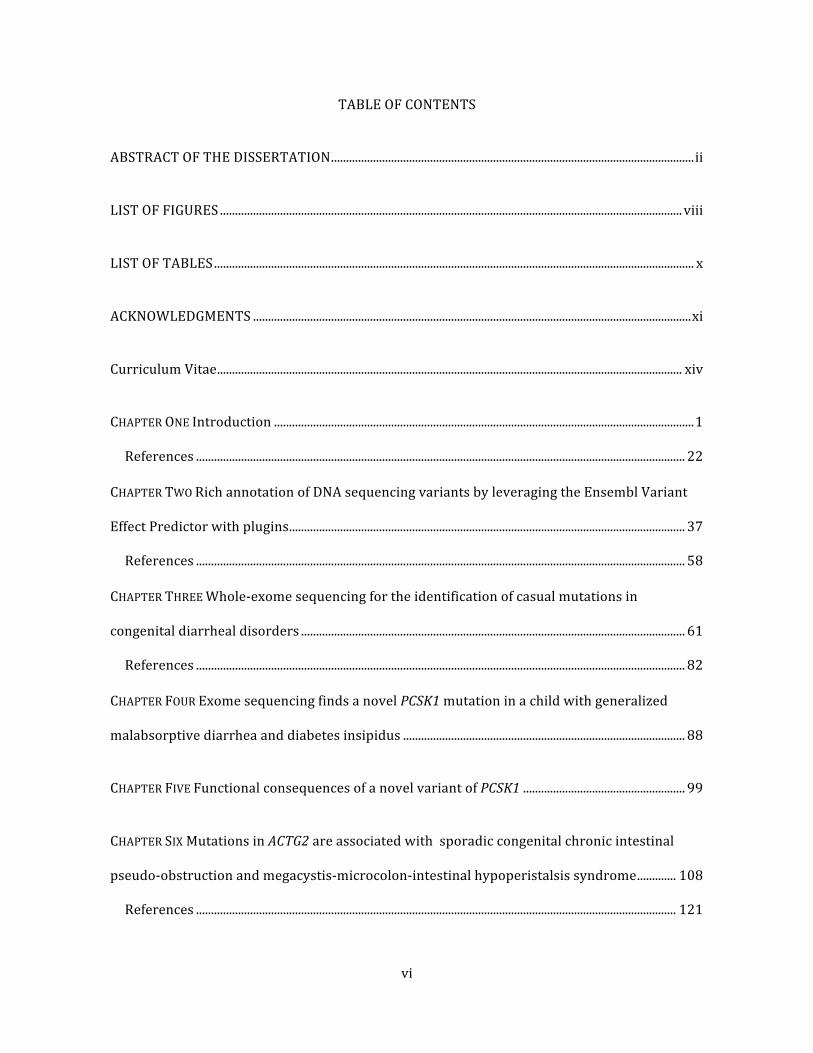
TABLE OF CONTENTS
ABSTRACT OF THE DISSERTATION ......................................................................................................................... ii
LIST OF FIGURES .......................................................................................................................................................... viii
LIST OF TABLES ................................................................................................................................................................ x
ACKNOWLEDGMENTS .................................................................................................................................................. xi
Curriculum Vitae ........................................................................................................................................................... xiv
CHAPTER ONE Introduction ............................................................................................................................................ 1
References ................................................................................................................................................................... 22
CHAPTER TWO Rich annotation of DNA sequencing variants by leveraging the Ensembl Variant
Effect Predictor with plugins .................................................................................................................................... 37
References ................................................................................................................................................................... 58
CHAPTER THREE Whole-‐exome sequencing for the identification of casual mutations in
congenital diarrheal disorders ................................................................................................................................ 61
References ................................................................................................................................................................... 82
CHAPTER FOUR Exome sequencing finds a novel PCSK1 mutation in a child with generalized
malabsorptive diarrhea and diabetes insipidus .............................................................................................. 88
CHAPTER FIVE Functional consequences of a novel variant of PCSK1 ...................................................... 99
CHAPTER SIX Mutations in ACTG2 are associated with sporadic congenital chronic intestinal
pseudo-‐obstruction and megacystis-‐microcolon-‐intestinal hypoperistalsis syndrome ............. 108
References ................................................................................................................................................................ 121

vii
CHAPTER SEVEN Mutations in the RNA exosome component gene EXOSC3 cause pontocerebellar
hypoplasia and spinal motor neuron degeneration .................................................................................... 128
CHAPTER EIGHT Conclusions .................................................................................................................................... 149
References ................................................................................................................................................................ 157
viii
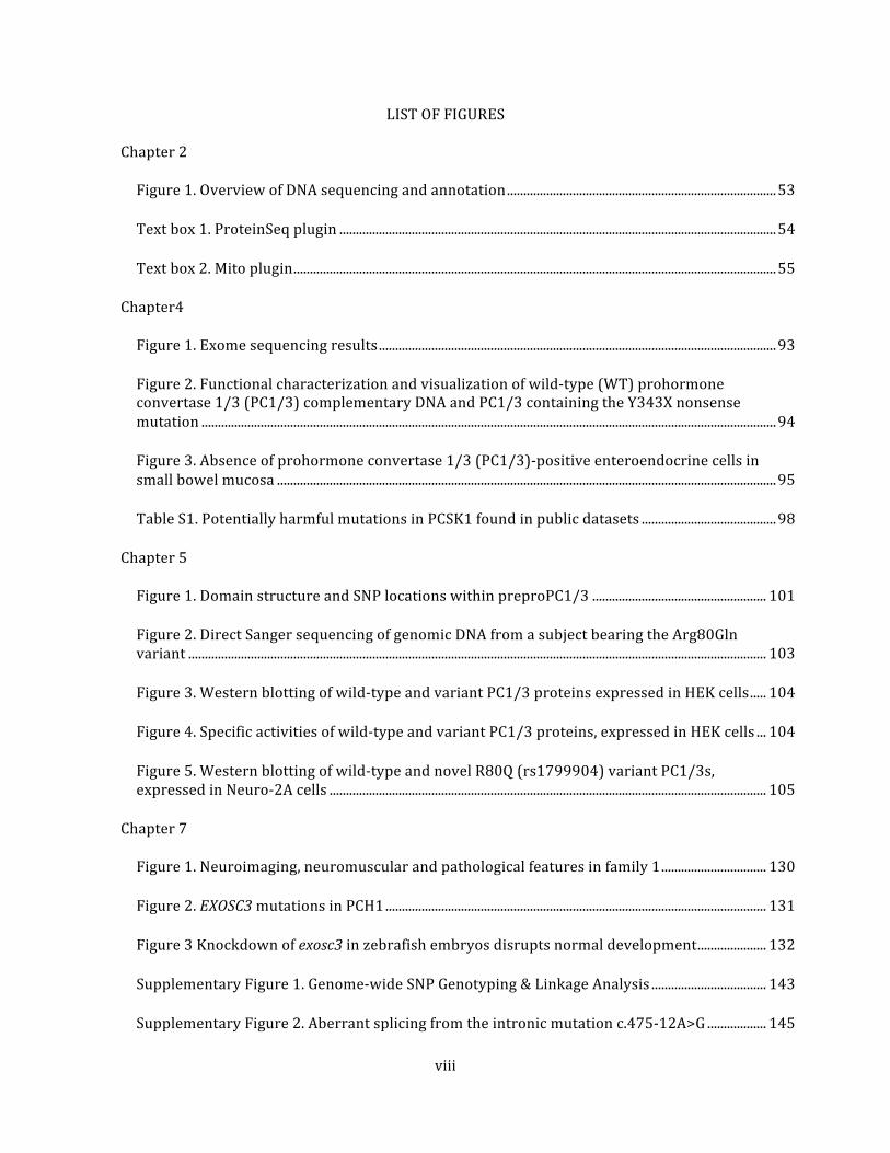
LIST OF FIGURES
Chapter 2
Figure 1. Overview of DNA sequencing and annotation .................................................................................. 53
Text box 1. ProteinSeq plugin ..................................................................................................................................... 54
Text box 2. Mito plugin ................................................................................................................................................... 55
Chapter4
Figure 1. Exome sequencing results ......................................................................................................................... 93
Figure 2. Functional characterization and visualization of wild-‐type (WT) prohormone convertase 1/3 (PC1/3) complementary DNA and PC1/3 containing the Y343X nonsense mutation ............................................................................................................................................................................... 94
Figure 3. Absence of prohormone convertase 1/3 (PC1/3)-‐positive enteroendocrine cells in small bowel mucosa ........................................................................................................................................................ 95
Table S1. Potentially harmful mutations in PCSK1 found in public datasets ......................................... 98
Chapter 5
Figure 1. Domain structure and SNP locations within preproPC1/3 ..................................................... 101
Figure 2. Direct Sanger sequencing of genomic DNA from a subject bearing the Arg80Gln variant ................................................................................................................................................................................ 103
Figure 3. Western blotting of wild-‐type and variant PC1/3 proteins expressed in HEK cells ..... 104
Figure 4. Specific activities of wild-‐type and variant PC1/3 proteins, expressed in HEK cells ... 104
Figure 5. Western blotting of wild-‐type and novel R80Q (rs1799904) variant PC1/3s, expressed in Neuro-‐2A cells ..................................................................................................................................... 105
Chapter 7
Figure 1. Neuroimaging, neuromuscular and pathological features in family 1 ................................ 130
Figure 2. EXOSC3 mutations in PCH1 .................................................................................................................... 131
Figure 3 Knockdown of exosc3 in zebrafish embryos disrupts normal development ..................... 132
Supplementary Figure 1. Genome-‐wide SNP Genotyping & Linkage Analysis ................................... 143
Supplementary Figure 2. Aberrant splicing from the intronic mutation c.475-‐12A>G .................. 145

ix
Supplementary Figure 3. rRNA processing ........................................................................................................ 146
Supplementary Figure 4. Exosc3 expression in zebrafish embryos by whole-‐mount in situ hybridization ................................................................................................................................................................... 147
x
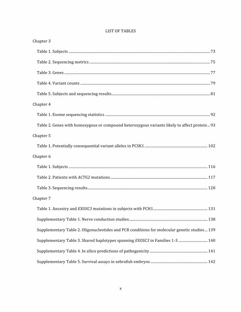
LIST OF TABLES
Chapter 3
Table 1. Subjects ............................................................................................................................................................... 73
Table 2. Sequencing metrics ........................................................................................................................................ 75
Table 3. Genes .................................................................................................................................................................... 77
Table 4. Variant counts .................................................................................................................................................. 79
Table 5. Subjects and sequencing results ............................................................................................................... 81
Chapter 4
Table 1. Exome sequencing statistics ...................................................................................................................... 92
Table 2. Genes with homozygous or compound heterozygous variants likely to affect protein ... 93
Chapter 5
Table 1. Potentially consequential variant alleles in PCSK1 ....................................................................... 102
Chapter 6
Table 1. Subjects ............................................................................................................................................................ 116
Table 2. Patients with ACTG2 mutations ............................................................................................................. 117
Table 3. Sequencing results ....................................................................................................................................... 120
Chapter 7
Table 1. Ancestry and EXOSC3 mutations in subjects with PCH1 ............................................................. 131
Supplementary Table 1. Nerve conduction studies ........................................................................................ 138
Supplementary Table 2. Oligonucleotides and PCR conditions for molecular genetic studies ... 139
Supplementary Table 3. Shared haplotypes spanning EXOSC3 in Families 1-‐3 ................................. 140
Supplementary Table 4. In silico predictions of pathogenicity ................................................................. 141
Supplementary Table 5. Survival assays in zebrafish embryos ................................................................ 142
xi
ACKNOWLEDGMENTS
Chapter 2 is a manuscript, “Rich Annotation of DNA Sequencing Variants by Leveraging the
Ensembl Variant Effect Predictor with Plugins”, which has been submitted to Briefings in
Bioinformatics and is currently under review. It is presented with the permission of the co-‐
authors, S. Paige Taylor, Aliz R. Rao, Martín G. Martín, and Stanley F. Nelson.
Chapter 3 is an in preparation first author manuscript, “Whole-‐Exome Sequencing for the
Identification of Casual Mutations in Congenital Diarrheal Disorders”. I acknowledge my co-‐
authors, Robert Venick, Pam Brown, Rune Rose Tronstad, David Brumbaugh, Vered
Pinsk, Robert Bandsma, Jeff Lewis, John Kerne, Anja Koren Jeverica, Anupama Chawla, Jerry
Levine, Stanley F. Nelson, and Martín G. Martín.
Chapter 4 is a published first author paper presented with permission from the Journal of
Pediatric Gastroenterology and Nutrition and the co-‐authors. Yourshaw M, Solorzano-‐Vargas
RS, Pickett LA, Lindberg I, Wang J, Cortina G, Pawlikowska-‐Haddal A, Baron H, Venick RS,
Nelson SF, Martin MG. Exome Sequencing Finds a Novel PCSK1 Mutation in a Child With
Generalized Malabsorptive Diarrhea and Diabetes Insipidus. Journal of pediatric
gastroenterology and nutrition. 2013;57(6):759-‐67. Epub 2013/11/28. doi:
10.1097/MPG.0b013e3182a8ae6c. PubMed PMID: 24280991.
Chapter 5 is a published paper presented with permission from PLOS ONE and the co-‐authors.
Pickett LA, Yourshaw M, Albornoz V, Chen Z, Solorzano-‐Vargas RS, Nelson SF, Martin MG,
Lindberg I. Functional consequences of a novel variant of PCSK1. PLoS One. 2013;8(1):e55065.
xii
doi: 10.1371/journal.pone.0055065. PubMed PMID: 23383060; PubMed Central PMCID:
PMC3557230.
Chapter 6 is an in preparation co-‐first author manuscript, “Mutations in ACTG2 are associated
with sporadic congenital chronic intestinal pseudo-‐obstruction”. I acknowledge my co-‐authors,
Antia Sicolo (who contributed equally to the work), Robert Venick, Laurie Reyen, Doug Farmer,
Jorge Vargas, Stanley F. Nelson, and Martín G. Martín.
Chapter 7 is a co-‐first author published paper presented with permission from Nature Genetics
and the co-‐authors. Wan J, Yourshaw M, Mamsa H, Rudnik-‐Schoneborn S, Menezes MP, Hong JE,
Leong DW, Senderek J, Salman MS, Chitayat D, Seeman P, von Moers A, Graul-‐Neumann L,
Kornberg AJ, Castro-‐Gago M, Sobrido MJ, Sanefuji M, Shieh PB, Salamon N, Kim RC, Vinters HV,
Chen Z, Zerres K, Ryan MM, Nelson SF, Jen JC. Mutations in the RNA exosome component gene
EXOSC3 cause pontocerebellar hypoplasia and spinal motor neuron degeneration. Nat Genet.
2012;44(6):704-‐8. Epub 2012/05/01. doi: 10.1038/ng.2254. PubMed PMID: 22544365;
PubMed Central PMCID: PMC3366034.
PIs and project directors are in bold.
I thank my committee members Rita M. Cantor, Lars Drier, Jake Lusis, and Stanley F. Nelson for
their guidance during this work, and my collaborators Martín Martín, Joanna Jen, and Berit
Kerner for their generosity with interesting and worthwhile projects. I also thank my friends
and colleagues for their help and tolerance throughout my research: Vivian Chang (who
introduced me to Dominion and Elle), Zugen Chen, Bobby Chin, Michael Clark, Ascia Eskin, Bret
Harry, Nils Homer, Janeen Ishii, Hane Lee, Hafsa Mamsa, Barry Merriman, Tove Olafsen, Aliz

xiii
Raksi Rao, Sergio Solorzano-‐Vargas, Cris Sosa, Kevin Squire, Sara Stanford, Sam Strom, Paige
Taylor (especially for a memorable World Series game one at Fenway), Traci Toy, Chris
Walthers, Jenny Wang, Richard Wang, Jijun Wan, Anna Wu, Jen Zieba, and Judith Zimmerman. I
am especially grateful to my colleague and BFF Genevieve Kendall for her inspiration,
friendship, advice, and assistance.
xiv
Curriculum Vitae
Michael Yourshaw
Education
Harvard College AB 1963
Harvard Law School JD 1971
Colorado State University BS 2006
Employment
Neaera Consulting Group, LLC, Fort Collins CO 2004-‐2006
Engineering Computer Consultants, Inc., Fort Collins CO 1999-‐2004
Wiley, Rein & Fielding, Washington DC 1983-‐1999
Kirkland & Ellis, Washington DC 1971-‐1983
United States Air Force 1964-‐1969
United Fruit Company, Boston MA 1963-‐1964
George Washington University, Washington DC 1962
United States Army 1960,1961
Publications
Wan J, Yourshaw M, Mamsa H, Rudnik-‐Schoneborn S, Menezes MP, Hong JE, Leong DW,
Senderek J, Salman MS, Chitayat D, Seeman P, von Moers A, Graul-‐Neumann L,
Kornberg AJ, Castro-‐Gago M, Sobrido MJ, Sanefuji M, Shieh PB, Salamon N, Kim RC,
Vinters HV, Chen Z, Zerres K, Ryan MM, Nelson SF, Jen JC. Mutations in the RNA
exosome component gene EXOSC3 cause pontocerebellar hypoplasia and spinal motor
neuron degeneration. Nat Genet. 2012;44(6):704-‐8. Epub 2012/05/01. doi:
10.1038/ng.2254. PubMed PMID: 22544365; PubMed Central PMCID: PMC3366034.
xv
Yourshaw M, Solorzano-‐Vargas RS, Pickett LA, Lindberg I, Wang J, Cortina G,
Pawlikowska-‐Haddal A, Baron H, Venick RS, Nelson SF, Martin MG. Exome Sequencing
Finds a Novel PCSK1 Mutation in a Child With Generalized Malabsorptive Diarrhea and
Diabetes Insipidus. Journal of pediatric gastroenterology and nutrition.
2013;57(6):759-‐67. Epub 2013/11/28. doi: 10.1097/MPG.0b013e3182a8ae6c.
PubMed PMID: 24280991.
Martin MG, Lindberg I, Solorzano-‐Vargas RS, Wang J, Avitzur Y, Bandsma R, Sokollik C,
Lawrence S, Pickett LA, Chen Z, Egritas O, Dalgic B, Albornoz V, de Ridder L, Hulst J, Gok
F, Aydogan A, Al-‐Hussaini A, Gok DE, Yourshaw M, Wu SV, Cortina G, Stanford S, Georgia
S. Congenital proprotein convertase 1/3 deficiency causes malabsorptive diarrhea and
other endocrinopathies in a pediatric cohort. Gastroenterology. 2013;145(1):138-‐48.
doi: 10.1053/j.gastro.2013.03.048. PubMed PMID: 23562752.
Pickett LA, Yourshaw M, Albornoz V, Chen Z, Solorzano-‐Vargas RS, Nelson SF, Martin
MG, Lindberg I. Functional consequences of a novel variant of PCSK1. PLoS One.
2013;8(1):e55065. doi: 10.1371/journal.pone.0055065. PubMed PMID: 23383060;
PubMed Central PMCID: PMC3557230.
Rudnik-‐Schoneborn S, Senderek J, Jen JC, Houge G, Seeman P, Puchmajerova A, Graul-‐
Neumann L, Seidel U, Korinthenberg R, Kirschner J, Seeger J, Ryan MM, Muntoni F,
Steinlin M, Sztriha L, Colomer J, Hubner C, Brockmann K, Van Maldergem L, Schiff M,
Holzinger A, Barth P, Reardon W, Yourshaw M, Nelson SF, Eggermann T, Zerres K.
Pontocerebellar hypoplasia type 1: clinical spectrum and relevance of EXOSC3
xvi
mutations. Neurology. 2013;80(5):438-‐46. doi: 10.1212/WNL.0b013e31827f0f66.
PubMed PMID: 23284067; PubMed Central PMCID: PMC3590055.
Kerner B, Rao AR, Christensen B, Dandekar S, Yourshaw M, Nelson SF. Rare genomic
variants link bipolar disorder to CREB regulated intracellular signaling pathways.
Frontiers in Psychiatry. 2013. doi: 10.3389/fpsyt.2013.00154.

1
CHAPTER ONE
Introduction
2
1.1 High-‐throughput exome sequencing and rare Mendelian disorders
The basic laws of monogenic inheritance were first set forth by Gregor Mendel in 1865
(1). In 1910 Thomas Hunt Morgan discovered that chromosomes were the physical and
mechanistic basis of Mendelian inheritance (2) and in 1953 Watson and Crick determined the
double helix structure of DNA (3). After these fundamental discoveries a framework for
associating genetic mutations with human disease was in place, but DNA could not be reliably
sequenced in the laboratory for several decades. In 1974 Fredrick Sanger developed the chain-‐
terminator method of DNA sequencing (4). However, even today one of the most advanced
Sanger sequencing machines (the ABI 3730xl) can produce only 2100 kilobases of data per
day, or 0.07% of the human genome. Two further developments revolutionized the field of
medical genetics: publication of the complete human genome in 2004 (5-‐7) based on
automated Sanger technology; and the development of next generation high-‐throughput DNA
sequencing technologies such as the Solexa/Illumina Genome Analyzer in 2006. In contrast to
Sanger sequencing, the Genome Analyzer could sequence a billion bases per run in ten days
(8). Today, the widely used Illumina HiSeq 2500 next-‐generation DNA sequencing platform can
generate 100 billon bases in 27 hours, enough to resequenced an entire human genome.
While Sanger sequencing processes a single fragment at a time, high throughput
platforms process billions of fragments in parallel. A DNA library is prepared by random
fragmentation of genomic DNA into templates, which are then ligated to adapters and PCR
amplified (9). The templates are immobilized on a solid support (glass slides or beads) and
clonally amplified in situ by solid phase amplification, specifically bridge PCR on the Illumina
platform, to create billions of colonies (10). The Illumina platform then performs one cycle of
synthetic sequencing with fluorescent reversible terminator deoxyribonucleotides for each
base position in parallel on all colonies. Images of the slide surface are analyzed to generate
high-‐confidence base calls (11).
3
The availability of this powerful technology inspired us to tackle daunting problems in
medical genetics that hitherto would have been extremely difficult to solve. Specifically, we
were interested in gaining a better understanding of the molecular basis of inherited diseases.
Genetic diseases are simplistically divided into those that are polygenic, complex, and common,
on the one hand, and monogenic, simple, and rare, on the other. Multiple genes acting through
a complex network of pathways control complex diseases. Monogenic diseases are often called
‘Mendelian disorders’ because they have certain characteristics of the garden peas studied by
Mendel: a particular genotype at a locus is both necessary and sufficient for a phenotype to be
expressed under the normal range of genetic and environmental backgrounds (12). Humans
inherit two copies of the 22 non-‐sex chromosomes (autosomes), one from each parent, and
thus may have the same allele (homozygosity) or two different alleles (heterozygosity) at a
given locus. Mendelian patterns of disease inheritance may be dominant, where an affected
individual need only inherit a disease causing allele from one parent, or recessive, where both
parents must transmit a disease causing allele. The type of chromosome further categorizes
Mendelian patterns: the non-‐sex chromosomes (autosomes) and the X or Y sex chromosomes.
Disease can also be inherited in a not strictly Mendelian manner via the maternally transmitted
mitochondrial chromosome. The reality is more complex than this simplified description, as a
single specific allele, different alleles in the same gene, and different alleles in different genes
all give rise to Mendelian inheritance patterns in a given pedigree. Moreover, a genetically
caused disease may arise in an individual sporadically as a result of a de novo mutation,
typically a dominant acting one.
The reference human genome is a haploid representation of each chromosome, with a
relatively complete list of its nucleotides, assembled from a number of individuals chosen
primarily for reasons of technical sequencing quality and not intended to represent a typical or
perfectly healthy person. High-‐throughput sequencing will find a large number of variants
4
from the reference in every subject, most of which are phenotypically insignificant.
Accordingly, we decided at the outset to explore this new technology by focusing on rare,
debilitating, congenital diseases with a distinct phenotype, to empower us to find, in a small
sample size, a causative allele from among thousands of false positives. First, a distinct
phenotype observed in early childhood is more likely to be caused by inborn genetic variants
than by environmental factors acting over a lifetime. Second, a rare incidence of a disorder
implies that it is caused by a rare allele, and we could therefore filter out variants that were
sufficiently common in the population that they could not possibly cause the disease. Third,
individuals affected by a severely debilitating condition in childhood would not generally have
survived to reproductive age absent heroic interventions with modern medicine, which would
insure that recessive alleles would remain heterozygous in the population and dominant acting
alleles would not be found. Fourth, if the pedigree is consistent with autosomal recessive
inheritance or dominant acting de novo mutations, sequencing candidate variants in parents
and affected or unaffected siblings would allow us to exclude variants that did not segregate
with the disease. We made a further simplifying assumption that the disease would be fully
penetrant, i.e., a person with the genotype would always manifest the disease. Congenital
malabsorptive diarrhea, intestinal pseudo-‐obstruction, and pontocerebellar hypoplasia
satisfied these criteria. A presented in the following chapters, we found homozygous or
compound heterozygous mutations exhibiting recessive inheritance and dominant acting de
novo heterozygous mutations in these diseases. These disorders may be distinguished from
comparatively common diseases, such as obesity, heart disease, type 2 diabetes mellitus, and
others, which typically manifest well after infancy. It is hypothesized that the genetic
component of these diseases can explained by a combination of common variants in one or
more genes, each with a small effect or perhaps by multiple extremely rare variants with a
stronger effect. This class of disease has been studied with large case-‐control associations
5
studies, which search for the difference in allele frequency of polymorphic markers between
unrelated groups of affected and unaffected individuals or within families. Association studies
may require costly genotyping of thousands of individuals to have sufficient power to detect
subtle effects and, like linkage analysis, lack resolution at the nucleotide level.
A practical concern regarding the use of high-‐throughput sequencing for modest
studies with limited budgets is achieving sufficient sequencing depth, i.e., the number of
independent fragments that support a genotype call at a given locus. Multiple observations per
base are necessary (a minimum of 10-‐20 to sensitively detect heterozygous variants) to make
it likely that both alleles of a heterozygous locus will be observed and also to account for
inevitable errors in the sequencing process. Although the per base cost of sequencing is low,
there are more than 6 billion bases in a diploid human genome. Thus, the cost of sequencing
the genomes of many cases at a depth sufficient to detect rare variants is considerable.
Furthermore, because the number of reads varies by orders of magnitude among loci, it is
necessary to get a mean coverage >100X to achieve 20X coverage for >90% of targeted bases.
For this reason, we initially explored the development of strategies to enrich sequencing
libraries for regions of interest in order to minimize the cost of sequencing less informative
regions. This was a well-‐recognized difficulty and commercial reagents became available to
meet the demand, obviating the need for us to pursue an in-‐house solution. A reasonable
strategy for the discovery of rare alleles responsible for Mendelian phenotypes is to sequence
only the protein coding regions of the genome (the ‘exome’), which represent fewer than 2% of
all bases (15). Most known genetic causes of Mendelian diseases affect protein-‐coding regions
(16), and there is reason to believe that many rare missense alleles and small insertions and
deletions (indels) in the exome have a functional consequence or are damaging (17).
Promoters, enhancers, short RNAs, and other regulatory elements outside the exome doubtless
govern some disorders, but variants in these regions are comparatively difficult to interpret.
6
Thus, the exome is an attractive target for an initial sub-‐genomic screen (18). Another
consideration is that regions outside the coding DNA sequences (CCDs) have been noted to
perform less efficiently in capture sequence experiments (19). When linkage or homozygosity
mapping identify a particular region of interest it is possible to develop a custom probe set to
enrich the sequencing library for only that region. Nonetheless, it still may be cost-‐effective to
use a standard exome probe set unless a large number of samples are involved.
A library can be enriched for exome fragments with a capture method that uses
biotinylated probes or 'baits' (RNA in the Agilent kit, DNA in the Illumina and Nimblegen kits)
to fish targets out of a 'pond' of DNA fragments. In the Agilent process RNA is transcribed from
PCR-‐amplified oligodeoxynucleotides originally synthesized on a microarray, generating
sufficient bait for multiple captures at concentrations high enough to drive the hybridization
(20). After the library and probes are hybridized, magnetic beads on the probes select probe-‐
exome hybrids for sequencing. In our experience ~76% of bases map on or near a bait and the
baited region is enriched 32-‐fold relative to the remainder of the genomic background.
A high-‐throughput sequencing instrument has a minimum unit of production
determined by how many DNA clusters are processed in parallel. For example, in a typical
configuration of an Illumina HiSeq 2500 instrument, two flowcells can run simultaneously,
each flowcell being divided into eight lanes. Thus one lane is the minimum platform unit and a
single lane can sequence up to 180 million paired end fragments. This is sufficient sequencing
capacity to get satisfactory coverage on three or more exomes. Accordingly, we could sequence
more samples for almost the same cost if we could multiplex samples in a lane and
computationally identify the samples in downstream processing. We developed a method of
Hamming code based DNA barcodes that were concatenated to the adapters, but switched to
commercially available barcoded adapters when they became available to allow efficient
exome sequencing.
7
1.2 Annotation of variants identified by high-‐throughput exome sequencing
In almost all of the experiments we performed, the sequencing platform produced
paired-‐end reads of 100 bases from either end of library fragments that are ~700 bases long.
Extensive downstream processing is necessary to transform the unmapped raw reads into a
useful dataset for variant discovery. The steps included: demultiplexing barcoded reads to
separate reads by sample; removing PCR duplicates to prevent overrepresented fragments
from biasing allele counts; recalibrating base quality scores to improve accuracy by analyzing
the covariation among reported quality score, position within read, dinucleotide, and
probability of mismatching the reference genome; mapping the fragments to the GRCh37
human reference genome; calling genotypes; assigning a well-‐calibrated probability of being
true to each variant call in a call set (under a Gaussian mixture model using the variables
inbreeding coefficient, quality by depth, mapping quality map sum test, mapping quality, read
position rank sum test, and Fisher strand); and homozygosity block identification. These
functions are performed by several software packages, including Picard (21), Novoalign (22),
the Genome Analysis Toolkit (23, 24), Samtools (25), and PLINK (26, 27). We developed
pipeline software that could keep track of case IDs, samples, libraries, machine runs, lanes,
barcodes and other experiment-‐related metadata, coordinate the parallel execution of the
programs on a compute cluster, and manage the output files. The pipeline software was
written in Python and Scala, is modular, and requires minimal manual intervention once the
metadata has been entered. An SQL Server stores metadata and the program results for
downstream analysis.
The output of such a sequencing pipeline is a Variant Call Format (VCF) (28, 29) file
that succinctly and systematically describes the genomic location, dbSNP ID, reference and
alternate alleles, genotype, and other information related to each variant. For an exome, a VCF
8
file typically consists of over 20,000 individual protein coding variants, and >50,000 records to
account for the effects of variants on different transcripts of the same gene.
A basic VCF file does not contain most of the information that will be needed by a
physician or researcher, such as the transcript and gene that contain the variant, the effect, if
any, on protein encoding (synonymous, missense, nonsense) or structure, the likelihood that
the variant is damaging, association with diseases or phenotypes, tissue expression data, or
phenotypes in model organisms. There are several applications that can add such annotations
to a VCF file, each with strengths an weaknesses (30). One characteristic of most of these tools
is that they have little or no flexibility to include customized user-‐defined annotations.
Furthermore, while on-‐line tools, such as SeattleSeq (15), have the advantage of simplicity of
use, they may not be appropriate for confidential patient data or proprietary intellectual
property.
We developed a custom annotator, which we call ‘VAX’ (Variant Annotator eXtras) that
runs on local servers, is not heavily dependent on an outside researcher for software
development and maintenance, and has a simple, modular mechanism for adding new features.
We used the Ensembl Variant Effect Predictor (VEP) (31) as an engine. The VEP annotates
variants with transcript and protein consequences including estimates of the extent of protein
damage as computed by SIFT (32-36), PolyPhen (37-39), and Condel (40) We incorporated
additional annotations from datasets such as Online Mendelian Inheritance in Man (OMIM)
(41), the Human Gene Mutation Database (HGMD pro, BIOBASE Biological Databases), the
Universal Protein Resource (UniProt) (42), KEGG Pathways (43), RefGene (44), the MitoCarta
Inventory of Mammalian Mitochondrial Genes (45), Mouse Genome Informatics (MGI) (46)(47)
and the Human Protein Atlas (HPA) (47), as well as allele frequencies and statistics on the
number of damaging variants per gene.
9
Several factors were decisive in adopting the Ensembl VEP as the underlying engine.
Ensembl, a joint scientific project between the European Bioinformatics Institute and the
Wellcome Trust Sanger Institute, provides access to genomic annotation for numerous species
stored on a MySQL database that can be accessed programmatically via a Perl application
programming interface (API). The database is supported by a large professional organization,
is updated regularly, and can be accessed remotely or by downloading a local copy. The
Ensembl database and VEP have a large and active user community, which provide excellent
and timely advice and support. The VEP is a mature open source Perl script that can be run
locally, connected either to the remote Ensembl database or a local copy thereof, or with some
limitations used with a local cache.
In addition to its use in our research projects, VAX is used routinely for CLIA/CAP-‐
accredited whole-‐exome sequencing by the UCLA Clinical Genomics Center, which has
processed more than 1000 exomes to date (48). VAX is also used by other researchers at UCLA,
for example in a study of bipolar disorder in a family of four affected siblings (14). This study
identified variants in genes that encode proteins with significant regulatory roles in the
ERK/MAPK and CREB-‐regulated intracellular signaling pathways and supported the
hypothesis that multiple rare, damaging mutations in genes functionally related to a common
signaling pathway may contribute to the manifestation of bipolar disorder.
1.3 Congenital gastrointestinal disorders
Congenital gastroenterological disorders may be caused by genetic mutations,
environmental factors, or a combination of both. These disorders fall into three broad
categories: diarrheal, motility, and obstructive.
Congenital diarrheal disorders (CDDs) are a set of enteropathies caused by inherited or
sporadic genetic mutations that generally manifest soon after birth or in early childhood. The
presenting symptom is chronic diarrhea that often requires total parenteral nutrition (TPN).
10
These patients frequently endure a complex and costly diagnostic odyssey that commonly fails
to produce a definitive diagnosis (49).
Congenital motility disorders are a heterogenous group of disorders affecting gut
neuromuscular function, which typically present with symptoms of vomiting, constipation or
diarrhea, and abdominal pain (50). These disorders account for a significant portion of
pediatric cases of intestinal failure (51). Hirschsprung disease (aganglionic megacolon) is a
complex genetic disease caused by both rare and common mutations in RET and related genes.
Linkage analysis, homozygosity mapping, and case-‐control association studies have all
contributed to identifying these genes (52). Non-‐Hirschsprung cases of congenital intestinal
motility failure are grouped under the term ‘chronic intestinal pseudo-‐obstruction’ (CIPO) and
also include a related disorder, megacystis-‐microcolon-‐intestinal hypoperistalsis syndrome
(MMIH).
Congenital intestinal obstructive disorders (atresia and stenosis) involve narrowed,
blocked or disconnected intestine. Although genetic mutations are believed to be responsible
for some congenital intestinal atresias and the disorder is associated with cystic fibrosis and
Down syndrome, no genetic cause was discovered without the use of exome sequencing.
In the current research we assessed whole-‐exome sequencing as a method for
identifying casual mutations in CDD patients, and for the discovery of novel genes that cause
CIPO.
1.4 A pilot study of high-‐throughput exome sequencing to identify the molecular basis of
congenital diarrheal disorders
Congenital diarrheal disorders (CDDs) are rare diseases with serious, even life-‐
threatening, consequences that impose massive diagnostic and treatment costs as well as great
emotional stress on patients and their families. Until recently, little was known of the genetic
etiology of these diseases, yet identification of a casual mutation can lead to improved
11
management of the disease and inform research efforts to develop new treatment modalities.
Prior to the availability of exome sequencing, CDDs with known genetic causes included
secretory chloride diarrhea caused by mutations in the SLC26A3 gene (53), microvillus
inclusion disease caused by mutations in the MYO5B gene (54), a syndromic form of congenital
secretory sodium diarrhea caused by mutations in the SPINT2 gene (55), malabsorptive
congenital diarrhea caused by mutations in the NEUROG3 gene (56), congenital tufting
enteropathy, caused by mutations in the EPCAM gene (57), and early-‐onset chronic diarrhea,
caused by mutations in the GUCY2C gene (58).
The discovery of the aforementioned genes involved arduous genetic and molecular
studies. In the 1990s genome wide linkage analysis, using 300-‐500 microsatellite markers,
offered a method of identifying disease susceptibility loci. This approach was time-‐consuming
and limited to a resolution of down to about 10Mb, a length, which can harbor over 100 genes
(59). To achieve adequate power to find linkage it was necessary to genotype one or more
extended families or a very large number of nuclear families. For example, linkage
disequilibrium and genetic linkage as determined by this technique in Finnish families
indicated that an unknown gene near the cystic fibrosis transmembrane regulator gene (CFTR)
was probably associated with secretory chloride diarrhea (60). Subsequently, cloning of the
linkage region identified four known genes, two of which were considered to be functionally
relevant (61). Finally, segregation of mutations in the SLC26A3 gene with the disorder in a
large number of patients confirmed that such mutations cause the disorder (53). The
development of highly parallel genotyping based on arrays with probes for thousands of single
nucleotide polymorphisms (SNPs) enabled more efficient genotyping with a 10-‐fold or better
improvement in resolution compared to microsatellite base methods. This technology, applied
to extended kindreds, enabled the discovery of mutations in the causative genes for microvillus
inclusion disease, syndromic congenital secretory sodium diarrhea, congenital tufting
12
enteropathy, and early-‐onset chronic diarrhea. A candidate gene resequencing approach,
founded on a mouse model, identified mutations in NEUROG3 as the cause of malabsorptive
congenital diarrhea (56).
Despite much progress in uncovering the molecular basis for this class of diseases
there remained many patients with strong indications of a genetic etiology for whom no
mutation in these genes could be identified. We hypothesized that a large fraction of these
cases had undetected mutations in genes known to be associated with CDD, and others would
have mutations in discoverable novel genes. An additional working hypothesis was that some
casual mutations would be in the protein coding portion of the genome, and the phenotype
would be recessive the mutation would have high penetrance. Accordingly, we sought to
determine the effectiveness of whole-‐exome sequencing to identify, in genes that have been
reported to be associated with CDD, the molecular causes of the disease in a cohort of patients
with CDDs that had defied conventional diagnostic methods.
We chose 45 patients from 38 families for exome sequencing. Inclusion criteria were a
diagnosis of congenital diarrhea and probability that the disease had a genetic cause (typically
consanguineous parents or affected family members). Patients were excluded if they had a
confirmed genetic diagnosis or a clinical presentation already suggesting a mutation in a gene
known to cause congenital diarrhea. These patients were of diverse ethnic backgrounds and
had clinical presentations of generalized malabsorptive diarrhea, selective nutrient
malabsorption, secretory diarrhea, and infantile IBD.
We performed whole-‐exome sequencing, as described above, validated variants found
in candidate genes with Sanger sequencing, and Sanger sequenced available relatives to ensure
that genotypes segregated with the phenotype. We originally sequenced seven cases in five
families on the ABI SOLiD platform. This method identified a casual mutation in PCSK1 in one
case (62), but failed to find interesting variants in the other four families. The SOLiD data
13
appeared to be quite noisy (about twice the expected number of raw variants), so we
resequenced the unsolved cases and all further cases on the Illumina platform. In all, we
identified 31 different mutations in 21 families (27 cases) that we considered likely to be the
cause of the disease because they were deemed to be damaging and were found in genes
known to associated with the phenotype or reported in the literature during the course of the
study. We concluded that exome sequencing would be valuable for diagnosis of CDDs in a
clinical setting. Importantly, we identified novel candidate genes in many of the remaining
cases; work is in progress to confirm these findings by functional studies in vitro, in model
organisms, and/or in a model of intestinal tissue developed from patient intestinal stem cells
or differentiated embryonic stem cell cultures.
1.5 PC1/3 deficiency
Before our study, three reported cases indicated that the gene proprotein convertase
subtilisin/kexin type 1 (PCSK1), which encodes the neuroendocrine convertase 1 precursor
protein (PC1/3), is involved in disorders characterized by abnormal enteroendocrine
development or function that manifest in generalized malabsorption (63-‐65). PC1/3 is a
calcium-‐dependent serine endoprotease essential for the conversion of a variety of
prohormones into their bioactive forms. It has a well-‐defined role of processing proinsulin in β
cells of the pancreas (66); it is expressed richly in endocrine cells in the gut, where its function
is obscure.
One of the patients in our CDD cohort was a perplexing child with congenital
malabsorptive diarrhea and other presumably unrelated clinical problems (62). The patient (of
consanguineous parentage) was initially assessed at three weeks of age for recurrent diarrhea
and associated metabolic acidosis. At six days of age he was transferred to the intensive care
nursery due to poor peripheral perfusion and indirect hyperbilirubinemia. DNA genotyping of
CFTR for cystic fibrosis was negative for the 97 mutations most commonly observed. He was
14
hospitalized 21 times before age three, including presentations of hypovolemic shock with
profound metabolic acidosis, central venous catheter occlusions, heparin-‐induced
thrombocytopenia, multiple deep venous thrombi, pneumonia, hyperglycemia, and left
ventricular dysfunction.
Exome sequencing analysis as described above identified a novel Tyr343Ter mutation
in PCSK1 that terminated the protein within its catalytic domain. This nonsense mutation
rendered the gene product undetectable in either cells or secreted into media, probably due to
nonsense-mediated decay, and a caused a total lack of enzyme activity. Immunohistochemistry for
PC1/3-expressing enteroendocrine cells was negative.
The identification of a mutation in PCSK1 suggested a specific clinical diagnosis that
includes diabetes insipidus (DI) as a component. Indeed, follow up with the patient confirmed
the presence of DI, and intranasal desmopressin (DDAVP) improved the patient’s condition
significantly.
Contemporaneously with the pilot exome study, we determined the clinical features of
13 other children with PC1/3 deficiency caused by PCSK1 mutations (67). We performed
Sanger sequencing analysis of PCSK1 and measured enzymatic activity of recombinant PC1/3
proteins. We identified a pattern of endocrinopathies that develop in an age-‐dependent
manner. Neonates had severe malabsorptive diarrhea and failure to thrive, required prolonged
parenteral nutrition support, and had high mortality. Additional endocrine abnormalities
developed as the disease progressed, including diabetes insipidus, growth hormone deficiency,
primary hypogonadism, adrenal insufficiency, and hypothyroidism.
We further explored the PC1/3 landscape by searching for potentially consequential
variants in the dbSNP (68), 1000 Genomes Project (69), NHLBI (70), and NIEHS (71) datasets.
We found that a novel Arg80GLN variant (rs1799904) both exhibits adverse effects on PC1/3
activity and is prevalent in the population at a low level, suggesting that further biochemical
15
and genetic analysis would be warranted to assess its contribution to the risk of metabolic
disease within the general population.
1.6 Application of high-‐throughput exome sequencing to identify a probable genetic cause of
sporadic chronic intestinal pseudo-‐obstruction
In addition to the CDD cohort, we also sequenced a cohort of 20 cases diagnosed with
CIPO or MMIH in the hope of finding one or more novel genes responsible for these rare
conditions. Chronic intestinal pseudo-‐obstruction (CIPO) is a heterogenous set of diseases
characterized by repetitive episodes or continuous symptoms of intestinal obstruction, in the
absence of a lesion that occludes the lumen of the gut (50, 51). A small fraction of cases are
secondary to organic, systemic, or metabolic diseases, but the majority are primary and may be
myopathic, mesenchymopathic, or neuropathic, depending upon whether predominant
abnormalities are found in the enteric nervous system, interstitial cells of Cajal (ICC), or
intestinal smooth muscle (72). A related disorder, megacystis-‐microcolon-‐intestinal
hypoperistalsis syndrome (MMIH), is characterized by constipation and urinary retention,
microcolon, giant bladder (megacystis), intestinal hypoperistalis, hydronephrosis, and dilated
small bowel (73).
Congenital forms of CIPO are rare and can be life-‐threatening; congenital CIPO is an
important cause of intestinal failure, for which the only treatment may be complete visceral
transplantation (74). Congenital CIPO may sometimes be due to prenatal exposure to toxins
such as alcohol or narcotics. A handful of familial cases of CIPO have been reported with
autosomal dominant (with variable penetrance), autosomal recessive, and X-‐linked modes of
inheritance (75-‐85). It is well known that mutations in mitochondrial tRNA genes, POLG
(polymerase (DNA directed), gamma), and TYMP (thymidine phosphorylase), which are
expressed in the mitochondrion, cause a severe form of CIPO requiring frequent and long-‐term
parenteral nutrition and with frequently fatal digestive and neurologic complications.
16
Mitochondrial disorders may account for ~19% of CIPO cases (86). Contrawise, it is rare for
CIPO to be the principal clinical manifestation of a mitochondrial disorder (87). Primary
defects of the mitochondrial oxidative phosphorylation pathway are phenotypically
heterogenous, and affecting multiple organs, typically with nervous system and skeletal or
ocular muscle dysfunction (88). Mitochondrial neurogastrointestinal encephalomyopathy
(MNGIE) is a rare, autosomal recessive syndrome due to the loss of thymidine phosphorylase
activity associated with loss-‐of-‐function mutations in TYMP (89-‐93). Mutations in POLG, the
mitochondrial myopathy, epilepsy, lactic acidosis, and strokelike episodes (‘MELAS’) mutation
in the tRNAleu(UUR) gene, or mutations in the tRNAlys gene are sometimes associated with CIPO
(85, 94-‐101). Still, congenital CIPO is usually sporadic and prior to the advent of exome
sequencing, no non-‐mitochondrial gene had been convincingly associated with primary
sporadic CIPO.
We hypothesized for this study that the mode of inheritance would be recessive, de
novo, or mitochondrial, and that mutation effects would be fully penetrant. Identifying de novo
variants with a dominant effect on phenotype typically requires sequencing of parent-‐child
trios to eliminate the large number of potential heterozygous variants. Unfortunately, we did
not have parental DNA for most of these patients, but we believed there was a reasonable
chance we could narrow down the potential de novo candidates by looking for mutations in the
same gene in multiple patients. We further hypothesized that casual variants would be found
in the protein coding or splicing regions of genes. Because failure of muscle function in the
intestinal wall was believed to be a common cause of CIPO, we were particularly alert for
mutations in genes that might affect muscle cell function, such as myosins, actins, and proteins
that bind or regulate myosins and actins.
A few genes are present on the small circular mitochondrial DNA but many more are
encoded by nuclear DNA and then localize to the mitochondrion. We developed a
17
mitochondrial gene annotation in the VAX program for this study, based on the MitoCarta
inventory of 1098 mouse genes encoding proteins with strong support of mitochondrial
localization (45). We also developed an SQL query that generates a matrix of genes and cases
to graphically display the number and ID of cases mutated in each gene. During the course of
our study Lehtonen et al. reported that a missense variant in ACTG, encoding γ-‐enteric actin,
segregated in a Finnish family with autosomal dominant familial visceral myopathy (FVM), a
disorder that is subsumed within the broad definition of CIPO (102). Thus, we were
particularly interested in mutations in this gene, and identified nine novel de novo mutations
in the CIPO cohort. We also found a potential mitochondrial gene (POLG) compound
heterozygous mutation of interest in the mitochondrial gene (POLG), but have not yet
confirmed whether it is causal.
1.7 Application of high-‐throughput exome sequencing to discover an unsuspected gene, EXOSC3,
that causes pontocerebellar hypoplasia and spinal motor neuron degeneration
Another exome sequencing project involved the search for mutations causing a
mysterious neuromuscular disorder that affected four siblings in a large family (103). The
children were floppy at birth, had ocular motor apraxia, progressive muscle wasting, distal
contractures, progressive microcephaly, growth retardation and global developmental delay,
and never reached any motor milestone or spoke. Initially we were unable to categorize this
condition, but after receiving the autopsy report on one subject, we suspected pontocerebellar
hypoplasia (PCH), which is characterized by cerebellar hypoplasia or atrophy, variable pontine
atrophy and progressive microcephaly with global developmental delay (104). Pontocerebellar
hypoplasia type1 (PCH1) is a distinctive subtype of PCH, characterized by diffuse muscle
wasting that is secondary to spinal cord anterior horn cell loss and cerebellar hypoplasia (105-‐
108). Diagnosis of PCH1 is often delayed or never made because the combination of cerebellar
and spinal motor neuron degeneration is not commonly recognized, and the presentation of
18
diffuse weakness and devastating brain involvement is atypical of classical proximal spinal
muscular atrophy (SMA) (109). The literature contains only a handful of descriptions of case
series (110-‐113) and reports of PCH1 (114-‐120). Prior to our study, a causative gene had not
been identified in the majority of individuals with PCH1. Recessive mutations have been found
in VRK1 (encoding vaccinia-‐related kinase 1) (121), RARS2 (encoding mitochondrial arginyl-‐
tRNA synthetase 2) (104) and TSEN54 (encoding tRNA splicing endonuclease 54) (122) in
single individuals with PCH1. In PCH without SMA, TSEN54 mutations account for most cases
of PCH2 and PCH4 (104, 123), and RARS2 mutations have been found in two families with
PCH6 (124, 125).
Array-‐based identity by descent analysis of the four affected siblings, three healthy
siblings, and their parents, highlighted candidate regions in four sub-‐chromosomal loci with
more than 100 candidate genes in total. Exome sequencing (Illumina IIx single end 76 base
reads) of the four affected siblings yielded a single candidate variant, g.9:37783990T>G
(c.395A>C, p.Asp132Ala) in the EXOSC3 gene (encoding exosome component 3). The variant
was homozygous in all four affected siblings, segregated with the disorder upon Sanger
sequencing of unaffected relatives, and was within one of the intervals identical by descent in
all affected siblings; the parents were heterozygous for the variant. The variant was at a
completely conserved locus.
Exosome component 3, also known as the ribosomal RNA–processing protein 40
(Rrp40), is a core component of the human RNA exosome complex (distinct from exosome
vesicles). RNA exosomes are multi-‐subunit complexes conserved throughout evolution (126)
and are emerging as the major cellular machinery for processing, surveillance and turnover of
a diverse spectrum of coding and noncoding RNA substrates essential for viability (127). The
exosome’s nine subunits are arranged in a two-‐layered ring; the bottom ‘hexamer’ layer is
19
formed by six subunits. Rrp40 is one of three RNA binding subunits that comprise the ‘cap’ of
the exosome complex (128, 129).
Eight probands with PCH1 out of twelve additional families had homozygous or
compound heterozygous mutations in EXOSC3 and all available parents were heterozygous.
The Asp132Ala mutation was present in six of these families, homozygous in three families and
compound heterozygous in another three. One case was homozygous for Gly31Ala, another
compound heterozygous for Gly31Ala and Trp238Arg, and the remainder compound
heterozygous for Asp132Ala plus 99fs*11, Ala139Pro, or intronic c.475–12A>G causing exon
skipping. Genotyping the original family and two others revealed an identical short 1 cM region
flanking the g.9:37783990T>G locus, suggesting a distant ancestry for the mutation.
Interestingly, another founder mutation, Gly31Ala, also seen in two of our cases, was recently
identified as a cause of severe PCH1 among the Czech Roma (130).
Knockdown of exosc3 expression in zebrafish embryos by antisense morpholinos led to
a dose-‐dependent phenotype of a short, curved spine and small brain with poor motility and
even death by 3 days post fertilization. Whole-‐mount in situ hybridization showed decreased
expression of atoh1a (a marker specific for dorsal hindbrain progenitors) in the upper and
lower rhombic lips and a lack of expression of pvalb7, which is specific for differentiated
cerebellar Purkinje neurons (131). The abnormal phenotype from exosc3-‐specific morpholino
injection was largely rescued by co-‐injection with wild-‐type zebrafish exosc3 mRNA whereas
co-‐injection with mRNA containing the mutation was ineffective in rescue.
In a companion study, biallelic mutations in EXOSC3 were detected in 10 of 27 families
(37%) (132). The mutation-‐positive subjects typically presented with normal pregnancy,
normal birth measurements, and relative preservation of brainstem and cortical structures.
Psychomotor retardation was profound in all patients but lifespan was variable, with 3
subjects surviving beyond the late teens. Abnormal oculomotor function was commonly
20
observed in patients surviving beyond the first year. Major clinical features previously
reported in PCH1, including intrauterine abnormalities, postnatal hypoventilation and feeding
difficulties, joint contractures, and neonatal death, were rarely observed in mutation-‐positive
infants but were typical among the mutation-‐negative subjects, indicating that variability in
survival and clinical severity is correlated with the genotype.
The same homozygous Asp132Ala mutation as that in our original family was reported
in four patients with muscle hypertonia, developmental delay, spinal anterior horn
involvement, and prolonged survival, consistent with a milder form of PCH1, suggesting
phenotypic variability possibly by caused by protective factors in the genetic background
(133). Another recently reported case from Bangladesh with Asp132Ala and a novel Val80Phe
mutation suffered from intellectual disability, early onset spasticity, and cerebellar atrophy
(134).
1.8 Overview of the chapters
Chapter 1 is this introduction. Chapter 2 describes the methods used in the VAX
program for rich annotation of DNA sequencing variants by leveraging the Ensembl Variant
Effect Predictor with plugins. This paper was submitted to Briefings in Bioinformatics and is
under review. Chapter 3 reports the results of a pilot study to use whole-‐exome sequencing for
the identification of casual mutations in congenital diarrheal disorders. Chapter 4 describes in
more detail one of the cases analyzed in the pilot study of Chapter 3, where exome sequencing
found a novel PCSK1 mutation in a child with generalized malabsorptive diarrhea and diabetes
insipidus. This paper was published in the Journal of Pediatric Gastroenterology and Nutrition
(62). Chapter 5 contains an overview of PCSK1 variants in the human population and the
functional consequences of a novel variant of PCSK1. This paper was published in PLoS One
(135). Chapter 6 presents evidence from exome sequencing that mutations in ACTG2 are a
probable significant cause of chronic intestinal pseudo-‐obstruction. Chapter 8 shows the
21
application of whole-‐exome sequencing in the field of neurology and describes our discovery
that mutations in the RNA exosome component gene EXOSC3 cause pontocerebellar hypoplasia
and spinal motor neuron degeneration. This paper was published in Nature Genetics (103).
Chapter 10 is the conclusion.
22
References
1. Mendel G. Versuche über Plflanzen-‐hybriden. Des naturforschenden Vereines; 1865; Brünn: Verhandlungen des naturforschenden Vereines in Brünn; 1866.
2. Morgan TH. Sex Limited Inheritance in Drosophila. Science. 1910;32(812):120-‐2. Epub 1910/07/22. doi: 10.1126/science.32.812.120. PubMed PMID: 17759620.
3. Watson JD, Crick FH. Molecular structure of nucleic acids; a structure for deoxyribose nucleic acid. Nature. 1953;171(4356):737-‐8. Epub 1953/04/25. PubMed PMID: 13054692.
4. Sanger F, Nicklen S, Coulson AR. DNA sequencing with chain-‐terminating inhibitors. Proc Natl Acad Sci U S A. 1977;74(12):5463-‐7. Epub 1977/12/01. PubMed PMID: 271968; PubMed Central PMCID: PMC431765.
5. International Human Genome Sequencing C. Finishing the euchromatic sequence of the human genome. Nature. 2004;431(7011):931-‐45. Epub 2004/10/22. doi: 10.1038/nature03001. PubMed PMID: 15496913.
6. Lander ES, Linton LM, Birren B, Nusbaum C, Zody MC, Baldwin J, Devon K, Dewar K, Doyle M, FitzHugh W, Funke R, Gage D, Harris K, Heaford A, Howland J, Kann L, Lehoczky J, LeVine R, McEwan P, McKernan K, Meldrim J, Mesirov JP, Miranda C, Morris W, Naylor J, Raymond C, Rosetti M, Santos R, Sheridan A, Sougnez C, Stange-‐Thomann N, Stojanovic N, Subramanian A, Wyman D, Rogers J, Sulston J, Ainscough R, Beck S, Bentley D, Burton J, Clee C, Carter N, Coulson A, Deadman R, Deloukas P, Dunham A, Dunham I, Durbin R, French L, Grafham D, Gregory S, Hubbard T, Humphray S, Hunt A, Jones M, Lloyd C, McMurray A, Matthews L, Mercer S, Milne S, Mullikin JC, Mungall A, Plumb R, Ross M, Shownkeen R, Sims S, Waterston RH, Wilson RK, Hillier LW, McPherson JD, Marra MA, Mardis ER, Fulton LA, Chinwalla AT, Pepin KH, Gish WR, Chissoe SL, Wendl MC, Delehaunty KD, Miner TL, Delehaunty A, Kramer JB, Cook LL, Fulton RS, Johnson DL, Minx PJ, Clifton SW, Hawkins T, Branscomb E, Predki P, Richardson P, Wenning S, Slezak T, Doggett N, Cheng JF, Olsen A, Lucas S, Elkin C, Uberbacher E, Frazier M, Gibbs RA, Muzny DM, Scherer SE, Bouck JB, Sodergren EJ, Worley KC, Rives CM, Gorrell JH, Metzker ML, Naylor SL, Kucherlapati RS, Nelson DL, Weinstock GM, Sakaki Y, Fujiyama A, Hattori M, Yada T, Toyoda A, Itoh T, Kawagoe C, Watanabe H, Totoki Y, Taylor T, Weissenbach J, Heilig R, Saurin W, Artiguenave F, Brottier P, Bruls T, Pelletier E, Robert C, Wincker P, Smith DR, Doucette-‐Stamm L, Rubenfield M, Weinstock K, Lee HM, Dubois J, Rosenthal A, Platzer M, Nyakatura G, Taudien S, Rump A, Yang H, Yu J, Wang J, Huang G, Gu J, Hood L, Rowen L, Madan A, Qin S, Davis RW, Federspiel NA, Abola AP, Proctor MJ, Myers RM, Schmutz J, Dickson M, Grimwood J, Cox DR, Olson MV, Kaul R, Raymond C, Shimizu N, Kawasaki K, Minoshima S, Evans GA, Athanasiou M, Schultz R, Roe BA, Chen F, Pan H, Ramser J, Lehrach H, Reinhardt R, McCombie WR, de la Bastide M, Dedhia N, Blocker H, Hornischer K, Nordsiek G, Agarwala R, Aravind L, Bailey JA, Bateman A, Batzoglou S, Birney E, Bork P, Brown DG, Burge CB, Cerutti L, Chen HC, Church D, Clamp M, Copley RR, Doerks T, Eddy SR, Eichler EE, Furey TS, Galagan J, Gilbert JG, Harmon C, Hayashizaki Y, Haussler D, Hermjakob H, Hokamp K, Jang W, Johnson LS, Jones TA, Kasif S, Kaspryzk A, Kennedy S, Kent WJ, Kitts P, Koonin EV, Korf I, Kulp D, Lancet D, Lowe TM, McLysaght A, Mikkelsen T, Moran JV, Mulder N, Pollara VJ, Ponting CP, Schuler G, Schultz J, Slater G, Smit AF, Stupka E, Szustakowski J, Thierry-‐Mieg D, Thierry-‐Mieg J, Wagner L, Wallis J, Wheeler R, Williams A, Wolf YI, Wolfe KH, Yang SP, Yeh RF, Collins F, Guyer MS, Peterson J, Felsenfeld A, Wetterstrand KA, Patrinos A, Morgan MJ, de Jong P, Catanese JJ, Osoegawa K, Shizuya H, Choi S, Chen YJ, International Human Genome Sequencing C. Initial
23
sequencing and analysis of the human genome. Nature. 2001;409(6822):860-‐921. Epub 2001/03/10. doi: 10.1038/35057062. PubMed PMID: 11237011.
7. Venter JC, Adams MD, Myers EW, Li PW, Mural RJ, Sutton GG, Smith HO, Yandell M, Evans CA, Holt RA, Gocayne JD, Amanatides P, Ballew RM, Huson DH, Wortman JR, Zhang Q, Kodira CD, Zheng XH, Chen L, Skupski M, Subramanian G, Thomas PD, Zhang J, Gabor Miklos GL, Nelson C, Broder S, Clark AG, Nadeau J, McKusick VA, Zinder N, Levine AJ, Roberts RJ, Simon M, Slayman C, Hunkapiller M, Bolanos R, Delcher A, Dew I, Fasulo D, Flanigan M, Florea L, Halpern A, Hannenhalli S, Kravitz S, Levy S, Mobarry C, Reinert K, Remington K, Abu-‐Threideh J, Beasley E, Biddick K, Bonazzi V, Brandon R, Cargill M, Chandramouliswaran I, Charlab R, Chaturvedi K, Deng Z, Di Francesco V, Dunn P, Eilbeck K, Evangelista C, Gabrielian AE, Gan W, Ge W, Gong F, Gu Z, Guan P, Heiman TJ, Higgins ME, Ji RR, Ke Z, Ketchum KA, Lai Z, Lei Y, Li Z, Li J, Liang Y, Lin X, Lu F, Merkulov GV, Milshina N, Moore HM, Naik AK, Narayan VA, Neelam B, Nusskern D, Rusch DB, Salzberg S, Shao W, Shue B, Sun J, Wang Z, Wang A, Wang X, Wang J, Wei M, Wides R, Xiao C, Yan C, Yao A, Ye J, Zhan M, Zhang W, Zhang H, Zhao Q, Zheng L, Zhong F, Zhong W, Zhu S, Zhao S, Gilbert D, Baumhueter S, Spier G, Carter C, Cravchik A, Woodage T, Ali F, An H, Awe A, Baldwin D, Baden H, Barnstead M, Barrow I, Beeson K, Busam D, Carver A, Center A, Cheng ML, Curry L, Danaher S, Davenport L, Desilets R, Dietz S, Dodson K, Doup L, Ferriera S, Garg N, Gluecksmann A, Hart B, Haynes J, Haynes C, Heiner C, Hladun S, Hostin D, Houck J, Howland T, Ibegwam C, Johnson J, Kalush F, Kline L, Koduru S, Love A, Mann F, May D, McCawley S, McIntosh T, McMullen I, Moy M, Moy L, Murphy B, Nelson K, Pfannkoch C, Pratts E, Puri V, Qureshi H, Reardon M, Rodriguez R, Rogers YH, Romblad D, Ruhfel B, Scott R, Sitter C, Smallwood M, Stewart E, Strong R, Suh E, Thomas R, Tint NN, Tse S, Vech C, Wang G, Wetter J, Williams S, Williams M, Windsor S, Winn-‐Deen E, Wolfe K, Zaveri J, Zaveri K, Abril JF, Guigo R, Campbell MJ, Sjolander KV, Karlak B, Kejariwal A, Mi H, Lazareva B, Hatton T, Narechania A, Diemer K, Muruganujan A, Guo N, Sato S, Bafna V, Istrail S, Lippert R, Schwartz R, Walenz B, Yooseph S, Allen D, Basu A, Baxendale J, Blick L, Caminha M, Carnes-‐Stine J, Caulk P, Chiang YH, Coyne M, Dahlke C, Mays A, Dombroski M, Donnelly M, Ely D, Esparham S, Fosler C, Gire H, Glanowski S, Glasser K, Glodek A, Gorokhov M, Graham K, Gropman B, Harris M, Heil J, Henderson S, Hoover J, Jennings D, Jordan C, Jordan J, Kasha J, Kagan L, Kraft C, Levitsky A, Lewis M, Liu X, Lopez J, Ma D, Majoros W, McDaniel J, Murphy S, Newman M, Nguyen T, Nguyen N, Nodell M, Pan S, Peck J, Peterson M, Rowe W, Sanders R, Scott J, Simpson M, Smith T, Sprague A, Stockwell T, Turner R, Venter E, Wang M, Wen M, Wu D, Wu M, Xia A, Zandieh A, Zhu X. The sequence of the human genome. Science. 2001;291(5507):1304-‐51. Epub 2001/02/22. doi: 10.1126/science.1058040. PubMed PMID: 11181995.
8. Illumina Inc. Solexa Technology 2013 [cited 2013 2013-‐11-‐23]. Available from: http://www.illumina.com/technology/solexa_technology.ilmn.
9. Shendure J, Ji H. Next-‐generation DNA sequencing. Nat Biotechnol. 2008;26(10):1135-‐45. doi: 10.1038/nbt1486. PubMed PMID: 18846087.
10. Fedurco M, Romieu A, Williams S, Lawrence I, Turcatti G. BTA, a novel reagent for DNA attachment on glass and efficient generation of solid-‐phase amplified DNA colonies. Nucleic Acids Res. 2006;34(3):e22. Epub 2006/02/14. doi: 10.1093/nar/gnj023. PubMed PMID: 16473845; PubMed Central PMCID: PMC1363783.
11. Bentley DR, Balasubramanian S, Swerdlow HP, Smith GP, Milton J, Brown CG, Hall KP, Evers DJ, Barnes CL, Bignell HR, Boutell JM, Bryant J, Carter RJ, Keira Cheetham R, Cox AJ, Ellis DJ, Flatbush MR, Gormley NA, Humphray SJ, Irving LJ, Karbelashvili MS, Kirk SM, Li H, Liu X,
24
Maisinger KS, Murray LJ, Obradovic B, Ost T, Parkinson ML, Pratt MR, Rasolonjatovo IM, Reed MT, Rigatti R, Rodighiero C, Ross MT, Sabot A, Sankar SV, Scally A, Schroth GP, Smith ME, Smith VP, Spiridou A, Torrance PE, Tzonev SS, Vermaas EH, Walter K, Wu X, Zhang L, Alam MD, Anastasi C, Aniebo IC, Bailey DM, Bancarz IR, Banerjee S, Barbour SG, Baybayan PA, Benoit VA, Benson KF, Bevis C, Black PJ, Boodhun A, Brennan JS, Bridgham JA, Brown RC, Brown AA, Buermann DH, Bundu AA, Burrows JC, Carter NP, Castillo N, Chiara ECM, Chang S, Neil Cooley R, Crake NR, Dada OO, Diakoumakos KD, Dominguez-‐Fernandez B, Earnshaw DJ, Egbujor UC, Elmore DW, Etchin SS, Ewan MR, Fedurco M, Fraser LJ, Fuentes Fajardo KV, Scott Furey W, George D, Gietzen KJ, Goddard CP, Golda GS, Granieri PA, Green DE, Gustafson DL, Hansen NF, Harnish K, Haudenschild CD, Heyer NI, Hims MM, Ho JT, Horgan AM, Hoschler K, Hurwitz S, Ivanov DV, Johnson MQ, James T, Huw Jones TA, Kang GD, Kerelska TH, Kersey AD, Khrebtukova I, Kindwall AP, Kingsbury Z, Kokko-‐Gonzales PI, Kumar A, Laurent MA, Lawley CT, Lee SE, Lee X, Liao AK, Loch JA, Lok M, Luo S, Mammen RM, Martin JW, McCauley PG, McNitt P, Mehta P, Moon KW, Mullens JW, Newington T, Ning Z, Ling Ng B, Novo SM, O'Neill MJ, Osborne MA, Osnowski A, Ostadan O, Paraschos LL, Pickering L, Pike AC, Pike AC, Chris Pinkard D, Pliskin DP, Podhasky J, Quijano VJ, Raczy C, Rae VH, Rawlings SR, Chiva Rodriguez A, Roe PM, Rogers J, Rogert Bacigalupo MC, Romanov N, Romieu A, Roth RK, Rourke NJ, Ruediger ST, Rusman E, Sanches-‐Kuiper RM, Schenker MR, Seoane JM, Shaw RJ, Shiver MK, Short SW, Sizto NL, Sluis JP, Smith MA, Ernest Sohna Sohna J, Spence EJ, Stevens K, Sutton N, Szajkowski L, Tregidgo CL, Turcatti G, Vandevondele S, Verhovsky Y, Virk SM, Wakelin S, Walcott GC, Wang J, Worsley GJ, Yan J, Yau L, Zuerlein M, Rogers J, Mullikin JC, Hurles ME, McCooke NJ, West JS, Oaks FL, Lundberg PL, Klenerman D, Durbin R, Smith AJ. Accurate whole human genome sequencing using reversible terminator chemistry. Nature. 2008;456(7218):53-‐9. Epub 2008/11/07. doi: 10.1038/nature07517. PubMed PMID: 18987734; PubMed Central PMCID: PMC2581791.
12. Strachan T, Read AP, Strachan T. Human molecular genetics. 4th ed. New York: Garland Science; 2011. xxv, 781 p. p.
13. Goodwin FK, Jamison KR. Manic-‐Depressive Illness. New York: Oxford University Press; 1990.
14. Kerner B, Rao AR, Christensen B, Dandekar S, Yourshaw M, Nelson SF. Rare genomic variants link bipolar disorder to CREB regulated intracellular signaling pathways. Frontiers in Psychiatry. 2013. doi: 10.3389/fpsyt.2013.00154.
15. Ng SB, Turner EH, Robertson PD, Flygare SD, Bigham AW, Lee C, Shaffer T, Wong M, Bhattacharjee A, Eichler EE, Bamshad M, Nickerson DA, Shendure J. Targeted capture and massively parallel sequencing of 12 human exomes. Nature. 2009;461(7261):272-‐6. Epub 2009/08/18. doi: nature08250 [pii] 10.1038/nature08250. PubMed PMID: 19684571.
16. Stenson PD. The Human Gene Mutation Database: 2008 update. Genome Med. 2009;1:13.
17. Kryukov GV, Pennacchio LA, Sunyaev SR. Most rare missense alleles are deleterious in humans: implications for complex disease and association studies. Am J Hum Genet. 2007;80(4):727-‐39. Epub 2007/03/16. doi: 10.1086/513473. PubMed PMID: 17357078; PubMed Central PMCID: PMC1852724.
25
18. Bamshad MJ, Ng SB, Bigham AW, Tabor HK, Emond MJ, Nickerson DA, Shendure J. Exome sequencing as a tool for Mendelian disease gene discovery. Nat Rev Genet. 2011;12(11):745-‐55. Epub 2011/09/29. doi: 10.1038/nrg3031. PubMed PMID: 21946919.
19. Bainbridge MN, Wang M, Wu Y, Newsham I, Muzny DM, Jefferies JL, Albert TJ, Burgess DL, Gibbs RA. Targeted enrichment beyond the consensus coding DNA sequence exome reveals exons with higher variant densities. Genome Biol. 2011;12(7):R68. Epub 2011/07/27. doi: 10.1186/gb-‐2011-‐12-‐7-‐r68. PubMed PMID: 21787409.
20. Gnirke A, Melnikov A, Maguire J, Rogov P, LeProust EM, Brockman W, Fennell T, Giannoukos G, Fisher S, Russ C, Gabriel S, Jaffe DB, Lander ES, Nusbaum C. Solution hybrid selection with ultra-‐long oligonucleotides for massively parallel targeted sequencing. Nat Biotechnol. 2009;27(2):182-‐9. Epub 2009/02/03. doi: nbt.1523 [pii] 10.1038/nbt.1523. PubMed PMID: 19182786; PubMed Central PMCID: PMC2663421.
21. Picard 2013 [updated 2013-‐11-‐18]. Available from: http://picard.sourceforge.net.
22. Hercus C. Novocraft.com Novocraft 2013. Available from: http://www.novocraft.com/main/index.php.
23. DePristo MA, Banks E, Poplin R, Garimella KV, Maguire JR, Hartl C, Philippakis AA, del Angel G, Rivas MA, Hanna M, McKenna A, Fennell TJ, Kernytsky AM, Sivachenko AY, Cibulskis K, Gabriel SB, Altshuler D, Daly MJ. A framework for variation discovery and genotyping using next-‐generation DNA sequencing data. Nat Genet. 2011;43(5):491-‐8. Epub 2011/04/12. doi: 10.1038/ng.806. PubMed PMID: 21478889; PubMed Central PMCID: PMC3083463.
24. McKenna A, Hanna M, Banks E, Sivachenko A, Cibulskis K, Kernytsky A, Garimella K, Altshuler D, Gabriel S, Daly M, DePristo MA. The Genome Analysis Toolkit: a MapReduce framework for analyzing next-‐generation DNA sequencing data. Genome Res. 2010;20(9):1297-‐303. Epub 2010/07/21. doi: 10.1101/gr.107524.110. PubMed PMID: 20644199; PubMed Central PMCID: PMC2928508.
25. Li H. The Sequence Alignment/Map format and SAMtools. Bioinformatics. 2009;25:2078-‐9.
26. Purcell S. PLINK 1.07. Available from: http://pngu.mgh.harvard.edu/purcell/plink/.
27. Purcell S, Neale B, Todd-‐Brown K, Thomas L, Ferreira MA, Bender D, Maller J, Sklar P, de Bakker PI, Daly MJ, Sham PC. PLINK: a tool set for whole-‐genome association and population-‐based linkage analyses. Am J Hum Genet. 2007;81(3):559-‐75. Epub 2007/08/19. doi: 10.1086/519795. PubMed PMID: 17701901; PubMed Central PMCID: PMC1950838.
28. 1000 Genomes Project. VCF (Variant Call Format) version 4.1 2013 [updated 2013-‐10-‐09]. Available from: http://www.1000genomes.org/wiki/Analysis/Variant Call Format/vcf-‐variant-‐call-‐format-‐version-‐41.
29. Danecek P, Auton A, Abecasis G, Albers CA, Banks E, DePristo MA, Handsaker RE, Lunter G, Marth GT, Sherry ST, McVean G, Durbin R. The variant call format and VCFtools. Bioinformatics. 2011;27(15):2156-‐8. Epub 2011/06/10. doi: 10.1093/bioinformatics/btr330. PubMed PMID: 21653522; PubMed Central PMCID: PMC3137218.
26
30. Pabinger S, Dander A, Fischer M, Snajder R, Sperk M, Efremova M, Krabichler B, Speicher MR, Zschocke J, Trajanoski Z. A survey of tools for variant analysis of next-‐generation genome sequencing data. Brief Bioinform. 2013. doi: 10.1093/bib/bbs086. PubMed PMID: 23341494.
31. McLaren W, Pritchard B, Rios D, Chen Y, Flicek P, Cunningham F. Deriving the consequences of genomic variants with the Ensembl API and SNP Effect Predictor. Bioinformatics. 2010;26(16):2069-‐70. Epub 2010/06/22. doi: btq330 [pii] 10.1093/bioinformatics/btq330. PubMed PMID: 20562413; PubMed Central PMCID: PMC2916720.
32. Kumar P, Henikoff S, Ng PC. Predicting the effects of coding non-‐synonymous variants on protein function using the SIFT algorithm. Nature protocols. 2009;4(7):1073-‐81. Epub 2009/06/30. doi: 10.1038/nprot.2009.86. PubMed PMID: 19561590.
33. Ng PC, Henikoff S. Predicting deleterious amino acid substitutions. Genome Res. 2001;11(5):863-‐74. Epub 2001/05/05. doi: 10.1101/gr.176601. PubMed PMID: 11337480; PubMed Central PMCID: PMC311071.
34. Ng PC, Henikoff S. Accounting for human polymorphisms predicted to affect protein function. Genome Res. 2002;12(3):436-‐46. Epub 2002/03/05. doi: 10.1101/gr.212802. PubMed PMID: 11875032; PubMed Central PMCID: PMC155281.
35. Ng PC, Henikoff S. SIFT: Predicting amino acid changes that affect protein function. Nucleic Acids Res. 2003;31(13):3812-‐4. Epub 2003/06/26. PubMed PMID: 12824425; PubMed Central PMCID: PMC168916.
36. Ng PC, Henikoff S. Predicting the effects of amino acid substitutions on protein function. Annual review of genomics and human genetics. 2006;7:61-‐80. Epub 2006/07/11. doi: 10.1146/annurev.genom.7.080505.115630. PubMed PMID: 16824020.
37. Ramensky V, Bork P, Sunyaev S. Human non-‐synonymous SNPs: server and survey. Nucleic Acids Res. 2002;30(17):3894-‐900. Epub 2002/08/31. PubMed PMID: 12202775; PubMed Central PMCID: PMC137415.
38. Sunyaev S, Ramensky V, Bork P. Towards a structural basis of human non-‐synonymous single nucleotide polymorphisms. Trends Genet. 2000;16(5):198-‐200. Epub 2000/04/27. PubMed PMID: 10782110.
39. Sunyaev S, Ramensky V, Koch I, Lathe W, 3rd, Kondrashov AS, Bork P. Prediction of deleterious human alleles. Hum Mol Genet. 2001;10(6):591-‐7. Epub 2001/03/07. PubMed PMID: 11230178.
40. Gonzalez-‐Perez A, Lopez-‐Bigas N. Improving the assessment of the outcome of nonsynonymous SNVs with a consensus deleteriousness score, Condel. Am J Hum Genet. 2011;88(4):440-‐9. Epub 2011/04/05. doi: 10.1016/j.ajhg.2011.03.004. PubMed PMID: 21457909; PubMed Central PMCID: PMC3071923.
41. Online Mendelian Inheritance in Man OMIM®. Online Mendelian Inheritance in Man, OMIM® Baltimore, MD: McKusick-‐Nathans Institute of Genetic Medicine, Johns Hopkins University; [2013-‐11-‐04]. Available from: http://omim.org/.

27
42. Apweiler R, Martin MJ, O'Donovan C, Magrane M, Alam-‐Faruque Y, Alpi E, Antunes R, Arganiska J, Casanova EB, Bely B, Bingley M, Bonilla C, Britto R, Bursteinas B, Chan WM, Chavali G, Cibrian-‐Uhalte E, Da Silva A, De Giorgi M, Dimmer E, Fazzini F, Gane P, Fedotov A, Castro LG, Garmiri P, Hatton-‐Ellis E, Hieta R, Huntley R, Jacobsen J, Jones R, Legge D, Liu WD, Luo J, MacDougall A, Mutowo P, Nightingale A, Orchard S, Patient S, Pichler K, Poggioli D, Pundir S, Pureza L, Qi GY, Rosanoff S, Sawford T, Sehra H, Turner E, Volynkin V, Wardell T, Watkins X, Zellner H, Corbett M, Donnelly M, van Rensburg P, Goujon M, McWilliam H, Lopez R, Xenarios I, Bougueleret L, Bridge A, Poux S, Redaschi N, Auchincloss A, Axelsen K, Bansal P, Baratin D, Binz PA, Blatter MC, Boeckmann B, Bolleman J, Boutet E, Breuza L, de Castro E, Cerutti L, Coudert E, Cuche B, Doche M, Dornevil D, Duvaud S, Estreicher A, Famiglietti L, Feuermann M, Gasteiger E, Gehant S, Gerritsen V, Gos A, Gruaz-‐Gumowski N, Hinz U, Hulo C, James J, Jungo F, Keller G, Lara V, Lemercier P, Lew J, Lieberherr D, Martin X, Masson P, Morgat A, Neto T, Paesano S, Pedruzzi I, Pilbout S, Pozzato M, Pruess M, Rivoire C, Roechert B, Schneider M, Sigrist C, Sonesson K, Staehli S, Stutz A, Sundaram S, Tognolli M, Verbregue L, Veuthey AL, Zerara M, Wu CH, Arighi CN, Arminski L, Chen CM, Chen YX, Huang HZ, Kukreja A, Laiho K, McGarvey P, Natale DA, Natarajan TG, Roberts NV, Suzek BE, Vinayaka CR, Wang QH, Wang YQ, Yeh LS, Yerramalla MS, Zhang J, Consortium U. Update on activities at the Universal Protein Resource (UniProt) in 2013. Nucleic Acids Research. 2013;41(D1):D43-‐D7. doi: Doi 10.1093/Nar/Gks1068. PubMed PMID: WOS:000312893300007.
43. Kanehisa M, Goto S, Kawashima S, Nakaya A. The KEGG databases at GenomeNet. Nucleic Acids Res. 2002;30(1):42-‐6. PubMed PMID: 11752249; PubMed Central PMCID: PMC99091.
44. Pruitt KD, Tatusova T, Brown GR, Maglott DR. NCBI Reference Sequences (RefSeq): current status, new features and genome annotation policy. Nucleic Acids Res. 2012;40(Database issue):D130-‐5. doi: 10.1093/nar/gkr1079. PubMed PMID: 22121212; PubMed Central PMCID: PMC3245008.
45. Pagliarini DJ, Calvo SE, Chang B, Sheth SA, Vafai SB, Ong SE, Walford GA, Sugiana C, Boneh A, Chen WK, Hill DE, Vidal M, Evans JG, Thorburn DR, Carr SA, Mootha VK. A mitochondrial protein compendium elucidates complex I disease biology. Cell. 2008;134(1):112-‐23. doi: 10.1016/j.cell.2008.06.016. PubMed PMID: 18614015; PubMed Central PMCID: PMC2778844.
46. Eppig JT, Blake JA, Bult CJ, Kadin JA, Richardson JE, Mouse Genome Database G. The Mouse Genome Database (MGD): comprehensive resource for genetics and genomics of the laboratory mouse. Nucleic Acids Res. 2012;40(Database issue):D881-‐6. doi: 10.1093/nar/gkr974. PubMed PMID: 22075990; PubMed Central PMCID: PMC3245042.
47. Uhlen M, Oksvold P, Fagerberg L, Lundberg E, Jonasson K, Forsberg M, Zwahlen M, Kampf C, Wester K, Hober S, Wernerus H, Bjorling L, Ponten F. Towards a knowledge-‐based Human Protein Atlas. Nat Biotechnol. 2010;28(12):1248-‐50. doi: 10.1038/nbt1210-‐1248. PubMed PMID: 21139605.
48. Lee H, Nelson SF. Rethinking clinical practice: clinical implementation of exome sequencing. Pers Med. 2012;9(8):785-‐7. doi: Doi 10.2217/Pme.12.101. PubMed PMID: WOS:000311977800001.
28
49. Terrin G, Tomaiuolo R, Passariello A, Elce A, Amato F, Di Costanzo M, Castaldo G, Canani RB. Congenital diarrheal disorders: an updated diagnostic approach. International journal of molecular sciences. 2012;13(4):4168-‐85. doi: 10.3390/ijms13044168. PubMed PMID: 22605972; PubMed Central PMCID: PMC3344208.
50. Hyman P, Thapar N. Chronic Intestinal Pseudo-‐Obstruction. In: Faure C, Di Lorenzo C, Thapar N, editors. Pediatric Neurogastroenterology: Humana Press; 2013. p. 257-‐70.
51. Antonucci A, Fronzoni L, Cogliandro L, Cogliandro RF, Caputo C, De Giorgio R, Pallotti F, Barbara G, Corinaldesi R, Stanghellini V. Chronic intestinal pseudo-‐obstruction. World journal of gastroenterology : WJG. 2008;14(19):2953-‐61. PubMed PMID: 18494042; PubMed Central PMCID: PMC2712158.
52. Alves MM, Sribudiani Y, Brouwer RW, Amiel J, Antinolo G, Borrego S, Ceccherini I, Chakravarti A, Fernandez RM, Garcia-‐Barcelo MM, Griseri P, Lyonnet S, Tam PK, van Ijcken WF, Eggen BJ, te Meerman GJ, Hofstra RM. Contribution of rare and common variants determine complex diseases-‐Hirschsprung disease as a model. Developmental biology. 2013;382(1):320-‐9. Epub 2013/05/28. doi: 10.1016/j.ydbio.2013.05.019. PubMed PMID: 23707863.
53. Hoglund P, Haila S, Socha J, Tomaszewski L, Saarialho-‐Kere U, Karjalainen-‐Lindsberg ML, Airola K, Holmberg C, de la Chapelle A, Kere J. Mutations of the Down-‐regulated in adenoma (DRA) gene cause congenital chloride diarrhoea. Nat Genet. 1996;14(3):316-‐9. Epub 1996/11/01. doi: 10.1038/ng1196-‐316. PubMed PMID: 8896562.
54. Muller T, Hess MW, Schiefermeier N, Pfaller K, Ebner HL, Heinz-‐Erian P, Ponstingl H, Partsch J, Rollinghoff B, Kohler H, Berger T, Lenhartz H, Schlenck B, Houwen RJ, Taylor CJ, Zoller H, Lechner S, Goulet O, Utermann G, Ruemmele FM, Huber LA, Janecke AR. MYO5B mutations cause microvillus inclusion disease and disrupt epithelial cell polarity. Nat Genet. 2008;40(10):1163-‐5. doi: 10.1038/ng.225. PubMed PMID: 18724368.
55. Heinz-‐Erian P, Muller T, Krabichler B, Schranz M, Becker C, Ruschendorf F, Nurnberg P, Rossier B, Vujic M, Booth IW, Holmberg C, Wijmenga C, Grigelioniene G, Kneepkens CM, Rosipal S, Mistrik M, Kappler M, Michaud L, Doczy LC, Siu VM, Krantz M, Zoller H, Utermann G, Janecke AR. Mutations in SPINT2 cause a syndromic form of congenital sodium diarrhea. Am J Hum Genet. 2009;84(2):188-‐96. Epub 2009/02/03. doi: 10.1016/j.ajhg.2009.01.004. PubMed PMID: 19185281; PubMed Central PMCID: PMC2668003.
56. Wang J, Cortina G, Wu SV, Tran R, Cho JH, Tsai MJ, Bailey TJ, Jamrich M, Ament ME, Treem WR, Hill ID, Vargas JH, Gershman G, Farmer DG, Reyen L, Martin MG. Mutant neurogenin-‐3 in congenital malabsorptive diarrhea. N Engl J Med. 2006;355(3):270-‐80. doi: 10.1056/NEJMoa054288. PubMed PMID: 16855267.
57. Sivagnanam M, Mueller JL, Lee H, Chen Z, Nelson SF, Turner D, Zlotkin SH, Pencharz PB, Ngan BY, Libiger O, Schork NJ, Lavine JE, Taylor S, Newbury RO, Kolodner RD, Hoffman HM. Identification of EpCAM as the gene for congenital tufting enteropathy. Gastroenterology. 2008;135(2):429-‐37. doi: Doi 10.1053/J.Gastro.2008.05.036. PubMed PMID: ISI:000258439900020.
58. Fiskerstrand T, Arshad N, Haukanes BI, Tronstad RR, Pham KD, Johansson S, Havik B, Tonder SL, Levy SE, Brackman D, Boman H, Biswas KH, Apold J, Hovdenak N, Visweswariah SS,
29
Knappskog PM. Familial diarrhea syndrome caused by an activating GUCY2C mutation. N Engl J Med. 2012;366(17):1586-‐95. Epub 2012/03/23. doi: 10.1056/NEJMoa1110132. PubMed PMID: 22436048.
59. Hearne CM, Ghosh S, Todd JA. Microsatellites for Linkage Analysis of Genetic-‐Traits. Trends in Genetics. 1992;8(8):288-‐94. doi: Doi 10.1016/0168-‐9525(92)90137-‐S. PubMed PMID: WOS:A1992JE96800009.
60. Kere J, Sistonen P, Holmberg C, de la Chapelle A. The gene for congenital chloride diarrhea maps close to but is distinct from the gene for cystic fibrosis transmembrane conductance regulator. Proc Natl Acad Sci U S A. 1993;90(22):10686-‐9. Epub 1993/11/15. PubMed PMID: 7504277; PubMed Central PMCID: PMC47842.
61. Hoglund P, Haila S, Scherer SW, Tsui LC, Green ED, Weissenbach J, Holmberg C, de la Chapelle A, Kere J. Positional candidate genes for congenital chloride diarrhea suggested by high-‐resolution physical mapping in chromosome region 7q31. Genome Res. 1996;6(3):202-‐10. Epub 1996/03/01. PubMed PMID: 8963897.
62. Yourshaw M, Solorzano-‐Vargas RS, Pickett LA, Lindberg I, Wang J, Cortina G, Pawlikowska-‐Haddal A, Baron H, Venick RS, Nelson SF, Martin MG. Exome Sequencing Finds a Novel PCSK1 Mutation in a Child With Generalized Malabsorptive Diarrhea and Diabetes Insipidus. Journal of pediatric gastroenterology and nutrition. 2013;57(6):759-‐67. Epub 2013/11/28. doi: 10.1097/MPG.0b013e3182a8ae6c. PubMed PMID: 24280991.
63. Jackson RS, Creemers JW, Farooqi IS, Raffin-‐Sanson ML, Varro A, Dockray GJ, Holst JJ, Brubaker PL, Corvol P, Polonsky KS, Ostrega D, Becker KL, Bertagna X, Hutton JC, White A, Dattani MT, Hussain K, Middleton SJ, Nicole TM, Milla PJ, Lindley KJ, O'Rahilly S. Small-‐intestinal dysfunction accompanies the complex endocrinopathy of human proprotein convertase 1 deficiency. The Journal of clinical investigation. 2003;112(10):1550-‐60. Epub 2003/11/18. doi: 10.1172/JCI18784. PubMed PMID: 14617756; PubMed Central PMCID: PMC259128.
64. Jackson RS, Creemers JW, Ohagi S, Raffin-‐Sanson ML, Sanders L, Montague CT, Hutton JC, O'Rahilly S. Obesity and impaired prohormone processing associated with mutations in the human prohormone convertase 1 gene. Nat Genet. 1997;16(3):303-‐6. Epub 1997/07/01. doi: 10.1038/ng0797-‐303. PubMed PMID: 9207799.
65. O'Rahilly S, Gray H, Humphreys PJ, Krook A, Polonsky KS, White A, Gibson S, Taylor K, Carr C. Brief report: impaired processing of prohormones associated with abnormalities of glucose homeostasis and adrenal function. N Engl J Med. 1995;333(21):1386-‐90. Epub 1995/11/23. doi: 10.1056/NEJM199511233332104. PubMed PMID: 7477119.
66. Hoshino AL, Iris. Peptide Biosynthesis: Prohormone Convertases 1/3 and 2. In: Fricker LDD, Lakshmi, editor. Colloquium Series on Neuropeptides. 1 ed: Morgan and Claypool Life Sciences Publishers; 2012.
67. Martin MG, Lindberg I, Solorzano-‐Vargas RS, Wang J, Avitzur Y, Bandsma R, Sokollik C, Lawrence S, Pickett LA, Chen Z, Egritas O, Dalgic B, Albornoz V, de Ridder L, Hulst J, Gok F, Aydogan A, Al-‐Hussaini A, Gok DE, Yourshaw M, Wu SV, Cortina G, Stanford S, Georgia S. Congenital proprotein convertase 1/3 deficiency causes malabsorptive diarrhea and other
30
endocrinopathies in a pediatric cohort. Gastroenterology. 2013;145(1):138-‐48. doi: 10.1053/j.gastro.2013.03.048. PubMed PMID: 23562752.
68. Sherry ST, Ward MH, Kholodov M, Baker J, Phan L, Smigielski EM, Sirotkin K. dbSNP: the NCBI database of genetic variation. Nucleic Acids Res. 2001;29(1):308-‐11. Epub 2000/01/11. PubMed PMID: 11125122; PubMed Central PMCID: PMC29783.
69. Genomes Project Consortium, Abecasis GR, Altshuler D, Auton A, Brooks LD, Durbin RM, Gibbs RA, Hurles ME, McVean GA. A map of human genome variation from population-‐scale sequencing. Nature. 2010;467(7319):1061-‐73. Epub 2010/10/29. doi: 10.1038/nature09534. PubMed PMID: 20981092; PubMed Central PMCID: PMC3042601.
70. Exome Variant Server [Internet]. NHLBI Exome Sequencing Project (ESP). 2011 [cited 2011-‐09-‐10]. Available from: http://evs.gs.washington.edu/EVS/.
71. NIEHS Exome Variant Server [Internet]. NIEHS Environmental Genome Project. 2011 [cited 2011-‐09-‐21]. Available from: http://evs.gs.washington.edu/niehsExome/.
72. Gisser J, Gariepy C. Genetics of Motility Disorder: Gastroesophageal Reflux, Triple A Syndrome, Hirschsprung Disease, and Chronic Intestinal Pseudo-‐Obstruction. In: Faure C, Di Lorenzo C, Thapar N, editors. Pediatric Neurogastroenterology: Humana Press; 2013. p. 203-‐16.
73. Berdon WE, Baker DH, Blanc WA, Gay B, Santulli TV, Donovan C. Megacystis-‐microcolon-‐intestinal hypoperistalsis syndrome: a new cause of intestinal obstruction in the newborn. Report of radiologic findings in five newborn girls. AJR American journal of roentgenology. 1976;126(5):957-‐64. Epub 1976/05/01. doi: 10.2214/ajr.126.5.957. PubMed PMID: 178239.
74. Stanghellini V, Cogliandro RF, De Giorgio R, Barbara G, Cremon C, Antonucci A, Fronzoni L, Cogliandro L, Naponelli V, Serra M, Corinaldesi R. Natural history of intestinal failure induced by chronic idiopathic intestinal pseudo-‐obstruction. Transplantation proceedings. 2010;42(1):15-‐8. doi: 10.1016/j.transproceed.2009.12.017. PubMed PMID: 20172271.
75. Anuras S, Mitros FA, Nowak TV, Ionasescu VV, Gurll NJ, Christensen J, Green JB. A familial visceral myopathy with external ophthalmoplegia and autosomal recessive transmission. Gastroenterology. 1983;84(2):346-‐53. Epub 1983/02/01. PubMed PMID: 6687359.
76. Auricchio A, Brancolini V, Casari G, Milla PJ, Smith VV, Devoto M, Ballabio A. The locus for a novel syndromic form of neuronal intestinal pseudoobstruction maps to Xq28. Am J Hum Genet. 1996;58(4):743-‐8. Epub 1996/04/01. PubMed PMID: 8644737; PubMed Central PMCID: PMC1914695.
77. Faulk DL, Anuras S, Gardner GD, Mitros FA, Summers RW, Christensen J. A familial visceral myopathy. Annals of internal medicine. 1978;89(5 Pt 1):600-‐6. Epub 1978/11/01. PubMed PMID: 717927.

31
78. Ionasescu V, Thompson SH, Ionasescu R, Searby C, Anuras S, Christensen J, Mitros F, Hart M, Bosch P. Inherited ophthalmoplegia with intestinal pseudo-‐obstruction. Journal of the neurological sciences. 1983;59(2):215-‐28. Epub 1983/05/01. PubMed PMID: 6687898.
79. Mayer EA, Schuffler MD, Rotter JI, Hanna P, Mogard M. Familial visceral neuropathy with autosomal dominant transmission. Gastroenterology. 1986;91(6):1528-‐35. Epub 1986/12/01. PubMed PMID: 3770377.
80. Patel H, Norman MG, Perry TL, Berry KE. Multiple system atrophy with neuronal intranuclear hyaline inclusions. Report of a case and review of the literature. Journal of the neurological sciences. 1985;67(1):57-‐65. Epub 1985/01/01. PubMed PMID: 2580060.
81. Roy AD, Bharucha H, Nevin NC, Odling-‐Smee GW. Idiopathic intestinal pseudo-‐obstruction: a familial visceral neuropathy. Clin Genet. 1980;18(4):291-‐7. Epub 1980/10/01. PubMed PMID: 7438508.
82. Schuffler MD, Bird TD, Sumi SM, Cook A. A familial neuronal disease presenting as intestinal pseudoobstruction. Gastroenterology. 1978;75(5):889-‐98. Epub 1978/11/01. PubMed PMID: 212342.
83. Schuffler MD, Lowe MC, Bill AH. Studies of idiopathic intestinal pseudoobstruction. I. Hereditary hollow visceral myopathy: clinical and pathological studies. Gastroenterology. 1977;73(2):327-‐38. Epub 1977/08/01. PubMed PMID: 873134.
84. Schuffler MD, Pope CE, 2nd. Studies of idiopathic intestinal pseudoobstruction. II. Hereditary hollow visceral myopathy: family studies. Gastroenterology. 1977;73(2):339-‐44. Epub 1977/08/01. PubMed PMID: 873135.
85. Van Goethem G, Schwartz M, Lofgren A, Dermaut B, Van Broeckhoven C, Vissing J. Novel POLG mutations in progressive external ophthalmoplegia mimicking mitochondrial neurogastrointestinal encephalomyopathy. Eur J Hum Genet. 2003;11(7):547-‐9. Epub 2003/06/26. doi: 10.1038/sj.ejhg.5201002. PubMed PMID: 12825077.
86. Amiot A, Tchikviladze M, Joly F, Slama A, Hatem DC, Jardel C, Messing B, Lombes A. Frequency of mitochondrial defects in patients with chronic intestinal pseudo-‐obstruction. Gastroenterology. 2009;137(1):101-‐9. Epub 2009/04/07. doi: 10.1053/j.gastro.2009.03.054. PubMed PMID: 19344718.
87. Chinnery PF, Turnbull DM. Clinical features, investigation, and management of patients with defects of mitochondrial DNA. J Neurol Neurosurg Psychiatry. 1997;63(5):559-‐63. Epub 1998/01/04. PubMed PMID: 9408091; PubMed Central PMCID: PMC2169824.
88. DiMauro S. Mitochondrial diseases. Biochimica et biophysica acta. 2004;1658(1-‐2):80-‐8. Epub 2004/07/30. doi: 10.1016/j.bbabio.2004.03.014. PubMed PMID: 15282178.
89. Blondon H, Polivka M, Joly F, Flourie B, Mikol J, Messing B. Digestive smooth muscle mitochondrial myopathy in patients with mitochondrial-‐neuro-‐gastro-‐intestinal encephalomyopathy (MNGIE). Gastroenterologie clinique et biologique. 2005;29(8-‐9):773-‐8. Epub 2005/11/19. PubMed PMID: 16294144.

32
90. Hirano M, Marti R, Spinazzola A, Nishino I, Nishigaki Y. Thymidine phosphorylase deficiency causes MNGIE: an autosomal recessive mitochondrial disorder. Nucleosides, nucleotides & nucleic acids. 2004;23(8-‐9):1217-‐25. Epub 2004/12/02. doi: 10.1081/NCN-‐200027485. PubMed PMID: 15571233.
91. Hirano M, Nishino I, Nishigaki Y, Marti R. Thymidine phosphorylase gene mutations cause mitochondrial neurogastrointestinal encephalomyopathy (MNGIE). Internal medicine. 2006;45(19):1103. Epub 2006/11/02. PubMed PMID: 17077575.
92. Nishino I, Spinazzola A, Hirano M. Thymidine phosphorylase gene mutations in MNGIE, a human mitochondrial disorder. Science. 1999;283(5402):689-‐92. Epub 1999/01/29. PubMed PMID: 9924029.
93. Slama A, Lacroix C, Plante-‐Bordeneuve V, Lombes A, Conti M, Reimund JM, Auxenfants E, Crenn P, Laforet P, Joannard A, Seguy D, Pillant H, Joly P, Haut S, Messing B, Said G, Legrand A, Guiochon-‐Mantel A. Thymidine phosphorylase gene mutations in patients with mitochondrial neurogastrointestinal encephalomyopathy syndrome. Molecular genetics and metabolism. 2005;84(4):326-‐31. Epub 2005/03/23. doi: 10.1016/j.ymgme.2004.12.004. PubMed PMID: 15781193.
94. Chang TM, Chi CS, Tsai CR, Lee HF, Li MC. Paralytic ileus in MELAS with phenotypic features of MNGIE. Pediatric neurology. 2004;31(5):374-‐7. Epub 2004/11/03. doi: 10.1016/j.pediatrneurol.2004.05.009. PubMed PMID: 15519124.
95. Chinnery PF, Jones S, Sviland L, Andrews RM, Parsons TJ, Turnbull DM, Bindoff LA. Mitochondrial enteropathy: the primary pathology may not be within the gastrointestinal tract. Gut. 2001;48(1):121-‐4. Epub 2000/12/15. PubMed PMID: 11115833; PubMed Central PMCID: PMC1728165.
96. Filosto M, Mancuso M, Nishigaki Y, Pancrudo J, Harati Y, Gooch C, Mankodi A, Bayne L, Bonilla E, Shanske S, Hirano M, DiMauro S. Clinical and genetic heterogeneity in progressive external ophthalmoplegia due to mutations in polymerase gamma. Archives of neurology. 2003;60(9):1279-‐84. Epub 2003/09/17. doi: 10.1001/archneur.60.9.1279. PubMed PMID: 12975295.
97. Garcia-‐Velasco A, Gomez-‐Escalonilla C, Guerra-‐Vales JM, Cabello A, Campos Y, Arenas J. Intestinal pseudo-‐obstruction and urinary retention: cardinal features of a mitochondrial DNA-‐related disease. Journal of internal medicine. 2003;253(3):381-‐5. Epub 2003/02/27. PubMed PMID: 12603507.
98. Li JY, Kong KW, Chang MH, Cheung SC, Lee HC, Pang CY, Wei YH. MELAS syndrome associated with a tandem duplication in the D-‐loop of mitochondrial DNA. Acta neurologica Scandinavica. 1996;93(6):450-‐5. Epub 1996/06/01. PubMed PMID: 8836308.
99. Mancuso M, Filosto M, Oh SJ, DiMauro S. A novel polymerase gamma mutation in a family with ophthalmoplegia, neuropathy, and Parkinsonism. Archives of neurology. 2004;61(11):1777-‐9. Epub 2004/11/10. doi: 10.1001/archneur.61.11.1777. PubMed PMID: 15534189.
33
100. Tanji K, Gamez J, Cervera C, Mearin F, Ortega A, de la Torre J, Montoya J, Andreu AL, DiMauro S, Bonilla E. The A8344G mutation in mitochondrial DNA associated with stroke-‐like episodes and gastrointestinal dysfunction. Acta neuropathologica. 2003;105(1):69-‐75. Epub 2002/12/10. doi: 10.1007/s00401-‐002-‐0604-‐y. PubMed PMID: 12471464.
101. Van Goethem G, Luoma P, Rantamaki M, Al Memar A, Kaakkola S, Hackman P, Krahe R, Lofgren A, Martin JJ, De Jonghe P, Suomalainen A, Udd B, Van Broeckhoven C. POLG mutations in neurodegenerative disorders with ataxia but no muscle involvement. Neurology. 2004;63(7):1251-‐7. Epub 2004/10/13. PubMed PMID: 15477547.
102. Lehtonen HJ, Sipponen T, Tojkander S, Karikoski R, Jarvinen H, Laing NG, Lappalainen P, Aaltonen LA, Tuupanen S. Segregation of a missense variant in enteric smooth muscle actin gamma-‐2 with autosomal dominant familial visceral myopathy. Gastroenterology. 2012;143(6):1482-‐91 e3. doi: 10.1053/j.gastro.2012.08.045. PubMed PMID: 22960657.
103. Wan J, Yourshaw M, Mamsa H, Rudnik-‐Schoneborn S, Menezes MP, Hong JE, Leong DW, Senderek J, Salman MS, Chitayat D, Seeman P, von Moers A, Graul-‐Neumann L, Kornberg AJ, Castro-‐Gago M, Sobrido MJ, Sanefuji M, Shieh PB, Salamon N, Kim RC, Vinters HV, Chen Z, Zerres K, Ryan MM, Nelson SF, Jen JC. Mutations in the RNA exosome component gene EXOSC3 cause pontocerebellar hypoplasia and spinal motor neuron degeneration. Nat Genet. 2012;44(6):704-‐8. Epub 2012/05/01. doi: 10.1038/ng.2254. PubMed PMID: 22544365; PubMed Central PMCID: PMC3366034.
104. Namavar Y, Barth PG, Poll-‐The BT, Baas F. Classification, diagnosis and potential mechanisms in pontocerebellar hypoplasia. Orphanet journal of rare diseases. 2011;6:50. Epub 2011/07/14. doi: 10.1186/1750-‐1172-‐6-‐50. PubMed PMID: 21749694; PubMed Central PMCID: PMC3159098.
105. Barth PG. Pontocerebellar hypoplasias. An overview of a group of inherited neurodegenerative disorders with fetal onset. Brain & development. 1993;15(6):411-‐22. Epub 1993/11/01. PubMed PMID: 8147499.
106. de Leon GA, Grover WD, D'Cruz CA. Amyotrophic cerebellar hypoplasia: a specific form of infantile spinal atrophy. Acta neuropathologica. 1984;63(4):282-‐6. Epub 1984/01/01. PubMed PMID: 6475488.
107. Goutieres F, Aicardi J, Farkas E. Anterior horn cell disease associated with pontocerebellar hypoplasia in infants. J Neurol Neurosurg Psychiatry. 1977;40(4):370-‐8. Epub 1977/04/01. PubMed PMID: 874513; PubMed Central PMCID: PMC492704.
108. Norman RM. Cerebellar hypoplasia in Werdnig-‐Hoffmann disease. Archives of disease in childhood. 1961;36:96-‐101. Epub 1961/02/01. PubMed PMID: 13729575; PubMed Central PMCID: PMC2012675.
109. Melki J, Lefebvre S, Burglen L, Burlet P, Clermont O, Millasseau P, Reboullet S, Benichou B, Zeviani M, Le Paslier D, et al. De novo and inherited deletions of the 5q13 region in spinal muscular atrophies. Science. 1994;264(5164):1474-‐7. Epub 1994/06/03. PubMed PMID: 7910982.
34
110. Gorgen-‐Pauly U, Sperner J, Reiss I, Gehl HB, Reusche E. Familial pontocerebellar hypoplasia type I with anterior horn cell disease. European journal of paediatric neurology : EJPN : official journal of the European Paediatric Neurology Society. 1999;3(1):33-‐8. Epub 2000/03/22. doi: 10.1053/ejpn.1999.0177. PubMed PMID: 10727190.
111. Muntoni F, Goodwin F, Sewry C, Cox P, Cowan F, Airaksinen E, Patel S, Ignatius J, Dubowitz V. Clinical spectrum and diagnostic difficulties of infantile ponto-‐cerebellar hypoplasia type 1. Neuropediatrics. 1999;30(5):243-‐8. Epub 1999/12/22. doi: 10.1055/s-‐2007-‐973498. PubMed PMID: 10598835.
112. Rudnik-‐Schoneborn S, Sztriha L, Aithala GR, Houge G, Laegreid LM, Seeger J, Huppke M, Wirth B, Zerres K. Extended phenotype of pontocerebellar hypoplasia with infantile spinal muscular atrophy. American journal of medical genetics Part A. 2003;117A(1):10-‐7. Epub 2003/01/28. doi: 10.1002/ajmg.a.10863. PubMed PMID: 12548734.
113. Ryan MM, Cooke-‐Yarborough CM, Procopis PG, Ouvrier RA. Anterior horn cell disease and olivopontocerebellar hypoplasia. Pediatric neurology. 2000;23(2):180-‐4. Epub 2000/10/06. PubMed PMID: 11020648.
114. Chou SM, Gilbert EF, Chun RW, Laxova R, Tuffli GA, Sufit RL, Krassikot N. Infantile olivopontocerebellar atrophy with spinal muscular atrophy (infantile OPCA + SMA). Clinical neuropathology. 1990;9(1):21-‐32. Epub 1990/01/01. PubMed PMID: 2407400.
115. Gomez-‐Lado C, Eiris-‐Punal J, Vazquez-‐Lopez ME, Castro-‐Gago M. [Pontocerebellar hypoplasia type I and mitochondrial pathology]. Revista de neurologia. 2007;45(10):639-‐40. Epub 2007/11/17. PubMed PMID: 18008272.
116. Lev D, Michelson-‐Kerman M, Vinkler C, Blumkin L, Shalev SA, Lerman-‐Sagie T. Infantile onset progressive cerebellar atrophy and anterior horn cell degeneration-‐-‐a late onset variant of PCH-‐1? European journal of paediatric neurology : EJPN : official journal of the European Paediatric Neurology Society. 2008;12(2):97-‐101. Epub 2007/08/08. doi: 10.1016/j.ejpn.2007.06.005. PubMed PMID: 17681808.
117. Salman MS, Blaser S, Buncic JR, Westall CA, Heon E, Becker L. Pontocerebellar hypoplasia type 1: new leads for an earlier diagnosis. Journal of child neurology. 2003;18(3):220-‐5. Epub 2003/05/07. PubMed PMID: 12731647.
118. Sanefuji M, Kira R, Matsumoto K, Gondo K, Torisu H, Kawakami H, Iwaki T, Hara T. Autopsy case of later-‐onset pontocerebellar hypoplasia type 1: pontine atrophy and pyramidal tract involvement. Journal of child neurology. 2010;25(11):1429-‐34. Epub 2010/06/19. doi: 10.1177/0883073810372991. PubMed PMID: 20558670.
119. Szabo N, Szabo H, Hortobagyi T, Turi S, Sztriha L. Pontocerebellar hypoplasia type 1. Pediatric neurology. 2008;39(4):286-‐8. Epub 2008/09/23. doi: 10.1016/j.pediatrneurol.2008.06.017. PubMed PMID: 18805371.
120. Tsao CY, Mendell J, Sahenk Z, Rusin J, Boue D. Hypotonia, weakness, and pontocerebellar hypoplasia in siblings. Seminars in pediatric neurology. 2008;15(4):151-‐3. Epub 2008/12/17. doi: 10.1016/j.spen.2008.09.001. PubMed PMID: 19073313.
35
121. Renbaum P, Kellerman E, Jaron R, Geiger D, Segel R, Lee M, King MC, Levy-‐Lahad E. Spinal muscular atrophy with pontocerebellar hypoplasia is caused by a mutation in the VRK1 gene. Am J Hum Genet. 2009;85(2):281-‐9. Epub 2009/08/04. doi: 10.1016/j.ajhg.2009.07.006. PubMed PMID: 19646678; PubMed Central PMCID: PMC2725266.
122. Simonati A, Cassandrini D, Bazan D, Santorelli FM. TSEN54 mutation in a child with pontocerebellar hypoplasia type 1. Acta neuropathologica. 2011;121(5):671-‐3. Epub 2011/04/07. doi: 10.1007/s00401-‐011-‐0823-‐1. PubMed PMID: 21468723.
123. Budde BS, Namavar Y, Barth PG, Poll-‐The BT, Nurnberg G, Becker C, van Ruissen F, Weterman MA, Fluiter K, te Beek ET, Aronica E, van der Knaap MS, Hohne W, Toliat MR, Crow YJ, Steinling M, Voit T, Roelenso F, Brussel W, Brockmann K, Kyllerman M, Boltshauser E, Hammersen G, Willemsen M, Basel-‐Vanagaite L, Krageloh-‐Mann I, de Vries LS, Sztriha L, Muntoni F, Ferrie CD, Battini R, Hennekam RC, Grillo E, Beemer FA, Stoets LM, Wollnik B, Nurnberg P, Baas F. tRNA splicing endonuclease mutations cause pontocerebellar hypoplasia. Nat Genet. 2008;40(9):1113-‐8. Epub 2008/08/20. doi: 10.1038/ng.204. PubMed PMID: 18711368.
124. Edvardson S, Shaag A, Kolesnikova O, Gomori JM, Tarassov I, Einbinder T, Saada A, Elpeleg O. Deleterious mutation in the mitochondrial arginyl-‐transfer RNA synthetase gene is associated with pontocerebellar hypoplasia. Am J Hum Genet. 2007;81(4):857-‐62. Epub 2007/09/12. doi: 10.1086/521227. PubMed PMID: 17847012; PubMed Central PMCID: PMC2227936.
125. Rankin J, Brown R, Dobyns WB, Harington J, Patel J, Quinn M, Brown G. Pontocerebellar hypoplasia type 6: A British case with PEHO-‐like features. American journal of medical genetics Part A. 2010;152A(8):2079-‐84. Epub 2010/07/17. doi: 10.1002/ajmg.a.33531. PubMed PMID: 20635367.
126. Mitchell P, Petfalski E, Shevchenko A, Mann M, Tollervey D. The exosome: a conserved eukaryotic RNA processing complex containing multiple 3'-‐-‐>5' exoribonucleases. Cell. 1997;91(4):457-‐66. Epub 1997/12/09. PubMed PMID: 9390555.
127. Jensen TH. RNA exosome. Preface. Advances in experimental medicine and biology. 2010;702:v-‐vi. Epub 2010/01/01. PubMed PMID: 21618869.
128. Chlebowski A, Lubas M, Jensen TH, Dziembowski A. RNA decay machines: the exosome. Biochimica et biophysica acta. 2013;1829(6-‐7):552-‐60. Epub 2013/01/29. doi: 10.1016/j.bbagrm.2013.01.006. PubMed PMID: 23352926.
129. Liu Q, Greimann JC, Lima CD. Reconstitution, activities, and structure of the eukaryotic RNA exosome. Cell. 2006;127(6):1223-‐37. Epub 2006/12/19. doi: 10.1016/j.cell.2006.10.037. PubMed PMID: 17174896.
130. Schwabova J, Brozkova DS, Petrak B, Mojzisova M, Pavlickova K, Haberlova J, Mrazkova L, Hedvicakova P, Hornofova L, Kaluzova M, Fencl F, Krutova M, Zamecnik J, Seeman P. Homozygous EXOSC3 Mutation c.92G-‐-‐>C, p.G31A is a Founder Mutation Causing Severe Pontocerebellar Hypoplasia Type 1 Among the Czech Roma. Journal of neurogenetics. 2013. Epub 2013/07/26. doi: 10.3109/01677063.2013.814651. PubMed PMID: 23883322.
36
131. Kani S, Bae YK, Shimizu T, Tanabe K, Satou C, Parsons MJ, Scott E, Higashijima S, Hibi M. Proneural gene-‐linked neurogenesis in zebrafish cerebellum. Developmental biology. 2010;343(1-‐2):1-‐17. Epub 2010/04/15. doi: 10.1016/j.ydbio.2010.03.024. PubMed PMID: 20388506.
132. Rudnik-‐Schoneborn S, Senderek J, Jen JC, Houge G, Seeman P, Puchmajerova A, Graul-‐Neumann L, Seidel U, Korinthenberg R, Kirschner J, Seeger J, Ryan MM, Muntoni F, Steinlin M, Sztriha L, Colomer J, Hubner C, Brockmann K, Van Maldergem L, Schiff M, Holzinger A, Barth P, Reardon W, Yourshaw M, Nelson SF, Eggermann T, Zerres K. Pontocerebellar hypoplasia type 1: clinical spectrum and relevance of EXOSC3 mutations. Neurology. 2013;80(5):438-‐46. doi: 10.1212/WNL.0b013e31827f0f66. PubMed PMID: 23284067; PubMed Central PMCID: PMC3590055.
133. Biancheri R, Cassandrini D, Pinto F, Trovato R, Di Rocco M, Mirabelli-‐Badenier M, Pedemonte M, Panicucci C, Trucks H, Sander T, Zara F, Rossi A, Striano P, Minetti C, Santorelli FM. EXOSC3 mutations in isolated cerebellar hypoplasia and spinal anterior horn involvement. Journal of neurology. 2013;260(7):1866-‐70. Epub 2013/04/09. doi: 10.1007/s00415-‐013-‐6896-‐0. PubMed PMID: 23564332.
134. Zanni G, Scotton C, Passarelli C, Fang M, Barresi S, Dallapiccola B, Wu B, Gualandi F, Ferlini A, Bertini E, Wei W. Exome sequencing in a family with intellectual disability, early onset spasticity, and cerebellar atrophy detects a novel mutation in EXOSC3. Neurogenetics. 2013;14(3-‐4):247-‐50. Epub 2013/08/27. doi: 10.1007/s10048-‐013-‐0371-‐z. PubMed PMID: 23975261.
135. Pickett LA, Yourshaw M, Albornoz V, Chen Z, Solorzano-‐Vargas RS, Nelson SF, Martin MG, Lindberg I. Functional consequences of a novel variant of PCSK1. PLoS One. 2013;8(1):e55065. doi: 10.1371/journal.pone.0055065. PubMed PMID: 23383060; PubMed Central PMCID: PMC3557230.
37
CHAPTER TWO
Rich annotation of DNA sequencing variants
by leveraging the Ensembl Variant Effect Predictor with plugins
38
Authors
Michael Yourshaw, BS, JD, Department of Human Genetics, David Geffen School of
Medicine, University of California Los Angeles, Los Angeles, CA 90025, USA; S. Paige Taylor, BS,
Department of Human Genetics, David Geffen School of Medicine, University of California Los
Angeles, Los Angeles, CA 90025, USA; Aliz R. Rao, MS, Bioinformatics Interdepartmental
Program, University of California Los Angeles, Los Angeles, CA 90025, USA; Martín G. Martín,
MD, MPP, Department of Pediatrics, Division of Gastroenterology and Nutrition, Mattel
Children's Hospital and the David Geffen School of Medicine, University of California Los
Angeles, Los Angeles, CA 90095, USA; Stanley F. Nelson, MD, Department of Human Genetics
and Department of Pathology and Laboratory Medicine, David Geffen School of Medicine,
University of California Los Angeles, Los Angeles, CA 90025, USA
Biographical notes
Michael Yourshaw is a Ph.D. student at the Department of Human Genetics, David
Geffen School of Medicine, University of California Los Angeles. His research interests include
next-‐generation sequencing and rare Mendelian disorders of the intestine and nervous system.
S. Paige Taylor is a Ph.D. student at the Department of Human Genetics, David Geffen
School of Medicine, University of California Los Angeles. Her research interests include
dissecting the molecular basis of genetic disease, investigating the structure and function of
primary cilia, and understanding the regulation of developmental signaling pathways.
Aliz R. Rao is a Ph.D. student at the Bioinformatics Interdepartmental Program at the
University of California, Los Angeles. Her research interests include improving variant
interpretation and gene prioritization techniques, and complex psychiatric diseases.
Martín G. Martín (MD, MPP) is a Professor in the Department of Pediatrics, Division of
Gastroenterology, David Geffen School of Medicine, University of California Los Angeles. His
39
research interests include monogenic forms of Pediatric intestinal failure and intestinal stem
cell biology.
Stanley F. Nelson (MD) is a Professor in the Department of Human Genetics and the
Department of Pathology and Laboratory Medicine, David Geffen School of Medicine,
University of California Los Angeles and co-‐director of the UCLA Clinical Genomics Center. His
research interests include next-‐generation sequencing and rare Mendelian disorders including
Duchenne muscular dystrophy.
Correspondence
Michael Yourshaw, Department of Human Genetics, David Geffen School of Medicine,
University of California Los Angeles, Los Angeles, CA 90025, USA. Email:
[email protected]; telephone: 310-‐825-‐7920; fax: 310-‐794-‐5446.
Disclosures
None of the authors have potential financial, professional, or personal conflicts to
disclose.
Abstract
High-‐throughput DNA sequencing has become a mainstay for the discovery of genomic
variants that may cause disease or affect phenotype. A next-‐generation sequencing pipeline
typically identifies thousands of variants in each sample. A particular challenge is the
annotation of each variant in a way that is useful to downstream consumers of the data, such as
clinical sequencing centers or researchers. Such users may require that all data storage and
analysis remain on secure local servers to protect patient confidentiality or intellectual
property, may have unique and changing needs to draw upon a variety of annotation datasets,
and may prefer not to rely on closed source applications beyond their control. Here we
describe scalable methods for using the plug-‐in capability of the Ensembl Variant Effect
40
Predictor to enrich its basic set of variant annotations with additional data on genes, function,
conservation, expression, diseases, pathways, and protein structure, and describe an extensible
framework for easily adding additional custom datasets.
Keywords
Ensembl Variant Effect Predictor; plugin; annotation; DNA sequencing; database
Introduction
The recent development of technology to sequence the entire genome of an individual
at moderate cost is revolutionizing clinical genetics and greatly accelerating the discovery of
new genetic causes of disease (1, 2). Next generation sequencing (NGS) platforms now provide
clinical laboratories with the ability to sequence in a single process nearly all of the thousands
of genes known to be causal of human Mendelian diseases at a cost comparable to that of
sequencing a single disease gene by conventional Sanger sequencing (3). Similarly, researchers
can use NGS for an unbiased examination of all genes and regulatory features in order to
discover the relationship of unsuspected genes and pathways to diseases or traits with
unknown causes (4). In operation, a modern NGS platform typically reads the sequence of over
one hundred million short DNA fragments extracted from an individual’s blood, saliva, or other
tissue. These fragments may have been enriched during library preparation for protein coding
regions (the “exome”) or for targeted regions, such as those known to be involved in a class of
diseases. Mature algorithms have been developed to align these short reads to a reference
genome, assign read and mapping quality scores, and genotype loci that vary from the
reference. The output of such a sequencing pipeline is a Variant Call Format (VCF) (5, 6) file
that succinctly and systematically describes the genomic location, dbSNP ID, reference and
alternate alleles, genotype, and other information related to each variant (Figure 1a). Where
the entire exome was sequenced, a VCF file typically consists of over 20,000 individual protein
coding variant records.
41
A basic VCF file does not contain most of the information that will be needed by a
physician or researcher, such as the transcript and gene that contain the variant, the effect, if
any, on protein encoding (synonymous, missense, nonsense) or structure, the likelihood that
the variant is damaging, association with diseases or phenotypes, or tissue expression data.
There are several applications that can add such annotations to a VCF file, each with strengths
and weaknesses, and these have been reviewed elsewhere (7). One characteristic of most of
these tools is that they have little or no flexibility to include customized user-‐defined
annotations. Furthermore, on-‐line tools, such as SeattleSeq (8), have the advantage of
simplicity of use but they may not be appropriate for confidential patient data or proprietary
intellectual property.
Here we present an approach for developing a custom annotator that can be run on
local servers, is not heavily dependent on an outside single researcher or small group for
software development and maintenance, and has a simple, modular mechanism for adding new
features. Thus, instead of a stand-‐alone software package, our goal is to share “how-‐to”
directions for using the plug-‐in capability of the Ensembl Variant Effect Predictor (VEP) (9) to
enrich its basic set of variant annotations with additional data from datasets such as Online
Mendelian Inheritance in Man (OMIM), the Human Gene Mutation Database (HGMD pro), the
Universal Protein Resource (UniProt), KEGG Pathways, RefSeq, the MitoCarta Inventory of
Mammalian Mitochondrial Genes, the Catalogue of Somatic Mutations in Cancer (COSMIC),
Mouse Genome Informatics (MGI) and the Human Protein Atlas (HPA).
To satisfy the needs of our laboratory research projects and the initiation of the UCLA
Clinical Genomics Center we elected to use the Ensembl database and VEP as the basis of a
custom annotator, which we call “VAX” (Variant Annotator Extras). Several factors were
decisive in adopting this approach. Ensembl, a joint scientific project between the European
Bioinformatics Institute and the Wellcome Trust Sanger Institute, provides access to genomic
42
annotation for numerous species stored on a MySQL database that can be accessed
programmatically via a Perl application programming interface (API). The database is
supported by a large professional organization, is updated regularly, and can be accessed
remotely or by downloading a local copy. The Ensembl database and VEP have a large and
active user community, and provide excellent and timely advice and support. The VEP is a
mature open source Perl script that can be run locally, connected either to the remote Ensembl
database or a local copy thereof, or with some limitations used with a local cache. Without any
modifications, VEP produces many useful annotations, including genes affected by the variants,
consequence of variants on the protein sequence, minor allele frequencies in the population,
and SIFT/PolyPhen scores. VEP plugins are a powerful way to extend, filter, and manipulate
the output of the VEP, and form the foundation of our methods for integrating diverse datasets
into our VAX annotation pipeline.
With the guidelines we present here, a research laboratory or clinical sequencing
center, with access to a modest data processing infrastructure and having easily acquired basic
Perl and SQL programming skills, can implement a custom annotation system similar to VAX.
The following sections describe 1) installation of the data and programs from Ensembl that are
needed to run the Variant Effect Predictor and plugin basics, 2) methods for altering VEP
output for downstream entry into a relational database, 3) enriching basic annotations using
Ensembl, 4) examples of how to implement several useful annotations from non-‐Ensembl
databases, and 5) additional considerations for variant analysis. Computer code for the
modules described herein is in the supplemental file vax_code.tar.gz, an index for which is in
vax_code_contents.docx. The code is available under a GNU Public License on an “as-‐is” basis;
users should expect to invest additional effort in adapting this code to their particular systems
and needs.
43
Base installation and plugins
Ensembl data and VEP. VAX consists of a locally installed MySQL database system,
which hosts the Ensembl database and custom data used by the annotator, local installations of
the Ensembl Perl API and VEP, and a library of custom VEP plugins (Figure 1b). We
downloaded Ensembl data from the Ensembl FTP site in the form of tab-‐delimited text files for
importing into MySQL (download_ensembl_databases.sh). We installed the Ensembl API and
VEP according to instructions on the Ensembl web site
[http://www.ensembl.org/info/docs/api/api_installation.html].
VEP plugin interfaces. Adding functionality to VEP via plugins is straightforward and
should be within the abilities of any researcher who has a working knowledge of the Perl
programming language. ProteinSeq (Text box 1) is a simple plugin that, for each variant, adds
an annotation of the amino acid sequence of the gene. The ‘use base’ line tells the plugin to
inherit the properties of a base class defined by VEP, allowing the module to interact with VEP
via well-‐defined methods. The ‘new’ method is called once by VEP to initialize the plugin. One-‐
time code, such as establishing a database connection, would be placed in the ‘new’ method.
The ‘version’ method returns the version of VEP for which the plugin was designed, and the
‘feature_types’ method tells VEP only to call the plugin for variants that are within transcripts.
The ‘get_header_info’ method defines the annotation. The ‘run’ method is where the plugin
processes each given variation-‐allele-‐feature combination. In the example, $tva gets the
transcript variation annotation object from VEP, which contains all information necessary to
identify the variant’s genomic context. The plugin uses the $tva object to access the related
translation object, and then returns the amino acid sequence of the translation to VEP, where
the annotation will be output in the Extra column. There are a number of additional examples
of plugins available through the Ensembl web site (https://github.com/ensembl-‐
variation/VEP_plugins).
44
Database connection. For convenience and efficiency, we implemented a single plugin,
vw.pm, to establish a connection to non-‐Ensembl MySQL databases, and a non-‐plugin module,
vax.pm, for commonly used functions such as get_unique, which removes duplicates from lists
of annotations.
Cross-‐references. External databases may use different gene IDs and number
chromosomes differently from Ensembl. The ensemb_xref2db.pl script builds a table of gene,
transcript, and protein cross references, and the faidx_decoy.txt table is useful to interconvert
human genome build GRCh37 (1000 Genomes decoy version) and UCSC build hg19
chromosomes.
Database-‐friendly output. By default, VEP places many of its annotations as key-‐value
pairs in the Extra column, for example, the amino acid sequence from the ProteinSeq plugin
example and a SIFT score of likelihood of protein damage might be represented as
‘ProteinSequence= MEAEESE…SLVRDS;SIFT=tolerated(0.34)’. For use in downstream analysis
it may be more convenient to separate data into one column for each annotation, and even to
create two columns for an annotation like SIFT: one for the verbal description and one for the
numerical score. This approach works well for Excel spreadsheets and is almost essential for
relational databases.
ExtraCols plugin. VEP outputs many annotations, such as HGVS coding and protein
sequence names and the SIFT, PolyPhen, and Condel values, as key-‐value pairs in the Extra
column. The ExtraCols plugin (ExtraCols.pm) adds selected additional columns to the output
file for each key, making the values more easily accessible to database queries. The command
“use Bio::EnsEMBL::Variation::Utils::VEP qw(@OUTPUT_COLS);” gives the plugin access to
VEP’s list of columns that will appear in the output, and the get_header_info method
demonstrates the technique for adding additional column headers. In the ‘new’ method, the
45
plugin places data for each Extra annotation into its own column, and also separates text from
numbers for the SIFT, PolyPhen, and Condel scores.
VCFCols plugin. VEP’s output format does not preserve all of the columns originally
present in the input VCF file, and represents insertions and deletions in a way that is not
directly comparable to the VCF standard use of POS, REF and ALT. Some downstream
applications require data in the original VCF form. The VCFCols plugin (VCFCols.pm) modifies
the VEP output format to include all input VCF columns. The ‘new’ method scans the input VCF
file to identify the columns and stores their names in $self-‐>{_vcf_cols} for future use by the
‘run’ method. The get_header_info method adds output columns and the ‘run’ method places
data from the original input data line into these columns.
Additional Ensembl annotations
The real power of plugins starts with the ability to add additional annotations from
Ensembl’s own rich data collection, as in the ProteinSeq plugin example, above. Two plugins,
adapted from the NGS-‐SNP collection of command-‐line scripts (10), and a plugin to get gene
and variant phenotype data, illustrate this.
Protein plugin. The Protein plugin (Protein.pm), derived from NGS-‐SNP, adds several
useful annotations. Protein_Length is helpful for analysis when considering where in the
protein a variant falls and the likelihood of protein mutation. Protein_Length_Decrease(%),
Protein_Sequence_Lost, Protein_Length_Increase(%), and Protein_Sequence_Gained lend
perspective to stop_gained and stop_lost variants. Reference_Splice_Site, and
Variant_Splice_Site clarify the effect of mutations in the essential splice site region. The plugin
computes each of these from the consequence annotation and the amino acid sequence. The
Overlapping_Protein_Domains annotation presents all the domain features annotated in
Ensembl’s translation object that overlap the variant locus.
46
Alignment plugin. NGS-‐SNP’s annotations for detailed comparisons with orthologous
sequences is, to our knowledge, unique among existing variant annotation tools. The Alignment
plugin (Alignment.pm) adapts portions of NGS-‐SNP to function as a VEP plugin, and illustrates
the use of the Ensembl comparative genomics (Compara) database, which is not implemented
in the basic VEP. The ‘new’ method accepts additional parameters used by the plugin and
establishes connections to the compara database. This plugin calculates three values that are
useful for evaluating the conservation of amino acid residues. Alignment_Score_Change is the
alignment score for the variant amino acid vs. the orthologous amino acids minus the
alignment score for the reference amino acid vs. the orthologous amino acids. C_blosum is
a measure of the conservation of the reference amino acid with the aligned amino acids in
orthologous sequences using the C_blosum formula given in (11). Context_Conservation is the
average percent identity obtained when the region of the reference protein containing the SNP-‐
affected residue is aligned with the orthologous region from other species. Additionally, this
plugin generates an alignment of amino acids in orthologous species, ordered by evolutionary
distance from humans as calculated from the phylogenetic tree obtained from Ensembl(12).
This data is displayed in two compact columns: Amino_Acids_In_Orthologues lists the amino
acids and Orthologue_Species lists the species from which sequences were obtained to
generate the alignment as well as the three numerical measures.
Phenotypes plugin. The Phenotypes plugin (Phenotypes.pm) creates columns for
phenotypes associated with a gene or variant locus, cancer associations from COSMIC, and the
public HGMD dataset, sourced from Ensembl (a plugin for the commercial HGMD Pro dataset is
discussed below).
External Databases
It is possible to access some remote databases directly from within a VEP plugin, but in
our experience this presents two difficulties: first, throughput may be slow, and second,
47
annotating multiple whole exome variant calls may place an undue burden on remote servers.
Consequently, we elected to create local copies of external datasets, and accepted the burden of
performing regular (usually quarterly) updates. Updating can be somewhat automated,
limiting the human workload to a few hours per year. To access external data we use the
MySQL ISAM engine, also used by Ensembl, for reasons of user familiarity, speed, and cost, but
any other database system with a Perl interface can easily interface with VEP plugins using
code similar to that in the vw plugin. Using external databases within Ensembl plugins
generally requires 1) obtaining data from an external source, 2) preprocessing the data, 3)
loading MySQL table(s), and 4) developing a plugin to access the database and produce output.
A simple example of this process is the Mito plugin (Text box 2), which produces a
column named MT that contains ‘1’ if the gene is annotated by MitoCarta (13) as being found in
mitochondria, otherwise blank. The first step is to download the Human.MitoCarta.xls file from
http://www.broadinstitute.org/pubs/MitoCarta/human.mitocarta.html. The second,
preprocessing, step is to select from the SYM (gene symbol) column only those genes with a ‘1’
in the MITOCARTA_LIST column (indicating strong support of mitochondrial localization) and
remove any duplicates. Step 3 involves creating a MySQL table named ‘mitocarta_gene’ with a
single column, ‘mito_gene’, which contains the selected mitochondrial gene symbols. For
robust and secure access we also create a stored procedure named ‘get_mitocarta_gene’ that
takes a gene symbol as input and returns a list of one or zero matching symbols. Finally, the
plugin creates an MT column with its get_header_info method and populates the column for
each variant in its ‘run’ method by querying the database, using the database connection
established by the generic vw plugin. Note the use of the $line_hash parameter passed from
VEP to store data in the output line.
GeneIDs plugin. The GeneIDs plugin (GeneIDs.pm) creates columns for the
chromosomal strand containing the transcribed gene, The Ensembl permanent gene identifier
48
(ENSG), a gene description, a RefSeq gene summary, the Entrez gene name, the UniProt KB_AC
and ID, Gene Ontology references, and mitochondrial location. With two exceptions, these
columns are populated from information in Ensembl. The RefSeq summary column contains
brief summary paragraphs for more than 5000 well-‐understood genes, and is an excellent
starting point for an analyst when first confronted with an unfamiliar gene (14). This plugin is
implemented by downloading RefSeqGene and refseqgene*.genomic.gbff.gz from the NCBI ftp
site (download_refseq_data.sh), converting to a database friendly text file (refgene2db.py), and
adding cross-‐references to Ensembl gene names (refseq_ensg_cross-‐references.sql). The MT
column is created as described above for the Mito plugin.
OMIM plugin. The OMIM plugin annotates gene associations with Mendelian disorders
from OMIM. (15) To build the dataset locally, download the OMIM data files
(download_omim_data.sh), convert to database tables (omim2db.py), and import genemap.txt
into MySQL.
DiseasesPhenotypes plugin. The DiseasesPhenotypes plugin creates a convenience
column intended to contain the union of all annotations of disease and phenotype associations
from the HGMD, OMIM, Phenotypes, and UniProt plugins. This plugin provides an easy way for
a relational database to scan all of these annotations with one query. It is an example of how
one plugin can create a column that will be populated by other plugins.
HGMD plugin. The HGMD plugin obtains gene and locus disease associations from the
commercial professional version of The Human Gene Mutation Database (HGMD®) (BIOBASE
Biological Databases). This plugin requires a local installation of the database as documented
in its distribution. Stored procedures for access to the data by a plugin are included in
plugin_stored_procs.sql.
HPA plugin. The HPA plugin creates annotations of gene expression by tissue, cell type,
and subcellular location from The Human Protein Atlas. (16) To install the HPA plugin,
49
download the normal_tissue and subcellular_location tables (download_hpa_data.sh) and
import them into MySQL.
KEGG plugin. The KEGG plugin annotates gene participation in molecular pathways and
interaction networks from the KEGG Pathway database. (17) Due to usage restrictions on the
database, the gene_pathways table must be created with a Perl script from the KEGG SOAP
server (kegg_pathways.pl). After importing the table into MySQL, create a new table that is
indexed by Ensembl gene IDs for use by the plugin (gene_pathways2ensg_kegg.sql).
MousePhenotypes plugin. Especially when a gene has no known associated phenotypes
in humans, it is important to consider whether there is a phenotype in a model organism. The
Mouse Genome Informatics (MGI) database(18) has extensive annotations of phenotypes
observed in mice when genes orthologous to human genes are mutated or knocked out. The
MousePhenotypes plugin annotates variants with all known mouse phenotypes in equivalent
human genes. The plugin requires downloading the HMD_HumanPhenotype.rpt and
VOC_MammalianPhenotype.rpt files (download_mgi_data.sh), cleaning up the format
(MGI_mouse_phenotype_files.py), and loading the two tables into MySQL. Many other model
organisms have similar human gene/phenotype datasets for which VEP plugins could be
developed by similar methods.
UniProt plugin. The UniProt plugin creates columns for many of the extensive protein
annotations in the UniProt database (19). These include VARIANT, MUTAGEN, SITES,
OTHER_OVERLAPPING_FEATURES, ALLERGEN, ALTERNATIVE_PRODUCTS,
CATALYTIC_ACTIVITY, CAUTION, COFACTOR, DE, DEVELOPMENTAL_STAGE, DISEASE,
DOMAIN, ENZYME_REGULATION, FUNCTION, GeneNames, GO, GO_term, INDUCTION,
INTERACTION, KEGG, KEYWORDS, MIM_gene, MIM_phenotype, MISCELLANEOUS, PATHWAY,
Pathway_Interaction, PE, POLYMORPHISM, PTM, Reactome, RecName, RefSeq_NM, RefSeq_NP,
RNA_EDITING, SEQUENCE_CAUTION, SIMILARITY, SUBCELLULAR_LOCATION, SUBUNIT,
50
TISSUE_SPECIFICITY, UCSC, and WEB_RESOURCE. When available, the plugin will annotate
specific protein features that overlap the variant. Install this plugin by downloading the
UniProt data in its unique format (download_uniprot_data.sh), convert to database tables
(uniprot2db.pl), load into MySQL, and create Ensembl transcript ID indexed tables
(uniprot2enst_uniprot.sql).
Additional considerations
VEP plugin-‐based annotations can work well for producing output that will be
reviewed directly or in an Excel spreadsheet. Even greater analytical power is available if
annotated variants are used as input to downstream applications, possibly including relational
databases. The ExtraCols and VCFCols plugins enable output formatting that is more conducive
to relational database analysis by ensuring that each column of contains a single discrete unit
of data. Two other issues with the way the VCF input format provides for genotypes of multiple
samples can complicate use of the data in some downstream applications. First, the VCF format
requires that each genotyped sample’s genotype and related data be stored in one column per
sample. Second, because samples may have different alternate alleles at a given locus, the ALT
column must contain a list of all observed alleles. Therefore, we preprocess VCF files before
running the VEP to create two files: 1) a VCF file without sample genotype columns and with
each alternate allele on a separate line (this file serves as input to VEP); 2) a file listing each
sample’s genotype on a separate line. After annotation by VEP we relate sample genotypes and
annotations in a database system. During preprocessing we also split the input into a number
of smaller files in order to run VEP in parallel for faster throughput. Although none of these
steps are essential to operation of a successful annotation pipeline based on VEP plugins, we
include our preprocessing program as an example for those who may be interested
(vcf2vax.py).
51
Conclusion
A local installation of the Ensembl databases and Perl API provides a robust and
flexible framework for annotating DNA sequencing variants from many different data sources
using Variant Effect Predictor plugin modules. We have outlined the design and usage of VEP
plugins for a number of widely used databases. In addition, modules may be easily designed for
incorporating annotations from any external dataset that is kept in a flat file or relational
database, such as the Zebrafish Model Organism database (20), and the Rat Genome database
(21). We have used the VAX system for the discovery of the causes of rare Mendelian diseases
and genes involved in psychiatric disorders. (4, 22, 23). VAX is used routinely for CLIA/CAP-‐
accredited whole exome sequencing by the UCLA Clinical Genomics Center, which has
processed more than 1000 exomes to date (24). An example of VAX output, demo.vax, is in the
vax_code.tar.gz file.
Key Points
• Richly annotated variants produced by next-‐generation sequencing are the
foundation of modern clinical sequencing and gene discovery research.
• Ensembl Variant Effect Predictor (VEP) plugins provide a robust and flexible
framework for annotating DNA sequencing variants.
• VEP plugins are Perl scripts that can use the extensive data in Ensembl, such as
comparative genomics and variant annotations.
• Custom VEP plugins can associate variants with data from diverse external
sources.
• An annotation pipeline incorporating VEP plugins is within the reach of small
laboratories and clinical sequencing centers

52
Funding
This work was supported by a grant from the California Institute of Regenerative
Medicine (CIRM), RT2-‐01985 to MGM.
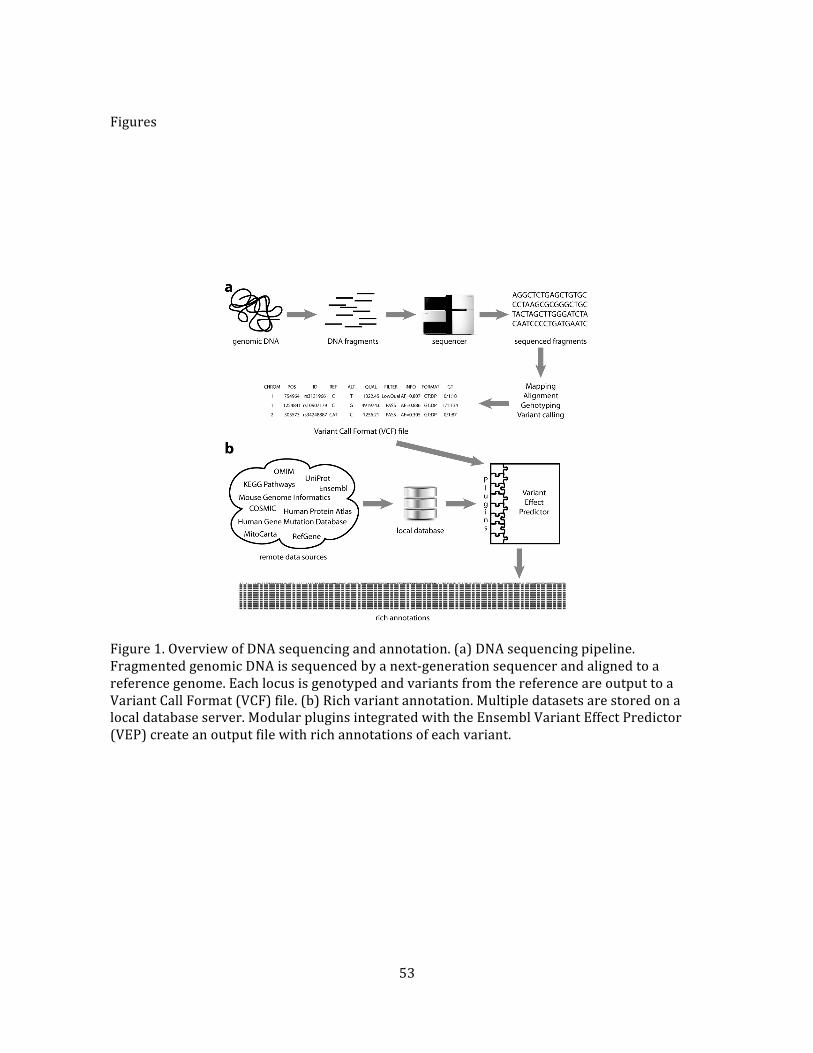
53
Figures
Figure 1. Overview of DNA sequencing and annotation. (a) DNA sequencing pipeline. Fragmented genomic DNA is sequenced by a next-‐generation sequencer and aligned to a reference genome. Each locus is genotyped and variants from the reference are output to a Variant Call Format (VCF) file. (b) Rich variant annotation. Multiple datasets are stored on a local database server. Modular plugins integrated with the Ensembl Variant Effect Predictor (VEP) create an output file with rich annotations of each variant.
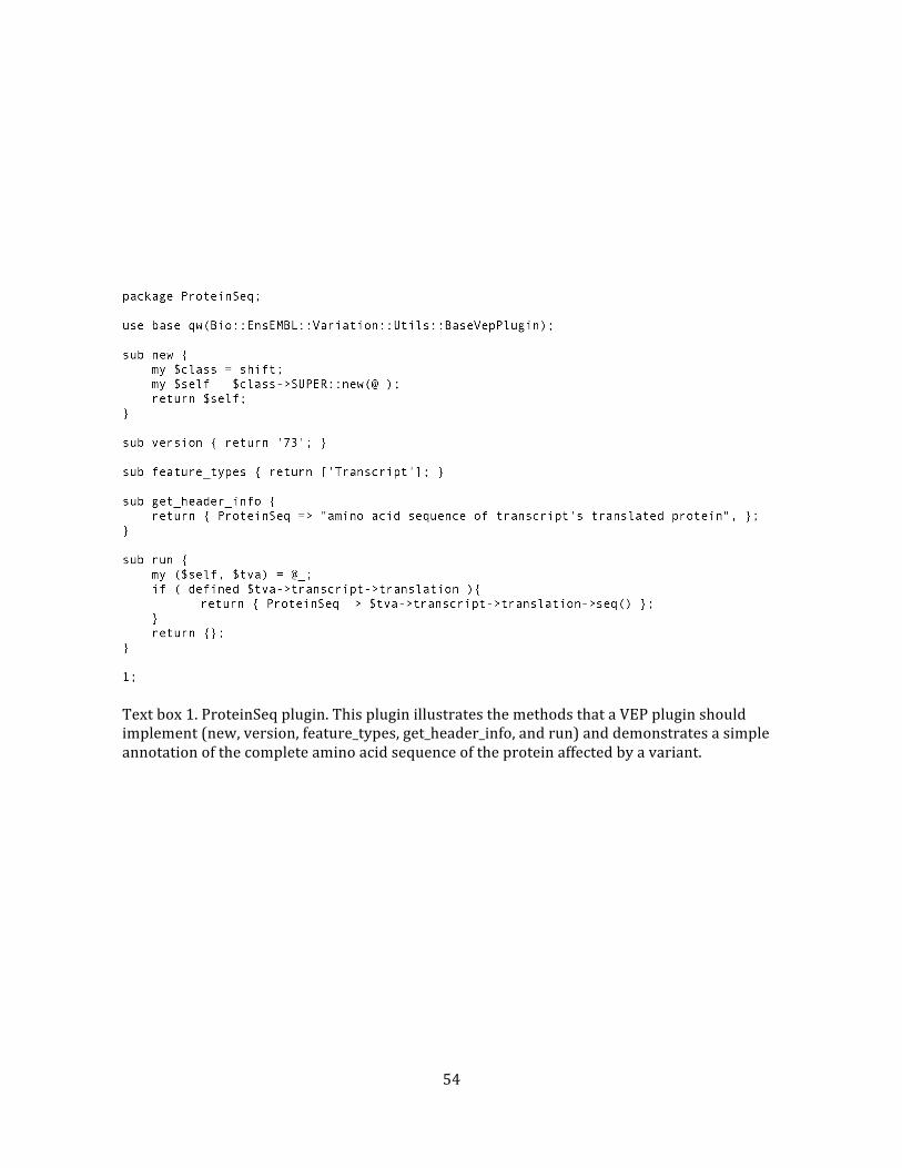
54
Text box 1. ProteinSeq plugin. This plugin illustrates the methods that a VEP plugin should implement (new, version, feature_types, get_header_info, and run) and demonstrates a simple annotation of the complete amino acid sequence of the protein affected by a variant.
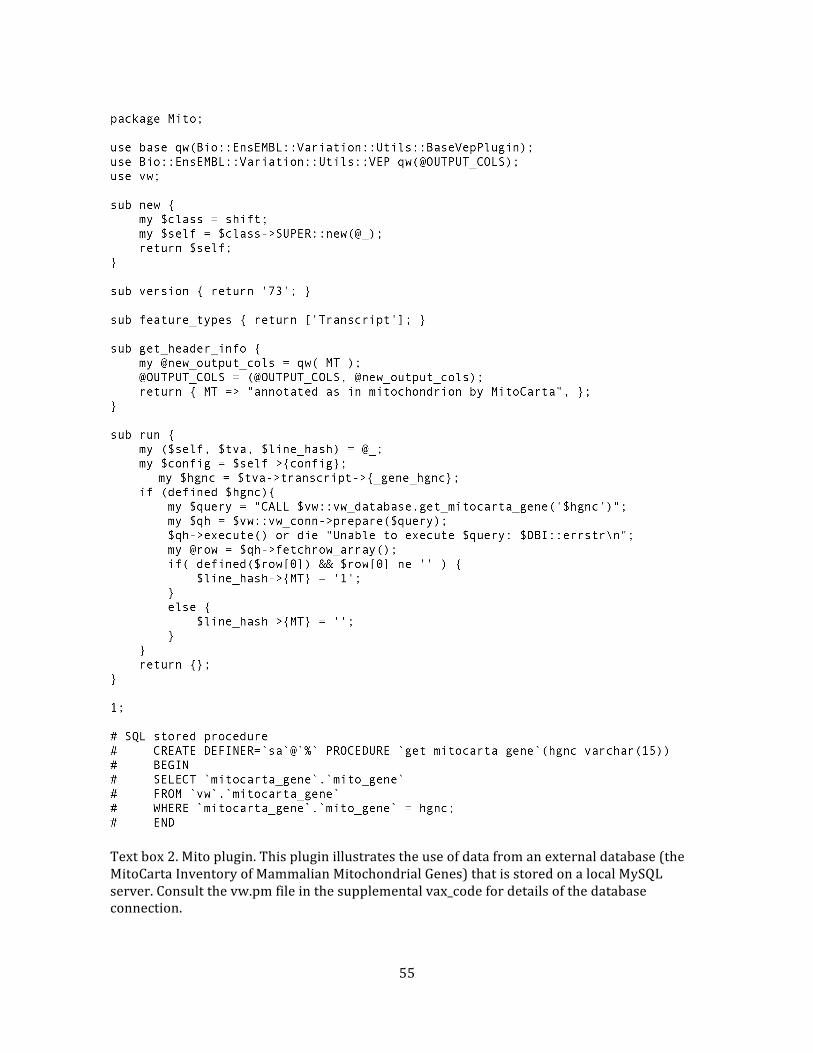
55
Text box 2. Mito plugin. This plugin illustrates the use of data from an external database (the MitoCarta Inventory of Mammalian Mitochondrial Genes) that is stored on a local MySQL server. Consult the vw.pm file in the supplemental vax_code for details of the database connection.
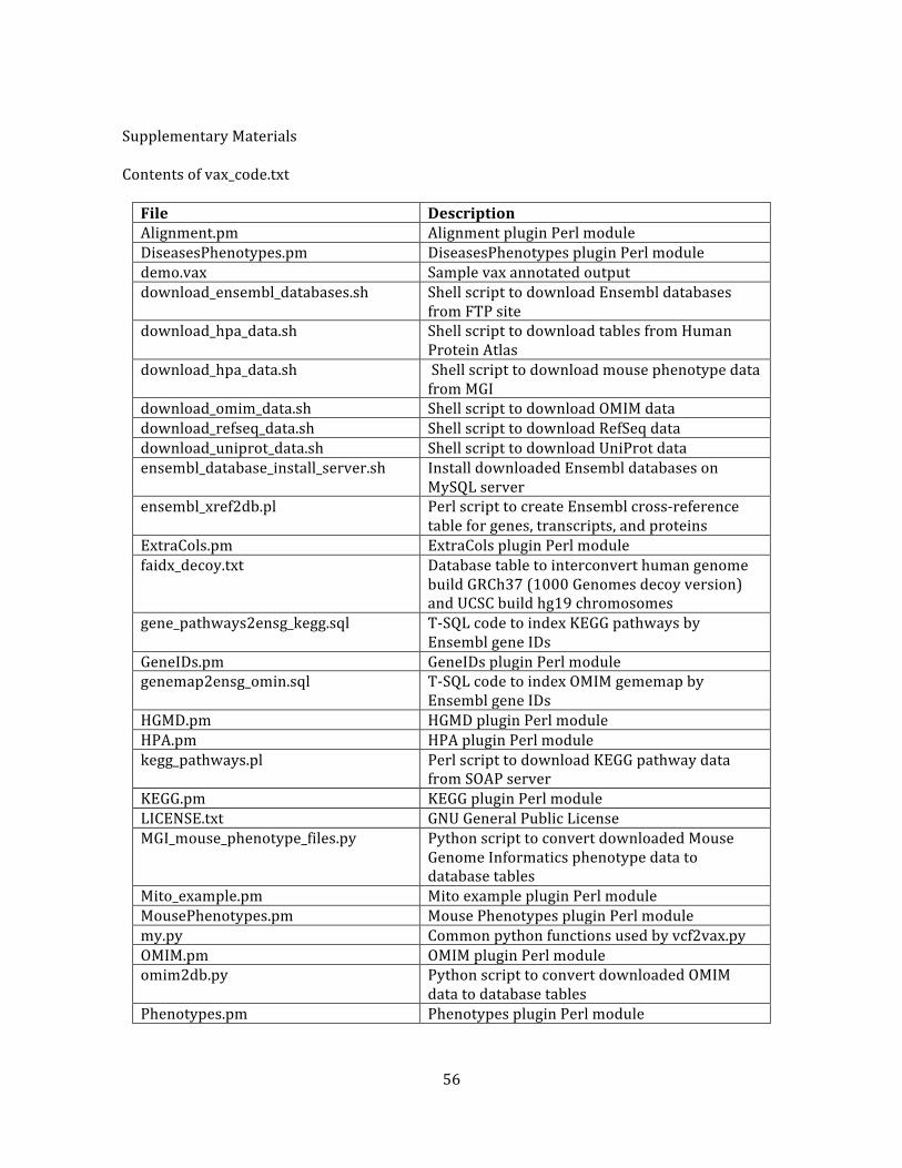
56
Supplementary Materials
Contents of vax_code.txt
File Description Alignment.pm Alignment plugin Perl module DiseasesPhenotypes.pm DiseasesPhenotypes plugin Perl module demo.vax Sample vax annotated output download_ensembl_databases.sh Shell script to download Ensembl databases
from FTP site download_hpa_data.sh Shell script to download tables from Human
Protein Atlas download_hpa_data.sh Shell script to download mouse phenotype data
from MGI download_omim_data.sh Shell script to download OMIM data download_refseq_data.sh Shell script to download RefSeq data download_uniprot_data.sh Shell script to download UniProt data ensembl_database_install_server.sh Install downloaded Ensembl databases on
MySQL server ensembl_xref2db.pl Perl script to create Ensembl cross-‐reference
table for genes, transcripts, and proteins ExtraCols.pm ExtraCols plugin Perl module faidx_decoy.txt Database table to interconvert human genome
build GRCh37 (1000 Genomes decoy version) and UCSC build hg19 chromosomes
gene_pathways2ensg_kegg.sql T-‐SQL code to index KEGG pathways by Ensembl gene IDs
GeneIDs.pm GeneIDs plugin Perl module genemap2ensg_omin.sql T-‐SQL code to index OMIM gememap by
Ensembl gene IDs HGMD.pm HGMD plugin Perl module HPA.pm HPA plugin Perl module kegg_pathways.pl Perl script to download KEGG pathway data
from SOAP server KEGG.pm KEGG plugin Perl module LICENSE.txt GNU General Public License MGI_mouse_phenotype_files.py Python script to convert downloaded Mouse
Genome Informatics phenotype data to database tables
Mito_example.pm Mito example plugin Perl module MousePhenotypes.pm Mouse Phenotypes plugin Perl module my.py Common python functions used by vcf2vax.py OMIM.pm OMIM plugin Perl module omim2db.py Python script to convert downloaded OMIM
data to database tables Phenotypes.pm Phenotypes plugin Perl module
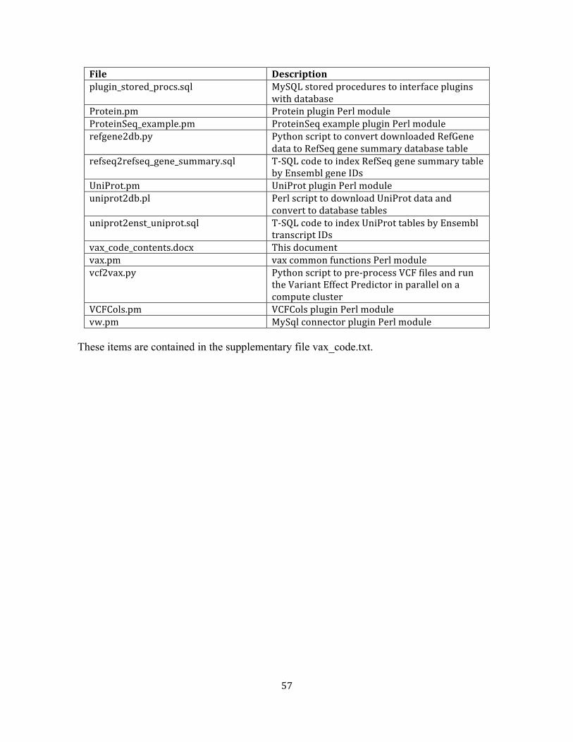
57
File Description plugin_stored_procs.sql MySQL stored procedures to interface plugins
with database Protein.pm Protein plugin Perl module ProteinSeq_example.pm ProteinSeq example plugin Perl module refgene2db.py Python script to convert downloaded RefGene
data to RefSeq gene summary database table refseq2refseq_gene_summary.sql T-‐SQL code to index RefSeq gene summary table
by Ensembl gene IDs UniProt.pm UniProt plugin Perl module uniprot2db.pl Perl script to download UniProt data and
convert to database tables uniprot2enst_uniprot.sql T-‐SQL code to index UniProt tables by Ensembl
transcript IDs vax_code_contents.docx This document vax.pm vax common functions Perl module vcf2vax.py Python script to pre-‐process VCF files and run
the Variant Effect Predictor in parallel on a compute cluster
VCFCols.pm VCFCols plugin Perl module vw.pm MySql connector plugin Perl module
These items are contained in the supplementary file vax_code.txt.
58
References
1. Gonzaga-‐Jauregui C, Lupski JR, Gibbs RA. Human genome sequencing in health and disease. Annual review of medicine. 2012;63:35-‐61. doi: 10.1146/annurev-‐med-‐051010-‐162644. PubMed PMID: 22248320; PubMed Central PMCID: PMC3656720.
2. Shendure J, Ji H. Next-‐generation DNA sequencing. Nat Biotechnol. 2008;26(10):1135-‐45. doi: 10.1038/nbt1486. PubMed PMID: 18846087.
3. Bamshad MJ, Ng SB, Bigham AW, Tabor HK, Emond MJ, Nickerson DA, Shendure J. Exome sequencing as a tool for Mendelian disease gene discovery. Nat Rev Genet. 2011;12(11):745-‐55. Epub 2011/09/29. doi: 10.1038/nrg3031. PubMed PMID: 21946919.
4. Wan J, Yourshaw M, Mamsa H, Rudnik-‐Schoneborn S, Menezes MP, Hong JE, Leong DW, Senderek J, Salman MS, Chitayat D, Seeman P, von Moers A, Graul-‐Neumann L, Kornberg AJ, Castro-‐Gago M, Sobrido MJ, Sanefuji M, Shieh PB, Salamon N, Kim RC, Vinters HV, Chen Z, Zerres K, Ryan MM, Nelson SF, Jen JC. Mutations in the RNA exosome component gene EXOSC3 cause pontocerebellar hypoplasia and spinal motor neuron degeneration. Nat Genet. 2012;44(6):704-‐8. Epub 2012/05/01. doi: 10.1038/ng.2254. PubMed PMID: 22544365; PubMed Central PMCID: PMC3366034.
5. 1000 Genomes Project. VCF (Variant Call Format) version 4.1 2013 [updated 2013-‐10-‐09]. Available from: http://www.1000genomes.org/wiki/Analysis/Variant Call Format/vcf-‐variant-‐call-‐format-‐version-‐41.
6. Danecek P, Auton A, Abecasis G, Albers CA, Banks E, DePristo MA, Handsaker RE, Lunter G, Marth GT, Sherry ST, McVean G, Durbin R. The variant call format and VCFtools. Bioinformatics. 2011;27(15):2156-‐8. Epub 2011/06/10. doi: 10.1093/bioinformatics/btr330. PubMed PMID: 21653522; PubMed Central PMCID: PMC3137218.
7. Pabinger S, Dander A, Fischer M, Snajder R, Sperk M, Efremova M, Krabichler B, Speicher MR, Zschocke J, Trajanoski Z. A survey of tools for variant analysis of next-‐generation genome sequencing data. Brief Bioinform. 2013. doi: 10.1093/bib/bbs086. PubMed PMID: 23341494.
8. Ng SB, Turner EH, Robertson PD, Flygare SD, Bigham AW, Lee C, Shaffer T, Wong M, Bhattacharjee A, Eichler EE, Bamshad M, Nickerson DA, Shendure J. Targeted capture and massively parallel sequencing of 12 human exomes. Nature. 2009;461(7261):272-‐6. Epub 2009/08/18. doi: nature08250 [pii] 10.1038/nature08250. PubMed PMID: 19684571.
9. McLaren W, Pritchard B, Rios D, Chen Y, Flicek P, Cunningham F. Deriving the consequences of genomic variants with the Ensembl API and SNP Effect Predictor. Bioinformatics. 2010;26(16):2069-‐70. Epub 2010/06/22. doi: btq330 [pii] 10.1093/bioinformatics/btq330. PubMed PMID: 20562413; PubMed Central PMCID: PMC2916720.
10. Grant JR, Arantes AS, Liao X, Stothard P. In-‐depth annotation of SNPs arising from resequencing projects using NGS-‐SNP. Bioinformatics. 2011;27(16):2300-‐1. Epub 2011/06/24.
59
doi: 10.1093/bioinformatics/btr372. PubMed PMID: 21697123; PubMed Central PMCID: PMC3150039.
11. Kowarsch A, Fuchs A, Frishman D, Pagel P. Correlated mutations: a hallmark of phenotypic amino acid substitutions. PLoS computational biology. 2010;6(9). doi: 10.1371/journal.pcbi.1000923. PubMed PMID: 20862353; PubMed Central PMCID: PMC2940720.
12. Ensembl. Ensembl/UCSC phylogenetic tree [2013-‐07-‐26]. Available from: http://tinyurl.com/ensembltree.
13. Pagliarini DJ, Calvo SE, Chang B, Sheth SA, Vafai SB, Ong SE, Walford GA, Sugiana C, Boneh A, Chen WK, Hill DE, Vidal M, Evans JG, Thorburn DR, Carr SA, Mootha VK. A mitochondrial protein compendium elucidates complex I disease biology. Cell. 2008;134(1):112-‐23. doi: 10.1016/j.cell.2008.06.016. PubMed PMID: 18614015; PubMed Central PMCID: PMC2778844.
14. Pruitt KD, Tatusova T, Brown GR, Maglott DR. NCBI Reference Sequences (RefSeq): current status, new features and genome annotation policy. Nucleic Acids Res. 2012;40(Database issue):D130-‐5. doi: 10.1093/nar/gkr1079. PubMed PMID: 22121212; PubMed Central PMCID: PMC3245008.
15. Online Mendelian Inheritance in Man OMIM®. Online Mendelian Inheritance in Man, OMIM® Baltimore, MD: McKusick-‐Nathans Institute of Genetic Medicine, Johns Hopkins University; [2013-‐11-‐04]. Available from: http://omim.org/.
16. Uhlen M, Oksvold P, Fagerberg L, Lundberg E, Jonasson K, Forsberg M, Zwahlen M, Kampf C, Wester K, Hober S, Wernerus H, Bjorling L, Ponten F. Towards a knowledge-‐based Human Protein Atlas. Nat Biotechnol. 2010;28(12):1248-‐50. doi: 10.1038/nbt1210-‐1248. PubMed PMID: 21139605.
17. Kanehisa M, Goto S, Kawashima S, Nakaya A. The KEGG databases at GenomeNet. Nucleic Acids Res. 2002;30(1):42-‐6. PubMed PMID: 11752249; PubMed Central PMCID: PMC99091.
18. Eppig JT, Blake JA, Bult CJ, Kadin JA, Richardson JE, Mouse Genome Database G. The Mouse Genome Database (MGD): comprehensive resource for genetics and genomics of the laboratory mouse. Nucleic Acids Res. 2012;40(Database issue):D881-‐6. doi: 10.1093/nar/gkr974. PubMed PMID: 22075990; PubMed Central PMCID: PMC3245042.
19. Apweiler R, Martin MJ, O'Donovan C, Magrane M, Alam-‐Faruque Y, Alpi E, Antunes R, Arganiska J, Casanova EB, Bely B, Bingley M, Bonilla C, Britto R, Bursteinas B, Chan WM, Chavali G, Cibrian-‐Uhalte E, Da Silva A, De Giorgi M, Dimmer E, Fazzini F, Gane P, Fedotov A, Castro LG, Garmiri P, Hatton-‐Ellis E, Hieta R, Huntley R, Jacobsen J, Jones R, Legge D, Liu WD, Luo J, MacDougall A, Mutowo P, Nightingale A, Orchard S, Patient S, Pichler K, Poggioli D, Pundir S, Pureza L, Qi GY, Rosanoff S, Sawford T, Sehra H, Turner E, Volynkin V, Wardell T, Watkins X, Zellner H, Corbett M, Donnelly M, van Rensburg P, Goujon M, McWilliam H, Lopez R, Xenarios I, Bougueleret L, Bridge A, Poux S, Redaschi N, Auchincloss A, Axelsen K, Bansal P, Baratin D, Binz PA, Blatter MC, Boeckmann B, Bolleman J, Boutet E, Breuza L, de Castro E, Cerutti L, Coudert E, Cuche B, Doche M, Dornevil D, Duvaud S, Estreicher A, Famiglietti L, Feuermann M, Gasteiger E,
60
Gehant S, Gerritsen V, Gos A, Gruaz-‐Gumowski N, Hinz U, Hulo C, James J, Jungo F, Keller G, Lara V, Lemercier P, Lew J, Lieberherr D, Martin X, Masson P, Morgat A, Neto T, Paesano S, Pedruzzi I, Pilbout S, Pozzato M, Pruess M, Rivoire C, Roechert B, Schneider M, Sigrist C, Sonesson K, Staehli S, Stutz A, Sundaram S, Tognolli M, Verbregue L, Veuthey AL, Zerara M, Wu CH, Arighi CN, Arminski L, Chen CM, Chen YX, Huang HZ, Kukreja A, Laiho K, McGarvey P, Natale DA, Natarajan TG, Roberts NV, Suzek BE, Vinayaka CR, Wang QH, Wang YQ, Yeh LS, Yerramalla MS, Zhang J, Consortium U. Update on activities at the Universal Protein Resource (UniProt) in 2013. Nucleic Acids Research. 2013;41(D1):D43-‐D7. doi: Doi 10.1093/Nar/Gks1068. PubMed PMID: WOS:000312893300007.
20. Howe DG, Bradford YM, Conlin T, Eagle AE, Fashena D, Frazer K, Knight J, Mani P, Martin R, Moxon SA, Paddock H, Pich C, Ramachandran S, Ruef BJ, Ruzicka L, Schaper K, Shao X, Singer A, Sprunger B, Van Slyke CE, Westerfield M. ZFIN, the Zebrafish Model Organism Database: increased support for mutants and transgenics. Nucleic Acids Res. 2013;41(Database issue):D854-‐60. doi: 10.1093/nar/gks938. PubMed PMID: 23074187; PubMed Central PMCID: PMC3531097.
21. Laulederkind SJ, Hayman GT, Wang SJ, Smith JR, Lowry TF, Nigam R, Petri V, de Pons J, Dwinell MR, Shimoyama M, Munzenmaier DH, Worthey EA, Jacob HJ. The Rat Genome Database 2013-‐-‐data, tools and users. Brief Bioinform. 2013;14(4):520-‐6. doi: 10.1093/bib/bbt007. PubMed PMID: 23434633; PubMed Central PMCID: PMC3713714.
22. Kerner B, Rao AR, Christensen B, Dandekar S, Yourshaw M, Nelson SF. Rare genomic variants link bipolar disorder to CREB regulated intracellular signaling pathways. Frontiers in Psychiatry. 2013. doi: 10.3389/fpsyt.2013.00154.
23. Yourshaw M, Solorzano-‐Vargas RS, Pickett LA, Lindberg I, Wang J, Cortina G, Pawlikowska-‐Haddal A, Baron H, Venick RS, Nelson SF. Exome Sequencing Finds a Novel PCSK1 Mutation in a Child With Generalized Malabsorptive Diarrhea and Diabetes Insipidus. Journal of pediatric gastroenterology and nutrition. 2013. doi: 10.1097/MPG.0b013e3182a8ae6c.
24. Lee H, Nelson SF. Rethinking clinical practice: clinical implementation of exome sequencing. Pers Med. 2012;9(8):785-‐7. doi: Doi 10.2217/Pme.12.101. PubMed PMID: WOS:000311977800001.
61
CHAPTER THREE
Whole-‐exome sequencing for the identification of casual mutations
in congenital diarrheal disorders
62
Abstract
Background. Congenital diarrheal disorders (CDDs) are rare diseases with serious, even
life-‐threatening, consequences. Whole-‐exome sequencing enables an unbiased search for
genetic variants that cause the disease.
Methods. We developed a bioinformatic pipeline and analytical methods for whole-‐
exome sequencing to discover variants in the protein coding region of genes associated with
CDDs.
Results. We sequenced 45 probands from diverse ethnic backgrounds who were
diagnosed with a variety of CDDs of probable, but unknown genetic cause, and searched for
damaging mutations in genes known to be associated with CDDs. Patients had been diagnosed
with generalized malabsorptive diarrhea (n=33), selective nutrient malabsorption (n=5),
secretory diarrhea (n=3), and infantile IBD (n=4). Although most cases had thorough
diagnostic workups, sometimes including sequencing of suspected genes, none had a
convincing genetic finding. Surprisingly, we found a mutation in a known gene, or a gene that
was reported during the course of the study, that is likely to cause disease in 27 cases (60%).
The genes implicated were ADAM17, DGAT1, EPCAM, IL10RA, MALT1, MYO5B, NEUROG3, PCSK1,
SI, SKIV2L, SLC26A3, and SLC5A1. While six of the mutations were previously reported, 25 of
them were novel.
Conclusions. Whole-‐exome sequencing is an effective approach for the identification of
casual mutations in known CDD genes that may escape detection with standard practice
involving a complex diagnostic workup and targeted gene sequencing.
Introduction
Congenital diarrheal disorders (CDDs) are rare diseases with serious, even life-‐
threatening, consequences that impose massive diagnostic and treatment costs as well as great
63
emotional stress on patients and their families. These patients frequently endure a complex
and costly diagnostic odyssey that often fails to produce a definitive diagnosis (1). Affected
families may be unaware of appropriate treatments and prognosis (2) and lack information on
the recurrence risk in subsequent pregnancies. Until recently, little was known of the genetic
etiology of these diseases, yet identification of a casual mutation can lead to improved
management of the disease and inform research efforts to develop new treatment modalities.
Importantly, identifying new genes provides a better understanding of the molecular pathways
and mechanisms responsible for the specific disease and other phenotypes governed by the
same pathways. For example, mutations in the gene PCSK1 (encoding neuroendocrine
convertase 1 precursor) can cause a severe form of generalized malabsorptive diarrhea, while
milder variants are associated with obesity (3).
Although rare, these diseases impose serious emotional and economic burdens on
families and society. Children with CDDs often require long-‐term parenteral nutrition in order
to absorb sufficient nutrients to sustain normal growth and development. Parenteral nutrition,
however, does not address the primary cause of the disorder and its prolonged use is
associated with numerous complications, including loss of central venous access, sepsis,
intestinal failure associated liver disease, poor health related quality of life, multivisceral
transplantation, and death. Economically, the expenditure of home parenteral nutrition alone
is estimated in the range of $100,000-‐$150,000 per year, and the cost of transplantation is
~$1.5-‐1.9 million for the first year alone (4).
CDDs are a group of enteropathies caused by inherited or sporadic genetic mutations
that generally manifest soon after birth or in early childhood. The primary presenting
symptom is chronic diarrhea that often requires total parenteral nutrition. Mutations in
number of genes are known to cause CDDs, but many cases, even those that harbor a mutation
in a known gene, do not receive a genetic diagnosis.
64
The discovery of most of the known genes involved arduous genetic and molecular
studies. In the 1990s genome-‐wide linkage analysis, using 300-‐500 microsatellite markers,
offered a method of identifying disease susceptibility loci. This approach was time-‐consuming
and limited to a resolution of down to about 10Mb, a length, which can harbor several genes
(5). To achieve adequate power it was necessary to genotype one or more extended families or
a very large number of nuclear families. For example, linkage disequilibrium as well as genetic
linkage, as determined by this technique in Finnish families, indicated that an unknown gene
near the cystic fibrosis transmembrane regulator gene (CFTR) was probably associated with
secretory chloride diarrhea (6). Subsequently, cloning of the linkage region identified four
known genes, two of which were considered to be functionally relevant (7). Finally,
segregation of mutations in the SLC26A3 gene with the disorder in a large number of patients
confirmed that such mutations cause the disorder (8). The development of highly parallel
genotyping based on arrays with probes for thousands of single nucleotide polymorphisms
(SNPs) enabled more efficient genotyping with a 10-‐fold or better improvement in resolution
(9) This technology, applied to extended kindreds, enabled the discovery of mutations of the
causative genes for microvillus inclusion disease (10), syndromic congenital secretory sodium
diarrhea (11), congenital tufting enteropathy (12), and early-‐onset chronic diarrhea (13). A
candidate gene resequencing approach, founded on a mouse model, identified mutations in
NEUROG3 as the cause of malabsorptive congenital diarrhea (14).
The paradigm of earlier techniques for mutation discovery required first, a narrowing
down the genome by application of candidate gene resequencing, linkage analysis,
homozygosity mapping, or case-‐control association studies, followed by a search for causative
mutations within a small part of the genome. Specific clinical tests would then be developed to
assess for the presence of a known mutation. The advent of affordable technology to sequence
a human genome has rapidly changed physicians’ and researchers’ approach across a wide
65
range of congenital disorders. It is now possible to sequence all the protein-‐coding DNA (the
“exome”) of a patient at a cost that approaches the price of clinical sequencing of a single gene
by traditional methods (15). For example, the widely used Illumina 2500 next-‐generation DNA
sequencing platform can sequence an entire genome in 27 hours for under $10,000. Thus, next-‐
generation sequencing technology enables researchers and clinicians to examine almost all
mutations in the whole genome, or the protein encoding portion thereof, at a cost much lower
than that of previous locus-‐specific methods. In particular, whole-‐exome sequencing is a
transformative technology that may alter the clinician’s approach to the evaluation of CDD
patients (16). Accordingly, we sought to determine the effectiveness of whole-‐exome
sequencing to identify, in genes that have been reported to be associated with CDD, the
molecular causes of the disease in a cohort of patients with congenital gastrointestinal
disorders that had defied conventional diagnostic methods.
Methods
Subjects. Samples for exome sequencing were identified from the UCLA Pediatric Diarrhea
Database, which includes samples referred for clinical diagnosis or research since 2004, and
was approved by our institutional review board. Inclusion criteria for the database were a
history of chronic (>2 mo) severe diarrhea during childhood (<18 y), although subjects with
various causes of short-‐bowel syndrome, inflammatory bowel disease (but not infantile IBD),
celiac disease, and pancreatic insufficiency were excluded. The database contains more than
172 kindreds comprising 194 children with chronic diarrhea, of which 163 cases were
classified as congenital in origin. Approximately 25 of the subjects were identified with various
forms of selective malabsorptive diarrhea and 133 were classified with the generalized form of
malabsorption.
66
We chose 45 patients from 38 families for exome sequencing. Inclusion criteria were a
diagnosis of congenital diarrhea and probability that the disease had a genetic cause
(consanguineous parents or affected family members). Patients were excluded if they had a
confirmed genetic diagnosis or a clinical presentation strongly suggesting a mutation in a gene
known to cause congenital diarrhea. These patients were of diverse ethnic backgrounds and
had clinical presentations of generalized malabsorptive diarrhea (n=33), selective nutrient
malabsorption (n=5), secretory diarrhea (n=3), and infantile IBD (n=4) [Table 1].
Whole-‐exome sequencing. Over the course of this study we evaluated several
methodologies for sequencing library preparation and sequencing platform. Genomic DNA
from probands, and in some cases family members, was either fragmented by sonication and
ligated to Illumina bar-‐coded adapters or fragmented and ligated in a single step with Illumina
engineered transposases, and then in either case, the fragments were amplified by PCR.
Fragments were then enriched for the protein coding portion of the genome by hybridization
to probes from either the Agilent SureSelect XT Human All Exon 50Mb, Illumina TruSeq Exome,
or Illumina Nextera ExpandedExome enrichment kits. Different kits were used for assessment
of coverage and sample preparation efficiency. The exome-‐enriched library was sequenced for
100x100 paired end reads on an Illumina Genome Analyzer 2000 or 2500 platform to a mean
coverage depth of 116X, with 85% of RefGene CDS and essential splice sites having at least 20X
coverage [Table 2] (Data not shown for subject 45, who was sequenced on an Applied
Biosystems SOLiD 4 System instrument as described previously (2)).
Variants that were predicted to be damaging in genes known to be associated with CDD
were validated by Sanger sequencing. Sanger sequencing of relatives from whom DNA was
available confirmed segregation of the variant allele with the disorder.
Data analysis. We converted sequenced reads from the native bcl files to the FastQ format
by the Illumina bcl2fastq program. We processed the FastQ files to create aligned bam files with in-
67
house pipeline software. Briefly, we aligned the reads to build GRCh37 of the human genome (17)
with Novoalign (http://www.novocraft.com) to obtain a mean of 120 million uniquely aligned
100x100 paired end reads per sample after removing PCR duplicates [Table 2]. We recalibrated base
quality scores to improve accuracy by analyzing the covariation among reported quality score,
position within read, dinucleotide, and probability of mismatching the reference genome using the
Genome Analysis Toolkit (GATK) (18, 19). We used the GATK Unified Genotyper and Haplotype
Caller tools to genotype single nucleotide variants and indels and recalibrated variant quality score
recalibration with the GATK Variant Quality Score Recalibrator to assign probabilities to each
variant call. We obtained the variant consequences on transcripts and proteins with the Ensembl
Variant Effect Predictor (VEP) (19), and estimated the extent of protein damage with SIFT (20-24),
PolyPhen (25-27), and Condel (28). We further annotated variants with additional data from Online
Mendelian Inheritance in Man (OMIM) (29), the Human Gene Mutation Database (HGMD pro,
BIOBASE Biological Databases), the Universal Protein Resource (UniProt) (30), KEGG Pathways
(31), RefGene (32), the MitoCarta Inventory of Mammalian Mitochondrial Genes (33), Mouse
Genome Informatics (MGI) (34) and the Human Protein Atlas (HPA) (35) using in-‐house
plugins for the VEP.
We hypothesized for this study that the mode of inheritance would be recessive
(homozygous or compound heterozygous) and mutation effects would be fully penetrant.
Further hypothesizing that casual variants would be found in the protein coding or splicing
regions of genes, we filtered to include only splice acceptor or donor, stop gained or lost,
frameshift, initiator codon, inframe insertion or deletion, missense, or splice region variants
(Ensembl consequence rank >13). We also removed variants with a minor allele frequency
>0.5% in the combined 1000 Genomes (36) and NHLBI (37) datasets to remove variants that,
given the rare incidence of CDDs, are too frequent in the population to cause these disorders. In
addition, we removed variants that were observed in more than 2 (homozygous model) or 8
68
(compound heterozygous model) samples from ~150 unaffected control exomes, to eliminate
false positives caused by technical artifacts. We ranked variants for likelihood of damage using
multiple factors. We deemed a splice acceptor or donor variant, stop gained, and frameshift, to
be probably damaging. We prioritized missense single nucleotide variants (SNVs) by SIFT,
PolyPhen, and Condel predictions, and variants in the splice site region near the acceptor and
donor by GERP conservation scores. In the case of compound heterozygous variants we
assigned the priority of the second ranked variant. In families where we had sequenced
members other than the proband, we filtered out all variants that were inconsistent with fully
penetrant Mendelian inheritance. Finally, for this phase of the study, we selected variants in
genes that were known or suspected of being involved in congenital diarrheal disorders [Table
3] and had reported mutations that cause a phenotype consistent with that of the patient.
Results
After filtering by consequence rank, allele frequency, and controls and eliminating
variants that did not segregate with the disease, the probands had 1-‐237 variants in all genes
and a mean of two filtered variants in one of the genes known or suspected to be associated
with CDDs (range 0-‐5) [Table 4]. After applying bioinformatic metrics of protein damage and
conservation, reviewing the literature, and comparing the expected phenotype of a mutation in
a gene to the proband’s presenting phenotype, we identified 31 different mutations in 21
families (27 cases) that we considered to be the likely cause of the disease [Table 5]. The genes
(and number of families) implicated were ADAM17 (n=1), DGAT1 (n=2), EPCAM (n=3), IL10RA
(n=1), MALT1 (n=1), MYO5B (n=2), NEUROG3 (n=3), PCSK1 (n=2), SI (n=3), SKIV2L (n=1),
SLC26A3 (n=1), and SLC5A1 (n=1). Six of these variants were previously known to cause a
CDD, but the other 25 are novel. All of these mutations had a recessive mode of inheritance;
mutations in a gene were homozygous in 13 probands and compound heterozygous in the
69
remaining 9 (one proband had mutations in two genes). The variant consequences included
large deletion (n=1), splice donor (n=2), stop gained (n=4), frameshift (n=5), missense (n=17),
and splice region (n=2). In summary, we identified a mutation highly likely to cause the
patient’s phenotype in 60% of the probands that we sequenced.
Discussion
Whole-‐exome sequencing identified a probable molecular explanation for the disorder
in a majority of cases, thus suggesting the value of this approach for diagnosis of CDDs in a
clinical setting. Certified clinical sequencing of the genes and in some cases clinical follow up to
confirm genotype/phenotype correlations would be needed to confirm these findings but each
phenotype, as recorded in the UCLA Pediatric Diarrhea Database, is consistent with the
reported phenotypes associated with mutations in the identified casual genes. For example,
loss of function mutations in the metallopeptidase domain 17 gene (ADAM17) cause neonatal-‐
onset inflammatory skin and bowel disease with diarrhea (38). A rare splice site mutation in
diacylglycerol O-‐acyltransferase (DGAT1) has been linked to CDD (39). The epithelial cell
adhesion molecule (EPCAM) is the casual gene for congenital tufting enteropathy (12).
Mutations in the interlukin-‐10 receptor, alpha gene (IL10RA) cause a form of early onset
inflammatory bowel disease (40). The mucosa associated lymphoid tissue lymphoma
translocation gene (MALT1), which has been implicated in combined immunodeficiency (41),
can also present with generalized malabsorptive diarrhea. Microvillus inclusion disease is
caused by mutations in the myosin VB (MYO5B) gene (10). Congenital malabsorptive diarrhea
is caused by mutations in the transcription factor neurogenin 3 (NEUROG3) (14). Proprotein
convertase 1/3 (PC1/3) deficiency, an autosomal-‐recessive disorder caused by rare mutations
in the proprotein convertase subtilisin/kexin type 1 (PCSK1) gene frequently cause
generalized malabsorptive diarrhea in childhood (42). Congenital sucrase-‐isomaltase
70
deficiency is a selective nutrient malabsorption disorder caused by mutations in the sucrase-‐
isomaltase (alpha-‐glucosidase) (SI) gene (43). The superkiller viralicidic activity 2-‐like (S.
cerevisiae) gene (SKIV2L) causes trichohepatoenteric syndrome-‐2, a severe disease
characterized by intrauterine growth retardation, facial dysmorphism, hair abnormalities,
intractable diarrhea, and immunodeficiency (44). Congenital chloride diarrhea is characterized
by excretion of large amounts of watery stool containing high levels of chloride; it is caused by
mutations in solute carrier family 26, member 3 (SLC26A3) (8). An intestinal monosaccharide
transporter deficiency known as glucose/galactose malabsorption is caused by mutations in
solute carrier family 5 (sodium/glucose cotransporter), member 1 (SLC5A1) (45).
Infants presenting with CDD often endure multiple hospitalizations, a batteries of
indirect, redundant, and expensive tests, and the life threatening risks of PN, as well as even
more serious procedures such as multivisceral transplantation. Whole-‐exome sequencing, now
becoming widely available from certified diagnostic centers (46, 47), is a revolutionary
technology that that should be considered early in a clinician’s evaluation of patients with CDD.
However, our study also demonstrates that much remains unknown about the genetic
etiology of CDDs, as 40% of the probands we sequenced did not yield a clear molecular finding,
although many interesting and novel candidate genes have been identified. There are several
factors contributing to this “genetic dark matter”. Most importantly, the cellular pathways
involved in the development and function of the intestine are not fully understood and many
genes are yet to be identified that contribute to the risk of CDDs. The present study generated a
number of candidate genes that are now the subject of active research.
A second consideration is that many de novo cases of CDD without a family history of
disease or evidence of consanguinity are likely be caused by sporadic mutations with dominant
effect and possibly found in novel genes. Before exome sequencing, it was technically difficult
to identify this class of mutation in a single case, and identifying a variant in an autosomal
71
dominant mode of inheritance would usually require that the variant did not have a serious
effect on reproductive fitness and was, therefore, present in a large kindred whose DNA was
available to researchers. Accordingly, we are aware of only two genes reported before exome
sequencing was introduced where mutations cause a CDD, namely, protease, serine, 1 (Trypsin
1) (PRSS1) which causes hereditary pancreatitis (48), and autoimmune regulator (AIRE),
which apparently caused autoimmune polyendocrinopathy syndrome type I with a dominant
pattern of inheritance in a single family (49). Exome sequencing of a single proband usually
returns too many heterozygous candidate variants (mean 315 in our study) to be practically
studied for their effect on function. However, exome sequencing of both parents and an
affected child allows de novo mutations to be identified from a few candidates (50). A third
source of genetic dark matter is the 98% of the human genome that is not in the exome, which
includes promoters, enhancers, short RNAs, and other regulatory elements. Identifying and
characterizing these features is an active area of research, but at this time the size of the
genome to be sequenced and the lack of data on these features limits our ability to determine
how they affect phenotypes.
Because some of the cases in this study were in consanguineous families, we
considered the approach of using a high density microarray to locate regions of homozygosity
in the patients’ genome, then developing a set of custom capture probes to select that region
for deep sequencing, or to sequence all exons in the region with the traditional Sanger method.
However this would require a different array for each family and was unlikely to be cost
effective. Furthermore, we wished to develop and evaluate an unbiased analytical pipeline that
could be used for many other genetic patterns in addition to homozygosity arising from
inbreeding.
Our success in identifying a majority of the probable causative mutations by screening
with a relatively short list of known genes raises the question of whether it would be more
72
efficient to perform targeted exome sequencing for these genes instead of whole-‐exome
sequencing. Comparing the costs of these two options is rather complex and depends on
variable such as type of sequencing platform (high throughput or low throughput), caseload
and batching factors, and the relative cost of a standard probe set versus a custom panel.
Another factor to consider is that new genes will be discovered and a targeted panel will
become obsolescent. Finally, whole-‐exome sequencing has the potential to generate novel
candidate genes as a side effect, and with proper consenting and safeguards, this data can
usefully be employed in a research setting.
In sum, we believe that with present technology, and for several years to come, whole-‐
exome sequencing is an effective approach for the identification of casual mutations in known
CDD genes that may escape detection with standard practice involving a complex diagnostic
workup and targeted gene sequencing. We envision that portions of the standard metabolic
panels and other urine, blood and radiographic tests will be used less frequently once exome
sequencing becomes fully implemented into clinical practice. More directed phenotypic
evaluation will be possible after a molecular basis for the gene is known, rather than the
shotgun approaches typically employed currently for children with rare genetic conditions.
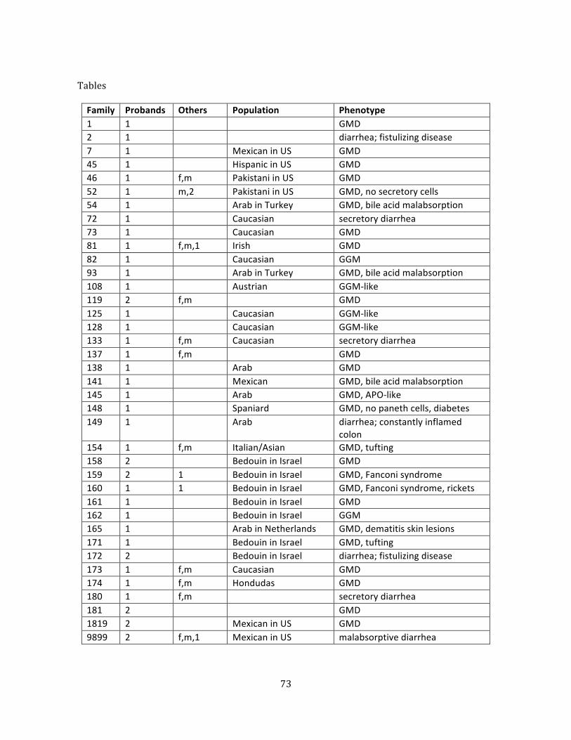
73
Tables
Family Probands Others Population Phenotype 1 1 GMD 2 1 diarrhea; fistulizing disease 7 1 Mexican in US GMD 45 1 Hispanic in US GMD 46 1 f,m Pakistani in US GMD 52 1 m,2 Pakistani in US GMD, no secretory cells 54 1 Arab in Turkey GMD, bile acid malabsorption 72 1 Caucasian secretory diarrhea 73 1 Caucasian GMD 81 1 f,m,1 Irish GMD 82 1 Caucasian GGM 93 1 Arab in Turkey GMD, bile acid malabsorption 108 1 Austrian GGM-‐like 119 2 f,m GMD 125 1 Caucasian GGM-‐like 128 1 Caucasian GGM-‐like 133 1 f,m Caucasian secretory diarrhea 137 1 f,m GMD 138 1 Arab GMD 141 1 Mexican GMD, bile acid malabsorption 145 1 Arab GMD, APO-‐like 148 1 Spaniard GMD, no paneth cells, diabetes 149 1 Arab diarrhea; constantly inflamed
colon 154 1 f,m Italian/Asian GMD, tufting 158 2 Bedouin in Israel GMD 159 2 1 Bedouin in Israel GMD, Fanconi syndrome 160 1 1 Bedouin in Israel GMD, Fanconi syndrome, rickets 161 1 Bedouin in Israel GMD 162 1 Bedouin in Israel GGM 165 1 Arab in Netherlands GMD, dematitis skin lesions 171 1 Bedouin in Israel GMD, tufting 172 2 Bedouin in Israel diarrhea; fistulizing disease 173 1 f,m Caucasian GMD 174 1 f,m Hondudas GMD 180 1 f,m secretory diarrhea 181 2 GMD 1819 2 Mexican in US GMD 9899 2 f,m,1 Mexican in US malabsorptive diarrhea

74
Table 1. Subjects. Children afffected with a congenital diarrhea disorder that were sequenced in this study. Probands: number of affected siblings; Others: other relatives sequenced (father, mother, number of unaffected siblings); Population: self-‐reported ethnic background, when available; Phenotype: presenting diagnosis prior to sequencing, GMD generalized malabsorptive diarrhea, GGM glucose galactose malabsorption.
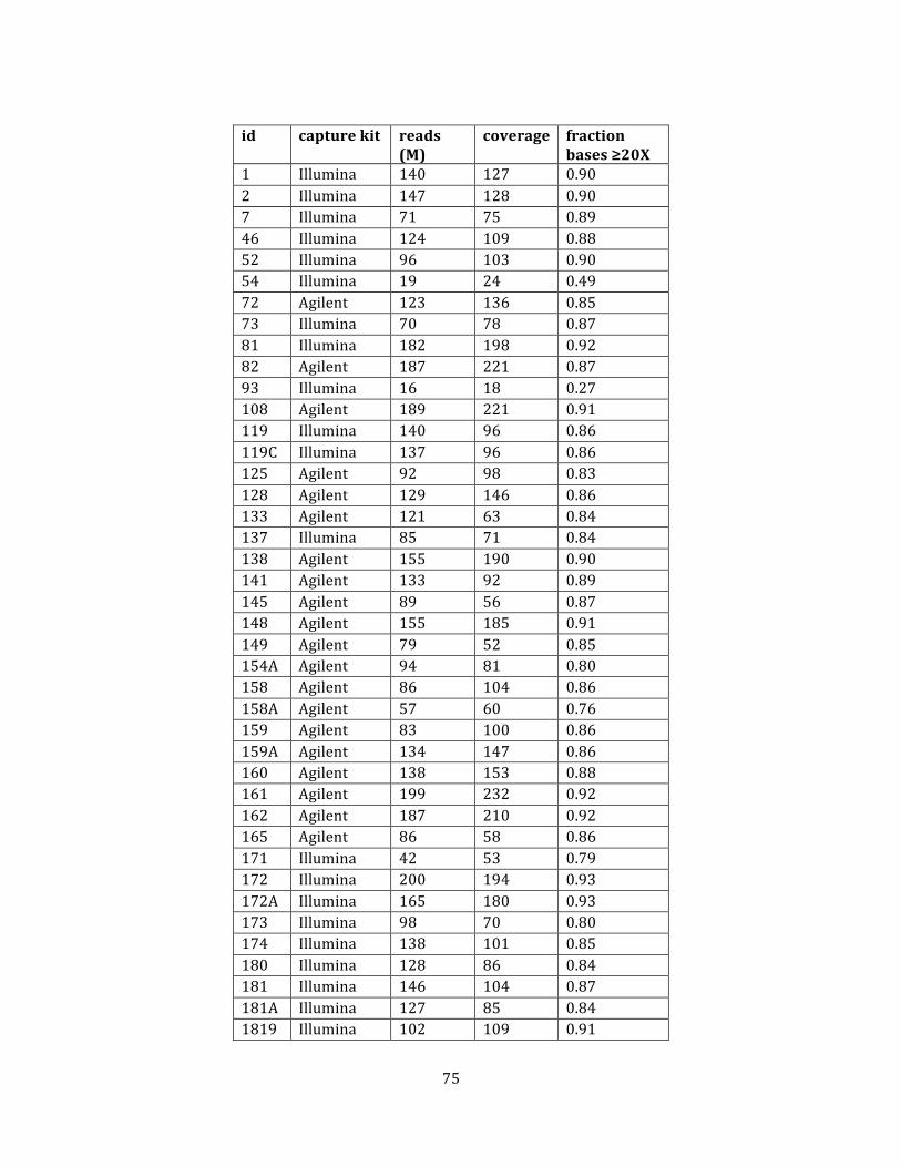
75
id capture kit reads
(M) coverage fraction
bases ≥20X 1 Illumina 140 127 0.90 2 Illumina 147 128 0.90 7 Illumina 71 75 0.89 46 Illumina 124 109 0.88 52 Illumina 96 103 0.90 54 Illumina 19 24 0.49 72 Agilent 123 136 0.85 73 Illumina 70 78 0.87 81 Illumina 182 198 0.92 82 Agilent 187 221 0.87 93 Illumina 16 18 0.27 108 Agilent 189 221 0.91 119 Illumina 140 96 0.86 119C Illumina 137 96 0.86 125 Agilent 92 98 0.83 128 Agilent 129 146 0.86 133 Agilent 121 63 0.84 137 Illumina 85 71 0.84 138 Agilent 155 190 0.90 141 Agilent 133 92 0.89 145 Agilent 89 56 0.87 148 Agilent 155 185 0.91 149 Agilent 79 52 0.85 154A Agilent 94 81 0.80 158 Agilent 86 104 0.86 158A Agilent 57 60 0.76 159 Agilent 83 100 0.86 159A Agilent 134 147 0.86 160 Agilent 138 153 0.88 161 Agilent 199 232 0.92 162 Agilent 187 210 0.92 165 Agilent 86 58 0.86 171 Illumina 42 53 0.79 172 Illumina 200 194 0.93 172A Illumina 165 180 0.93 173 Illumina 98 70 0.80 174 Illumina 138 101 0.85 180 Illumina 128 86 0.84 181 Illumina 146 104 0.87 181A Illumina 127 85 0.84 1819 Illumina 102 109 0.91
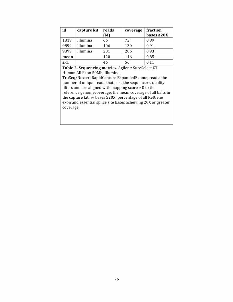
76
id capture kit reads (M)
coverage fraction bases ≥20X
1819 Illumina 66 72 0.89 9899 Illumina 106 130 0.91 9899 Illumina 201 206 0.93 mean 120 116 0.85 s.d. 46 56 0.11 Table 2. Sequencing metrics. Agilent: SureSelect XT Human All Exon 50Mb; Illumina: TruSeq/NexteraRapidCapture ExpandedExome; reads: the number of unique reads that pass the sequencer's quality filters and are aligned with mapping score > 0 to the reference genomecoverage: the mean coverage of all baits in the capture kit; % bases ≥20X: percentage of all RefGene exon and essential splice site bases acheiving 20X or greater coverage.
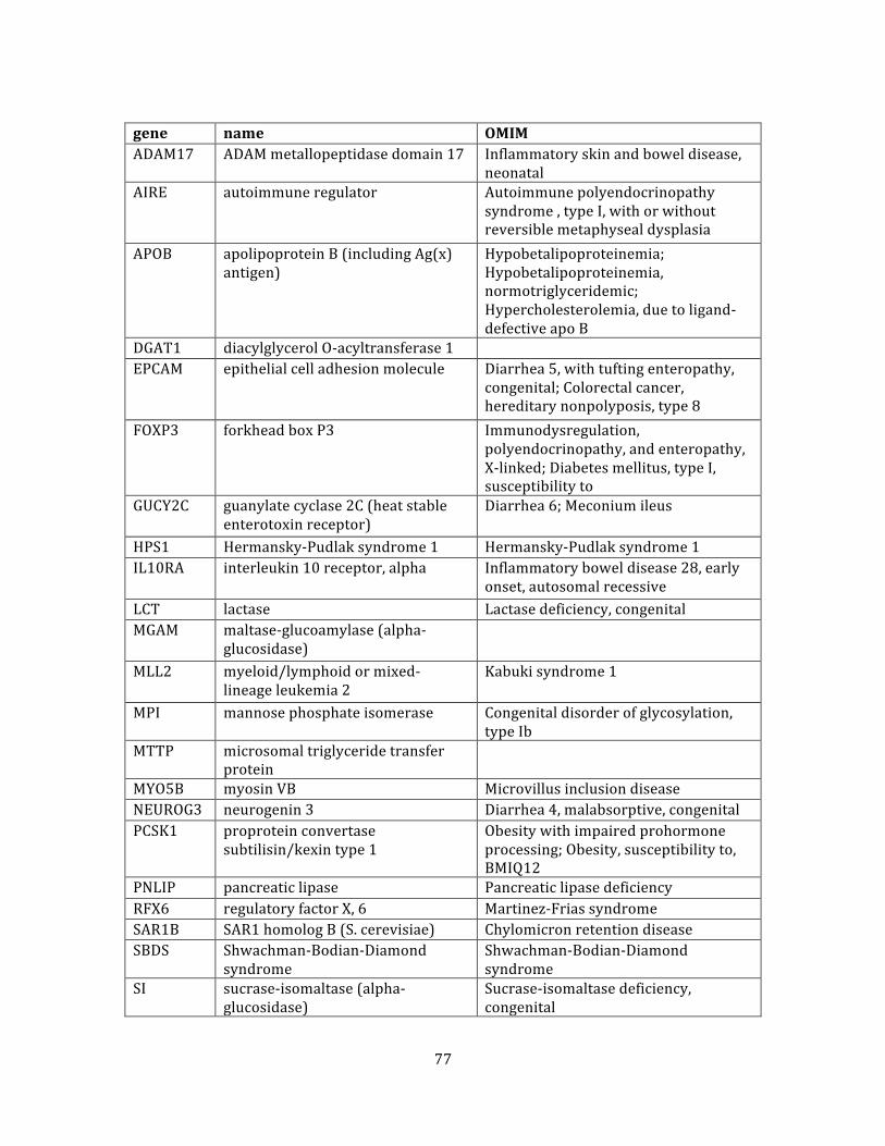
77
gene name OMIM ADAM17 ADAM metallopeptidase domain 17 Inflammatory skin and bowel disease,
neonatal AIRE autoimmune regulator Autoimmune polyendocrinopathy
syndrome , type I, with or without reversible metaphyseal dysplasia
APOB apolipoprotein B (including Ag(x) antigen)
Hypobetalipoproteinemia; Hypobetalipoproteinemia, normotriglyceridemic; Hypercholesterolemia, due to ligand-‐defective apo B
DGAT1 diacylglycerol O-‐acyltransferase 1 EPCAM epithelial cell adhesion molecule Diarrhea 5, with tufting enteropathy,
congenital; Colorectal cancer, hereditary nonpolyposis, type 8
FOXP3 forkhead box P3 Immunodysregulation, polyendocrinopathy, and enteropathy, X-‐linked; Diabetes mellitus, type I, susceptibility to
GUCY2C guanylate cyclase 2C (heat stable enterotoxin receptor)
Diarrhea 6; Meconium ileus
HPS1 Hermansky-‐Pudlak syndrome 1 Hermansky-‐Pudlak syndrome 1 IL10RA interleukin 10 receptor, alpha Inflammatory bowel disease 28, early
onset, autosomal recessive LCT lactase Lactase deficiency, congenital MGAM maltase-‐glucoamylase (alpha-‐
glucosidase)
MLL2 myeloid/lymphoid or mixed-‐lineage leukemia 2
Kabuki syndrome 1
MPI mannose phosphate isomerase Congenital disorder of glycosylation, type Ib
MTTP microsomal triglyceride transfer protein
MYO5B myosin VB Microvillus inclusion disease NEUROG3 neurogenin 3 Diarrhea 4, malabsorptive, congenital PCSK1 proprotein convertase
subtilisin/kexin type 1 Obesity with impaired prohormone processing; Obesity, susceptibility to, BMIQ12
PNLIP pancreatic lipase Pancreatic lipase deficiency RFX6 regulatory factor X, 6 Martinez-‐Frias syndrome SAR1B SAR1 homolog B (S. cerevisiae) Chylomicron retention disease SBDS Shwachman-‐Bodian-‐Diamond
syndrome Shwachman-‐Bodian-‐Diamond syndrome
SI sucrase-‐isomaltase (alpha-‐glucosidase)
Sucrase-‐isomaltase deficiency, congenital
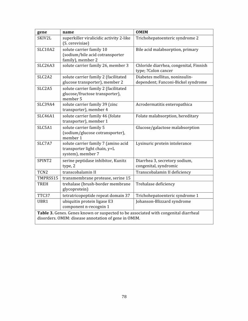
78
gene name OMIM SKIV2L superkiller viralicidic activity 2-‐like
(S. cerevisiae) Trichohepatoenteric syndrome 2
SLC10A2 solute carrier family 10 (sodium/bile acid cotransporter family), member 2
Bile acid malabsorption, primary
SLC26A3 solute carrier family 26, member 3 Chloride diarrhea, congenital, Finnish type; ?Colon cancer
SLC2A2 solute carrier family 2 (facilitated glucose transporter), member 2
Diabetes mellitus, noninsulin-‐dependent; Fanconi-‐Bickel syndrome
SLC2A5 solute carrier family 2 (facilitated glucose/fructose transporter), member 5
SLC39A4 solute carrier family 39 (zinc transporter), member 4
Acrodermatitis enteropathica
SLC46A1 solute carrier family 46 (folate transporter), member 1
Folate malabsorption, hereditary
SLC5A1 solute carrier family 5 (sodium/glucose cotransporter), member 1
Glucose/galactose malabsorption
SLC7A7 solute carrier family 7 (amino acid transporter light chain, y+L system), member 7
Lysinuric protein intolerance
SPINT2 serine peptidase inhibitor, Kunitz type, 2
Diarrhea 3, secretory sodium, congenital, syndromic
TCN2 transcobalamin II Transcobalamin II deficiency TMPRSS15 transmembrane protease, serine 15 TREH trehalase (brush-‐border membrane
glycoprotein) Trehalase deficiency
TTC37 tetratricopeptide repeat domain 37 Trichohepatoenteric syndrome 1 UBR1 ubiquitin protein ligase E3
component n-‐recognin 1 Johanson-‐Blizzard syndrome
Table 3. Genes. Genes known or suspected to be associated with congenital diarrheal disorders. OMIM: disease annotation of gene in OMIM.
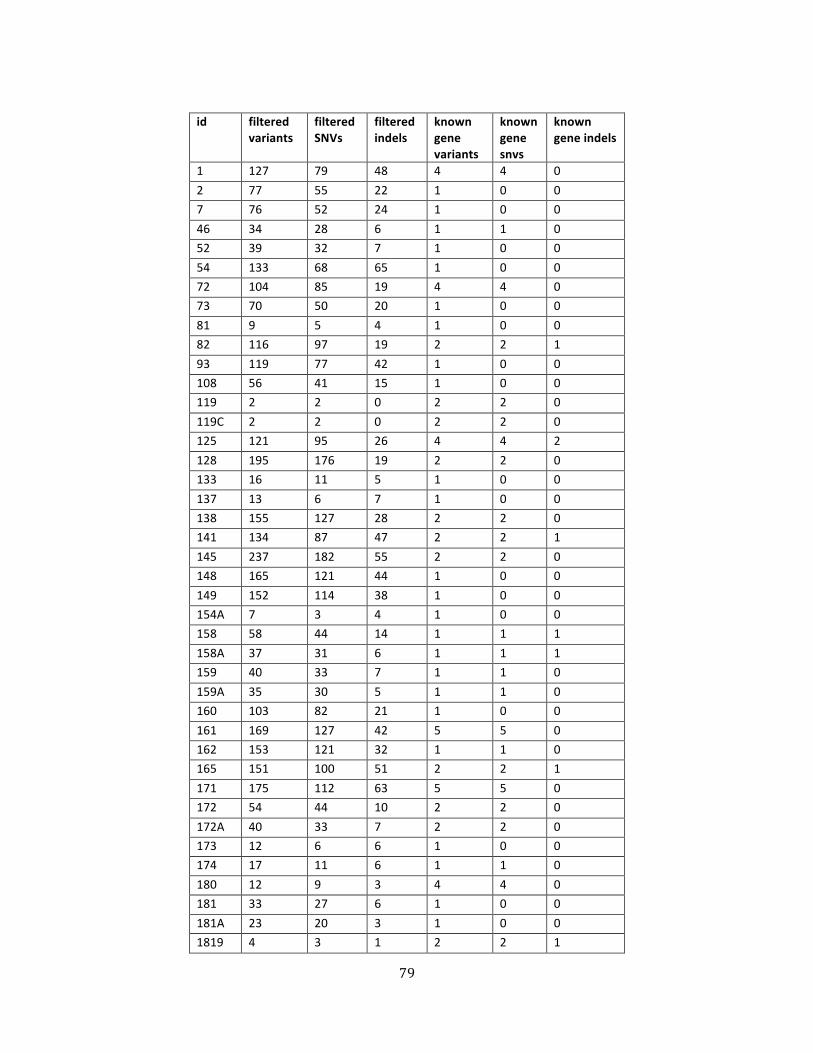
79
id filtered
variants filtered SNVs
filtered indels
known gene variants
known gene snvs
known gene indels
1 127 79 48 4 4 0 2 77 55 22 1 0 0 7 76 52 24 1 0 0 46 34 28 6 1 1 0 52 39 32 7 1 0 0 54 133 68 65 1 0 0 72 104 85 19 4 4 0 73 70 50 20 1 0 0 81 9 5 4 1 0 0 82 116 97 19 2 2 1 93 119 77 42 1 0 0 108 56 41 15 1 0 0 119 2 2 0 2 2 0 119C 2 2 0 2 2 0 125 121 95 26 4 4 2 128 195 176 19 2 2 0 133 16 11 5 1 0 0 137 13 6 7 1 0 0 138 155 127 28 2 2 0 141 134 87 47 2 2 1 145 237 182 55 2 2 0 148 165 121 44 1 0 0 149 152 114 38 1 0 0 154A 7 3 4 1 0 0 158 58 44 14 1 1 1 158A 37 31 6 1 1 1 159 40 33 7 1 1 0 159A 35 30 5 1 1 0 160 103 82 21 1 0 0 161 169 127 42 5 5 0 162 153 121 32 1 1 0 165 151 100 51 2 2 1 171 175 112 63 5 5 0 172 54 44 10 2 2 0 172A 40 33 7 2 2 0 173 12 6 6 1 0 0 174 17 11 6 1 1 0 180 12 9 3 4 4 0 181 33 27 6 1 0 0 181A 23 20 3 1 0 0 1819 4 3 1 2 2 1
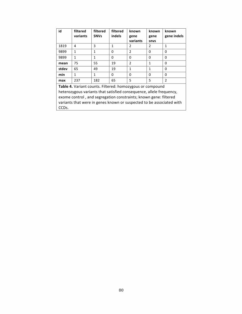
80
id filtered variants
filtered SNVs
filtered indels
known gene variants
known gene snvs
known gene indels
1819 4 3 1 2 2 1 9899 1 1 0 2 0 0 9899 1 1 0 0 0 0 mean 75 55 19 2 1 0 stdev 65 49 19 1 1 0 min 1 1 0 0 0 0 max 237 182 65 5 5 2
Table 4. Variant counts. Filtered: homozygous or compound heterozygous variants that satisfied consequence, allele frequency, exome control , and segregation constraints; known gene: filtered variants that were in genes known or suspected to be associated with CCDs.
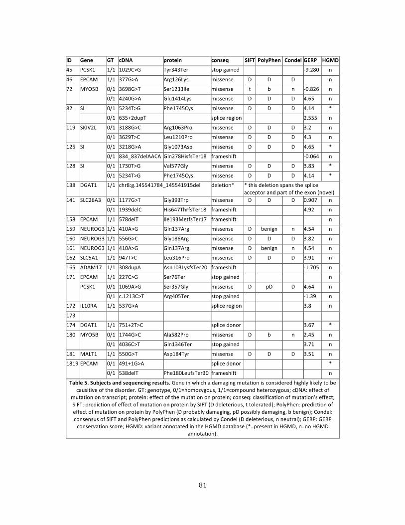
81
ID Gene GT cDNA protein conseq SIFT PolyPhen Condel GERP HGMD
45 PCSK1 1/1 1029C>G Tyr343Ter stop gained -‐9.280 n
46 EPCAM 1/1 377G>A Arg126Lys missense D D D n
72 MYO5B 0/1 3698G>T Ser1233Ile missense t b n -‐0.826 n
0/1 4240G>A Glu1414Lys missense D D D 4.65 n
82 SI 0/1 5234T>G Phe1745Cys missense D D D 4.14 *
0/1 635+2dupT splice region 2.555 n
119 SKIV2L 0/1 3188G>C Arg1063Pro missense D D D 3.2 n
0/1 3629T>C Leu1210Pro missense D D D 4.3 n
125 SI 0/1 3218G>A Gly1073Asp missense D D D 4.65 *
0/1 834_837delAACA Gln278HisfsTer18 frameshift -‐0.064 n
128 SI 0/1 1730T>G Val577Gly missense D D D 3.83 *
0/1 5234T>G Phe1745Cys missense D D D 4.14 *
138 DGAT1 1/1 chr8:g.145541784_145541915del
deletion* * this deletion spans the splice acceptor and part of the exon (novel)
141 SLC26A3 0/1 1177G>T Gly393Trp missense D D D 0.907 n
0/1 1939delC His647ThrfsTer18 frameshift 4.92 n
158 EPCAM 1/1 578delT Ile193MetfsTer17 frameshift n
159 NEUROG3 1/1 410A>G Gln137Arg missense D benign n 4.54 n
160 NEUROG3 1/1 556G>C Gly186Arg missense D D D 3.82 n
161 NEUROG3 1/1 410A>G Gln137Arg missense D benign n 4.54 n
162 SLC5A1 1/1 947T>C Leu316Pro missense D D D 3.91 n
165 ADAM17 1/1 308dupA Asn103LysfsTer20 frameshift -‐1.705 n
171 EPCAM 1/1 227C>G Ser76Ter stop gained n
PCSK1 0/1 1069A>G Ser357Gly missense D pD D 4.64 n
0/1 c.1213C>T Arg405Ter stop gained -‐1.39 n
172 IL10RA 1/1 537G>A splice region 3.8 n
173
174 DGAT1 1/1 751+2T>C splice donor 3.67 *
180 MYO5B 0/1 1744G>C Ala582Pro missense D b n 2.45 n
0/1 4036C>T Gln1346Ter stop gained 3.71 n
181 MALT1 1/1 550G>T Asp184Tyr missense D D D 3.51 n
1819 EPCAM 0/1 491+1G>A splice donor *
0/1 538delT Phe180LeufsTer30 frameshift n
Table 5. Subjects and sequencing results. Gene in which a damaging mutation is considered highly likely to be causitive of the disorder. GT: genotype, 0/1=homozygous, 1/1=compound heterozygous; cDNA: effect of
mutation on transcript; protein: effect of the mutation on protein; conseq: classification of mutation's effect; SIFT: prediction of effect of mutation on protein by SIFT (D deleterious, t tolerated); PolyPhen: prediction of effect of mutation on protein by PolyPhen (D probably damaging, pD possibly damaging, b benign); Condel: consensus of SIFT and PolyPhen predictions as calculated by Condel (D deleterious, n neutral); GERP: GERP conservation score; HGMD: variant annotated in the HGMD database (*=present in HGMD, n=no HGMD
annotation).
82
References
1. Terrin G, Tomaiuolo R, Passariello A, Elce A, Amato F, Di Costanzo M, Castaldo G, Canani RB. Congenital diarrheal disorders: an updated diagnostic approach. International journal of molecular sciences. 2012;13(4):4168-‐85. doi: 10.3390/ijms13044168. PubMed PMID: 22605972; PubMed Central PMCID: PMC3344208.
2. Yourshaw M, Solorzano-‐Vargas RS, Pickett LA, Lindberg I, Wang J, Cortina G, Pawlikowska-‐Haddal A, Baron H, Venick RS, Nelson SF. Exome Sequencing Finds a Novel PCSK1 Mutation in a Child With Generalized Malabsorptive Diarrhea and Diabetes Insipidus. Journal of pediatric gastroenterology and nutrition. 2013. doi: 10.1097/MPG.0b013e3182a8ae6c.
3. Jackson RS, Creemers JW, Ohagi S, Raffin-‐Sanson ML, Sanders L, Montague CT, Hutton JC, O'Rahilly S. Obesity and impaired prohormone processing associated with mutations in the human prohormone convertase 1 gene. Nat Genet. 1997;16(3):303-‐6. Epub 1997/07/01. doi: 10.1038/ng0797-‐303. PubMed PMID: 9207799.
4. Spencer AU, Kovacevich D, McKinney-‐Barnett M, Hair D, Canham J, Maksym C, Teitelbaum DH. Pediatric short-‐bowel syndrome: the cost of comprehensive care. The American journal of clinical nutrition. 2008;88(6):1552-‐9. Epub 2008/12/10. doi: 10.3945/ajcn.2008.26007. PubMed PMID: 19064515.
5. Hearne CM, Ghosh S, Todd JA. Microsatellites for Linkage Analysis of Genetic-‐Traits. Trends in Genetics. 1992;8(8):288-‐94. doi: Doi 10.1016/0168-‐9525(92)90137-‐S. PubMed PMID: WOS:A1992JE96800009.
6. Kere J, Sistonen P, Holmberg C, de la Chapelle A. The gene for congenital chloride diarrhea maps close to but is distinct from the gene for cystic fibrosis transmembrane conductance regulator. Proc Natl Acad Sci U S A. 1993;90(22):10686-‐9. Epub 1993/11/15. PubMed PMID: 7504277; PubMed Central PMCID: PMC47842.
7. Hoglund P, Haila S, Scherer SW, Tsui LC, Green ED, Weissenbach J, Holmberg C, de la Chapelle A, Kere J. Positional candidate genes for congenital chloride diarrhea suggested by high-‐resolution physical mapping in chromosome region 7q31. Genome Res. 1996;6(3):202-‐10. Epub 1996/03/01. PubMed PMID: 8963897.
8. Hoglund P, Haila S, Socha J, Tomaszewski L, Saarialho-‐Kere U, Karjalainen-‐Lindsberg ML, Airola K, Holmberg C, de la Chapelle A, Kere J. Mutations of the Down-‐regulated in adenoma (DRA) gene cause congenital chloride diarrhoea. Nat Genet. 1996;14(3):316-‐9. Epub 1996/11/01. doi: 10.1038/ng1196-‐316. PubMed PMID: 8896562.
9. Sellick GS, Longman C, Tolmie J, Newbury-‐Ecob R, Geenhalgh L, Hughes S, Whiteford M, Garrett C, Houlston RS. Genomewide linkage searches for Mendelian disease loci can be efficiently conducted using high-‐density SNP genotyping arrays. Nucleic Acids Res. 2004;32(20):e164. Epub 2004/11/25. doi: 10.1093/nar/gnh163. PubMed PMID: 15561999; PubMed Central PMCID: PMC534642.
10. Muller T, Hess MW, Schiefermeier N, Pfaller K, Ebner HL, Heinz-‐Erian P, Ponstingl H, Partsch J, Rollinghoff B, Kohler H, Berger T, Lenhartz H, Schlenck B, Houwen RJ, Taylor CJ, Zoller H, Lechner S, Goulet O, Utermann G, Ruemmele FM, Huber LA, Janecke AR. MYO5B
83
mutations cause microvillus inclusion disease and disrupt epithelial cell polarity. Nat Genet. 2008;40(10):1163-‐5. doi: 10.1038/ng.225. PubMed PMID: 18724368.
11. Heinz-‐Erian P, Muller T, Krabichler B, Schranz M, Becker C, Ruschendorf F, Nurnberg P, Rossier B, Vujic M, Booth IW, Holmberg C, Wijmenga C, Grigelioniene G, Kneepkens CM, Rosipal S, Mistrik M, Kappler M, Michaud L, Doczy LC, Siu VM, Krantz M, Zoller H, Utermann G, Janecke AR. Mutations in SPINT2 cause a syndromic form of congenital sodium diarrhea. Am J Hum Genet. 2009;84(2):188-‐96. Epub 2009/02/03. doi: 10.1016/j.ajhg.2009.01.004. PubMed PMID: 19185281; PubMed Central PMCID: PMC2668003.
12. Sivagnanam M, Mueller JL, Lee H, Chen Z, Nelson SF, Turner D, Zlotkin SH, Pencharz PB, Ngan BY, Libiger O, Schork NJ, Lavine JE, Taylor S, Newbury RO, Kolodner RD, Hoffman HM. Identification of EpCAM as the gene for congenital tufting enteropathy. Gastroenterology. 2008;135(2):429-‐37. doi: Doi 10.1053/J.Gastro.2008.05.036. PubMed PMID: ISI:000258439900020.
13. Fiskerstrand T, Arshad N, Haukanes BI, Tronstad RR, Pham KD, Johansson S, Havik B, Tonder SL, Levy SE, Brackman D, Boman H, Biswas KH, Apold J, Hovdenak N, Visweswariah SS, Knappskog PM. Familial diarrhea syndrome caused by an activating GUCY2C mutation. N Engl J Med. 2012;366(17):1586-‐95. Epub 2012/03/23. doi: 10.1056/NEJMoa1110132. PubMed PMID: 22436048.
14. Wang J, Cortina G, Wu SV, Tran R, Cho JH, Tsai MJ, Bailey TJ, Jamrich M, Ament ME, Treem WR, Hill ID, Vargas JH, Gershman G, Farmer DG, Reyen L, Martin MG. Mutant neurogenin-‐3 in congenital malabsorptive diarrhea. N Engl J Med. 2006;355(3):270-‐80. doi: 10.1056/NEJMoa054288. PubMed PMID: 16855267.
15. Bamshad MJ, Ng SB, Bigham AW, Tabor HK, Emond MJ, Nickerson DA, Shendure J. Exome sequencing as a tool for Mendelian disease gene discovery. Nat Rev Genet. 2011;12(11):745-‐55. Epub 2011/09/29. doi: 10.1038/nrg3031. PubMed PMID: 21946919.
16. Maxmen A. Exome sequencing deciphers rare diseases. Cell. 2011;144(5):635-‐7. Epub 2011/03/08. doi: 10.1016/j.cell.2011.02.033. PubMed PMID: 21376225.
17. International Human Genome Sequencing Consortium. Finishing the euchromatic sequence of the human genome. Nature. 2004;431(7011):931-‐45. Epub 2004/10/22. doi: 10.1038/nature03001. PubMed PMID: 15496913.
18. DePristo MA, Banks E, Poplin R, Garimella KV, Maguire JR, Hartl C, Philippakis AA, del Angel G, Rivas MA, Hanna M, McKenna A, Fennell TJ, Kernytsky AM, Sivachenko AY, Cibulskis K, Gabriel SB, Altshuler D, Daly MJ. A framework for variation discovery and genotyping using next-‐generation DNA sequencing data. Nat Genet. 2011;43(5):491-‐8. Epub 2011/04/12. doi: 10.1038/ng.806. PubMed PMID: 21478889; PubMed Central PMCID: PMC3083463.
19. McKenna A, Hanna M, Banks E, Sivachenko A, Cibulskis K, Kernytsky A, Garimella K, Altshuler D, Gabriel S, Daly M, DePristo MA. The Genome Analysis Toolkit: a MapReduce framework for analyzing next-‐generation DNA sequencing data. Genome Res. 2010;20(9):1297-‐303. Epub 2010/07/21. doi: 10.1101/gr.107524.110. PubMed PMID: 20644199; PubMed Central PMCID: PMC2928508.
84
20. Kumar P, Henikoff S, Ng PC. Predicting the effects of coding non-‐synonymous variants on protein function using the SIFT algorithm. Nature protocols. 2009;4(7):1073-‐81. Epub 2009/06/30. doi: 10.1038/nprot.2009.86. PubMed PMID: 19561590.
21. Ng PC, Henikoff S. Predicting deleterious amino acid substitutions. Genome Res. 2001;11(5):863-‐74. Epub 2001/05/05. doi: 10.1101/gr.176601. PubMed PMID: 11337480; PubMed Central PMCID: PMC311071.
22. Ng PC, Henikoff S. Accounting for human polymorphisms predicted to affect protein function. Genome Res. 2002;12(3):436-‐46. Epub 2002/03/05. doi: 10.1101/gr.212802. PubMed PMID: 11875032; PubMed Central PMCID: PMC155281.
23. Ng PC, Henikoff S. SIFT: Predicting amino acid changes that affect protein function. Nucleic Acids Res. 2003;31(13):3812-‐4. Epub 2003/06/26. PubMed PMID: 12824425; PubMed Central PMCID: PMC168916.
24. Ng PC, Henikoff S. Predicting the effects of amino acid substitutions on protein function. Annual review of genomics and human genetics. 2006;7:61-‐80. Epub 2006/07/11. doi: 10.1146/annurev.genom.7.080505.115630. PubMed PMID: 16824020.
25. Ramensky V, Bork P, Sunyaev S. Human non-‐synonymous SNPs: server and survey. Nucleic Acids Res. 2002;30(17):3894-‐900. Epub 2002/08/31. PubMed PMID: 12202775; PubMed Central PMCID: PMC137415.
26. Sunyaev S, Ramensky V, Bork P. Towards a structural basis of human non-‐synonymous single nucleotide polymorphisms. Trends Genet. 2000;16(5):198-‐200. Epub 2000/04/27. PubMed PMID: 10782110.
27. Sunyaev S, Ramensky V, Koch I, Lathe W, 3rd, Kondrashov AS, Bork P. Prediction of deleterious human alleles. Hum Mol Genet. 2001;10(6):591-‐7. Epub 2001/03/07. PubMed PMID: 11230178.
28. Gonzalez-‐Perez A, Lopez-‐Bigas N. Improving the assessment of the outcome of nonsynonymous SNVs with a consensus deleteriousness score, Condel. Am J Hum Genet. 2011;88(4):440-‐9. Epub 2011/04/05. doi: 10.1016/j.ajhg.2011.03.004. PubMed PMID: 21457909; PubMed Central PMCID: PMC3071923.
29. Online Mendelian Inheritance in Man OMIM®. Online Mendelian Inheritance in Man, OMIM® Baltimore, MD: McKusick-‐Nathans Institute of Genetic Medicine, Johns Hopkins University; [2013-‐11-‐04]. Available from: http://omim.org/.
30. Apweiler R, Martin MJ, O'Donovan C, Magrane M, Alam-‐Faruque Y, Alpi E, Antunes R, Arganiska J, Casanova EB, Bely B, Bingley M, Bonilla C, Britto R, Bursteinas B, Chan WM, Chavali G, Cibrian-‐Uhalte E, Da Silva A, De Giorgi M, Dimmer E, Fazzini F, Gane P, Fedotov A, Castro LG, Garmiri P, Hatton-‐Ellis E, Hieta R, Huntley R, Jacobsen J, Jones R, Legge D, Liu WD, Luo J, MacDougall A, Mutowo P, Nightingale A, Orchard S, Patient S, Pichler K, Poggioli D, Pundir S, Pureza L, Qi GY, Rosanoff S, Sawford T, Sehra H, Turner E, Volynkin V, Wardell T, Watkins X, Zellner H, Corbett M, Donnelly M, van Rensburg P, Goujon M, McWilliam H, Lopez R, Xenarios I, Bougueleret L, Bridge A, Poux S, Redaschi N, Auchincloss A, Axelsen K, Bansal P, Baratin D, Binz PA, Blatter MC, Boeckmann B, Bolleman J, Boutet E, Breuza L, de Castro E, Cerutti L, Coudert E,
85
Cuche B, Doche M, Dornevil D, Duvaud S, Estreicher A, Famiglietti L, Feuermann M, Gasteiger E, Gehant S, Gerritsen V, Gos A, Gruaz-‐Gumowski N, Hinz U, Hulo C, James J, Jungo F, Keller G, Lara V, Lemercier P, Lew J, Lieberherr D, Martin X, Masson P, Morgat A, Neto T, Paesano S, Pedruzzi I, Pilbout S, Pozzato M, Pruess M, Rivoire C, Roechert B, Schneider M, Sigrist C, Sonesson K, Staehli S, Stutz A, Sundaram S, Tognolli M, Verbregue L, Veuthey AL, Zerara M, Wu CH, Arighi CN, Arminski L, Chen CM, Chen YX, Huang HZ, Kukreja A, Laiho K, McGarvey P, Natale DA, Natarajan TG, Roberts NV, Suzek BE, Vinayaka CR, Wang QH, Wang YQ, Yeh LS, Yerramalla MS, Zhang J, Consortium U. Update on activities at the Universal Protein Resource (UniProt) in 2013. Nucleic Acids Research. 2013;41(D1):D43-‐D7. doi: Doi 10.1093/Nar/Gks1068. PubMed PMID: WOS:000312893300007.
31. Kanehisa M, Goto S, Kawashima S, Nakaya A. The KEGG databases at GenomeNet. Nucleic Acids Res. 2002;30(1):42-‐6. PubMed PMID: 11752249; PubMed Central PMCID: PMC99091.
32. Pruitt KD, Tatusova T, Brown GR, Maglott DR. NCBI Reference Sequences (RefSeq): current status, new features and genome annotation policy. Nucleic Acids Res. 2012;40(Database issue):D130-‐5. doi: 10.1093/nar/gkr1079. PubMed PMID: 22121212; PubMed Central PMCID: PMC3245008.
33. Pagliarini DJ, Calvo SE, Chang B, Sheth SA, Vafai SB, Ong SE, Walford GA, Sugiana C, Boneh A, Chen WK, Hill DE, Vidal M, Evans JG, Thorburn DR, Carr SA, Mootha VK. A mitochondrial protein compendium elucidates complex I disease biology. Cell. 2008;134(1):112-‐23. doi: 10.1016/j.cell.2008.06.016. PubMed PMID: 18614015; PubMed Central PMCID: PMC2778844.
34. Eppig JT, Blake JA, Bult CJ, Kadin JA, Richardson JE, Mouse Genome Database G. The Mouse Genome Database (MGD): comprehensive resource for genetics and genomics of the laboratory mouse. Nucleic Acids Res. 2012;40(Database issue):D881-‐6. doi: 10.1093/nar/gkr974. PubMed PMID: 22075990; PubMed Central PMCID: PMC3245042.
35. Uhlen M, Oksvold P, Fagerberg L, Lundberg E, Jonasson K, Forsberg M, Zwahlen M, Kampf C, Wester K, Hober S, Wernerus H, Bjorling L, Ponten F. Towards a knowledge-‐based Human Protein Atlas. Nat Biotechnol. 2010;28(12):1248-‐50. doi: 10.1038/nbt1210-‐1248. PubMed PMID: 21139605.
36. 1000 Genomes Project. VCF (Variant Call Format) version 4.1 2013 [updated 2013-‐10-‐09]. Available from: http://www.1000genomes.org/wiki/Analysis/Variant Call Format/vcf-‐variant-‐call-‐format-‐version-‐41.
37. Exome Variant Server [Internet]. NHLBI Exome Sequencing Project (ESP). 2011 [cited 2011-‐09-‐10]. Available from: http://evs.gs.washington.edu/EVS/.
38. Blaydon DC, Biancheri P, Di WL, Plagnol V, Cabral RM, Brooke MA, van Heel DA, Ruschendorf F, Toynbee M, Walne A, O'Toole EA, Martin JE, Lindley K, Vulliamy T, Abrams DJ, MacDonald TT, Harper JI, Kelsell DP. Inflammatory skin and bowel disease linked to ADAM17 deletion. N Engl J Med. 2011;365(16):1502-‐8. Epub 2011/10/21. doi: 10.1056/NEJMoa1100721. PubMed PMID: 22010916.
86
39. Haas JT, Winter HS, Lim E, Kirby A, Blumenstiel B, DeFelice M, Gabriel S, Jalas C, Branski D, Grueter CA, Toporovski MS, Walther TC, Daly MJ, Farese RV, Jr. DGAT1 mutation is linked to a congenital diarrheal disorder. The Journal of clinical investigation. 2012;122(12):4680-‐4. doi: 10.1172/JCI64873. PubMed PMID: 23114594; PubMed Central PMCID: PMC3533555.
40. Glocker EO, Kotlarz D, Boztug K, Gertz EM, Schaffer AA, Noyan F, Perro M, Diestelhorst J, Allroth A, Murugan D, Hatscher N, Pfeifer D, Sykora KW, Sauer M, Kreipe H, Lacher M, Nustede R, Woellner C, Baumann U, Salzer U, Koletzko S, Shah N, Segal AW, Sauerbrey A, Buderus S, Snapper SB, Grimbacher B, Klein C. Inflammatory bowel disease and mutations affecting the interleukin-‐10 receptor. N Engl J Med. 2009;361(21):2033-‐45. Epub 2009/11/06. doi: 10.1056/NEJMoa0907206. PubMed PMID: 19890111; PubMed Central PMCID: PMC2787406.
41. Jabara HH, Ohsumi T, Chou J, Massaad MJ, Benson H, Megarbane A, Chouery E, Mikhael R, Gorka O, Gewies A, Portales P, Nakayama T, Hosokawa H, Revy P, Herrod H, Le Deist F, Lefranc G, Ruland J, Geha RS. A homozygous mucosa-‐associated lymphoid tissue 1 (MALT1) mutation in a family with combined immunodeficiency. The Journal of allergy and clinical immunology. 2013;132(1):151-‐8. Epub 2013/06/04. doi: 10.1016/j.jaci.2013.04.047. PubMed PMID: 23727036; PubMed Central PMCID: PMC3700575.
42. Martin MG, Lindberg I, Solorzano-‐Vargas RS, Wang J, Avitzur Y, Bandsma R, Sokollik C, Lawrence S, Pickett LA, Chen Z, Egritas O, Dalgic B, Albornoz V, de Ridder L, Hulst J, Gok F, Aydogan A, Al-‐Hussaini A, Gok DE, Yourshaw M, Wu SV, Cortina G, Stanford S, Georgia S. Congenital proprotein convertase 1/3 deficiency causes malabsorptive diarrhea and other endocrinopathies in a pediatric cohort. Gastroenterology. 2013;145(1):138-‐48. doi: 10.1053/j.gastro.2013.03.048. PubMed PMID: 23562752.
43. Ouwendijk J, Moolenaar CE, Peters WJ, Hollenberg CP, Ginsel LA, Fransen JA, Naim HY. Congenital sucrase-‐isomaltase deficiency. Identification of a glutamine to proline substitution that leads to a transport block of sucrase-‐isomaltase in a pre-‐Golgi compartment. The Journal of clinical investigation. 1996;97(3):633-‐41. Epub 1996/02/01. doi: 10.1172/JCI118459. PubMed PMID: 8609217; PubMed Central PMCID: PMC507098.
44. Fabre A, Charroux B, Martinez-‐Vinson C, Roquelaure B, Odul E, Sayar E, Smith H, Colomb V, Andre N, Hugot JP, Goulet O, Lacoste C, Sarles J, Royet J, Levy N, Badens C. SKIV2L mutations cause syndromic diarrhea, or trichohepatoenteric syndrome. Am J Hum Genet. 2012;90(4):689-‐92. Epub 2012/03/27. doi: 10.1016/j.ajhg.2012.02.009. PubMed PMID: 22444670; PubMed Central PMCID: PMC3322239.
45. Turk E, Zabel B, Mundlos S, Dyer J, Wright EM. Glucose/galactose malabsorption caused by a defect in the Na+/glucose cotransporter. Nature. 1991;350(6316):354-‐6. Epub 1991/03/28. doi: 10.1038/350354a0. PubMed PMID: 2008213.
46. Lee H, Nelson SF. Rethinking clinical practice: clinical implementation of exome sequencing. Pers Med. 2012;9(8):785-‐7. doi: Doi 10.2217/Pme.12.101. PubMed PMID: WOS:000311977800001.
47. Yang J, Duan S, Zhong R, Yin J, Pu J, Ke J, Lu X, Zou L, Zhang H, Zhu Z, Wang D, Xiao H, Guo A, Xia J, Miao X, Tang S, Wang G. Exome sequencing identified NRG3 as a novel susceptible
87
gene of Hirschsprung's disease in a Chinese population. Molecular neurobiology. 2013;47(3):957-‐66. doi: 10.1007/s12035-‐012-‐8392-‐4. PubMed PMID: 23315268.
48. Whitcomb DC, Preston RA, Aston CE, Sossenheimer MJ, Barua PS, Zhang Y, Wong-‐Chong A, White GJ, Wood PG, Gates LK, Jr., Ulrich C, Martin SP, Post JC, Ehrlich GD. A gene for hereditary pancreatitis maps to chromosome 7q35. Gastroenterology. 1996;110(6):1975-‐80. Epub 1996/06/01. PubMed PMID: 8964426.
49. Cetani F, Barbesino G, Borsari S, Pardi E, Cianferotti L, Pinchera A, Marcocci C. A novel mutation of the autoimmune regulator gene in an Italian kindred with autoimmune polyendocrinopathy-‐candidiasis-‐ectodermal dystrophy, acting in a dominant fashion and strongly cosegregating with hypothyroid autoimmune thyroiditis. The Journal of clinical endocrinology and metabolism. 2001;86(10):4747-‐52. Epub 2001/10/16. PubMed PMID: 11600535.
50. Koboldt DC, Steinberg KM, Larson DE, Wilson RK, Mardis ER. The next-‐generation sequencing revolution and its impact on genomics. Cell. 2013;155(1):27-‐38. Epub 2013/10/01. doi: 10.1016/j.cell.2013.09.006. PubMed PMID: 24074859.
88
CHAPTER FOUR
Exome sequencing finds a novel PCSK1 mutation
in a child with generalized malabsorptive diarrhea and diabetes insipidus
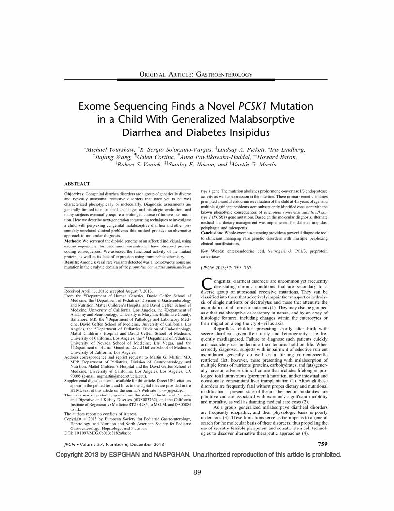
89
Copyright 2013 by ESPGHAN and NASPGHAN. Unauthorized reproduction of this article is prohibited.
Exome Sequencing Finds a Novel PCSK1 Mutationin a Child With Generalized Malabsorptive
Diarrhea and Diabetes Insipidus!Michael Yourshaw, yR. Sergio Solorzano-Vargas, zLindsay A. Pickett, zIris Lindberg,yJiafang Wang, !Galen Cortina, #Anna Pawlikowska-Haddal, !!Howard Baron,
yRobert S. Venick, zzStanley F. Nelson, and yMartın G. Martın
ABSTRACT
Objectives: Congenital diarrhea disorders are a group of genetically diverseand typically autosomal recessive disorders that have yet to be wellcharacterized phenotypically or molecularly. Diagnostic assessments aregenerally limited to nutritional challenges and histologic evaluation, andmany subjects eventually require a prolonged course of intravenous nutri-tion. Here we describe next-generation sequencing techniques to investigatea child with perplexing congenital malabsorptive diarrhea and other pre-sumably unrelated clinical problems; this method provides an alternativeapproach to molecular diagnosis.Methods: We screened the diploid genome of an affected individual, usingexome sequencing, for uncommon variants that have observed protein-coding consequences. We assessed the functional activity of the mutantprotein, as well as its lack of expression using immunohistochemistry.Results: Among several rare variants detected was a homozygous nonsensemutation in the catalytic domain of the proprotein convertase subtilisin/kexin
type 1 gene. The mutation abolishes prohormone convertase 1/3 endoproteaseactivity as well as expression in the intestine. These primary genetic findingsprompted a careful endocrine reevaluation of the child at 4.5 years of age, andmultiple significant problems were subsequently identified consistent with theknown phenotypic consequences of proprotein convertase subtilisin/kexin
type 1 (PCSK1) gene mutations. Based on the molecular diagnosis, alternatemedical and dietary management was implemented for diabetes insipidus,polyphagia, and micropenis.Conclusions: Whole-exome sequencing provides a powerful diagnostic toolto clinicians managing rare genetic disorders with multiple perplexingclinical manifestations.
Key Words: enteroendocrine cell, Neurogenin-3, PC1/3, proproteinconvertases
(JPGN 2013;57: 759–767)
C ongenital diarrheal disorders are uncommon yet frequentlydevastating chronic conditions that are secondary to a
diverse group of autosomal recessive mutations. They can beclassified into those that selectively impair the transport or hydroly-sis of single nutrients or electrolytes and those that attenuate theassimilation of all forms of nutrients (1). They may also be groupedas either malabsorptive or secretory in nature, and by an array ofhistologic features, including changes within the enterocytes ortheir migration along the crypt–villus axis.
Regardless, children presenting shortly after birth withsevere diarrhea—given their rarity and heterogeneity—are fre-quently misdiagnosed. Failure to diagnose such patients quicklyand accurately can undermine their tenuous hold on life. Whencorrectly diagnosed, subjects with impairment of selective nutrientassimilation generally do well on a lifelong nutrient-specificrestricted diet; however, those presenting with malabsorption ofmultiple forms of nutrients (proteins, carbohydrates, and fats) gener-ally have an adverse clinical course that includes lifelong or pro-longed total intravenous (parenteral) nutrition, and/or intestinal andoccasionally concomitant liver transplantation (1). Although thesedisorders are frequently fatal without proper dietary and nutritionalmodifications, present state-of-the-art therapeutic modalities areprimitive and are associated with extremely significant morbidityand mortality, as well as daunting medical care costs (2).
As a group, generalized malabsorptive diarrheal disordersare frequently idiopathic, and their physiologic basis is poorlyunderstood (3). These limitations serve as the impetus to a generalsearch for the molecular basis of these disorders, thus propelling theuse of recently feasible pluripotent and somatic stem cell technol-ogies to discover alternative therapeutic approaches (4).
Received April 13, 2013; accepted August 7, 2013.From the !Department of Human Genetics, David Geffen School of
Medicine, the yDepartment of Pediatrics, Division of Gastroenterologyand Nutrition, Mattel Children’s Hospital and David Geffen School ofMedicine, University of California, Los Angeles, the zDepartment ofAnatomy and Neurobiology, University of Maryland-Baltimore County,Baltimore, MD, the !Department of Pathology and Laboratory Medi-cine, David Geffen School of Medicine, University of California, LosAngeles, the #Department of Pediatrics, Division of Endocrinology,Mattel Children’s Hospital and David Geffen School of Medicine,University of California, Los Angeles, the !!Department of Pediatrics,University of Nevada School of Medicine, Las Vegas, and thezzDepartment of Human Genetics, David Geffen School of Medicine,University of California, Los Angeles.
Address correspondence and reprint requests to Martın G. Martın, MD,MPP, Department of Pediatrics, Division of Gastroenterology andNutrition, Mattel Children’s Hospital and the David Geffen School ofMedicine, University of California, Los Angeles, Los Angeles, CA90095 (e-mail: [email protected]).
Supplemental digital content is available for this article. Direct URL citationsappear in the printed text, and links to the digital files are provided in theHTML text of this article on the journal’s Web site (www.jpgn.org).
This work was supported by grants from the National Institute of Diabetesand Digestive and Kidney Diseases (#DK083762), and the CaliforniaInstitute of Regenerative Medicine RT2-01985, to M.G.M. and DA05084to I.L.
The authors report no conflicts of interest.Copyright # 2013 by European Society for Pediatric Gastroenterology,
Hepatology, and Nutrition and North American Society for PediatricGastroenterology, Hepatology, and Nutrition
DOI: 10.1097/MPG.0b013e3182a8ae6c
ORIGINAL ARTICLE: GASTROENTEROLOGY
JPGN " Volume 57, Number 6, December 2013 759
90
Copyright 2013 by ESPGHAN and NASPGHAN. Unauthorized reproduction of this article is prohibited.
In some cases, the histology and nutrient absorption charac-teristics may point to the possibility of dysfunction in known genes(Neurogenin-3 [NEUROG3], SGLT1, EPCAM, MYO5B, SPINT2,TTC37, SKIV2L, ADAM17), which can then be directly sequencedto identify likely causative mutations (5–11). Often, however,clinical evidence may be insufficient to implicate known genesor sequencing of candidate genes fails to reveal damaging mutationsresulting from genetic heterogeneity. These aspects, as is typical ofall rare disorders, greatly impede efficient and timely diagnosis.Recent advances in sequencing technology now make it possible tosequence the coding portion and essential splice sites of approxi-mately 95% of all protein-coding bases of all known genes (the‘‘exome’’) at a cost comparable to clinical sequencing tests of asingle gene (12,13). Thus, an unbiased scan of the exome candiscover known and novel mutations in known genes and alsomutations in hitherto unsuspected genes in a manner that efficientlydirects clinical care. Here we show an example of the use of whole-exome sequencing to identify the causative mutation in a child withcongential diarrhea.
Prohormone convertase 1/3 (PC1/3) is a calcium-dependentserine endoprotease that converts proinsulin and other prohormonesinto active forms (14). PC1/3 is highly expressed in the smallintestine. PC1/3 deficiency, resulting from the mutations in the pro-protein convertase subtilisin/kexin type 1 (PCSK1) gene, can prevententeroendocrine cells from producing functional hormonesand cause generalized malabsorption and a variety of systemicendocrinopathies that develop in an age-dependent fashion (15).The mechanism by which PC1/3 deficiency causes malabsorptionis not well understood, but it may be that a novel peptide, ormultiple redundant peptides, processed by PC1/3 enhance nutrientassimilation.
METHODS
DNA SequencingWe prepared an exon-enriched sequencing library following
Agilent Technologies (Santa Clara, CA) SureSelect Target Enrich-ment System for the Applied Biosystems (Foster City, CA) SOLiDSystem protocol (version 1.5.1). Briefly, genomic DNA extractedfrom patient saliva with an Oragene DNA Collection kit (DNA-Genotek, Kanata, Canada) was ultrasonically sheared (Covaris,Woburn, MA) into !125 bp fragments. After fragment end repair,ligation of adapters, and gel-size selection for!175 bp product, thelibrary was nick translated and amplified by 12 polymerase chainreaction (PCR) cycles. The library was hybridized in solution toRNA probes from the 3-Mb SureSelect All Human Exon kit(Agilent G3361), covering 1.22% of the human genome containingthe exons of the Consensus CDS genes (16). The exon-enrichedlibrary was selected by magnetic bead separation, further amplifiedby 12 cycles of PCR, and clonally amplified on beads by emulsionPCR (Applied Biosystems SOLiD 4 System Templated BeadPreparation Guide [March 2010]). Fragment sequencing by ligationwas performed on a SOLiD 4 System (Applied Biosystems SOLiD4 System Instrument Operation Guide [March 2010]), whichyielded !108 million 50-base reads.
Bioinformatics Analysis
We aligned the sequenced reads to build GRCh37 of thehuman genome (17) with Novoalign (http://www.novocraft.com) toobtain !31 million uniquely aligned 50-base reads (!1.5G bases)after removing PCR duplicates. !1.2G bases were within thetargeted exome with a mean coverage of !33X at each targetedposition, achieving at least 10X coverage of 67% of the codingsequence of annotated protein-coding genes per Ensembl
(Wellcome Trust Sanger Institute, Hinxton, UK) (18). Base qualityscores were recalibrated to improve accuracy by analyzing thecovariation among reported quality score, position within read,dinucleotide, and probability of mismatching the reference genomeusing the Genome Analysis Toolkit (GATK) (19,20). The GATKUnified Genotyper was used to genotype single-nucleotide variants(SNVs) and indels. The GATK variant quality score recalibrationmodule was used to assign probabilities to each variant call. Inaddition to variants that passed quality checks, for improvedsensitivity, we retained for downstream analysis a tranche ofvariants that had a likelihood "1% of being false-positives. Variantconsequences were determined by the Ensembl Variant EffectPredictor (20), and the extent of protein damage was estimatedwith SIFT (Sorting Intolerant From Tolerant) (21–25), PolyPhen(Polymorphism Phenotyping) (26–28), and Condel (ConsensusDeleteriousness) (29).
Development of Wild-Type and Mutant PC1/3Expression Vectors
Human PC1/3 complement DNA (cDNA) was generatedfrom RNA isolated from human pancreatic carcinoid (BON) cells.The wild-type PC1/3 was amplified by using an oligonucleotide thatcontained a KpnI site, and 20 nucleotides of PC1/3 from thetranslational start site. A 30 oligonucleotide contained an XhoI site,and a FLAG amino acid (DYKDDDDK) sequence introduced at thestop site of the wild-type and Y343X mutant cDNA. The wild-typecDNA was subcloned into the KpnI and XhoI restriction-digestedpcDNA3 vector and clones were screened. A single clone, desig-nated WT PC1/3, was used to generate the Y343X mutant clone,and the entire clone was sequenced to verify that only the targetedvariant was altered.
Transient Transfection of Expression Vectors
HEK293 cells at a density of 2# 105 cells per well in 24-wellplates were transfected with plasmids encoding WT PC1/3 or Y343Xin triplicatewells. Cells were transfected with 200 ng of plasmid DNAper well using Lipofectamine Reagent (Invitrogen, Carlsbad, CA).Five hours posttransfection, 1 mL of growth medium was added toeach well and incubation continued for an additional 24 hours.Postincubation, cells were washed with phosphate-buffered salineand 0.3 mL of Opti-MEM (Invitrogen) containing 100-mg/mL bovineaprotinin was added to each well. Cells were incubated for anadditional 24 hours before conditioned medium and cells wereharvested. Conditioned medium was analyzed first by enzyme assay;both cells and medium were then subjected to sodium dodecyl sulfate-polyacrylamide gel electrophoresis followed by Western blotting.Expression in both types of samples was assessed using primaryantiserum against the amino terminus of mature PC1/3 followed byhorseradish peroxidase–coupled secondary antiserum (30). Immuno-reactive protein visualization was accomplished using the Super-Signal West Femto Maximum Sensitivity Substrate kit (ThermoScientific, Rockford, IL).
Enzyme Assay
Enzymatic activity of secreted recombinant PC1/3 proteinspresent in conditioned medium obtained from transiently trans-fected HEK293 cells was measured in triplicate 50-mL reactions ina 96-well polypropylene plate containing 25 mL of conditionedmedium, 200 mmol/L substrate (pyr-RTKR-amc [7-amino-4-methylcoumarin]), 100 mmol/L of sodium acetate, pH 5.5, 2mmol/L of CaCl2, 0.1% of Brij 35, and a protease inhibitor cocktail
Yourshaw et al JPGN $ Volume 57, Number 6, December 2013
760 www.jpgn.org
91
Copyright 2013 by ESPGHAN and NASPGHAN. Unauthorized reproduction of this article is prohibited.
(final concentrations 1 mmol/L of pepstatin, 0.28 mmol/L of tosylphenylalanyl chloromethylketone, 10 mmol/L of E-64, and 0.14mmol/L of Na-tosyl-Lys-chloromethylketone). Reaction mixtureswere incubated at 378C and fluorescence measurements (380 nmexcitation, 460 emission) were taken under kinetic conditions every20 seconds for 1 hour in a Fluoroskan fluorometer (ThermoScientific). Maximum rates were calculated.
Histology and Immunohistochemistry
Small bowel and colonic mucosal biopsies were stained withhematoxylin and eosin and immunohistochemistry (IHC) with anti-serum to chromogranin A (CGA) as previously described (31).Briefly for IHC, sections were cut at 2 to 3 mm, deparaffinized,and endogenous peroxidase activity was quenched using 0.5% hydro-gen peroxide. Heat-induced epitope retrieval was performed. Slideswere placed on a Dako Autostainer and then incubated sequentially inprimary anti-CGA (DakoCytomation, Campbellfield, Australia) for30 minutes in rabbit anti-mouse immunoglobulin, followed byEnvisionþ (DakoCytomation). Diaminobenzidine and hydrogenperoxide were used as the substrates for the peroxidase enzyme.Similarly, PCSK1 staining was performed manually using a humananti-PCSK1 mouse monoclonal antibody (Novus/Biologicals,Littleton, CO); this antiserum is directed against the C-terminusand would be expected to be lost in a C-terminal truncation mutant.Antigen retrieval was performed in citrate buffer pH 6.0 at 958C for30 minutes in a steamer. Tissue was stained with the primary antibody(1:250) at room temperature for 45 minutes. Secondary antibody wasused as obtained directly from the company and samples incubated atroom temperature for 45 minutes (DakoCytomation). Diaminoben-zidine incubation time was 10 minutes at room temperature.
RESULTS
Clinical History Before Exome SequencingThe patient was initially assessed at 3 weeks of age for
recurrent diarrhea and associated metabolic acidosis. He was a41-weeks’ gestational age male infant born to a 17-year-old gravida1 female and a biological father said to be the mother’s father’s firstcousin. There was no history of substance abuse, prenatal infec-tions, or other complications during the pregnancy. The baby wasborn at the appropriate size for gestational age with normal Apgarscores. At 6 days of age, he was transferred to the intensive carenursery because of poor peripheral perfusion and indirect hyper-bilirubinemia, with an initial bicarbonate level of 8 and an anion gapof 13. Other liver enzymes (aspartate aminotransferase, alaninetransaminase, and alkaline phosphatase) were normal, and urinewas trace positive for reducing substances. The infant was treatedfor possible sepsis with antibiotics. Blood and urine cultures weresubsequently negative.
Because of the initial presentation, metabolic laboratoryvalues were sent twice. These included urine organic acids, serumamino acids, acylcarnitine profile, lactate and pyruvate as well asserum ammonia levels. These tests were normal. He was treated inthe interim with carnitine supplementation. Two newborn statemetabolic screens were normal for congenital adrenal hyperplasia,hypothyroidism, and disorders of amino acid, organic acid, and fattyacid oxidation. When feedings were stopped, the diarrhea ceased,but feeds were subsequently resumed using a standard milkprotein–based formula.
Because of the extremely early onset of the diarrhea, con-genital disorders were considered and stool was evaluated for redu-cing substances, pH, qualitative fat, and elastase-1 level, as well as forwhite and red blood cells. These were normal or negative. DNAgenotyping of CFTR for cystic fibrosis was negative for the
97 mutations most commonly observed. Serum immunoreactivetrypsinogen, serum a1-antitrypsin levels, phenotyping, and an ultra-sound of the liver and gallbladder were normal.
Nephrology consultation for possible renal tubular acidosisresulted in brief treatments with oral bicarbonate replacement, butthis was stopped when it was believed that no renal tubular issuewas present. The patient received amoxicillin prophylaxis for 1urinary tract infection during his neonatal intensive care unit courseand grade 2 bilateral vesicoureteral reflux on a voiding cystoure-throgram study.
Both upper and lower gastrointestinal (GI) endoscopiesrevealed no gross or microscopic abnormalities in duodenal, gastric,and colonic biopsies, and electron microscopy of small bowelbiopsies showed no ultrastructural abnormalities. Disaccharidaselevels were normal (more detailed information can be found athttp://links.lww.com/MPG/A254 and http://links.lww.com/MPG/A255).
The patient had several bouts of acute-onset acidosis requir-ing several boluses of sodium bicarbonate and fluids. He wasdischarged home at 3 months of age. Upon discharge, he wasplaced on Elecare (amino acid–based formula, Abbott Nutrition)and gaining weight adequately despite multiple interruptions of hisfeeding schedule for intolerance and diarrhea and multiple stooltests were positive for Clostridium difficile toxin. Even on a casein-hydrolyzed formula, he had gross blood in his stool, which dis-sipated on Elecare. His medications upon discharge includedamoxicillin for urinary tract infection prophylaxis, multivitaminwith iron, and metronidazole to complete a course for the stoolC difficile.
Five weeks after discharge, he presented to a differenthospital with a reported 3-day history of diarrhea and was foundto be in hypovolemic shock with profound metabolic acidosis andan initial bicarbonate level of only 4.1. His serum sodium level was163, and his chloride was 138. Before hydration, it was noted thathe had decreased 420 g in weight from his neonatal intensive careunit discharge weight of 4620 g. He was transferred to anotherchildren’s medical center for admission, had his first centralvenous (Broviac) catheter placed, and started receiving totalparenteral nutrition. At 6 months of age, he had significant failureto thrive with length<5%, and a weight of 5.1 kg (Z"3.75). Duringthis prolonged hospitalization, he was transferred to UCLAMedical Center for 1 month for additional evaluation and wasreturned to the transferring facility, where he remained hospitalizedfor an additional 3 weeks. Among various tests that were performed,the serum pancreatic polypeptide level was extremely elevated(>1600 pg/mL, normal <519) and serotonin was low (34 ng/mL,normal range 50–220); however, serum substance P (540 pg/mL,normal <1780), chromogranin A (26.2 ng/mL, normal <36.4), andvasoactive intestinal peptide (28.6 pg/mL, normal <50) levels werenormal for age. A proinsulin level was, unfortunately, not obtained atthat time.
He was readmitted to local community hospitals 19 timesduring the subsequent 31 months. Nine of these were emergencydepartment visits, 7 were inpatient stays, and 3 were simply outpatientcontacts for testing. He was subsequently placed into foster carebecause it was believed that many of his admissions were because ofinadequate care of his central venous line by his biological parents orbecause of lack of appropriate outpatient follow-up.
He subsequently had multiple problems with central venouscatheter occlusions and was diagnosed as having heparin-inducedthrombocytopenia and later as having a plasminogen inhibitordeficiency, which were believed to result in multiple deep venousthrombi, for which he was treated with enoxaparin and laterwarfarin. Given these significant thrombotic events, his centralvenous catheter was removed and a percutaneous gastrostomy tube
JPGN # Volume 57, Number 6, December 2013 Exome Sequencing Finds a Novel PCSK1 Mutation
www.jpgn.org 761
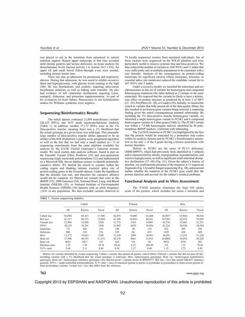
92
Copyright 2013 by ESPGHAN and NASPGHAN. Unauthorized reproduction of this article is prohibited.
was placed to aid in the transition from parenteral to enteralnutrition support. Repeat upper endoscopy at that time revealedmild chronic gastritis and lactase deficiency on tissue analysis fordisaccharidase levels (lactase activity 1.4, normal 24.5! 8.0). Anupper GI and small bowel follow-through x-ray were normal,including normal transit time.
There was also an admission for pneumonia and respiratorydistress. During that admission, he was noted to exhibit excessivethirst and hyperglycemia, with glucose levels running in the high100s. He was hypokalemic and acidotic, requiring intravenousbicarbonate infusions as well as baking soda enterally. He alsohad evidence of left ventricular dysfunction requiring Lasix,enalapril, Aldactone, and potassium supplementation. As part ofhis evaluation for heart failure, fluorescence in situ hybridizationstudies for Williams syndrome were negative.
Sequencing/Bioinformatics Results
The initial dataset contained 21,804 nonreference variants(20,129 SNVs) and 1675 small insertions/deletions [indels])(Table 1). In addition, another 17,172 SNVs were in the 1%false-positive tranche, meaning there was a 1% likelihood thatthe actual genotype at a given locus was wild-type. This unusuallylarge number of false-positive tranche alleles appeared to be anartifact of the SOLiD platform’s quality score assignment algorithmas well as the fact that we had only a small number (n¼ 7) ofsequencing experiments from the same platform available foranalysis by the GATK Unified Genotyper’s Gaussian mixturemodel. We used custom data analysis software, based in part onthe Ensembl Variant Effect Predictor (20) and next-generationsequencing-single nucleotide polymorphism (32) and implementedon a Microsoft SQL Server database system, to identify potentiallycausative alleles. We limited the search to variants within thecoding region and flanking intronic essential splice site ofprotein-coding genes in the Ensembl dataset. Under the hypothesisthat the disorder was rare, and therefore the causative allele(s)would not be common, we filtered out variants that were in thedbSNP (33), 1000 Genomes (34), National Heart, Lung, and BloodInstitute (NHLBI) (35), or National Institute of EnvironmentalHealth Sciences (NIEHS) (36) datasets with an allele frequency#0.01 in any population. We also excluded variants observed in
74 locally sequenced exomes from unrelated individuals. Six ofthese exomes were sequenced on the SOLiD platform and wereparticularly useful to remove systemic bias and false-positives. Wethus reduced the number of variants to 1043 SNVs and 11 indels thatwere sufficiently rare in multiple populations to be consistent with arare disorder. Analysis of the consequences on protein-codingtranscripts for significant adverse effects (nonsense, missense, oressential splice site mutations) reduced the candidate variant list to467 SNVs and 3 indels.
Under a recessive model, we searched the autosomes and sexchromosomes in this set of variants for homozygous and compoundheterozygous mutations (the latter defined as 2 variants in the sametranscript). We required that the variants be likely to have a deleter-ious effect on protein structure as predicted by at least 1 of SIFT(21–25), PolyPhen (26–28), or Condel (29)). Initially, we limited thesearch to variants that fully passed all of the data quality filters, butthis resulted in no homozygous variants being selected, a surprisingfinding given the stated consanguineous parental relationship. Byincluding the 1% false-positive tranche homozygous variants, weidentified a single homozygous variant in PCSK1 and 6 compoundheterozygous variants in 3 other genes (Table 2). The PCSK1 variantwas within a 7.5-Mb homozygous interval, identified by 49 poly-morphous dbSNP markers, consistent with inbreeding.
The Tyr343X mutation in PCSK1 was highlighted by the factthat the protein would be truncated by a premature stop codonwithin its catalytic domain and by PCSK1’s bioinformatics annota-tion as the only 1 of the 4 genes having a known association withhuman disorders.
Defects in PCSK1 are the cause of PC1/3 deficiency(MIM:600955), which had previously been identified in 3 subjectsand is characterized by obesity, hypogonadism, hypoadrenalism, andreactive hypoglycemia, as well as significant small-intestinal absorp-tive dysfunction (37–40) (Fig. 1C). Given the subject’s history ofdiarrhea, we confirmed the presence of the mutation in 71 sequencedfragments (Fig. 1A) and by Sanger sequencing (Fig. 1B), and assessedfurther whether the mutation of the PCSK1 gene could alter theprotein function and account for the subject’s medical problems.
Functional Analysis and In Vitro Assessment
The Y343X mutation eliminates the final 410 aminoacids of the protein, which includes the entire C-terminal and
TABLE 1. Exome sequencing statistics
Called Filtered Raw
All Known Novel All Known Novel All Known Novel
Called loci 10,2961 85,413 17,548 82,876 19,608 63,268 18,5837 10,5021 80,816Ref loci 81,157 66,133 15,024 61,104 16,843 44,261 14,2261 82,976 59,285Variant loci 21,804 19,280 2524 21,772 2765 19,007 43,576 22,045 21,531SNVs 20,129 18,291 1838 21,095 2639 18,456 41,224 20,930 20,294Insertions 775 465 310 148 30 118 923 495 428Deletions 900 524 376 529 96 433 1429 620 809Hets 13,172 10,863 2309 21,330 2389 18,941 34,502 13,252 21,250Hom ref 57,506 46,193 11,313 42,576 8661 33,915 10,0082 54,854 45,228Hom var 8632 8417 215 442 376 66 9074 8793 281Het/hom ratio 1.53 1.29 10.74 48.26 6.35 286.98 3.8 1.51 75.62Ti/Tv ratio 2.64 3.11 0.69 0.54 1.27 0.48 1.15 2.73 0.49
Metrics for variants identified by exome sequencing. Called¼ variants that passed all quality control filters; Filtered¼ variants that did not pass all QC,including variants with a 1% likelihood that the actual genotype is wild-type; Hets¼ heterozygous genotypes; Hom var¼ homozygous nonreferencegenotypes; Hom ref¼ homozygous reference genotypes (wt); Known/novel¼ variants in/not in dbSNP137; Ref loci¼ loci that match GRCh37 referencegenome; SNVs¼ single nucleotide polymorphisms; Ti/Tv ratio¼ ratio of transition (purine to purine or pyrimidine to pyrimidine) to transversion (purine to/from pyrimidine) variants; Variant loci¼ loci that differ from the reference.
Yourshaw et al JPGN $ Volume 57, Number 6, December 2013
762 www.jpgn.org
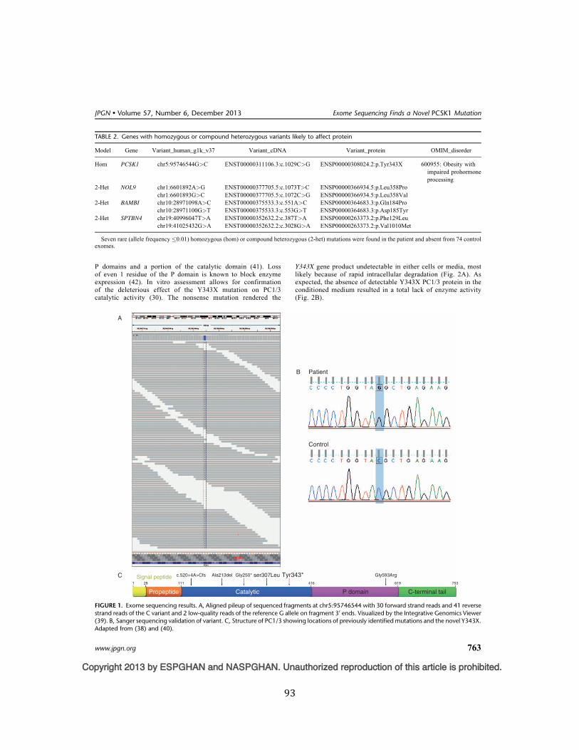
93
Copyright 2013 by ESPGHAN and NASPGHAN. Unauthorized reproduction of this article is prohibited.
P domains and a portion of the catalytic domain (41). Lossof even 1 residue of the P domain is known to block enzymeexpression (42). In vitro assessment allows for confirmationof the deleterious effect of the Y343X mutation on PC1/3catalytic activity (30). The nonsense mutation rendered the
Y343X gene product undetectable in either cells or media, mostlikely because of rapid intracellular degradation (Fig. 2A). Asexpected, the absence of detectable Y343X PC1/3 protein in theconditioned medium resulted in a total lack of enzyme activity(Fig. 2B).
TABLE 2. Genes with homozygous or compound heterozygous variants likely to affect protein
Model Gene Variant_human_g1k_v37 Variant_cDNA Variant_protein OMIM_disorder
Hom PCSK1 chr5:95746544G>C ENST00000311106.3:c.1029C>G ENSP00000308024.2:p.Tyr343X 600955: Obesity withimpaired prohormoneprocessing
2-Het NOL9 chr1:6601892A>G ENST00000377705.5:c.1073T>C ENSP00000366934.5:p.Leu358Prochr1:6601893G>C ENST00000377705.5:c.1072C>G ENSP00000366934.5:p.Leu358Val
2-Het BAMBI chr10:28971098A>C ENST00000375533.3:c.551A>C ENSP00000364683.3:p.Gln184Prochr10:28971100G>T ENST00000375533.3:c.553G>T ENSP00000364683.3:p.Asp185Tyr
2-Het SPTBN4 chr19:40996047T>A ENST00000352632.2:c.387T>A ENSP00000263373.2:p.Phe129Leuchr19:41025432G>A ENST00000352632.2:c.3028G>A ENSP00000263373.2:p.Val1010Met
Seven rare (allele frequency !0.01) homozygous (hom) or compound heterozygous (2-het) mutations were found in the patient and absent from 74 controlexomes.
PatientB
A
C
P domain
1 28 111 416 619
Gly593ArgGly250*Ala213delc.520+4A>CfsSignal peptide Tyr343*ser307Leu753
CatalyticPropeptide C-terminal tail
Control
FIGURE 1. Exome sequencing results. A, Aligned pileup of sequenced fragments at chr5:95746544 with 30 forward strand reads and 41 reversestrand reads of the C variant and 2 low-quality reads of the reference G allele on fragment 30 ends. Visualized by the Integrative Genomics Viewer(39). B, Sanger sequencing validation of variant. C, Structure of PC1/3 showing locations of previously identified mutations and the novel Y343X.Adapted from (38) and (40).
JPGN " Volume 57, Number 6, December 2013 Exome Sequencing Finds a Novel PCSK1 Mutation
www.jpgn.org 763
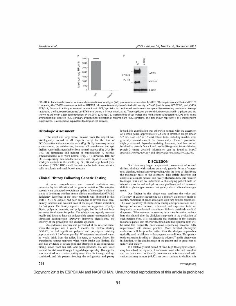
94
Copyright 2013 by ESPGHAN and NASPGHAN. Unauthorized reproduction of this article is prohibited.
Histologic Assessment
The small and large bowel mucosa from the subject washistologically normal in all respects except for the loss ofPC1/3-positive enteroendocrine cells (Fig. 3). By hematoxylin andeosin staining, the architecture, immune cell complement, and epi-thelium were indistinguishable from normal mucosa (Fig. 3A). ByIHC, the appearance and number of chromogranin A–positiveenteroendocrine cells were normal (Fig. 3B); however, IHC forPC1/3-expressing enteroendocrine cells was negative relative towild-type controls in the small (Fig. 3C, D) and large bowel (datanot shown). PC1/3 IHC should decorate a subset of enteroendocrinecells in colonic and small bowel mucosa.
Clinical History Following Genetic Testing
A more comprehensive and focused evaluation wasprompted by identification of the genetic mutation. The adoptiveparents were contacted to obtain an update of the subject’s clinicalstatus to determine whether known clinical manifestation of PC1/3deficiency described in the other probands was observed in thischild (15). The subject had been managed at several local com-munity facilities and was not seen at the major referral institutionfor >4 years. The family reported evidence suggestive of poly-dipsia, polyuria, enuresis, and polyphagia, but he had not beenevaluated for diabetes insipidus. The subject was urgently assessedlocally and found to have an undetectable serum vasopressin level.Intranasal desmopressin (DDAVP) improved significantly theseverity of the polydipsia and enuretic episodes.
An endocrine analysis was performed at the referral centerwhen the subject was 4 years, 5 months old. Before startingDDAVP, he had significant polyuria and polydipsia, drinkingapproximately 4 L of water per day. When parents restricted water,he would drink from the toilet, fish tank, or outdoor faucet. Heexperienced temper tantrums when water intake was limited. Healso had evidence of severe pica and attempted to eat intravenoustubing, wood from his crib, and paper products. He was toilettrained, but still went through 1 bag of diapers per day. His appetitewas described as excessive, eating more than his teenage siblingscombined, and his parents keeping the refrigerator and pantry
locked. His examination was otherwise normal, with the exceptionof a small penis: approximately 2.8 cm in stretched length (mean5.7 cm, Z of !2.5 is 3.5 cm). Blood tests, including insulin, weregenerally normal except for dramatically elevated proinsulin,slightly elevated thyroid-stimulating hormone, and low seruminsulin-like growth factor 1 and insulin-like growth factor–bindingprotein-3 (more detailed information can be found at http://links.lww.com/MPG/A254 and http://links.lww.com/MPG/A255).
DISCUSSIONOur laboratory began a systematic assessment of several
distinct kindreds with various putatively genetic forms of conge-nital diarrhea, using exome sequencing, with the hope of identifyingthe molecular basis of the disorders. This article describes ouranalysis of a single patient, and nicely illustrates how this researchtechnique was used to understand a challenging patient with aninherited disorder and multiple medical problems, and led to a moredefinitive phenotypic workup that greatly altered clinical manage-ment.
Our finding in this single case confirms the value andefficiency of exome sequencing as a primary diagnostic mode toidentify mutations of genes associated with rare clinical conditions.This case pointedly illustrates how multiple hospitalizations and abarrage of various indirect, redundant, and expensive tests arefrequently required—and sometimes fail—to establish medicaldiagnoses. Whole-exome sequencing is a transformative techno-logy that should alter the clinician’s approach to the evaluation ofsuch patients (43). It is conceivable that portions of the standardmetabolic panels and other urine, blood, and radiographic tests willbe used less frequently once exome sequencing becomes fullyimplemented into clinical practice. More directed phenotypicevaluation will be possible rather than the shotgun approachestypically used in children with rare genetic conditions. This pheno-typic evaluation is called a ‘‘diagnostic odyssey’’ and is often yearsin duration, to the disadvantage of the patient and at great cost tofamily and society.
In a relatively short period of time, high-throughput sequen-cing has solved the mystery of numerous novel inherited disordersand has been used to identify common variants associated withvarious primary tumors (44,45). As costs continue to decline, this
3500A
3000
2500
2000
Max
imum
rat
e R
FU
/min
1500
1000
500
0
WT
Y343X
B
87 kDa
PC1/3
Lysate
WT
Y34
3X
Medium
b-actin
FIGURE 2. Functional characterization and visualization of wild-type (WT) prohormone convertase 1/3 (PC1/3) complementary DNA and PC1/3containing the Y343X nonsense mutation. HEK293 cells were transiently transfected with empty pcDNA3 (not shown), WT PC1/3, and Y343XPC1/3. A, Enzymatic activity of secreted recombinant PC1/3 proteins in conditioned medium was compared by measuring maximum cleavagerates using the fluorogenic substrate pyr-RTKR-amc during a 1-hour kinetic assay. Three replicates per condition were assayed in triplicate and areshown as the mean" standard deviation, P<0.0017 (2-tailed). B, Western blot of cell lysates and media from transfected HEK293 cells, usingamino terminal–directed PC1/3 primary antiserum for detection of recombinant PC1/3 proteins. The data shown represent 1 of 3 independentexperiments. b-actin shows equivalent loading of cell extracts.
Yourshaw et al JPGN # Volume 57, Number 6, December 2013
764 www.jpgn.org
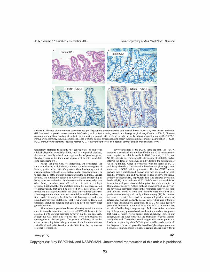
95
Copyright 2013 by ESPGHAN and NASPGHAN. Unauthorized reproduction of this article is prohibited.
technology promises to identify the genetic basis of numerousclinical diagnoses, especially those, such as congenital diarrhea,that can be causally related to a large number of possible genes,thereby bypassing the traditional approach of targeted candidategene sequencing (46).
Given the possibility of inbreeding, we considered theapproach of using a high-density microarray to locate regions ofhomozygosity in the patient’s genome, then developing a set ofcustom-capture probes to select that region for deep sequencing, orto sequence all of the exons in the region with the traditional Sangermethod. We ultimately decided on whole-exome sequencing asbeing more cost-effective. Furthermore, without knowledge thatother family members were affected, we did not have a highprevious likelihood that the mutation would be in a large regionof homozygosity that could be detected by a microarray. Eventhough we may hypothesize that this child’s disease was caused bya homozygous mutation, there was essentially no additional cost ortime needed to analyze the data for both homozygous and com-pound heterozygous mutations. Finally, we wished to develop anunbiased analytical pipeline that could be used for many othergenetic patterns.
Others have reported on the use of next-generation sequen-cing to identify mutations in a gene (SLC26A3) known to beassociated with chronic diarrhea; however, unlike our approach,sequencing was limited to regions that were homozygous byconsanguineous descent (46a). The general efficiency of whole-exome sequencing favors the generation of whole-exome data onvirtually all such patients as the most efficient and thorough meansof genetic evaluation.
Severe mutations of the PCSK1 gene are rare. The Y343Xmutation is novel and was not identified in the 7252 chromosomesthat comprise the publicly available 1000 Genomes, NHLBI, andNIEHS datasets, suggesting an allele frequency of<0.00014 and aninferred incidence of homozygous individuals in the population of<1 in 52 million, which is consistent with the rarity of PC1/3deficiency disorders. This mutation broadens the phenotypic con-sequences of PC1/3 deficiency disorders. The first PCSK1-mutantproband was a middle-aged woman who was evaluated for post-prandial hypoglycemia and was found to have obesity, hypogona-dotropic hypogonadism, hypoadrenalism, and elevated proinsulinlevels (47,48). A second case of PC1/3 deficiency was establishedin an infant with generalized malabsorptive diarrhea who expired at18 months of age (37). A third proband was described as a 6-year-old boy with a diarrheal condition that resembled the previous case,and intestinal biopsies from both children were described as apersistent enteropathy with patchy villous atrophy (38). In contrast,the subject reported here had no histopathological evidence ofenteropathy and had perfectly normal crypt-villus axis without apathologic inflammatory component (Fig. 3). We have recentlypresented findings on additional cases with PCSK1 mutations thatwe identified by Sanger sequencing (15). Retrospective question-ing of the primary proband confirmed similar diarrheal symptomsthat were certainly worse during early childhood (37). In ourpatient, as in the other 3 patients, the proinsulin level was signifi-cantly elevated. These data would suggest that serum proinsulinlevels and sequencing of the PCSK1 gene could be used to establishthe diagnosis; however, given the breadth of phenotypic presenta-tions, molecular diagnosis is likely to remain challenging without
APatient H&E
Patient PCSK1 Control PCSK1
Patient CgAB
C D
FIGURE 3. Absence of prohormone convertase 1/3 (PC1/3)-positive enteroendocrine cells in small bowel mucosa. A, Hematoxylin and eosin(H&E)–stained proprotein convertase subtilisin/kexin type 1 mutant showing normal morphology; original magnification !200. B, Chromo-granin A immunohistochemistry of mutant tissue showing a normal pattern of enteroendocrine cells; original magnification !400. C, PC1/3immunohistochemistry showing complete absence of PC1/3-positive enteroendocrine cells in the mutant tissue; original magnification!400. D,PC1/3 immunohistochemistry showing normal PC1/3 enteroendocrine cells in a healthy control; original magnification !400.
JPGN " Volume 57, Number 6, December 2013 Exome Sequencing Finds a Novel PCSK1 Mutation
www.jpgn.org 765
96
Copyright 2013 by ESPGHAN and NASPGHAN. Unauthorized reproduction of this article is prohibited.
implementation of broader approaches such as whole-exomesequencing.
To develop a sense of the mutational load of rare variants onPCSK1 in the whole population, we examined all 913 PCSK1variants reported in dbSNP and the 1000 Genomes, NHLBI, andNIEHS datasets, and found 47 variants causing nonsynonymouscodons or more serious consequences and having minor allelefrequencies !0.01 (supplementary Table S1, http://links.lww.-com/MPG/A255). Interestingly, common variants in the codingregion of PCSK1 are also associated with common forms of obesity(N221D, Q665E, S690T) and type 2 diabetes mellitus (Q665E,S690T) (49,50). A recent study suggests that PC1/3 deficiency isdependent on the dosage of PCSK1, and rare heterozygous mutationscan cause obesity (51). From the population data, rare protein-alteringmutations will be homozygous or compound heterozygous, resultingin substantial loss of PC1/3 activity in "86 individuals per million,and these individuals would be predicted to be at risk for a life-threatening PC1/3 deficiency. Additionally, an estimated 1 in 20individuals may harbor a modest PC1/3 deficiency, which maycontribute to PC1/3 deficiency–related obesity.
Exome sequencing will typically generate upwards of 20,000candidate variants from the reference genome in a given individual.A challenge for diagnosis by exome sequencing is to filter outvariants that cannot possibly cause the disease in question. Alleles,such as synonymous or intronic variants, can be eliminated withslight risk that they are false-negatives, as can alleles that are toofrequent in the population to be consistent with the incidence of thedisorder. Beyond that, prediction of the functional consequences ofnovel mutations remains a daunting task.
Enteric anendocrinosis is another inherited intestinal endo-crinopathy that has clinical features that resemble the early stages ofPC1/3 deficiency, including a generalized form of malabsorption(MIM:610370) (5). Homozygous mutations of NEUROG3 weredescribed in 3 probands, the intestines of which were devoid ofenteroendocrine cells, yet had an otherwise normal-appearingintestine. NEUROG3 is a basic helix-loop-helix transcriptionalfactor that is necessary and sufficient to drive endocrine celldevelopment in the pancreas and intestine (5). Although 2 of the3 cases in the initial report did not develop insulin-dependentdiabetes mellitus until preadolescent age, 2 recent cases describethe onset of diabetes during the neonatal period (52). Unlikepatients with PC1/3 deficiency, children with enteric anendocri-nosis do not appear to develop hypothalamic, pituitary, adrenal,thyroid, or gonadal insufficiencies.
Establishing the precise diagnosis of a congenital diarrhealcondition requires an intestinal biopsy and a thoughtful approachto dietary challenges. A differential diagnosis of this condition,when presenting with seemingly histologically normal intestinalmucosa, is mostly limited to specific defects of nutrient assim-ilation (digestive enzymes or transport proteins) or enteroendo-crinopathies. Enteroendocrinopathies are histologically subtle,are generally only discovered when specifically sought, andrequire immunohistochemical confirmation (31). Exome sequen-cing will certainly be used in the coming years to identify theinherited basis of novel diarrheal disorders and will likely bethe standard of practice for genotype testing of establisheddisorders. In our case, exome sequencing provided a diagnosisthat resulted in immediate changes in patient care and an improvedability to predict clinical progression, based on previous cases ofPC1/3 deficiency.
REFERENCES1. Martin MG, Wright EM. Congenital intestinal transport defects. In:
Walker WA, Goulet O, Kliegman RM, eds. Pediatric GastrointestinalDisease. 4th ed. Hamilton, Canada: BC Decker; 2004:898–921.
2. Fishbein TM. Intestinal transplantation. N Engl J Med 2009;361:998–1008.
3. Binder HJ. Causes of chronic diarrhea. N Engl J Med 2006;355:236–9.4. Spence JR, Mayhew CN, Rankin SA, et al. Directed differentiation of
human pluripotent stem cells into intestinal tissue in vitro. Nature2011;470:105–9.
5. Wang J, Cortina G, Wu SV, et al. Mutant neurogenin-3 in congenitalmalabsorptive diarrhea. N Engl J Med 2006;355:270–80.
6. Martin MG, Turk E, Lostao MP, et al. Defects in Naþ glucosecotransporter (SGLT1) trafficking and function cause glucose-galactosemalabsorption. Nat Genet 1996;12:216–20.
7. Sivagnanam M, Mueller JL, Lee H, et al. Identification of EpCAM as thegene for congenital tufting enteropathy. Gastroenterology 2008;135:429–37.
8. Muller T, Hess MW, Schiefermeier N, et al. MYO5B mutations causemicrovillus inclusion disease and disrupt epithelial cell polarity. NatGenet 2008;40:1163–5.
9. Hartley JL, Zachos NC, Dawood B, et al. Mutations in TTC37 causetrichohepatoenteric syndrome (phenotypic diarrhea of infancy). Gastro-enterology 2010;138:2388–982398 e1–2.
10. Heinz-Erian P, Muller T, Krabichler B, et al. Mutations in SPINT2 causea syndromic form of congenital sodium diarrhea. Am J Hum Genet2009;84:188–96.
11. Blaydon DC, Biancheri P, Di WL, et al. Inflammatory skin and boweldisease linked to ADAM17 deletion. N Engl J Med 2011;365:1502–8.
12. Shendure J. Next-generation human genetics. Genome Biol 2011;12:408.
13. Singleton AB. Exome sequencing: a transformative technology. LancetNeurol 2011;10:942–6.
14. Hoshino A, Lindberg I. Peptide biosynthesis: prohormone convertases1/3 and 2. In: Fricker LD, Devi L, eds. Colloquium Series onNeuropeptides. 1st ed. San Rafael, CA: Morgan & Claypool LifeSciences Publishers; 2004.
15. Martin MG, Lindberg I, Solorzano-Vargas RS, et al. Congenital propro-tein convertase 1/3 deficiency causes malabsorptive diarrhea and otherendocrinopathies in a pediatric cohort. Gastroenterology 2013;145: 138–48.
16. Pruitt KD, Harrow J, Harte RA, et al. The consensus coding sequence(CCDS) project: Identifying a common protein-coding gene set for thehuman and mouse genomes. Genome Res 2009;19:1316–23.
17. International Human Genome Sequencing Consortium. Finishing theeuchromatic sequence of the human genome. Nature 2004;431:931–45.
18. Flicek P, Amode MR, Barrell D, et al. Ensembl 2011. Nucleic Acids Res2011;39:D800–806.
19. DePristo MA, Banks E, Poplin R, et al. A framework for variationdiscovery and genotyping using next-generation DNA sequencing data.Nat Genet 2011;43:491–8.
20. McKenna A, Hanna M, Banks E, et al. The Genome Analysis Toolkit: aMapReduce framework for analyzing next-generation DNA sequencingdata. Genome Res 2010;20:1297–303.
21. Kumar P, Henikoff S, Ng PC. Predicting the effects of coding non-synonymous variants on protein function using the SIFT algorithm. NatProtoc 2009;4:1073–81.
22. Ng PC, Henikoff S. Predicting deleterious amino acid substitutions.Genome Res 2001;11:863–74.
23. Ng PC, Henikoff S. Accounting for human polymorphisms predicted toaffect protein function. Genome Res 2002;12:436–46.
24. Ng PC, Henikoff S. SIFT: predicting amino acid changes that affectprotein function. Nucleic Acids Res 2003;31:3812–4.
25. Ng PC, Henikoff S. Predicting the effects of amino acid substitutions onprotein function. Annu Rev Genomics Hum Genet 2006;7:61–80.
26. Ramensky V, Bork P, Sunyaev S. Human non-synonymous SNPs: serverand survey. Nucleic Acids Res 2002;30:3894–900.
27. Sunyaev S, Ramensky V, Bork P. Towards a structural basis of humannon-synonymous single nucleotide polymorphisms. Trends Genet2000;16:198–200.
28. Sunyaev S, Ramensky V, Koch I, et al. Prediction of deleterious humanalleles. Hum Mol Genet 2001;10:591–7.
29. Gonzalez-Perez A, Lopez-Bigas N. Improving the assessment of theoutcome of nonsynonymous SNVs with a consensus deleteriousnessscore, Condel. Am J Hum Genet 2011;88:440–9.
Yourshaw et al JPGN $ Volume 57, Number 6, December 2013
766 www.jpgn.org
97
Copyright 2013 by ESPGHAN and NASPGHAN. Unauthorized reproduction of this article is prohibited.
30. Vindrola O, Lindberg I. Biosynthesis of the prohormone convertasemPC1 in AtT-20 cells. Mol Endocrinol 1992;6:1088–94.
31. Cortina G, Smart CN, Farmer DG, et al. Enteroendocrine cell dysgenesisand malabsorption, a histopathologic and immunohistochemical char-acterization. Hum Pathol 2007;38:570–80.
32. Grant JR, Arantes AS, Liao X, et al. In-depth annotation of SNPs arisingfrom resequencing projects using NGS-SNP. Bioinformatics 2011;27:2300–1.
33. National Center for Biotechnology Information NLoM. Database ofSingle Nucleotide Polymorphisms (dbSNP Build ID: 134). VoldbSNP134. Bethesda, MD: National Center for Biotechnology Infor-mation, National Library of Medicine.
34. The 1000 Genomes Project ConsortiumA map of human genomevariation from population-scale sequencing. Nature 2010;467:1061–73.
35. NHLBI Exome Sequencing Project. Exome Variant Server. VolumeSeptember. Seattle, WA: NHLBI Exome Sequencing Project; 2011.
36. NIEHS Environmental Genome Project. NIEHS Exome Variant Server.Volume September. Seattle, WA: NIEHS Environmental Genome Pro-ject; 2011.
37. Jackson RS. Small-intestinal dysfunction accompanies the complexendocrinopathy of human proprotein convertase 1 deficiency. J ClinInvest 2003;112:1550–60.
38. Farooqi IS, Volders K, Stanhope R, et al. Hyperphagia and early-onsetobesity due to a novel homozygous missense mutation in prohormoneconvertase 1/3. J Clin Endocrinol Metab 2007;92:3369–73.
39. Robinson JT, Thorvaldsdottir H, Winckler W, et al. Integrative genomicsviewer. Nat Biotechnol 2011;29:24–6.
40. Steiner DF. The proprotein convertases. Curr Opin Chem Biol1998;2:31–9.
41. Ueda K, Lipkind GM, Zhou A, et al. Mutational analysis of predictedinteractions between the catalytic and P domains of prohormone con-vertase 3 (PC3/PC1). Proc Natl Acad Sci U S A 2003;100:5622–7.
42. Zhou A, Martin S, Lipkind G, et al. Regulatory roles of the P domain ofthe subtilisin-like prohormone convertases. J Biol Chem 1998;273:11107–14.
43. Maxmen A. Exome sequencing deciphers rare diseases. Cell 2011;144:635–7.
44. Ng SB, Buckingham KJ, Lee C, et al. Exome sequencing identifies thecause of a mendelian disorder. Nat Genet 2010;42:30–5.
45. Stransky N, Egloff AM, Tward AD, et al. The mutational landscape ofhead and neck squamous cell carcinoma. Science 2011;333:1157–60.
46. Lifton RP. Individual genomes on the horizon. N Engl J Med2010;362:1235–6.
46a. Choi M, Scholl UI, Ji W, et al. Genetic diagnosis by whole exomecapture and massively parallel DNA sequencing. Proc Natl Acad SciU S A 2009;106:19096–101.
47. Jackson RS, Creemers JW, Ohagi S, et al. Obesity and impairedprohormone processing associated with mutations in the human pro-hormone convertase 1 gene. Nat Genet 1997;16:303–6.
48. O’Rahilly S, Gray H, Humphreys PJ, et al. Brief report: impairedprocessing of prohormones associated with abnormalities of glucosehomeostasis and adrenal function. N Engl J Med 1995;333:1386–90.
49. Benzinou M, Creemers JW, Choquet H, et al. Common nonsynonymousvariants in PCSK1 confer risk of obesity. Nat Genet 2008;40:943–5.
50. Strawbridge RJ, Dupuis J, Prokopenko I, et al. Genome-wide associa-tion identifies nine common variants associated with fasting proinsulinlevels and provides new insights into the pathophysiology of type 2diabetes. Diabetes 2011;60:2624–34.
51. Creemers JWM, Choquet H, Stijnen P, et al. Heterozygous mutationscausing partial prohormone convertase 1 deficiency contribute to humanobesity. Diabetes 2012;61:383–90.
52. Gradwohl G, Dierich A, LeMeur M, et al. Neurogenin3 is required forthe development of the four endocrine cell lineages of the pancreas.Proc Natl Acad Sci U S A 2000;97:1607–11.
JPGN ! Volume 57, Number 6, December 2013 Exome Sequencing Finds a Novel PCSK1 Mutation
www.jpgn.org 767
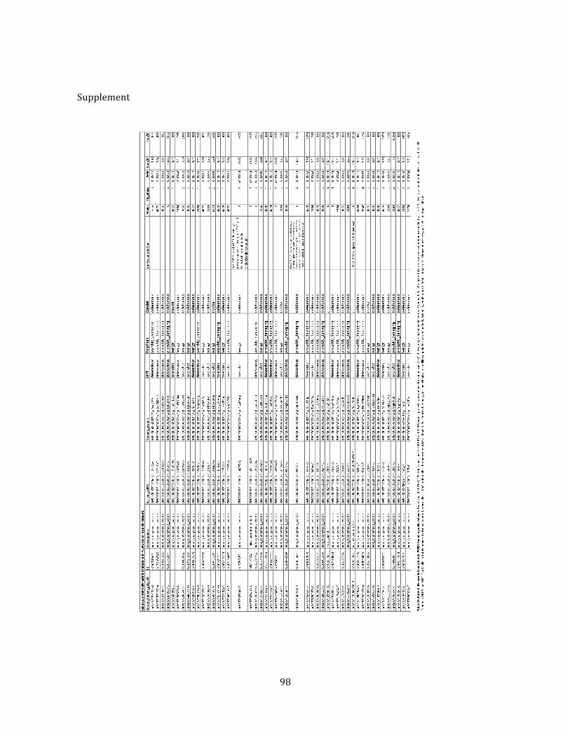
98
Supplement

99
CHAPTER FIVE
Functional consequences of a novel variant of PCSK1
100
Functional Consequences of a Novel Variant of PCSK1Lindsay A. Pickett3, Michael Yourshaw1, Valeria Albornoz3, Zijun Chen2, R. Sergio Solorzano-Vargas2,
Stanley F. Nelson1,4, Martın G. Martın2, Iris Lindberg3*
1 Department of Human Genetics, David Geffen School of Medicine, University of California Los Angeles, Los Angeles, California, United States of America, 2 Division of
Gastroenterology and Nutrition, Department of Pediatrics, Mattel Children’s Hospital and David Geffen School of Medicine, University of California Los Angeles, Los
Angeles, California, United States of America, 3 Department of Anatomy and Neurobiology, University of Maryland-Baltimore, Baltimore, Maryland, United States of
America, 4 Department of Pathology and Laboratory Medicine, David Geffen School of Medicine, University of California Los Angeles, Los Angeles California, United States
of America
Abstract
Background: Common single nucleotide polymorphisms (SNPs) in proprotein convertase subtilisin/kexin type 1 withmodest effects on PC1/3 in vitro have been associated with obesity in five genome-wide association studies and withdiabetes in one genome-wide association study. We here present a novel SNP and compare its biosynthesis, secretion andcatalytic activity to wild-type enzyme and to SNPs that have been linked to obesity.
Methodology/Principal Findings: A novel PC1/3 variant introducing an Arg to Gln amino acid substitution at residue 80(within the secondary cleavage site of the prodomain) (rs1799904) was studied. This novel variant was selected for analysisfrom the 1000 Genomes sequencing project based on its predicted deleterious effect on enzyme function and itscomparatively more frequent allele frequency. The actual existence of the R80Q (rs1799904) variant was verified by Sangersequencing. The effects of this novel variant on the biosynthesis, secretion, and catalytic activity were determined; thepreviously-described obesity risk SNPs N221D (rs6232), Q665E/S690T (rs6234/rs6235), and the Q665E and S690T SNPs(analyzed separately) were included for comparative purposes. The novel R80Q (rs1799904) variant described in this studyresulted in significantly detrimental effects on both the maturation and in vitro catalytic activity of PC1/3.
Conclusion/Significance: Our findings that this novel R80Q (rs1799904) variant both exhibits adverse effects on PC1/3activity and is prevalent in the population suggests that further biochemical and genetic analysis to assess its contributionto the risk of metabolic disease within the general population is warranted.
Citation: Pickett LA, Yourshaw M, Albornoz V, Chen Z, Solorzano-Vargas RS, et al. (2013) Functional Consequences of a Novel Variant of PCSK1. PLoS ONE 8(1):e55065. doi:10.1371/journal.pone.0055065
Editor: Ludmila Prokunina-Olsson, National Cancer Institute, National Institutes of Health, United States of America
Received May 29, 2012; Accepted December 22, 2012; Published January 28, 2013
Copyright: ! 2013 Pickett et al. This is an open-access article distributed under the terms of the Creative Commons Attribution License, which permitsunrestricted use, distribution, and reproduction in any medium, provided the original author and source are credited.
Funding: This work was supported by grants from the National Institute of Diabetes and Digestive and Kidney Diseases (#DK083762) and the California Instituteof Regenerative Medicine (CIRM), RT2-01985 to MGM; and a grant from the National Institute of Drug Abuse (#DA05084) to IL. The funders had no role in studydesign, data collection and analysis, decision to publish, or preparation of the manuscript.
Competing Interests: The authors have declared that no competing interests exist.
* E-mail: [email protected]
Introduction
Prohormone convertase 1/3 is a calcium-dependent serineendoprotease essential for the conversion of a variety ofprohormones and neuropeptide precursors to their bioactiveforms. Human prohormone convertase 1/3 (PC1/3) is encoded bythe gene PCSK1, which is located on chromosome 5 and iscomprised of 14 exons [1]. PC1/3 is expressed in a subset ofendocrine and neuroendocrine tissues, cells equipped with aregulated secretory pathway. During transit through the secretorypathway, PC1/3 is first synthesized in the endoplasmic reticulum(ER) as an inactive 94 kDa zymogen composed of an N-terminalsignal peptide, a prodomain which serves as an intramolecularchaperone and inhibitor; a catalytic domain which accomplishessubstrate hydrolysis; a P (homo B) domain which contributes toenzymatic properties; and a carboxyl-terminal (CT) domainwhich, when removed by partial or complete in trans proteolyticprocessing, results in a much more active, but also less stable,enzymatic form (reviewed in [2] (Figure 1). PC1/3 is abundantlyexpressed in the arcuate and paraventricular nuclei of the
hypothalamus [3,4], tissues that are known to mediate satietyand hunger signals [5]. Substrates of PC1/3, such as proinsulin,proglucagon, proghrelin, agouti-related protein, pro-neuropeptideY, provasopressin and proopiomelanocortin are responsible for theregulation of absorption, metabolism and acquisition (appetite) ofnutrients [6,7,8,9,10,11,12,13,14].
Deficiencies in PC1/3 frequently lead to imbalances inprohormone processing that result in an array of metabolicphenotypes, previously investigated both in mouse models and inhumans. Three human subjects have been described with anautosomal recessive disorder (MIM:600955) associated with severemutations of PC1/3 resulting in early-onset obesity, hyperphagia,hypoadrenalism, reactive hypoglycemia, malabsorptive diarrhea,and hypogonadism [15,16,17]. Interestingly, the PC1/3 nullmouse model, unlike the PC1/3-deficient human, is not obese.Although of normal weight at birth, PC1/3 null mice have a highpost-natal mortality rate, and those that do survive have asignificant reduction in body mass as compared to wild-typeanimals by the age of 6 weeks. The stunted growth of PC1/3 nullmice is believed to be due at least in part to reduced processing of
PLOS ONE | www.plosone.org 1 January 2013 | Volume 8 | Issue 1 | e55065
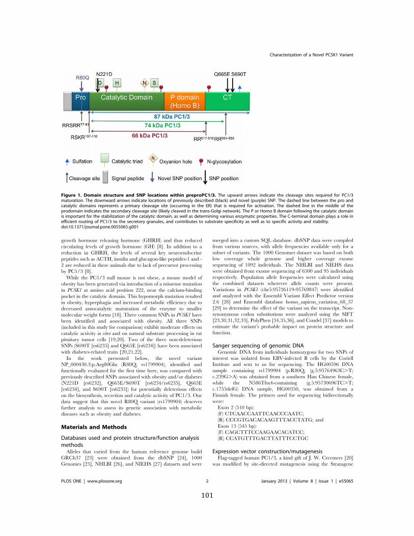
101
growth hormone releasing hormone (GHRH) and thus reducedcirculating levels of growth hormone (GH) [8]. In addition to areduction in GHRH, the levels of several key neuroendocrinepeptides such as ACTH, insulin and glucagon-like peptides-1 and -2 are reduced in these animals due to lack of precursor processingby PC1/3 [8].
While the PC1/3 null mouse is not obese, a mouse model ofobesity has been generated via introduction of a missense mutationin PCSK1 at amino acid position 222, near the calcium-bindingpocket in the catalytic domain. This hypomorph mutation resultedin obesity, hyperphagia and increased metabolic efficiency due todecreased autocatalytic maturation of the enzyme to smallermolecular weight forms [18]. Three common SNPs in PCSK1 havebeen identified and associated with obesity. All three SNPs(included in this study for comparison) exhibit moderate effects oncatalytic activity in vitro and on natural substrate processing in ratpituitary tumor cells [19,20]. Two of the three non-deleteriousSNPs (S690T [rs6235] and Q665E [rs6234]) have been associatedwith diabetes-related traits [20,21,22].
In the work presented below, the novel variantNP_000430.3:p.Arg80Gln (R80Q; rs1799904), identified andfunctionally evaluated for the first time here, was compared withpreviously described SNPs associated with obesity and/or diabetes(N221D [rs6232], Q665E/S690T [rs6234/rs6235], Q665E[rs6234], and S690T [rs6235]) for potentially deleterious effectson the biosynthesis, secretion and catalytic activity of PC1/3. Ourdata suggest that this novel R80Q variant (rs1799904) deservesfurther analysis to assess its genetic association with metabolicdiseases such as obesity and diabetes.
Materials and Methods
Databases used and protein structure/function analysismethods
Alleles that varied from the human reference genome buildGRCh37 [23] were obtained from the dbSNP [24], 1000Genomes [25], NHLBI [26], and NIEHS [27] datasets and were
merged into a custom SQL database. dbSNP data were compiledfrom various sources, with allele frequencies available only for asubset of variants. The 1000 Genomes dataset was based on bothlow coverage whole genome and higher coverage exomesequencing of 1092 individuals. The NHLBI and NIEHS datawere obtained from exome sequencing of 6500 and 95 individualsrespectively. Population allele frequencies were calculated usingthe combined datasets wherever allele counts were present.Variations in PCSK1 (chr5:95726119-95769847) were identifiedand analyzed with the Ensembl Variant Effect Predictor version2.6 [28] and Ensembl database homo_sapiens_variation_68_37[29] to determine the effect of the variant on the transcript. Non-synonymous codon substitutions were analyzed using the SIFT[23,30,31,32,33], PolyPhen [34,35,36], and Condel [37] models toestimate the variant’s probable impact on protein structure andfunction.
Sanger sequencing of genomic DNAGenomic DNA from individuals homozygous for two SNPs of
interest was isolated from EBV-infected B cells by the CoriellInstitute and sent to us for sequencing. The HG00596 DNAsample containing rs1799904 (p.R80Q; (g.5:95764963C.T;c.239G.A) was obtained from a southern Han Chinese female,while the N586Tfsx4-containing (g.5:95730696TC.T;c.1755delG) DNA sample, HG00350, was obtained from aFinnish female. The primers used for sequencing bidirectionallywere:
Exon 2 (510 bp):(F) CTCAACCAATTCAACCCAATC;(R) CCCGTGACACAAGTTTACCTATG; andExon 13 (545 bp):(F) CAGCTTTCCAAGAACACATCC;(R) CCATGTTTGACTTATTTCCTGC
Expression vector construction/mutagenesisFlag-tagged human PC1/3, a kind gift of J. W. Creemers [20]
was modified by site-directed mutagenesis using the Stratagene
Figure 1. Domain structure and SNP locations within preproPC1/3. The upward arrows indicate the cleavage sites required for PC1/3maturation. The downward arrows indicate locations of previously described (black) and novel (purple) SNP. The dashed line between the pro andcatalytic domains represents a primary cleavage site (occurring in the ER) that is required for activation. The dashed line in the middle of theprodomain indicates the secondary cleavage site (likely cleaved in the trans-Golgi network). The P or Homo B domain following the catalytic domainis important for the stabilization of the catalytic domain, as well as determining various enzymatic properties. The C-terminal domain plays a role inefficient routing of PC1/3 to the secretory granules, and contributes to substrate specificity as well as to specific activity and stability.doi:10.1371/journal.pone.0055065.g001
Characterization of a Novel PCSK1 Variant
PLOS ONE | www.plosone.org 2 January 2013 | Volume 8 | Issue 1 | e55065
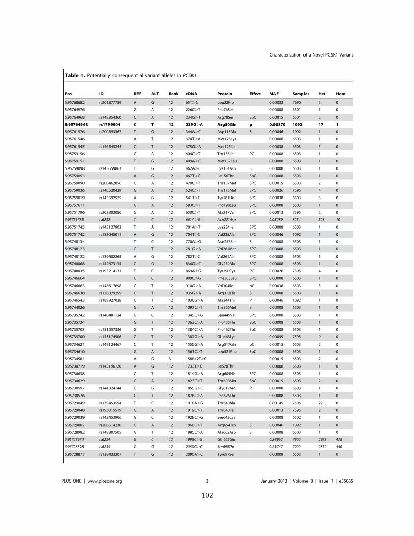
102
Table 1. Potentially consequential variant alleles in PCSK1.
Pos ID REF ALT Rank cDNA Protein Effect MAF Samples Het Hom
5:95768682 rs201377789 A G 12 65T.C Leu22Pro 0.00033 7690 5 0
5:95764976 G A 12 226C.T Pro76Ser 0.00008 6501 1 0
5:95764968 rs148354360 C A 12 234G.T Arg78Ser SpC 0.00015 6501 2 0
5:95764963 rs1799904 C T 12 239G.A Arg80Gln p 0.00870 1092 17 1
5:95761576 rs200893367 T G 12 344A.C Asp115Ala S 0.00046 1092 1 0
5:95761546 A T 12 374T.A Met125Lys 0.00008 6503 1 0
5:95761545 rs146545244 C T 12 375G.A Met125Ile 0.00038 6503 5 0
5:95759156 G A 12 404C.T Thr135Ile PC 0.00008 6503 1 0
5:95759151 T G 12 409A.C Met137Leu 0.00008 6503 1 0
5:95759098 rs145659863 T G 12 462A.C Lys154Asn S 0.00008 6503 1 0
5:95759093 A G 12 467T.C Ile156Thr SpC 0.00008 6503 1 0
5:95759090 rs200462856 G A 12 470C.T Thr157Met SPC 0.00015 6503 2 0
5:95759036 rs140520429 G A 12 524C.T Thr175Met SPC 0.00026 7595 4 0
5:95759019 rs145592525 A G 12 541T.C Tyr181His SPC 0.00038 6503 5 0
5:95757611 G A 12 593C.T Pro198Leu SPC 0.00008 6503 1 0
5:95751796 rs202203086 G A 12 650C.T Ala217Val SPC 0.00013 7595 2 0
5:95751785 rs6232 T C 12 661A.G Asn221Asp 0.03289 8254 523 10
5:95751745 rs145127903 T A 12 701A.T Lys234Ile SPC 0.00008 6503 1 0
5:95751742 rs183045011 A G 12 704T.C Val235Ala SPC 0.00046 1092 1 0
5:95748134 T C 12 770A.G Asn257Ser S 0.00008 6503 1 0
5:95748123 C T 12 781G.A Val261Met SPC 0.00008 6503 1 0
5:95748122 rs139602265 A G 12 782T.C Val261Ala SPC 0.00008 6503 1 0
5:95748068 rs142673134 C G 12 836G.C Gly279Ala SPC 0.00008 6503 1 0
5:95748035 rs193214131 T C 12 869A.G Tyr290Cys PC 0.00026 7595 4 0
5:95746664 G C 12 909C.G Phe303Leu SPC 0.00008 6503 1 0
5:95746663 rs148617898 C T 12 910G.A Val304Ile pC 0.00038 6503 5 0
5:95746638 rs138879299 C T 12 935G.A Arg312His S 0.00008 6503 1 0
5:95746543 rs189927028 C T 12 1030G.A Ala344Thr P 0.00046 1092 1 0
5:95744026 G A 12 1097C.T Thr366Met S 0.00008 6503 1 0
5:95735742 rs140481124 G C 12 1345C.G Leu449Val SPC 0.00008 6503 1 0
5:95735724 G T 12 1363C.A Pro455Thr SpC 0.00008 6503 1 0
5:95735703 rs151257336 G T 12 1384C.A Pro462Thr SpC 0.00008 6503 1 0
5:95735700 rs143174906 C T 12 1387G.A Glu463Lys 0.00059 7595 9 0
5:95734621 rs149124467 C T 12 1550G.A Arg517Gln pC 0.00015 6503 2 0
5:95734610 G A 12 1561C.T Leu521Phe SpC 0.00008 6503 1 0
5:95734581 A G 3 1588+2T.C - 0.00015 6503 2 0
5:95730719 rs145196120 A G 12 1733T.C Ile578Thr 0.00008 6503 1 0
5:95730638 C T 12 1814G.A Arg605His SPC 0.00008 6503 1 0
5:95730629 G A 12 1823C.T Thr608Met SpC 0.00015 6503 2 0
5:95730597 rs144324144 C G 12 1855G.C Gly619Arg P 0.00008 6503 1 0
5:95730576 G T 12 1876C.A Pro626Thr 0.00008 6503 1 0
5:95729049 rs139453594 T C 12 1918A.G Thr640Ala 0.00145 7595 22 0
5:95729048 rs193015519 G A 12 1919C.T Thr640Ile 0.00013 7595 2 0
5:95729039 rs142453906 G C 12 1928C.G Ser643Cys 0.00008 6502 1 0
5:95729007 rs200614230 G A 12 1960C.T Arg654Trp S 0.00046 1092 1 0
5:95728982 rs148807505 G T 12 1985C.A Ala662Asp S 0.00008 6503 1 0
5:95728974 rs6234 G C 12 1993C.G Gln665Glu 0.24962 7900 2988 478
5:95728898 rs6235 C G 12 2069G.C Ser690Thr 0.23747 7900 2852 450
5:95728877 rs138433207 T G 12 2090A.C Tyr697Ser 0.00008 6503 1 0
Characterization of a Novel PCSK1 Variant
PLOS ONE | www.plosone.org 3 January 2013 | Volume 8 | Issue 1 | e55065
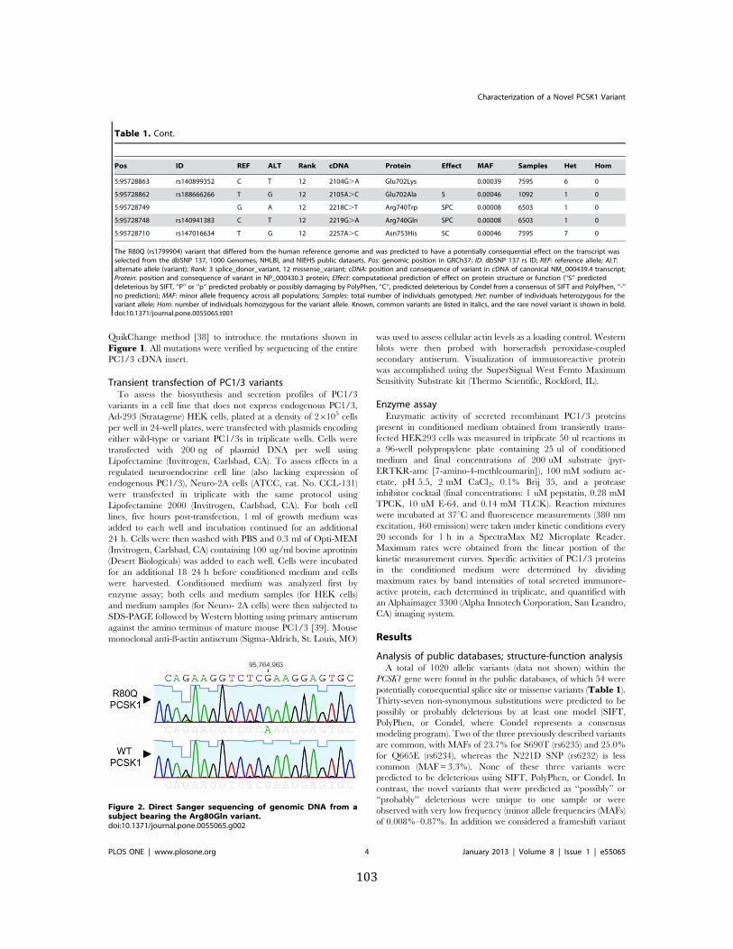
103
QuikChange method [38] to introduce the mutations shown inFigure 1. All mutations were verified by sequencing of the entirePC1/3 cDNA insert.
Transient transfection of PC1/3 variantsTo assess the biosynthesis and secretion profiles of PC1/3
variants in a cell line that does not express endogenous PC1/3,Ad-293 (Stratagene) HEK cells, plated at a density of 26105 cellsper well in 24-well plates, were transfected with plasmids encodingeither wild-type or variant PC1/3s in triplicate wells. Cells weretransfected with 200 ng of plasmid DNA per well usingLipofectamine (Invitrogen, Carlsbad, CA). To assess effects in aregulated neuroendocrine cell line (also lacking expression ofendogenous PC1/3), Neuro-2A cells (ATCC, cat. No. CCL-131)were transfected in triplicate with the same protocol usingLipofectamine 2000 (Invitrogen, Carlsbad, CA). For both celllines, five hours post-transfection, 1 ml of growth medium wasadded to each well and incubation continued for an additional24 h. Cells were then washed with PBS and 0.3 ml of Opti-MEM(Invitrogen, Carlsbad, CA) containing 100 ug/ml bovine aprotinin(Desert Biologicals) was added to each well. Cells were incubatedfor an additional 18–24 h before conditioned medium and cellswere harvested. Conditioned medium was analyzed first byenzyme assay; both cells and medium samples (for HEK cells)and medium samples (for Neuro- 2A cells) were then subjected toSDS-PAGE followed by Western blotting using primary antiserumagainst the amino terminus of mature mouse PC1/3 [39]. Mousemonoclonal anti-ß-actin antiserum (Sigma-Aldrich, St. Louis, MO)
was used to assess cellular actin levels as a loading control. Westernblots were then probed with horseradish peroxidase-coupledsecondary antiserum. Visualization of immunoreactive proteinwas accomplished using the SuperSignal West Femto MaximumSensitivity Substrate kit (Thermo Scientific, Rockford, IL).
Enzyme assayEnzymatic activity of secreted recombinant PC1/3 proteins
present in conditioned medium obtained from transiently trans-fected HEK293 cells was measured in triplicate 50 ul reactions ina 96-well polypropylene plate containing 25 ul of conditionedmedium and final concentrations of 200 uM substrate (pyr-ERTKR-amc [7-amino-4-methlcoumarin]), 100 mM sodium ac-etate, pH 5.5, 2 mM CaCl2, 0.1% Brij 35, and a proteaseinhibitor cocktail (final concentrations: 1 uM pepstatin, 0.28 mMTPCK, 10 uM E-64, and 0.14 mM TLCK). Reaction mixtureswere incubated at 37uC and fluorescence measurements (380 nmexcitation, 460 emission) were taken under kinetic conditions every20 seconds for 1 h in a SpectraMax M2 Microplate Reader.Maximum rates were obtained from the linear portion of thekinetic measurement curves. Specific activities of PC1/3 proteinsin the conditioned medium were determined by dividingmaximum rates by band intensities of total secreted immunore-active protein, each determined in triplicate, and quantified withan Alphaimager 3300 (Alpha Innotech Corporation, San Leandro,CA) imaging system.
Results
Analysis of public databases; structure-function analysisA total of 1020 allelic variants (data not shown) within the
PCSK1 gene were found in the public databases, of which 54 werepotentially consequential splice site or missense variants (Table 1).Thirty-seven non-synonymous substitutions were predicted to bepossibly or probably deleterious by at least one model (SIFT,PolyPhen, or Condel, where Condel represents a consensusmodeling program). Two of the three previously described variantsare common, with MAFs of 23.7% for S690T (rs6235) and 25.0%for Q665E (rs6234), whereas the N221D SNP (rs6232) is lesscommon (MAF = 3.3%). None of these three variants werepredicted to be deleterious using SIFT, PolyPhen, or Condel. Incontrast, the novel variants that were predicted as ‘‘possibly’’ or‘‘probably’’ deleterious were unique to one sample or wereobserved with very low frequency (minor allele frequencies (MAFs)of 0.008%–0.87%. In addition we considered a frameshift variant
Table 1. Cont.
Pos ID REF ALT Rank cDNA Protein Effect MAF Samples Het Hom
5:95728863 rs140899352 C T 12 2104G.A Glu702Lys 0.00039 7595 6 0
5:95728862 rs188666266 T G 12 2105A.C Glu702Ala S 0.00046 1092 1 0
5:95728749 G A 12 2218C.T Arg740Trp SPC 0.00008 6503 1 0
5:95728748 rs140941383 C T 12 2219G.A Arg740Gln SPC 0.00008 6503 1 0
5:95728710 rs147016634 T G 12 2257A.C Asn753His SC 0.00046 7595 7 0
The R80Q (rs1799904) variant that differed from the human reference genome and was predicted to have a potentially consequential effect on the transcript wasselected from the dbSNP 137, 1000 Genomes, NHLBI, and NIEHS public datasets. Pos: genomic position in GRCh37; ID: dbSNP 137 rs ID; REF: reference allele; ALT:alternate allele (variant); Rank: 3 splice_donor_variant, 12 missense_variant; cDNA: position and consequence of variant in cDNA of canonical NM_000439.4 transcript;Protein: position and consequence of variant in NP_000430.3 protein; Effect: computational prediction of effect on protein structure or function (‘‘S’’ predicteddeleterious by SIFT, ‘‘P’’ or ‘‘p’’ predicted probably or possibly damaging by PolyPhen, ‘‘C’’, predicted deleterious by Condel from a consensus of SIFT and PolyPhen, ‘‘-’’no prediction); MAF: minor allele frequency across all populations; Samples: total number of individuals genotyped; Het: number of individuals heterozygous for thevariant allele; Hom: number of individuals homozygous for the variant allele. Known, common variants are listed in italics, and the rare novel variant is shown in bold.doi:10.1371/journal.pone.0055065.t001
Figure 2. Direct Sanger sequencing of genomic DNA from asubject bearing the Arg80Gln variant.doi:10.1371/journal.pone.0055065.g002
Characterization of a Novel PCSK1 Variant
PLOS ONE | www.plosone.org 4 January 2013 | Volume 8 | Issue 1 | e55065
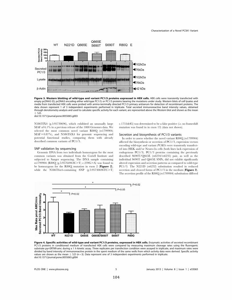
104
N586TfX4 (g.5:95730696), which exhibited an unusually largeMAF of 6.1% in a previous release of the 1000 Genomes data. Weselected the most common novel variant R80Q (rs1799904;MAF = 0.87%), and N586TfsX4 for genomic sequencing andpotential functional studies, comparing them with alreadydescribed common variants of PC1/3.
SNP validation by sequencingGenomic DNA from two individuals homozygous for the most
common variants was obtained from the Coriell Institute andsubjected to Sanger sequencing. The DNA sample containingrs1799904 (R80Q (g.5:95764963C.T; c.239G.A) was found tobe homozygous for the R80Q mutation in exon 2 (Figure 2),while the N586Tfsx4-containing SNP (g.5:95730696TC.T;
c.1755delG) was determined to be a false positive (i.e. no frameshiftmutation was found in in exon 13) (data not shown).
Secretion and biosynthesis of PC1/3 variantsIn order to assess whether the novel variant R80Q (rs1799904)
affected the biosynthesis or secretion of PC1/3, expression vectorsencoding wild-type and variant PCSK1s were transiently transfect-ed into HEK and/or Neuro-2a cells (both lines lack expression ofendogenous PC1/3). PC1/3 proteins containing the previouslydescribed S690T/Q665E (rs6234/rs6235) pair, as well as theindividual S690T and Q665E SNPs, did not exhibit significantlyaltered expression and secretion patterns as compared to wild-typePC1/3. The N221D (rs6232) substitution resulted in reducedsecretion and cleaved forms of PC1/3 in the medium (Figure 3).The secretion profile of the R80Q (rs1799904) substitution differed
Figure 3. Western blotting of wild-type and variant PC1/3 proteins expressed in HEK cells. HEK cells were transiently transfected withempty pcDNA3 (E); pcDNA3 encoding either wild-type PC1/3; or PC1/3 proteins bearing the mutations under study. Western blots of cell lysates andmedia from transfected HEK cells were probed with amino-terminally directed PC1/3 primary antiserum for detection of recombinant proteins. Thedata shown represent 1 of 3 independent experiments performed in triplicate. Total secreted immunoreactive band intensity values, obtainedthrough densitometry analysis and used to calculate specific activity for each variant, are represented above the Western blot and shown as the mean6 S.D.doi:10.1371/journal.pone.0055065.g003
Figure 4. Specific activities of wild-type and variant PC1/3 proteins, expressed in HEK cells. Enzymatic activities of secreted recombinantPC1/3 proteins in conditioned medium of transfected HEK cells were compared by measuring maximum cleavage rates using the fluorogenicsubstrate pyr-ERTKR-amc during a 1 h kinetic assay. Three replicates per transfection condition were assayed in triplicate, and maximum rates weredivided by band intensity of immunoreactive protein in the spent medium of the same wells from which activity data were derived. Specific activityvalues are shown as the mean 6 S.D (n = 3). Data represent one of 3 independent experiments performed in triplicate.doi:10.1371/journal.pone.0055065.g004
Characterization of a Novel PCSK1 Variant
PLOS ONE | www.plosone.org 5 January 2013 | Volume 8 | Issue 1 | e55065
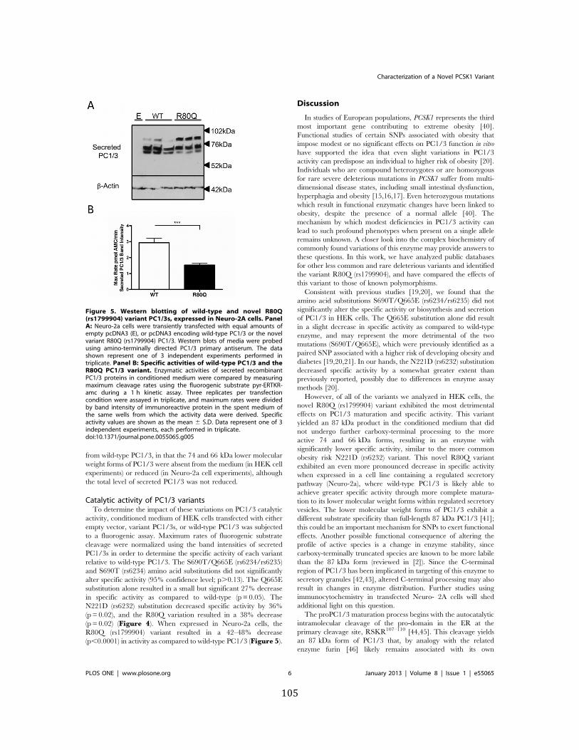
105
from wild-type PC1/3, in that the 74 and 66 kDa lower molecularweight forms of PC1/3 were absent from the medium (in HEK cellexperiments) or reduced (in Neuro-2a cell experiments), althoughthe total level of secreted PC1/3 was not reduced.
Catalytic activity of PC1/3 variantsTo determine the impact of these variations on PC1/3 catalytic
activity, conditioned medium of HEK cells transfected with eitherempty vector, variant PC1/3s, or wild-type PC1/3 was subjectedto a fluorogenic assay. Maximum rates of fluorogenic substratecleavage were normalized using the band intensities of secretedPC1/3s in order to determine the specific activity of each variantrelative to wild-type PC1/3. The S690T/Q665E (rs6234/rs6235)and S690T (rs6234) amino acid substitutions did not significantlyalter specific activity (95% confidence level; p.0.13). The Q665Esubstitution alone resulted in a small but significant 27% decreasein specific activity as compared to wild-type (p = 0.05). TheN221D (rs6232) substitution decreased specific activity by 36%(p = 0.02), and the R80Q variation resulted in a 38% decrease(p = 0.02) (Figure 4). When expressed in Neuro-2a cells, theR80Q (rs1799904) variant resulted in a 42–48% decrease(p,0.0001) in activity as compared to wild-type PC1/3 (Figure 5).
Discussion
In studies of European populations, PCSK1 represents the thirdmost important gene contributing to extreme obesity [40].Functional studies of certain SNPs associated with obesity thatimpose modest or no significant effects on PC1/3 function in vitrohave supported the idea that even slight variations in PC1/3activity can predispose an individual to higher risk of obesity [20].Individuals who are compound heterozygotes or are homozygousfor rare severe deleterious mutations in PCSK1 suffer from multi-dimensional disease states, including small intestinal dysfunction,hyperphagia and obesity [15,16,17]. Even heterozygous mutationswhich result in functional enzymatic changes have been linked toobesity, despite the presence of a normal allele [40]. Themechanism by which modest deficiencies in PC1/3 activity canlead to such profound phenotypes when present on a single alleleremains unknown. A closer look into the complex biochemistry ofcommonly found variations of this enzyme may provide answers tothese questions. In this work, we have analyzed public databasesfor other less common and rare deleterious variants and identifiedthe variant R80Q (rs1799904), and have compared the effects ofthis variant to those of known polymorphisms.
Consistent with previous studies [19,20], we found that theamino acid substitutions S690T/Q665E (rs6234/rs6235) did notsignificantly alter the specific activity or biosynthesis and secretionof PC1/3 in HEK cells. The Q665E substitution alone did resultin a slight decrease in specific activity as compared to wild-typeenzyme, and may represent the more detrimental of the twomutations (S690T/Q665E), which were previously identified as apaired SNP associated with a higher risk of developing obesity anddiabetes [19,20,21]. In our hands, the N221D (rs6232) substitutiondecreased specific activity by a somewhat greater extent thanpreviously reported, possibly due to differences in enzyme assaymethods [20].
However, of all of the variants we analyzed in HEK cells, thenovel R80Q (rs1799904) variant exhibited the most detrimentaleffects on PC1/3 maturation and specific activity. This variantyielded an 87 kDa product in the conditioned medium that didnot undergo further carboxy-terminal processing to the moreactive 74 and 66 kDa forms, resulting in an enzyme withsignificantly lower specific activity, similar to the more commonobesity risk N221D (rs6232) variant. This novel R80Q variantexhibited an even more pronounced decrease in specific activitywhen expressed in a cell line containing a regulated secretorypathway (Neuro-2a), where wild-type PC1/3 is likely able toachieve greater specific activity through more complete matura-tion to its lower molecular weight forms within regulated secretoryvesicles. The lower molecular weight forms of PC1/3 exhibit adifferent substrate specificity than full-length 87 kDa PC1/3 [41];this could be an important mechanism for SNPs to exert functionaleffects. Another possible functional consequence of altering theprofile of active species is a change in enzyme stability, sincecarboxy-terminally truncated species are known to be more labilethan the 87 kDa form (reviewed in [2]). Since the C-terminalregion of PC1/3 has been implicated in targeting of this enzyme tosecretory granules [42,43], altered C-terminal processing may alsoresult in changes in enzyme distribution. Further studies usingimmunocytochemistry in transfected Neuro- 2A cells will shedadditional light on this question.
The proPC1/3 maturation process begins with the autocatalyticintramolecular cleavage of the pro-domain in the ER at theprimary cleavage site, RSKR107–110 [44,45]. This cleavage yieldsan 87 kDa form of PC1/3 that, by analogy with the relatedenzyme furin [46] likely remains associated with its own
Figure 5. Western blotting of wild-type and novel R80Q(rs1799904) variant PC1/3s, expressed in Neuro-2A cells. PanelA: Neuro-2a cells were transiently transfected with equal amounts ofempty pcDNA3 (E), or pcDNA3 encoding wild-type PC1/3 or the novelvariant R80Q (rs1799904) PC1/3. Western blots of media were probedusing amino-terminally directed PC1/3 primary antiserum. The datashown represent one of 3 independent experiments performed intriplicate. Panel B: Specific activities of wild-type PC1/3 and theR80Q PC1/3 variant. Enzymatic activities of secreted recombinantPC1/3 proteins in conditioned medium were compared by measuringmaximum cleavage rates using the fluorogenic substrate pyr-ERTKR-amc during a 1 h kinetic assay. Three replicates per transfectioncondition were assayed in triplicate, and maximum rates were dividedby band intensity of immunoreactive protein in the spent medium ofthe same wells from which the activity data were derived. Specificactivity values are shown as the mean 6 S.D. Data represent one of 3independent experiments, each performed in triplicate.doi:10.1371/journal.pone.0055065.g005
Characterization of a Novel PCSK1 Variant
PLOS ONE | www.plosone.org 6 January 2013 | Volume 8 | Issue 1 | e55065
106
prodomain through non-covalent interactions until its arrival atthe trans-Golgi network. Although this has not yet been strictlydemonstrated for PC1/3, the PC1/3 prodomain most likely assistsin the folding of the catalytic domain and in enzyme inhibitionduring secretory pathway transport [47,48,49,50,51,52]. If prodo-main processing of PC1/3 occurs similarly to that of furin, trans-Golgi network protonation of a histidine in the vicinity of thesecondary cleavage site (RRSRR77–81) then results in secondarysite cleavage at R81, followed by dissociation of prodomainfragments from PC1/3 [53,54]. The inhibitory role of theprodomain is of particular interest to this study when we considerthe location of the R80Q (rs1799904) substitution within thesecondary cleavage site of the prodomain (Figure 1). Independentstudies have shown that alteration of mouse proPC1/3 prodomainresidues either within or surrounding cleavage motifs can affectpropeptide processing; the in vitro proteolytic conversion of anR80A mutant propeptide (the same residue as the R80Q variantstudied here) by wild-type enzyme was impaired compared towild-type propeptide [44]. Given this finding, our lack ofidentification of propeptide-bearing R80Q PC1/3 is puzzling.We have previously found that a portion of newly synthesizedproPC1/3 is subjected to endoplasmic reticulum- associateddegradation [52]; this might represent the fate of this molecularspecies. Collectively, these data support the idea that residueswithin the secondary cleavage site, including the novel variantstudied here, contribute to the proper processing of proPC1/3.
The novel R80Q (rs1799904) variant (MAF = 0.87%) is aboutone-third as common as the N221D (rs6232) SNP (MAF = 3.3%).Although less common, the R80Q variant should be subjected to
further analysis to evaluate its influence on insulin sensitivity,proinsulin conversion and the risk of developing obesity, similarlyto the effect of the N221D (rs6232) SNP [20,22]. We note that 119individuals in the public datasets have other, less common andrare variants of PCSK1, most of which are predicted to have somedetrimental effect on protein function. This mutational burden onthe population is not trivial and may also play a role insusceptibility to obesity or other disorders. The importance ofrare variants in common disorders is not clear at present, butadvances in massively parallel sequencing and computationalanalysis may soon shed additional light on this question.
In conclusion, we show that the novel PCSK1 variant R80Q(rs1799904) exhibits deleterious effects on PC1/3 maturation. ThisPC1/3 variant exhibits decreased catalytic activity as compared towild-type PC1/3 and to previously described obesity risk SNPs;therefore, it may contribute to a higher risk of metabolic disease inthe general population. Our results suggest that further study ofless common and rare variations in PCSK1 from both biochemicaland genetic standpoints will be useful in elucidating themechanisms by which variant PC1/3s contribute to metabolicdiseases such as obesity and diabetes.
Author Contributions
Conceived and designed the experiments: MY MGM IL. Performed theexperiments: LAP MY VA ZC RSS. Analyzed the data: LAP VA MY ZCRSS SN MGM IL. Contributed reagents/materials/analysis tools: MGMIL. Wrote the paper: LAP MY MGM IL.
References
1. Seidah NG, Mattei MG, Gaspar L, Benjannet S, Mbikay M, et al. (1991)Chromosomal assignments of the genes for neuroendocrine convertase PC1(NEC1) to human 5q15-21, neuroendocrine convertase PC2 (NEC2) to human20p11.1-11.2, and furin (mouse 7[D1-E2] region). Genomics 11: 103–107.
2. Hoshino A, Lindberg I (2012) Peptide Biosynthesis: Prohormone Convertases 1/3 and 2. In: Fricker LD, Devi L, editors: Morgan & Claypool Life SciencesPublishers.
3. Schafer MK, Day R, Cullinan WE, Chretien M, Seidah NG, et al. (1993) Geneexpression of prohormone and proprotein convertases in the rat CNS: acomparative in situ hybridization analysis. J Neurosci 13: 1258–1279.
4. Dong W, Seidel B, Marcinkiewicz M, Chretien M, Seidah NG, et al. (1997)Cellular localization of the prohormone convertases in the hypothalamicparaventricular and supraoptic nuclei: selective regulation of PC1 in corticotro-phin-releasing hormone parvocellular neurons mediated by glucocorticoids.J Neurosci 17: 563–575.
5. Wynne K, Stanley S, McGowan B, Bloom S (2005) Appetite control.J Endocrinol 184: 291–318.
6. Smeekens S, Montag AG, Thomas G, Albiges-Rizo C, Carrol R, et al. (1992)Proinsulin processing by the subtilisin-related proprotein convertases furin, PC2,and PC3. Proc Natl Acad Sci USA 89: 8822–8826.
7. Rouille Y, Kantengwa S, Irminger JC, Halban PA (1997) Role of theprohormone convertases in the processing of proglucagon to glucagon-likepeptides. J Biol Chem 72: 32810–32816.
8. Zhu X, Zhou A, Dey A, Norrbom C, Carroll R, et al. (2002) Disruption of PC1/3 expression in mice causes dwarfism and multiple neuroendocrine peptideprocessing defects. Proc Natl Acad Sci USA 99: 10293–10298.
9. Zhu X, Cao Y, Voogd K, Steiner DF (2006) On the processing of proghrelin toghrelin. J Biol Chem 281: 38867–38870.
10. Creemers JW, Pritchard LE, Gyte A, Le Rouzic P, Meulemans S, et al. (2006)Agouti-related protein is posttranslationally cleaved by proprotein convertase 1to generate agouti-related protein (AGRP)83-132: interaction betweenAGRP83-132 and melanocortin receptors cannot be influenced by syndecan-3. Endocrinology 147: 1621–1631.
11. Brakch N, Rist B, Beck-Sickinger AG, Goenaga J, Wittek R, et al. (1997) Role ofprohormone convertases in pro-neuropeptide Y processing: coexpression and invitro kinetic investigations. Biochemistry 36: 16309–16320.
12. Coates LC, Birch NP (1998) Differential cleavage of provasopressin by the majormolecular forms of SPC3. J Neurochem 70: 1670–1678.
13. Benjannet S, Rondeau N, Day R, Chretien M, Seidah NG (1991) PC1 and PC2are proprotein convertases capable of cleaving proopiomelanocortin at distinctpairs of basic residues. Proc Natl Acad Sci U S A 88: 3564–3568.
14. Thomas L, Leduc R, Thorne BA, Smeekens SP, Steiner D, et al. (1991) Kex2-like endoproteases PC2 and PC3 accurately cleave a model prohormone inmammalian cells: evidence for a common core of neuroendocrine processingenzymes. Proc Natl Acad Sci USA 88: 5297–5301.
15. Jackson RS, Creemers JW, Farooqi IS, Raffin-Sanson ML, Varro A, et al. (2003)Small-intestinal dysfunction accompanies the complex endocrinopathy ofhuman proprotein convertase 1 deficiency. J Clin Invest 112: 1550–1560.
16. Jackson RS, Creemers JW, Ohagi S, Raffin-Sanson ML, Sanders L, et al. (1997)Obesity and impaired prohormone processing associated with mutations in thehuman prohormone convertase 1 gene. Nat Genet 16: 303–306.
17. Farooqi IS, Volders K, Stanhope R, Heuschkel R, White A, et al. (2007)Hyperphagia and early-onset obesity due to a novel homozygous missensemutation in prohormone convertase 1/3. J Clin Endocrinol Metab 92: 3369–3373.
18. Lloyd DJ, Bohan S, Gekakis N (2006) Obesity, hyperphagia and increasedmetabolic efficiency in Pc1 mutant mice. Hum Mol Genet 15: 1884–1893.
19. Mbikay M, Sirois F, Nkongolo KK, Basak A, Chretien M (2011) Effects ofrs6234/rs6235 and rs6232/rs6234/rs6235 PCSK1 single-nucleotide polymor-phism clusters on proprotein convertase 1/3 biosynthesis and activity. MolGenet Metab 104: 682–687.
20. Benzinou M, Creemers JW, Choquet H, Lobbens S, Dina C, et al. (2008)Common nonsynonymous variants in PCSK1 confer risk of obesity. Nat Genet40: 943–945.
21. Strawbridge RJ, Dupuis J, Prokopenko I, Barker A, Ahlqvist E, et al. (2011)Genome-wide association identifies nine common variants associated withfasting proinsulin levels and provides new insights into the pathophysiology oftype 2 diabetes. Diabetes 60: 2624–2634.
22. Heni M, Haupt A, Schafer SA, Ketterer C, Thamer C, et al. (2010) Associationof obesity risk SNPs in PCSK1 with insulin sensitivity and proinsulin conversion.BMC Med Genet 11: 86.
23. (2004) Finishing the euchromatic sequence of the human genome. Nature 431:931–945.
24. Sherry ST, Ward MH, Kholodov M, Baker J, Phan L, et al. (2001) dbSNP: theNCBI database of genetic variation. Nucleic Acids Res 29: 308–311.
25. (2010) A map of human genome variation from population-scale sequencing.Nature 467: 1061–1073.
26. NHLBI Exome Sequencing Project (2011) Exome Variant Server. Seattle WA:NHLBI Exome Sequencing Project (ESP).
27. NIEHS Environmental Genome Project (2011) NIEHS Exome Variant Server.Seattle WA: NIEHS Environmental Genome Project.
Characterization of a Novel PCSK1 Variant
PLOS ONE | www.plosone.org 7 January 2013 | Volume 8 | Issue 1 | e55065
107
28. McKenna A, Hanna M, Banks E, Sivachenko A, Cibulskis K, et al. (2010) TheGenome Analysis Toolkit: a MapReduce framework for analyzing next-generation DNA sequencing data. Genome Res 20: 1297–1303.
29. Flicek P, Amode MR, Barrell D, Beal K, Brent S, et al. (2011) Ensembl 2011.Nucleic Acids Res 39: D800–806.
30. Kumar P, Henikoff S, Ng PC (2009) Predicting the effects of coding non-synonymous variants on protein function using the SIFT algorithm. NatureProtocols 4: 1073–1082.
31. Ng PC, Henikoff S (2002) Accounting for human polymorphisms predicted toaffect protein function. Genome Research 12: 436–446.
32. Ng CP, Henikoff S (2003) SIFT: Predicting amino acid changes that affectprotein function. Nucleic Acids Research 31: 3812–3814.
33. Ng PC, Henikoff S (2006) Predicting the effects of amino acid substitutions onprotein function. Annual Review of Genomics and Human Genetics 7: 61–80.
34. Ramensky V, Bork P, Sunyaev S (2002) Human non-synonymous SNPs: serverand survey. Nucleic Acids Res 30: 3894–3900.
35. Sunyaev S, Ramensky V, Bork P (2000) Towards a structural basis of humannon-synonymous single nucleotide polymorphisms. Trends Genet 16: 198–200.
36. Sunyaev S, Ramensky V, Koch I, Lathe W, 3rd, Kondrashov AS, et al. (2001)Prediction of deleterious human alleles. Human Molecular Genetics 10: 591–597.
37. Gonzalez-Perez A, Lopez-Bigas N (2011) Improving the assessment of theoutcome of nonsynonymous SNVs with a consensus deleteriousness score,Condel. American Journal of Human Genetics 88: 440–449.
38. Braman J, Papworth C, Greener A (1996) Site-directed mutagenesis usingdouble-stranded plasmid DNA templates. Methods Mol Biol 57: 31–44.
39. Vindrola O, Lindberg I (1992) Biosynthesis of the prohormone convertasemPC1 in AtT-20 cells. Mol Endocrinol 6: 1088–1094.
40. Creemers JW, Choquet H, Stijnen P, Vatin V, Pigeyre M, et al. (2012)Heterozygous mutations causing partial prohormone convertase 1 deficiencycontribute to human obesity. Diabetes 61: 383–390.
41. Zhou Y, Rovere C, Kitabgi P, Lindberg I (1995) Mutational analysis of PC1(SPC3) in PC12 cells. 66-kDa PC1 is fully functional. J Biol Chem 270: 24702–24706.
42. Zhou A, Mains RE (1994) Endoproteolytic processing of proopiomelanocortinand prohormone convertases 1 and 2 in neuroendocrine cells overexpressingprohormone convertases 1 or 2. J Biol Chem 269: 17440–17447.
43. Bernard N, Kitabgi P, Rovere-Jovene C (2003) The Arg617–Arg618 cleavagesite in the C-terminal domain of PC1 plays a major role in the processing andtargeting of the enzyme within the regulated secretory pathway. J Neurochem85: 1592–1603.
44. Rabah N, Gauthier D, Wilkes BC, Gauthier DJ, Lazure C (2006) Single aminoacid substitution in the PC1/3 propeptide can induce significant modifications ofits inhibitory profile toward its cognate enzyme. J Biol Chem 281: 7556–7567.
45. Goodman LJ, Gorman CM (1994) Autoproteolytic activation of the mouseprohormone convertase mPC1. Biochem Biophys Res Commun 201: 795–804.
46. Anderson ED, VanSlyke JK, Thulin CD, Jean F, Thomas G (1997) Activation ofthe furin endoprotease is a multiple-step process: requirements for acidificationand internal propeptide cleavage. EMBO J 16: 1508–1518.
47. Creemers JW, Vey M, Schafer W, Ayoubi TA, Roebroek AJ, et al. (1995)Endoproteolytic cleavage of its propeptide is a prerequisite for efficient transportof furin out of the endoplasmic reticulum. J Biol Chem 270: 2695–2702.
48. Mains RE, Milgram SL, Keutman HT, Eipper BA (1995) The NH2-terminalproregion of peptidylglycine a-amidating monooxygenase facilitates the secretionof soluble proteins. Mol Endocrinol 9: 3–13.
49. Apletalina EV, Juliano MA, Juliano L, Lindberg I (2000) Structure-functionanalysis of the 7B2 CT peptide. Biochem Biophys Res Commun 267: 940–942.
50. Muller L, Cameron A, Fortenberry Y, Apletalina EV, Lindberg I (2000)Processing and sorting of the prohormone convertase 2 propeptide. J Biol Chem275: 39213–39222.
51. Bissonnette L, Charest G, Longpre JM, Lavigne P, Leduc R (2004) Identificationof furin pro-region determinants involved in folding and activation. Biochem J379: 757–763.
52. Lee SN, Prodhomme E, Lindberg I (2004) Prohormone convertase 1 (PC1)processing and sorting: effect of PC1 propeptide and proSAAS. J Endocrinol182: 353–364.
53. Tangrea MA, Bryan PN, Sari N, Orban J (2002) Solution structure of the pro-hormone convertase 1 pro-domain from Mus musculus. J Mol Biol 320: 801–812.
54. Benjannet S, Rondeau N, Paquet L, Boudreault A, Lazure C, et al. (1993)Comparative biosynthesis, covalent post-translational modifications and effi-ciency of prosegment cleavage of the prohormone convertases PC1 and PC2:glycosylation, sulphation and identification of the intracellular site of prosegmentcleavage of PC1 and PC2. Biochem J 294 (Pt 3): 735–743.
Characterization of a Novel PCSK1 Variant
PLOS ONE | www.plosone.org 8 January 2013 | Volume 8 | Issue 1 | e55065
108
CHAPTER SIX
Mutations in ACTG2 are associated with
sporadic congenital chronic intestinal pseudo-‐obstruction
and megacystis-‐microcolon-‐intestinal hypoperistalsis syndrome
109
ABSTRACT
Chronic intestinal pseudo-‐obstruction (CIPO) is a serious motility dysfunction
syndrome characterized by symptoms of intestinal obstruction in the absence of any
mechanical blockage. It is a major cause of intestinal failure. With whole-‐exome sequencing in a
cohort of 20 patients with congenital CIPO or MMIH, we identified a subset of 10 cases with
potentially damaging de-‐novo mutations at highly conserved loci in the ACTG2 gene. In light of
a recent finding that a mutation in ACTG2 caused familial visceral myopathy in a Finnish family,
we conclude that ACTG2 also governs a significant proportion of cases of sporadic congenital
CIPO.
INTRODUCTION
Chronic intestinal pseudo-‐obstruction (CIPO) is a heterogenous set of diseases
characterized by repetitive episodes or continuous symptoms of intestinal obstruction, in the
absence of a lesion that occludes the lumen of the gut (1, 2). A small fraction of cases are
secondary to organic, systemic, or metabolic diseases, but the majority are primary and may be
myopathic, mesenchymopathic, or neuropathic, depending upon whether predominant
abnormalities are found in the enteric nervous system, Interstitial Cells of Cajal (ICC), or
intestinal smooth muscle (3). A related disorder, megacystis-‐microcolon-‐intestinal
hypoperistalsis syndrome (MMIH), is characterized by constipation and urinary retention,
microcolon, giant bladder (megacystis), intestinal hypoperistalis, hydronephrosis, and dilated
small bowel (4).
Congenital forms of CIPO are rare and can be life-‐threatening; congenital CIPO is an
important cause of intestinal failure, for which the only treatment may be complete visceral
transplantation (5). Congenital CIPO may sometimes be due to prenatal exposure to toxins
such as alcohol or narcotics. A handful of familial cases of CIPO have been reported with
110
autosomal dominant (with variable penetrance), autosomal recessive, and X-‐linked modes of
inheritance (6-‐16). It is well known that mutations in mitochondrial tRNA genes, POLG
(polymerase (DNA directed), gamma), and TYMP (thymidine phosphorylase), which are
expressed in the mitochondrion, cause a severe form of CIPO requiring frequent and long-‐term
parenteral nutrition and with frequently fatal digestive and neurologic complications.
Mitochondrial disorders may account for ~19% of CIPO cases (17). Contrawise, it is rare for
CIPO to be the principal clinical manifestation of a mitochondrial disorder (18). Primary
defects of the mitochondrial oxidative phosphorylation pathway are phenotypically
heterogenous, and affecting multiple organs, typically with nervous system and skeletal or
ocular muscle dysfunction (19). Mitochondrial neurogastrointestinal encephalomyopathy
(MNGIE) is a rare, autosomal recessive syndrome due to the loss of thymidine phosphorylase
activity associated with loss-‐of-‐function mutations in TYMP (20-‐24). Mutations in POLG, the
mitochondrial myopathy, epilepsy, lactic acidosis, and strokelike episodes (‘MELAS’) mutation
in the tRNAleu(UUR) gene (MT-‐TL1), or mutations in the tRNAlys gene (MT-‐TK) are sometimes
associated with CIPO (16, 25-‐32). Still, congenital CIPO is usually sporadic and prior to the
advent of exome sequencing, no non-‐mitochondrial gene had been convincingly associated
with primary sporadic CIPO.
METHODS
Samples were selected from 20 patients seen at UCLA, and were approved by our
institutional review board. We chose 18 patients for exome sequencing; we sequenced only
one of monozygotic twins in one family and a second patient entered the study after exome
sequencing had concluded. Inclusion criteria were a diagnosis of chronic severe CIPO or MMIH.
These patients were of diverse ethnic backgrounds and had clinical presentations of myopathic
CIPO (n=6), neuropathic CIPO (n=5), idiopathic CIPO (n=3), and megacystis-‐microcolon-‐
111
intestinal hypoperistalsis syndrome (MMIH) (n=4). The phenotype of several patients was
complex with syndromic presentations of developmental delay (n=1), severe neurological
problems (n=1), or microcephaly and mental retardation (n=2 siblings) [Table 1].
Genomic DNA from probands, and in some cases unaffected family members, was
either fragmented by sonication and ligated to Illumina bar-‐coded adapters or fragmented and
ligated in a single step with Illumina engineered transposases, and then in either case, the
fragments were amplified by PCR. Fragments were then enriched for the protein coding
portion of the genome by hybridization to probes from either the Agilent SureSelect XT Human
All Exon 50Mb, Illumina TruSeq Exome, or Illumina Nextera Expanded Exome enrichment kits.
The exome-‐enriched library was sequenced for 100x100 paired end reads on an Illumina
Genome Analyzer 2000 or 2500 platform to a mean coverage depth of 97X, with 84% of
RefGene CDS and essential splice sites having at least 20X coverage. Sanger sequencing
confirmed candidate variants and segregation of the variant allele with the disorder in
relatives from whom DNA was available.
We converted sequenced reads from the native bcl files to the FastQ format by the Illumina
bcl2fastq program. We processed the FastQ files to create aligned bam files with in-house pipeline
software. Briefly, we aligned the reads to build GRCh37 of the human genome (33) with Novoalign
(http://www.novocraft.com) to obtain a mean of 93 million uniquely aligned 100x100 paired end
reads per sample after removing PCR duplicates. We recalibrated base quality scores to improve
accuracy by analyzing the covariation among reported quality score, position within read,
dinucleotide, and probability of mismatching the reference genome using the Genome Analysis
Toolkit (GATK) (34, 35). We used the GATK Unified Genotyper and Haplotype Caller tools to
genotype single nucleotide variants and indels and recalibrated variant quality score recalibration
with the GATK Variant Quality Score Recalibrator to assign probabilities to each variant call. We
obtained the variant consequences on transcripts and proteins with the Ensembl Variant Effect
112
Predictor (VEP) (35), estimated the extent of protein damage with SIFT (36-40), PolyPhen (41-43),
and Condel (44), and computed GERP conservation scores (45). We further annotated variants with
additional data from Online Mendelian Inheritance in Man (OMIM) (46), the Human Gene
Mutation Database (HGMD pro, BIOBASE Biological Databases), the Universal Protein Resource
(UniProt) (47), KEGG Pathways (48), RefGene (49), the MitoCarta Inventory of Mammalian
Mitochondrial Genes (50), Mouse Genome Informatics (MGI) (51) and the Human Protein
Atlas (HPA) (52) using in-‐house plugins for the VEP.
We hypothesized for this study that the mode of inheritance would be recessive, de
novo, or mitochondrial, with a dominant effect on phenotype and that mutation effects would
be fully penetrant. Further hypothesizing that casual variants would be found in the protein
coding or splicing regions of genes, we filtered to include only splice acceptor or donor, stop
gained or lost, frameshift, initiator codon, inframe insertion or deletion, missense, or splice
region variants. We removed variants that, given the rare incidence of CIPO, are too frequent in
the population to cause this disorder; under the recessive model we removed variants with a
minor allele frequency (MAF) >0.5% in the combined 1000 Genomes (53) and NHLBI (54)
datasets and with a MAF of >0.1% under the de-‐novo model. In addition, we removed variants
that were observed in more than 2 (homozygous and de-‐novo models) or 8 (compound
heterozygous model) samples from ~150 unaffected control exomes, to eliminate false
positives caused by technical artifacts. We ranked variants for likelihood of damage using
multiple factors. We deemed a splice acceptor or donor variant, stop gained, and frameshift, to
be probably damaging. We prioritized missense single nucleotide variants (SNVs) by SIFT,
PolyPhen, and Condel predictions, and variants in the splice site region near the acceptor and
donor by GERP conservation scores. In the case of compound heterozygous variants we
assigned the priority of the second ranked variant. In families where we had sequenced
members other than the proband, we filtered out all variants that were inconsistent with fully
113
penetrant Mendelian inheritance under the recessive model or present in any unaffected
relative under the de-‐novo model. We hypothesized that there would be genotypic
heterogeneity among the cases, so we looked for cases that shared predicted damaging
mutations in the same gene.
RESULTS
The probands had a mean of ~93,000 variations from the GRCh37 reference genome
before filtering. After filtering by consequence rank, allele frequency, and controls and
eliminating variants that did not segregate with the disease, the probands had a mean of 85
variants consistent with either the recessive or de-‐novo models. Looking for genes that had de-‐
novo mutations in multiple cases, our attention focused on ACTG2 (encoding actin, gamma-‐
enteric smooth muscle precursor), in which there were seven different de-‐novo mutations in
seven cases and no controls. ACTG2 was of interest because of actin’s involvement in muscle
function (55) and particularly because γ-‐enteric actin is the dominant isoform of actin
expressed in the jejunum (56). We performed Sanger sequencing on ACTG2 exons in all cases
and found two additional mutations, one that had been missed in exome sequencing and one in
a case added to the study after exome sequencing was complete, as well as confirming the
presence of a mutation in the unsequenced twin [Tables 2 & 3]. Each of the mutations was
predicted to be deleterious by the Condel consensus statistic based on PolyPhen and SIFT and
all were at highly conserved loci with GERP scores ≥3.59. All variants were confirmed by
Sanger sequencing, and were not found in unaffected relatives. No variant was found in ~150
control exomes, nor in the 1000 Genomes or NHLBI datasets.
114
DISCUSSION
During the course of our study Lehtonen et al. reported that a missense variant
(Arg148Ser) in ACTG segregated in a Finnish family with autosomal dominant familial visceral
myopathy (FVM), a disorder that is subsumed within the broad definition of CIPO (57).
Although none of the mutations we identified overlapped with the FVM mutations, our results
along with the FVM finding, make a strong case that mutations in γ-‐enteric actin have a
profound effect on intestinal motility and may be a predominant cause of sporadic and familial
intestinal failure due to pseudo-‐obstruction.
Nonetheless, ACTG mutations explain only 50% of the cases we examined, suggesting
that CIPO must be genetically heterogeneous. Moreover, we found ACTG2 mutations in patients
initially suspected to be myopathic, neuropathic, or having MIMH syndrome. This may be
explained in part by the challenges in diagnosing this class of disorders and in part by
phenotypic heterogeneity. There may also be identifiable genotype-‐phenotype correlations,
which may become apparent when a larger number of cases are genotyped. Interestingly, none
of the cases in our study with syndromic CIPO had detectable mutations in ACTG2.
A large study (n=115) observed absent or partial staining for smooth muscle α-‐actin in
the jejunal circular muscle layer of in 24% of CIPO patients (58). This report, which found no
differences in staining of γ-‐actin, may indicate a different molecular basis for another class of
patients than those with γ-‐ actin mutations. It is also possible that the near sequence identity of
α-‐ and γ-‐actin made it difficult to distinguish the isoforms, even though the antibodies used
purported to be specific. Another study also found α-‐actin deficiencies in CIPO patient tissue
(59), but that finding was not replicated (60).
The molecular mechanism by which disrupted actin affects enteric smooth muscle cells
remains to be discovered. Now that ten different probably disease-‐causing ACTG2 mutations
have been identified it will be interesting to discover how each of these variants affects the cell.
115
Mutations in other actin isoforms at homologous loci to those reported here can cause disease.
The ACTA2 Arg258Cys mutation, homologous to ACTG2 Arg257Cys, causes thoracic aneurisms
and dissections (61); ACTA1 Arg258His causes nemaline myopathy (62); and ACTA2 Arg258His
causes thoracic aortic disease & strokes (63). Several other loci when mutated in ACTA1 or
ACTA2 to different amino acids than those reported here also cause disease [Table 3] (64-‐68).
This suggests the possibility of common molecular mechanisms at work, which manifest
variously in different tissues where other actin isoforms are expressed. However, determining
the these mechanisms will be challenging (69).
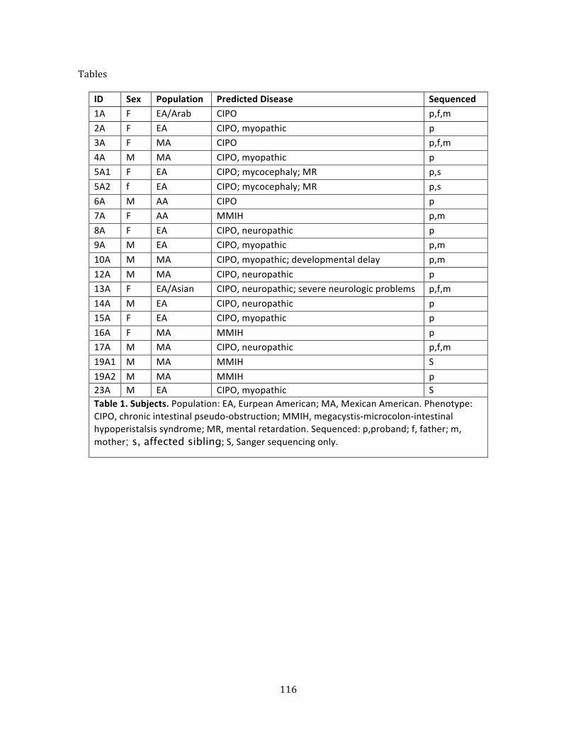
116
Tables
ID Sex Population Predicted Disease Sequenced 1A F EA/Arab CIPO p,f,m 2A F EA CIPO, myopathic p 3A F MA CIPO p,f,m 4A M MA CIPO, myopathic p 5A1 F EA CIPO; mycocephaly; MR p,s 5A2 f EA CIPO; mycocephaly; MR p,s 6A M AA CIPO p 7A F AA MMIH p,m 8A F EA CIPO, neuropathic p 9A M EA CIPO, myopathic p,m 10A M MA CIPO, myopathic; developmental delay p,m 12A M MA CIPO, neuropathic p 13A F EA/Asian CIPO, neuropathic; severe neurologic problems p,f,m 14A M EA CIPO, neuropathic p 15A F EA CIPO, myopathic p 16A F MA MMIH p 17A M MA CIPO, neuropathic p,f,m 19A1 M MA MMIH S 19A2 M MA MMIH p 23A M EA CIPO, myopathic S Table 1. Subjects. Population: EA, Eurpean American; MA, Mexican American. Phenotype: CIPO, chronic intestinal pseudo-‐obstruction; MMIH, megacystis-‐microcolon-‐intestinal hypoperistalsis syndrome; MR, mental retardation. Sequenced: p,proband; f, father; m, mother; s, affected sibling; S, Sanger sequencing only.
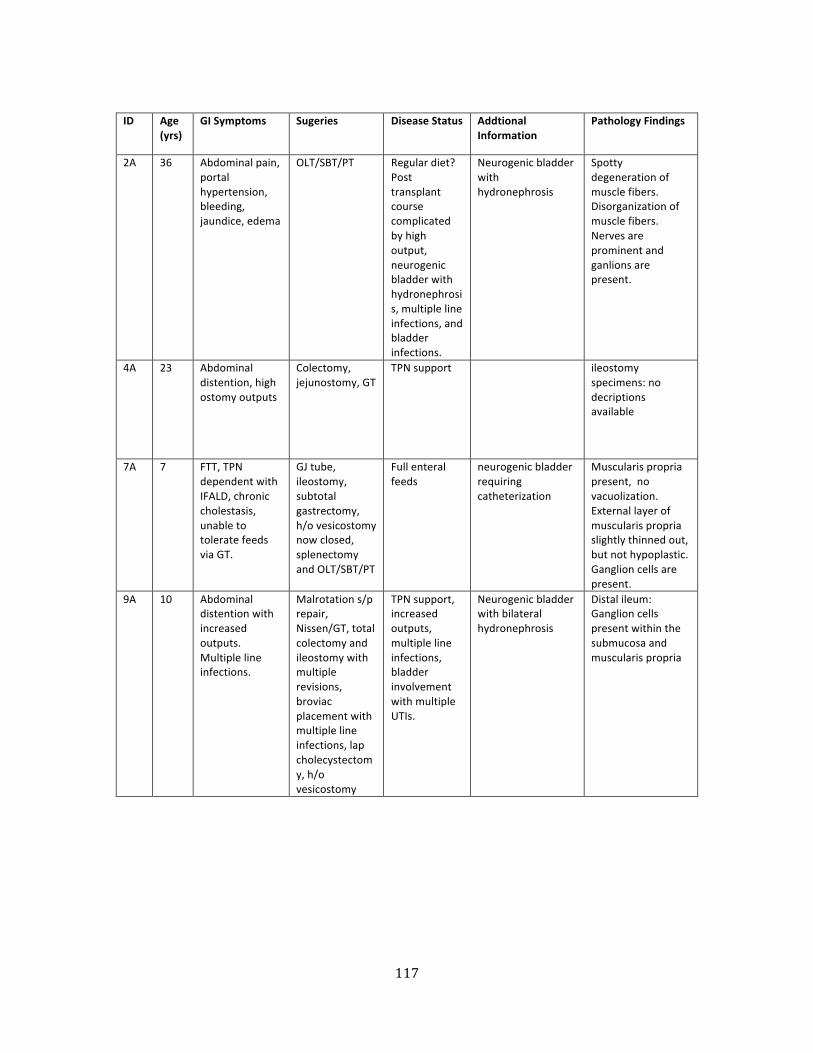
117
ID Age
(yrs) GI Symptoms Sugeries Disease Status Addtional
Information Pathology Findings
2A 36 Abdominal pain, portal hypertension, bleeding, jaundice, edema
OLT/SBT/PT Regular diet? Post transplant course complicated by high output, neurogenic bladder with hydronephrosis, multiple line infections, and bladder infections.
Neurogenic bladder with hydronephrosis
Spotty degeneration of muscle fibers. Disorganization of muscle fibers. Nerves are prominent and ganlions are present.
4A 23 Abdominal distention, high ostomy outputs
Colectomy, jejunostomy, GT
TPN support ileostomy specimens: no decriptions available
7A 7 FTT, TPN dependent with IFALD, chronic cholestasis, unable to tolerate feeds via GT.
GJ tube, ileostomy, subtotal gastrectomy, h/o vesicostomy now closed, splenectomy and OLT/SBT/PT
Full enteral feeds
neurogenic bladder requiring catheterization
Muscularis propria present, no vacuolization. External layer of muscularis propria slightly thinned out, but not hypoplastic. Ganglion cells are present.
9A 10 Abdominal distention with increased outputs. Multiple line infections.
Malrotation s/p repair, Nissen/GT, total colectomy and ileostomy with multiple revisions, broviac placement with multiple line infections, lap cholecystectomy, h/o vesicostomy
TPN support, increased outputs, multiple line infections, bladder involvement with multiple UTIs.
Neurogenic bladder with bilateral hydronephrosis
Distal ileum: Ganglion cells present within the submucosa and muscularis propria
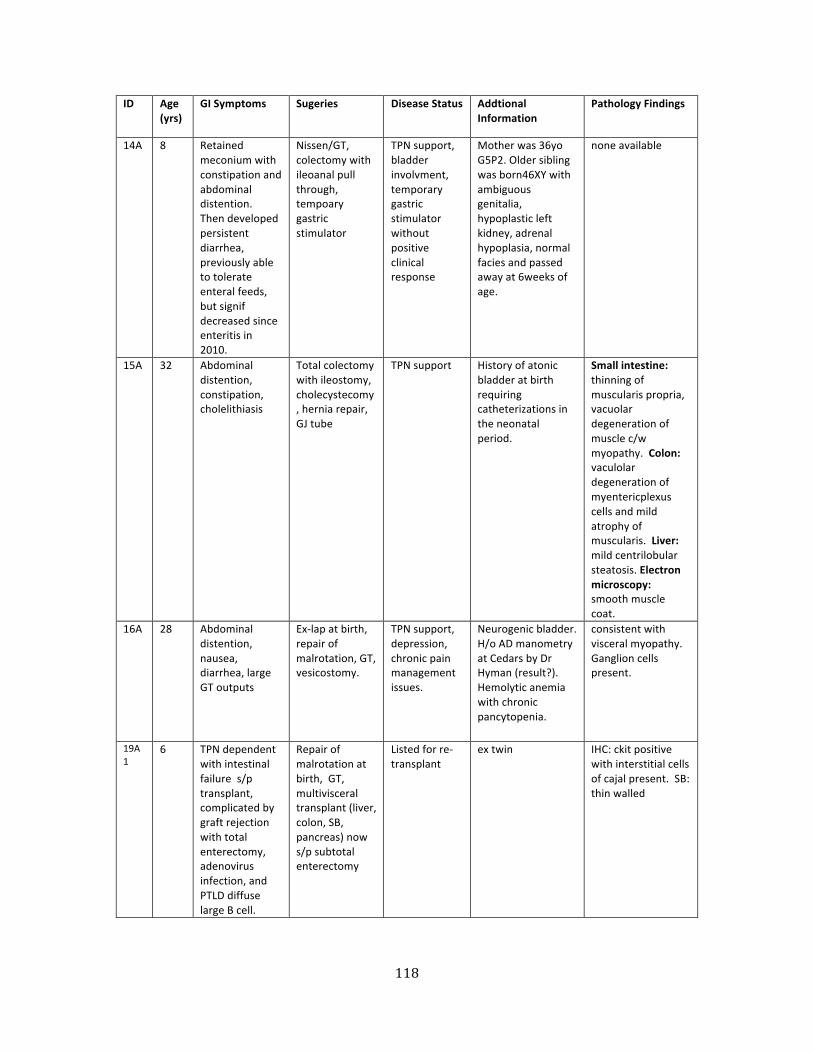
118
ID Age (yrs)
GI Symptoms Sugeries Disease Status Addtional Information
Pathology Findings
14A 8 Retained meconium with constipation and abdominal distention. Then developed persistent diarrhea, previously able to tolerate enteral feeds, but signif decreased since enteritis in 2010.
Nissen/GT, colectomy with ileoanal pull through, tempoary gastric stimulator
TPN support, bladder involvment, temporary gastric stimulator without positive clinical response
Mother was 36yo G5P2. Older sibling was born46XY with ambiguous genitalia, hypoplastic left kidney, adrenal hypoplasia, normal facies and passed away at 6weeks of age.
none available
15A 32 Abdominal distention, constipation, cholelithiasis
Total colectomy with ileostomy, cholecystecomy, hernia repair, GJ tube
TPN support History of atonic bladder at birth requiring catheterizations in the neonatal period.
Small intestine: thinning of muscularis propria, vacuolar degeneration of muscle c/w myopathy. Colon: vaculolar degeneration of myentericplexus cells and mild atrophy of muscularis. Liver: mild centrilobular steatosis. Electron microscopy: smooth muscle coat.
16A 28 Abdominal distention, nausea, diarrhea, large GT outputs
Ex-‐lap at birth, repair of malrotation, GT, vesicostomy.
TPN support, depression, chronic pain management issues.
Neurogenic bladder. H/o AD manometry at Cedars by Dr Hyman (result?). Hemolytic anemia with chronic pancytopenia.
consistent with visceral myopathy. Ganglion cells present.
19A1
6 TPN dependent with intestinal failure s/p transplant, complicated by graft rejection with total enterectomy, adenovirus infection, and PTLD diffuse large B cell.
Repair of malrotation at birth, GT, multivisceral transplant (liver, colon, SB, pancreas) now s/p subtotal enterectomy
Listed for re-‐transplant
ex twin IHC: ckit positive with interstitial cells of cajal present. SB: thin walled
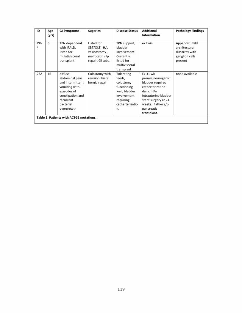
119
ID Age (yrs)
GI Symptoms Sugeries Disease Status Addtional Information
Pathology Findings
19A2
6 TPN dependent with IFALD, listed for mulativisceral transplant.
Listed for SBT/OLT. H/o vesicostomy , malrotatin s/p repair, GJ tube.
TPN support, bladder involvement. Currently listed for multivisceral transplant
ex twin Appendix: mild architectural dissarray with ganglion cells present
23A 16 diffuse abdominal pain and intermittent vomiting with episodes of constipation and recurrent bacterial overgrowth
Colostomy with revision, hiatal hernia repair
Tolerating feeds, colostomy functioning well, bladder involvement requiring catherterization.
Ex 31 wk premie,neurogenic bladder requires catherterization daily. H/o intrauterine bladder stent surgery at 24 weeks. Father s/p pancreatic transplant.
none available
Table 2. Patients with ACTG2 mutations.
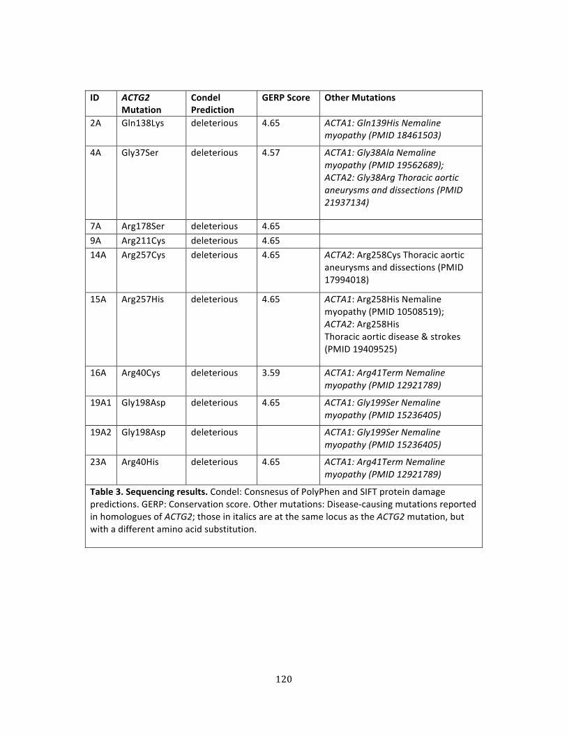
120
ID ACTG2 Mutation
Condel Prediction
GERP Score Other Mutations
2A Gln138Lys deleterious 4.65 ACTA1: Gln139His Nemaline myopathy (PMID 18461503)
4A Gly37Ser deleterious 4.57 ACTA1: Gly38Ala Nemaline myopathy (PMID 19562689); ACTA2: Gly38Arg Thoracic aortic aneurysms and dissections (PMID 21937134)
7A Arg178Ser deleterious 4.65 9A Arg211Cys deleterious 4.65 14A Arg257Cys deleterious 4.65 ACTA2: Arg258Cys Thoracic aortic
aneurysms and dissections (PMID 17994018)
15A Arg257His deleterious 4.65 ACTA1: Arg258His Nemaline myopathy (PMID 10508519); ACTA2: Arg258His Thoracic aortic disease & strokes (PMID 19409525)
16A Arg40Cys deleterious 3.59 ACTA1: Arg41Term Nemaline myopathy (PMID 12921789)
19A1 Gly198Asp deleterious 4.65 ACTA1: Gly199Ser Nemaline myopathy (PMID 15236405)
19A2 Gly198Asp deleterious ACTA1: Gly199Ser Nemaline myopathy (PMID 15236405)
23A Arg40His deleterious 4.65 ACTA1: Arg41Term Nemaline myopathy (PMID 12921789)
Table 3. Sequencing results. Condel: Consnesus of PolyPhen and SIFT protein damage predictions. GERP: Conservation score. Other mutations: Disease-‐causing mutations reported in homologues of ACTG2; those in italics are at the same locus as the ACTG2 mutation, but with a different amino acid substitution.
121
References
1. Antonucci A, Fronzoni L, Cogliandro L, Cogliandro RF, Caputo C, De Giorgio R, Pallotti F, Barbara G, Corinaldesi R, Stanghellini V. Chronic intestinal pseudo-‐obstruction. World journal of gastroenterology : WJG. 2008;14(19):2953-‐61. PubMed PMID: 18494042; PubMed Central PMCID: PMC2712158.
2. Hyman P, Thapar N. Chronic Intestinal Pseudo-‐Obstruction. In: Faure C, Di Lorenzo C, Thapar N, editors. Pediatric Neurogastroenterology: Humana Press; 2013. p. 257-‐70.
3. Gisser J, Gariepy C. Genetics of Motility Disorder: Gastroesophageal Reflux, Triple A Syndrome, Hirschsprung Disease, and Chronic Intestinal Pseudo-‐Obstruction. In: Faure C, Di Lorenzo C, Thapar N, editors. Pediatric Neurogastroenterology: Humana Press; 2013. p. 203-‐16.
4. Berdon WE, Baker DH, Blanc WA, Gay B, Santulli TV, Donovan C. Megacystis-‐microcolon-‐intestinal hypoperistalsis syndrome: a new cause of intestinal obstruction in the newborn. Report of radiologic findings in five newborn girls. AJR American journal of roentgenology. 1976;126(5):957-‐64. Epub 1976/05/01. doi: 10.2214/ajr.126.5.957. PubMed PMID: 178239.
5. Stanghellini V, Cogliandro RF, De Giorgio R, Barbara G, Cremon C, Antonucci A, Fronzoni L, Cogliandro L, Naponelli V, Serra M, Corinaldesi R. Natural history of intestinal failure induced by chronic idiopathic intestinal pseudo-‐obstruction. Transplantation proceedings. 2010;42(1):15-‐8. doi: 10.1016/j.transproceed.2009.12.017. PubMed PMID: 20172271.
6. Anuras S, Mitros FA, Nowak TV, Ionasescu VV, Gurll NJ, Christensen J, Green JB. A familial visceral myopathy with external ophthalmoplegia and autosomal recessive transmission. Gastroenterology. 1983;84(2):346-‐53. Epub 1983/02/01. PubMed PMID: 6687359.
7. Auricchio A, Brancolini V, Casari G, Milla PJ, Smith VV, Devoto M, Ballabio A. The locus for a novel syndromic form of neuronal intestinal pseudoobstruction maps to Xq28. Am J Hum Genet. 1996;58(4):743-‐8. Epub 1996/04/01. PubMed PMID: 8644737; PubMed Central PMCID: PMC1914695.
8. Faulk DL, Anuras S, Gardner GD, Mitros FA, Summers RW, Christensen J. A familial visceral myopathy. Annals of internal medicine. 1978;89(5 Pt 1):600-‐6. Epub 1978/11/01. PubMed PMID: 717927.
9. Ionasescu V, Thompson SH, Ionasescu R, Searby C, Anuras S, Christensen J, Mitros F, Hart M, Bosch P. Inherited ophthalmoplegia with intestinal pseudo-‐obstruction. Journal of the neurological sciences. 1983;59(2):215-‐28. Epub 1983/05/01. PubMed PMID: 6687898.
10. Mayer EA, Schuffler MD, Rotter JI, Hanna P, Mogard M. Familial visceral neuropathy with autosomal dominant transmission. Gastroenterology. 1986;91(6):1528-‐35. Epub 1986/12/01. PubMed PMID: 3770377.
122
11. Patel H, Norman MG, Perry TL, Berry KE. Multiple system atrophy with neuronal intranuclear hyaline inclusions. Report of a case and review of the literature. Journal of the neurological sciences. 1985;67(1):57-‐65. Epub 1985/01/01. PubMed PMID: 2580060.
12. Roy AD, Bharucha H, Nevin NC, Odling-‐Smee GW. Idiopathic intestinal pseudo-‐obstruction: a familial visceral neuropathy. Clin Genet. 1980;18(4):291-‐7. Epub 1980/10/01. PubMed PMID: 7438508.
13. Schuffler MD, Bird TD, Sumi SM, Cook A. A familial neuronal disease presenting as intestinal pseudoobstruction. Gastroenterology. 1978;75(5):889-‐98. Epub 1978/11/01. PubMed PMID: 212342.
14. Schuffler MD, Lowe MC, Bill AH. Studies of idiopathic intestinal pseudoobstruction. I. Hereditary hollow visceral myopathy: clinical and pathological studies. Gastroenterology. 1977;73(2):327-‐38. Epub 1977/08/01. PubMed PMID: 873134.
15. Schuffler MD, Pope CE, 2nd. Studies of idiopathic intestinal pseudoobstruction. II. Hereditary hollow visceral myopathy: family studies. Gastroenterology. 1977;73(2):339-‐44. Epub 1977/08/01. PubMed PMID: 873135.
16. Van Goethem G, Schwartz M, Lofgren A, Dermaut B, Van Broeckhoven C, Vissing J. Novel POLG mutations in progressive external ophthalmoplegia mimicking mitochondrial neurogastrointestinal encephalomyopathy. Eur J Hum Genet. 2003;11(7):547-‐9. Epub 2003/06/26. doi: 10.1038/sj.ejhg.5201002. PubMed PMID: 12825077.
17. Amiot A, Tchikviladze M, Joly F, Slama A, Hatem DC, Jardel C, Messing B, Lombes A. Frequency of mitochondrial defects in patients with chronic intestinal pseudo-‐obstruction. Gastroenterology. 2009;137(1):101-‐9. Epub 2009/04/07. doi: 10.1053/j.gastro.2009.03.054. PubMed PMID: 19344718.
18. Chinnery PF, Turnbull DM. Clinical features, investigation, and management of patients with defects of mitochondrial DNA. J Neurol Neurosurg Psychiatry. 1997;63(5):559-‐63. Epub 1998/01/04. PubMed PMID: 9408091; PubMed Central PMCID: PMC2169824.
19. DiMauro S. Mitochondrial diseases. Biochimica et biophysica acta. 2004;1658(1-‐2):80-‐8. Epub 2004/07/30. doi: 10.1016/j.bbabio.2004.03.014. PubMed PMID: 15282178.
20. Blondon H, Polivka M, Joly F, Flourie B, Mikol J, Messing B. Digestive smooth muscle mitochondrial myopathy in patients with mitochondrial-‐neuro-‐gastro-‐intestinal encephalomyopathy (MNGIE). Gastroenterologie clinique et biologique. 2005;29(8-‐9):773-‐8. Epub 2005/11/19. PubMed PMID: 16294144.
21. Hirano M, Marti R, Spinazzola A, Nishino I, Nishigaki Y. Thymidine phosphorylase deficiency causes MNGIE: an autosomal recessive mitochondrial disorder. Nucleosides, nucleotides & nucleic acids. 2004;23(8-‐9):1217-‐25. Epub 2004/12/02. doi: 10.1081/NCN-‐200027485. PubMed PMID: 15571233.
22. Hirano M, Nishino I, Nishigaki Y, Marti R. Thymidine phosphorylase gene mutations cause mitochondrial neurogastrointestinal encephalomyopathy (MNGIE). Internal medicine. 2006;45(19):1103. Epub 2006/11/02. PubMed PMID: 17077575.
123
23. Nishino I, Spinazzola A, Hirano M. Thymidine phosphorylase gene mutations in MNGIE, a human mitochondrial disorder. Science. 1999;283(5402):689-‐92. Epub 1999/01/29. PubMed PMID: 9924029.
24. Slama A, Lacroix C, Plante-‐Bordeneuve V, Lombes A, Conti M, Reimund JM, Auxenfants E, Crenn P, Laforet P, Joannard A, Seguy D, Pillant H, Joly P, Haut S, Messing B, Said G, Legrand A, Guiochon-‐Mantel A. Thymidine phosphorylase gene mutations in patients with mitochondrial neurogastrointestinal encephalomyopathy syndrome. Molecular genetics and metabolism. 2005;84(4):326-‐31. Epub 2005/03/23. doi: 10.1016/j.ymgme.2004.12.004. PubMed PMID: 15781193.
25. Chang TM, Chi CS, Tsai CR, Lee HF, Li MC. Paralytic ileus in MELAS with phenotypic features of MNGIE. Pediatric neurology. 2004;31(5):374-‐7. Epub 2004/11/03. doi: 10.1016/j.pediatrneurol.2004.05.009. PubMed PMID: 15519124.
26. Chinnery PF, Jones S, Sviland L, Andrews RM, Parsons TJ, Turnbull DM, Bindoff LA. Mitochondrial enteropathy: the primary pathology may not be within the gastrointestinal tract. Gut. 2001;48(1):121-‐4. Epub 2000/12/15. PubMed PMID: 11115833; PubMed Central PMCID: PMC1728165.
27. Filosto M, Mancuso M, Nishigaki Y, Pancrudo J, Harati Y, Gooch C, Mankodi A, Bayne L, Bonilla E, Shanske S, Hirano M, DiMauro S. Clinical and genetic heterogeneity in progressive external ophthalmoplegia due to mutations in polymerase gamma. Archives of neurology. 2003;60(9):1279-‐84. Epub 2003/09/17. doi: 10.1001/archneur.60.9.1279. PubMed PMID: 12975295.
28. Garcia-‐Velasco A, Gomez-‐Escalonilla C, Guerra-‐Vales JM, Cabello A, Campos Y, Arenas J. Intestinal pseudo-‐obstruction and urinary retention: cardinal features of a mitochondrial DNA-‐related disease. Journal of internal medicine. 2003;253(3):381-‐5. Epub 2003/02/27. PubMed PMID: 12603507.
29. Li JY, Kong KW, Chang MH, Cheung SC, Lee HC, Pang CY, Wei YH. MELAS syndrome associated with a tandem duplication in the D-‐loop of mitochondrial DNA. Acta neurologica Scandinavica. 1996;93(6):450-‐5. Epub 1996/06/01. PubMed PMID: 8836308.
30. Mancuso M, Filosto M, Oh SJ, DiMauro S. A novel polymerase gamma mutation in a family with ophthalmoplegia, neuropathy, and Parkinsonism. Archives of neurology. 2004;61(11):1777-‐9. Epub 2004/11/10. doi: 10.1001/archneur.61.11.1777. PubMed PMID: 15534189.
31. Tanji K, Gamez J, Cervera C, Mearin F, Ortega A, de la Torre J, Montoya J, Andreu AL, DiMauro S, Bonilla E. The A8344G mutation in mitochondrial DNA associated with stroke-‐like episodes and gastrointestinal dysfunction. Acta neuropathologica. 2003;105(1):69-‐75. Epub 2002/12/10. doi: 10.1007/s00401-‐002-‐0604-‐y. PubMed PMID: 12471464.
32. Van Goethem G, Luoma P, Rantamaki M, Al Memar A, Kaakkola S, Hackman P, Krahe R, Lofgren A, Martin JJ, De Jonghe P, Suomalainen A, Udd B, Van Broeckhoven C. POLG mutations in neurodegenerative disorders with ataxia but no muscle involvement. Neurology. 2004;63(7):1251-‐7. Epub 2004/10/13. PubMed PMID: 15477547.
124
33. International Human Genome Sequencing Consortium. Finishing the euchromatic sequence of the human genome. Nature. 2004;431(7011):931-‐45. Epub 2004/10/22. doi: 10.1038/nature03001. PubMed PMID: 15496913.
34. DePristo MA, Banks E, Poplin R, Garimella KV, Maguire JR, Hartl C, Philippakis AA, del Angel G, Rivas MA, Hanna M, McKenna A, Fennell TJ, Kernytsky AM, Sivachenko AY, Cibulskis K, Gabriel SB, Altshuler D, Daly MJ. A framework for variation discovery and genotyping using next-‐generation DNA sequencing data. Nat Genet. 2011;43(5):491-‐8. Epub 2011/04/12. doi: 10.1038/ng.806. PubMed PMID: 21478889; PubMed Central PMCID: PMC3083463.
35. McKenna A, Hanna M, Banks E, Sivachenko A, Cibulskis K, Kernytsky A, Garimella K, Altshuler D, Gabriel S, Daly M, DePristo MA. The Genome Analysis Toolkit: a MapReduce framework for analyzing next-‐generation DNA sequencing data. Genome Res. 2010;20(9):1297-‐303. Epub 2010/07/21. doi: 10.1101/gr.107524.110. PubMed PMID: 20644199; PubMed Central PMCID: PMC2928508.
36. Kumar P, Henikoff S, Ng PC. Predicting the effects of coding non-‐synonymous variants on protein function using the SIFT algorithm. Nature protocols. 2009;4(7):1073-‐81. Epub 2009/06/30. doi: 10.1038/nprot.2009.86. PubMed PMID: 19561590.
37. Ng PC, Henikoff S. Predicting deleterious amino acid substitutions. Genome Res. 2001;11(5):863-‐74. Epub 2001/05/05. doi: 10.1101/gr.176601. PubMed PMID: 11337480; PubMed Central PMCID: PMC311071.
38. Ng PC, Henikoff S. Accounting for human polymorphisms predicted to affect protein function. Genome Res. 2002;12(3):436-‐46. Epub 2002/03/05. doi: 10.1101/gr.212802. PubMed PMID: 11875032; PubMed Central PMCID: PMC155281.
39. Ng PC, Henikoff S. SIFT: Predicting amino acid changes that affect protein function. Nucleic Acids Res. 2003;31(13):3812-‐4. Epub 2003/06/26. PubMed PMID: 12824425; PubMed Central PMCID: PMC168916.
40. Ng PC, Henikoff S. Predicting the effects of amino acid substitutions on protein function. Annual review of genomics and human genetics. 2006;7:61-‐80. Epub 2006/07/11. doi: 10.1146/annurev.genom.7.080505.115630. PubMed PMID: 16824020.
41. Ramensky V, Bork P, Sunyaev S. Human non-‐synonymous SNPs: server and survey. Nucleic Acids Res. 2002;30(17):3894-‐900. Epub 2002/08/31. PubMed PMID: 12202775; PubMed Central PMCID: PMC137415.
42. Sunyaev S, Ramensky V, Bork P. Towards a structural basis of human non-‐synonymous single nucleotide polymorphisms. Trends Genet. 2000;16(5):198-‐200. Epub 2000/04/27. PubMed PMID: 10782110.
43. Sunyaev S, Ramensky V, Koch I, Lathe W, 3rd, Kondrashov AS, Bork P. Prediction of deleterious human alleles. Hum Mol Genet. 2001;10(6):591-‐7. Epub 2001/03/07. PubMed PMID: 11230178.
44. Gonzalez-‐Perez A, Lopez-‐Bigas N. Improving the assessment of the outcome of nonsynonymous SNVs with a consensus deleteriousness score, Condel. Am J Hum Genet.
125
2011;88(4):440-‐9. Epub 2011/04/05. doi: 10.1016/j.ajhg.2011.03.004. PubMed PMID: 21457909; PubMed Central PMCID: PMC3071923.
45. Cooper GM, Brudno M, Program NCS, Green ED, Batzoglou S, Sidow A. Quantitative estimates of sequence divergence for comparative analyses of mammalian genomes. Genome Res. 2003;13(5):813-‐20. Epub 2003/05/03. doi: 10.1101/gr.1064503. PubMed PMID: 12727901; PubMed Central PMCID: PMC430923.
46. Online Mendelian Inheritance in Man OMIM®. Online Mendelian Inheritance in Man, OMIM® Baltimore, MD: McKusick-‐Nathans Institute of Genetic Medicine, Johns Hopkins University; [2013-‐11-‐04]. Available from: http://omim.org/.
47. Apweiler R, Martin MJ, O'Donovan C, Magrane M, Alam-‐Faruque Y, Alpi E, Antunes R, Arganiska J, Casanova EB, Bely B, Bingley M, Bonilla C, Britto R, Bursteinas B, Chan WM, Chavali G, Cibrian-‐Uhalte E, Da Silva A, De Giorgi M, Dimmer E, Fazzini F, Gane P, Fedotov A, Castro LG, Garmiri P, Hatton-‐Ellis E, Hieta R, Huntley R, Jacobsen J, Jones R, Legge D, Liu WD, Luo J, MacDougall A, Mutowo P, Nightingale A, Orchard S, Patient S, Pichler K, Poggioli D, Pundir S, Pureza L, Qi GY, Rosanoff S, Sawford T, Sehra H, Turner E, Volynkin V, Wardell T, Watkins X, Zellner H, Corbett M, Donnelly M, van Rensburg P, Goujon M, McWilliam H, Lopez R, Xenarios I, Bougueleret L, Bridge A, Poux S, Redaschi N, Auchincloss A, Axelsen K, Bansal P, Baratin D, Binz PA, Blatter MC, Boeckmann B, Bolleman J, Boutet E, Breuza L, de Castro E, Cerutti L, Coudert E, Cuche B, Doche M, Dornevil D, Duvaud S, Estreicher A, Famiglietti L, Feuermann M, Gasteiger E, Gehant S, Gerritsen V, Gos A, Gruaz-‐Gumowski N, Hinz U, Hulo C, James J, Jungo F, Keller G, Lara V, Lemercier P, Lew J, Lieberherr D, Martin X, Masson P, Morgat A, Neto T, Paesano S, Pedruzzi I, Pilbout S, Pozzato M, Pruess M, Rivoire C, Roechert B, Schneider M, Sigrist C, Sonesson K, Staehli S, Stutz A, Sundaram S, Tognolli M, Verbregue L, Veuthey AL, Zerara M, Wu CH, Arighi CN, Arminski L, Chen CM, Chen YX, Huang HZ, Kukreja A, Laiho K, McGarvey P, Natale DA, Natarajan TG, Roberts NV, Suzek BE, Vinayaka CR, Wang QH, Wang YQ, Yeh LS, Yerramalla MS, Zhang J, Consortium U. Update on activities at the Universal Protein Resource (UniProt) in 2013. Nucleic Acids Research. 2013;41(D1):D43-‐D7. doi: Doi 10.1093/Nar/Gks1068. PubMed PMID: WOS:000312893300007.
48. Kanehisa M, Goto S, Kawashima S, Nakaya A. The KEGG databases at GenomeNet. Nucleic Acids Res. 2002;30(1):42-‐6. PubMed PMID: 11752249; PubMed Central PMCID: PMC99091.
49. Pruitt KD, Tatusova T, Brown GR, Maglott DR. NCBI Reference Sequences (RefSeq): current status, new features and genome annotation policy. Nucleic Acids Res. 2012;40(Database issue):D130-‐5. doi: 10.1093/nar/gkr1079. PubMed PMID: 22121212; PubMed Central PMCID: PMC3245008.
50. Pagliarini DJ, Calvo SE, Chang B, Sheth SA, Vafai SB, Ong SE, Walford GA, Sugiana C, Boneh A, Chen WK, Hill DE, Vidal M, Evans JG, Thorburn DR, Carr SA, Mootha VK. A mitochondrial protein compendium elucidates complex I disease biology. Cell. 2008;134(1):112-‐23. doi: 10.1016/j.cell.2008.06.016. PubMed PMID: 18614015; PubMed Central PMCID: PMC2778844.
51. Eppig JT, Blake JA, Bult CJ, Kadin JA, Richardson JE, Mouse Genome Database G. The Mouse Genome Database (MGD): comprehensive resource for genetics and genomics of the
126
laboratory mouse. Nucleic Acids Res. 2012;40(Database issue):D881-‐6. doi: 10.1093/nar/gkr974. PubMed PMID: 22075990; PubMed Central PMCID: PMC3245042.
52. Uhlen M, Oksvold P, Fagerberg L, Lundberg E, Jonasson K, Forsberg M, Zwahlen M, Kampf C, Wester K, Hober S, Wernerus H, Bjorling L, Ponten F. Towards a knowledge-‐based Human Protein Atlas. Nat Biotechnol. 2010;28(12):1248-‐50. doi: 10.1038/nbt1210-‐1248. PubMed PMID: 21139605.
53. 1000 Genomes Project. VCF (Variant Call Format) version 4.1 2013 [updated 2013-‐10-‐09]. Available from: http://www.1000genomes.org/wiki/Analysis/Variant Call Format/vcf-‐variant-‐call-‐format-‐version-‐41.
54. Exome Variant Server [Internet]. NHLBI Exome Sequencing Project (ESP). 2011 [cited 2011-‐09-‐10]. Available from: http://evs.gs.washington.edu/EVS/.
55. Dominguez R, Holmes KC. Actin structure and function. Annual review of biophysics. 2011;40:169-‐86. doi: 10.1146/annurev-‐biophys-‐042910-‐155359. PubMed PMID: 21314430; PubMed Central PMCID: PMC3130349.
56. Fatigati V, Murphy RA. Actin and tropomyosin variants in smooth muscles. Dependence on tissue type. The Journal of biological chemistry. 1984;259(23):14383-‐8. Epub 1984/12/10. PubMed PMID: 6501298.
57. Lehtonen HJ, Sipponen T, Tojkander S, Karikoski R, Jarvinen H, Laing NG, Lappalainen P, Aaltonen LA, Tuupanen S. Segregation of a missense variant in enteric smooth muscle actin gamma-‐2 with autosomal dominant familial visceral myopathy. Gastroenterology. 2012;143(6):1482-‐91 e3. doi: 10.1053/j.gastro.2012.08.045. PubMed PMID: 22960657.
58. Knowles CH, Silk DB, Darzi A, Veress B, Feakins R, Raimundo AH, Crompton T, Browning EC, Lindberg G, Martin JE. Deranged smooth muscle alpha-‐actin as a biomarker of intestinal pseudo-‐obstruction: a controlled multinational case series. Gut. 2004;53(11):1583-‐9. doi: 10.1136/gut.2003.037275. PubMed PMID: 15479676; PubMed Central PMCID: PMC1774262.
59. Smith VV, Lake BD, Kamm MA, Nicholls RJ. Intestinal pseudo-‐obstruction with deficient smooth muscle alpha-‐actin. Histopathology. 1992;21(6):535-‐42. Epub 1992/12/01. PubMed PMID: 1468752.
60. Gamba E, Carr NJ, Bateman AC. Deficient alpha smooth muscle actin expression as a cause of intestinal pseudo-‐obstruction: fact or fiction? Journal of clinical pathology. 2004;57(11):1168-‐71. doi: 10.1136/jcp.2003.015297. PubMed PMID: 15509678; PubMed Central PMCID: PMC1770492.
61. Guo DC, Pannu H, Tran-‐Fadulu V, Papke CL, Yu RK, Avidan N, Bourgeois S, Estrera AL, Safi HJ, Sparks E, Amor D, Ades L, McConnell V, Willoughby CE, Abuelo D, Willing M, Lewis RA, Kim DH, Scherer S, Tung PP, Ahn C, Buja LM, Raman CS, Shete SS, Milewicz DM. Mutations in smooth muscle alpha-‐actin (ACTA2) lead to thoracic aortic aneurysms and dissections. Nat Genet. 2007;39(12):1488-‐93. Epub 2007/11/13. doi: 10.1038/ng.2007.6. PubMed PMID: 17994018.
127
62. Nowak KJ, Wattanasirichaigoon D, Goebel HH, Wilce M, Pelin K, Donner K, Jacob RL, Hubner C, Oexle K, Anderson JR, Verity CM, North KN, Iannaccone ST, Muller CR, Nurnberg P, Muntoni F, Sewry C, Hughes I, Sutphen R, Lacson AG, Swoboda KJ, Vigneron J, Wallgren-‐Pettersson C, Beggs AH, Laing NG. Mutations in the skeletal muscle alpha-‐actin gene in patients with actin myopathy and nemaline myopathy. Nat Genet. 1999;23(2):208-‐12. doi: 10.1038/13837. PubMed PMID: 10508519.
63. Guo DC, Papke CL, Tran-‐Fadulu V, Regalado ES, Avidan N, Johnson RJ, Kim DH, Pannu H, Willing MC, Sparks E, Pyeritz RE, Singh MN, Dalman RL, Grotta JC, Marian AJ, Boerwinkle EA, Frazier LQ, LeMaire SA, Coselli JS, Estrera AL, Safi HJ, Veeraraghavan S, Muzny DM, Wheeler DA, Willerson JT, Yu RK, Shete SS, Scherer SE, Raman CS, Buja LM, Milewicz DM. Mutations in smooth muscle alpha-‐actin (ACTA2) cause coronary artery disease, stroke, and Moyamoya disease, along with thoracic aortic disease. Am J Hum Genet. 2009;84(5):617-‐27. Epub 2009/05/05. doi: 10.1016/j.ajhg.2009.04.007. PubMed PMID: 19409525; PubMed Central PMCID: PMC2680995.
64. Koy A, Ilkovski B, Laing N, North K, Weis J, Neuen-‐Jacob E, Mayatepek E, Voit T. Nemaline myopathy with exclusively intranuclear rods and a novel mutation in ACTA1 (Q139H). Neuropediatrics. 2007;38(6):282-‐6. Epub 2008/05/08. doi: 10.1055/s-‐2008-‐1065356. PubMed PMID: 18461503.
65. Laing NG, Dye DE, Wallgren-‐Pettersson C, Richard G, Monnier N, Lillis S, Winder TL, Lochmuller H, Graziano C, Mitrani-‐Rosenbaum S, Twomey D, Sparrow JC, Beggs AH, Nowak KJ. Mutations and polymorphisms of the skeletal muscle alpha-‐actin gene (ACTA1). Hum Mutat. 2009;30(9):1267-‐77. Epub 2009/06/30. doi: 10.1002/humu.21059. PubMed PMID: 19562689; PubMed Central PMCID: PMC2784950.
66. Renard M, Callewaert B, Baetens M, Campens L, MacDermot K, Fryns JP, Bonduelle M, Dietz HC, Gaspar IM, Cavaco D, Stattin EL, Schrander-‐Stumpel C, Coucke P, Loeys B, De Paepe A, De Backer J. Novel MYH11 and ACTA2 mutations reveal a role for enhanced TGFbeta signaling in FTAAD. International journal of cardiology. 2013;165(2):314-‐21. Epub 2011/09/23. doi: 10.1016/j.ijcard.2011.08.079. PubMed PMID: 21937134; PubMed Central PMCID: PMC3253210.
67. Sparrow JC, Nowak KJ, Durling HJ, Beggs AH, Wallgren-‐Pettersson C, Romero N, Nonaka I, Laing NG. Muscle disease caused by mutations in the skeletal muscle alpha-‐actin gene (ACTA1). Neuromuscular disorders : NMD. 2003;13(7-‐8):519-‐31. Epub 2003/08/19. PubMed PMID: 12921789.
68. Agrawal PB, Strickland CD, Midgett C, Morales A, Newburger DE, Poulos MA, Tomczak KK, Ryan MM, Iannaccone ST, Crawford TO, Laing NG, Beggs AH. Heterogeneity of nemaline myopathy cases with skeletal muscle alpha-‐actin gene mutations. Ann Neurol. 2004;56(1):86-‐96. Epub 2004/07/06. doi: 10.1002/ana.20157. PubMed PMID: 15236405.
69. Rubenstein PA, Mayer EA. Familial visceral myopathies: from symptom-‐based syndromes to actin-‐related diseases. Gastroenterology. 2012;143(6):1420-‐3. doi: 10.1053/j.gastro.2012.10.031. PubMed PMID: 23085350.

128
CHAPTER SEVEN
Mutations in the RNA exosome component gene EXOSC3
cause pontocerebellar hypoplasia and spinal motor neuron degeneration
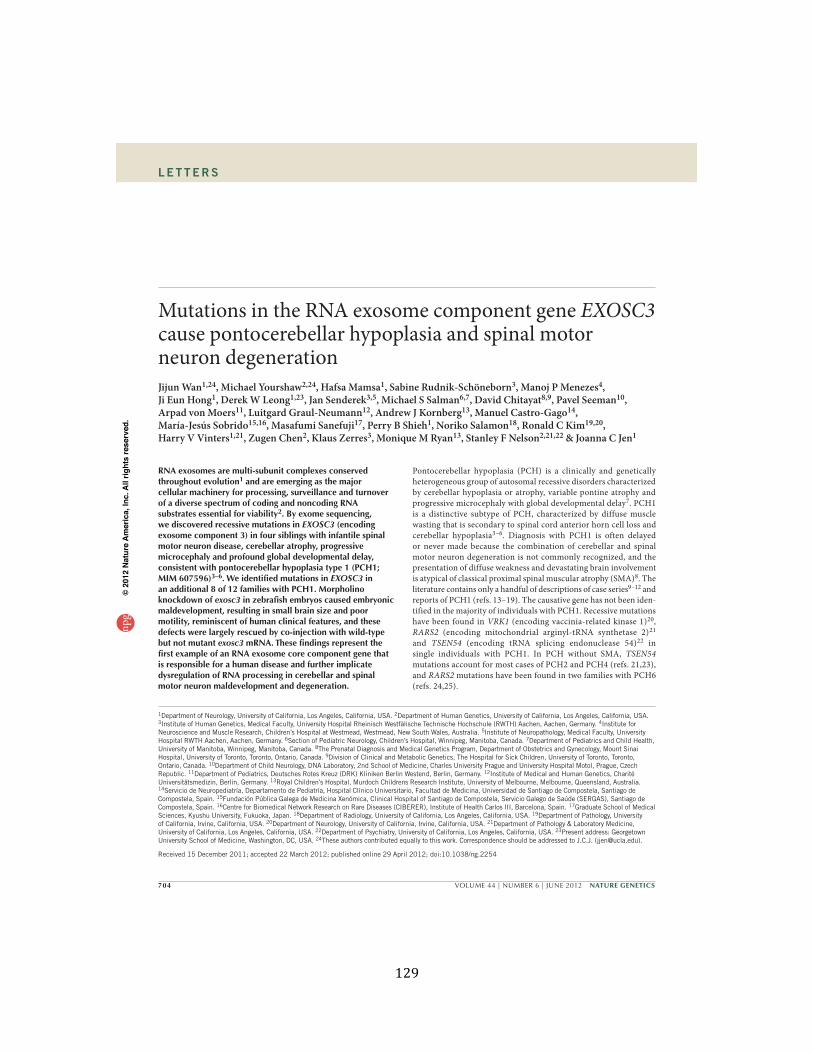
129
704 VOLUME 44 | NUMBER 6 | JUNE 2012 NATURE GENETICS
L E T T E R S
RNA exosomes are multi-subunit complexes conserved throughout evolution1 and are emerging as the major cellular machinery for processing, surveillance and turnover of a diverse spectrum of coding and noncoding RNA substrates essential for viability2. By exome sequencing, we discovered recessive mutations in EXOSC3 (encoding exosome component 3) in four siblings with infantile spinal motor neuron disease, cerebellar atrophy, progressive microcephaly and profound global developmental delay, consistent with pontocerebellar hypoplasia type 1 (PCH1; MIM 607596)3–6. We identified mutations in EXOSC3 in an additional 8 of 12 families with PCH1. Morpholino knockdown of exosc3 in zebrafish embryos caused embryonic maldevelopment, resulting in small brain size and poor motility, reminiscent of human clinical features, and these defects were largely rescued by co-injection with wild-type but not mutant exosc3 mRNA. These findings represent the first example of an RNA exosome core component gene that is responsible for a human disease and further implicate dysregulation of RNA processing in cerebellar and spinal motor neuron maldevelopment and degeneration.
Pontocerebellar hypoplasia (PCH) is a clinically and genetically heterogeneous group of autosomal recessive disorders characterized by cerebellar hypoplasia or atrophy, variable pontine atrophy and progressive microcephaly with global developmental delay7. PCH1 is a distinctive subtype of PCH, characterized by diffuse muscle wasting that is secondary to spinal cord anterior horn cell loss and cerebellar hypoplasia3–6. Diagnosis with PCH1 is often delayed or never made because the combination of cerebellar and spinal motor neuron degeneration is not commonly recognized, and the presentation of diffuse weakness and devastating brain involvement is atypical of classical proximal spinal muscular atrophy (SMA)8. The literature contains only a handful of descriptions of case series9–12 and reports of PCH1 (refs. 13–19). The causative gene has not been iden-tified in the majority of individuals with PCH1. Recessive mutations have been found in VRK1 (encoding vaccinia-related kinase 1)20, RARS2 (encoding mitochondrial arginyl-tRNA synthetase 2)21 and TSEN54 (encoding tRNA splicing endonuclease 54)22 in single individuals with PCH1. In PCH without SMA, TSEN54 mutations account for most cases of PCH2 and PCH4 (refs. 21,23), and RARS2 mutations have been found in two families with PCH6 (refs. 24,25).
Mutations in the RNA exosome component gene EXOSC3 cause pontocerebellar hypoplasia and spinal motor neuron degenerationJijun Wan1,24, Michael Yourshaw2,24, Hafsa Mamsa1, Sabine Rudnik-Schöneborn3, Manoj P Menezes4, Ji Eun Hong1, Derek W Leong1,23, Jan Senderek3,5, Michael S Salman6,7, David Chitayat8,9, Pavel Seeman10, Arpad von Moers11, Luitgard Graul-Neumann12, Andrew J Kornberg13, Manuel Castro-Gago14, María-Jesús Sobrido15,16, Masafumi Sanefuji17, Perry B Shieh1, Noriko Salamon18, Ronald C Kim19,20, Harry V Vinters1,21, Zugen Chen2, Klaus Zerres3, Monique M Ryan13, Stanley F Nelson2,21,22 & Joanna C Jen1
1Department of Neurology, University of California, Los Angeles, California, USA. 2Department of Human Genetics, University of California, Los Angeles, California, USA. 3Institute of Human Genetics, Medical Faculty, University Hospital Rheinisch Westfälische Technische Hochschule (RWTH) Aachen, Aachen, Germany. 4Institute for Neuroscience and Muscle Research, Children’s Hospital at Westmead, Westmead, New South Wales, Australia. 5Institute of Neuropathology, Medical Faculty, University Hospital RWTH Aachen, Aachen, Germany. 6Section of Pediatric Neurology, Children’s Hospital, Winnipeg, Manitoba, Canada. 7Department of Pediatrics and Child Health, University of Manitoba, Winnipeg, Manitoba, Canada. 8The Prenatal Diagnosis and Medical Genetics Program, Department of Obstetrics and Gynecology, Mount Sinai Hospital, University of Toronto, Toronto, Ontario, Canada. 9Division of Clinical and Metabolic Genetics, The Hospital for Sick Children, University of Toronto, Toronto, Ontario, Canada. 10Department of Child Neurology, DNA Laboratory, 2nd School of Medicine, Charles University Prague and University Hospital Motol, Prague, Czech Republic. 11Department of Pediatrics, Deutsches Rotes Kreuz (DRK) Kliniken Berlin Westend, Berlin, Germany. 12Institute of Medical and Human Genetics, Charité Universitätsmedizin, Berlin, Germany. 13Royal Children’s Hospital, Murdoch Childrens Research Institute, University of Melbourne, Melbourne, Queensland, Australia. 14Servicio de Neuropediatría, Departamento de Pediatría, Hospital Clínico Universitario, Facultad de Medicina, Universidad de Santiago de Compostela, Santiago de Compostela, Spain. 15Fundación Pública Galega de Medicina Xenómica, Clinical Hospital of Santiago de Compostela, Servicio Galego de Saúde (SERGAS), Santiago de Compostela, Spain. 16Centre for Biomedical Network Research on Rare Diseases (CIBERER), Institute of Health Carlos III, Barcelona, Spain. 17Graduate School of Medical Sciences, Kyushu University, Fukuoka, Japan. 18Department of Radiology, University of California, Los Angeles, California, USA. 19Department of Pathology, University of California, Irvine, California, USA. 20Department of Neurology, University of California, Irvine, California, USA. 21Department of Pathology & Laboratory Medicine, University of California, Los Angeles, California, USA. 22Department of Psychiatry, University of California, Los Angeles, California, USA. 23Present address: Georgetown University School of Medicine, Washington, DC, USA. 24These authors contributed equally to this work. Correspondence should be addressed to J.C.J. ([email protected]).
Received 15 December 2011; accepted 22 March 2012; published online 29 April 2012; doi:10.1038/ng.2254
npg
© 2
012
Nat
ure
Am
eric
a, In
c. A
ll rig
hts
rese
rved
.
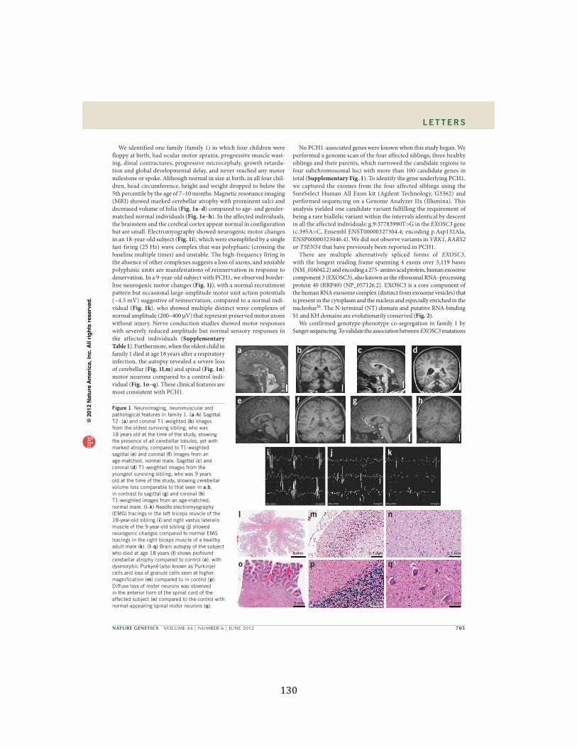
130
NATURE GENETICS VOLUME 44 | NUMBER 6 | JUNE 2012 705
L E T T E R S
We identified one family (family 1) in which four children were floppy at birth, had ocular motor apraxia, progressive muscle wast-ing, distal contractures, progressive microcephaly, growth retarda-tion and global developmental delay, and never reached any motor milestone or spoke. Although normal in size at birth, in all four chil-dren, head circumference, height and weight dropped to below the 5th percentile by the age of 7–10 months. Magnetic resonance imaging (MRI) showed marked cerebellar atrophy with prominent sulci and decreased volume of folia (Fig. 1a–d) compared to age- and gender-matched normal individuals (Fig. 1e–h). In the affected individuals, the brainstem and the cerebral cortex appear normal in configuration but are small. Electromyography showed neurogenic motor changes in an 18-year-old subject (Fig. 1i), which were exemplified by a single fast-firing (25 Hz) wave complex that was polyphasic (crossing the baseline multiple times) and unstable. The high-frequency firing in the absence of other complexes suggests a loss of axons, and unstable polyphasic units are manifestations of reinnervation in response to denervation. In a 9-year-old subject with PCH1, we observed border-line neurogenic motor changes (Fig. 1j), with a normal recruitment pattern but occasional large-amplitude motor unit action potentials (~4.5 mV) suggestive of reinnervation, compared to a normal indi-vidual (Fig. 1k), who showed multiple distinct wave complexes of normal amplitude (200–400 V) that represent preserved motor axons without injury. Nerve conduction studies showed motor responses with severely reduced amplitude but normal sensory responses in the affected individuals (Supplementary Table 1). Furthermore, when the oldest child in family 1 died at age 18 years after a respiratory infection, the autopsy revealed a severe loss of cerebellar (Fig. 1l,m) and spinal (Fig. 1n) motor neurons compared to a control indi-vidual (Fig. 1o–q). These clinical features are most consistent with PCH1.
No PCH1-associated genes were known when this study began. We performed a genome scan of the four affected siblings, three healthy siblings and their parents, which narrowed the candidate regions to four subchromosomal loci with more than 100 candidate genes in total (Supplementary Fig. 1). To identify the gene underlying PCH1, we captured the exomes from the four affected siblings using the SureSelect Human All Exon kit (Agilent Technology, G3362) and performed sequencing on a Genome Analyzer IIx (Illumina). This analysis yielded one candidate variant fulfilling the requirement of being a rare biallelic variant within the intervals identical by descent in all the affected individuals: g.9:37783990T>G in the EXOSC3 gene (c.395A>C, Ensembl ENST00000327304.4; encoding p.Asp132Ala, ENSP00000323046.4). We did not observe variants in VRK1, RARS2 or TSEN54 that have previously been reported in PCH1.
There are multiple alternatively spliced forms of EXOSC3, with the longest reading frame spanning 4 exons over 5,119 bases (NM_016042.2) and encoding a 275–amino acid protein, human exosome component 3 (EXOSC3), also known as the ribosomal RNA–processing protein 40 (RRP40) (NP_057126.2). EXOSC3 is a core component of the human RNA exosome complex (distinct from exosome vesicles) that is present in the cytoplasm and the nucleus and especially enriched in the nucleolus26. The N-terminal (NT) domain and putative RNA-binding S1 and KH domains are evolutionarily conserved (Fig. 2).
We confirmed genotype-phenotype co-segregation in family 1 by Sanger sequencing. To validate the association between EXOSC3 mutations
ba
2 cm
c d
e
5 mm
l
5 mm
o
0.1 mm
p
0.1 mm
q0.1 mm
m
0.1 mm
n500 µV/Div 10 mg/Div
i
500 µV/Div 10 mg/Div
j
200 µV/Div 10 mg/Div
k
f g hFigure 1 Neuroimaging, neuromuscular and pathological features in family 1. (a–h) Sagittal T2- (a) and coronal T1-weighted (b) images from the oldest surviving sibling, who was 18 years old at the time of the study, showing the presence of all cerebellar lobules, yet with marked atrophy, compared to T1-weighted sagittal (e) and coronal (f) images from an age-matched, normal male. Sagittal (c) and coronal (d) T1-weighted images from the youngest surviving sibling, who was 9 years old at the time of the study, showing cerebellar volume loss comparable to that seen in a,b, in contrast to sagittal (g) and coronal (h) T1-weighted images from an age-matched, normal male. (i–k) Needle electromyography (EMG) tracings in the left triceps muscle of the 18-year-old sibling (i) and right vastus lateralis muscle of the 9-year-old sibling (j) showed neurogenic changes compared to normal EMG tracings in the right biceps muscle of a healthy adult male (k). (l–q) Brain autopsy of the subject who died at age 18 years (l) shows profound cerebellar atrophy compared to control (o), with dysmorphic Purkyne (also known as Purkinje) cells and loss of granule cells seen at higher magnification (m) compared to in control (p). Diffuse loss of motor neurons was observed in the anterior horn of the spinal cord of the affected subject (n) compared to the control with normal-appearing spinal motor neurons (q).
npg
© 2
012
Nat
ure
Am
eric
a, In
c. A
ll rig
hts
rese
rved
.
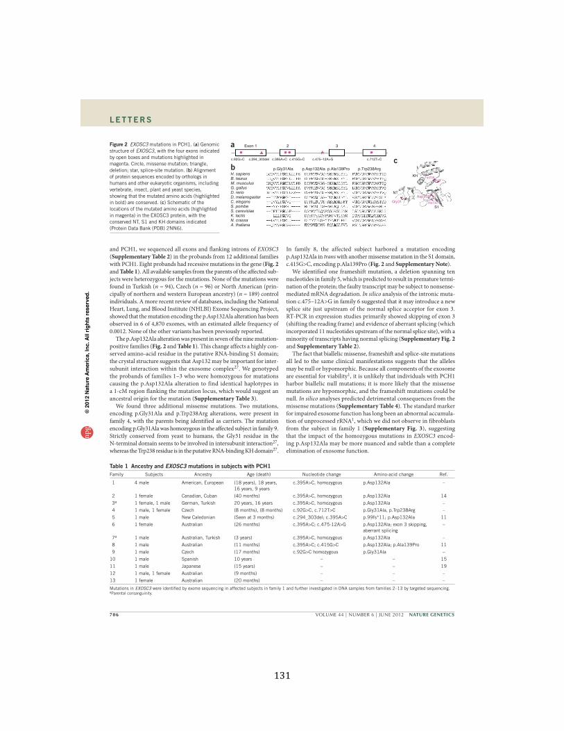
131
706 VOLUME 44 | NUMBER 6 | JUNE 2012 NATURE GENETICS
L E T T E R S
and PCH1, we sequenced all exons and flanking introns of EXOSC3 (Supplementary Table 2) in the probands from 12 additional families with PCH1. Eight probands had recessive mutations in the gene (Fig. 2 and Table 1). All available samples from the parents of the affected sub-jects were heterozygous for the mutations. None of the mutations were found in Turkish (n = 94), Czech (n = 96) or North American (prin-cipally of northern and western European ancestry) (n = 189) control individuals. A more recent review of databases, including the National Heart, Lung, and Blood Institute (NHLBI) Exome Sequencing Project, showed that the mutation encoding the p.Asp132Ala alteration has been observed in 6 of 4,870 exomes, with an estimated allele frequency of 0.0012. None of the other variants has been previously reported.
The p.Asp132Ala alteration was present in seven of the nine mutation- positive families (Fig. 2 and Table 1). This change affects a highly con-served amino-acid residue in the putative RNA-binding S1 domain; the crystal structure suggests that Asp132 may be important for inter-subunit interaction within the exosome complex27. We genotyped the probands of families 1–3 who were homozygous for mutations causing the p.Asp132Ala alteration to find identical haplotypes in a 1-cM region flanking the mutation locus, which would suggest an ancestral origin for the mutation (Supplementary Table 3).
We found three additional missense mutations. Two mutations, encoding p.Gly31Ala and p.Trp238Arg alterations, were present in family 4, with the parents being identified as carriers. The mutation encoding p.Gly31Ala was homozygous in the affected subject in family 9. Strictly conserved from yeast to humans, the Gly31 residue in the N-terminal domain seems to be involved in intersubunit interaction27, whereas the Trp238 residue is in the putative RNA-binding KH domain27.
In family 8, the affected subject harbored a mutation encoding p.Asp132Ala in trans with another missense mutation in the S1 domain, c.415G>C, encoding p.Ala139Pro (Fig. 2 and Supplementary Note).
We identified one frameshift mutation, a deletion spanning ten nucleotides in family 5, which is predicted to result in premature termi-nation of the protein; the faulty transcript may be subject to nonsense- mediated mRNA degradation. In silico analysis of the intronic muta-tion c.475–12A>G in family 6 suggested that it may introduce a new splice site just upstream of the normal splice acceptor for exon 3. RT-PCR in expression studies primarily showed skipping of exon 3 (shifting the reading frame) and evidence of aberrant splicing (which incorporated 11 nucleotides upstream of the normal splice site), with a minority of transcripts having normal splicing (Supplementary Fig. 2 and Supplementary Table 2).
The fact that biallelic missense, frameshift and splice-site mutations all led to the same clinical manifestations suggests that the alleles may be null or hypomorphic. Because all components of the exosome are essential for viability1, it is unlikely that individuals with PCH1 harbor biallelic null mutations; it is more likely that the missense mutations are hypomorphic, and the frameshift mutations could be null. In silico analyses predicted detrimental consequences from the missense mutations (Supplementary Table 4). The standard marker for impaired exosome function has long been an abnormal accumula-tion of unprocessed rRNA1, which we did not observe in fibroblasts from the subject in family 1 (Supplementary Fig. 3), suggesting that the impact of the homozygous mutations in EXOSC3 encod-ing p.Asp132Ala may be more nuanced and subtle than a complete elimination of exosome function.
KH Trp238
Gly31Asp132
Ala139
NT
S1
Exon 1
c.92G>C c.294_303del c.395A>C c.415G>C c.475–12A>G c.712T>C
2 3 4
p.Gly31AlaH. sapiensB. taurusM. musculusG. gallusD. rerioD. melanogasterC. elegansS. pombeS. cerevisiaeK. lactisN. crassaA. thaliana
p.Asp132Ala p.Ala139Pro p.Trp238Arg
a
bc
Figure 2 EXOSC3 mutations in PCH1. (a) Genomic structure of EXOSC3, with the four exons indicated by open boxes and mutations highlighted in magenta. Circle, missense mutation; triangle, deletion; star, splice-site mutation. (b) Alignment of protein sequences encoded by orthologs in humans and other eukaryotic organisms, including vertebrate, insect, plant and yeast species, showing that the mutated amino acids (highlighted in bold) are conserved. (c) Schematic of the locations of the mutated amino acids (highlighted in magenta) in the EXOSC3 protein, with the conserved NT, S1 and KH domains indicated (Protein Data Bank (PDB) 2NN6).
Table 1 Ancestry and EXOSC3 mutations in subjects with PCH1Family Subjects Ancestry Age (death) Nucleotide change Amino-acid change Ref.
1 4 male American, European (18 years), 18 years, 16 years, 9 years
c.395A>C, homozygous p.Asp132Ala –
2 1 female Canadian, Cuban (40 months) c.395A>C, homozygous p.Asp132Ala 14 3a 1 female, 1 male German, Turkish 20 years, 16 years c.395A>C, homozygous p.Asp132Ala – 4 1 male, 1 female Czech (8 months), (8 months) c.92G>C, c.712T>C p.Gly31Ala, p.Trp238Arg – 5 1 male New Caledonian (Seen at 3 months) c.294_303del; c.395A>C p.99fs*11; p.Asp132Ala 11 6 1 female Australian (26 months) c.395A>C; c.475-12A>G p.Asp132Ala; exon 3 skipping,
aberrant splicing–
7a 1 male Australian, Turkish (3 years) c.395A>C, homozygous p.Asp132Ala – 8 1 male Australian (11 months) c.395A>C; c.415G>C p.Asp132Ala; p.Ala139Pro 11 9 1 male Czech (17 months) c.92G>C homozygous p.Gly31Ala –10 1 male Spanish 10 years – – 1511 1 male Japanese (15 years) – – 1912 1 male, 1 female Australian (9 months) – – –13 1 female Australian (20 months) – – –
Mutations in EXOSC3 were identified by exome sequencing in affected subjects in family 1 and further investigated in DNA samples from families 2–13 by targeted sequencing. aParental consanguinity.
npg
© 2
012
Nat
ure
Am
eric
a, In
c. A
ll rig
hts
rese
rved
.
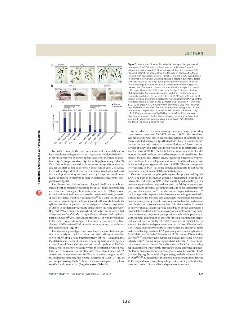
132
NATURE GENETICS VOLUME 44 | NUMBER 6 | JUNE 2012 707
L E T T E R S
To further examine the functional effects of the mutations, we knocked down endogenous exosc3 expression (NM_001029961.1) in zebrafish embryos by exosc3-specific antisense morpholino injec-tion (Fig. 3, Supplementary Fig. 4 and Supplementary Table 2). Zebrafish embryos injected with antisense morpholinos directed against the start codon or the splice-donor site of exon 2 of exosc3 led to a dose-dependent phenotype of a short, curved spine and small brain with poor motility and even death by 3 days post fertilization (d.p.f.) compared to embryos injected with nonspecific, control mor-pholinos (Fig. 3a).
The observation of shrunken or collapsed hindbrain in embryos injected with morpholinos targeting the splice-donor site prompted us to further investigate hindbrain-specific cells. Whole-mount in situ hybridization showed decreased expression of atoh1a (a marker specific for dorsal hindbrain progenitors)28 by 1 d.p.f. in the upper and lower rhombic lips in embryos injected with morpholinos to the splice-donor site compared to the normal pattern of robust expression of atoh1a in hindbrain progenitors in the control-injected embryos28 (Fig. 3b). Whole-mount in situ hybridization further showed a lack of expression of pvalb7, which is specific for differentiated cerebellar Purkinje neurons28, by 3 d.p.f. in embryos injected with morpholinos to the splice-donor site compared to normal expression in distinct clusters of differentiated Purkinje cells in embryos injected with con-trol morpholinos (Fig. 3b).
The abnormal phenotype from exosc3-specific morpholino injec-tion was largely rescued by co-injection with wild-type zebrafish exosc3 mRNA (Fig. 3c and Supplementary Table 5), suggesting that the detrimental effects of the antisense morpholinos were specific to exosc3 knockdown. Co-injection with wild-type human EXOSC3 mRNA, which shares 67% identity with the zebrafish ortholog, was less effective in rescue. Co-injection with zebrafish or human mRNA containing the mutations was ineffective in rescue, suggesting that the mutations disrupted the normal function of EXOSC3 (Fig. 3c and Supplementary Table 5). Survival data of embryos 1–3 d.p.f. are stratified and summarized (Supplementary Table 5).
We have discovered disease-causing mutations in a gene encoding the exosome component EXOSC3 leading to PCH1 with combined cerebellar and spinal motor neuron degeneration of infantile onset. There is clinical heterogeneity. Affected individuals in families 1 and 3 do not present with primary hypoventilation and have survived beyond infancy and early childhood, which is exceptionally unu-sual for classical PCH1 (refs. 7,23). Furthermore, in families 1 and 2, autopsy showed profound cerebellar atrophy and variable involve-ment of the pons and inferior olives, suggesting a degenerative proc-ess in addition to a developmental disorder. Additional studies will facilitate endophenotype stratification of PCH1. There is clear genetic heterogeneity in PCH1, as some affected individuals do not harbor mutations in any known PCH1-associated genes.
RNA exosomes are the principal enzymes that process and degrade RNA. The bulk of the human genome is transcribed to produce an extraordinary diversity of RNA29. The versatility and specificity of the exosome regulate the activity and maintain the fidelity of gene expres-sion. Although exosomes are immunogenic in some individuals with polymyositis-scleroderma30,31 or chronic myelogenous leukemia32,33, the findings in this report are the first to our knowledge to establish a pathogenic role for exosome core component mutations in human dis-ease. Despite a growing effort to examine exosome function and subunit contribution, its substrates have not been fully characterized in humans or in lower animals, and the specific contribution of each component is incompletely understood. The discovery of naturally occurring muta-tions in exosome component genes provides a valuable opportunity to define subunit contribution to exosome function. Our findings suggest that normal function of the EXOSC3 component is essential for the survival of cerebellar and spinal motor neurons. Of note, RNA dysregula-tion is increasingly understood to be important in the etiology of motor and cerebellar degeneration. RNA processing defects are implicated in SMN1 deficiency in SMA8. Mutations in RNA- and/or DNA-binding proteins34–37 and pathogenic repeat expansions generating RNA that is likely toxic38,39 cause amyotrophic lateral sclerosis (ALS), an adult-onset motor neuron disease. Gain of function of RNA from noncoding repeat expansions was recently proposed to cause combined spinocer-ebellar and brainstem motor neuron degeneration of late onset in SCA36 (ref. 40). Dysregulation of tRNA processing underlies other subtypes of PCH21,23,24. Elucidation of the pathological mechanism underlying PCH1 may lead to new insights regarding RNA processing in the develop-ment and survival of cerebellar and spinal motor neurons.
a
b
c
Control URL LRL
URL
LRL
SPL*
*
Control SPL
100 µm
atoh1a pvalb7
Control
3 ng 1.5 ng 3 ng 1.5 ng 3 ng
1 mm
AUG SPL
1.0
0.5
0
(289) (233) (151) (202) (209) (174)** * * * *(75) (149) (98)
SPL + zW
T mRNA
SPL + zG
A mRNA
SPL + zD
A mRNA
SPL + zW
R mRNA
SPL + hW
T mRNA
SPL + hG
A mRNA
SPL + hD
A mRNA
SPL + hW
R mRNA
SPL + G
FP mRNA
Phe
noty
pe fr
actio
n
NormalAbnormal
Figure 3 Knockdown of exosc3 in zebrafish embryos disrupts normal development. (a) Zebrafish embryos injected with exosc3-specific antisense morpholinos AUG (directed against the start codon) or SPL (directed against the splice-donor site for exon 2) compared to those injected with nonspecific control. (b) Whole-mount in situ hybridization in embryos injected with SPL morpholinos in lateral view (inset, dorsal view with rostral to the left) showing diminished expression of dorsal hindbrain progenitor–specific marker atoh1a and cerebellar-specific marker pvalb7 compared to embryos injected with nonspecific control. URL, upper rhombic lip; LRL, lower rhombic lip; *, distinct clusters of differentiated Purkinje cells in embryos 3 d.p.f. (c) Survival data from embryos 3 d.p.f. co-injected with 3 ng of SPL and with 240 pg of human EXOSC3 or zebrafish exosc3 mRNA versus GFP mRNA as control from three separate experiments. z, zebrafish; h, human; WT, wild-type EXOSC3 or exosc3; GA, mutant mRNA encoding p.Gly31Ala in human or p.Gly20Ala in zebrafish; DA, mutant mRNA encoding p.Asp132Ala in human or p.Asp102Ala in zebrafish; WR, mutant mRNA encoding p.Trp238Arg in human or p.Trp208Arg in zebrafish. Embryos were classified as normal (blue) or abnormal (gray; including embryos that were mildly abnormal, severely abnormal or dead). *P < 0.0001, two-tailed Pearson’s -squared test.
npg
© 2
012
Nat
ure
Am
eric
a, In
c. A
ll rig
hts
rese
rved
.
133
708 VOLUME 44 | NUMBER 6 | JUNE 2012 NATURE GENETICS
URLs. NHLBI Exome Sequencing Project, http://evs.gs.washington.edu/EVS/; Novocraft, http://www.novocraft.com; Ensembl, http://www.ensembl.org/; dbSNP132, http://www.ncbi.nlm.nih.gov/projects/SNP/; 1000 Genome Project, http://www.1000genomes.org/.
METHODSMethods and any associated references are available in the online version of the paper.
Note: Supplementary information is available in the online version of the paper.
ACKNOWLEDGMENTSThe authors are grateful for the generosity of the families who participated in this study. The sequencing and analytical work were supported by the Bioinformatics and Genomics Core of the UCLA Muscular Dystrophy Core Center (US National Institutes of Health (NIH)/National Institute of Arthritis and Musculoskeletal and Skin Diseases (NIAMS) P30AR057230) within the Center for Duchenne Muscular Dystrophy. We appreciate fibroblasts from three subjects from the Eunice Kennedy Shriver National Institute of Child Health & Human Development (NICHD) Brain and Tissue Bank for Developmental Disorders. We acknowledge the contribution of clinical data by P. Carpenter (University of California, Irvine). The work was supported by the NIH/National Eye Institute (NEI) (R01 EY015311 and NINDS R01 NS064183 to J.C.J.), Deutsche Forschungsgemeinschaft (Ru-746/1-1 to S.R.-S. and K.Z.), IZKF Aachen (N5-4 to S.R.-S. and J.S.), the Australian National Health and Medical Research Council (NHMRC) Centre for Research Excellence (M.M.R.), the Internal Grant Agency of the Ministry of Health of the Czech Republic (NS 10552-3 to P.S.) and Xunta de Galicia–Plan Galego de InvestigaCión, Desenvolvemento e Innovación Tecnolóxica (INCITE) (10PXIB9101280PR to M.-J.S.).
AUTHOR CONTRIBUTIONSS.F.N. and J.C.J. designed the study. M.Y., Z.C. and S.F.N. analyzed data from SNP genotyping and exome sequencing. S.R.-S., M.P.M., J.S., M.S.S., D.C., P.S., A.v.M., L.G.-N., A.J.K., M.C.-G., M.-J.S., M.S., P.B.S., N.S., R.C.K., H.V.V., K.Z. and M.M.R. provided and analyzed clinical material from subjects. H.M., J.E.H. and D.W.L. performed and analyzed data from Sanger sequencing of subjects and controls. H.M. and J.E.H. generated constructs and performed molecular genetics studies. J.W. performed and analyzed the functional studies in zebrafish. All authors contributed to the manuscript written by J.C.J.
COMPETING FINANCIAL INTERESTSThe authors declare no competing financial interests.
Published online at http://www.nature.com/doifinder/10.1038/ng.2254. Reprints and permissions information is available online at http://www.nature.com/reprints/index.html.
1. Mitchell, P., Petfalski, E., Shevchenko, A., Mann, M. & Tollervey, D. The exosome: a conserved eukaryotic RNA processing complex containing multiple 3 5 exoribonucleases. Cell 91, 457–466 (1997).
2. Jensen, T.H. RNA exosome. Adv. Exp. Med. Biol. 702 (2010).3. Norman, R.M. Cerebellar hypoplasia in Werdnig-Hoffmann disease. Arch. Dis. Child.
36, 96–101 (1961).4. Goutières, F., Aicardi, J. & Farkas, E. Anterior horn cell disease associated with
pontocerebellar hypoplasia in infants. J. Neurol. Neurosurg. Psychiatry 40, 370–378 (1977).
5. de Leén, G.A., Grover, W.D. & D’Cruz, C.A. Amyotrophic cerebellar hypoplasia: a specific form of infantile spinal atrophy. Acta Neuropathol. 63, 282–286 (1984).
6. Barth, P.G. Pontocerebellar hypoplasias. An overview of a group of inherited neurodegenerative disorders with fetal onset. Brain Dev. 15, 411–422 (1993).
7. Namavar, Y., Barth, P.G., Poll-The, B.T. & Baas, F. Classification, diagnosis and potential mechanisms in pontocerebellar hypoplasia. Orphanet J. Rare Dis. 6, 50 (2011).
8. Melki, J. et al. De novo and inherited deletions of the 5q13 region in spinal muscular atrophies. Science 264, 1474–1477 (1994).
9. Görgen-Pauly, U., Sperner, J., Reiss, I., Gehl, H.B. & Reusche, E. Familial pontocerebellar hypoplasia type I with anterior horn cell disease. Eur. J. Paediatr. Neurol. 3, 33–38 (1999).
10. Muntoni, F. et al. Clinical spectrum and diagnostic difficulties of infantile ponto-cerebellar hypoplasia type 1. Neuropediatrics 30, 243–248 (1999).
11. Ryan, M.M., Cooke-Yarborough, C.M., Procopis, P.G. & Ouvrier, R.A. Anterior horn cell disease and olivopontocerebellar hypoplasia. Pediatr. Neurol. 23, 180–184 (2000).
12. Rudnik-Schöneborn, S. et al. Extended phenotype of pontocerebellar hypoplasia with infantile spinal muscular atrophy. Am. J. Med. Genet. A 117A, 10–17 (2003).
13. Chou, S.M. et al. Infantile olivopontocerebellar atrophy with spinal muscular atrophy (infantile OPCA + SMA). Clin. Neuropathol. 9, 21–32 (1990).
14. Salman, M.S. et al. Pontocerebellar hypoplasia type 1: new leads for an earlier diagnosis. J. Child Neurol. 18, 220–225 (2003).
15. Gómez-Lado, C., Eiris-Punal, J., Vazquez-Lopez, M.E. & Castro-Gago, M. Pontocerebellar hypoplasia type I and mitochondrial pathology. Rev. Neurol. 45, 639–640 (2007).
16. Lev, D. et al. Infantile onset progressive cerebellar atrophy and anterior horn cell degeneration—a late onset variant of PCH-1? Eur. J. Paediatr. Neurol. 12, 97–101 (2008).
17. Szabó, N., Szabo, H., Hortobagyi, T., Turi, S. & Sztriha, L. Pontocerebellar hypoplasia type 1. Pediatr. Neurol. 39, 286–288 (2008).
18. Tsao, C.Y., Mendell, J., Sahenk, Z., Rusin, J. & Boue, D. Hypotonia, weakness, and pontocerebellar hypoplasia in siblings. Semin. Pediatr. Neurol. 15, 151–153 (2008).
19. Sanefuji, M. et al. Autopsy case of later-onset pontocerebellar hypoplasia type 1: pontine atrophy and pyramidal tract involvement. J. Child Neurol. 25, 1429–1434 (2010).
20. Renbaum, P. et al. Spinal muscular atrophy with pontocerebellar hypoplasia is caused by a mutation in the VRK1 gene. Am. J. Hum. Genet. 85, 281–289 (2009).
21. Namavar, Y. et al. Clinical, neuroradiological and genetic findings in pontocerebellar hypoplasia. Brain 134, 143–156 (2011).
22. Simonati, A., Cassandrini, D., Bazan, D. & Santorelli, F.M. TSEN54 mutation in a child with pontocerebellar hypoplasia type 1. Acta Neuropathol. 121, 671–673 (2011).
23. Budde, B.S. et al. tRNA splicing endonuclease mutations cause pontocerebellar hypoplasia. Nat. Genet. 40, 1113–1118 (2008).
24. Edvardson, S. et al. Deleterious mutation in the mitochondrial arginyl-transfer RNA synthetase gene is associated with pontocerebellar hypoplasia. Am. J. Hum. Genet. 81, 857–862 (2007).
25. Rankin, J. et al. Pontocerebellar hypoplasia type 6: a British case with PEHO-like features. Am. J. Med. Genet. A 152A, 2079–2084 (2010).
26. Brouwer, R. et al. Three novel components of the human exosome. J. Biol. Chem. 276, 6177–6184 (2001).
27. Liu, Q., Greimann, J.C. & Lima, C.D. Reconstitution, activities, and structure of the eukaryotic RNA exosome. Cell 127, 1223–1237 (2006).
28. Kani, S. et al. Proneural gene–linked neurogenesis in zebrafish cerebellum. Dev. Biol. 343, 1–17 (2010).
29. Birney, E. et al. Identification and analysis of functional elements in 1% of the human genome by the ENCODE pilot project. Nature 447, 799–816 (2007).
30. Wolfe, J.F., Adelstein, E. & Sharp, G.C. Antinuclear antibody with distinct specificity for polymyositis. J. Clin. Invest. 59, 176–178 (1977).
31. Allmang, C. et al. The yeast exosome and human PM-Scl are related complexes of 3 5 exonucleases. Genes Dev. 13, 2148–2158 (1999).
32. Yang, X.F. et al. CML28 is a broadly immunogenic antigen, which is overexpressed in tumor cells. Cancer Res. 62, 5517–5522 (2002).
33. Xie, L.H. et al. Activation of cytotoxic T lymphocytes against CML28-bearing tumors by dendritic cells transduced with a recombinant adeno-associated virus encoding the CML28 gene. Cancer Immunol. Immunother. 57, 1029–1038 (2008).
34. Kabashi, E. et al. TARDBP mutations in individuals with sporadic and familial amyotrophic lateral sclerosis. Nat. Genet. 40, 572–574 (2008).
35. Sreedharan, J. et al. TDP-43 mutations in familial and sporadic amyotrophic lateral sclerosis. Science 319, 1668–1672 (2008).
36. Kwiatkowski, T.J. Jr. et al. Mutations in the FUS/TLS gene on chromosome 16 cause familial amyotrophic lateral sclerosis. Science 323, 1205–1208 (2009).
37. Vance, C. et al. Mutations in FUS, an RNA processing protein, cause familial amyotrophic lateral sclerosis type 6. Science 323, 1208–1211 (2009).
38. DeJesus-Hernandez, M. et al. Expanded GGGGCC hexanucleotide repeat in noncoding region of C9ORF72 causes chromosome 9p–linked FTD and ALS. Neuron 72, 245–256 (2011).
39. Renton, A.E. et al. A hexanucleotide repeat expansion in C9ORF72 is the cause of chromosome 9p21–linked ALS-FTD. Neuron 72, 257–268 (2011).
40. Kobayashi, H. et al. Expansion of intronic GGCCTG hexanucleotide repeat in NOP56 causes SCA36, a type of spinocerebellar ataxia accompanied by motor neuron involvement. Am. J. Hum. Genet. 89, 121–130 (2011).
L E T T E R S
npg
© 2
012
Nat
ure
Am
eric
a, In
c. A
ll rig
hts
rese
rved
.
134
NATURE GENETICSdoi:10.1038/ng.2254
ONLINE METHODSClinical characterization. PCH1 was diagnosed in 13 unrelated families from around the world, with documented congenital combined cerebellar and spinal motor neuron disease. Almost all affected subjects were hypotonic from birth. All had spontaneous breathing. Neurogenic muscle atrophy with spinal motor neuron disease was confirmed by EMG, muscle biopsy or autopsy. Many sub-jects developed progressive microcephaly, with prominent cerebellar atrophy and variable involvement of the brainstem, as determined by MRI or autopsy.
Genetic analysis. DNA was extracted from peripheral blood, with consent from all participants and their legal guardians, using standard methods. The study was approved by the University of California, Los Angeles (UCLA) Institutional Review Board. For exome sequencing, each library produced approximately 28 million single-end 76-bp reads. The mean coverage of bases in the target exomes was 23×. Raw reads that passed Illumina’s quality filters were aligned to the reference human genome Build 37 with Novoalign from Novocraft. The GATK UnifiedGenotyper was used to call single-nucleotide variants and indels41,42. Each case had ~15,000 variants not present in the GRCh37 reference human genome, amounting to 19,098 total variants in the 4 cases. We limited the search to variants within the coding regions or flanking intronic essential splice sites of protein-coding genes in the Ensembl data set. Under the hypothesis that the disorder was rare and the causative allele(s) would therefore not be common, we filtered out variants that were in dbSNP132 (refs. 43,44) and the 1000 Genomes Project45, leaving ~400 variants in each case and a total of 699 variants. Under a recessive model, we searched for homozygous variants and compound heterozygous variants (defined as 2 variants in the same transcript) that were shared by all 4 cases (15 and 10 vari-ants, respectively). To compensate for bias in our own analytical system, we then filtered out variants that we had identified in 25 exomes from unrelated, unaffected subjects. Sanger sequencing for further validation was performed using standard protocols (Supplementary Table 2).
Zebrafish morpholino injection and in situ hybridization. Cloning, muta-genesis and in vitro mRNA synthesis were performed using standard protocols (Supplementary Table 2). Zebrafish embryos were provided by the UCLA Zebrafish Core Facility, and procedures were approved by the UCLA Animal Research Committee. Fish were maintained at 28 °C using a 14-h light/10-h dark cycle and bred to obtain embryos. Morpholino oligonucleotides (Gene Tools) were designed to block translation initiation or the splice-donor site of exon 2 of zebrafish exosc3 pre-mRNA (Supplementary Table 2). We obtained a standard control from Gene Tools. We used fine glass needles and a micro-injector to perform injections of embryos at the one-cell stage. The injec-tion volume ranged from 0.5 to 2.0 nl at a concentration of 3 ng/nl. Embryos were incubated in E3 medium (5 mM NaCl, 0.17 mM KCl, 0.33 mM CaCl2, 0.33 mM MgSO4 and 0.00001% methylene blue; Sigma) at 28 °C.
Whole-mount in situ hybridization was performed, and the expression of specific genes was detected using an alkaline phosphatase–conjugated antibody against digoxigenin (DIG) and a chromogenic substrate, as described previously46.
Riboprobe generation. To generate the antisense probe, full-length zebrafish exosc3 cDNA was digested with EcoRI and NotI and ligated into pCR-Blunt II-TOPO (Invitrogen) with T4 DNA ligase. The sense probe was transcribed using full-length zebrafish exosc3 in pcDNA3.1/Zeo(+) (Invitrogen).
Full-length zebrafish pvalb7 (clone 7087368, Thermo Scientific Open Biosystems) was digested with EcoRI and NotI and ligated into pCR-Blunt II-TOPO with T4 DNA ligase. The sense probe was transcribed in vitro from the SP6 promoter, and the antisense was transcribed from the T7 promoter.
Full-length zebrafish atoh1a (clone 7428977; Thermo Scientific Open Biosystems) was digested with EcoRI and NotI and ligated into pcDNA3.1/Zeo(+) to generate the sense probe and pcDNA3.1/Zeo(−) (Invitrogen) for the antisense probe.
RNA probes for in situ hybridization were generated with the mMESSAGE mMACHINE SP6 or mMESSAGE mMACHINE T7 Ultra kits (Ambion), sub-stituting the DIG RNA labeling Mix (Roche Applied Science) for the NTP mix supplied in the kit. All probes were analyzed by denaturing agarose gel electrophoresis and quantified by NanoDrop.
Assessing functional impact on EXOSC3 splicing. We obtained full-length EXOSC3 constructs (all exons and intervening introns) by PCR amplification using Phusion High-Fidelity DNA Polymerase (NEB; Supplementary Table 2) from genomic DNA of a normal subject and the proband of family 6 (harbor-ing the c.475–12A>G mutation). Gel-extracted amplicons were cloned into pcDNA3.1/Zeo(+) (linearized with BamHI and XhoI) by using the In-Fusion HD Cloning System (Clonetech). The full-length clones were confirmed by bidirectional Sanger sequencing.
The wild-type or mutant full-length constructs were introduced into HeLa cells by transfection with Lipofectamine 2000 (Invitrogen). Two days after transfection, RNA was extracted using TRIzol Reagent (Invitrogen). cDNA was generated using the Transcriptor First Strand cDNA Synthesis kit (Roche Applied Science) and the BGH reverse primer–binding site located upstream of the BGH polyadenylation signal on pcDNA3.1/Zeo(+) (Supplementary Fig. 2). Reverse transcription from the BGHr site ensured that cDNA was synthesized exclusively from exogenous mRNA and eliminated transcription from endogenous EXOSC3. cDNA (1 l) was PCR amplified with Phusion High-Fidelity DNA Polymerase and the hEXOSC3-c.423f and hEXOSC3-c.670r primers (Supplementary Fig. 2 and Supplementary Table 2). RT-PCR products were resolved on a 12% polyacrylamide gel and visualized by GelRed (Biotium). We observed multiple products for both wild-type and mutant constructs. To ascertain the identity of each product, amplicons were cloned into pCR-Blunt II-TOPO and directly sequenced.
Cloning, mutagenesis and in vitro transcription. Full-length human EXOSC3 cDNA (clone 3346075; Thermo Scientific Open Biosystems) was digested with EcoRI and XhoI and ligated into pcDNA3.1/Zeo(+)with T4 DNA ligase. Full-length zebrafish exosc3 (clone 7282897; Thermo Scientific Open Biosystems) was digested with EcoRI and NotI and ligated into pcDNA3.1/Zeo(+) with T4 DNA ligase. Specific missense mutations were introduced into the wild-type cDNA constructs with the QuikChange II XL Site-Directed Mutagenesis kit (Agilent Technologies; Supplementary Table 2). All clones were fully sequenced bidirectionally by Sanger sequencing. In vitro transcription was performed with the mMESSAGE mMACHINE T7 Ultra kit. Resulting mRNA was analyzed by denaturing agarose gel electrophoresis and quantified by NanoDrop.
Cell culture, siRNA transfection and RNA extraction. All medium components were purchased from Invitrogen. HeLa cells were cultured in DMEM supple-mented with 10% FBS at 37 °C and 5% CO2. Cells were transfected every 48 h over 6 d with 60 nM siRNA duplexes (sense: 5 -CACGCACAGUACUAGGUCATT-3 ) with Lipofectamine 2000 or left untreated, as detailed previously47. Cells were lysed in TRIzol Reagent, and RNA was extracted from the aqueous phase and protein from the remaining organic phase.
Fibroblasts were grown in DMEM supplemented with 20% FBS, 100 nM MEM Non-Essential Amino Acids, 1 mM GlutaMAX, 100 U penicillin and 100 g streptomycin at 37 °C and 5% CO2. Total RNA was extracted with TRIzol Reagent.
RNA blots. RNA blotting was performed following standard protocols. RNA integrity was analyzed by denaturing agarose gel electrophoresis, and RNA was quantified by NanoDrop. Total RNA (4 g) was resolved on a 6% denaturing polyacrylamide gel and transferred to positively charged nylon membrane (Roche). RNA was cross-linked to the membrane, and blots were hybridized with DIG-labeled, locked nucleic acid (LNA)-modified antisense oligonu-cleotide probe to 5.8S rRNA (5 -CGAAGTGTCGATGATCAAT-3×DIG-3 ; LNA in bold). Bound probes were detected with an alkaline phosphatase– conjugated antibody to DIG (1:10,000 dilution) and CSPD chemiluminescence (Roche Applied Science).
Protein blots. Protein concentration was determined using the Micro BCA Protein Assay kit (Pierce). Lysates (10 g) were separated by SDS-PAGE and transferred to a nitrocellulose membrane. Protein blotting was performed following standard protocols with mouse monoclonal antibody to EXOSC3 (Santa Cruz Biotechnology, sc-166568; 1:400 dilution) and with secondary horseradish peroxidase–conjugated horse antibody to mouse IgG (Vector Laboratories, PI-2000; 1:5,000 dilution). Blots were subsequently stripped
npg
© 2
012
Nat
ure
Am
eric
a, In
c. A
ll rig
hts
rese
rved
.
135
NATURE GENETICS doi:10.1038/ng.2254
and reprobed with chicken antibody to GAPDH (Millipore, ab2302; 1:1,000 dilution) and secondary horseradish peroxidase–conjugated goat antibody to chicken IgY (Abcam, ab97150; 1:5,000 dilution). Bound antibodies were visualized with Amersham ECL Plus Western Blotting Detection Reagents.
41. McKenna, A. et al. The Genome Analysis Toolkit: a MapReduce framework for analyzing next-generation DNA sequencing data. Genome Res. 20, 1297–1303 (2010).
42. DePristo, M.A. et al. A framework for variation discovery and genotyping using next-generation DNA sequencing data. Nat. Genet. 43, 491–498 (2011).
43. Biesecker, L.G. et al. The ClinSeq Project: piloting large-scale genome sequencing for research in genomic medicine. Genome Res. 19, 1665–1674 (2009).
44. Bhagwat, M. Searching NCBI’s dbSNP database. Curr. Protoc. Bioinformatics Chapter 1, Unit 1.19 (2010).
45. 1000 Genomes Project Consortium. A map of human genome variation from population-scale sequencing. Nature 467, 1061–1073 (2010).
46. Thisse, C. & Thisse, B. High-resolution in situ hybridization to whole-mount zebrafish embryos. Nat. Protoc. 3, 59–69 (2008).
47. Preker, P. et al. RNA exosome depletion reveals transcription upstream of active human promoters. Science 322, 1851–1854 (2008).
npg
© 2
012
Nat
ure
Am
eric
a, In
c. A
ll rig
hts
rese
rved
.
136
*These�authors�contributed�equally.�†Current�address:�Georgetown�University�School�of�Medicine,�Washington�D.C.,�U.S.A.���Corresponding�Author:��J.�C.�Jen�UCLA�Neurology�710�Westwood�Plaza�Los�Angeles,�CA�90095Ͳ1769�TEL�310�825�3731�email�[email protected]���Supplementary�Note�Ascertainment�of�biallelic�mutations�in�families�5�and�8�by�longͲrange�PCR�Since�parental�DNA�was�not�available�for�these�two�families,�we�determined�that�the�mutations�were�in�trans�by�long�range�sequencing.�In�Family�5,�we�amplified�in�the�patient�the�genomic�sequence�spanning�exons�1�and�2�(Supplementary�Table�2)�with�Phusion�HighͲFidelity�DNA�Polymerase�(NEB)�and�cloned�the�gelͲextracted�1344�bp�product�into�pCRͲBlunt�IIͲTOPO�(Invitrogen)�for�transformation�of�competent�bacterial�cells.��The�two�mutations�were�found�in�separate�clones,�as�confirmed�by�direct�sequencing�of�purified�plasmids.��For�Family�8,�we�PCR�amplified�the�entire�gene�genomic�DNA�with�Phusion�HighͲFidelity�DNA�Polymerase�(NEB)�and�primers�hEXOSC3ͲATGf�and�hEXOSC3ͲRcr�(Supplementary�Table�2).��The�gel�extracted�amplicon�was�cloned�into�pcDNA3.1/Zeo(+)�(Invitrogen)�with�InͲFusion®�HD�Cloning�System�(Clonetech)�and�transformed�into�competent�bacterial�cells.��Plasmids�from�multiple�colonies�were�sequenced�to�ascertain�that�the�two�mutations�resided�in�different�clones.������
Nature Genetics: doi:10.1038/ng.2254
137
SUPPLEMENTARY�INFORMATION��Mutations�in�the�RNA�exosome�component�gene�EXOSC3�cause�pontocerebellar�hypoplasia�and�spinal�motor�neuron�degeneration��Jijun�Wan*1,�Michael�Yourshaw*2,�Hafsa�Mamsa1,�Sabine�RudnikͲSchöneborn3,�Manoj�P.�Menezes4,�Ji�Eun�Hong1,�Derek�W.�Leong1†,�Jan�Senderek3,5,�Michael�S.�Salman6,�David�Chitayat7,8,�Pavel�Seeman9,�Arpad�von�Moers10,�Luitgard�GraulͲNeumann11,�Andrew�J.�Kornberg12,�Manuel�CastroͲGago13,�MaríaͲJesús�Sobrido14,15,�Masafumi�Sanefuji16,�Perry�B.�Shieh1,�Noriko�Salamon17,�Ronald�C.�Kim18,�19,�Harry�V.�Vinters1,20,�Zugen�Chen2,�Klaus�Zerres3,�Monique�M.�Ryan12,�Stanley�F.�Nelson2,�20,�21,�&�Joanna�C.�Jen1.��1Department�of�Neurology,�University�of�California,�Los�Angeles,�U.S.A.�2Department�of�Human�Genetics,�University�of�California,�Los�Angeles,�U.S.A.�3Institute�of�Human�Genetics,�Medical�Faculty,�University�Hospital�Rheinisch�Westfälische�Technische�Hochschule�(RWTH)�Aachen,�Germany��4�Institute�for�Neuroscience�and�Muscle�Research,�Children's�Hospital�at�Westmead,�Westmead,�Australia.�5Institute�of�Neuropathology,�Medical�Faculty,�University�Hospital�RWTH�Aachen,�Germany�6Section�of�Pediatric�Neurology,�Children's�Hospital�&�Department�of�Pediatrics�and�Child�Health,�University�of�Manitoba,�Winnipeg,�Manitoba,�Canada��7Mount�Sinai�Hospital,�The�Prenatal�Diagnosis�and�Medical�Genetics�Program,�Department�of�Obstetrics�and�Gynecology,�University�of�Toronto,�Toronto,�Ontario,�Canada;��8The�Hospital�for�Sick�Children,�Division�of�Clinical�and�Metabolic�Genetics,�Toronto,�Ontario,�Canada�9Department�of�Child�Neurology,�DNA�Laboratory,�2nd�School�of�Medicine,�Charles�University�Prague�and�University�Hospital�Motol,�the�Czech�Republic�10Department�of�Pediatrics,�DRKͲKliniken�Westend,�Berlin,�Germany�11Institute�of�Medical�and�Human�Genetics,�Charité�Universitätsmedizin,�Berlin,�Germany�12�Royal�Children's�Hospital,�Murdoch�Childrens�Research�Institute,�University�of�Melbourne,�Melbourne,�Australia�13�Servicio�de�Neuropediatría,�Departamento�de�Pediatría,�Hospital�Clínico�Universitario,�Facultad�de�Medicina,�Universidad�de�Santiago�de�Compostela,�Santiago�de�Compostela,�Spain.�14�Fundación�Pública�Galega�de�Medicina�Xenómica,�Clinical�Hospital�of�Santiago�de�Compostela,�Servicio�Galego�de�Saúde�(SERGAS),�Santiago�de�Compostela,�Spain.�15Center�for�Network�Research�on�Rare�Disorders�(CIBERER),�Institute�of�Health�Carlos�III,�Barcelona,�Spain�16Graduate�School�of�Medical�Sciences,�Kyushu�University,�Fukuoka,�Japan�17Department�of�Radiology,�University�of�California,�Los�Angeles,�U.S.A.�18Department�of�Pathology,�University�of�California,�Irvine,�U.S.A.�19Department�of�Neurology,�University�of�California,�Irvine,�U.S.A.�20�Department�of�Pathology�&�Laboratory�Medicine,�University�of�California,�Los�Angeles,�U.S.A.�21�Department�of�Psychiatry,�University�of�California,�Los�Angeles,�U.S.A.���
Nature Genetics: doi:10.1038/ng.2254
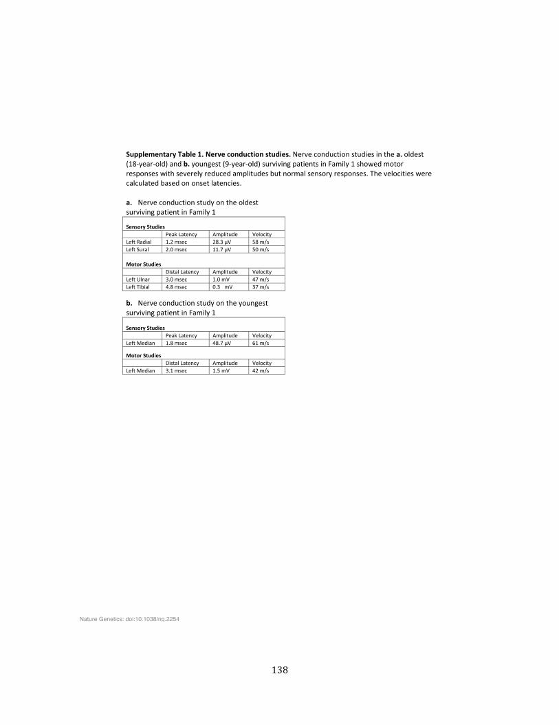
138
Supplementary�Table�1.�Nerve�conduction�studies.�Nerve�conduction�studies�in�the�a.�oldest�(18ͲyearͲold)�and�b.�youngest�(9ͲyearͲold)�surviving�patients�in�Family�1�showed�motor�responses�with�severely�reduced�amplitudes�but�normal�sensory�responses.�The�velocities�were�calculated�based�on�onset�latencies.��a. Nerve�conduction�study�on�the�oldest��surviving�patient�in�Family�1�
Sensory�Studies�� Peak�Latency� Amplitude� Velocity�Left�Radial� 1.2�msec� 28.3�µV� 58�m/s�Left�Sural� 2.0�msec� 11.7�µV� 50�m/s�
Motor�Studies�� Distal�Latency� Amplitude� Velocity�Left�Ulnar� 3.0�msec� 1.0�mV� 47�m/s�Left�Tibial� 4.8�msec� 0.3 mV� 37�m/s��
b. Nerve�conduction�study�on�the�youngest��surviving�patient�in�Family�1�
Sensory�Studies�� Peak�Latency� Amplitude� Velocity�Left�Median� 1.8�msec� 48.7�µV� 61�m/s�
Motor�Studies�� Distal�Latency� Amplitude� Velocity�Left�Median� 3.1�msec� 1.5�mV� 42�m/s��
���
Nature Genetics: doi:10.1038/ng.2254
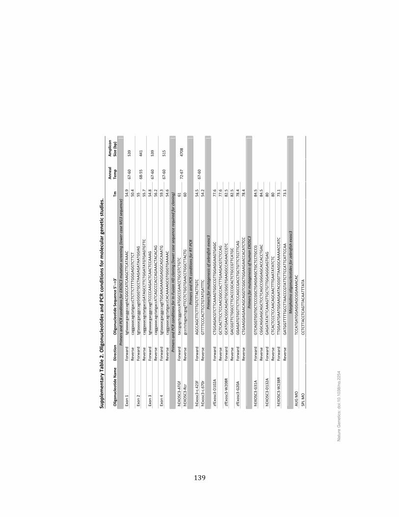
139
Supp
lemen
tary�Tab
le�2.�O
ligon
ucleotides�and
�PCR
�con
dition
s�for�molecular�gen
etic�studies.�
Oligon
ucleotide�Nam
e�Direction
�Oligon
ucleotide�Sequ
ence�5'ͲͲ>3'��
Tm�
Ann
eal�
Temp��
Amplicon
�Size�(b
p)�
Prim
ers�an
d�PC
R�cond
ition
s�for�E
XOSC3�mutation�screening�(lo
wer�case�M13
�sequence)�
Exon
�1�
Forw
ard�
tgtaaaacgacggccagtACG
GCC
ATC
AAGCT
TCATA
AAC�
54.9�
��Re
verse�
caggaaacagctatgaccCT
CTTC
TTTTGGGAGGTC
TTCT
�50
.4�
67Ͳ60�
539�
Exon
�2�
Forw
ard�
tgtaaaacgacggccagtGGGGTG
CCTA
AGAGATA
ATG
GAG�
55�
��Re
verse�
caggaaacagctatgaccGATA
GCC
TTCT
GGATA
TGTG
AGTG
TTC�
55.7�
68Ͳ55�
441�
Exon
�3�
Forw
ard�
tgtaaaacgacggccagtTC
CCCA
AGACT
CAACT
CCAAAG�
54.8�
��Re
verse�
caggaaacagctatgaccATC
AGCC
CACC
AGAAACT
ACA
CAG�
56.2�
67Ͳ60�
539�
Exon
�4�
Forw
ard�
tgtaaaacgacggccagtTG
GAAGAAAGGAGGCA
GCA
AATG
�59
.3�
��Re
verse�
caggaaacagctatgaccCA
CAAAAGCG
TGGGTG
AAAAC�
54.6�
67Ͳ60�
515�
Prim
ers�an
d�PC
R�cond
ition
s�for�InͲFusion
�HD�cloning
�(low
er�case�sequ
ence�re
quire
d�for�cloning
)�
hEXO
SC3ͲATG
f�Forw
ard�
taccgagctcggatccATG
GCC
GAACC
TGCG
TCTG
TC�
61�
hEXO
SC3ͲRcr�
Reverse�
gccctctagactcgagTTCC
TCTG
GTG
AACC
TGGCT
TACT
G�
60�
72Ͳ67�
4708
�
Prim
ers�an
d�PC
R�cond
ition
s�for�RT
ͲPCR
�
hExosc3Ͳc.423f�
Forw
ard�
AGCC
AGCT
TCTTTG
TCTTACT
TGTC
�54
.5�
hExosc3Ͳc.670r�
Reverse�
GTTTTCC
CACT
TCCT
GTA
TGATTTC
�54
.2�
67Ͳ60�
��
Prim
ers�for�m
utag
enesis�of�zebrafish�exosc3�
zfExosc3ͲD102A
�Forw
ard�
CTGGAGACG
TCTTCA
AAGTG
GCC
GTTGGAGGAAGTG
AGC�
77.6�
��Re
verse�
GCT
CACT
TCCT
CCAACG
GCC
ACT
TTGAAGACG
TCTC
CAG�
77.6�
zfExosc3ͲW
208R
��Forw
ard�
GCA
TGAACG
GCA
GAGTG
CGGGTG
AAGGCC
AGAACC
GTC
�82
.5�
��Re
verse�
GACG
GTTCT
GGCC
TTCA
CCCG
CACTCT
GCC
GTTCA
TGC�
82.5�
zfExosc3ͲG20
A��
Forw
ard�
GGAGATG
TGGTTCT
TCCA
GCC
GACC
TGCT
GTTCT
CCTTCA
G�
78.4�
��Re
verse�
CTGAAGGAGAACA
GCA
GGTC
GGCT
GGAAGAACC
ACA
TCTC
C�78
.4�
����
Prim
ers�for�m
utag
enesis�of�h
uman
�EXO
SC3�
hEXO
SC3ͲG31
A�
Forw
ard�
GTC
AGGTG
GTG
CTCC
CGGCT
GAGGAGCT
GCT
CCTG
CCG�
84.5�
��Re
verse�
CGGCA
GGAGCA
GCT
CCTC
AGCC
GGGAGCA
CCACC
TGAC�
84.5�
hEXO
SC3ͲD132A
�Forw
ard�
GAGATA
TATTCA
AAGTTGCT
GTTGGAGGGAGTG
AG�
80�
��Re
verse�
CTCA
CTCC
CTCC
AACA
GCA
ACT
TTGAATA
TATC
TC�
80�
hEXO
SC3ͲW238R
�Forw
ard�
TTGGAATG
AATG
GAAGAATA
CGGGTTAAGGCA
AAAACC
ATC
�73
.1�
��Re
verse�
GATG
GTTTTTG
CCTTAACC
CGTA
TTCT
TCCA
TTCA
TTCC
AA�
73.1�
����
Morph
olino�oligon
ucleotides�fo
r�zebrafish�exosc3�
AUG�M
O�
��TC
CATG
ATG
GAGGAGCG
GAAAACA
C���
��
SPL�MO�
��CC
TCTTACC
TCAGTTACA
ATTTA
TA�
����
��
Nat
ure
Gen
etic
s: d
oi:1
0.10
38/n
g.22
54
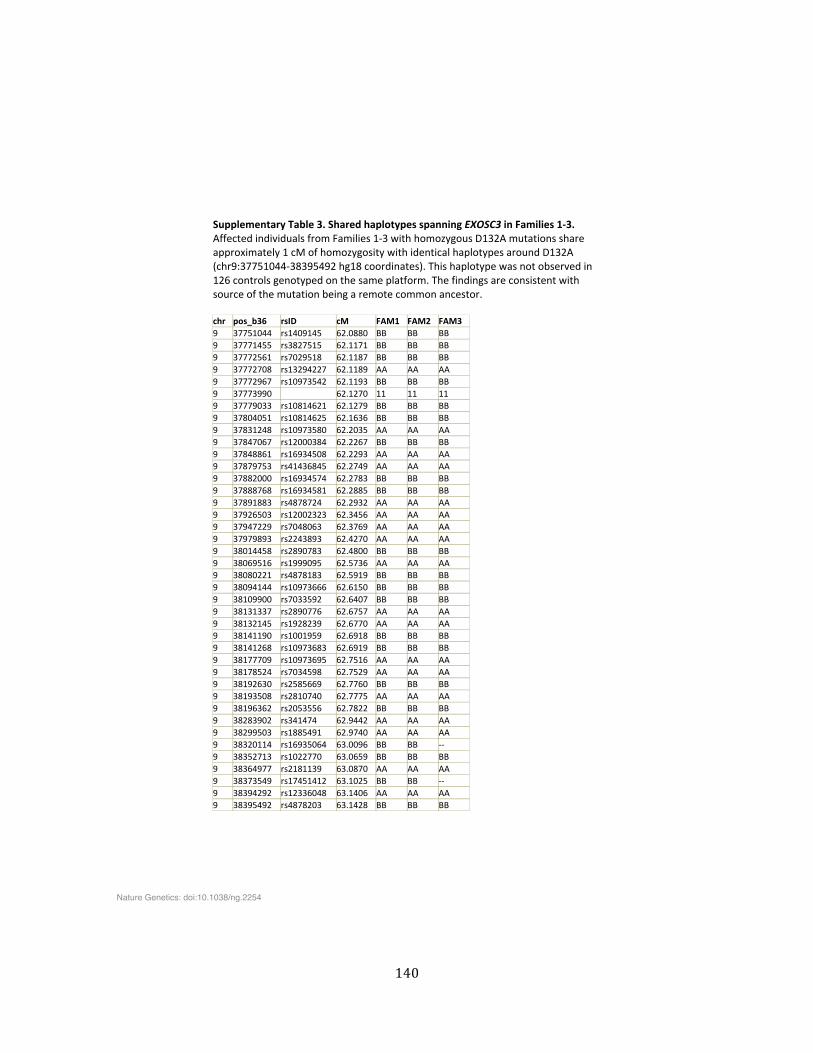
140
Supplementary�Table�3.�Shared�haplotypes�spanning�EXOSC3�in�Families�1Ͳ3.�Affected�individuals�from�Families�1Ͳ3�with�homozygous�D132A�mutations�share�approximately�1�cM�of�homozygosity�with�identical�haplotypes�around�D132A�(chr9:37751044Ͳ38395492�hg18�coordinates).�This�haplotype�was�not�observed�in�126�controls�genotyped�on�the�same�platform.�The�findings�are�consistent�with�source�of�the�mutation�being�a�remote�common�ancestor.��chr� pos_b36� rsID� cM� FAM1� FAM2� FAM3�9� 37751044� rs1409145� 62.0880� BB� BB� BB�9� 37771455� rs3827515� 62.1171� BB� BB� BB�9� 37772561� rs7029518� 62.1187� BB� BB� BB�9� 37772708� rs13294227� 62.1189� AA� AA� AA�9� 37772967� rs10973542� 62.1193� BB� BB� BB�9� 37773990� �� 62.1270� 11� 11� 11�9� 37779033� rs10814621� 62.1279� BB� BB� BB�9� 37804051� rs10814625� 62.1636� BB� BB� BB�9� 37831248� rs10973580� 62.2035� AA� AA� AA�9� 37847067� rs12000384� 62.2267� BB� BB� BB�9� 37848861� rs16934508� 62.2293� AA� AA� AA�9� 37879753� rs41436845� 62.2749� AA� AA� AA�9� 37882000� rs16934574� 62.2783� BB� BB� BB�9� 37888768� rs16934581� 62.2885� BB� BB� BB�9� 37891883� rs4878724� 62.2932� AA� AA� AA�9� 37926503� rs12002323� 62.3456� AA� AA� AA�9� 37947229� rs7048063� 62.3769� AA� AA� AA�9� 37979893� rs2243893� 62.4270� AA� AA� AA�9� 38014458� rs2890783� 62.4800� BB� BB� BB�9� 38069516� rs1999095� 62.5736� AA� AA� AA�9� 38080221� rs4878183� 62.5919� BB� BB� BB�9� 38094144� rs10973666� 62.6150� BB� BB� BB�9� 38109900� rs7033592� 62.6407� BB� BB� BB�9� 38131337� rs2890776� 62.6757� AA� AA� AA�9� 38132145� rs1928239� 62.6770� AA� AA� AA�9� 38141190� rs1001959� 62.6918� BB� BB� BB�9� 38141268� rs10973683� 62.6919� BB� BB� BB�9� 38177709� rs10973695� 62.7516� AA� AA� AA�9� 38178524� rs7034598� 62.7529� AA� AA� AA�9� 38192630� rs2585669� 62.7760� BB� BB� BB�9� 38193508� rs2810740� 62.7775� AA� AA� AA�9� 38196362� rs2053556� 62.7822� BB� BB� BB�9� 38283902� rs341474� 62.9442� AA� AA� AA�9� 38299503� rs1885491� 62.9740� AA� AA� AA�9� 38320114� rs16935064� 63.0096� BB� BB� ͲͲ�9� 38352713� rs1022770� 63.0659� BB� BB� BB�9� 38364977� rs2181139� 63.0870� AA� AA� AA�9� 38373549� rs17451412� 63.1025� BB� BB� ͲͲ�9� 38394292� rs12336048� 63.1406� AA� AA� AA�9� 38395492� rs4878203� 63.1428� BB� BB� BB�
�
Nature Genetics: doi:10.1038/ng.2254
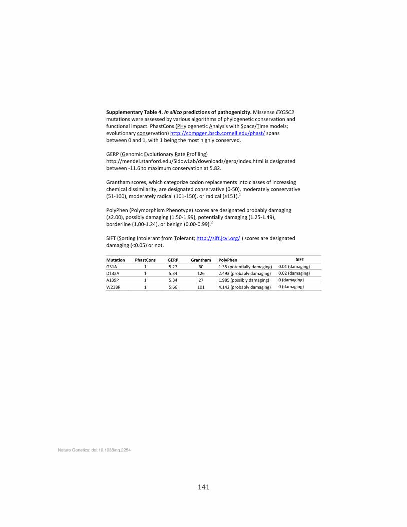
141
Supplementary�Table�4.�In�silico�predictions�of�pathogenicity.�Missense�EXOSC3�mutations�were�assessed�by�various�algorithms�of�phylogenetic�conservation�and�functional�impact.�PhastCons�(PHylogenetic�Analysis�with�Space/Time�models;�evolutionary�conservation)�http://compgen.bscb.cornell.edu/phast/�spans�between�0�and�1,�with�1�being�the�most�highly�conserved.����GERP�(Genomic�Evolutionary�Rate�Profiling)�http://mendel.stanford.edu/SidowLab/downloads/gerp/index.html�is�designated�between�Ͳ11.6�to�maximum�conservation�at�5.82.���Grantham�scores,�which�categorize�codon�replacements�into�classes�of�increasing�chemical�dissimilarity,�are�designated�conservative�(0Ͳ50),�moderately�conservative�(51Ͳ100),�moderately�radical�(101Ͳ150),�or�radical�(ш151).1���PolyPhen�(Polymorphism�Phenotype)�scores�are�designated�probably�damaging�(ш2.00),�possibly�damaging�(1.50Ͳ1.99),�potentially�damaging�(1.25Ͳ1.49),�borderline�(1.00Ͳ1.24),�or�benign�(0.00Ͳ0.99).2��SIFT�(Sorting�Intolerant�from�Tolerant;�http://sift.jcvi.org/�)�scores�are�designated�damaging�(<0.05)�or�not.��Mutation� PhastCons� GERP�� Grantham�� PolyPhen� SIFT�
G31A� 1� 5.27� 60� 1.35�(potentially�damaging)� 0.01�(damaging)�
D132A� 1� 5.34� 126� 2.493�(probably�damaging)� 0.02�(damaging)�
A139P� 1� 5.34� 27� 1.985�(possibly�damaging)� 0�(damaging)�
W238R� 1� 5.66� 101� 4.142�(probably�damaging)� 0�(damaging)�
���
Nature Genetics: doi:10.1038/ng.2254
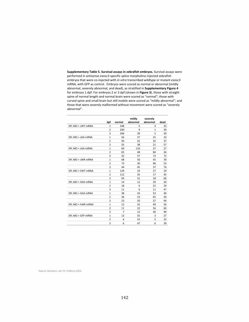
142
Supplementary�Table�5.�Survival�assays�in�zebrafish�embryos.�Survival�assays�were�performed�in�antisense�exosc3Ͳspecific�splice�morpholinoͲinjected�zebrafish�embryos�that�were�coͲinjected�with�in�vitro�transcribed�wildtype�or�mutant�exosc3�mRNA,�with�GFP�as�control.��Embryos�were�scored�as�normal�or�abnormal�(mildly�abnormal,�severely�abnormal,�and�dead),�as�stratified�in�Supplementary�Figure�4�for�embryos�1�dpf.�For�embryos�2�or�3�dpf�(shown�in�Figure�3),�those�with�straight�spine�of�normal�length�and�normal�brain�were�scored�as�“normal”;�those�with�curved�spine�and�small�brain�but�still�mobile�were�scored�as�“mildly�abnormal”;�and�those�that�were�severely�malformed�without�movement�were�scored�as�“severely�abnormal”.��
�� dpf� normal�mildly�
abnormal�severely�abnormal� dead�
SPL�MO�+�zWT�mRNA� 1� 248� 5� 3� 33��� 2� 240� 9� 1� 39��� 3� 206� 39� 5� 39�SPL�MO�+�zDA�mRNA� 1� 56� 37� 25� 33��� 2� 50� 32� 32� 37��� 3� 35� 38� 21� 57�SPL�MO�+�zGA�mRNA� 1� 69� 110� 27� 27��� 2� 65� 48� 84� 36��� 3� 32� 57� 72� 72�SPL�MO�+�zWR�mRNA� 1� 68� 50� 45� 39��� 2� 73� 30� 46� 53��� 3� 44� 45� 37� 76�SPL�MO�+�hWT�mRNA� 1� 129� 24� 27� 29��� 2� 112� 35� 17� 45��� 3� 94� 31� 18� 66�SPL�MO�+�hDA�mRNA� 1� 14� 12� 29� 20��� 2� 18� 6� 22� 29��� 3� 11� 6� 11� 47�SPL�MO�+�hGA�mRNA� 1� 38� 43� 53� 40��� 2� 36� 23� 65� 50��� 3� 23� 20� 37� 94�SPL�MO�+�hWR�mRNA� 1� 12� 32� 49� 56��� 2� 11� 22� 56� 60��� 3� 7� 13� 30� 99�SPL�MO�+�GFP�mRNA� 1� 13� 55� 3� 27��� 2� 6� 55� 5� 32��� 3� 6� 47� 6� 39�
Nature Genetics: doi:10.1038/ng.2254
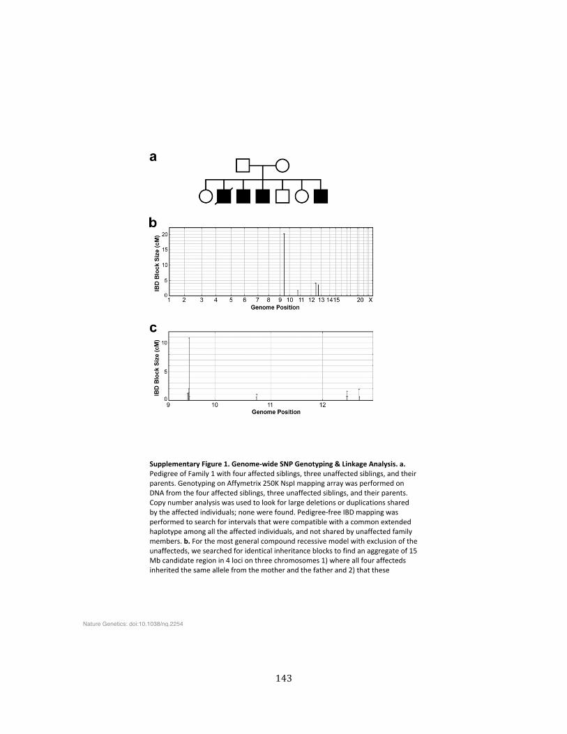
143
��
���Supplementary�Figure�1.�GenomeͲwide�SNP�Genotyping�&�Linkage�Analysis.�a.�Pedigree�of�Family�1�with�four�affected�siblings,�three�unaffected�siblings,�and�their�parents.�Genotyping�on�Affymetrix�250K�NspI�mapping�array�was�performed�on�DNA�from�the�four�affected�siblings,�three�unaffected�siblings,�and�their�parents.�Copy�number�analysis�was�used�to�look�for�large�deletions�or�duplications�shared�by�the�affected�individuals;�none�were�found.�PedigreeͲfree�IBD�mapping�was�performed�to�search�for�intervals�that�were�compatible�with�a�common�extended�haplotype�among�all�the�affected�individuals,�and�not�shared�by�unaffected�family�members.�b.�For�the�most�general�compound�recessive�model�with�exclusion�of�the�unaffecteds,�we�searched�for�identical�inheritance�blocks�to�find�an�aggregate�of�15�Mb�candidate�region�in�4�loci�on�three�chromosomes�1)�where�all�four�affecteds�inherited�the�same�allele�from�the�mother�and�the�father�and�2)�that�these�
Nature Genetics: doi:10.1038/ng.2254
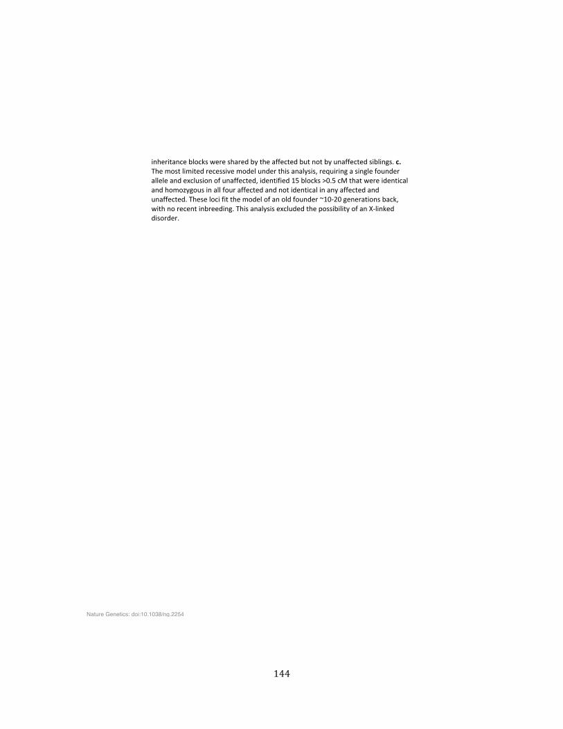
144
inheritance�blocks�were�shared�by�the�affected�but�not�by�unaffected�siblings.�c.�The�most�limited�recessive�model�under�this�analysis,�requiring�a�single�founder�allele�and�exclusion�of�unaffected,�identified�15�blocks�>0.5�cM�that�were�identical�and�homozygous�in�all�four�affected�and�not�identical�in�any�affected�and�unaffected.�These�loci�fit�the�model�of�an�old�founder�~10Ͳ20�generations�back,�with�no�recent�inbreeding.�This�analysis�excluded�the�possibility�of�an�XͲlinked�disorder.��
Nature Genetics: doi:10.1038/ng.2254
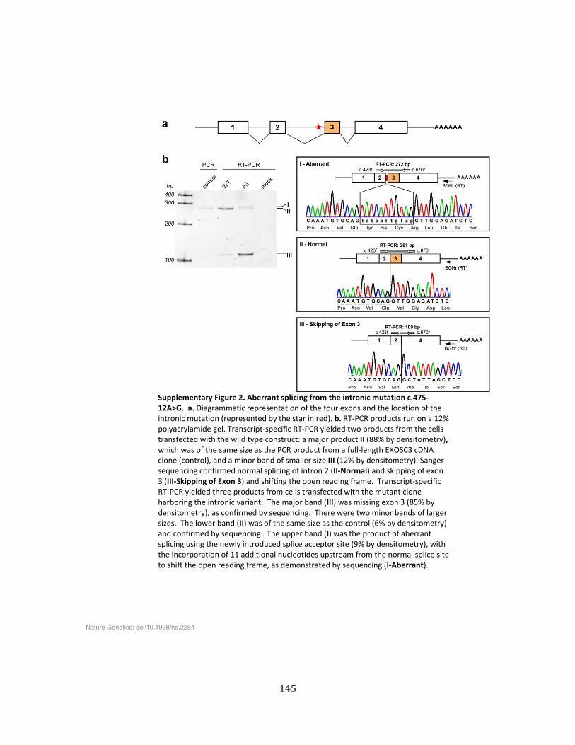
145
�Supplementary�Figure�2.�Aberrant�splicing�from�the�intronic�mutation�c.475Ͳ12A>G.��a.�Diagrammatic�representation�of�the�four�exons�and�the�location�of�the�intronic�mutation�(represented�by�the�star�in�red).�b.�RTͲPCR�products�run�on�a�12%�polyacrylamide�gel.�TranscriptͲspecific�RTͲPCR�yielded�two�products�from�the�cells�transfected�with�the�wild�type�construct:�a�major�product�II�(88%�by�densitometry),�which�was�of�the�same�size�as�the�PCR�product�from�a�fullͲlength�EXOSC3�cDNA�clone�(control),�and�a�minor�band�of�smaller�size�III�(12%�by�densitometry).�Sanger�sequencing�confirmed�normal�splicing�of�intron�2�(IIͲNormal)�and�skipping�of�exon�3�(IIIͲSkipping�of�Exon�3)�and�shifting�the�open�reading�frame.��TranscriptͲspecific�RTͲPCR�yielded�three�products�from�cells�transfected�with�the�mutant�clone�harboring�the�intronic�variant.��The�major�band�(III)�was�missing�exon�3�(85%�by�densitometry),�as�confirmed�by�sequencing.��There�were�two�minor�bands�of�larger�sizes.��The�lower�band�(II)�was�of�the�same�size�as�the�control�(6%�by�densitometry)�and�confirmed�by�sequencing.��The�upper�band�(I)�was�the�product�of�aberrant�splicing�using�the�newly�introduced�splice�acceptor�site�(9%�by�densitometry),�with�the�incorporation�of�11�additional�nucleotides�upstream�from�the�normal�splice�site�to�shift�the�open�reading�frame,�as�demonstrated�by�sequencing�(IͲAberrant).�
Nature Genetics: doi:10.1038/ng.2254
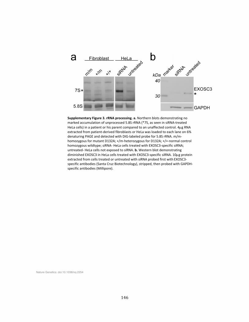
146
��Supplementary�Figure�3.�rRNA�processing.�a.�Northern�blots�demonstrating�no�marked�accumulation�of�unprocessed�5.8S�rRNA�(*7S,�as�seen�in�siRNAͲtreated�HeLa�cells)�in�a�patient�or�his�parent�compared�to�an�unaffected�control.�4Pg�RNA�extracted�from�patientͲderived�fibroblasts�or�HeLa�was�loaded�to�each�lane�on�6%�denaturing�PAGE�and�detected�with�DIGͲlabeled�probe�for�5.8S�rRNA.�m/mͲhomozygous�for�mutant�D132A;�+/mͲheterozygous�for�D132A;�+/+Ͳnormal�control�homozygous�wildtype;�siRNAͲ�HeLa�cells�treated�with�EXOSC3Ͳspecific�siRNA;�untreatedͲ�HeLa�cells�not�exposed�to�siRNA.�b.�Western�blot�demonstrating�diminished�EXOSC3�in�HeLa�cells�treated�with�EXOSC3Ͳspecific�siRNA.�10Pg�protein�extracted�from�cells�treated�or�untreated�with�siRNA�probed�first�with�EXOSC3Ͳspecific�antibodies�(Santa�Cruz�Biotechnology),�stripped,�then�probed�with�GAPDHͲspecific�antibodies�(Millipore).���
Nature Genetics: doi:10.1038/ng.2254
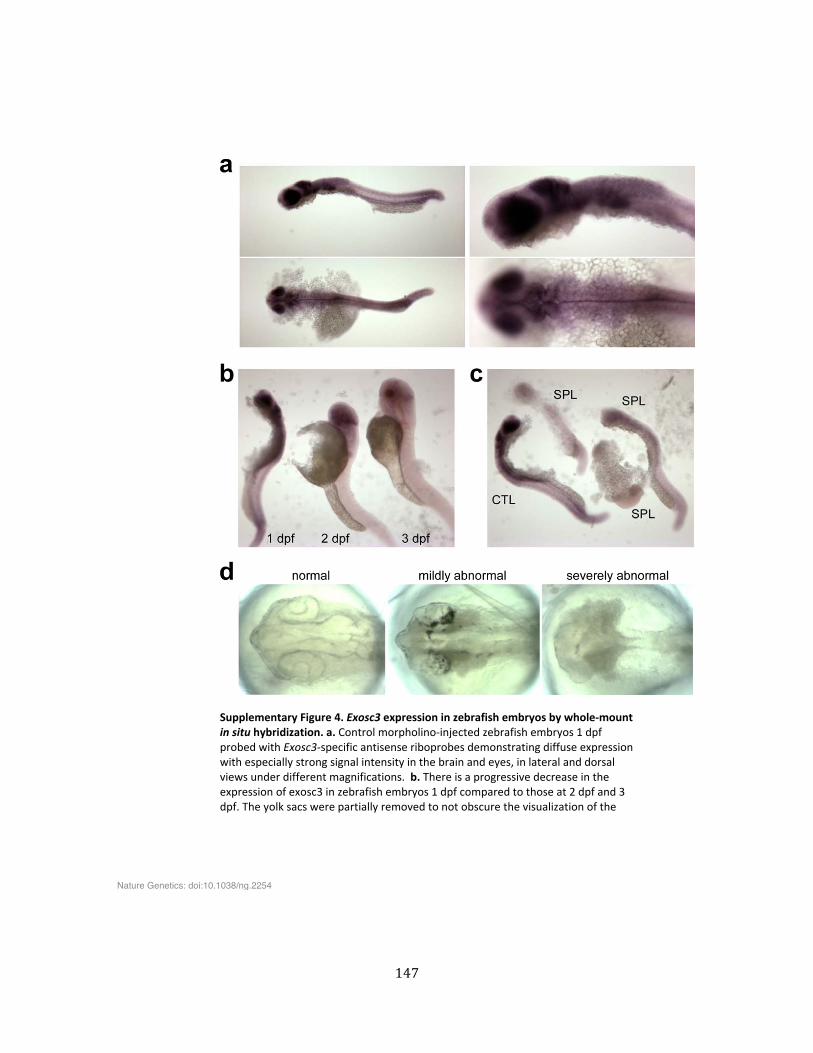
147
��Supplementary�Figure�4.�Exosc3�expression�in�zebrafish�embryos�by�wholeͲmount�in�situ�hybridization.�a.�Control�morpholinoͲinjected�zebrafish�embryos�1�dpf�probed�with�Exosc3Ͳspecific�antisense�riboprobes�demonstrating�diffuse�expression�with�especially�strong�signal�intensity�in�the�brain�and�eyes,�in�lateral�and�dorsal�views�under�different�magnifications.��b.�There�is�a�progressive�decrease�in�the�expression�of�exosc3�in�zebrafish�embryos�1�dpf�compared�to�those�at�2�dpf�and�3�dpf.�The�yolk�sacs�were�partially�removed�to�not�obscure�the�visualization�of�the�
Nature Genetics: doi:10.1038/ng.2254
148
embryos.�c.�Zebrafish�embryos�1�dpf�injected�with�antisense�morpholinos�targeting�the�splice�site�of�exosc3�(SPL)�led�to�a�marked�but�variable�decrease�in�the�expression�of�exosc3�compared�to�those�injected�with�control�morpholinos�(CTL).�There�was�higher�expression�of�exosc3�in�the�embryo�on�the�far�right�compared�to�the�other�two�SPLͲinjected�embryos.�d.�Light�microscopic�examination�in�dorsal�view�of�unstained�live�chorionͲenveloped�zebrafish�embryos�1�dpf�revealed�variable�phenotypes.��Embryos�with�smaller�brain�and�eyes�are�scored�as�mildly�abnormal.��Those�embryos�with�very�small�brain�and�malformed�eyes�as�well�as�a�very�small�hindbrain�and�thin�spinal�cord�are�scored�as�severely�abnormal.���References��1. Li�WH,�Wu�CI,�Luo�CC.�Nonrandomness�of�point�mutation�as�reflected�in�
nucleotide�substitutions�in�pseudogenes�and�its�evolutionary�implications.�J�Mol�Evol�21,�58–71(1984).�
2. Xi�T,�Jones�IM,�Mohrenweiser�HW.�Many�amino�acid�substitution�variants�identified�in�DNA�repair�genes�during�human�population�screenings�are�predicted�to�impact�protein�function.�Genomics�83,�970–979�(2004).�
���
Nature Genetics: doi:10.1038/ng.2254

149
CHAPTER EIGHT
Conclusions
150
General conclusions
Eric Lander, first author of the 2001 paper that presented the initial sequencing of the
human genome (1), more recently noted that “[t]he human genome has had a certain tendency
to incite passion and excess” (2). The work presented here justifies a certain passion for
unlocking the secrets of genetic diseases and suggests caution against excessive expectations.
At the outset we sought to employ a new and powerful technology to understand the molecular
basis of severe congenital disorders of the intestine and brain. In this endeavor we had success:
we pinpointed many unsuspected mutations interfering with intestinal absorption in genes
that already were known to medicine; we identified the probable, and possibly predominant,
genetic cause of sporadic congenital intestinal motility disorders; and we discovered a
surprising link between RNA processing and cerebellar and spinal motor neuron survival. In a
few instances, this knowledge can immediately translate into improved disease management;
in all, it can bring closure to patients and families, provide guidance for pregnancy planning,
and can open doors for further basic research on the underlying mechanisms and pathways
and for the development of new treatments.
During the course of this project the field of high throughput sequencing matured
rapidly. Cost per base plummeted and read lengths and read quality improved as a result of
technical improvements in sequencing platforms (3). Software developers have produced tools
for read quality assessment (4-‐12), mapping and alignment of short reads (13-‐24), genotyping
and variant identification(25-‐42), and variant annotation(43-‐103). The 1000 Genomes project
and NHLBI released exome sequencing results for data from thousands of individuals (104,
105). Nonetheless, many challenges remain for the discovery of rare variants that cause
Mendelian diseases. Foremost among these is the difficulty of obtaining sufficient numbers of
samples from consented and well phenotyped individuals. Power to detect a single casual gene
for a Mendelian disorder rises dramatically when two unrelated samples are sequenced. Multi-‐
151
center projects such as a proposed study of intestinal failure can address this problem. Another
practical concern is storing the large and growing amount of aligned data; hardware,
maintenance, and physical infrastructure are costly, and benefit less from economies of scale
and innovation than has the generation of sequencing data.
Existing tools reliably detect single nucleotide variants with acceptable false positive
rates. The detection of small indels is somewhat less reliable, but software tools such as the
GATK Haplotype Caller are improving in both sensitivity and specificity. A number of tools
attempt to call larger scale duplications and deletions from exome data (106-‐114), but there is
little consistency among them in our experience. Exon capture variability may prove to
introduce too much noise for this technique to become reliable. Complex rearrangements can
be identified with paired end reads of whole genome data (115-‐131), but most such events
cannot be detected with exome sequencing.
As this research progressed, we became aware of a number of limitations and pitfalls.
Most importantly, at the outset we underestimated the likelihood that the diseases we studied
might be due to sporadic de novo mutations. The prevailing medical genetics literature has
doubtless underestimated the contribution of de novo mutations to disease burden because of
the difficulty of identifying such mutations in the absence of a priori reasons to investigate a
particular genomic region. Most discoveries had been made in extended families showing
autosomal dominant or recessive, X-‐linked, or mitochondrial inheritance patterns. Indeed, in
the congenital diarrhea disorders (CDDs) pilot study, all of the mutations we identified have a
recessive effect on phenotype. But this was unsurprising precisely because we looked for
mutations in genes reported in the literature, which typically had been found by a family
linkage methodology. Our identification of de novo dominant acting mutations in half of the
chronic intestinal pseudo-‐obstruction (CIPO) cohort highlighted the possible importance of
sporadic mutations more generally in families with no prior history of disease. And in the CIPO
152
cases, publication of a milder autosomal dominant familial form of CIPO helped us weed out
several other candidate genes in which members of the CIPO cohort shared mutations.
The estimated baseline rate of germline mutation in the human genome is currently
believed to be 1.18 x 10-‐8 per nucleotide or 74 de novo mutations per generation of which
~1.19 mutations per generation affect the coding region and ~78% of these (~0.9 mutations)
are predicted to alter the protein sequence (132-‐137). The rate is highly variable within and
between families (138), and increases with paternal age (139). Although there may only be a
few true potentially damaging de novo variants in each exome, there will be many seeming
possibilities in the exome sequencing results of a single individual because of the large number
of inherited heterozygous variants, which can be filtered out by sequencing trios (both parents
and affected child) and excluding any variants that are seen in either parent. False positives
are a more intractable problem. These can be due to false positive variant calls in the proband
or by false negative variant calls in the parents causing the variant in the proband falsely seem
to be de novo. We sequenced only a few trios in the CDD (n=8) and CIPO (n=4) studies.
Coincidentally none of the CIPO trios had ACTG2 mutations. Moreover, even with filtering it can
be difficult to screen out false positives without loosing sensitivity. For example, using our
standard filters for allele frequency, annotation, and segregation filters, we observed a mean of
39 putative variants per proband across all trios, but with stricter mapping quality
(QUAL>=500) and allele balance (40-‐60%) filters, as might be used for clinical sequencing, the
mean number of de novos mutation calls was just two. Although this approximates the ‘true’
number of de novo germline variants, if we had applied strict filtering to the CIPO cohort, we
would not have found the ACTG2 mutations in three of the cases. Further work may be needed
to find the right balance between sensitivity and specificity in variant detection, and the
balance may be different in a clinical context where avoiding false positives is critical, versus a
research context where sensitivity to novel findings may take precedence.
153
We also failed to find with exome sequencing an ACTG2 mutation in one of the cases
that we later confirmed by Sanger sequencing. This may have been due to the relatively poor
coverage of this sample (mean=76X, 86%>=20X), illustrating that variability of coverage is
another pitfall of whole-‐exome sequencing. Moreover, we used two different modules from the
Genome Analysis Toolkit (GATK) for genotyping, Unified Genotyper and Haplotype Caller. The
latter is now recommended by the GATK’s developers for projects such as the present work;
unlike the Unified Genotyper, the Haplotype Caller performs a local de novo assembly to
resolve misaligned reads and is said to be more accurate and more sensitive to indels. In our
experience, the Haplotype caller succeeds in calling more and longer indels. Nonetheless, with
respect to the de novo mutations in the CIPO cases, both methods found five of the variants, but
only the Unified Genotyper selected two others, yet Sanger sequencing confirmed all of these.
Even more challenging than the technical issues, is the process of determining whether
novel mutations, especially missense or splice regulatory region mutations, are truly harmful.
One report found that human genomes contain over 1000 apparently loss-‐of-‐function variants,
of which ~100 (~20 homozygous) were deemed genuine after strict filtering for false
positives; many genes appeared to be tolerant of mutations that would be predicted to be
severely damaging (140). Much work remains to be done to algorithmically predict which
variants are genuinely damaging and likely to be responsible for disease. Ideally, one would
want to demonstrate that a variant causes the expected phenotype in vivo, but this can be
costly and time consuming. For example, we were able to confirm the effect of the EXOSC3
mutation by experiments in a model organism. In this case, with four affected siblings, whole-‐
exome sequencing produced only one candidate that needed to be confirmed. More typically,
we find a number of mutations that are predicted to be damaging and the effort involved in
narrowing down the candidates may be daunting. Many researchers are now attempting to
develop improved bioinformatic methods for predicting whether a given variant is likely to
154
cause harm to the organism. We expect some improvements in this area, but it is unlikely to
become a panacea.
Finding the same mutation, or other damaging mutations in the same gene, as we did
with CIPO, is quite powerful. The difficulty, however, is that the class of disorders we study are
quite rare, limiting the number of cases available for study. One estimate suggests that exome
sequencing of two to five unrelated subjects is necessary for adequate power to identify rare
dominant-‐acting variants in casual genes (141). Furthermore, despite sharing common
syndromes, the diseases are genetically and phenotypically heterogeneous. A large multi-‐
center program to sequence and rigorously phenotype many subjects, such as one we have
proposed elsewhere for intestinal failure, is a promising approach.
It also will be necessary to develop high-‐throughput screening methods for each class
of disease. We are currently developing methods to model the intestine in cell cultures derived
from embryonic stem cells, induced pluripotent stem cells reprogrammed from patient
fibroblasts, and intestinal progenitor stem cells from patient biopsy tissue. We are developing
methods for overexpression of mutant and wild-‐type alleles in these systems, and evaluating
the resulting phenotypes. In the near future, methods for specifically editing the genome, such
as TALENs or CRISPR/Cas systems (142) will permit more precise recapitulation of the disease
state and the development of methods to repair patient-‐derived organoids looking towards
future therapeutic modalities. High throughput assays with model organisms could be a
powerful method for quickly and inexpensively screening candidate mutations. For example,
libraries of gene knockdown morpholinos for use with zebrafish, or siRNA for use with C.
elegans, can be used to run many experiments in parallel. Still, mapping human diseases onto
other organisms is not straightforward, and a major screening program will involve much
effort and expense.
155
Specific conclusions
Our VAX method, which uses of local installation of the Ensembl databases and Perl
API, provides a robust and flexible framework for annotating DNA sequencing variants from
many different data sources using Variant Effect Predictor plugin modules. We have outlined
the design and usage of VEP plugins for a number of widely used databases. In addition,
modules may be easily designed for incorporating annotations from any external dataset that
is kept in a flat file or relational database, such as the Zebrafish Model Organism database
(143), and the Rat Genome database (144). We have used the VAX system for the discovery of
the causes of rare Mendelian diseases and genes involved in psychiatric disorders. (145-‐147).
VAX is used routinely for CLIA/CAP-‐accredited whole exome sequencing by the UCLA Clinical
Genomics Center, which has processed more than 1000 exomes to date (148).
The CDD pilot project successfully identified a probable molecular explanation for the
disorder in a majority of cases, thus suggesting the value of this approach for diagnosis of CDDs
in a clinical setting. The case of the child with a PCSK1 mutation, illustrates how timely use of
whole-‐exome sequencing could reduce medical costs and patient misery. However, our study
also demonstrates that much remains unknown about the genetic etiology of CDDs, as 40% of
the probands we sequenced did not yield a clear molecular finding, although many interesting
and novel candidate genes have been identified. There are several factors contributing to this
“genetic dark matter”. Most importantly, the cellular pathways involved in the development
and function of the intestine are not fully understood and many genes are yet to be identified
that contribute to the risk of CDDs. The present study generated a number of candidate genes
that are now the subject of active research. As discussed above, de novo mutations, which the
study was not well powered to identify, may account for some of the unsolved cases. A third
source of genetic dark matter is the 98% of the human genome that is not in the exome, which
includes promoters, enhancers, short RNAs, and other regulatory elements. Identifying and
156
characterizing these features is an active area of research, but at this time the size of the
genome to be sequenced and the lack of data on these features limits our ability to determine
how they affect phenotypes.
Finding that familial visceral myopathy in one family and a subset of CIPO cases appear
to have a common genetic cause resolves a longstanding mystery. Our finding at this point is
preliminary and it will be necessary in future work to demonstrate at the cellular or organism
level that each mutation causes a disordered phenotype. Beyond that, much remains to be
discovered about the actual molecular mechanism that causes a point mutation in one form of
actin to have a dominant effect. It could involve disruption of actin filament polymerizability or
stability, altered or blocked interactions with actin regulatory proteins, changes to
actin/myosin interactions, or weakening of actin filament structure (149).
Finally, the finding that the RNA exosome seems to play a unique role in the survival of
cerebellar and spinal motor neurons ties in with work on understanding how RNA regulation is
involved in motor and cerebellar degeneration.
157
References
1. Lander ES, Linton LM, Birren B, Nusbaum C, Zody MC, Baldwin J, Devon K, Dewar K, Doyle M, FitzHugh W, Funke R, Gage D, Harris K, Heaford A, Howland J, Kann L, Lehoczky J, LeVine R, McEwan P, McKernan K, Meldrim J, Mesirov JP, Miranda C, Morris W, Naylor J, Raymond C, Rosetti M, Santos R, Sheridan A, Sougnez C, Stange-‐Thomann N, Stojanovic N, Subramanian A, Wyman D, Rogers J, Sulston J, Ainscough R, Beck S, Bentley D, Burton J, Clee C, Carter N, Coulson A, Deadman R, Deloukas P, Dunham A, Dunham I, Durbin R, French L, Grafham D, Gregory S, Hubbard T, Humphray S, Hunt A, Jones M, Lloyd C, McMurray A, Matthews L, Mercer S, Milne S, Mullikin JC, Mungall A, Plumb R, Ross M, Shownkeen R, Sims S, Waterston RH, Wilson RK, Hillier LW, McPherson JD, Marra MA, Mardis ER, Fulton LA, Chinwalla AT, Pepin KH, Gish WR, Chissoe SL, Wendl MC, Delehaunty KD, Miner TL, Delehaunty A, Kramer JB, Cook LL, Fulton RS, Johnson DL, Minx PJ, Clifton SW, Hawkins T, Branscomb E, Predki P, Richardson P, Wenning S, Slezak T, Doggett N, Cheng JF, Olsen A, Lucas S, Elkin C, Uberbacher E, Frazier M, Gibbs RA, Muzny DM, Scherer SE, Bouck JB, Sodergren EJ, Worley KC, Rives CM, Gorrell JH, Metzker ML, Naylor SL, Kucherlapati RS, Nelson DL, Weinstock GM, Sakaki Y, Fujiyama A, Hattori M, Yada T, Toyoda A, Itoh T, Kawagoe C, Watanabe H, Totoki Y, Taylor T, Weissenbach J, Heilig R, Saurin W, Artiguenave F, Brottier P, Bruls T, Pelletier E, Robert C, Wincker P, Smith DR, Doucette-‐Stamm L, Rubenfield M, Weinstock K, Lee HM, Dubois J, Rosenthal A, Platzer M, Nyakatura G, Taudien S, Rump A, Yang H, Yu J, Wang J, Huang G, Gu J, Hood L, Rowen L, Madan A, Qin S, Davis RW, Federspiel NA, Abola AP, Proctor MJ, Myers RM, Schmutz J, Dickson M, Grimwood J, Cox DR, Olson MV, Kaul R, Raymond C, Shimizu N, Kawasaki K, Minoshima S, Evans GA, Athanasiou M, Schultz R, Roe BA, Chen F, Pan H, Ramser J, Lehrach H, Reinhardt R, McCombie WR, de la Bastide M, Dedhia N, Blocker H, Hornischer K, Nordsiek G, Agarwala R, Aravind L, Bailey JA, Bateman A, Batzoglou S, Birney E, Bork P, Brown DG, Burge CB, Cerutti L, Chen HC, Church D, Clamp M, Copley RR, Doerks T, Eddy SR, Eichler EE, Furey TS, Galagan J, Gilbert JG, Harmon C, Hayashizaki Y, Haussler D, Hermjakob H, Hokamp K, Jang W, Johnson LS, Jones TA, Kasif S, Kaspryzk A, Kennedy S, Kent WJ, Kitts P, Koonin EV, Korf I, Kulp D, Lancet D, Lowe TM, McLysaght A, Mikkelsen T, Moran JV, Mulder N, Pollara VJ, Ponting CP, Schuler G, Schultz J, Slater G, Smit AF, Stupka E, Szustakowski J, Thierry-‐Mieg D, Thierry-‐Mieg J, Wagner L, Wallis J, Wheeler R, Williams A, Wolf YI, Wolfe KH, Yang SP, Yeh RF, Collins F, Guyer MS, Peterson J, Felsenfeld A, Wetterstrand KA, Patrinos A, Morgan MJ, de Jong P, Catanese JJ, Osoegawa K, Shizuya H, Choi S, Chen YJ, International Human Genome Sequencing C. Initial sequencing and analysis of the human genome. Nature. 2001;409(6822):860-‐921. Epub 2001/03/10. doi: 10.1038/35057062. PubMed PMID: 11237011.
2. Lander ES. Initial impact of the sequencing of the human genome. Nature. 2011;470(7333):187-‐97. Epub 2011/02/11. doi: 10.1038/nature09792. PubMed PMID: 21307931.
3. Mardis ER. Next-‐generation sequencing platforms. Annual review of analytical chemistry. 2013;6:287-‐303. Epub 2013/04/09. doi: 10.1146/annurev-‐anchem-‐062012-‐092628. PubMed PMID: 23560931.
4. Blankenberg D, Gordon A, Von Kuster G, Coraor N, Taylor J, Nekrutenko A, Galaxy T. Manipulation of FASTQ data with Galaxy. Bioinformatics. 2010;26(14):1783-‐5. Epub 2010/06/22. doi: 10.1093/bioinformatics/btq281. PubMed PMID: 20562416; PubMed Central PMCID: PMC2894519.
158
5. Cibulskis K, McKenna A, Fennell T, Banks E, DePristo M, Getz G. ContEst: estimating cross-‐contamination of human samples in next-‐generation sequencing data. Bioinformatics. 2011;27(18):2601-‐2. Epub 2011/08/02. doi: 10.1093/bioinformatics/btr446. PubMed PMID: 21803805; PubMed Central PMCID: PMC3167057.
6. Cox MP, Peterson DA, Biggs PJ. SolexaQA: At-‐a-‐glance quality assessment of Illumina second-‐generation sequencing data. BMC Bioinformatics. 2010;11:485. Epub 2010/09/30. doi: 10.1186/1471-‐2105-‐11-‐485. PubMed PMID: 20875133; PubMed Central PMCID: PMC2956736.
7. Dai M, Thompson RC, Maher C, Contreras-‐Galindo R, Kaplan MH, Markovitz DM, Omenn G, Meng F. NGSQC: cross-‐platform quality analysis pipeline for deep sequencing data. BMC Genomics. 2010;11 Suppl 4:S7. Epub 2010/12/22. doi: 10.1186/1471-‐2164-‐11-‐S4-‐S7. PubMed PMID: 21143816; PubMed Central PMCID: PMC3005923.
8. Dohm JC, Lottaz C, Borodina T, Himmelbauer H. Substantial biases in ultra-‐short read data sets from high-‐throughput DNA sequencing. Nucleic Acids Res. 2008;36(16):e105. Epub 2008/07/29. doi: 10.1093/nar/gkn425. PubMed PMID: 18660515; PubMed Central PMCID: PMC2532726.
9. Dolan PC, Denver DR. TileQC: a system for tile-‐based quality control of Solexa data. BMC Bioinformatics. 2008;9:250. Epub 2008/05/30. doi: 10.1186/1471-‐2105-‐9-‐250. PubMed PMID: 18507856; PubMed Central PMCID: PMC2443380.
10. Martinez-‐Alcantara A, Ballesteros E, Feng C, Rojas M, Koshinsky H, Fofanov VY, Havlak P, Fofanov Y. PIQA: pipeline for Illumina G1 genome analyzer data quality assessment. Bioinformatics. 2009;25(18):2438-‐9. Epub 2009/07/16. doi: 10.1093/bioinformatics/btp429. PubMed PMID: 19602525; PubMed Central PMCID: PMC2735671.
11. Planet E, Attolini CS, Reina O, Flores O, Rossell D. htSeqTools: high-‐throughput sequencing quality control, processing and visualization in R. Bioinformatics. 2012;28(4):589-‐90. Epub 2011/12/27. doi: 10.1093/bioinformatics/btr700. PubMed PMID: 22199381.
12. Schmieder R, Edwards R. Quality control and preprocessing of metagenomic datasets. Bioinformatics. 2011;27(6):863-‐4. Epub 2011/02/01. doi: 10.1093/bioinformatics/btr026. PubMed PMID: 21278185; PubMed Central PMCID: PMC3051327.
13. Alkan C, Kidd JM, Marques-‐Bonet T, Aksay G, Antonacci F, Hormozdiari F, Kitzman JO, Baker C, Malig M, Mutlu O, Sahinalp SC, Gibbs RA, Eichler EE. Personalized copy number and segmental duplication maps using next-‐generation sequencing. Nat Genet. 2009;41(10):1061-‐7. Epub 2009/09/01. doi: 10.1038/ng.437. PubMed PMID: 19718026; PubMed Central PMCID: PMC2875196.
14. Galinsky VL. YOABS: yet other aligner of biological sequences-‐-‐an efficient linearly scaling nucleotide aligner. Bioinformatics. 2012;28(8):1070-‐7. Epub 2012/03/10. doi: 10.1093/bioinformatics/bts102. PubMed PMID: 22402614.
15. Homer N, Merriman B, Nelson SF. BFAST: an alignment tool for large scale genome resequencing. PLoS One. 2009;4(11):e7767. Epub 2009/11/13. doi:
159
10.1371/journal.pone.0007767. PubMed PMID: 19907642; PubMed Central PMCID: PMC2770639.
16. Langmead B, Salzberg SL. Fast gapped-‐read alignment with Bowtie 2. Nat Methods. 2012;9(4):357-‐9. Epub 2012/03/06. doi: 10.1038/nmeth.1923. PubMed PMID: 22388286; PubMed Central PMCID: PMC3322381.
17. Langmead B, Trapnell C, Pop M, Salzberg SL. Ultrafast and memory-‐efficient alignment of short DNA sequences to the human genome. Genome Biol. 2009;10(3):R25. Epub 2009/03/06. doi: 10.1186/gb-‐2009-‐10-‐3-‐r25. PubMed PMID: 19261174; PubMed Central PMCID: PMC2690996.
18. Li H, Durbin R. Fast and accurate short read alignment with Burrows-‐Wheeler transform. Bioinformatics. 2009;25(14):1754-‐60. Epub 2009/05/20. doi: 10.1093/bioinformatics/btp324. PubMed PMID: 19451168; PubMed Central PMCID: PMC2705234.
19. Li H, Durbin R. Fast and accurate long-‐read alignment with Burrows-‐Wheeler transform. Bioinformatics. 2010;26(5):589-‐95. Epub 2010/01/19. doi: 10.1093/bioinformatics/btp698. PubMed PMID: 20080505; PubMed Central PMCID: PMC2828108.
20. Li H, Ruan J, Durbin R. Mapping short DNA sequencing reads and calling variants using mapping quality scores. Genome Res. 2008;18(11):1851-‐8. Epub 2008/08/21. doi: 10.1101/gr.078212.108. PubMed PMID: 18714091; PubMed Central PMCID: PMC2577856.
21. Li R, Yu C, Li Y, Lam TW, Yiu SM, Kristiansen K, Wang J. SOAP2: an improved ultrafast tool for short read alignment. Bioinformatics. 2009;25(15):1966-‐7. Epub 2009/06/06. doi: 10.1093/bioinformatics/btp336. PubMed PMID: 19497933.
22. Lunter G, Goodson M. Stampy: a statistical algorithm for sensitive and fast mapping of Illumina sequence reads. Genome Res. 2011;21(6):936-‐9. Epub 2010/10/29. doi: 10.1101/gr.111120.110. PubMed PMID: 20980556; PubMed Central PMCID: PMC3106326.
23. Ning Z, Cox AJ, Mullikin JC. SSAHA: a fast search method for large DNA databases. Genome Res. 2001;11(10):1725-‐9. Epub 2001/10/10. doi: 10.1101/gr.194201. PubMed PMID: 11591649; PubMed Central PMCID: PMC311141.
24. Yu X, Guda K, Willis J, Veigl M, Wang Z, Markowitz S, Adams MD, Sun S. How do alignment programs perform on sequencing data with varying qualities and from repetitive regions? BioData mining. 2012;5(1):6. Epub 2012/06/20. doi: 10.1186/1756-‐0381-‐5-‐6. PubMed PMID: 22709551; PubMed Central PMCID: PMC3414812.
25. Albers CA, Lunter G, MacArthur DG, McVean G, Ouwehand WH, Durbin R. Dindel: accurate indel calls from short-‐read data. Genome Res. 2011;21(6):961-‐73.
26. Altmann A, Weber P, Quast C, Rex-‐Haffner M, Binder EB, Muller-‐Myhsok B. vipR: variant identification in pooled DNA using R. Bioinformatics. 2011;27(13):77-‐84.
160
27. Bansal VK, Misra MC, Krishna A, Kumar S, Garg P, Khan RN, Loli A, Jindal V. Pancreatic hydatid cyst masquerading as cystic neoplasm of pancreas. Trop Gastroenterol. 2010;31(4):335-‐7.
28. Browning BL, Yu Z. Simultaneous genotype calling and haplotype phasing improves genotype accuracy and reduces false-‐positive associations for genome-‐wide association studies. Am J Hum Genet. 2009;85(6):847-‐61.
29. Challis D, Yu J, Evani US, Jackson AR, Paithankar S, Coarfa C, Milosavljevic A, Gibbs RA, Yu F. An integrative variant analysis suite for whole exome next-‐generation sequencing data. BMC Bioinformatics. 2012;13:8-‐.
30. Chen K, McLellan MD, Ding L, Wendl MC, Kasai Y, Wilson RK, Mardis ER. PolyScan: an automatic indel and SNP detection approach to the analysis of human resequencing data. Genome Res. 2007;17(5):659-‐66.
31. Dalca AV, Rumble SM, Levy S, Brudno M. VARiD: a variation detection framework for color-‐space and letter-‐space platforms. Bioinformatics. 2010;26(12):343-‐9.
32. DePristo MA, Banks E, Poplin R, Garimella KV, Maguire JR, Hartl C, Philippakis AA, del Angel G, Rivas MA, Hanna M, McKenna A, Fennell TJ, Kernytsky AM, Sivachenko AY, Cibulskis K, Gabriel SB, Altshuler D, Daly MJ. A framework for variation discovery and genotyping using next-‐generation DNA sequencing data. Nat Genet. 2011;43(5):491-‐8.
33. Edmonson MN, Zhang J, Yan C, Finney RP, Meerzaman DM, Buetow KH. Bambino: a variant detector and alignment viewer for next-‐generation sequencing data in the SAM/BAM format. Bioinformatics. 2011;27(6):865-‐6.
34. Goya R, Sun MGF, Morin RD, Leung G, Ha G, Wiegand KC, Senz J, Crisan A, Marra MA, Hirst M, Huntsman D, Murphy KP, Aparicio S, Shah SP. SNVMix: predicting single nucleotide variants from next-‐generation sequencing of tumors. Bioinformatics. 2010;26(6):730-‐6.
35. Howie BN, Donnelly P, Marchini J. A flexible and accurate genotype imputation method for the next generation of genome-‐wide association studies. PLoS Genet. 2009;5(6).
36. Iqbal Z, Caccamo M, Turner I, Flicek P, McVean G. De novo assembly and genotyping of variants using colored de Bruijn graphs. Nat Genet. 2012;44(2):226-‐32.
37. Le SQ, Durbin R. SNP detection and genotyping from low-‐coverage sequencing data on multiple diploid samples. Genome Res. 2011;21(6):952-‐60.
38. Lee S, Hormozdiari F, Alkan C, Brudno M. MoDIL: detecting small indels from clone-‐end sequencing with mixtures of distributions. Nat Methods. 2009;6(7):473-‐4.
39. Li H, Handsaker B, Wysoker A, Fennell T, Ruan J, Homer N, Marth G, Abecasis G, Durbin R. The Sequence Alignment/Map format and SAMtools. Bioinformatics. 2009;25(16):2078-‐9.
40. Li Y, Willer CJ, Ding J, Scheet P, Abecasis GR. MaCH: using sequence and genotype data to estimate haplotypes and unobserved genotypes. Genet Epidemiol. 2010;34(8):816-‐34.
161
41. Malhis N, Jones SJM. High quality SNP calling using Illumina data at shallow coverage. Bioinformatics. 2010;26(8):1029-‐35.
42. Rivas MA, Beaudoin M, Gardet A, Stevens C, Sharma Y, Zhang CK, Boucher G, Ripke S, Ellinghaus D, Burtt N, Fennell T, Kirby A, Latiano A, Goyette P, Green T, Halfvarson J, Haritunians T, Korn JM, Kuruvilla F, Lagace C, Neale B, Lo KS, Schumm P, Torkvist L, Dubinsky MC, Brant SR, Silverberg MS, Duerr RH, Altshuler D, Gabriel S, Lettre G, Franke A, D'Amato M, McGovern DPB, Cho JH, Rioux JD, Xavier RJ, Daly MJ. Deep resequencing of GWAS loci identifies independent rare variants associated with inflammatory bowel disease. Nat Genet. 2011;43(11):1066-‐73.
43. Adzhubei IA, Schmidt S, Peshkin L, Ramensky VE, Gerasimova A, Bork P, Kondrashov AS, Sunyaev SR. A method and server for predicting damaging missense mutations. Nat Methods. 2010;7(4):248-‐9.
44. Asthana S, Roytberg M, Stamatoyannopoulos J, Sunyaev S. Analysis of sequence conservation at nucleotide resolution. PLoS computational biology. 2007;3(12).
45. Banerji S, Cibulskis K, Rangel-‐Escareno C, Brown KK, Carter SL, Frederick AM, Lawrence MS, Sivachenko AY, Sougnez C, Zou L, Cortes ML, Fernandez-‐Lopez JC, Peng S, Ardlie KG, Auclair D, Bautista-‐Pina V, Duke F, Francis J, Jung J, Maffuz-‐Aziz A, Onofrio RC, Parkin M, Pho NH, Quintanar-‐Jurado V, Ramos AH, Rebollar-‐Vega R, Rodriguez-‐Cuevas S, Romero-‐Cordoba SL, Schumacher SE, Stransky N, Thompson KM, Uribe-‐Figueroa L, Baselga J, Beroukhim R, Polyak K, Sgroi DC, Richardson AL, Jimenez-‐Sanchez G, Lander ES, Gabriel SB, Garraway LA, Golub TR, Melendez-‐Zajgla J, Toker A, Getz G, Hidalgo-‐Miranda A, Meyerson M. Sequence analysis of mutations and translocations across breast cancer subtypes. Nature. 2012;486(7403):405-‐9.
46. Bao H, Guo H, Wang J, Zhou R, Lu X, Shi S. MapView: visualization of short reads alignment on a desktop computer. Bioinformatics. 2009;25(12):1554-‐5.
47. Bromberg Y, Rost B. SNAP: predict effect of non-‐synonymous polymorphisms on function. Nucleic Acids Res. 2007;35(11):3823-‐35.
48. Calabrese R, Capriotti E, Fariselli P, Martelli PL, Casadio R. Functional annotations improve the predictive score of human disease-‐related mutations in proteins. Hum Mutat. 2009;30(8):1237-‐44.
49. Capriotti E, Arbiza L, Casadio R, Dopazo J, Dopazo H, Marti-‐Renom MA. Use of estimated evolutionary strength at the codon level improves the prediction of disease-‐related protein mutations in humans. Hum Mutat. 2008;29(1):198-‐204.
50. Capriotti E, Calabrese R, Casadio R. Predicting the insurgence of human genetic diseases associated to single point protein mutations with support vector machines and evolutionary information. Bioinformatics. 2006;22(22):2729-‐34.
51. Capriotti E, Fariselli P, Calabrese R, Casadio R. Predicting protein stability changes from sequences using support vector machines. Bioinformatics. 2005;21 Suppl 2:54-‐8.
52. Cartegni L, Wang J, Zhu Z, Zhang MQ, Krainer AR. ESEfinder: A web resource to identify exonic splicing enhancers. Nucleic Acids Res. 2003;31(13):3568-‐71.
162
53. Carter SL, Cibulskis K, Helman E, McKenna A, Shen H, Zack T, Laird PW, Onofrio RC, Winckler W, Weir BA, Beroukhim R, Pellman D, Levine DA, Lander ES, Meyerson M, Getz G. Absolute quantification of somatic DNA alterations in human cancer. Nat Biotechnol. 2012;30(5):413-‐21.
54. Chelala C, Khan A, Lemoine NR. SNPnexus: a web database for functional annotation of newly discovered and public domain single nucleotide polymorphisms. Bioinformatics. 2009;25(5):655-‐61.
55. Cingolani P, Patel VM, Coon M, Nguyen T, Land SJ, Ruden DM, Lu X. Using Drosophila melanogaster as a Model for Genotoxic Chemical Mutational Studies with a New Program, SnpSift. Front Genet. 2012;3:35-‐.
56. Davydov EV, Goode DL, Sirota M, Cooper GM, Sidow A, Batzoglou S. Identifying a high fraction of the human genome to be under selective constraint using GERP++. PLoS computational biology. 2010;6(12).
57. Fairbrother WG, Yeh R-‐F, Sharp PA, Burge CB. Predictive identification of exonic splicing enhancers in human genes. Science. 2002;297(5583):1007-‐13.
58. Freimuth RR, Stormo GD, McLeod HL. PolyMAPr: programs for polymorphism database mining, annotation, and functional analysis. Hum Mutat. 2005;25(2):110-‐7.
59. Gamazon ER, Zhang W, Konkashbaev A, Duan S, Kistner EO, Nicolae DL, Dolan ME, Cox NJ. SCAN: SNP and copy number annotation. Bioinformatics. 2010;26(2):259-‐62.
60. Garber M, Guttman M, Clamp M, Zody MC, Friedman N, Xie X. Identifying novel constrained elements by exploiting biased substitution patterns. Bioinformatics. 2009;25(12):54-‐62.
61. Ge D, Ruzzo EK, Shianna KV, He M, Pelak K, Heinzen EL, Need AC, Cirulli ET, Maia JM, Dickson SP, Zhu M, Singh A, Allen AS, Goldstein DB. SVA: software for annotating and visualizing sequenced human genomes. Bioinformatics. 2011;27(14):1998-‐2000.
62. Grant JR, Arantes AS, Liao X, Stothard P. In-‐depth annotation of SNPs arising from resequencing projects using NGS-‐SNP. Bioinformatics. 2011;27(16):2300-‐1.
63. Grover D, Woodfield AS, Verma R, Zandi PP, Levinson DF, Potash JB. QuickSNP: an automated web server for selection of tagSNPs. Nucleic Acids Res. 2007;35(Web Server issue):115-‐20.
64. Guerois R, Nielsen JE, Serrano L. Predicting changes in the stability of proteins and protein complexes: a study of more than 1000 mutations. Journal of molecular biology. 2002;320(2):369-‐87.
65. Han A, Kang HJ, Cho Y, Lee S, Kim YJ, Gong S. SNP@Domain: a web resource of single nucleotide polymorphisms (SNPs) within protein domain structures and sequences. Nucleic Acids Res. 2006;34(Web Server issue):642-‐4.
66. Hemminger BM, Saelim B, Sullivan PF. TAMAL: an integrated approach to choosing SNPs for genetic studies of human complex traits. Bioinformatics. 2006;22(5):626-‐7.
163
67. Hubisz MJ, Pollard KS, Siepel A. PHAST and RPHAST: phylogenetic analysis with space/time models. Brief Bioinform. 2011;12(1):41-‐51.
68. Jegga AG, Chen J, Gowrisankar S, Deshmukh MA, Gudivada R, Kong S, Kaimal V, Aronow BJ. GenomeTrafac: a whole genome resource for the detection of transcription factor binding site clusters associated with conventional and microRNA encoding genes conserved between mouse and human gene orthologs. Nucleic Acids Res. 2007;35(Database issue):116-‐21.
69. Kang HJ, Choi KO, Kim B-‐D, Kim S, Kim YJ. FESD: a Functional Element SNPs Database in human. Nucleic Acids Res. 2005;33(Database issue):518-‐22.
70. Karchin R, Diekhans M, Kelly L, Thomas DJ, Pieper U, Eswar N, Haussler D, Sali A. LS-‐SNP: large-‐scale annotation of coding non-‐synonymous SNPs based on multiple information sources. Bioinformatics. 2005;21(12):2814-‐20.
71. Kumar P, Henikoff S, Ng PC. Predicting the effects of coding non-‐synonymous variants on protein function using the SIFT algorithm. Nature protocols. 2009;4(7):1073-‐81.
72. Li B, Krishnan VG, Mort ME, Xin F, Kamati KK, Cooper DN, Mooney SD, Radivojac P. Automated inference of molecular mechanisms of disease from amino acid substitutions. Bioinformatics. 2009;25(21):2744-‐50.
73. Liu X, Jian X, Boerwinkle E. dbNSFP: a lightweight database of human nonsynonymous SNPs and their functional predictions. Hum Mutat. 2011;32(8):894-‐9.
74. Makarov V, O'Grady T, Cai G, Lihm J, Buxbaum JD, Yoon S. AnnTools: a comprehensive and versatile annotation toolkit for genomic variants. Bioinformatics. 2012;28(5):724-‐5.
75. Masso M, Vaisman II. AUTO-‐MUTE: web-‐based tools for predicting stability changes in proteins due to single amino acid replacements. Protein Eng Des Sel. 2010;23(8):683-‐7.
76. Mathe E, Olivier M, Kato S, Ishioka C, Hainaut P, Tavtigian SV. Computational approaches for predicting the biological effect of p53 missense mutations: a comparison of three sequence analysis based methods. Nucleic Acids Res. 2006;34(5):1317-‐25.
77. McLaren W, Pritchard B, Rios D, Chen Y, Flicek P, Cunningham F. Deriving the consequences of genomic variants with the Ensembl API and SNP Effect Predictor. Bioinformatics. 2010;26(16):2069-‐70.
78. Mermel CH, Schumacher SE, Hill B, Meyerson ML, Beroukhim R, Getz G. GISTIC2.0 facilitates sensitive and confident localization of the targets of focal somatic copy-‐number alteration in human cancers. Genome Biol. 2011;12(4).
79. Parthiban V, Gromiha MM, Schomburg D. CUPSAT: prediction of protein stability upon point mutations. Nucleic Acids Res. 2006;34(Web Server issue):239-‐42.
80. Reva B, Antipin Y, Sander C. Predicting the functional impact of protein mutations: application to cancer genomics. Nucleic Acids Res. 2011;39(17).
81. Riva A, Kohane IS. A SNP-‐centric database for the investigation of the human genome. BMC Bioinformatics. 2004;5:33-‐.
164
82. Saccone SF, Quan J, Mehta G, Bolze R, Thomas P, Deelman E, Tischfield JA, Rice JP. New tools and methods for direct programmatic access to the dbSNP relational database. Nucleic Acids Res. 2011;39(Database issue):901-‐7.
83. Schaefer C, Meier A, Rost B, Bromberg Y. SNPdbe: constructing an nsSNP functional impacts database. Bioinformatics. 2012;28(4):601-‐2.
84. Schmitt AO, Assmus J, Bortfeldt RH, Brockmann GA. CandiSNPer: a web tool for the identification of candidate SNPs for causal variants. Bioinformatics. 2010;26(7):969-‐70.
85. Schwarz JM, Rodelsperger C, Schuelke M, Seelow D. MutationTaster evaluates disease-‐causing potential of sequence alterations. Nat Methods. 2010;7(8):575-‐6.
86. Shetty AC, Athri P, Mondal K, Horner VL, Steinberg KM, Patel V, Caspary T, Cutler DJ, Zwick ME. SeqAnt: a web service to rapidly identify and annotate DNA sequence variations. BMC Bioinformatics. 2010;11:471-‐.
87. Stitziel NO, Binkowski TA, Tseng YY, Kasif S, Liang J. topoSNP: a topographic database of non-‐synonymous single nucleotide polymorphisms with and without known disease association. Nucleic Acids Res. 2004;32(Database issue):520-‐2.
88. Stone EA, Sidow A. Physicochemical constraint violation by missense substitutions mediates impairment of protein function and disease severity. Genome Res. 2005;15(7):978-‐86.
89. Stoyanovich J, Pe'er I. MutaGeneSys: estimating individual disease susceptibility based on genome-‐wide SNP array data. Bioinformatics. 2008;24(3):440-‐2.
90. Thomas PD, Campbell MJ, Kejariwal A, Mi H, Karlak B, Daverman R, Diemer K, Muruganujan A, Narechania A. PANTHER: a library of protein families and subfamilies indexed by function. Genome Res. 2003;13(9):2129-‐41.
91. Tian J, Wu N, Guo X, Guo J, Zhang J, Fan Y. Predicting the phenotypic effects of non-‐synonymous single nucleotide polymorphisms based on support vector machines. BMC Bioinformatics. 2007;8:450-‐.
92. Uzun A, Leslin CM, Abyzov A, Ilyin V. Structure SNP (StSNP): a web server for mapping and modeling nsSNPs on protein structures with linkage to metabolic pathways. Nucleic Acids Res. 2007;35(Web Server issue):384-‐92.
93. Venselaar H, Te Beek TAH, Kuipers RKP, Hekkelman ML, Vriend G. Protein structure analysis of mutations causing inheritable diseases. An e-‐Science approach with life scientist friendly interfaces. BMC Bioinformatics. 2010;11:548-‐.
94. Wainreb G, Ashkenazy H, Bromberg Y, Starovolsky-‐Shitrit A, Haliloglu T, Ruppin E, Avraham KB, Rost B, Ben-‐Tal N. MuD: an interactive web server for the prediction of non-‐neutral substitutions using protein structural data. Nucleic Acids Res. 2010;38(Web Server issue):523-‐8.
165
95. Wang J, Ronaghi M, Chong SS, Lee CGL. pfSNP: An integrated potentially functional SNP resource that facilitates hypotheses generation through knowledge syntheses. Hum Mutat. 2011;32(1):19-‐24.
96. Wang L, Liu S, Niu T, Xu X. SNPHunter: a bioinformatic software for single nucleotide polymorphism data acquisition and management. BMC Bioinformatics. 2005;6:60-‐.
97. Wang P, Dai M, Xuan W, McEachin RC, Jackson AU, Scott LJ, Athey B, Watson SJ, Meng F. SNP Function Portal: a web database for exploring the function implication of SNP alleles. Bioinformatics. 2006;22(14):523-‐9.
98. Wong WC, Kim D, Carter H, Diekhans M, Ryan MC, Karchin R. CHASM and SNVBox: toolkit for detecting biologically important single nucleotide mutations in cancer. Bioinformatics. 2011;27(15):2147-‐8.
99. Xu H, Gregory SG, Hauser ER, Stenger JE, Pericak-‐Vance MA, Vance JM, Zuchner S, Hauser MA. SNPselector: a web tool for selecting SNPs for genetic association studies. Bioinformatics. 2005;21(22):4181-‐6.
100. Ye K, Schulz MH, Long Q, Apweiler R, Ning Z. Pindel: a pattern growth approach to detect break points of large deletions and medium sized insertions from paired-‐end short reads. Bioinformatics. 2009;25(21):2865-‐71.
101. Yeo G, Hoon S, Venkatesh B, Burge CB. Variation in sequence and organization of splicing regulatory elements in vertebrate genes. Proc Natl Acad Sci U S A. 2004;101(44):15700-‐5.
102. Yue P, Melamud E, Moult J. SNPs3D: candidate gene and SNP selection for association studies. BMC Bioinformatics. 2006;7:166-‐.
103. Zhang XHF, Kangsamaksin T, Chao MSP, Banerjee JK, Chasin LA. Exon inclusion is dependent on predictable exonic splicing enhancers. Molecular and cellular biology. 2005;25(16):7323-‐32.
104. Genomes Project Consortium, Abecasis GR, Altshuler D, Auton A, Brooks LD, Durbin RM, Gibbs RA, Hurles ME, McVean GA. A map of human genome variation from population-‐scale sequencing. Nature. 2010;467(7319):1061-‐73. Epub 2010/10/29. doi: 10.1038/nature09534. PubMed PMID: 20981092; PubMed Central PMCID: PMC3042601.
105. Exome Variant Server [Internet]. NHLBI Exome Sequencing Project (ESP). 2011 [cited 2011-‐09-‐10]. Available from: http://evs.gs.washington.edu/EVS/.
106. Abyzov A, Urban AE, Snyder M, Gerstein M. CNVnator: an approach to discover, genotype, and characterize typical and atypical CNVs from family and population genome sequencing. Genome Res. 2011;21(6):974-‐84.
107. Chiang DY, Getz G, Jaffe DB, O'Kelly MJT, Zhao X, Carter SL, Russ C, Nusbaum C, Meyerson M, Lander ES. High-‐resolution mapping of copy-‐number alterations with massively parallel sequencing. Nat Methods. 2009;6(1):99-‐9103.
166
108. Ivakhno S, Royce T, Cox AJ, Evers DJ, Cheetham RK, Tavare S. CNAseg-‐-‐a novel framework for identification of copy number changes in cancer from second-‐generation sequencing data. Bioinformatics. 2010;26(24):3051-‐8.
109. Li J, Lupat R, Amarasinghe KC, Thompson ER, Doyle MA, Ryland GL, Tothill RW, Halgamuge SK, Campbell IG, Gorringe KL. CONTRA: copy number analysis for targeted resequencing. Bioinformatics. 2012;28(10):1307-‐13.
110. Medvedev P, Fiume M, Dzamba M, Smith T, Brudno M. Detecting copy number variation with mated short reads. Genome Res. 2010;20(11):1613-‐22.
111. Sathirapongsasuti JF, Lee H, Horst BAJ, Brunner G, Cochran AJ, Binder S, Quackenbush J, Nelson SF. Exome sequencing-‐based copy-‐number variation and loss of heterozygosity detection: ExomeCNV. Bioinformatics. 2011;27(19):2648-‐54.
112. Waszak SM, Hasin Y, Zichner T, Olender T, Keydar I, Khen M, Stutz AM, Schlattl A, Lancet D, Korbel JO. Systematic inference of copy-‐number genotypes from personal genome sequencing data reveals extensive olfactory receptor gene content diversity. PLoS computational biology. 2010;6(11).
113. Xie C, Tammi MT. CNV-‐seq, a new method to detect copy number variation using high-‐throughput sequencing. BMC Bioinformatics. 2009;10:80-‐.
114. Yoon S, Xuan Z, Makarov V, Ye K, Sebat J. Sensitive and accurate detection of copy number variants using read depth of coverage. Genome Res. 2009;19(9):1586-‐92.
115. Chen K, Wallis JW, McLellan MD, Larson DE, Kalicki JM, Pohl CS, McGrath SD, Wendl MC, Zhang Q, Locke DP, Shi X, Fulton RS, Ley TJ, Wilson RK, Ding L, Mardis ER. BreakDancer: an algorithm for high-‐resolution mapping of genomic structural variation. Nat Methods. 2009;6(9):677-‐81.
116. Clark MJ, Homer N, O'Connor BD, Chen Z, Eskin A, Lee H, Merriman B, Nelson SF. U87MG decoded: the genomic sequence of a cytogenetically aberrant human cancer cell line. PLoS Genet. 2010;6(1).
117. Ge H, Liu K, Juan T, Fang F, Newman M, Hoeck W. FusionMap: detecting fusion genes from next-‐generation sequencing data at base-‐pair resolution. Bioinformatics. 2011;27(14):1922-‐8.
118. Ha G, Roth A, Lai D, Bashashati A, Ding J, Goya R, Giuliany R, Rosner J, Oloumi A, Shumansky K, Chin S-‐F, Turashvili G, Hirst M, Caldas C, Marra MA, Aparicio S, Shah SP. Integrative analysis of genome-‐wide loss of heterozygosity and monoallelic expression at nucleotide resolution reveals disrupted pathways in triple-‐negative breast cancer. Genome Res. 2012;22(10):1995-‐2007.
119. Hormozdiari F, Alkan C, Ventura M, Hajirasouliha I, Malig M, Hach F, Yorukoglu D, Dao P, Bakhshi M, Sahinalp SC, Eichler EE. Alu repeat discovery and characterization within human genomes. Genome Res. 2011;21(6):840-‐9.
167
120. Karakoc E, Alkan C, O'Roak BJ, Dennis MY, Vives L, Mark K, Rieder MJ, Nickerson DA, Eichler EE. Detection of structural variants and indels within exome data. Nat Methods. 2012;9(2):176-‐8.
121. Korbel JO, Abyzov A, Mu XJ, Carriero N, Cayting P, Zhang Z, Snyder M, Gerstein MB. PEMer: a computational framework with simulation-‐based error models for inferring genomic structural variants from massive paired-‐end sequencing data. Genome Biol. 2009;10(2).
122. Lam HYK, Mu XJ, Stutz AM, Tanzer A, Cayting PD, Snyder M, Kim PM, Korbel JO, Gerstein MB. Nucleotide-‐resolution analysis of structural variants using BreakSeq and a breakpoint library. Nat Biotechnol. 2010;28(1):47-‐55.
123. Marschall T, Costa IG, Canzar S, Bauer M, Klau GW, Schliep A, Schonhuth A. CLEVER: clique-‐enumerating variant finder. Bioinformatics. 2012;28(22):2875-‐82.
124. Mills RE, Walter K, Stewart C, Handsaker RE, Chen K, Alkan C, Abyzov A, Yoon SC, Ye K, Cheetham RK, Chinwalla A, Conrad DF, Fu Y, Grubert F, Hajirasouliha I, Hormozdiari F, Iakoucheva LM, Iqbal Z, Kang S, Kidd JM, Konkel MK, Korn J, Khurana E, Kural D, Lam HYK, Leng J, Li R, Li Y, Lin C-‐Y, Luo R, Mu XJ, Nemesh J, Peckham HE, Rausch T, Scally A, Shi X, Stromberg MP, Stutz AM, Urban AE, Walker JA, Wu J, Zhang Y, Zhang ZD, Batzer MA, Ding L, Marth GT, McVean G, Sebat J, Snyder M, Wang J, Ye K, Eichler EE, Gerstein MB, Hurles ME, Lee C, McCarroll SA, Korbel JO. Mapping copy number variation by population-‐scale genome sequencing. Nature. 2011;470(7332):59-‐65.
125. Quinlan AR, Clark RA, Sokolova S, Leibowitz ML, Zhang Y, Hurles ME, Mell JC, Hall IM. Genome-‐wide mapping and assembly of structural variant breakpoints in the mouse genome. Genome Res. 2010;20(5):623-‐35.
126. Sindi SS, Onal S, Peng LC, Wu H-‐T, Raphael BJ. An integrative probabilistic model for identification of structural variation in sequencing data. Genome Biol. 2012;13(3).
127. Sun R, Love MI, Zemojtel T, Emde A-‐K, Chung H-‐R, Vingron M, Haas SA. Breakpointer: using local mapping artifacts to support sequence breakpoint discovery from single-‐end reads. Bioinformatics. 2012;28(7):1024-‐5.
128. Suzuki S, Yasuda T, Shiraishi Y, Miyano S, Nagasaki M. ClipCrop: a tool for detecting structural variations with single-‐base resolution using soft-‐clipping information. BMC Bioinformatics. 2011;12 Suppl 14.
129. Wang J, Mullighan CG, Easton J, Roberts S, Heatley SL, Ma J, Rusch MC, Chen K, Harris CC, Ding L, Holmfeldt L, Payne-‐Turner D, Fan X, Wei L, Zhao D, Obenauer JC, Naeve C, Mardis ER, Wilson RK, Downing JR, Zhang J. CREST maps somatic structural variation in cancer genomes with base-‐pair resolution. Nat Methods. 2011;8(8):652-‐4.
130. Wong K, Keane TM, Stalker J, Adams DJ. Enhanced structural variant and breakpoint detection using SVMerge by integration of multiple detection methods and local assembly. Genome Biol. 2010;11(12).
131. Zeitouni B, Boeva V, Janoueix-‐Lerosey I, Loeillet S, Legoix-‐ne P, Nicolas A, Delattre O, Barillot E. SVDetect: a tool to identify genomic structural variations from paired-‐end and mate-‐pair sequencing data. Bioinformatics. 2010;26(15):1895-‐6.
168
132. de Ligt J, Veltman JA, Vissers LE. Point mutations as a source of de novo genetic disease. Current opinion in genetics & development. 2013;23(3):257-‐63. Epub 2013/03/05. doi: 10.1016/j.gde.2013.01.007. PubMed PMID: 23453690.
133. Iossifov I, Ronemus M, Levy D, Wang Z, Hakker I, Rosenbaum J, Yamrom B, Lee YH, Narzisi G, Leotta A, Kendall J, Grabowska E, Ma B, Marks S, Rodgers L, Stepansky A, Troge J, Andrews P, Bekritsky M, Pradhan K, Ghiban E, Kramer M, Parla J, Demeter R, Fulton LL, Fulton RS, Magrini VJ, Ye K, Darnell JC, Darnell RB, Mardis ER, Wilson RK, Schatz MC, McCombie WR, Wigler M. De novo gene disruptions in children on the autistic spectrum. Neuron. 2012;74(2):285-‐99. Epub 2012/05/01. doi: 10.1016/j.neuron.2012.04.009. PubMed PMID: 22542183; PubMed Central PMCID: PMC3619976.
134. Neale BM, Kou Y, Liu L, Ma'ayan A, Samocha KE, Sabo A, Lin CF, Stevens C, Wang LS, Makarov V, Polak P, Yoon S, Maguire J, Crawford EL, Campbell NG, Geller ET, Valladares O, Schafer C, Liu H, Zhao T, Cai G, Lihm J, Dannenfelser R, Jabado O, Peralta Z, Nagaswamy U, Muzny D, Reid JG, Newsham I, Wu Y, Lewis L, Han Y, Voight BF, Lim E, Rossin E, Kirby A, Flannick J, Fromer M, Shakir K, Fennell T, Garimella K, Banks E, Poplin R, Gabriel S, DePristo M, Wimbish JR, Boone BE, Levy SE, Betancur C, Sunyaev S, Boerwinkle E, Buxbaum JD, Cook EH, Jr., Devlin B, Gibbs RA, Roeder K, Schellenberg GD, Sutcliffe JS, Daly MJ. Patterns and rates of exonic de novo mutations in autism spectrum disorders. Nature. 2012;485(7397):242-‐5. Epub 2012/04/13. doi: 10.1038/nature11011. PubMed PMID: 22495311; PubMed Central PMCID: PMC3613847.
135. O'Roak BJ, Deriziotis P, Lee C, Vives L, Schwartz JJ, Girirajan S, Karakoc E, Mackenzie AP, Ng SB, Baker C, Rieder MJ, Nickerson DA, Bernier R, Fisher SE, Shendure J, Eichler EE. Exome sequencing in sporadic autism spectrum disorders identifies severe de novo mutations. Nat Genet. 2011;43(6):585-‐9. Epub 2011/05/17. doi: 10.1038/ng.835. PubMed PMID: 21572417; PubMed Central PMCID: PMC3115696.
136. Sanders SJ, Murtha MT, Gupta AR, Murdoch JD, Raubeson MJ, Willsey AJ, Ercan-‐Sencicek AG, DiLullo NM, Parikshak NN, Stein JL, Walker MF, Ober GT, Teran NA, Song Y, El-‐Fishawy P, Murtha RC, Choi M, Overton JD, Bjornson RD, Carriero NJ, Meyer KA, Bilguvar K, Mane SM, Sestan N, Lifton RP, Gunel M, Roeder K, Geschwind DH, Devlin B, State MW. De novo mutations revealed by whole-‐exome sequencing are strongly associated with autism. Nature. 2012;485(7397):237-‐41. Epub 2012/04/13. doi: 10.1038/nature10945. PubMed PMID: 22495306; PubMed Central PMCID: PMC3667984.
137. Veltman JA, Brunner HG. De novo mutations in human genetic disease. Nat Rev Genet. 2012;13(8):565-‐75. Epub 2012/07/19. doi: 10.1038/nrg3241. PubMed PMID: 22805709.
138. Conrad DF, Keebler JE, DePristo MA, Lindsay SJ, Zhang Y, Casals F, Idaghdour Y, Hartl CL, Torroja C, Garimella KV, Zilversmit M, Cartwright R, Rouleau GA, Daly M, Stone EA, Hurles ME, Awadalla P, Genomes P. Variation in genome-‐wide mutation rates within and between human families. Nat Genet. 2011;43(7):712-‐4. Epub 2011/06/15. doi: 10.1038/ng.862. PubMed PMID: 21666693; PubMed Central PMCID: PMC3322360.
139. Kong A, Frigge ML, Masson G, Besenbacher S, Sulem P, Magnusson G, Gudjonsson SA, Sigurdsson A, Jonasdottir A, Jonasdottir A, Wong WS, Sigurdsson G, Walters GB, Steinberg S, Helgason H, Thorleifsson G, Gudbjartsson DF, Helgason A, Magnusson OT, Thorsteinsdottir U, Stefansson K. Rate of de novo mutations and the importance of father's age to disease risk.
169
Nature. 2012;488(7412):471-‐5. Epub 2012/08/24. doi: 10.1038/nature11396. PubMed PMID: 22914163; PubMed Central PMCID: PMC3548427.
140. MacArthur DG, Balasubramanian S, Frankish A, Huang N, Morris J, Walter K, Jostins L, Habegger L, Pickrell JK, Montgomery SB, Albers CA, Zhang ZD, Conrad DF, Lunter G, Zheng H, Ayub Q, DePristo MA, Banks E, Hu M, Handsaker RE, Rosenfeld JA, Fromer M, Jin M, Mu XJ, Khurana E, Ye K, Kay M, Saunders GI, Suner MM, Hunt T, Barnes IH, Amid C, Carvalho-‐Silva DR, Bignell AH, Snow C, Yngvadottir B, Bumpstead S, Cooper DN, Xue Y, Romero IG, Genomes Project C, Wang J, Li Y, Gibbs RA, McCarroll SA, Dermitzakis ET, Pritchard JK, Barrett JC, Harrow J, Hurles ME, Gerstein MB, Tyler-‐Smith C. A systematic survey of loss-‐of-‐function variants in human protein-‐coding genes. Science. 2012;335(6070):823-‐8. Epub 2012/02/22. doi: 10.1126/science.1215040. PubMed PMID: 22344438; PubMed Central PMCID: PMC3299548.
141. Bamshad MJ, Ng SB, Bigham AW, Tabor HK, Emond MJ, Nickerson DA, Shendure J. Exome sequencing as a tool for Mendelian disease gene discovery. Nat Rev Genet. 2011;12(11):745-‐55. Epub 2011/09/29. doi: 10.1038/nrg3031. PubMed PMID: 21946919.
142. Gaj T, Gersbach CA, Barbas CF, 3rd. ZFN, TALEN, and CRISPR/Cas-‐based methods for genome engineering. Trends in biotechnology. 2013. doi: 10.1016/j.tibtech.2013.04.004. PubMed PMID: 23664777.
143. Howe DG, Bradford YM, Conlin T, Eagle AE, Fashena D, Frazer K, Knight J, Mani P, Martin R, Moxon SA, Paddock H, Pich C, Ramachandran S, Ruef BJ, Ruzicka L, Schaper K, Shao X, Singer A, Sprunger B, Van Slyke CE, Westerfield M. ZFIN, the Zebrafish Model Organism Database: increased support for mutants and transgenics. Nucleic Acids Res. 2013;41(Database issue):D854-‐60. doi: 10.1093/nar/gks938. PubMed PMID: 23074187; PubMed Central PMCID: PMC3531097.
144. Laulederkind SJ, Hayman GT, Wang SJ, Smith JR, Lowry TF, Nigam R, Petri V, de Pons J, Dwinell MR, Shimoyama M, Munzenmaier DH, Worthey EA, Jacob HJ. The Rat Genome Database 2013-‐-‐data, tools and users. Brief Bioinform. 2013;14(4):520-‐6. doi: 10.1093/bib/bbt007. PubMed PMID: 23434633; PubMed Central PMCID: PMC3713714.
145. Kerner B, Rao AR, Christensen B, Dandekar S, Yourshaw M, Nelson SF. Rare genomic variants link bipolar disorder to CREB regulated intracellular signaling pathways. Frontiers in Psychiatry. 2013. doi: 10.3389/fpsyt.2013.00154.
146. Wan J, Yourshaw M, Mamsa H, Rudnik-‐Schoneborn S, Menezes MP, Hong JE, Leong DW, Senderek J, Salman MS, Chitayat D, Seeman P, von Moers A, Graul-‐Neumann L, Kornberg AJ, Castro-‐Gago M, Sobrido MJ, Sanefuji M, Shieh PB, Salamon N, Kim RC, Vinters HV, Chen Z, Zerres K, Ryan MM, Nelson SF, Jen JC. Mutations in the RNA exosome component gene EXOSC3 cause pontocerebellar hypoplasia and spinal motor neuron degeneration. Nat Genet. 2012;44(6):704-‐8. Epub 2012/05/01. doi: 10.1038/ng.2254. PubMed PMID: 22544365; PubMed Central PMCID: PMC3366034.
147. Yourshaw M, Solorzano-‐Vargas RS, Pickett LA, Lindberg I, Wang J, Cortina G, Pawlikowska-‐Haddal A, Baron H, Venick RS, Nelson SF. Exome Sequencing Finds a Novel PCSK1 Mutation in a Child With Generalized Malabsorptive Diarrhea and Diabetes Insipidus. Journal of pediatric gastroenterology and nutrition. 2013. doi: 10.1097/MPG.0b013e3182a8ae6c.

170
148. Lee H, Nelson SF. Rethinking clinical practice: clinical implementation of exome sequencing. Pers Med. 2012;9(8):785-‐7. doi: Doi 10.2217/Pme.12.101. PubMed PMID: WOS:000311977800001.
149. Rubenstein PA, Mayer EA. Familial visceral myopathies: from symptom-‐based syndromes to actin-‐related diseases. Gastroenterology. 2012;143(6):1420-‐3. doi: 10.1053/j.gastro.2012.10.031. PubMed PMID: 23085350.