Large-Scale Analysis of Plasmid Relationships through Gene-Sharing Networks
Upload
independentCategory
view
0download
0
International Journal of Applied Science and Engineering 2009. 7, 1: 25-41
Int. J. Appl. Sci. Eng., 2009. 7, 1 25
The Discoveery of Agarolytic Bacterium with Agrarse Gene Containing Plasmid, and Sone Enzymology Characteristics
Yu-Fong Siea, Hui-Chun Yangb, and Yen Leea *
aNational Taitung University, Life science Institute. 684 Sec.1, Chung Hua Rd.
Taitung, Taiwan. 950 bTaitung Hospital, Department of Health, Executive Yuan. 1, Wu Chuan St.
Taitung, Taiwan. 950 Abstract: An aerobic, Gram negative extracellular agarase producing bacillus was discovered in Taitung, Taiwan. It has a small plasmid containing an agarase gene with high enzyme activity. This bacterium was identified as Alcaligenes sp. strain Yen (AY744384) by 16S RNA sequenc-ing comparison and MicroScan-AS4 automatic microorganism analyzing data. The intact plas-mid was successfully transformed into competent E. coli HB 101 cells for the first time. The en-zyme was purified from culture supernatant. The enzyme had a specific agarase activity of 406 U/mg. The plasmid was estimated 18.4 kb by comparing to electrophoresis markers. This aga-rase couldn’t degrade ι-carrageenan and κ-carrageenan, but could degrade sodium alginate. The enzyme’s acting target was β-1,4 bond, so, the enzyme was recognized as β type. This is a dis-covery of a previously undescribed small agarolytic plasmid and the first time transformation of the intact agarase gene containing plasmid into E.coli. Some enzyme characteristics were also described. Keywords: agarolytic bacterium; agarase plasmid; Alcaligenes sp. strain Yen
* Corresponding author; e-mail: [email protected] Accepted for Publication: March 12, 2009
© 2009 Chaoyang University of Technology, ISSN 1727-2394
1. Introduction Agar, a polysaccharide present in the cell
walls of some red algae is composed of agarose (70%) and agaropectin (30%). Agarose is a linear polysaccharide composed of alternating residues of 3-O-linked β-D-galactopyranose and 4-O- linked 3,6-anhydro-α-L-galactopyranose [1]. Agar-degrading bacteria may utilize agar as sole carbon and energy source [2]. Agar can be degraded by several bacterial strains, including Agarviorans albus strain MKT106T [3], Alteromonas sp. strain C-1 [4,5] Pseudoalteromonas citrea [6], Pseudoalteromonas antarctica N-1 [7],
Pseudoalteromonas sp.strain CKT1[8], Pseudoalteromonas agarivorans [9], Thalassomonas strain JAMB-A33 [10], Thalassomonas agarivorans [11], Reichenbachia agariperfornas KMM 3252T
[12], Zobellia amurskyensis; Zobellia laminariae; Zobellia russellii [13,14], Cellulophaga pucifica [15], Echinicola pacifica [16], Microbulbifer strain JAMB-A7 [17], Bacillus sp. MK03 [18], and Paenibacillus spp. [19] etc. Agarase can be used to degrade the cell
walls of marine algae for extraction of labile substances with biological activities and for

Yu-Fong Sie, Hui-Chun Yang, and Yen Lee
26 Int. J. Appl. Sci. Eng., 2009. 7, 1
the preparation of protoplasts [20], as well as isolation of monoclonal hybrids [21]. The polysaccharide fractions can be applied for functional foods [22]. Agarase have applications in food, cosmetics, and medical industries by degrading agar [23]. The polysaccharides produced by hydrolysis of agar can promote immunity in mice by abdominal injection or feeding [22]. Some researches have shown that adding 5% agaropectin to diet suppressed significantly the increasing in cholesterol level in plasma of rats. Anti-hypercholesterolemic effect of rats also was observed [24]. Agarase also could be adapted for molecular biology applications such as extraction of DNA or RNA fragments from agarose gel [25]. In this study, we described the isolation of a
plasmid from an agarolytic bacterium Alcaligenes sp. strain Yen. The bacterium could produce secretory agarase with high agar-degrading ability. The bacterium was purified from a Taitung city ditch (Taiwan). The plasmid has been transformed into competent E. coli HB 101 cell and the transformed cells could form obvious pits on agar surface no more than 18hrs after incubation. We also described the zymogram characterized of the extracellular agarase. We report a previously undescribed small plasmid containing an agarase gene with high agarase activity and for the first transformation of intact agarase gene containing plasmid into E.coli. 2. Materials and Methods Sample collection and screening of agar-degrading microorganisms For the screening of agarolytic bacteria, city
drainer water samples from National Taitung University Taitung City Campus front road side ditch were collected(30 cm wide, 50 cm in depth, with 20 cm deep ordinary house waste water)and spread on C-10 agar plates.
The compositions for C-10 medium were (per liter): HEPES (N-2-hydroxyethyl- piperazine-N-2-ethanesulfonic acid) 1.2 g, MgSO4.7H2O 0.25 g, K2HPO4 0.05 g, Ca(NO3)2
.4H2O 0.025 g, KNO3 1.0 g, Na2-EDTA (Na2-ethylenediaminetetraacetic acid) 0.01 g, Fe2(SO4)3
.6H2O 0.004 g. For plating, 1.5% agar (Difco) was added. The plates were incubated at 37 overnight. ℃Colonies formed depressions or pits and clearing zones on agar surface were picked and re-purified by the same cultural method. Identification of the isolates Isolates were identified through its
morphological and physiological properties according to Bergey’s Manual of Systematic Bacteriology [26] and MicroScan-AS4 automatic microorganism analyzing system (Baxter Diagnostics, Inc., West Sacramento, Calif. USA. Bacterial identification is based on measurement of the turbidity or color change in the wells of the MicroScan panel. The automated methods directly match to standard biochemical manual methods). The 16S rRNA was amplified by using MicroSeq
500-16S rDNA Bacterial Identification PCR Kit. One and half kb length 16S rDNA fragment enhancements were collected. PCR products were checked by electrophoresis. The DNA sequencing was done by using Applied Biosystems Instruments 310(ABI 310). The sequence was compared to NCBI GenBank database using BLAST. Assay for agarase activity of purified enzyme The isolates were inoculated in 1 liter size
Erlenmeyer flasks containing 500 ml of C-10 medium with 0.1% agar and cultured overnight at 37 with shaking 100 rpm. ℃Cultures were supplemented with 0.1 mM phenylmethyl-sulfonyl fluoride(PMSF) and the cells were centrifuged at 7,000 xg for 30 min at 4 to remove bacterial debris. ℃ The

The Discoveery of Agarolytic Bacterium with Agrarse Gene Containing Plasmid, and Sone Enzymology Characteristics
Int. J. Appl. Sci. Eng., 2009. 7, 1 27
supernatants were brought to 80% saturation with solid ammonium sulfate, the mixture was stirred overnight then centrifuged at 8,000 xg for 45 min, 4 . Pellets were resuspended in ℃1 ml of 20 mM Tris-HCl buffer pH 8.0 and dialyzed against the same buffer at 4 ℃overnight. The purified enzymes were collected after dialyzation. The dialysate was loaded onto a Sephadex G-100 column (1 x 50 cm) equilibrated with 50 mM Tris-HCl buffer. Elution was carried out with the same buffer. Active fractions were concentrated by 30 kDa MW protein collection centrifuge tubes and then stored at 4 ℃. Protein quantification Protein concentrations were determined at
OD595 by Bradford’s method [27], with a Bio-Rad Protein Assay Kit and using bovine serum albumin as the standard. Electrophoresis analysis for agarase Purified agarase sample (30 μl) were mixed
with 10 μl of 4-fold sodium dodecyl sulfate (4x SDS) loading buffer, denatured at 95 ℃for 3 min. The sample was loaded onto gradient polyacrylaminde gel (4~20% Tris-HEPES-SDS), Bio-Rad broad range protein marker was used. Electrophoresis run for 45 min at 20 . Protein bands were ℃stained with Coomassie Brilliant Blue R-250. Assay of agarase activity Agarase activity was measured by the
method of Miller [28] under standard assay condition, and compared to a standard (Sigma A 6306 agarase from Pseudomonas atlantica) using D-galactose reduction equivalents. Enzyme sol (50 μl) was added to 450 μl of sodium phosphate buffer pH 7.0 containing 0.2 % agarose. After incubation at 42 for ℃50 min, 1.0 mL DNS (3,5-dinitrosalicylic acid) and K-Na tartrate sol. was added, the reaction was stopped by heating the tube in boiling
water for 5 min. at 100 , then cool to room ℃temperature, measured OD546 , compared to D-galactose standard curve. One unit of agarase activity was defined as liberating 1 μM of D-galactose per min. Effect of pH on the activity One hundred µL(10 U/mL) enzyme sol was
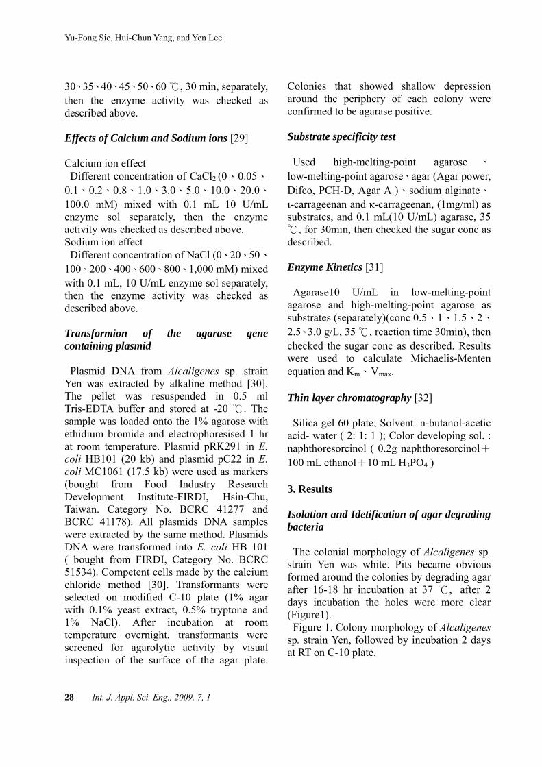
added to different pH buffer, 0.9 mL each separately, at 35 , 30 min, then checked the ℃reduced sugar concentration. Citrate-Na2HPO4 buffer (50 mM) ( pH Value 3.0 4.0 5.0); Tris-HCl buffer (50 mM) (pH Value 6.0 6.5 7.0 7.5); Sodium phosphate buffer (50mM) (pH Value 7.0 7.5 8.5); Phosphate buffer (50mM) (pH Value 8.0 8.5); Glycine-NaOH (50mM) (pH Value 9.0 10.0) were used. Effect of temperature on the stability and activity Made 5U/mL enzyme mixture with 50 mM
different pH of citrate-Na2HPO4 buffer, pH 3.0-5.0; Tris-HCl buffer, pH 6.0-7.5; Sodium phosphate buffer pH 7.5-8.0; phosphate buffer, pH 8.5; glycine-NaOH buffer, pH 9.0-10.0 , 4 , 24 hr. Then 0.1mL of different pH ℃enzyme sol was mixed with 0.2% agarose sol substrate (0.9 mL, separately, 35 , 30 min, ℃and then the reduced sugar conc were checked. The effects of the temperature [18] Low-melting-point agarose 0.2%, 0.9 mL
and 0.2% high-melting-point agarose, in 20、
25、30、35、40、45、50、55、60 were ℃
preheated 5 min, then 0.1 mL(10 U/mL) enzyme sol was added, 30min. and then the reduced sugar conc were checked. Temperature stability Enzyme sol 10 U/mL were set in 20、25、
Yu-Fong Sie, Hui-Chun Yang, and Yen Lee
28 Int. J. Appl. Sci. Eng., 2009. 7, 1
30、35、40、45、50、60 ℃, 30 min, separately, then the enzyme activity was checked as described above. Effects of Calcium and Sodium ions [29] Calcium ion effect Different concentration of CaCl2 (0、0.05、
0.1、0.2、0.8、1.0、3.0、5.0、10.0、20.0、
100.0 mM) mixed with 0.1 mL 10 U/mL enzyme sol separately, then the enzyme activity was checked as described above. Sodium ion effect Different concentration of NaCl (0、20、50、
100、200、400、600、800、1,000 mM) mixed with 0.1 mL, 10 U/mL enzyme sol separately, then the enzyme activity was checked as described above. Transformion of the agarase gene containing plasmid Plasmid DNA from Alcaligenes sp. strain
Yen was extracted by alkaline method [30]. The pellet was resuspended in 0.5 ml Tris-EDTA buffer and stored at -20 ℃. The sample was loaded onto the 1% agarose with ethidium bromide and electrophoresised 1 hr at room temperature. Plasmid pRK291 in E. coli HB101 (20 kb) and plasmid pC22 in E. coli MC1061 (17.5 kb) were used as markers (bought from Food Industry Research Development Institute-FIRDI, Hsin-Chu, Taiwan. Category No. BCRC 41277 and BCRC 41178). All plasmids DNA samples were extracted by the same method. Plasmids DNA were transformed into E. coli HB 101 ( bought from FIRDI, Category No. BCRC 51534). Competent cells made by the calcium chloride method [30]. Transformants were selected on modified C-10 plate (1% agar with 0.1% yeast extract, 0.5% tryptone and 1% NaCl). After incubation at room temperature overnight, transformants were screened for agarolytic activity by visual inspection of the surface of the agar plate.
Colonies that showed shallow depression around the periphery of each colony were confirmed to be agarase positive. Substrate specificity test Used high-melting-point agarose 、
low-melting-point agarose、agar (Agar power, Difco, PCH-D, Agar A )、sodium alginate、
ι-carrageenan and κ-carrageenan, (1mg/ml) as substrates, and 0.1 mL(10 U/mL) agarase, 35
, for 30min, then checked the sugar conc as ℃described. Enzyme Kinetics [31]
Agarase10 U/mL in low-melting-point agarose and high-melting-point agarose as substrates (separately)(conc 0.5、1、1.5、2、
2.5、3.0 g/L, 35 , reaction time 30min), then ℃
checked the sugar conc as described. Results were used to calculate Michaelis-Menten equation and Km、Vmax. Thin layer chromatography [32] Silica gel 60 plate; Solvent: n-butanol-acetic
acid- water ( 2: 1: 1 ); Color developing sol. : naphthoresorcinol ( 0.2g naphthoresorcinol+ 100 mL ethanol+10 mL H3PO4 ) 3. Results Isolation and Idetification of agar degrading bacteria The colonial morphology of Alcaligenes sp.
strain Yen was white. Pits became obvious formed around the colonies by degrading agar after 16-18 hr incubation at 37 ,℃ after 2 days incubation the holes were more clear (Figure1). Figure 1. Colony morphology of Alcaligenes
sp. strain Yen, followed by incubation 2 days at RT on C-10 plate.

The Discoveery of Agarolytic Bacterium with Agrarse Gene Containing Plasmid, and Sone Enzymology Characteristics
Int. J. Appl. Sci. Eng., 2009. 7, 1 29
Figure 1. Colony morphology of Alcaligenes sp.
strain Yen, after incubation for 2 days at RT on C-10 plate.
Bacterium characteristics The strain was Gram-negative rods,
non-fermentative and highly motile. It was positive for oxidase, citrate, malonate, tartrate, acetamide and cetrimide, negative for the production of indole and H2S. The strain was susceptible to penicillin, kanamycin, nitrofu-rantoin and tobramycin. The result of several biochemical tests were listed in Table 1. MicroScan AS4 system identified this bacte-
rium and asigned it as Alcaligenes sp. with 97.4% similarity. This finding was compara-ble to the results of 16S RNA comparison. However our strain showed agarolytic activity. Based on these data and the suggestion of GenBank, we assigned our strain as Alcali-genes sp. strain Yen (NCBI GenBank acces-sion number AY744384). 16S RNA Sequence was obtained CTGGCTCAGATTGAACGCTAGCGGGATGCCTTACACATGCAAGTCGAACGGCAGCACGGACTTCGGTCTGGTGGCGAGTGGCGAACGGGTGAGTAATGTATCGGAACGTGCCCAGTAGCGGGGGATAACTACGCGAAAGCGTAGCTAATACCGCATACGCCCTACGGGGGAAAGCAGGGGATCGCAAGACCTTGCACTATTGGAGCGGCCGATATCGGATTAGCTAGTTGGTGGGGTAACGGCTCACCAAGGCGACGATCCGTAGCTGGTT
TGAGAGGACGACCAGCCACACTGGGACTGAGACACGGCCCAGACTCCTACGGGAGGCAGCAGTGGGGAATTTTGGACAATGGGGGAAACCCTGATCCAGCCATCCCGCGTGTGCGATGAAGGCCTTCGGGTTGTAAAGCACTTTTGGCAGGAAAGAAACGTCGCGGGTTAATACCCCGCGGAACTGACGGTACCTGCAGAATAAGCACCGGCTAACTACGTGCCAGCAGCCGCGGTAATACGTAGGGTGCAAGCGTTAATCGGAATTACTGGGCGTAAAGCGTGCGCAGGCGGTTCGGAAAGAAAGATGTGAAATCCCAGAGCTTAACTTTGGAACTGCATTTTTAACTACCGAGCTAGAGTGTGTCAGAGGGAGGTGGAATTCCGCGTGTAGCAGTGAAATGCGTAGATATGCGGAGGAACACCGATGGCGAAGGCAGCCTCCTGGGATAACACTGACGCTCATGCACGAAAGCGTGGGGAGCAAACAGGATTAGATACCCTGGTAGTCCACGCCCTAAACGATGTCAACTAGCTGTTGGGGCCTTCGGGCCTTGGTAGCGCAGCTAACGCGTGAAGTTGACCGCCTGGGGAGTACGGTCGCAAGATTAAAACTCAAAGGAATTGACGGGGACCCGCACAAGCGGTGGATGATGTGGATTAATTCGATGCAACGCGAAAAACCTTAYCTACCCTTGACATGTCTGGAATTCCGAAGAGATTTGGAAGTGCTCGCAAGAGAACCGGAACACAGGTGCTGCATGGCTGTCGTCAGCTCGTGTCGTGAGATGTTGGGTTAAGTCCCGCAACGAGCGCAACCCTTGTCATTAGTTGCTACGAAAGGGCACTCTAATGAGACTGCCGGTGACAAACCGGAGGAAGGTGGGGATGACGTCAAGTCCTCATGGCCCTTATGGGTAGGGCTTCACACGTCATACAATGGTCGGGACAGAGGGTCGCCAACCCGCGAGGGGGAGCCAATCCCAGAAACCCGATCGTAGTCCGGATCGCAGTCTGCAACTCGACTGCGTGAAGTCGGAATCGCTAGTAATCGCGGATCAGCATGTCGCGGTGAATACGTTCCCGGGTCTTGTACACACCGCCCGTCACACCATGGGAGTGGGTTTTACCAGAAGTAGTTAGCCTAACCGTAAGGGGGGCGATTACCACGGTAGGATTCATGACTGGGGTGAAGTCGTAACAAGGTAGCCGTATCGGAAGGTGTGGCTGGATCA

Yu-Fong Sie, Hui-Chun Yang, and Yen Lee
30 Int. J. Appl. Sci. Eng., 2009. 7, 1
The 16S rDNA sequence of the isolate shared 100% sequence similarity with Alcali-
genes sp. strain ON5 (GenBank accession number AJ306836).
Table 1. Biochemical characteristics of Alcaligenes sp. Strain Yen.
Symbols: +,positive reaction ; -,negative reaction; m,motile; S:susceptible; R:resistible
Characteristic tested Result
Motility m Glucose - Sucrose - Sorbitol - Raffinose - Rhamnose - Arabinose - Inositol - Adonitol - Melibiose - Oxidase +
Urea -
Hydrogen Sulfide - Indole - Lysine - Arginine -
Characteristic tested ResultOrnithine - Tryptophan Deaminase - Esculin - Voges-Proskauer - Citrate + Malonate + o-Nit rophenyl-beta-D-Galactopyranoside - Tartrate + Acetamide + Cetrimide + OF Glucose - Penicillin S Kanamycin S Colistin R Nitrofurantoin S Tobramycin S Nitrate +
Properties of purified agarase Electrophoresis result showed the purified
agarase has a molecular mass of 101 kDa as detected by SDS-PAGE (Figure 2). Figure 2. SDS-PAGE of purified agarase
form Alcaligenes sp. Strain Yen. Lane 1, mo-
lecular mass standard;Lane 2, purified aga-rase. Myosine (210 kDa), b-galactosidase (125 kDa), Bovine serum albumin (101 kDa), Ovalbumin (56 kDa), Carbonic anhydrase (36 kDa), Soybean trypsin inhibitor (29 kDa), Lysozyme (21 kDa) and Aprotinin (6.9 kDa) were used as markers.

The Discoveery of Agarolytic Bacterium with Agrarse Gene Containing Plasmid, and Sone Enzymology Characteristics
Int. J. Appl. Sci. Eng., 2009. 7, 1 31
Figure 2. SDS-PAGE of purified agarase form Alcaligenes sp. Strain Yen. Lane 1, molecular mass stan-
dard;Lane 2, purified agarase. Myosine (210 kDa), β-galactosidase (125 kDa), Bovine serum albumin (101 kDa), Ovalbumin (56 kDa), Carbonic anhydrase (36 kDa), Soybean trypsin inhibitor (29 kDa), Lysozyme (21 kDa) and Aprotinin (6.9 kDa) were used as markers.
The measured molecular mass was close to
those reported for agarase from Agarivorans sp. JAMB-A11 105 kDa [17], but differ from Pseudomonas sp. W7 89 kDa [33], and Al-teromnas sp. SY37-12 39.5 kDa [5] Effect of pH and temperature optimum on the activity The optimal pH for the enzyme in different
buffers were different (Figure 3). The enzyme stability for pH profiles was
shown in Figure 4. In pH 3.0 - 8.5, the enzyme was relatively
sTable, the enzyme activity decreased sharply as pH raised higher than 8.5. The enzyme ac-tivity was sTable between temperature 20 - 35 ℃ (Figure 5). Then the activity decreased as temperature
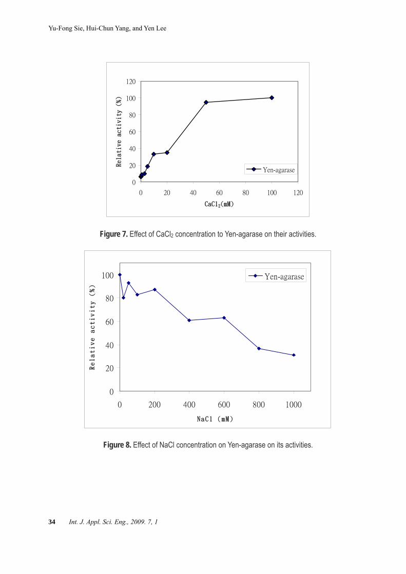
raised higher than 35 ℃. The optimal tem-perature for the enzyme was 42 ℃ (Figure 6). Effects of Calcium and Sodium ions Calcium ion could increase the activity for
Yen-agarase (Figure 7). Sodium ion could inhibit the activity for
Yen-agarase (Figure 8). Transformion of the agarase containing plasmid
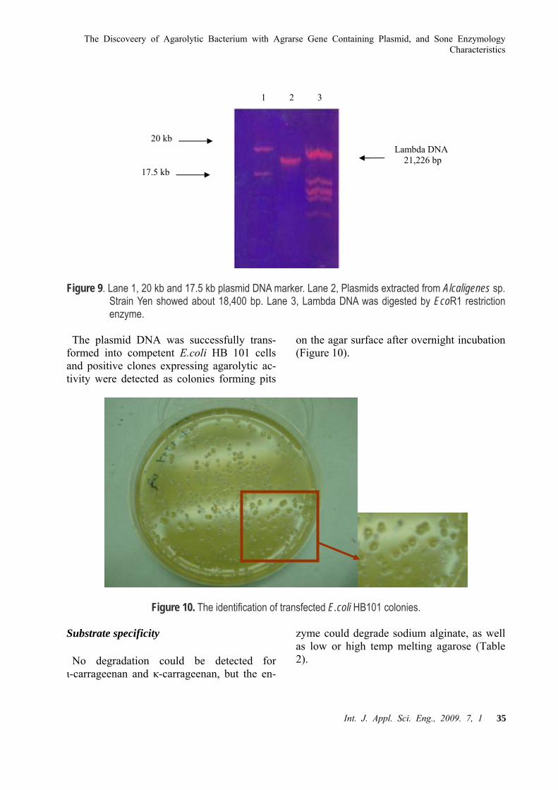
The plasmid DNA was successfully ex-tracted from Alcaligenes sp. strain Yen and electrophoresised on the gel. The size of the plasmid was estimated to be 18.4 kb (Figure 9).
1 2 kDa210
21
29
56
101
36
101
125
6.9

Yu-Fong Sie, Hui-Chun Yang, and Yen Lee
32 Int. J. Appl. Sci. Eng., 2009. 7, 1
Figure 9. Lane 1, 20 kb and 17.5 kb plasmid DNA marker. Lane 2, Plasmids extracted from Alcaligenes sp.. Strain Yen showed
about 18,400 bp. Lane 3, Lambda DNA was digested by EcoR1 restriction enzyme.
Figure 3. Optimum pH of Yen-agarase in various buffer sol.
0
20
40
60
80
100
2 3 4 5 6 7 8 9 10 11
pH
Relative acctivity (%)
Figure 4. Effect of pH on the stability of Yen-agarase.

The Discoveery of Agarolytic Bacterium with Agrarse Gene Containing Plasmid, and Sone Enzymology Characteristics
Int. J. Appl. Sci. Eng., 2009. 7, 1 33
0
20
40
60
80
100
20 30 40 50 60 70
Temperature (℃)
Relative activity (%)
Figure 5. Thermostability of Yen-agarase.
Figure 6. Optimum temperature of Yen-agarase.
Yu-Fong Sie, Hui-Chun Yang, and Yen Lee
34 Int. J. Appl. Sci. Eng., 2009. 7, 1
0
20
40
60
80
100
120
0 20 40 60 80 100 120
CaCl2(mM)
Relative activity (%)
Yen-agarase
Figure 7. Effect of CaCl2 concentration to Yen-agarase on their activities.
0
20
40
60
80
100
0 200 400 600 800 1000
NaCl (mM)
Relative activity (%)
Yen-agarase
Figure 8. Effect of NaCl concentration on Yen-agarase on its activities.
The Discoveery of Agarolytic Bacterium with Agrarse Gene Containing Plasmid, and Sone Enzymology Characteristics
Int. J. Appl. Sci. Eng., 2009. 7, 1 35
Figure 9. Lane 1, 20 kb and 17.5 kb plasmid DNA marker. Lane 2, Plasmids extracted from Alcaligenes sp. Strain Yen showed about 18,400 bp. Lane 3, Lambda DNA was digested by EcoR1 restriction enzyme.
The plasmid DNA was successfully trans-
formed into competent E.coli HB 101 cells and positive clones expressing agarolytic ac-tivity were detected as colonies forming pits
on the agar surface after overnight incubation (Figure 10).
Figure 10. The identification of transfected E.coli HB101 colonies. Substrate specificity
No degradation could be detected for ι-carrageenan and κ-carrageenan, but the en-
zyme could degrade sodium alginate, as well as low or high temp melting agarose (Table 2).
20 kb
17.5 kb
Lambda DNA 21,226 bp
1 2 3

Yu-Fong Sie, Hui-Chun Yang, and Yen Lee
36 Int. J. Appl. Sci. Eng., 2009. 7, 1
Table 2. Substrate specificity of agarase-Yen against nine polysaccharides 1: µmole D-galactose equivalent/min/mL. 2: Could not be determined.
Substrate Yen-agarases
High-melting-point agarose 51.451
Low-melting-point agarose 42.60
Difco agar 30.72
Agar power 24.84
Agar A 32.68
PCH-D agar 24.78
Sodium alginate 9.28
ι-Carrageenan ---2
κ-Carrageenan --- Km、Vmax High and low-melting-point agarose were
used as substrates separately, the catalytic rate was shown by Lineweaver-Burk plots and Michalis-Menten equations. When the high-melting-point agarose was used as sub-strate, Km was 2.76 g/L, Vmax was 68.03 μmole min-1(Figure 11)
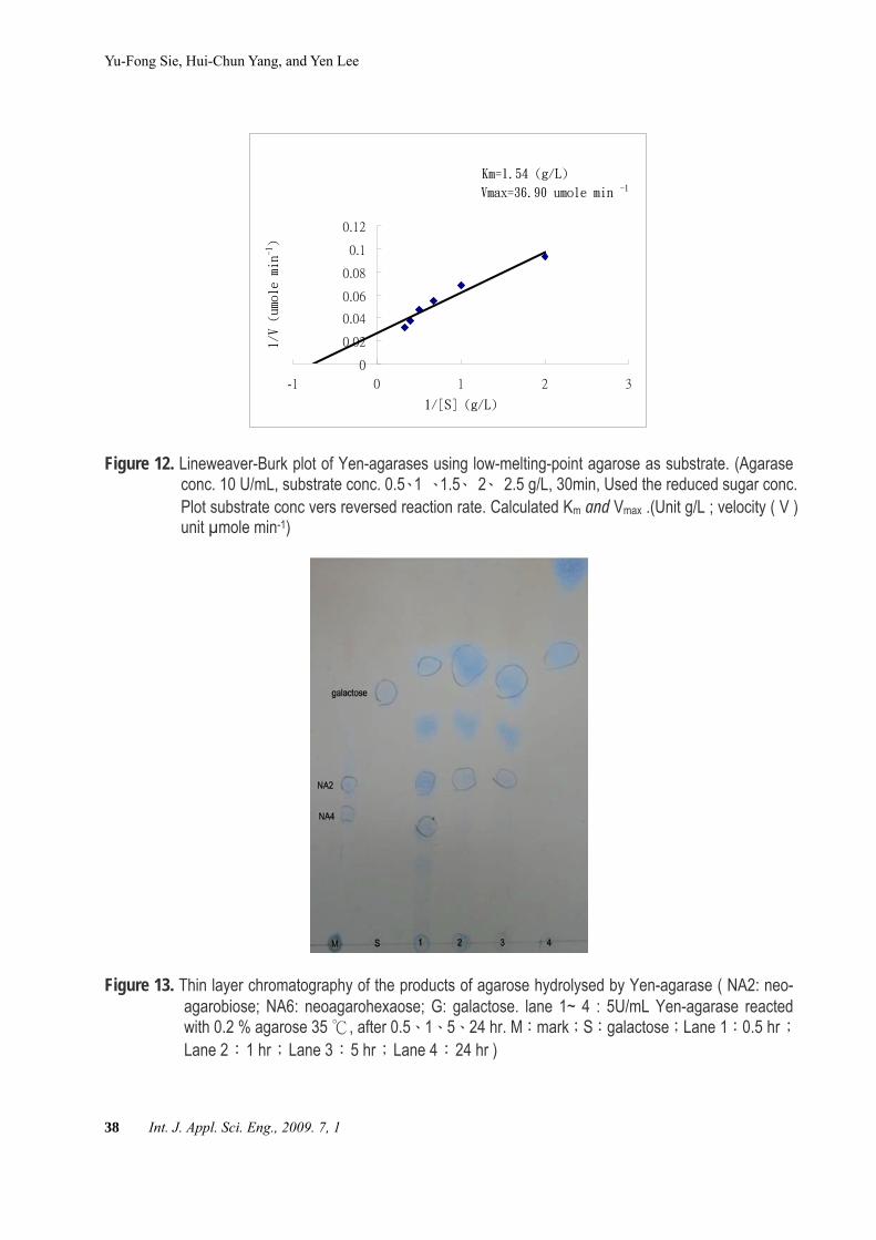
when low-melting-point agarose was used as substrate, Km was 1.54 g/L, Vmax was 36.9 μmole min-1(Figure 12). The Yen-agarase had higher affinity for low-melting-point agarose than high-melting-point agarose.Thin layer chromatography Neoagarobiose, neoagarohexaose and galac-
tose were detected (Figure 13). it was the re-sults of the beta type agarase. 4. Discussion Previous studies have showed that the
agar-degrading bacterium Paenibacillus spp.[19] took 10 days(28 ℃) to form de-
pressions on agar surface, and Pseudoaltero-monas antarctica N-1 [7] took 48 hr(25 ℃). Our strain shown pits on agar surface sur-rounding the colony in less then 18 hr. The purified enzyme shown high agarase activity as 406 U/mg. Pseudoalteromonas antarctica strain N-1 [7] had reported agarase activity 290 U/mg , Thalassomonas sp. strain JAMB A33 40.7 U/mg [10], Agarivorans sp. JAMB-A11 371 U/mg (Ohta et al. 2005a) and Microbulbifer strain JAMB-A7 398 U/mg [34] respectively. A marine bacterium JAMB-A94 had reported agarase activity 517 U/mg [35]. Alcaligenes sp. strain Yen had different pH maximal enzyme activity in different buffers. The optimal enzyme activity shown at 42 . ℃Compare to other agar-degrading bacteria. Agarase from Bacillus sp. MK03 had optimal pH at 7.6 [18], Thalassomonas sp. strain JAMB-A33 8.5 [10], Pseudomonas sp. SK38 9.0 [36]. JAMB-A94 had optima tem-perature at 55 ℃ [35], Microbulbifer strain JAMB-A7 50 ℃ [34]. The transformed E. coli HB101 cells had optima growth at 25-30
. This plasmid had the smallest size among ℃all plasmids containing an agarase gene that

The Discoveery of Agarolytic Bacterium with Agrarse Gene Containing Plasmid, and Sone Enzymology Characteristics
Int. J. Appl. Sci. Eng., 2009. 7, 1 37
have been found in nature. Some other find-ings were much larger than this [37]. This plasmid could be used to transform competent bacterial cells directly or be modified for cloning vectors. For the successfully trans-formation in E. coli HB101 and got agarase expression. The plasmid’s agarase gene pro-moter region should be recognized by E.coli HB101 transcription facilities. The agarase activity raised sharply at 30 - 42 , afterward ℃it decreased. The further study is to prepare a new cloning vector with agarase gene and an-other marker gene such as β-galactosidase or green fluorescent protein without antibiotic resistant gene as an innovative molecular bi-ology tool. The new tool can select positive colonies without using environmental con-taminating antibiotics. In this study, we used C-10 plates to culture the bacterium without adding organic material. This was the first time of all agarase research that used inor-
ganic material plate to select agarolytic bacte-ria. Agarases have low activity and low pro-ductivity are not economically useful for in-dustrial application. Our strain secreted exta-celluar agarase with high activity. It has a highly biotechnological significant agarolytic plasmid for industry application. The results showed Alcaligenes sp. strain Yen could use agar as its carbon and energy source. Trans-formed E. coli HB101 cells needed yeast ex-tract as supplement for growth. When yeast extract was not added in the medium, the transformed E. coli grew poorly and formed tiny pits on the agar surface. This is a report of finding an agarase secre-
tion bacterium with smallest agarolytic plas-mid ever been found, and the first time trans-formation of the intact agarolytic plasmid into E. coli and got agarase gene expression. The enzymology analysis was also reported.
Km = 2.76 (g/L)
Vmax = 68.03 umole min-1
-0.02
0
0.02
0.04
0.06
0.08
0.1
0.12
-1 -0.5 0 0.5 1 1.5 2
1/[S] (g/L)
1/V
(um
ole
min
-1)
Figure 11. Lineweaver-Burk plot of Yen-agarases using high-melting-point agarose as substrate. (Yen-garase conc.10 U/mL . Substrate conc. 0.5、1 、1.5、 2、 2.5 g/L ,30min, Used the reduced sugar conc. Plot substrate conc vers reversed reaction rate. Calculated Km and Vmax (unit g/L ; velocity( V ) µmole min-1)
Yu-Fong Sie, Hui-Chun Yang, and Yen Lee
38 Int. J. Appl. Sci. Eng., 2009. 7, 1
Km=1.54 (g/L)
Vmax=36.90 umole min -1
0
0.02
0.04
0.06
0.08
0.1
0.12
-1 0 1 2 3
1/[S] (g/L)
1/V (umole min-1)
Figure 12. Lineweaver-Burk plot of Yen-agarases using low-melting-point agarose as substrate. (Agarase conc. 10 U/mL, substrate conc. 0.5、1 、1.5、 2、 2.5 g/L, 30min, Used the reduced sugar conc. Plot substrate conc vers reversed reaction rate. Calculated Km and Vmax .(Unit g/L ; velocity ( V ) unit µmole min-1)
Figure 13. Thin layer chromatography of the products of agarose hydrolysed by Yen-agarase ( NA2: neo-agarobiose; NA6: neoagarohexaose; G: galactose. lane 1~ 4 : 5U/mL Yen-agarase reacted with 0.2 % agarose 35 , after 0.5℃ 、1、5、24 hr. M:mark;S:galactose;Lane 1:0.5 hr;Lane 2:1 hr;Lane 3:5 hr;Lane 4:24 hr )
The Discoveery of Agarolytic Bacterium with Agrarse Gene Containing Plasmid, and Sone Enzymology Characteristics
Int. J. Appl. Sci. Eng., 2009. 7, 1 39
Authors' contributions The bacterium was isolated by Yen Lee. Its
agarolytic plasmid and the plasmid was transformable into E. coli HB101 were also discovered by Yen Lee. The enzymological analysis were done cooperatively by Yu-Fong Sie and Hui-Chun Yang. References [ 1] Craigie, J. 1990. “Biology of the red al-
gae. Cell walls”. In K. M. Cole and R. G. Sheath(ed.), Cambridge University Press, New York, N.Y. 221-258.
[ 2] Allouch, J., Jam, M., Helbert, W., Bar-beyron, T., Kloareg, B., Henrissat, B. and Czjzek, M. 2003. The three-dimensional structures of two beta-agarases. Journal of Biological Chemistry, 278: 47171-47180.
[ 3] Kurahashi, M., and Yokota, A. 2004. Agarivorans albus gen. nov., sp. nov., a gamma-proteobacterium isolated from marine animals. International Journal of Ststematic Evolutionary Microbiol, 54: 693-697.
[ 4] Leon, O., Quintana, L., Peruzzo, G., and Slebe, J. C. 1992. Purification and Prop-erties of an Extracellular Agarase from Alteromonas sp. Strain C-1. Applied and Environmental Microbiology, 58: 4060-4063.
[ 5] Wang, J., Mou, H., Jiang, X., and Guan, H. 2006. Characterization of a novel beta-agarase from marine Alteromonas sp. SY37-12 and its degrading products. Applied Microbiology Biotechnology, 71: 833-839.
[ 6] Ivanova, E. P., Kiprianova, E. A., Mik-hailov, V. V., Levanova, G. F., Garagu-lya, A. D., Gorshkova, N. M., Vysotskii, M. V., Nicolau, D. V., Yumoto, N., Ta-guchi, T., and Yoshikawa, S. 1998. Phe-notypic diversity of Pseudoalteromonas citrea from different marine habitats and emendation of the description. Interna-
tional Journal of Systematic Bacteriol-ogy, 48 Pt. 1: 247-256.
[ 7] Vera, J., Alvarez, R., Murano, E., Slebe, J. C., and Leon, O. 1998. Identification of a marine agarolytic Pseudoalteromo-nas isolate and characterization of its ex-tracellular agarase. Applied and Envi-ronmental Microbiology, 64: 4378-4383.
[ 8] Chiura, H. X., and Tsukamoto, K. 2000. Purification and characterization of novel agarase secreted by marine bacte-rium, Pseudoalteromonas sp. strain CKT1. Microbial Environ, 64: 11-22.
[ 9] Romanenko, L. A., Zhukova, N. V., Rohde, M., Lysenko, A. M., Mikhailov, V. V., and Stackebrandt, E. 2003. Pseu-doalteromonas agarivorans sp. nov., a novel marine agarolytic bacterium. In-ternational Journal of Systematic Evolu-tionary Microbiol, 53: 125-131.
[10] Ohta, Y., Hatada, Y., Miyazaki, M., Nogi, Y., Ito, S., and Horikoshi, K. 2005b. Purification and characterization of a novel alpha-agarase from a Thalas-somonas sp. Current Microbiology, 50: 212-216.
[11] Jean, W. D., Shieh, W. Y., and Liu, T. Y. 2006. Thalassomonas agarivorans sp. nov., a marine agarolytic bacterium iso-lated from shallow coastal water of An-Ping Harbour, Taiwan, and emended description of the genus Thalassomonas. International Journal of Systematic Evo-lutionary Microbiol, 56: 1245-1250.
[12] Nedashkovskaya, O. I., Suzuki, M., Vy-sotskii, M. V., and Mikhailov, V. V. 2003. Reichenbachia agariperforans gen. nov., sp. nov., a novel marine bacterium in the phylum Cyto-phaga-Flavobacterium-Bacteroides. In-ternational Journal of Systematic Evolu-tionary Microbiol, 53: 81-85.
[13] Nedashkovskaya, O. I., Suzuki, M., Van-canneyt, M., Cleenwerck, I., Lysenko, A. M., Mikhailov, V. V., and Swings, J. 2004b. Zobellia amurskyensis sp. nov., Zobellia laminariae sp. nov. and Zobel-

Yu-Fong Sie, Hui-Chun Yang, and Yen Lee
40 Int. J. Appl. Sci. Eng., 2009. 7, 1
lia russellii sp. nov., novel marine bacte-ria of the family Flavobacteriaceae. In-ternational Journal of Ststematic Evolu-tionary Microbiol, 54: 1643-1648.
[14] Jam, M., Flament, D., Allouch, J., Potin, P., Thion, L., Kloareg, B., Czjzek, M., Helbert, W., Michel, G., and Barbeyron, T. 2005. The endo-beta-agarases AgaA and AgaB from the marine bacterium Zobellia galactanivorans: two paralogue enzymes with different molecular or-ganizations and catalytic behaviours. Biochemical Journal, 385: 703-713.
[15] Nedashkovskaya, O. I., Suzuki, M., Lysenko, A. M., Snauwaert, C., Van-canneyt, M., Swings, J., Vysotskii, M. V., and Mikhailov, V. V. 2004a. Cellulo-phaga pacifica sp. nov. International Journal of Ststematic Evolutionary Mi-crobiol, 54: 609-613.
[16] Nedashkovskaya, O. I., Kim, S. B., Van-canneyt, M., Lysenko, A. M., Shin, D. S., Park, M. S., Lee, K. H., Jung, W. J., Ka-linovskaya, N. I., Mikhailov, V. V., Bae, K.S., and Swings, J. 2006. Echinicola pacifica gen. nov., sp. nov., a novel flexibacterium isolated from the sea ur-chin Strongylocentrotus intermedius. In-ternational Journal of Ststematic Evolu-tionary Microbiol, 56: 953-958.
[17] Ohta, Y., Hatada, Y., Ito, S., and Horikoshi, K. 2005a. High-level expres-sion of a neoagarobiose-producing beta-agarase gene from Agarivorans sp. JAMB-A11 in Bacillus subtilis and en-zymic properties of the recombinant en-zyme. Biotechnology and Applied Bio-chemistry, 41: 183-191.
[18] Suzuki, H., Sawai, Y., Suzuki, T., and Kawai, K. 2003. Purification and char-acterization of an extracellular beta-agarase from Bacillus sp. MK03. Journal of Biosciences Bioeng, 95: 328-334.
[19] Hosoda, A., Sakai, M., and Kanazawa, S. 2003. Isolation and characterization of agar-degrading Paenibacillus spp. asso-
ciated with the rhizosphere of spinach. Bioscience Biotechnology and Biochem-istry, 67: 1048-1055.
[20] Araki, T., Lu, Z., and Morishita, T. 1998. Optimization of parameters for isolation of protoplasts from Gracilaria verrucosa (Rhodophyta). Journal of Marine Bio-technology, 6: 193-197.
[21] Gray, F., Kenney, J. S., and Dunne, J. F. 1995. Secretion capture and report web: use of affinity derivatized agarose mi-crodroplets for the selection of hybri-doma cells. Journal of Immunological Methods, 182: 155-163.
[22] Yoshizawa, Y., Ametani, A., Tsunehiro, J., Nomura, K., Itoh, M., Fukui, F., and Kaminogawa, S. 1995. Macrophage stimulation activity of the polysaccharide fraction from a marine alga (Porphyra yezoensis): structure-function relation-ships and improved solubility. Biosci-ence Biotechnology and Biochemistry, 59: 1933-1937.
[23] Kobayashi, R., Takisada, M., Suzuki, T., Kirimura, K., and Usami, S. 1997. Neo-agarobiose as a novel moisturizer with whitening effect. Bioscience Biotech-nology and Biochemistry, 61: 162-163.
[24] Yamamoto, Y., Nakagawa, H., and Hi-rose, K. 2002. Antihypercholestrolmic effets of agaropectin in rats. Journal of the Japanese Society for Nutrition Food Science and Technologe-Nippon Shoku-hin Kagaku Kogaku Kaishi, 55: 143-147.
[25] Osoegawa, K., Woon, P. Y., Zhao, B., Frengen, E., Tateno, M., Catanese, J. J., and de Jong, P. J. 1998. An improved approach for construction of bacterial ar-tificial chromosome libraries. Genomics 52: 1-8.
[26] Williams, S. T., Sharpe, M. E., and Holt, J. G. 1989. “Bergy's manual of system-atic bacteriology”, Vol. 4. Williams & Wilkins, Baltimore, Md.
[27] Bradford, M. M. 1976. A rapid and sen-sitive method for the quantitation of mi-crogram quantities of protein utilizing

The Discoveery of Agarolytic Bacterium with Agrarse Gene Containing Plasmid, and Sone Enzymology Characteristics
Int. J. Appl. Sci. Eng., 2009. 7, 1 41
the principle of protein-dye binding. Analytical Biochemistry, 72: 248-254.
[28] Miller, G. L. 1959. Use of dinitrosali-cylic acid reagent for deter for determi-nation of reducing sugar. Analytical Chemistry, 31: 426-428.
[29] Li., X., X., Dong, C., Zhao, Z., Chen., and F. Chen. 2003. Isolation and some properties of cellulose-degrading Vibrio sp. LX-3 with agar-liquefying ability from soil. World Journal of Microbiol-ogy & Biotechnology, 19:375-379.
[30] Ausubel, F. M., Brent, R., Kingston, R. E., Moore, D. D., Seidman, J. G., Smith, J. A., and Struhl, K. 1999. “Short Proto-cols in Molecular Biology”, Vol. 1. Wiley, New York. 22-27.
[31] Sugano, Y., H. Kodama, I., Terada, Y. Yamazaki., and M. Noma. 1994. Purifi-cation and characterization of a novel enzyme, (α-NAOS hydrolase) α-neoagarooligosac-charidehydrolase, from a marine bacterium, Vibrio sp. strain JT0107. Journal of Bacteriology, 176:6812-6818.
[32] Kirimura, K. N., Masuda, Y., Iwasaki, H., Nakagawa, R., Kobayashi, and S. Usami. 1999. Purification and characterization of a novel β-agarase from an alkalophilic bacterium, Alteromonas sp. E-1. Journal of Biosciences Bioeng, 87:436-441.
[33] Kong, J., Hwang, S. H., Kim, B. J., Bae, S. K., and Kim, J. D. 1997. Cloning and expression of an agarae gene from a ma-
rine bacterium Pseudomonas sp. W7. Biotechnology Letters, 19: 23-26.
[34] Ohta, Y., Hatada, Y., Nogi, Y., Miyazaki, M., Li, Z., Akita, M., Hidaka, Y., Goda, S., Ito, S., and Horikoshi, K. 2004a. En-zymatic properties and nucleotide and amino acid sequences of a thermosTable beta-agarase from a novel species of deep-sea Microbulbifer. Applied Micro-biology Biotechnology, 64: 505-514.
[35] Ohta, Y., Nogi, Y., Miyazaki, M., Li, Z., Hatada, Y., Ito, S., and Horikoshi, K. 2004b. Enzymatic properties and nucleo-tide and amino acid sequences of a thermosTable beta-agarase from the novel marine isolate, JAMB-A94. Bio-science Biotechnology and Biochemistry, 68: 1073-1081.
[36] Kang, N. Y., Choi, Y. L., Cho, Y. S., Kim, B. K., Jeon, B. S., Cha, J. Y., Kim, C. H., and Lee, Y. C. 2003. Cloning, ex-pression and characterization of a beta-agarase gene from a marine bacte-rium, Pseudomonas sp. SK38. Biotech-nology Letters, 25: 1165-1170.
[37] Zhong, Z., Toukdarian, A., Helinski, D., Knauf, V., Sykes, S., Wilkinson, J. E., O'Bryne, C., Shea, T., DeLoughery, C., and Caspi, R. 2001. Sequence analysis of a 101-kilobase plasmid required for agar degradation by a Microscilla isolate. Ap-plied and Environmental Microbiology, 67: 5771-9.
Copyright © 2022 FDOKUMEN