Xylanase Attachment to the Cell Wall of the Hyperthermophilic Bacterium Thermotoga maritima
10
Published Ahead of Print 14 December 2007. 2008, 190(4):1350. DOI: 10.1128/JB.01149-07. J. Bacteriol. Baumeister, Martin Armbrecht and Michael Valdez Wolfgang Liebl, Christoph Winterhalter, Wolfgang maritima Thermotoga Hyperthermophilic Bacterium Xylanase Attachment to the Cell Wall of the http://jb.asm.org/content/190/4/1350 Updated information and services can be found at: These include: REFERENCES http://jb.asm.org/content/190/4/1350#ref-list-1 at: This article cites 39 articles, 10 of which can be accessed free CONTENT ALERTS more» articles cite this article), Receive: RSS Feeds, eTOCs, free email alerts (when new http://journals.asm.org/site/misc/reprints.xhtml Information about commercial reprint orders: http://journals.asm.org/site/subscriptions/ To subscribe to to another ASM Journal go to: on October 6, 2014 by guest http://jb.asm.org/ Downloaded from on October 6, 2014 by guest http://jb.asm.org/ Downloaded from
-
Upload
independent -
Category
Documents
-
view
0 -
download
0
Transcript of Xylanase Attachment to the Cell Wall of the Hyperthermophilic Bacterium Thermotoga maritima

Published Ahead of Print 14 December 2007. 2008, 190(4):1350. DOI: 10.1128/JB.01149-07. J. Bacteriol.
Baumeister, Martin Armbrecht and Michael ValdezWolfgang Liebl, Christoph Winterhalter, Wolfgang
maritimaThermotogaHyperthermophilic Bacterium
Xylanase Attachment to the Cell Wall of the
http://jb.asm.org/content/190/4/1350Updated information and services can be found at:
These include:
REFERENCEShttp://jb.asm.org/content/190/4/1350#ref-list-1at:
This article cites 39 articles, 10 of which can be accessed free
CONTENT ALERTS more»articles cite this article),
Receive: RSS Feeds, eTOCs, free email alerts (when new
http://journals.asm.org/site/misc/reprints.xhtmlInformation about commercial reprint orders: http://journals.asm.org/site/subscriptions/To subscribe to to another ASM Journal go to:
on October 6, 2014 by guest
http://jb.asm.org/
Dow
nloaded from
on October 6, 2014 by guest
http://jb.asm.org/
Dow
nloaded from

JOURNAL OF BACTERIOLOGY, Feb. 2008, p. 1350–1358 Vol. 190, No. 40021-9193/08/$08.00�0 doi:10.1128/JB.01149-07Copyright © 2008, American Society for Microbiology. All Rights Reserved.
Xylanase Attachment to the Cell Wall of the HyperthermophilicBacterium Thermotoga maritima�
Wolfgang Liebl,1* Christoph Winterhalter,2 Wolfgang Baumeister,3Martin Armbrecht,1† and Michael Valdez1
Institut fur Mikrobiologie und Genetik, Georg-August-Universitat, Grisebachstr. 8, D-37077 Gottingen, Germany1;Wacker Chemie GmbH, Johannes Hess Str. 24, D-84489 Burghausen, Germany2; Max-Planck-Institut fur Biochemie,
Am Klopferspitz, D-82152 Martinsried, Germany3
Received 20 July 2007/Accepted 27 November 2007
The cellular localization and processing of the endo-xylanases (1,4-�-D-xylan-xylanohydrolase; EC 3.2.1.8) ofthe hyperthermophile Thermotoga maritima were investigated, in particular with respect to the unusual outermembrane (“toga”) of this gram-negative bacterium. XynB (40 kDa) was detected in the periplasmic fractionof T. maritima cells and in the culture supernatant. XynA (120 kDa) was partially released to the surroundingmedium, but most XynA remained cell associated. Immunogold labeling of thin sections revealed that cell-bound XynA was localized mainly in the outer membranes of T. maritima cells. Amino-terminal sequencing ofpurified membrane-bound XynA revealed processing of the signal peptide after the eighth residue, therebyleaving the hydrophobic core of the signal peptide attached to the enzyme. This mode of processing isreminiscent of type IV prepilin signal peptide cleavage. Removal of the entire XynA signal peptide wasnecessary for release from the cell because enzyme purified from the culture supernatant lacked 44 residuesat the N terminus, including the hydrophobic part of the signal peptide. We conclude that toga association ofXynA is mediated by residues 9 to 44 of the signal peptide. The biochemical and electron microscopiclocalization studies together with the amino-terminal processing data indicate that XynA is held at the cellsurface of T. maritima via a hydrophobic peptide anchor, which is highly unusual for an outer membraneprotein.
Thermotoga maritima was the first hyperthermophilic speciesof the domain Bacteria to be discovered (18), and to date it isthe best-studied bacterial hyperthermophile. For the organ-isms related to Thermotoga, the phylum Thermotogae was pro-posed (35). The hyperthermophilic bacteria represent a deepbranch within the phylogenetic tree of the Bacteria, suggestingthat these organisms may have retained archetypal featuresresembling those of primordial bacterial cells. This expectationis apparently supported by the fact that some of the biomol-ecules and properties of T. maritima have indeed revealedquite unusual features (19). Its peptidoglycan is thin and witha low degree of cross-linking, devoid of meso-diaminopimelicacid but containing D-lysine and L-lysine, which are absentfrom other gram-negative bacteria (17). A common morpho-logical characteristic of the members of the order Thermoto-gales is the presence of an outer sheath-like envelope (“toga”)(19). For cells of the genus Thermotoga, the toga forms largeballoons at the cell ends and in this way encloses a periplasmiccompartment with a volume comparable to or sometimes evenlarger than that of the cytoplasm. The only components of thetoga studied in any detail are the coiled-coil protein Omp� andthe porin Omp� of T. maritima (13, 14, 34). Previous studieshave indicated that besides Omp�, certain enzymes are also
probably localized in the toga (reference 36 and our unpub-lished results).
The genome of T. maritima strain MSB8 encodes all en-zymes needed for the degradation of complex xylans consistingof a poly-�-1,4-linked xylose backbone substituted with acetyl,arabinofuranosyl and 4-O-methylglucuronyl groups, i.e., xyla-nases, �-xylosidase, arabinofuranosidase, �-glucuronidase, andacetyxylan esterase. This paper deals with the localizationof the endo-xylanases (1,4-�-D-xylan-xylanohydrolase; EC3.2.1.8) XynB and, in particular, XynA of T. maritima strainMSB8. XynA is an extremely thermostable 120-kDa enzymewhose precursor sequence, based on the primary structurederived from the nucleotide sequence of the gene, is composedof an apparently typical N-terminal signal peptide, followed byfive domains in the order A1-A2-B-C1-C2 (42). The centralpart (domain B, �340 amino acids), which represents the cat-alytic domain, belongs to family 10 of glycoside hydrolases. TheN-terminal �150-amino-acid repeated domains (A1 and A2)have no significant similarity to the C-terminal �170-amino-acid repeated domains (C1 and C2). Domain A2 belongs tofamily 22 of carbohydrate-binding modules (CBMs) and hasbeen shown to display xylan-binding ability (24, 30). DomainC2 has been shown to represent a cellulose-binding module(42) which belongs to family 9 of CBMs and is unique in thatit specifically binds to the reducing end of cellulose and solublepolysaccharides as well as to a variety of mono-, di-, and oli-gosaccharides. After binding to microcrystalline cellulose, elu-tion of C2 can be achieved with a 0.2 M cellobiose solution (10,22, 32, 42). The data reported here clearly demonstrate thetoga association of the multidomain xylanase XynA and pro-
* Corresponding author. Mailing address: Institut fur Mikrobiologieund Genetik, Grisebachstr. 8, D-37077 Gottingen, Germany. Phone:49-551-393795. Fax: 49-551-394897. E-mail: [email protected].
† Present address: Eppendorf AG, Barkhausenweg 1, D-22339Hamburg, Germany.
� Published ahead of print on 14 December 2007.
1350
on October 6, 2014 by guest
http://jb.asm.org/
Dow
nloaded from

vide information about the processing and the mechanism ofattachment of this enzyme to the cell envelope of T. maritima.
MATERIALS AND METHODS
Strain and growth conditions. Thermotoga maritima strain MSB8 (DSM 3109)was propagated in Difco marine broth medium 2216 (Difco, Detroit, MI) sup-plemented with 0.25% soluble starch or xylose as described before (42). For largebatch cultures, cells from a fresh overnight culture were inoculated into a re-duced medium consisting of 0.5% peptone, 0.1% yeast extract, 0.25% solublestarch or xylose, 1% (vol/vol) Difco marine broth, 3% NaCl, 0.5% Na2S, and0.0001% resazurin dissolved in tap water and preheated to 80°C. Disruption ofT. maritima cells for the preparation of crude extracts was achieved by passingsuspensions of cells in 20 mM bis-Tris buffer (pH 6.2) through a French pressurecell (American Instrument Company, Silver Spring, MD) at 6.9 MPa.
Protein purification, analytical methods, and xylanase assay. Previously pub-lished methods (25, 26) were used for the determination of protein concentra-tions and sodium dodecyl sulfate-polyacrylamide gel electrophoresis (SDS-PAGE). Detection of protein bands with thermostable xylanase activity afterseparation by SDS-PAGE was done with a zymogram staining technique de-scribed before (42). Amino-terminal sequencing of protein samples via sequen-tial Edman degradation was done on an Applied Biosystems 477A sequencer.
For purification of XynA from T. maritima culture supernatant, the superna-tant obtained from 50 liters of culture was concentrated to a final volume of 300ml by ultrafiltration with a Sartocon ultrafiltration system followed by a secondstep with a Sartocon mini-ultrafiltration system (Sartorius, Gottingen, Germany),both equipped with cellulose triacetate membranes (10,000-Da cutoff). The con-centrated supernatant was applied to a column with a 400-ml bed volume packedwith microcrystalline cellulose. Washing of the column and elution of the 120-kDa enzyme with 0.1 M cellobiose were done as described previously (42).
For expression of xynA in Escherichia coli, a synthetic oligonucleotide primerwith the sequence 5�-GGACTGAATTCATGCAAGTCAGGAAGAGACGGGG-3� was used in combination with primer 6 (42) to generate via PCR a DNAfragment carrying the complete xynA coding region. The PCR product was cut atthe ends with EcoRI and SmaI before insertion into the accordingly double-digested Ptac expression vector pJF118ut (8), a derivative of pJF118EH (16). Thisplasmid construction, designated p86, was introduced into E. coli WCM105. Thesecreted recombinant xylanase was purified to apparent electrophoretic homo-geneity via ultrafiltration and cellulose affinity chromatography.
Standard assay mixtures for the determination of xylanase activity contained0.8% oat spelt xylan (Roth, Karlsruhe, Germany), 250 mM NaCl, 50 mM bis-Trisbuffer (pH 6.2), and appropriately diluted enzyme (in some cases in the form ofa suspension of washed cells; see below), and assays were performed in a totalvolume of 0.5 ml. Unless mentioned otherwise, incubation was carried out for 10min at 75°C. The amount of reducing groups liberated during the enzymatichydrolysis of xylan was quantitated by the dinitrosalicylic acid method (7) asdescribed previously (43). One unit of xylanase activity is the amount of enzymenecessary to liberate 1 �mol of reducing groups (as xylose equivalents) perminute.
Osmotic shock treatment of T. maritima cells. Cells of T. maritima (1 g) grownin medium with soluble starch (0.2%), oat spelt xylan (0.1%), or xylose (0.5%)were suspended in 8 ml of 500 mM glucose–1 mM EDTA–200 mM Tris-HCl, pH7.4. After addition of 0.8 ml of a lysozyme solution (20 mg ml�1) and 5 mlpolymyxin B sulfate (1 mg ml�1) and incubation at ambient temperature for 30min, 8 ml distilled water was added; 30 min later, 32 ml distilled water was added.The supernatant obtained after centrifugation for 10 min at 11,000 � g was usedas the periplasmic protein preparation. Control experiments demonstrated theefficient separation of the T. maritima periplasmic maltose-binding protein withthis spheroplasting and osmotic shock treatment (not shown).
Immunogold labeling and electron microscopy. A suspension of T. maritimacells from a fresh overnight culture was used for fixation (30 min, 20°C) with 2%paraformaldehyde, 1% glutaraldehyde, and 0.5% uranyl acetate. The cells wereembedded in 2% low-gelling agarose type VII (Sigma) before the preparationwas subjected to multistep infiltration with Lowicryl K4M (Lowi, Waldkraiburg,Germany) using the PLT method (23) as described before (14). After polymer-ization of the sample with UV light, thin sections (60 nm) were collected onnickel grids.
For immunolabeling, the nickel grids with the T. maritima thin sections wereincubated for 1 h in labeling solution (TBS [10 mM Tris {pH 7.5}, 0.15 M NaCl]supplemented with 0.05% Tween 20, 5% milk powder, and 0.5 M NaCl) and thenfor 3 h in labeling solution mixed with 0.01 volume of a polyclonal anti-XynAantiserum, which was a rabbit-derived preparation raised against a recombinanttruncated xylanase derivative consisting of the central catalytic domain of XynA
(42). After five brief washing steps in labeling solution, the grids were incubatedfor 1 h with anti-rabbit immunoglobulin G (IgG) gold (10 nm) conjugate (Sigma,Deisenhofen, Germany) diluted 1:10 to 1:50 in labeling solution. After washingrepeatedly in labeling solution and once in water (5 min), the grids were air driedfor 10 min. Poststaining was done by incubation for 10 min in 4% uranyl acetateand, after three washes with water, for 5 min in lead citrate, followed by a finalwash with water. After shadowing with carbon, electron micrographs were re-corded with a Philips CM12 apparatus at a nominal magnification of �16,000.
Immunofluorescence labeling and epifluorescence microscopy. Cells from afresh overnight culture of T. maritima grown in medium with 0.25% xylose werecollected by centrifugation and suspended in TBS. The cells were used forimmunolabeling either after fixation, which was accomplished by mixing thesuspension with 3 volumes TBS containing 4% paraformaldehyde and incubatingat 4°C for 16 h, or without prior fixation. For immunolabeling, 0.01 volume ofrabbit anti-XynA antiserum was added. After 90 min, excess primary antibodywas removed by washing the cells with TBS. The cells were then suspended inTBS and mixed with 0.025 volume fluorescein isothiocyanate–anti-rabbit anti-body conjugate (Sigma) for 90 min before immobilization on a gelatin-coatedglass slide. Fluorescence microscopy was carried out at 495 nm with a ZeissAxioplan epifluorescence microscope. In some experiments, immunolabeling ofthe fixed cells was preceded by successive 2-min incubation steps in 30%, 50%,and 70% ethanol. In another set of experiments, intact cells were directly im-mobilized on gelatin-coated microscopic slides, fixation with formaldehyde wasomitted, and the cells were air dried before immunolabeling on the slide surface.The apparent intactness of the cells was generally checked by phase-contrastmicroscopic examination. Dried cells and ethanol-treated cells displayed rela-tively weak phase contrast.
RESULTS
Cell-associated and extracellular xylanase production byThermotoga maritima. Upon growth of T. maritima strainMSB8 in medium supplemented with either soluble starch(0.2%, wt/vol), oat spelt xylan (0.1% wt/vol), or xylose (0.5%,wt/vol), 0.5 g cells from each culture were harvested, and thexylanase activities present in whole cells, in the soluble fractionfrom spheroplasting and osmotic shock treatment (periplasmicfraction), and in the non-shock-extractable fractions of thecells were compared by SDS-PAGE and zymogram staining(Fig. 1). While activity bands from starch-grown cells werebarely visible, both the 120-kDa XynA and the 40-kDa XynBwere found in xylan- or xylose-grown cells, in accordance withprevious results (42), confirming that xylanase production in T.maritima underlies an induction mechanism. Obviously, moreXynB was present after growth on xylan than after growth onxylose, which indicates that xylan-derived oligosaccharides aremore efficient inducers of XynB synthesis than the monosac-charide xylose. While most XynB was released from the cellswith spheroplast formation and osmotic shock (Fig. 1B and C,lanes 3), XynA (and variable amounts of smaller activity bandspresumably representing C-terminally shortened derivatives)remained cell bound during this procedure.
In several independent experiments where T. maritimastrain MSB8 was grown in xylose-supplemented medium, wehave observed that xylanase activity was present in the culturesupernatant. The activity in the supernatant represented about16% to 30% of the total xylanase activity (cell-associated plusextracellular) of the culture (data not shown). In order tocharacterize the extracellular xylanase(s), the supernatant ob-tained from 50 liters of culture was concentrated by ultrafil-tration (10,000-Da cutoff) as described in Materials andMethods. SDS-PAGE coupled with zymogram staining forthermoactive xylanase activity revealed two bands with mobil-ities of 120 kDa and 40 kDa (Fig. 2, lane 1). Thus, it appearedthat both T. maritima xylanases previously purified from cell
VOL. 190, 2008 XYLANASE ATTACHMENT TO T. MARITIMA TOGA 1351
on October 6, 2014 by guest
http://jb.asm.org/
Dow
nloaded from

extracts (42), i.e., XynA (120 kDa) and XynB (40 kDa), werealso present in the culture supernatant. In case the 120-kDasecreted enzyme was indeed XynA, which has been shown tocontain a cellulose-binding domain (42), cellulose affinity chro-matography was thought to be an adequate method for puri-fication. Indeed, it was possible to separate the secreted xyla-nases with this method (Fig. 2). The N-terminal sequencedetermined for the secreted 40-kDa xylanase was NH2-Phe-Gln-Asn-Val-Ser-Leu-Arg-Glu-Leu-Ala-Xxx-Lys-Leu-Asn-Ile-Tyr-Ile-Gly-Phe-Ala-Ala-Ile, which corresponds to posi-tions 20 to 38 of the XynB precursor sequence, with theexception of the first residue, which according to the transla-
tion of the xynB open reading frame is Ser and not Phe. Analiquot of the purified secreted 120-kDa enzyme was also sub-jected to N-terminal sequencing. The 14 N-terminal residueswere NH2-Gly-Asp-Ser-Ser-Leu-Glu-Thr-Val-Leu-Ala-Leu-Ser-Phe-Glu, which corresponds to positions 45 to 58 of thepre-XynA primary structure.
Upon expression of the complete xynA coding region in E.coli WCM105, which is a strain with a leaky outer membranethat releases proteins exported across the cytoplasmic mem-brane to the culture medium, the secreted recombinant xyla-nase was purified to apparent electrophoretic homogeneityand subjected to N-terminal sequencing. The 10 N-terminalresidues were NH2-Gly-Asp-Ser-Ser-Leu-Glu-Thr-Val-Leu-Ala, which corresponds to positions 45 to 54 of pre-XynA.Thus, the recombinant xylanase exported by E. coli had thesame N terminus as authentic XynA isolated from the T. ma-ritima MSB8 culture supernatant (data not shown). However,we observed that the signals obtained on the phenylthiohydan-toin amino acid analyzer during sequencing of the secretedrecombinant XynA were weaker than expected for the amountof protein used for the analysis, which could be an indication ofpartial N-terminal modification.
Amino-terminal amino acid sequence of membrane-boundXynA. Since previous experiments had indicated the presenceof XynA in the membrane fraction of T. maritima cells, amembrane fraction was prepared from 12 g of T. maritima cellsgrown in the presence of 0.25% xylose by French press lysis ofthe cells suspended in 30 mM bis-Tris (pH 7), removal ofremaining intact cells by centrifugation at 5,000 � g for 15 min,and high-speed centrifugation (140,000 � g) for 1.5 h in aTST41.14 swing-out rotor (Kontron Instruments). The mem-brane fraction was suspended in 50 mM bis-Tris (pH 6.2),recentrifuged, and solubilized in 50 mM bis-Tris (pH 6.2) con-taining 2% Triton X-100 with the aid of sonication. Of the totalxylanase activity of the crude lysate (note that not only XynAbut also XynB contributes to the total activity of the crudelysate), 43.5% was recovered from the membrane fraction (Ta-ble 1). SDS-PAGE analysis coupled with zymogram stainingfor xylanase activity showed that there was almost no XynB leftin this membrane fraction (Fig. 3). The 120-kDa xylanase
FIG. 1. Xylanase zymogram staining of SDS-polyacrylamide gel of fractions of T. maritima cells grown on soluble starch (A), oat spelt xylan(B), or xylose (C). Lanes 1, molecular mass marker proteins; lanes 2, total crude extract; lanes 3, soluble fraction after spheroplast preparation(cleared supernatant; periplasmic fraction); lanes 4, residual fraction after spheroplast preparation (nonperiplasmic cell-bound proteins). In eachgel, the samples applied to lanes 2, 3, and 4 were derived from identical amounts of cells. Dark bands are Coomassie blue-stained protein bands,and white bands against the gray background correspond to proteins with xylanase activity. The mobilities of full-length XynA (120 kDa) and XynB(40 kDa) are marked at the right.
FIG. 2. Zymogram of SDS-polyacrylamide gel with xylanases fromthe supernatant of a T. maritima MSB8 culture and their separation viacellulose affinity chromatography. Lane 1, concentrated culture super-natant (10 �g protein) before cellulose affinity chromatography; lane 2,material with no affinity to microcrystalline cellulose (100 �g protein);lane 3, XynA eluted from the cellulose affinity column with 0.1 Mcellobiose. Note that it is not possible to accurately estimate the rel-ative amounts of XynA and XynB on the basis of the intensity of theactive bands due to the fact that the two enzymes differ significantly inspecific activity, pH and temperature optima, and molecular mass (43).The numbers at the right indicate the positions of molecular massmarkers.
1352 LIEBL ET AL. J. BACTERIOL.
on October 6, 2014 by guest
http://jb.asm.org/
Dow
nloaded from

XynA was purified from the membrane preparation with thechromatographic steps described earlier (43). The enzymepreparation obtained was homogenous as judged by SDS-PAGE analysis. Seven cycles of automated N-terminal Edmandegradation yielded the sequence NH2-Leu-Leu-Asp-Val-Ser-Thr-Ala for the membrane-derived xylanase. This sequence ispresent once, i.e., at positions 9 to 15, in the pre-XynA primarystructure derived from the nucleotide sequence of the gene(42) and thus indicates proteolytic processing of the precursorafter residue Gly8, which is on the N-terminal side of thehydrophobic core region of the XynA signal peptide.
Immunogold labeling experiments for investigation of thesubcellular localization of XynA. The biochemical evidencepointing to the membrane localization of cell-associated XynAraised the question of whether the enzyme is associated withthe cytoplasmic membrane or the outer membrane of T. ma-ritima cells. In order to determine the precise subcellular lo-calization, the organism was grown in Difco marine broth sup-plemented with 0.25% xylose. The cells were harvested, fixed,and embedded as described in Materials and Methods beforethe preparation of thin sections (60 nm). A polyclonal anti-
serum raised against a recombinant truncated xylanase deriv-ative consisting of the central catalytic domain of XynA (42)was used to probe the xylanase in the thin sections. Specificallybound anti-XynA antibodies were labeled with anti-rabbit IgGconjugated with 10-nm gold particles, which were then de-tected via transmission electron microscopy (Fig. 4). The loca-tion of the gold spheres in relation to the cellular substructuresdemonstrated that XynA is evenly distributed along the entirecell envelope. The unique cellular morphology of T. maritima,particularly the large distance between the cytoplasmic mem-brane and the outer membrane at the cell poles, facilitates the
TABLE 1. Xylanase activity in fractions of a T. maritima crudecell extracta
Fraction Protein(mg)
Xylanase activity(U)b
% of totalactivity
Soluble 648 3,950 51.2Wash supernatant 52 406 5.3Membrane 68 3,360 43.5
Total 768 7,716 100.0
a The extract was obtained by disruption of cells by French press lysis followedby high-speed centrifugation (140,000 � g, 1.5 h). The membrane pellet waswashed once with 50 mM bis-Tris buffer, pH 6.2.
b Xylanase activities are expressed as units of oat spelt xylan-hydrolyzing ac-tivity and were determined in 100 mM sodium phosphate-citrate buffer (pH 6.2)at 90°C.
FIG. 3. SDS-PAGE analysis of crude extract and membrane pro-teins of T. maritima. See footnote a of Table 1 for details of samplepreparation. Lanes 1 and 5, crude extract; lanes 2 and 6, wash super-natant; lanes 3 and 7, washed membrane fraction; lane 4, molecularmass standard proteins. The sizes of the markers are indicated at theright. Ten micrograms of protein was applied to each lane. The left halfof the gel was stained with Coomassie brilliant blue, while the right halfof the gel was stained for xylanase activity.
FIG. 4. Electron micrograph of immunogold-labeled thin sections offixed, Lowicryl K4M-embedded Thermotoga maritima cells. The primaryantibody used for labeling of XynA was raised against the recombinantlysynthesized, purified catalytic domain of XynA. The secondary antibodywas an anti-rabbit IgG gold (10 nm) conjugate.
VOL. 190, 2008 XYLANASE ATTACHMENT TO T. MARITIMA TOGA 1353
on October 6, 2014 by guest
http://jb.asm.org/
Dow
nloaded from

interpretation of the immunogold labeling results. Since theouter membrane parts ballooning over the cell ends were la-beled as well as the cylindrical part of the cells, but on the otherhand, the cytoplasmic membrane at the cell ends carried nosignificant amount of label, it seems clear that XynA is local-ized mainly in the toga. The frequency of gold particles withinthe cytoplasmic and periplasmic compartments was similar tothe background level outside of the cells.
Immunofluorescence labeling and xylanase activity of intactT. maritima cells. The immunogold labeling method success-fully used as described above is not suited to discriminatewhether the antigen is located on the outer or the inner side ofthe toga of T. maritima cells. Therefore, we attempted to ad-dress this question by the determination of xylanase activitywith intact cells. For this purpose, cells from a fresh overnightculture in Difco marine broth supplemented with 0.25% xylosewere harvested and washed once with 3% NaCl, and half of thewashed cells were subjected to French press disintegration.Comparison of the total xylanase activity of the intact cells withthe activity of the crude lysate, as determined with the standardassay for XynA (43), revealed that the total activity of thecrude lysate was only 1.3-fold higher than the activity of theintact cells (data not shown), indicating the accessibility ofXynA to high-molecular-mass substrate. The 30% increase ofxylanase activity upon cell lysis can be attributed to the releaseof the 40-kDa enzyme XynB, which is partially cell associatedas it can be purified from a crude cell extract (43), apparentlylocalized mainly in the periplasm but not membrane bound(see above).
XynA in whole cells was inaccessible to immunofluorescencelabeling when untreated or formaldehyde-fixed T. maritimacells grown in the presence of xylose were used. In this case,fewer than 5% of the cells revealed fluorescence when exam-ined with an epifluorescence microscope after the labelingprocedure (Fig. 5). This could mean that the antigenic epitopesof the catalytic domain of XynA were not accessible for anti-
body binding, perhaps due to steric hindrance by the noncata-lytic domains of the enzyme itself or by other cell surfacestructures such as proteins or carbohydrates, or that XynA hasan inward orientation facing the periplasmic compartment.The latter possibility seems unlikely because most of the xyla-nase activity of whole cells of T. maritima was accessible fromthe outside (see previous paragraph). Alternatively, the treat-ment with anti-XynA serum could have released XynA fromthe cell surfaces of the nonfixed intact cells, which would alsoresult in nonfluorescent cells. On the other hand, when thecells were air dried on the microscopic slide or when formal-dehyde-fixed cells were treated with 70% ethanol, subsequentimmunofluorescence labeling was positive for 100% of thecells. Both methods of pretreatment did not affect the overallcell morphology, as judged by microscopic examination (notshown), but presumably caused (partial) dehydration/denatur-ation of toga components.
Finally we attempted to proteolytically degrade surface-ex-posed XynA from whole T. maritima cells. However, thismethod was not useful to characterize the orientation of XynA,since the proteinase K treatment (2 to 10 mg ml�1, 30 min,55°C) led to the loss of the rod-shaped morphology of morethan 95% of the T. maritima cells (not shown).
DISCUSSION
Localization of xylanases in T. maritima cells. The amountof xylanase released to the medium by T. maritima MSB8 cellswas in the range of 16 to 30% of the total activity (data notshown). The electrophoretic mobilities (120 kDa and 40 kDa,respectively) and cellulose-binding properties provided indi-rect evidence that the two xylanases found in the supernatant(Fig. 2) were extracellular derivatives of the same xylanases,XynA (120 kDa) and XynB (40 kDa), that initially were foundin crude cellular extracts prepared from washed T. maritimaMSB8 cells (43). N-terminal sequencing of the secreted 40-kDa xylanase revealed that this enzyme represented XynBdevoid of its 19-residue signal peptide. Thus, cleavage of theXynB precursor had occurred at the position predicted bysequence analysis with the program SignalP 3.0 (6). The soft-ware LipoP 1.0 (21) did not predict a lipoprotein cleavage site.It is currently unknown whether a specific mechanism fortransport across the outer membrane exists or whether non-specific phenomena such as leakage or autolysis lead to theappearance of XynB in the surrounding medium after its ex-port to the periplasmic space (Fig. 1). N-terminal sequencingof the purified secreted 120-kDa xylanase demonstrated thatthis enzyme was indeed a product of the same gene as cell-bound XynA. The secreted XynA species was processed afterresidue 44, i.e., on the C-terminal side of the hydrophobicregion of the signal peptide (Fig. 6) of pre-XynA.
More than 40% of the total cell-bound xylanase activity wasfound in the membrane fraction after high-speed centrifuga-tion of T. maritima crude extract (Fig. 3; Table 1). XynA mostlikely accounts for most of the membrane-bound activity (Fig.2). Also, the immunogold labeling experiments with polyclonalanti-XynA antibodies (Fig. 4) demonstrate the membrane as-sociation of XynA. We found uniformly distributed labelingalong the entire surface of the cells. From the lack of specificlabeling of the cytoplasmic membrane at the cell ends, where
FIG. 5. Epifluorescence micrographs of cells of T. maritima strainMSB8 grown in medium containing 0.25% xylose after immunolabel-ing with anti-XynA antiserum and anti-rabbit IgG fluorescein isothio-cyanate conjugate. (A) Formaldehyde-fixed cells labeled without eth-anol pretreatment. This micrograph represents a double exposureunder phase-contrast and epifluorescence microscope conditions inorder to visualize both fluorescent (arrowheads) and nonfluorescentcells. (B) Epifluorescence micrograph of cells labeled after pretreat-ment with ethanol as described in Materials and Methods. In this case,all cells displayed green fluorescence.
1354 LIEBL ET AL. J. BACTERIOL.
on October 6, 2014 by guest
http://jb.asm.org/
Dow
nloaded from

the outer membrane (toga) and the cytoplasmic membrane arefar apart, we conclude that the main subcellular location ofXynA is the toga. It cannot be excluded that part of XynA maybe (perhaps intermediately) bound to the cytoplasmic mem-brane along the cylindrical part of the cell. However, about77% of the total cell-associated xylanase activity is accessible tothe polymeric substrate from outside without disintegration ofthe cells (see below). This fact together with the immunode-tection of much of the XynA in the toga regions supports afunctional localization for access to extracellular xylan.
The high labeling density found in the immunogold-labeledultrathin sections (Fig. 4) indicates a large abundance of XynAmolecules in the toga, which is in agreement with earlier bio-chemical experiments. Based on the fact that about sixfoldpurification was sufficient to obtain 90% pure XynA from amembrane fraction of T. maritima cells (43) and assuming thatroughly 50% of the membrane fraction is outer membrane(which due to its ballooning at the cell ends is an underesti-mate), XynA under appropriate conditions of xylanase induc-tion may represent as much as one-third of the outer mem-brane proteins of T. maritima cells. Using different growthconditions, lower apparent levels of membrane-bound xylanasehave been observed (unpublished data).
In intact T. maritima cells, the xylanase was largely accessibleto polymeric oat spelt xylan. The total activity of a crude lysateprepared from washed T. maritima cells was only 1.3-foldhigher than the activity of a corresponding amount of intactcells, meaning that about 77% of the total xylanase activity ofT. maritima cells, as determined under optimal XynA assayconditions, is accessible from outside without cell disruption.Taking into account that part of the crude extract activity (butnot of the undisrupted whole-cell activity) must be attributedto the soluble 40-kDa xylanase XynB, it seems clear that toga-associated XynA must be responsible for most of the activitymeasured with washed whole cells. Previous studies on thetoga proteins of T. maritima, especially the porin Omp�, whichappears to have properties similar to those of other bacterialporins (13), make it seem unlikely that xylan can enter theperiplasm without prior cleavage. We conclude that XynA isfaced toward the exterior medium. Curiously, however, immu-nofluorescence labeling of XynA on untreated or formalde-hyde-fixed whole cells was not possible, indicating that underthese conditions the central catalytic domain of the enzyme(note that the antibodies were raised against this part of XynA)is inaccessible to antibody binding. Possibly, the noncatalytic
domains of XynA (24) or other cell surface structures such asproteins or carbohydrates prevented antibody binding. Alter-natively, treatment of the nonfixed cells with anti-XynA serummay have caused the release of XynA from the cell surface, aphenomenon that was reported to occur with pullulanasebound to Klebsiella oxytoca (K. pneumoniae) cells (12).
Mode of outer membrane (toga) anchorage of XynA. Somemembers of the Firmicutes (low-GC gram-positive bacteria)have cell wall-anchored xylanases with a modular structuresimilar to that of XynA, with a central glycoside hydrolasefamily 10 catalytic domain flanked on the N-terminal side byfamily 22 CBMs and on the C-terminal side by family 9 CBMs(e.g., Paenibacillus sp. Xyn5, Clostridium josui Xyn10A, Clos-tridium stercorarium Xyn10B, Thermoanaerobacterium thermo-sulfurigenes XynA, and Thermoanaerobacterium saccharolyti-cum XynA). Cell surface display of these enzymes is mediatedby two or three C-terminal S-layer-homologous domains of 50to 60 residues (15, 28) which are thought to bind to secondarycell wall polymers (4, 20, 44). In Rhodothermus marinus andrelated bacteria, a different, approximately 80-residue C-ter-minal domain is involved in cell attachment (31). Finally, someclostridial xylanases are cell wall bound within large enzymecomplexes called cellulosomes. We now show that in the gram-negative T. maritima, a member of a deeply branching phylo-genetic lineage, a completely different mechanism of xylanasecell surface display is used.
At first glance, the N terminus of pre-XynA looks like atypical standard signal peptide, with positively charged resi-dues near the N terminus, a �15-amino-acid hydrophobic coreregion, and a possible cleavage site for signal peptidase (pre-dicted as described by von Heijne [41] to be between Ala30 andAla31). However, in membrane-bound XynA, processing hadoccurred between Gly8 and Leu9, leaving the hydrophobiccore attached to the enzyme. On the other hand, the N termi-nus of the XynA species isolated from the culture supernatantbegan at residue 45 and therefore lacked the entire signalpeptide. We conclude that the N terminus of XynA (residues9 to 44), most likely the hydrophobic region of the signalpeptide, represents the anchor responsible for the attachmentof the enzyme to the outer membrane of T. maritima. Signalpeptide cleavage on the N- instead of the C-terminal side ofthe hydrophobic core, as found with toga-associated XynA, isreminiscent of type IV prepilin processing but to our knowl-edge is unprecedented for an outer membrane-bound enzyme.
FIG. 6. Summary of processing events observed with toga-associated and secreted XynA derivatives produced by T. maritima strain MSB8. Thehydrophobic core of the XynA signal peptide as well as charged residues and the predicted standard signal peptidase cleavage site (whichapparently is not utilized in T. maritima) are also indicated.
VOL. 190, 2008 XYLANASE ATTACHMENT TO T. MARITIMA TOGA 1355
on October 6, 2014 by guest
http://jb.asm.org/
Dow
nloaded from
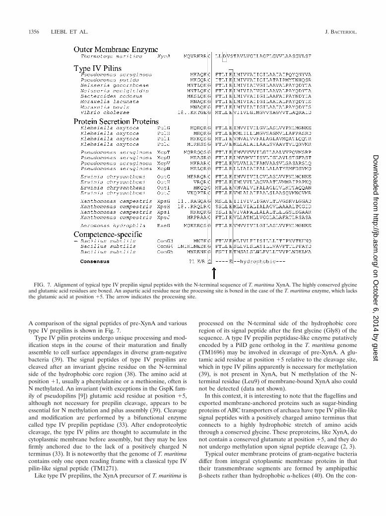
A comparison of the signal peptides of pre-XynA and varioustype IV prepilins is shown in Fig. 7.
Type IV pilin proteins undergo unique processing and mod-ification steps in the course of their maturation and finallyassemble to cell surface appendages in diverse gram-negativebacteria (39). The signal peptides of type IV prepilins arecleaved after an invariant glycine residue on the N-terminalside of the hydrophobic core region (38). The amino acid atposition �1, usually a phenylalanine or a methionine, often isN methylated. An invariant (with exceptions in the GspK fam-ily of pseudopilins [9]) glutamic acid residue at position �5,although not necessary for prepilin cleavage, appears to beessential for N methylation and pilus assembly (39). Cleavageand modification are performed by a bifunctional enzymecalled type IV prepilin peptidase (33). After endoproteolyticcleavage, the type IV pilins are thought to accumulate in thecytoplasmic membrane before assembly, but they may be lessfirmly anchored due to the lack of a positively charged Nterminus (33). It is noteworthy that the genome of T. maritimacontains only one open reading frame with a classical type IVpilin-like signal peptide (TM1271).
Like type IV prepilins, the XynA precursor of T. maritima is
processed on the N-terminal side of the hydrophobic coreregion of its signal peptide after the first glycine (Gly8) of thesequence. A type IV prepilin peptidase-like enzyme putativelyencoded by a PilD gene ortholog in the T. maritima genome(TM1696) may be involved in cleavage of pre-XynA. A glu-tamic acid residue at position �5 relative to the cleavage site,which in type IV pilins apparently is necessary for methylation(39), is not present in XynA, but N methylation of the N-terminal residue (Leu9) of membrane-bound XynA also couldnot be detected (data not shown).
In this context, it is interesting to note that the flagellins andexported membrane-anchored proteins such as sugar-bindingproteins of ABC transporters of archaea have type IV pilin-likesignal peptides with a positively charged amino terminus thatconnects to a highly hydrophobic stretch of amino acidsthrough a conserved glycine. These preproteins, like XynA, donot contain a conserved glutamate at position �5, and they donot undergo methylation upon signal peptide cleavage (2, 3).
Typical outer membrane proteins of gram-negative bacteriadiffer from integral cytoplasmic membrane proteins in thattheir transmembrane segments are formed by amphipathic�-sheets rather than hydrophobic �-helices (40). On the con-
FIG. 7. Alignment of typical type IV prepilin signal peptides with the N-terminal sequence of T. maritima XynA. The highly conserved glycineand glutamic acid residues are boxed. An aspartic acid residue near the processing site is boxed in the case of the T. maritima enzyme, which lacksthe glutamic acid at position �5. The arrow indicates the processing site.
1356 LIEBL ET AL. J. BACTERIOL.
on October 6, 2014 by guest
http://jb.asm.org/
Dow
nloaded from

trary, long stretches of hydrophobic amino acids can act asstop-transfer sequences, preventing release from the cytoplas-mic membrane and sorting to the outer membrane (1, 29, 37).A different common mode of attachment to the outer mem-brane is via N-terminal lipoprotein modification which resultsin a fatty-acylated cysteine residue at the extreme N terminusof the mature protein. Our data suggest that binding of thexylanase XynA to the outer membrane of T. maritima cells is
accomplished with neither of the two modes just discussed butrather via an N-terminal hydrophobic membrane anchor (Fig.8), which is highly unusual for an outer membrane protein.Interestingly, another protein of T. maritima, called Omp�, acoiled-coil protein which apparently spans the periplasmicspace, was proposed to be anchored to the outer membrane viaa carboxy-terminal hydrophobic peptide tail (14). Intriguingly,the hydrophobic core sequences of the postulated outer mem-brane anchors of XynA (N terminal) and Omp� (C terminal)bear striking similarity to one another, including three con-served glycine residues (Fig. 8). We are aware of only oneother case, i.e., the phospholipase PldA of E. coli, where exportto the outer membrane without proteolytic removal of theputative (but in our opinion far from optimal because quitehydrophilic) signal peptide has been suggested (11).
The unusual mode of outer membrane anchoring of T. ma-ritima XynA raises a number of interesting questions. (i) Whatare the sorting signals that lead to outer membrane localiza-tion? (ii) What does the terminal part of the secretion pathway,leading to the final location of the enzyme in the outer mem-brane, look like? (iii) Where and how does the processing ofpre-XynA after residue 44 (Fig. 6), which leads to the secretedXynA species found in the culture supernatant, take place?Clearly more work is necessary to come to a better understand-ing of protein transport and localization in the ancestral eu-bacterium T. maritima.
Interestingly, most of the amylase activity of T. maritimaMSB8 cells also is associated with the toga (reference 36 andour unpublished data). According to sequence data, the modeof attachment to the outer membrane of at least one �-amy-lase, AmyA, appears to be based on a bacterial lipoproteinmodification (27) and therefore is different from the case forXynA. Thus, the hyperthermophile T. maritima has variouspolysaccharide hydrolases anchored via different mechanismsin the outermost cell layer. The retention of these enzymes atthe cell surface is a plausible strategy to avoid the rapid loss ofsecreted enzymes in an extremely hot marine environment.Perhaps the unique ballooning of the outer membrane over theends of Thermotoga cells serves to enlarge the surface areaspiked with depolymerases.
ACKNOWLEDGMENTS
We thank B. Kuhlmorgen and B. Schumacher for skillful technicalassistance, F. Lottspeich for N-terminal sequencing of protein samples,and A. Engel for valuable advice during the course of immunogoldlabeling experiments and electron microscopy. We are also grateful toR. Amann for assistance with epifluorescence microscopy.
Financial support from the Deutsche Forschungsgemeinschaft (Li398/7), Consortium fur elektrochemische Chemie GmbH, Munich, andthe Federal Ministry for Education and Science (BMBF) to W.L. isgratefully acknowledged.
REFERENCES
1. Agterberg, M., H. Adriaanse, A. van Bruggen, M. Karperien, and J. Tommassen.1990. Outer membrane PhoE protein of Escherichia coli K-12 as an exposure vector:possibilities and limitations. Gene 88:37–45.
2. Albers, S.-V., W. N. Konings, and A. J. M. Driessen. 1999. A unique shortsignal sequence in membrane-anchored proteins of archaea. Mol. Microbiol.31:1595–1596.
3. Albers, S.-V., and A. J. M. Driessen. 2002. Signal peptides of secretedproteins of the archaeon Sulfolobus solfataricus: a genomic survey. Arch.Microbiol. 177:209–216.
4. Ali, M. K., T. Kimura, K. Sakka, and K. Ohmiya. 2001. The multidomainxylanase Xyn10B as a cellulose-binding protein in Clostridium stercorarium.FEMS Microbiol. Lett. 198:79–83.
FIG. 8. (A) Schematic model of the T. maritima cell envelope,showing the postulated mode of anchoring of XynA in the toga via ahydrophobic N-terminal insertion signal. The five-domain modularstructure of XynA consists of the central catalytic domain (B) flankedby repeated N-terminal domains (A1 and A2) and repeated C-terminaldomains (C1 and C2), which represent CBMs. The most abundantproteins of the T. maritima cell envelope, i.e., Omp� (a dimeric coiled-coil protein apparently spanning the periplasm [14]) and Omp� (aporin [13, 34]), are also indicated. The lipid content and compositionof the T. maritima outer membrane and the mode of association ofOmp� with murein or the cytoplasmic membrane are not clear.(B) Helical wheel representation of the hydrophobic cores of thepostulated outer membrane anchors of XynA and Omp� of T. mari-tima. Hydrophobic residues are shaded in light gray. In both cases theglycine residues (marked with asterisks) occupy similar positions in thestructure.
VOL. 190, 2008 XYLANASE ATTACHMENT TO T. MARITIMA TOGA 1357
on October 6, 2014 by guest
http://jb.asm.org/
Dow
nloaded from

5. Reference deleted.6. Bendtsen, J. D., H. Nielsen, G. von Heijne, and S. Brunak. 2004. Improved
prediction of signal peptides: SignalP 3.0. J. Mol. Biol. 340:783–795.7. Bernfeld, P. 1955. Amylases � and �. Methods Enzymol. 1:149–158.8. Binder, F. 1987. Genetische und biochemische Analyse der Cyclodextrin-
Glycosyl-Transferase aus Klebsiella pneumoniae M5�1. Ph.D. thesis. Ludwig-Maximilians-Universitat Munchen, Munich, Germany.
9. Bleves, S., R. Voulhoux, G. Michel, A. Lazdunski, J. Tommassen, and A.Filloux. 1998. The secretion apparatus of Pseudomonas aeruginosa: identifi-cation of a fifth pseudopilin, XcpX (GspK family). Mol. Microbiol. 27:31–40.
10. Boraston, A. B., A. L. Creagh, M. Alam Md, J. M. Kormos, P. Tomme, C. A.Haynes, A. J. Warren, and D. G. Kilburn. 2001. Binding specificity andthermodynamics of a family 9 carbohydrate-binding module from Thermo-toga maritima xylanase 10A. Biochemistry 40:6240–6247.
11. de Geus, P., M. Verheij, N. H. Riegman, W. P. M. Hoekstra, and G. H. deHaas. 1984. The pro- and mature forms of the E. coli K-12 outer membranephospholipase A are identical. EMBO J. 3:1799–1802.
12. d’Enfert, C., C. Chapon, and A. P. Pugsley. 1987. Export and secretion of thelipoprotein pullulanase by Klebsiella pneumoniae. Mol. Microbiol. 1:107–116.
13. Engel, A. M., M. Brunen, and W. Baumeister. 1993. The functional proper-ties of Omp�, a regularly arrayed porin of the hyperthermophilic bacteriumThermotoga maritima. FEMS Microbiol. Lett. 109:231–236.
14. Engel, A. M., Z. Cejka, A. Lupas, F. Lottspeich, and W. Baumeister. 1992.Isolation and cloning of Omp�, a coiled-coil protein spanning the periplas-mic space of the ancestral eubacterium Thermotoga maritima. EMBO J.11:4369–4378.
15. Fujino, T., P. Beguin, and J. P. Aubert. 1993. Organization of a Clostridiumthermocellum gene cluster encoding the cellulosomal scaffolding proteinCipA and a protein possibly involved in attachment of the cellulosome to thecell surface. J. Bacteriol. 175:1891–1899.
16. Furste, J. P., W. Pansegrau, R. Frank, H. Blocker, P. Scholz, M. Bagdasar-ian, and E. Lanka. 1986. Molecular cloning of the plasmid RP4 primaseregion in a multi-host-range tacP expression vector. Gene 48:119–131.
17. Huber, R., and M. Hannig. 2006. Thermotogales, p. 899–922. In M. Dworkinet al. (ed.), The prokaryotes. An evolving electronic resource for the micro-biological community, 3rd ed., vol. 7. Springer-Verlag, New York, NY.
18. Huber, R., T. A. Langworthy, H. Konig, M. Thomm, C. R. Woese, U. B.Sleytr, and K. O. Stetter. 1986. Thermotoga maritima sp. nov. represents anew genus of unique extremely thermophilic eubacteria growing up to 90°C.Arch. Microbiol. 144:324–333.
19. Huber, R., and K. O. Stetter. 1992. The order Thermotogales, p. 3809–3815.In A. Balows, H. G. Truper, M. Dworkin, W. Harder, and K. H. Schleifer(ed.), The prokaryotes. A handbook on the biology of bacteria, 2nd ed., vol.II. Ecophysiology, isolation, identification, applications. Springer-Verlag,New York, NY.
20. Ito, Y., T. Tomita, N. Roy, A. Nikano, N. Sugawara-Tomita, S. Watanabe, N.Okai, N. Abe, and Y. Kamio. 2003. Cloning, expression and cell surfacelocalization of Paenibacillus sp. strain W-61 xylanase 5, a multidomain xyla-nase. Appl. Environ Microbiol. 69:6969–6978.
21. Juncker, A. S., H. Willenbrock, G. von Heijne, H. Nielsen, S. Brunak, and A.Krogh. 2003. Prediction of lipoprotein signal peptides in Gram-negativebacteria. Protein Sci. 12:1652–1662.
22. Kavoosi, M., J. Meijer, E. Kwan, A. L. Creagh, D. G. Kilburn, and C. A.Haynes. 2004. Inexpensive one-step purification of polypeptides expressed inEscherichia coli as fusions with the family 9 carbohydrate-binding module ofxylanase 10A from T. maritima. J. Chromatogr. B 807:87–94.
23. Kellenberger, E., E. Carlemalm, W. Villiger, J. Roth, and R. M. Garavito.1980. Low denaturation embedding for electron microscopy of thin sections.Chem. Werke Lowi, Waldkraiburg, Germany.
24. Kleine, J., and W. Liebl. 2006. Comparative characterization of deletionderivatives of the modular xylanase XynA of Thermotoga maritima. Extremo-philes 10:373–381.
25. Liebl, W., R. Feil, J. Gabelsberger, J. Kellermann, and K. H. Schleifer. 1992.Purification and characterization of a novel thermostable 4-�-glucanotrans-
ferase of Thermotoga maritima cloned in Escherichia coli. Eur. J. Biochem.207:81–88.
26. Liebl, W., J. Gabelsberger, and K. H. Schleifer. 1994. Structural analysis ofthe bglA gene encoding a �-glucosidase of the hyperthermophilic bacteriumThermotoga maritima and comparison of the deduced amino acid sequencewith other �-1,4-glycosyl hydrolases. Mol. Gen. Genet. 242:111–115.
27. Liebl, W., I. Stemplinger, and P. Ruile. 1997. Properties and gene structureof the Thermotoga maritima �-amylase AmyA, a putative lipoprotein of ahyperthermophilic bacterium. J. Bacteriol. 179:941–948.
28. Lupas, A., H. Engelhardt, J. Peters, U. Santarius, S. Volker, and W.Baumeister. 1994. Domain structure of the Acetogenium kivui surface layerrevealed by electron crystallography and sequence analysis. J. Bacteriol.176:1224–1233.
29. MacIntyre, S., R. Freudl, M.-L. Eschbach, and U. Henning. 1988. An arti-ficial hydrophobic sequence functions as either an anchor or a signal se-quence at only one of two positions within the Escherichia coli outer mem-brane protein OmpA. J. Biol. Chem. 263:19053–19059.
30. Meissner, K., D. Wassenberg, and W. Liebl. 2000. The ‘thermostabilisingdomain’ of the modular xylanase XynA of the hyperthermophilic bacteriumThermotoga maritima represents a novel xylan-binding domain. Mol. Micro-biol. 36:898–912.
31. Nordberg Karlsson, E., M. Abou Hachem, S. Ramchuran, H. Costa, O.Holst, A. F. Svenningsen, and G. O. Hreggvidsson. 2004. The modularxylanase Xyn10A from Rhodothermus marinus is cell-attached, and its C-terminal domain has several putative homologues among cell-attached pro-teins within the phylum Bacteroidetes. FEMS Microbiol. Lett. 241:233–242.
32. Notenboom, V., A. B. Boraston, D. G. Kilburn, and D. R. Rose. 2001. Crystalstructure of the family 9 carbohydrate-binding module from Thermotogamaritima xylanase 10A in native and ligand-bound forms. Biochemistry 40:6248–6256.
33. Pugsley, A. P. 1993. The complete general secretory pathway in gram-neg-ative bacteria. Microbiol. Rev. 57:50–108.
34. Rachel, R., A. M. Engel, R. Huber, K. O. Stetter, and W. Baumeister. 1990.A porin is the main constituent of the cell envelope of the ancestral eubac-terium Thermotoga maritima. FEBS Lett. 262:64–68.
35. Reysenbach, A.-L. 2001. Phylum BII: Thermotogae phy. nov., p. 369. In D. R.Boone and R. W. Castenholz (ed.), Bergey’s manual of systematic bacteri-ology, 2nd ed. Springer-Verlag, New York, NY.
36. Schumann, J., A. Wrba, R. Jaenicke, and K. O. Stetter. 1991. Topographicaland enzymatic characterization of amylases from the extremely thermophiliceubacterium Thermotoga maritima. FEBS Lett. 282:122–126.
37. Shinkai, A., H. Yamada, T. Mizuno, and S. Mizushima. 1989. Insertion of asignal peptide-derived hydrophobic segment into the mature domain ofOmpC, an outer membrane protein, does not interfere with the export of thefollowing polypeptide chain across the cytoplasmic membrane of E. coli.J. Biochem. 106:323–330.
38. Strom, M. S., and S. Lory. 1991. Amino acid substitutions in pilin of Pseudo-monas aeruginosa. Effect on leader peptide cleavage, amino-terminal meth-ylation, and pilus assembly. J. Biol. Chem. 266:1656–1664.
39. Strom, M. S., and S. Lory. 1993. Structure-function and biogenesis of thetype IV pili. Annu. Rev. Microbiol. 47:565–596.
40. Tommassen, J., M. Struyve, and H. deCock. 1992. Export and assembly ofbacterial outer membrane proteins. Antonie van Leeuwenhoek. 61:81–85.
41. von Heijne, G. 1986. A new method for predicting signal sequence cleavagesites. Nucleic Acids Res. 14:4683–4690.
42. Winterhalter, C., P. Heinrich, A. Candussio, G. Wich, and W. Liebl. 1995.Identification of a novel cellulose-binding domain within the multi-domain120 kDa xylanase XynA of the hyperthermophilic bacterium Thermotogamaritima. Mol. Microbiol. 15:431–444.
43. Winterhalter, C., and W. Liebl. 1995. Two extremely thermostable xylanasesof the hyperthermophilic bacterium Thermotoga maritima strain MSB8.Appl. Environ Microbiol. 61:1810–1815.
44. Zhao, G., E. Ali, M. Sakka, T. Kimura, and K. Sakka. 2006. Binding ofS-layer homology modules from Clostridium thermocellum SdbA to pep-tidoglycans. Appl. Microbiol. Biotechnol. 70:464–469.
1358 LIEBL ET AL. J. BACTERIOL.
on October 6, 2014 by guest
http://jb.asm.org/
Dow
nloaded from