Bennett's Fracture - ISSH
10
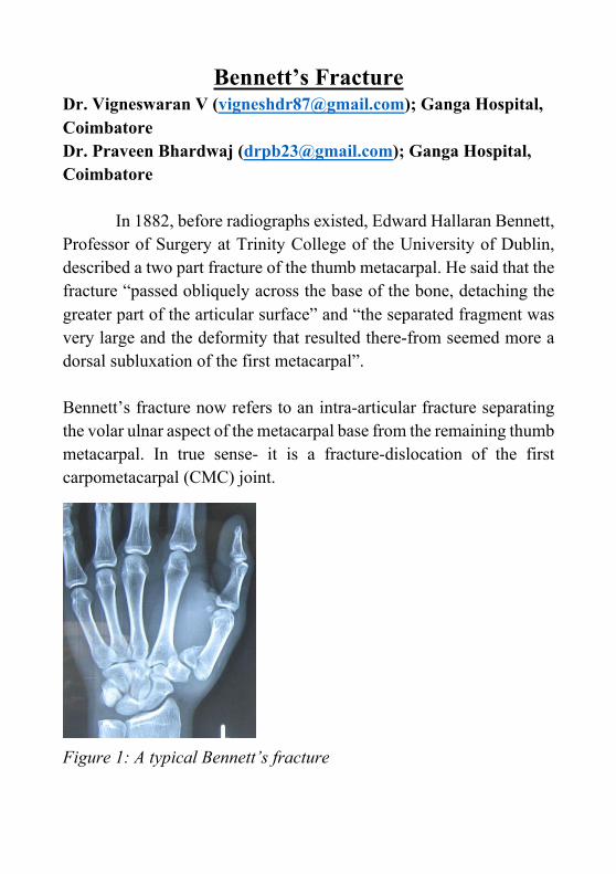
Bennett’s Fracture Dr. Vigneswaran V ([email protected]); Ganga Hospital, Coimbatore Dr. Praveen Bhardwaj ([email protected]); Ganga Hospital, Coimbatore In 1882, before radiographs existed, Edward Hallaran Bennett, Professor of Surgery at Trinity College of the University of Dublin, described a two part fracture of the thumb metacarpal. He said that the fracture “passed obliquely across the base of the bone, detaching the greater part of the articular surface” and “the separated fragment was very large and the deformity that resulted there-from seemed more a dorsal subluxation of the first metacarpal”. Bennett’s fracture now refers to an intra-articular fracture separating the volar ulnar aspect of the metacarpal base from the remaining thumb metacarpal. In true sense- it is a fracture-dislocation of the first carpometacarpal (CMC) joint. Figure 1: A typical Bennett’s fracture
-
Upload
khangminh22 -
Category
Documents
-
view
1 -
download
0
Transcript of Bennett's Fracture - ISSH
Bennett’s Fracture Dr. Vigneswaran V ([email protected]); Ganga Hospital, Coimbatore Dr. Praveen Bhardwaj ([email protected]); Ganga Hospital, Coimbatore
In 1882, before radiographs existed, Edward Hallaran Bennett, Professor of Surgery at Trinity College of the University of Dublin, described a two part fracture of the thumb metacarpal. He said that the fracture “passed obliquely across the base of the bone, detaching the greater part of the articular surface” and “the separated fragment was very large and the deformity that resulted there-from seemed more a dorsal subluxation of the first metacarpal”. Bennett’s fracture now refers to an intra-articular fracture separating the volar ulnar aspect of the metacarpal base from the remaining thumb metacarpal. In true sense- it is a fracture-dislocation of the first carpometacarpal (CMC) joint. Figure 1: A typical Bennett’s fracture
Incidence:
Approximately 25% of all the metacarpal fractures occur in the thumb metacarpal with 80% of them occurring at the base, with Bennett’s fracture being the commonest. The overall incidence of Bennett’s fracture is 1.4% of all the fractures within the hand.
Patho-mechanics:
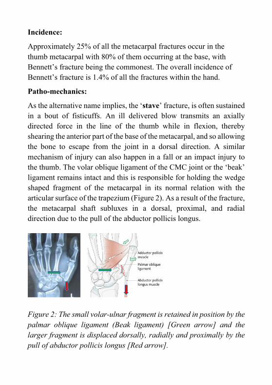
As the alternative name implies, the ‘stave’ fracture, is often sustained in a bout of fisticuffs. An ill delivered blow transmits an axially directed force in the line of the thumb while in flexion, thereby shearing the anterior part of the base of the metacarpal, and so allowing the bone to escape from the joint in a dorsal direction. A similar mechanism of injury can also happen in a fall or an impact injury to the thumb. The volar oblique ligament of the CMC joint or the ‘beak’ ligament remains intact and this is responsible for holding the wedge shaped fragment of the metacarpal in its normal relation with the articular surface of the trapezium (Figure 2). As a result of the fracture, the metacarpal shaft subluxes in a dorsal, proximal, and radial direction due to the pull of the abductor pollicis longus. Figure 2: The small volar-ulnar fragment is retained in position by the palmar oblique ligament (Beak ligament) [Green arrow] and the larger fragment is displaced dorsally, radially and proximally by the pull of abductor pollicis longus [Red arrow].
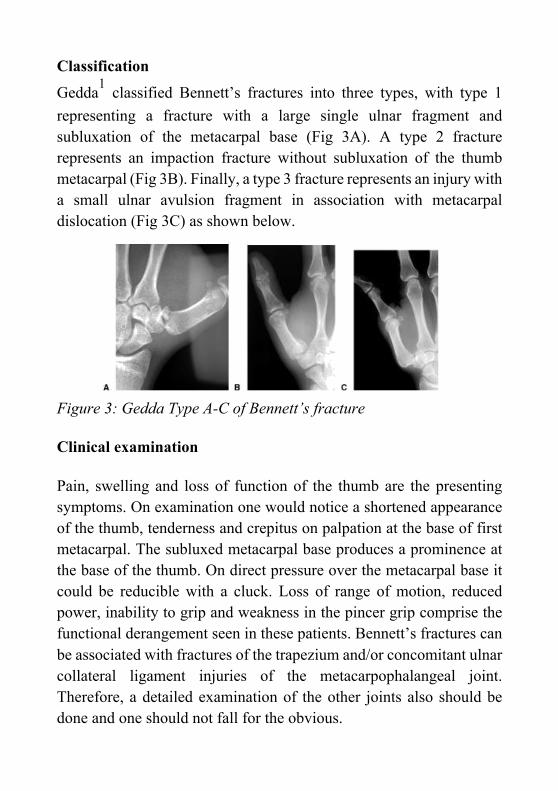
Classification Gedda1 classified Bennett’s fractures into three types, with type 1 representing a fracture with a large single ulnar fragment and subluxation of the metacarpal base (Fig 3A). A type 2 fracture represents an impaction fracture without subluxation of the thumb metacarpal (Fig 3B). Finally, a type 3 fracture represents an injury with a small ulnar avulsion fragment in association with metacarpal dislocation (Fig 3C) as shown below.
Figure 3: Gedda Type A-C of Bennett’s fracture Clinical examination Pain, swelling and loss of function of the thumb are the presenting symptoms. On examination one would notice a shortened appearance of the thumb, tenderness and crepitus on palpation at the base of first metacarpal. The subluxed metacarpal base produces a prominence at the base of the thumb. On direct pressure over the metacarpal base it could be reducible with a cluck. Loss of range of motion, reduced power, inability to grip and weakness in the pincer grip comprise the functional derangement seen in these patients. Bennett’s fractures can be associated with fractures of the trapezium and/or concomitant ulnar collateral ligament injuries of the metacarpophalangeal joint. Therefore, a detailed examination of the other joints also should be done and one should not fall for the obvious.
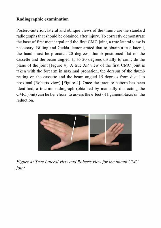
Radiographic examination Postero-anterior, lateral and oblique views of the thumb are the standard radiographs that should be obtained after injury. To correctly demonstrate the base of first metacarpal and the first CMC joint, a true lateral view is necessary. Billing and Gedda demonstrated that to obtain a true lateral, the hand must be pronated 20 degrees, thumb positioned flat on the cassette and the beam angled 15 to 20 degrees distally to coincide the plane of the joint [Figure 4]. A true AP view of the first CMC joint is taken with the forearm in maximal pronation, the dorsum of the thumb resting on the cassette and the beam angled 15 degrees from distal to proximal (Roberts view) [Figure 4]. Once the fracture pattern has been identified, a traction radiograph (obtained by manually distracting the CMC joint) can be beneficial to assess the effect of ligamentotaxis on the reduction. Figure 4: True Lateral view and Roberts view for the thumb CMC joint
Treatment The main objective of the treatment is to obtain and then maintain adequate fracture reduction to allow healing in an anatomical position. Conservative management of these fractures were common until
19702. Although historical reports have noted satisfactory outcomes with nonsurgical treatment, more recent studies have shown poor
outcomes with casting alone for this injury2. Conservative treatment with immobilization in a plaster cast is usually unable to maintain the reduction of this unstable injury. Hence, surgical stabilization is universally recommended which is capable of restoring congruence and joint stability. Surgical treatment includes closed reduction with percutaneous pinning, open reduction and internal fixation with pins or inter-fragmentary fixation, oblique pin traction and external fixation. Continuous traction with external fixation has been described but it is difficult to maintain the traction and reduction, it is uncomfortable and requires very close monitoring. Hence, it has fallen out of repute. Arthroscopic assisted reduction and internal fixation carries the advantages of a minimally invasive procedure, direct visual control of reduction and removal of the
interposed soft tissues3. In spite of these theoretical advantages it has not gained universal popularity because of logistic issues and in general good results with the conventional fixation techniques.
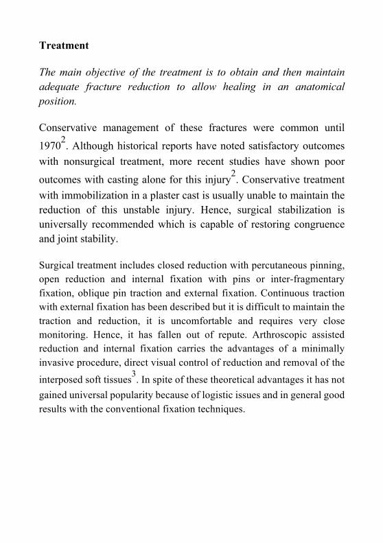
There are number of methods described for the closed reduction. The three commonly described techniques are as follows:
1. Axial traction, palmar abduction (to relax the abductor pollicis longus), and pronation of the thumb while applying external pressure over the metacarpal base [Authors’ Preferred Method].
1
3
2
4
Figure 5: The authors’ preferred method of closed reduction of the Bennett’s fracture. The maneuvers involved are – 1. Traction, 2. Palmar abduction, 3. Pronation, 4. Direct pressure over the base of the thumb metacarpal.
2. Flexion of the thumb MCP joint and applying pressure over
the first CMC joint in a palmar and ulnar direction.
3. ‘Screw home torque’ reduction technique described by
Edmunds4. In this technique, the surgeon places the first CMC joint in full passive opposition with palmar abduction and
pronation of the thumb metacarpal such that the plane of the thumb pulp lies opposite to that of the index and middle fingers as with a chuck grip position. This maneuver is believed to make the dorsal ligament complex tauter, facilitating anatomical reduction.
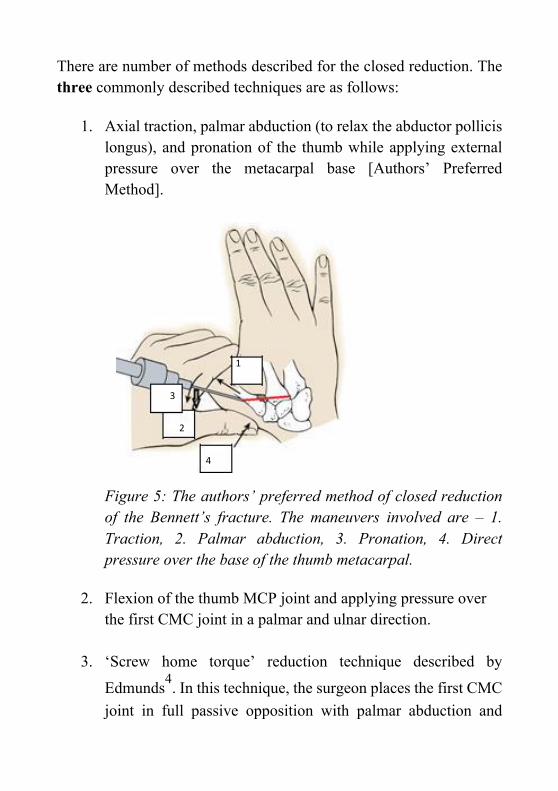
Closed reduction is generally easy to achieve. The reduction is maintained by percutaneous K-wires. The various configurations of the K-wire fixation described are- intermetacarpal fixation to the second metacarpal, as described by Wagner, Iselin and Adi or to the trapezium, as described by Wiggins- Bundens [Figure 6]. All the patterns have been found to provide a satisfactory stability. The intermetacarpal fixation technique avoids a wire across the articular surface of the CMC joint and it is the authors’ preferred. It is not necessary to have the wires across the fracture site into the volar-ulnar fragment and main aim would be to achieve joint reduction. Figure 6: The various configurations of the K-wire stabilization of the reduced Bennett’s fracture Wiggins- Bundens (A); Wagner (B); Iselin (C) and Adi (D).
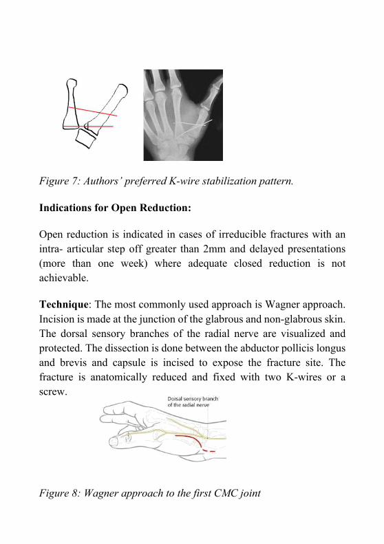
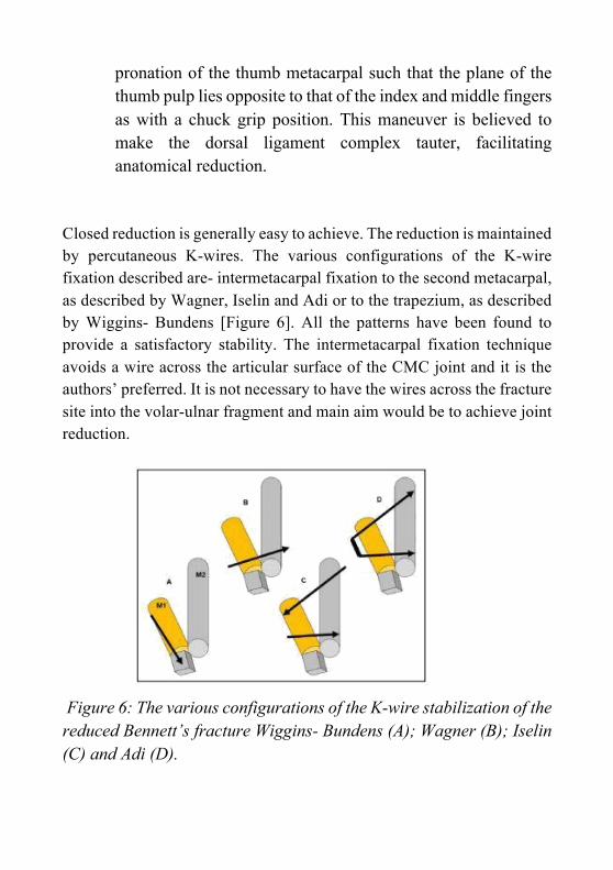
Figure 7: Authors’ preferred K-wire stabilization pattern. Indications for Open Reduction: Open reduction is indicated in cases of irreducible fractures with an intra- articular step off greater than 2mm and delayed presentations (more than one week) where adequate closed reduction is not achievable. Technique: The most commonly used approach is Wagner approach. Incision is made at the junction of the glabrous and non-glabrous skin. The dorsal sensory branches of the radial nerve are visualized and protected. The dissection is done between the abductor pollicis longus and brevis and capsule is incised to expose the fracture site. The fracture is anatomically reduced and fixed with two K-wires or a screw. Figure 8: Wagner approach to the first CMC joint
Rehabilitation: The thumb is immobilized in a below elbow thumb slab after fixation. The slab can be changed to a removable splint for convenience at two weeks. The K-wires are removed at four weeks. The thumb CMC joint movements are then started and the splint is maintained for another two weeks. Patient is advised to do opposition of thumb to the fingers and gradually start using his/her hand. At six weeks the hand is left free to use. Rigorous use of the hand is avoided for another 6 weeks. Owing to the lack of long term studies comparing various reduction methods (open vs closed) and stabilization techniques, definite protocol for the management of Bennett’s fracture is difficult to establish. The fundamental things need to be addressed are as follows: 1. Any mode of surgical fixation, whether open or percutaneous, to maintain reduction of the metacarpal at the first CMC joint would provide good result. 2. Fracture reduction techniques should include thumb pronation to aid in anatomic reduction of the metacarpal to the ulnar fragment. 3. Open reduction should be considered in those circumstances where greater than 2 mm of articular step-off persists despite attempts of closed reduction and in delayed presentation where adequate closed reduction is not achievable.
References:
1. Gedda KO, Moberg E. Open reduction and fixation of so called Bennett’s fracture. Acta
Orthop Scand. 1953;22:249-257.
2. Carlsen BT, Moran SL. Thumb trauma: Bennett fractures,
Rolando fractures, and ulnar collateral ligament injuries. J Hand Surg Am. 2009;34(5):945-52.
3. Solomon J, Culp RW. Arthroscopic Management of
Bennett Fracture. Hand Clin. 2017;33(4):787-794.
4. Edmunds JO.Traumatic dislocations and instability of the trapeziometacarpal joint of the thumb. Hand Clin. 2006;22(3):365-92.