Bacteria Patterns on Tonsillar Surface and Tonsillar Core ...
9
pathogens Article Bacteria Patterns on Tonsillar Surface and Tonsillar Core Tissue among Patients Scheduled for Tonsillectomy at Bugando Medical Centre, Mwanza, Tanzania Gustave Buname 1 , Gapto Aristides Kiwale 2 , Martha F. Mushi 2, * , Vitus Silago 2 , Peter Rambau 3 and Stephen E. Mshana 2 Citation: Buname, G.; Kiwale, G.A.; Mushi, M.F.; Silago, V.; Rambau, P.; Mshana, S.E. Bacteria Patterns on Tonsillar Surface and Tonsillar Core Tissue among Patients Scheduled for Tonsillectomy at Bugando Medical Centre, Mwanza, Tanzania. Pathogens 2021, 10, 1560. https://doi.org/ 10.3390/pathogens10121560 Academic Editor: Jilei Zhang Received: 21 August 2021 Accepted: 24 November 2021 Published: 30 November 2021 Publisher’s Note: MDPI stays neutral with regard to jurisdictional claims in published maps and institutional affil- iations. Copyright: © 2021 by the authors. Licensee MDPI, Basel, Switzerland. This article is an open access article distributed under the terms and conditions of the Creative Commons Attribution (CC BY) license (https:// creativecommons.org/licenses/by/ 4.0/). 1 Department of ENT, Bugando Medical Centre, Weill Bugando School of Medicine, Catholic University of Health and Allied Sciences, Mwanza P.O. Box 1464, Tanzania; [email protected] 2 Department of Microbiology and Immunology, Weill Bugando School of Medicine, Catholic University of Health and Allied Sciences, Mwanza P.O. Box 1464, Tanzania; [email protected] (G.A.K.); [email protected] (V.S.); [email protected] (S.E.M.) 3 Department of Pathology, Weill Bugando School of Medicine, Catholic University of Health and Allied Sciences, Mwanza P.O. Box 1464, Tanzania; [email protected] * Correspondence: [email protected]; Tel.: +255-282500881 Abstract: Background: Tonsillitis is an inflammation of the tonsils due to either viruses or bacteria. Here, we report the bacteria patterns on the tonsillar surface and tonsillar core tissue among patients scheduled for tonsillectomy at Bugando Medical Centre (BMC), Mwanza Tanzania. Methods: The study included 120 patients planned for tonsillectomy between April and July 2019. Swab samples from tonsillar surface pre-tonsillectomy and core post-tonsillectomy were collected. Culture was performed following the microbiology laboratory standard operating procedures. Data analysis was completed using STATA version 13, as per the study objectives. Results: The slight majority of participants were males (73; 60.83%) with median age of 6 years (interquartile range 4–11). The proportion of positive culture growth was higher on the surface than in core swab samples: 65 (54.2%) vs. 42 (35.0%), p = 0.003. The commonest bacterial pathogen detected from the surface and core were S. aureus in 29 (40.3%) and 22 (51.2%) participants, followed by S. pyogenes in 17 (23.6%) and 11 (25.6%), respectively. Methicillin-resistant Staphylococcus aureus (MRSA) was observed in 20/51 (39%) of isolates. Streptococcus pyogenes resistance to macrolides ranged from 8.3% for core isolates to 35.3% for surface isolates. Features suggestive of tonsillitis on histology were reported in 83 (73.5%) samples. Conclusion: More than two-thirds of patients undergoing tonsillectomy had a positive culture for possible bacterial pathogens. Staphylococcus aureus and Streptococcus pyogenes were the predominant bacteria detected with more than one third of Staphylococcus aureus being MRSA. More studies to investigate the treatment outcome of these patients are highly recommended. Keywords: tonsillar; Tanzania; core; surface; tonsillectomy; bacteria 1. Introduction Tonsillectomy is one of the most frequently performed otolaryngological operation worldwide [1]. Tonsillectomy can be performed at any age but is mainly performed among children [2,3]. Recurrent acute tonsillitis, with at least five or more attack episodes in a year, obstructive sleep apnea and peritonsillar abscess, is indicated in patients with adenotonsil- lar hypertrophy [4]. Recurrent acute tonsillitis is mainly associated with bacterial and/or viral infection of the tonsillar crypts or parenchyma, and is characterized by sore throat, fever, odynophagia, and leukocytosis, accompanied by congested tonsils with or without enlargement and tender jugulodigastric lymph nodes. Tonsil infection may occur primarily or secondarily as a result of upper respiratory tract infections commonly preceded by viral infections [5]. Several pathogens are implicated in tonsil infections including: Group A beta Pathogens 2021, 10, 1560. https://doi.org/10.3390/pathogens10121560 https://www.mdpi.com/journal/pathogens
-
Upload
khangminh22 -
Category
Documents
-
view
0 -
download
0
Transcript of Bacteria Patterns on Tonsillar Surface and Tonsillar Core ...

pathogens
Article
Bacteria Patterns on Tonsillar Surface and Tonsillar Core Tissueamong Patients Scheduled for Tonsillectomy at BugandoMedical Centre, Mwanza, Tanzania
Gustave Buname 1, Gapto Aristides Kiwale 2, Martha F. Mushi 2,* , Vitus Silago 2 , Peter Rambau 3
and Stephen E. Mshana 2
�����������������
Citation: Buname, G.; Kiwale, G.A.;
Mushi, M.F.; Silago, V.; Rambau, P.;
Mshana, S.E. Bacteria Patterns on
Tonsillar Surface and Tonsillar Core
Tissue among Patients Scheduled for
Tonsillectomy at Bugando Medical
Centre, Mwanza, Tanzania. Pathogens
2021, 10, 1560. https://doi.org/
10.3390/pathogens10121560
Academic Editor: Jilei Zhang
Received: 21 August 2021
Accepted: 24 November 2021
Published: 30 November 2021
Publisher’s Note: MDPI stays neutral
with regard to jurisdictional claims in
published maps and institutional affil-
iations.
Copyright: © 2021 by the authors.
Licensee MDPI, Basel, Switzerland.
This article is an open access article
distributed under the terms and
conditions of the Creative Commons
Attribution (CC BY) license (https://
creativecommons.org/licenses/by/
4.0/).
1 Department of ENT, Bugando Medical Centre, Weill Bugando School of Medicine, Catholic University ofHealth and Allied Sciences, Mwanza P.O. Box 1464, Tanzania; [email protected]
2 Department of Microbiology and Immunology, Weill Bugando School of Medicine, Catholic University ofHealth and Allied Sciences, Mwanza P.O. Box 1464, Tanzania; [email protected] (G.A.K.);[email protected] (V.S.); [email protected] (S.E.M.)
3 Department of Pathology, Weill Bugando School of Medicine, Catholic University of Health and AlliedSciences, Mwanza P.O. Box 1464, Tanzania; [email protected]
* Correspondence: [email protected]; Tel.: +255-282500881
Abstract: Background: Tonsillitis is an inflammation of the tonsils due to either viruses or bacteria.Here, we report the bacteria patterns on the tonsillar surface and tonsillar core tissue among patientsscheduled for tonsillectomy at Bugando Medical Centre (BMC), Mwanza Tanzania. Methods: Thestudy included 120 patients planned for tonsillectomy between April and July 2019. Swab samplesfrom tonsillar surface pre-tonsillectomy and core post-tonsillectomy were collected. Culture wasperformed following the microbiology laboratory standard operating procedures. Data analysiswas completed using STATA version 13, as per the study objectives. Results: The slight majorityof participants were males (73; 60.83%) with median age of 6 years (interquartile range 4–11). Theproportion of positive culture growth was higher on the surface than in core swab samples: 65 (54.2%)vs. 42 (35.0%), p = 0.003. The commonest bacterial pathogen detected from the surface and corewere S. aureus in 29 (40.3%) and 22 (51.2%) participants, followed by S. pyogenes in 17 (23.6%) and11 (25.6%), respectively. Methicillin-resistant Staphylococcus aureus (MRSA) was observed in 20/51(39%) of isolates. Streptococcus pyogenes resistance to macrolides ranged from 8.3% for core isolates to35.3% for surface isolates. Features suggestive of tonsillitis on histology were reported in 83 (73.5%)samples. Conclusion: More than two-thirds of patients undergoing tonsillectomy had a positiveculture for possible bacterial pathogens. Staphylococcus aureus and Streptococcus pyogenes were thepredominant bacteria detected with more than one third of Staphylococcus aureus being MRSA. Morestudies to investigate the treatment outcome of these patients are highly recommended.
Keywords: tonsillar; Tanzania; core; surface; tonsillectomy; bacteria
1. Introduction
Tonsillectomy is one of the most frequently performed otolaryngological operationworldwide [1]. Tonsillectomy can be performed at any age but is mainly performed amongchildren [2,3]. Recurrent acute tonsillitis, with at least five or more attack episodes in a year,obstructive sleep apnea and peritonsillar abscess, is indicated in patients with adenotonsil-lar hypertrophy [4]. Recurrent acute tonsillitis is mainly associated with bacterial and/orviral infection of the tonsillar crypts or parenchyma, and is characterized by sore throat,fever, odynophagia, and leukocytosis, accompanied by congested tonsils with or withoutenlargement and tender jugulodigastric lymph nodes. Tonsil infection may occur primarilyor secondarily as a result of upper respiratory tract infections commonly preceded by viralinfections [5]. Several pathogens are implicated in tonsil infections including: Group A beta
Pathogens 2021, 10, 1560. https://doi.org/10.3390/pathogens10121560 https://www.mdpi.com/journal/pathogens
Pathogens 2021, 10, 1560 2 of 9
hemolytic Streptococcus, alpha hemolytic Streptococcus, Hemophilus influenzae, Staphylococ-cus aureus, Enterococcus spp., Klebsiella pneumoniae, Brahmnella catarrhalis, Corynebacteriumspp. and anaerobes-like Peptostreptococci, Fusobacterium and Veillonella [6,7]. Neverthe-less, previous studies conducted worldwide reported the possibility of the tonsillar surfacebeing colonized by the microbial flora which are not implicated in the tonsil infections [6,8].The bacteria on the core of the tonsils are highly associated with tonsillitis and play a vitalrole in the recurrent nature of the infections [9]. Furthermore, the recurrent and chronicnature of tonsillitis is highly associated with the ability of the implicated pathogen toresist antibiotic therapy. Antibiotic resistance is reported to be highly linked with the lowconcentration of antibiotics in the tonsilar core tissue caused by scarring as a result ofrecurrent infections which impair the antibiotic diffusion [5,10,11]. Penicillin, resistantamong pharyngeal flora, also accounts for the recurrent and chronic nature of tonsillitis [12].In Tanzania, penicillin resistance is reported to range from 53% to 67.8% among S. pneumo-niae [13,14] one of the pathogens that causes tonsillitis. The steady increase in resistance isreported to be associated with irrational use of the antibiotic in the community [15].
The significant complications of tonsillitis in resource-limited settings is attributedto poor or lack of evidence-based and empiric antibiotic treatment guidelines, due tothe lack of data from local studies [16]. Recurrent tonsillitis poses a great health risk topatients including the post streptococcus (Rheumatic heart disease (RHD)) diseases. TheRHD is believed to be more prevalent in resource-limited settings such as in Tanzania [17].The effective treatment of recurrent tonsillitis highly depends on the ability to isolate theassociated pathogen and its susceptibility pattern. The primary aim of this study was todetermine patterns of Gram-positive bacteria on the tonsillar surface pre tonsillectomyand tonsillar core tissue post tonsillectomy, among patients scheduled for tonsillectomy atBugando Medical Centre (BMC), Mwanza, Tanzania, in order to obtain data to guide theempirical treatment of tonsillitis.
2. Results
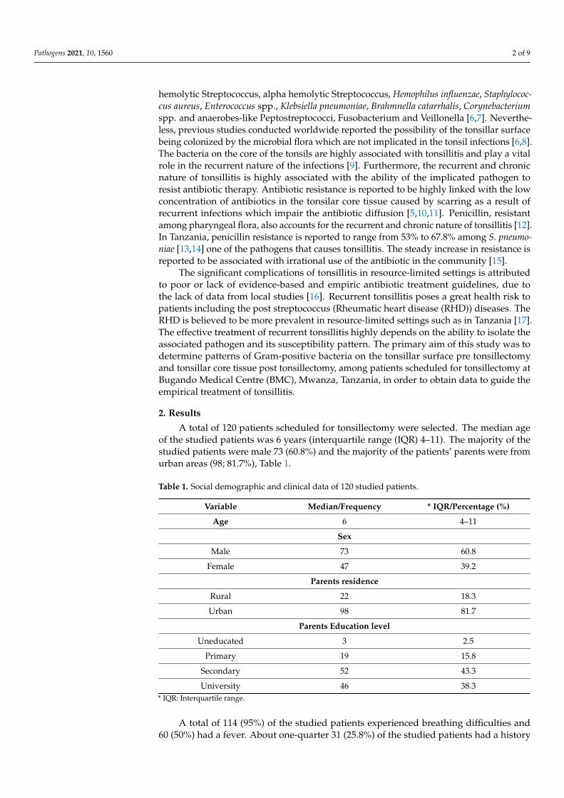
A total of 120 patients scheduled for tonsillectomy were selected. The median ageof the studied patients was 6 years (interquartile range (IQR) 4–11). The majority of thestudied patients were male 73 (60.8%) and the majority of the patients’ parents were fromurban areas (98; 81.7%), Table 1.
Table 1. Social demographic and clinical data of 120 studied patients.
Variable Median/Frequency * IQR/Percentage (%)
Age 6 4–11
Sex
Male 73 60.8
Female 47 39.2
Parents residence
Rural 22 18.3
Urban 98 81.7
Parents Education level
Uneducated 3 2.5
Primary 19 15.8
Secondary 52 43.3
University 46 38.3* IQR: Interquartile range.
A total of 114 (95%) of the studied patients experienced breathing difficulties and60 (50%) had a fever. About one-quarter 31 (25.8%) of the studied patients had a history
Pathogens 2021, 10, 1560 3 of 9
of hospital admission and three-quarters (90; 75%) had a history of antibiotic use due totonsillitis. The majority of participants reported the antibiotic used was decided by thehealth care workers (64; 71.1%), Table 2.
Table 2. Clinical characteristics and antibiotic use among study participants.
Variable Frequency Percentage
Breathing difficulty
Yes 114 95.0
No 6 5.0
Fever
Yes 60 50.0
No 60 50.0
Tonsillitis infection history in family
Yes 14 11.7
No 106 88.3
Previous operation?
Yes 11 9.2
No 109 90.8
History of hospital admission
Yes 31 25.8
No 89 74.2
Recurrent tonsillitis
Yes 99 82.5
No 21 17.5
Dysphagia
Yes 93 77.5
No 27 22.5
Antibiotic usage in past two months
Yes 90 75.0
No 30 25.0
Antibiotic usage decision
Health care worker 64 71.1
Self 26 28.9
Health care seeking
On symptom insert 64 53.3
Several days post infection 32 26.7
After drug failure 24 20.0
Patients AMR * awareness
Yes 29 24.2
No 91 75.8* AMR is antimicrobial resistance.
Out of 90 patients with a history of antibiotic use, 26 (28.9%) had a self-prescription. Atotal of 29 (24.2%) patients were aware of antimicrobial resistance. More than one-third ofpatients reportedly used penicillin (35; 38.7%), while macrolides were reported by more
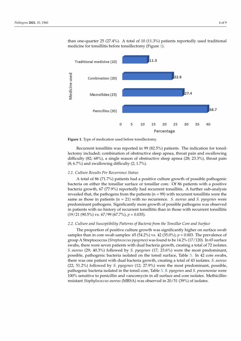
Pathogens 2021, 10, 1560 4 of 9
than one-quarter 25 (27.4%). A total of 10 (11.3%) patients reportedly used traditionalmedicine for tonsillitis before tonsillectomy (Figure 1).
Pathogens 2021, 10, x FOR PEER REVIEW 4 of 9
Figure 1. Type of medication used before tonsillectomy.
Recurrent tonsillitis was reported in 99 (82.5%) patients. The indication for tonsillec-
tomy included; combination of obstructive sleep apnea, throat pain and swallowing diffi-
culty (82; 68%), a single reason of obstructive sleep apnea (28; 23.3%), throat pain (8; 6.7%)
and swallowing difficulty (2; 1.7%).
2.1. Culture Results Per Recurrence Status
A total of 86 (71.7%) patients had a positive culture growth of possible pathogenic
bacteria on either the tonsillar surface or tonsillar core. Of 86 patients with a positive bac-
teria growth, 67 (77.9%) reportedly had recurrent tonsillitis. A further sub-analysis re-
vealed that, the pathogens from the patients (n = 99) with recurrent tonsillitis were the
same as those in patients (n = 21) with no recurrence. S. aureus and S. pyogenes were pre-
dominant pathogens. Significantly more growth of possible pathogens was observed in
patients with no history of recurrent tonsillitis than in those with recurrent tonsillitis
(19/21 (90.5%) vs. 67/99 (67.7%), p = 0.035).
2.2. Culture and Susceptibility Patterns of Bacteria from the Tonsillar Core and Surface
The proportion of positive culture growth was significantly higher on surface swab
samples than in core swab samples: 65 (54.2%) vs. 42 (35.0%); p = 0.003. The prevalence of
group A Streptococcus (Streptococcus pyogenes) was found to be 14.2% (17/120). In 65 sur-
face swabs, there were seven patients with dual bacteria growth, creating a total of 72
isolates. S. aureus (29; 40.3%) followed by S. pyogenes (17; 23.6%) were the most predomi-
nant, possible, pathogenic bacteria isolated on the tonsil surface, Table 3. In 42 core swabs,
there was one patient with dual bacteria growth, creating a total of 43 isolates. S. aureus
(22; 51.2%) followed by S. pyogenes (12; 27.9%) were the most predominant, possible, path-
ogenic bacteria isolated in the tonsil core, Table 3. S. pyogenes and S. pneumoniae were 100%
sensitive to penicillin and vancomycin in all surface and core isolates. Methicillin-resistant
Staphylococcus aureus (MRSA) was observed in 20/51 (39%) of isolates.
Figure 1. Type of medication used before tonsillectomy.
Recurrent tonsillitis was reported in 99 (82.5%) patients. The indication for tonsil-lectomy included; combination of obstructive sleep apnea, throat pain and swallowingdifficulty (82; 68%), a single reason of obstructive sleep apnea (28; 23.3%), throat pain(8; 6.7%) and swallowing difficulty (2; 1.7%).
2.1. Culture Results Per Recurrence Status
A total of 86 (71.7%) patients had a positive culture growth of possible pathogenicbacteria on either the tonsillar surface or tonsillar core. Of 86 patients with a positivebacteria growth, 67 (77.9%) reportedly had recurrent tonsillitis. A further sub-analysisrevealed that, the pathogens from the patients (n = 99) with recurrent tonsillitis were thesame as those in patients (n = 21) with no recurrence. S. aureus and S. pyogenes werepredominant pathogens. Significantly more growth of possible pathogens was observedin patients with no history of recurrent tonsillitis than in those with recurrent tonsillitis(19/21 (90.5%) vs. 67/99 (67.7%), p = 0.035).
2.2. Culture and Susceptibility Patterns of Bacteria from the Tonsillar Core and Surface
The proportion of positive culture growth was significantly higher on surface swabsamples than in core swab samples: 65 (54.2%) vs. 42 (35.0%); p = 0.003. The prevalence ofgroup A Streptococcus (Streptococcus pyogenes) was found to be 14.2% (17/120). In 65 surfaceswabs, there were seven patients with dual bacteria growth, creating a total of 72 isolates.S. aureus (29; 40.3%) followed by S. pyogenes (17; 23.6%) were the most predominant,possible, pathogenic bacteria isolated on the tonsil surface, Table 3. In 42 core swabs,there was one patient with dual bacteria growth, creating a total of 43 isolates. S. aureus(22; 51.2%) followed by S. pyogenes (12; 27.9%) were the most predominant, possible,pathogenic bacteria isolated in the tonsil core, Table 3. S. pyogenes and S. pneumoniae were100% sensitive to penicillin and vancomycin in all surface and core isolates. Methicillin-resistant Staphylococcus aureus (MRSA) was observed in 20/51 (39%) of isolates.

Pathogens 2021, 10, 1560 5 of 9
Table 3. Sensitivity patterns of Gram-positive bacteria isolated from the surface and core tonsillar swab.
Bacteria Isolated (n) E n(%) DA n(%) LZD n(%) SXT n(%) CIP n(%) CN n(%) AK n(%) FOX n(%)
Surface
S. aureus (29) 14 (48.3) 18 (62.1) 4 (13.8) 5 (17.2) 15 (51.7) 18 (62.1) 23 (79.3) 18 (62.1)
S. pyogenes (17) 11 (64.7) 12 (70.6) 10 (58.8) 0 (0.0) 11 (64.7) 14 (82.4) 14 (82.4)
S. viridans (15) 2 (13.3) 13 (86.7) 14 (93.3) 2 (13.3) 10 (66.7) 11 (73.3) 12 (80)
S. pneumoniae (4) 3 (75) 3 (75) 3 (75) 3 (75) 2 (50) 2 (50) 2 (50)
Enterococcous spp. (3) 2 (66.7) 2 (66.7) 2 (66.7) 1 (33.3) 3 (100) 3 (100) 2 (66.7)
Core
S. aureus (22) 9 (40.9) 12 (54.5) 12 (54.5) 8 (36.4) 13 (59.1) 14 (63.6) 15 (68.2) 13 (59.1)
S. pyogenes (12) 11 (91.7) 6 (50.0) 9 (75.0) 0 (0.0) 8 (66.7) 9 (75.0) 9 (75.0)
S. viridans (6) 3 (50.0) 5 (83.3) 4 (66.7) 1 (16.7) 2 (33.3) 3 (50.0) 3 (50.0)
E—erythromycin, DA—clindamycin, LZD—linezolod, SXT—trimethoprim-sulfamethoxazole, CIP—ciprofloxacin, AK—amikacin, FOX—cefoxitin, CN-Gentamicin.
2.3. Histology Results
Histological examinations of the 120 tonsillar tissues indicated that 83 (69%) hadfeatures suggestive of tonsillitis (Figure 2a,b). Seven (6%) tissues had features of reactivefollicular tissue hyperplasia (Figure 3). From 83 tissues samples with suggestive features oftonsillitis, 68 (79.1%) had a positive growth of possible, pathogenic bacteria. A significantlyhigh proportion of samples with features suggestive of tonsillitis in histology had a higherpathogenic bacteria growth than those without features suggestive of tonsillitis in histology:68 (79.1%) vs. 18 (20.9%); p < 0.001. Furthermore, a high proportion of tissue with suggestivefeatures of tonsillitis had a higher positive growth of possible pathogenic bacteria on thesurface than in the core: 64 (90.1%) vs. 31 (43.6%); p < 0.001. A total of 27 (38.5%) tissueshad a positive growth of possible pathogenic bacteria on both surface and core swabs.
1
(a) (b)
Figure 2. (a) Hematoxylin and eosin tissue stain showing tonsillar squamous epithelium infiltratedby lymphocytes × 40. (b) Hematoxylin and eosin tissue stain showing variably sized folliclesproliferation with prominent germinal centers, and fibrous tissue between the follicles × 4.
Tonsillar tissue, with features suggestive of tonsillitis in histological examinations,had a 4.8 probability of having positive culture results for possible pathogenic bacteria,compared to tonsillar tissue without features suggestive of tonsillitis (OR 4.8, 95% CI2.0–11.2, p < 0.001).

Pathogens 2021, 10, 1560 6 of 9Pathogens 2021, 10, x FOR PEER REVIEW 6 of 9
Figure 3. Histology results if tonsillar tissue.
3. Discussion
The local epidemiological data for bacteria in the core and surface of the tonsils of
patients clinically diagnosed with tonsillitis are important for establishing the proper em-
pirical management protocol of these patients. In response to the call of the World Health
Assembly for global action to combat RHD in 2018, this study ‘s contribution was to pro-
vide data on the patterns of bacteria found on the surface and core of tonsils among pa-
tients scheduled for tonsillectomy. Nearly half of the patients in the current study were
found to have possible pathogenic bacteria on the tonsillar surface with the predominance
of Staphylococcus aureus and Streptococcus pyogenes. Furthermore, we observed that more
than one-third of Staphylococcus aureus isolates were resistant to methicillin, signifying the
importance of local susceptibility data in guiding the antibiotic treatment for patients with
tonsillitis.
The findings from this study are significantly different from the study in Iraq involv-
ing 73 children in the age range of 3–10 years, which reported a prevalence of 58.9% [3].
Furthermore, this prevalence was significantly higher than 15.9%, as the previously re-
ported figure from a study conducted in Uganda [17]. The broad range of possible patho-
gens reported in the current study could explain this difference, since the study conducted
in Uganda was centered on only group A Streptococcus spp. The prevalence of group A
Streptococcus spp. observed in the current study is similar to what was reported in Uganda
signifying the importance of this pathogen in the pathogenesis of tonsillitis [17].
The current study found the prevalence of possible pathogenic bacteria in tonsillar
core to be 35.0%. This prevalence is almost similar to 40.7% which was previously reported
in India in the age group between 11 and 20 years [5]. In addition, the prevalence observed
in this study was significantly lower than the 90.4% reported from a study conducted in
Iraq [3]. These differences could be explained by the nature of the sample and geograph-
ical differences. The study in Iraq used fine-needle aspiration, which has higher ability to
concentrate the pathogen and increase the yield of the pathogen than the core swab used
Figure 3. Histology results if tonsillar tissue.
3. Discussion
The local epidemiological data for bacteria in the core and surface of the tonsils ofpatients clinically diagnosed with tonsillitis are important for establishing the properempirical management protocol of these patients. In response to the call of the WorldHealth Assembly for global action to combat RHD in 2018, this study ‘s contribution wasto provide data on the patterns of bacteria found on the surface and core of tonsils amongpatients scheduled for tonsillectomy. Nearly half of the patients in the current study werefound to have possible pathogenic bacteria on the tonsillar surface with the predominanceof Staphylococcus aureus and Streptococcus pyogenes. Furthermore, we observed that morethan one-third of Staphylococcus aureus isolates were resistant to methicillin, signifyingthe importance of local susceptibility data in guiding the antibiotic treatment for patientswith tonsillitis.
The findings from this study are significantly different from the study in Iraq involving73 children in the age range of 3–10 years, which reported a prevalence of 58.9% [3]. Fur-thermore, this prevalence was significantly higher than 15.9%, as the previously reportedfigure from a study conducted in Uganda [17]. The broad range of possible pathogensreported in the current study could explain this difference, since the study conductedin Uganda was centered on only group A Streptococcus spp. The prevalence of group AStreptococcus spp. observed in the current study is similar to what was reported in Ugandasignifying the importance of this pathogen in the pathogenesis of tonsillitis [17].
The current study found the prevalence of possible pathogenic bacteria in tonsillarcore to be 35.0%. This prevalence is almost similar to 40.7% which was previously reportedin India in the age group between 11 and 20 years [5]. In addition, the prevalence observedin this study was significantly lower than the 90.4% reported from a study conducted inIraq [3]. These differences could be explained by the nature of the sample and geographicaldifferences. The study in Iraq used fine-needle aspiration, which has higher ability toconcentrate the pathogen and increase the yield of the pathogen than the core swab used in
Pathogens 2021, 10, 1560 7 of 9
the current study. The fine needle aspiration culture is reported to have a 100% sensitivity,which is higher than the 82.9–92% sensitivity of the core swab culture [10,18].
As previously reported elsewhere [3,5,18], the current study found a mixed growth ofpossible pathogenic bacteria in seven tonsil surface swabs and one core swabs. The type ofbacteria species on the surface and in the core of the tonsils was similar in 22.1% of studiedpatients. This was also reported from previous studies [10,18].
Staphylococcus aureus and Streptococcus pyogenes were the predominant bacteria isolatedon both the tonsil surface and in the tonsil core. This was previously reported from otherstudies [5,10]. Methicillin-resistant Staphylococcus aureus (MRSA) was observed in 39%of isolates. This was within the range (28.6–50%) of MRSA reported by different studiesconducted in the same setting among patients with different infections [19–21]. A similarresistance trend was reported in India [5].
4. Limitations
The inability to perform anaerobic cultures of bacteria and tissue cultures for virusesmight lead to an underestimation of the reported prevalence. The use of patients scheduledfor tonsillectomy limits the reproducibility of the study results to the general populationof patients with tonsillitis. The study cannot give a detailed insight into the type andcomposition of traditional medicine used by patients as the data were not collected.
5. Conclusions and Recommendations
More than two-thirds of patients undergoing tonsillectomy at BMC had a positiveculture growth of possible pathogenic bacteria. Streptococcus pyogenes and Staphylococcusaureus were the predominant bacteria detected with more than one-third of Staphylococcusaureus being MRSA. More studies to investigate the treatment outcome of these patientsare highly recommended.
6. Methodology
This prospective, cross-sectional, hospital-based study was conducted among patientsscheduled for tonsillectomy between April and July 2019. The study was conducted in theotolaryngology department of BMC, a tertiary teaching hospital of the Catholic Universityof Health and Allied Health Sciences, which has a bed capacity of 1000. The otolaryngologydepartment of BMC performs about 70–80 tonsillectomies each month. The minimumsample size was obtained using a Kish Leslie formula (1965) with a 75% prevalence oftonsillitis from a study conducted in India, which analyzed the patterns of bacteria frompatients with tonsillitis [16].
Patients were serially recruited to the study until the sample size was reached. Thestudy excluded all patients with an indication of a tumor as judged by the attendingclinicians. Data (age, gender, employment status, and history of breathing difficulty, fever,hospital admission, dysphagia, history of family members with tonsillitis, antibiotic usagein the past two months and recurrent tonsillitis, etc.) were obtained using a pre-codedstructured questionnaire before the patient was anesthetized.
Tonsils were defined as the lymphoepithelial structures that provide a protectiveimmunological ring at the openings of both the digestive and respiratory tracts [22]. Inthe current study, recurrent tonsillitis was defined by having more than five episodes ayear [23]. Tonsil core was defined as the inner part of the tonsil, including the tonsil crypt,which was reached by dissecting the tonsil tissue.
6.1. Sample Collection and Processing
The surface of the tonsil was rubbed using sterile swab by the experienced surgeonwhile patient is under anesthesia. The tonsillectomy was conducted followed by thedissection of the tonsil to expose the core area and a second swab was taken immediately.All swabs were placed in the Stuart transport media and transported to microbiologylaboratory for processing within two hours of collection. All microbiological analyses
Pathogens 2021, 10, 1560 8 of 9
were conducted at the microbiology research laboratory of CUHAS following standardoperating procedures.
Swabs were inoculated on 5% sheep blood agar and chocolate agar. The plates wereincubated in the candle jar for 18–24 h at 37 ◦C. The identification of bacteria specieswas based on their growth morphology, hemolytic patterns in blood agar and biochem-ical/physiological tests such as catalase, coagulase, bile solubility test, and their sensi-tivity patterns to bacitracin, optochin and trimethoprim/sulphamethoxazole. Antimicro-bial susceptibility testing was conducted using Kirby-bauer disc diffusion technique for15 µg erythromycin(E), 10 µg linezolid(LZD), 10µg gentamicin(CN), 30 µg amikacin(AK),1.25/23.75 µg trimethoprim-sulfamethoxazole(SXT), 5 µg ciprofloxacin(CIP) and 2 µg clin-damycin(DA) [24]. Cefoxitin(FOX) discs (30µg) were used for detection of MRSA as perClinical Laboratory Standard Institute (CLSI) recommendations [25].
6.2. Histological Analysis
The excised tonsil was immediately immersed in the buffered formalin for completefixation then transferred to histology laboratory for processing by an experienced technolo-gist, and then read by a pathologist. In the histology laboratory, the tissue was processed,sectioned and stained by hematoxylin and eosin as previously documented [26].
6.3. Data Analysis
Data were entered on Excel spreadsheet for consistent checking and cleaning, andthen transferred to STATA version 13 for analysis. Categorical data were summarizedusing proportions while continuous data were summarized using median and interquartileranges. Frequency run was conducted to determine the proportions while two proportiontest was conducted to determine the level of differences in microbiological and histologicalresults. p value of less than 0.05 at 95% confidence interval was considered statisticallysignificant.
6.4. Ethical Considerations
The protocol of this study was ethically approved by the CUHAS/BMC researchethics and review committee (CREC) with certificate number 912/2019. For participantsaged under 18 years, informed consent from a parent and/or legal guardian was obtainedbefore selection. All the procedures were performed following the ethical guidelines.
Author Contributions: G.B., M.F.M. and S.E.M. designed the work. G.B. and G.A.K. selected patients,G.A.K., M.F.M., V.S., P.R. and S.E.M. performed laboratory investigations and interpreted the results.M.F.M., P.R. and S.E.M. analyzed and interpreted the data. M.F.M. wrote the first draft of themanuscript, which was critically reviewed by P.R. and S.E.M. All authors have read and agreed tothe published version of the manuscript.
Funding: This research received no external funding.
Institutional Review Board Statement: The protocol of this study was ethically approved by theJoint CUHAS/BMC research ethics and review committee (CREC) with certificate number 912/2019.
Informed Consent Statement: Written informed consent was obtained from the patient(s) in orderto publish this paper.
Data Availability Statement: The datasets used and/or analyzed during the current study areavailable from the corresponding author upon reasonable request.
Acknowledgments: The authors would like to acknowledge the support provided by the Departmentof ENT, Bugando Medical Centre/Weill Bugando School of Medicine, Catholic University of Healthand Allied Sciences and the Department of Microbiology and Immunology of the Catholic Universityof Health and Allied Sciences, Mwanza, Tanzania.
Conflicts of Interest: The authors declare no conflict of interest.

Pathogens 2021, 10, 1560 9 of 9
References1. Ingram, D.G.; Friedman, N.R. Toward adenotonsillectomy in children: A review for the general pediatrician. JAMA Pediatr. 2015,
169, 1155–1161. [CrossRef]2. Gul, M.; Okur, E.; Ciragil, P.; Yildirim, I.; Aral, M.; Kilic, M.A. The comparison of tonsillar surface and core cultures in recurrent
tonsillitis. Am. J. Otolaryngol. 2007, 28, 173–176. [CrossRef]3. Yousef, R.Y.; Yousef, R.Y. Comparison of the bacteriology of tonsil surface and core in bacterial profile isolated from children with
chronic tonsillitis. Med. J. Babylon 2010, 7, 52–57.4. Oburra, H.O.; Idenya, M. Frequency of adenotonsillectomy in some Nairobi hospitals. East Afr. Med. J. 2001, 78, 338. [CrossRef]
[PubMed]5. Agrawal, A.; Kumar, D.; Goyal, A.; Gupta, R.; Bhooshan, S. Bacteriological Evaluation and Their Antibiotic Sensitivity Pattern in
Tonsillitis. IOSR J. Dent. Med. Sci. 2014, 13, 51–55. [CrossRef]6. Bista, M.; Sinha, B.; Amatya, R.; Tuladhar, N.; Pokharel, B. Comparison of core and surface cultures in recurrent tonsillitis. J. Inst.
Med. 2007, 27, 60–65. [CrossRef]7. Brook, I.; Yocum, P.; Shah, K. Surface vs. core-tonsillar aerobic and anaerobic flora in recurrent tonsillitis. JAMA 1980, 244,
1696–1698. [CrossRef]8. Kumai, A.; Gupta, V.; Chandra, K.; Gupta, P.; Varshney, S. Clinico bacteriological evaluation of surface and core microflora in
chronic tonsillitis. Indian J. Otolaryngol. Head Neck Surg. 2005, 57, 118–120. [CrossRef] [PubMed]9. Kurien, M.; Sheelan, S.; Jeyaseelan, L.; Thomas, K. Fine needle aspiration in chronic tonsillitis: Reliable and valid diagnostic test.
J. Laryngol. Otol. 2003, 117, 973–975. [CrossRef] [PubMed]10. Loganathan, A.; Arumainathan, U.D.; Raman, R. Comparative study of bacteriology in recurrent tonsillitis among children and
adults. Singap. Med. J. 2006, 47, 271–275.11. Kardooni, M.; Mosavian, S.M.; Lotfinia, M.; Eghbalnejad, A.M.; Mofrad, M.S.; Saki, N.; Abshirini, H. Study of common bacterial
agents and antibiotic susceptibility in patients with chronic and repeated tonsillitis. J. Adv. Pharm. Educ. Res. 2020, 10, 90–95.12. Hadi, U.; El-Hajj, M.; Uwaydah, M.; Fuleihan, N.; Matar, G.M. Characteristics of pathogens recovered from the tonsils and
adenoids in a group of Lebanese children undergoing tonsillectomy and adenoidectomy. J. Appl. Res. Clin. Exp. Ther. 2005, 5, 473.13. Moyo, S.J.; Steinbakk, M.; Aboud, S.; Mkopi, N.; Kasubi, M.; Blomberg, B.; Manji, K.; Lyamuya, E.F.; Maselle, S.Y.; Langeland, N.
Penicillin resistance and serotype distribution of Streptococcus pneumoniae in nasopharyngeal carrier children under 5 years ofage in Dar es Salaam, Tanzania. J. Med. Microbiol. 2012, 61, 952–959. [CrossRef]
14. Emgård, M.; Msuya, S.E.; Nyombi, B.M.; Mosha, D.; Gonzales-Siles, L.; Nordén, R.; Geravandi, S.; Mosha, V.; Blomqvist, J.;Franzén, S.; et al. Carriage of penicillin-non-susceptible pneumococci among children in northern Tanzania in the 13-valentpneumococcal vaccine era. Int. J. Infect. Dis. 2019, 81, 156–166. [CrossRef]
15. Mboya, E.A.; Sanga, L.A.; Ngocho, J.S. Irrational use of antibiotics in the Moshi Municipality Northern Tanzania: A cross sectionalstudy. Pan Afr. Med. J. 2018, 31, 165. [CrossRef] [PubMed]
16. Vijayashree, M.; Viswanatha, B.; Sambamurthy, B. Clinical and Bacteriological Study of Acute Tonsillitis. IOSR J. Dent. Med. Sci.2014, 13, 37–43. [CrossRef]
17. DeWyer, A.; Scheel, A.; Webel, A.R.; Longenecker, C.T.; Kamarembo, J.; Aliku, T.; Engel, M.E.; Bowen, A.; Bwanga, F.; Hovis, I.;et al. Prevalence of group A β-hemolytic streptococcal throat carriage and prospective pilot surveillance of streptococcal sorethroat in Ugandan school children. Int. J. Infect. Dis. 2020, 93, 245–251. [CrossRef]
18. Sarkar, S.; Sil, A.; Sarkar, S.; Sikder, B. A comparison of tonsillar surface swabbing, fine-needle aspiration core sampling, anddissected tonsillar core biopsy culture in children with recurrent tonsillitis. Ear Nose Throat J. 2017, 96, E29–E32. [CrossRef]
19. Ahmed, M.; Mirambo, M.M.; Mushi, M.F.; Hokororo, A.; Mshana, S.E. Bacteremia caused by multidrug-resistant bacteria amonghospitalized malnourished children in Mwanza, Tanzania: A cross sectional study. BMC Res. Notes 2017, 10, 62. [CrossRef][PubMed]
20. Mushi, M.F.; Mwalutende, A.E.; Gilyoma, J.M.; Chalya, P.L.; Seni, J.; Mirambo, M.M.; Mshana, S.E. Predictors of diseasecomplications and treatment outcome among patients with chronic suppurative otitis media attending a tertiary hospital,Mwanza Tanzania. BMC Ear, Nose Throat Disord. 2016, 16, 1. [CrossRef] [PubMed]
21. Silago, V.; Mushi, M.F.; Remi, B.A.; Mwayi, A.; Swetala, S.; Mtemisika, C.I.; Mshana, S.E. Methicillin resistant Staphylococcusaureus causing osteomyelitis in a tertiary hospital, Mwanza, Tanzania. J. Orthop. Surg. Res. 2020, 15, 95–96. [CrossRef] [PubMed]
22. Perry, M.; Whyte, A. Immunology of the tonsils. Immunol. Today 1998, 19, 414–421. [CrossRef]23. McKerrow, W.S. Recurrent tonsillitis. Am. Fam. Physician 2002, 66, 1735. [PubMed]24. Bauer, A.W.; Kirby, W.M.; Sherris, J.C.; Turck, M. Antibiotic susceptibility testing by a standardized single disk method. Am. J.
Clin. Pathol. 1966, 45, 493–496. [CrossRef]25. Clinical Lab Standards Institute. Performance Standards for Antimicrobial Susceptibility Testing; Clinical Lab Standards Institute:
Wayne, PA, USA, 2016.26. Bieluch, V.M.; Chasin, W.D.; Martin, E.T.; Tally, F.P. Recurrent Tonsillitis: Histologic and Bacteriologic Evaluation. Ann. Otol.
Rhinol. Laryngol. 1989, 98, 332–335. [CrossRef] [PubMed]