การดูแลสุขภาพทารกในครรภ ในระยะคลอด Intrapartum Fetal Monitor

AUTISM: MATERNALLY DERIVED ANTIBODIES SPECIFIC FORFETAL BRAIN PROTEINS
Daniel Braunschweig1,7,8, Paul Ashwood2,7,8, Paula Krakowiak3,7,8, Irva Hertz-Picciotto3,7,8, Robin Hansen4,7,8, Lisa Croen5, Isaac N. Pessah6,7,8, and Judy Van deWater1,7,8,*
1 Division of Rheumatology, Allergy and Clinical Immunology, University of California at Davis. Davis, CA95616 USA
2Department of Medical Microbiology and Immunology, University of California at Davis. Davis, CA 95616USA
3 Department of Public Health Sciences, Division of Epidemiology, University of California at Davis, Davis,CA 95616 USA
4 Department of Pediatrics, University of California at Davis. Davis, CA 95616 USA
5Division of Research, Kaiser Permanente Northern California, Oakland, CA.
6Department of Veterinary Molecular Biosciences, University of California at Davis Davis, CA 95616 USA
7 The M.I.N.D. Institute, University of California at Davis. Davis, CA 95616 USA
8 NIEHS Center for Children's Environmental Health, University of California, Davis. Davis, CA 95616 USA
AbstractAutism is a profound disorder of neurodevelopment with poorly understood biological origins. Apotential role for maternal autoantibodies in the etiology of some cases of autism has been proposedin previous studies To investigate this hypothesis, maternal plasma antibodies against human fetaland adult brain proteins were analyzed by western blot in 61 mothers of children with autistic disorderand 102 controls matched for maternal age and birth year (62 mothers of typically developing children(TD) and 40 mothers of children with non-ASD developmental delays (DD)). We observed reactivityto two protein bands at approximately 73kDa and 37kDa in plasma from 7 of 61 (11.5%) mothersof children with autism (AU) against fetal but not adult brain, which was not noted in either controlgroup (TD; 0/62 p=0.0061 and DD; 0/40 p=0.0401). Further, the presence of reactivity to these twobands correlated with a diagnosis of behavioral regression in the child when compared to the TD(p=0.0019) and DD (0.0089) groups. Individual reactivity to the 37kDa band was observedsignificantly more often in the AU population compared with TD (p=0.0086) and DD (p=0.002)mothers, yielding a 5.69-fold odds ratio (95% confidence interval 2.09 - 15.51) associated with thisband. The presence of these antibodies in the plasma of some mothers of children with autism, aswell as the differential findings between mothers of children with early onset and regressive autismmay suggest an association between the transfer of IgG autoantibodies during earlyneurodevelopment and the risk of developing of autism in some children.
*Corresponding author: Division of Rheumatology/Allergy and Clinical Immunology University of California 451 E. Health SciencesDrive, Suite 6510 GBSF Davis, CA 95616 Phone: 530-752-2154 Fax: 530-752-4669 Email: [email protected]'s Disclaimer: This is a PDF file of an unedited manuscript that has been accepted for publication. As a service to our customerswe are providing this early version of the manuscript. The manuscript will undergo copyediting, typesetting, and review of the resultingproof before it is published in its final citable form. Please note that during the production process errors may be discovered which couldaffect the content, and all legal disclaimers that apply to the journal pertain.
NIH Public AccessAuthor ManuscriptNeurotoxicology. Author manuscript; available in PMC 2009 March 1.
Published in final edited form as:Neurotoxicology. 2008 March ; 29(2): 226–231. doi:10.1016/j.neuro.2007.10.010.
NIH
-PA Author Manuscript
NIH
-PA Author Manuscript
NIH
-PA Author Manuscript

KeywordsAutism; Maternal Antibodies; Regression; Autoantibodies
1. IntroductionThe autism spectrum disorders (ASD), manifest as highly variable combined deficits in socialinteraction, verbal and non-verbal communication, and often include the presence of repetitive,stereotypical and overly restrictive behaviors (APA, 1994). Despite the lack of clear etiologyfor the large majority of ASD cases, evidence from twin studies (Bailey et al., 1995) and familialincidence (Lauritsen et al., 2005) rates support a view of ASD as a largely, but not exclusively,genetic disorder. The potential role of the immune system in ASD has been addressed in severalstudies. These include reports of neuroglial activation and neuroinflammation in the CNS(Pardo et al., 2005), as well as plasma antibodies reactive to rodent neuronal tissue (Singer etal., 2006) in children with autism. However, there have been no systematic case-based studiesdescribing a direct relationship between maternal autoantibodies to human fetal neuronalproteins and the development of ASD.
The role of the maternal immune system in fetal neurodevelopment is an area of active research.It has long been known that in humans, maternal IgG isotype antibodies readily cross theplacenta to equip the immunologically naïve fetus with a subset of the maternal adaptivehumoral immune system proteins (Garty et al., 1994); these maternal IgG antibodies are knownto persist for up to six months post-natal (Heininger et al., 2006). However, together with IgGantibodies that are immunoprotective, autoantibodies that react to fetal ‘self’-proteins can alsocross the placenta. A recent report demonstrated maternal IgG antibody reactivity to rodentPurkinje cells in a mother of multiple children with ASD, as well as the presence of behavioraldeficits in pups of a mouse injected during gestation with her serum (Dalton et al., 2003). Inanother study, mothers of children with autism and their affected children were found to haveconsistent patterns of antibody reactivity against rat prenatal (day 18) brain proteins. Incontrast, unaffected children and control mothers had alternative patterns of reactivity(Zimmerman et al., 2006).
The preponderance of evidence suggests a pre-natal or early post-natal etiology for autism,potentially involving errant developmental cues. Advances in understanding the role ofimmune system components during fetal neurodevelopment combined with the cross-talkbetween the maternal and fetal immune systems, led us to investigate the profiles ofautoantibody reactivity in mothers of children with autism and to compare them with profilesfrom mothers of typically developing children and from mothers of children with otherdevelopmental disorders excluding autism.
2. Materials and Methods2.1 Study Subjects
This case-control study examined 61 mothers of children with autism and 102 control mothersenrolled through the Center for Children's Environmental Health (CCEH) as part of the ongoingCHARGE (Childhood Autism Risks from Genetics and Environment) study at the M.I.N.D.Institute at the University of California at Davis (Hertz-Picciotto et al., 2006). The CHARGEstudy population was sampled from three strata: children considered to have autism (AU),children selected from the general population who were typically developing (TD), andchildren with developmental disabilities without autism (DD). The families were recruited forthis study without bias for any medical or demographic factors.
Braunschweig et al. Page 2
Neurotoxicology. Author manuscript; available in PMC 2009 March 1.
NIH
-PA Author Manuscript
NIH
-PA Author Manuscript
NIH
-PA Author Manuscript
To confirm and further detail the initial diagnosis, all children were assessed at the UC DavisM.I.N.D. Institute. The diagnosis of autism was confirmed for all cases using the AutismDiagnostic Interview-Revised (Lord et al., 1997) and the Autism Diagnostic ObservationSchedule, modules 1, 2 or 3 (DiLavore et al., 1995; Joseph et al., 2002; Lord et al., 2001;Owley et al., 2001). The ADI-R provides a standardized, semi-structured interview and adiagnostic algorithm for the DSM-IV(APA, 1994) and the ICD-10 definitions of autism(Steinhausen and Erdin, 1992; WHO, 1992). The ADOS is a semi-structured, standardizedassessment in which the researcher observes the social interaction, communication, play andimaginative use of materials for children suspected of having ASD. Final autism case diagnosiswas defined as meeting criteria on the communication, social, and repetitive behaviors domainsof the ADI-R and scoring at or above the cut-off for autistic disorder on the ADOS modules 1or 2. The Social Communication Questionnaire was used to screen for behavioral anddevelopmental characteristics of ASD among the subjects with developmental disabilities andamong the general population typically developing controls; children who scored above thescreening cut-off were fully assessed using the ADI-R and ADOS. Those who met criteria forautistic disorder were classified as AU; similarly any general population children who metcriteria for DD based on the Vineland Scales of Adaptive Behavior (Sparrow et al., 1984) andthe Mullen Scales of Early Learning (Mullen, 1995) were classified as DD. Controls who didnot meet criteria for ASD or for DD were classified as typically developing (TD). Using clinicalcharacteristics reported in the Early Development Questionnaire (Ozonoff et al., 2005) andanswers to questions regarding loss of language (Q11) and social skills (Q25) of the ADI-R,the autism population was further divided into two groups based on the clinical onset of autisticsymptoms; firstly, children with regression who initially developed normally, but subsequentlylost acquired skills (n=36; (31 male, 5 female)) and secondly, children with early onset autismcharacterized by early deficits in the requisite behavioral domains (n=25; (24 male, 1 female)).The study protocol followed the ethical guidelines of the most recent Declaration of Helsinki(Edinburgh, 2000), and was approved by the Institutional Review Boards of the UC DavisSchool of Medicine and the State of California, and written informed consent was obtained forall participants enrolled in the study.
Following informed consent, plasma samples were collected from mothers of children meetingthe above enrollment criteria. For this analysis we matched control maternal samples with AUcase maternal samples for maternal age and parity as well as the age of offspring (Table 1).Mothers of AU children were considered without regard to sibling developmental status. TDmothers were excluded from this analysis if any of their children were diagnosed with adevelopmental disorder. Paternal half-siblings of case and control children were not includedin total children (parity) and birth order demographics.
Detailed information regarding current autoimmune disease status for first-degree relativeswas collected at the clinic visit by questionnaire (Hertz-Picciotto et al., 2006). A list ofautoimmune conditions was reviewed with the parent by the clinician and descriptions wereprovided where needed.
2.2 Sample CollectionMaternal blood was collected in yellow top acid citrate dextrose tubes (BD Diagnostic, FranklinLakes, NJ). Plasma was separated from cells, coded, and aliquoted to minimize freeze/thawcycles and stored at −80°C until use.
2.3 Western blot analysisWestern blots were performed as described elsewhere (Cabanlit et al., 2007 (In Press)). Briefly,300 μg human fetal brain protein medley (Clontech, Mountain View, CA), prepared from apooled sample of 63 spontaneously aborted male and female fetuses 20-40 weeks gestation,
Braunschweig et al. Page 3
Neurotoxicology. Author manuscript; available in PMC 2009 March 1.
NIH
-PA Author Manuscript
NIH
-PA Author Manuscript
NIH
-PA Author Manuscript

was separated under reducing conditions on 4-15% SDS-polyacrylamide prep gels (Bio-Rad,Hercules, CA) and transferred electrophoretically to 0.2μm-pore nitrocellulose membranes(Whatman, Florham Park, NJ) at 35V for 14hrs. Magic mark XP molecular weight marker(Invitrogen, Carlsbad, CA) was used in the single marker lane allowing chemiluminescentvisualization of marker bands from 20 to 220kDa. After transfer, the blots were blocked in10% Casein Block (Pierce Biotechnology, Rockford, IL) and then cut into strips including theMW marker and 24 fetal brain strips. Strips were placed in mini-incubation trays (Bio-Rad,Hercules, CA) on a rocking platform with 700μl of 1:400 maternal plasma diluted in PBS/0.05% Tween 20/0.5% Casein Block (PBSTC) for 1.5hrs and then washed in PBS/0.05%Tween 20 (PBST) 5 times for 5 minutes each. Zymax horseradish peroxidase conjugated Goatanti-Human IgG (Invitrogen, Carlsbad, CA) diluted 1:25,000 in PBST was added and stripswere incubated for 30 minutes with rocking. Following secondary antibody incubation, stripswere washed 5 times for 5 minutes with PBST and subsequently incubated with SuperSignalWest Pico (Pierce Biotechnology, Rockford, IL) chemiluminescent substrate for 5 minutes.The strips were then removed from the incubation trays and arranged on a glass plate forimaging using a FluorChem 8900 with AlphaEaseFC software (Alpha Innotech, San Leandro,CA) with a 1, 3, 5 minute stacked movie acquisition. Reactivity against proteins from controltissues including human adult brain (Clontech, Mountain View, CA), duodenum (Clontech,Mountain View, CA) and human kidney (Clontech, Mountain View, CA), was assessed using300 μg/gel as described in the above technique.
Band presence and apparent molecular weight were determined using the image analysiscapabilities of the AlphaEaseFC software. After defining the loading well position (proteinmigration start-point) and the dye front (end-point), relative migration (Rf) of each of themolecular weight markers was calculated. A point-by-point curve fit was applied to the Rf ofthe molecular weight markers and was used to determine the molecular weight of bands ofmaternal immunoreactivity to fetal brain. The presence of IgG heavy- and light-chain bandsat approximately 25kDa and 50 kDa, arising from reactivity of the secondary antibody toendogenous IgG present in the protein preparations, provided an internal reference for eachsample strip and was used to verify uniform protein migration. Blots were analyzed completelybefore revealing the diagnosis of the child. Bands were considered to be the same betweensamples when <4% difference was observed in Rf. The threshold for assigning the presenceof a band was a two-fold higher densitometry reading above background on the strip.
2.4 Statistical AnalysisStatistical analysis was carried out with SAS statistical analysis software (SAS Institute Inc.Cary, NC). Comparisons of experimental groups were made using a Fisher's exact test appliedto all bands individually and in all possible combinations to determine individual as well asgrouped associations with diagnosis. Differences were considered significant at p < 0.05.Because of the presence of zero values, an odds ratio and 95% confidence interval could onlybe calculated for the 37kDa band.
3. Results3.1 Band Prevalence
Autoreactivity to a protein at approximately 37kDa was observed in the plasma of 16/61mothers of AU children (26.2%) (Figure 1 and Table 2) compared with 1/40 mothers of DDchildren (2.5%; p=0.0023), and 5/62 mothers of TD children (8.1%; p=0.0086) (Table 2).Furthermore, the presence of the 37kDa band yielded a significantly elevated odds ratio of 5.69(95% confidence interval: 2.09 – 15.51) when compared with the TD group. Of particular note,reactivity against proteins at both 37kDa and 73kDa was observed only in mothers of AUchildren, yielding highly significant statistical differences between mothers of AU children
Braunschweig et al. Page 4
Neurotoxicology. Author manuscript; available in PMC 2009 March 1.
NIH
-PA Author Manuscript
NIH
-PA Author Manuscript
NIH
-PA Author Manuscript
and mothers of TD children (7/61 vs. 0/62; p=0.0061) and mothers of DD children (0/40;p=0.0401, Table 2). The presence of these bands did not correlate with maternal age or historyof autoimmune disease, nor with child birth order or child IQ (data not shown).
3.2 Band reactivity and clinical onset of autismWhen band prevalence was analyzed based on the pattern of clinical onset of autistic behaviorsin children, 6/7 (86%) of the AU mothers that exhibited reactivity to the pair of bands at 37kDaand 73kDa had children with the regressive phenotype (Table 2). Moreover, this trend was alsoevident regarding reactivity to the 37kDa and 73kDa bands separately (Table 2). In contrast,the antibody response of the mothers of early onset children was only significantly differentfrom the TD and DD groups for the 37kDa band alone (Table 2). Finally, no association wasobserved between the presence of autoreactivity to fetal brain antigens and history ofautoimmune disease in the maternal populations at the time of blood draw.
3.3 Maternal antibody reactivity to control proteinsAs a control for tissue specificity of these maternal antibodies, we analyzed several other tissueprotein extracts for reactivity patterns. Duodenum, a highly innervated compartment of the GItract, was chosen as a non-CNS tissue with substantial levels of neuronal proteins. We alsoexamined reactivity of maternal IgG to kidney protein extract, and found, as with duodenum,an absence of the 37kDa and 73kDa protein pattern of reactivity. Similarly, although faintbands were sometimes observed at 73kD, the specific pattern of both the 37kDa and 73kDabands was not seen for human adult brain protein extract (Figure 2).
4. DiscussionHerein we present a detailed analysis of maternal antibodies to human fetal brain in a largecohort of mothers for whom detailed familial information is available. The presence ofautoantibodies to proteins at 37kDa and 73kDa occurred significantly more often in AUmothers when compared with two distinct control populations. Bands that were shown to bedifferent between autism and controls were seen in more than a quarter of mothers who hadchildren with autism. The fact that these bands were not found in all mothers of children withautism further emphasizes the heterogeneity that is widely reported in autism, and the varietyof etiologic mechanisms that likely exist (Hertz-Picciotto et al., 2006). Furthermore, whilenone of the individual bands were noted exclusively in mothers of children with AU, thesimultaneous presence of the 37kDa and 73kDa protein bands was unique to the AU group.
Previous studies have also suggested a role for maternal antibodies in the etiology of somecases of autism (Dalton et al., 2003). Moreover, Zimmerman et al. (2006) recently reporteddiffering patterns of serum immunoreactivity to pre-natal rat brain between mothers of childrenwith autism and mothers of control children. Furthermore, the authors demonstrated thatimmunoreactivity persisted in maternal circulation for up to 18 years post-delivery(Zimmerman et al., 2006). Interestingly, the group differences in brain reactivity patterns wereobserved only with pre-natal rat brain protein and not post-natal (day 8) rat brain protein. Thepatterns described in the Zimmerman study differ from those presented in the current report,which is possibly due to disparities between rat and human brain proteins, or differences insample processing. However, the presence of maternal antibody reactivity against neuronalprotein associated with an outcome of autism in the child, is consistent across the two studies.
The transplacental passage of maternal IgG isotype antibodies has long been known as amechanism for fetal immune instruction (Garty et al., 1994) and protection (Harris et al.,2006; Simister, 2003). A recently described organelle in the placental epithelium that expressesthe low affinity IgG receptor, FcγRIIb, as well as the IgG receptor and transport protein FcRn,
Braunschweig et al. Page 5
Neurotoxicology. Author manuscript; available in PMC 2009 March 1.
NIH
-PA Author Manuscript
NIH
-PA Author Manuscript
NIH
-PA Author Manuscript
appears to provide a dedicated transport mechanism for maternal IgG to enter fetal circulation(Mishima et al., 2006). Detectable levels of maternal IgG are present in fetal circulation asearly as 18 weeks gestation, and by 38 weeks gestation, fetal levels are comparable withmaternal levels. Interestingly, neonatal IgG, which is overwhelmingly maternal in origin, isseen at levels exceeding the maternal concentration at delivery and persists at detectable levelsup to 6 months post-delivery (Garty et al., 1994). Further, the presence of IgG heavy- and light-chain bands in the fetal brain immunoblots supports the transport of maternal IgG into the fetalbrain during gestation. Thus the window of exposure to maternal IgG coincides substantiallywith critical periods of early neurodevelopment.
Despite the beneficial nature of the majority of maternal IgG received by the fetus, a numberof neonatal autoimmune diseases have been demonstrated to result from pathogenic maternalIgG. Notably, the presence of maternal anti-Ro/SS-A and anti-La/SS-B antibodies causeneonatal lupus syndrome, often leading to congenital heart block (Tincani et al., 2006) Inaddition, cases of neonatal anti-phospholipid syndrome (APS), mediated through maternalautoantibodies, have been observed in the newborn infants of mothers with primary APS(Soares Rolim et al., 2006). Finally, abnormal thyroid function is often noted in infants bornto mother with Hashimoto's thyroiditis or Graves' disease, caused by placental transfer ofmaternal anti-thyroid antibodies(Fu et al., 2005). Typically, symptoms of neonatal thyroiditisresolve as maternal antibodies are cleared from the circulation of the infant.
Our data suggest that the presence of maternal autoantibodies to fetal brain proteins ofapproximately 37kDa and 73kDa molecular weight confers an elevated risk for autism. Weperformed western blots using several other protein sources in order to determine the tissuespecificity of these autoantibodies. Interestingly, the reactivity observed towards fetal brainprotein was not seen among kidney, duodenum or adult brain proteins. This finding suggeststhe possibility that the autoreactivity described herein is targeted towards proteins expressedexclusively, or at substantially higher levels, during fetal development.
Maternal plasma collection for the current retrospective study occurred on average 3.5 yearsafter the birth of the affected child, and approximately 18 months after a diagnosis of autism.As circulating antibody titers are known to vary over time based on the immunological stateof the individual (Toptygina et al., 2005), the maternal antibody profile observed at the timeof the registration of her child into the CHARGE study may be slightly different than duringgestation. However, it has been demonstrated that antibodies, such as those generated inresponse to vaccination, can persist for many years due to the maintenance and subsequentpolyclonal reactivation of memory B cells (Shinefield et al., 2002). It is currently unknownwhether or not successive children from those mothers with reactivity to fetal brain will haveautism. A longitudinal analysis of subsequent offspring from these control mothers will allowus to resolve this issue.
Increasing attention has been given to the notion that autism, as a spectrum of disorders, likelyencompasses numerous, etiologically distinct behavioral phenotypes. Our observation ofmaternal reactivity to two protein bands at 37kDa and 73kDa more frequently in mothers ofAU children exhibiting behavioral regression than in those with early onset AU may help toelucidate biologic mechanisms contributing to phenotypic variance in ASD. Assuming that theobserved maternal autoantibody reactivity was also present during the prenatal and/or earlypostnatal period, the association of autoantibodies to neural antigens with delayed onset autismappears paradoxical. While beyond the scope of the present study, this could be explained bya pathogenic mechanism involving the interference of maternal autoantibodies withneurodevelopmental pathways for which compensatory mechanisms exist, but are ultimatelyoverwhelmed, leading to disease symptoms. Such a pathogenesis is noted in Rett syndrome,where mutations in the gene Mecp2 manifest in behavioral regression around 18 months of age
Braunschweig et al. Page 6
Neurotoxicology. Author manuscript; available in PMC 2009 March 1.
NIH
-PA Author Manuscript
NIH
-PA Author Manuscript
NIH
-PA Author Manuscript

(Williamson and Christodoulou, 2006). Finally, it is important to note that the presence ofmaternal autoantibodies to both the 37 kDa and 73 kDa proteins does not provide an etiologicmechanism for all cases of regressive autism, and their presence is strongly associated withthe regressive phenotype only in a sub-population of individuals.
These data provide evidence for an association between the presence of maternal immunesystem biomarkers and a diagnosis of autism in a subset of children. The presence of specificanti-fetal brain antibodies in the circulation of mothers during pregnancy may be a potentialtrigger that, when paired with genetic susceptibility, is sufficient to induce a downstream effecton neurodevelopment leading to autism. At present, we are investigating maternal plasmareactivity against fetal brain in a prospective cohort to determine the effect of the gestationalautoantibody profile as it relates to an outcome of autism. Furthermore, work is currently underway to determine the protein targets of these antibodies, the identification of which will allowus to better understand potential pathogenic mechanisms as well as create specific screeningassays.
AcknowledgementsGrant support: NIEHS 1 P01 ES11269-01, the U.S. Environmental Protection Agency (U.S. EPA) through theScience to Achieve Results (STAR) program (Grant R829388), the UC Davis M.I.N.D. Institute,
ReferencesAPA. Diagnostic and statistical manual of mental disorders: DSM-IV. American Psychiatric Association;
Washington, DC: 1994. p. 886Bailey A, Le Couteur A, Gottesman I, Bolton P, Simonoff E, Yuzda E, Rutter M. Autism as a strongly
genetic disorder: evidence from a British twin study. Psychol Med 1995;25:63–77. [PubMed:7792363]
Cabanlit M, Wills S, Goines P, Ashwood P, Van de Water J. Brain-specific Autoantibodies in the Plasmaof Subjects with Autistic Spectrum Disorders. PNYAS. 2007In Press
Dalton P, Deacon R, Blamire A, Pike M, McKinlay I, Stein J, Styles P, Vincent A. Maternal neuronalantibodies associated with autism and a language disorder. Ann Neurol 2003;53:533–537. [PubMed:12666123]
DiLavore PC, Lord C, Rutter M. The pre-linguistic autism diagnostic observation schedule. J AutismDev Disord 1995;25:355–379. [PubMed: 7592249]
Fu J, Jiang Y, Liang L, Zhu H. Risk factors of primary thyroid dysfunction in early infants born to motherswith autoimmune thyroid disease. Acta Paediatr 2005;94:1043–1048. [PubMed: 16188847]
Garty BZ, Ludomirsky A, Danon YL, Peter JB, Douglas SD. Placental transfer of immunoglobulin Gsubclasses. Clin Diagn Lab Immunol 1994;1:667–669. [PubMed: 8556518]
Harris NL, Spoerri I, Schopfer JF, Nembrini C, Merky P, Massacand J, Urban JF Jr. Lamarre A, BurkiK, Odermatt B, Zinkernagel RM, Macpherson AJ. Mechanisms of neonatal mucosal antibodyprotection. J Immunol 2006;177:6256–6262. [PubMed: 17056555]
Heininger U, Desgrandchamps D, Schaad UB. Seroprevalence of Varicella-Zoster virus IgG antibodiesin Swiss children during the first 16 months of age. Vaccine 2006;24:3258–3260. [PubMed: 16459000]
Hertz-Picciotto I, Croen LA, Hansen R, Jones CR, van de Water J, Pessah IN. The CHARGE study: anepidemiologic investigation of genetic and environmental factors contributing to autism. EnvironHealth Perspect 2006;114:1119–1125. [PubMed: 16835068]
Joseph RM, Tager-Flusberg H, Lord C. Cognitive profiles and social-communicative functioning inchildren with autism spectrum disorder. J Child Psychol Psychiatry 2002;43:807–821. [PubMed:12236615]
Lauritsen MB, Pedersen CB, Mortensen PB. Effects of familial risk factors and place of birth on the riskof autism: a nationwide register-based study. J Child Psychol Psychiatry 2005;46:963–971. [PubMed:16108999]
Braunschweig et al. Page 7
Neurotoxicology. Author manuscript; available in PMC 2009 March 1.
NIH
-PA Author Manuscript
NIH
-PA Author Manuscript
NIH
-PA Author Manuscript
Lord C, Leventhal BL, Cook EH Jr. Quantifying the phenotype in autism spectrum disorders. Am J MedGenet 2001;105:36–38. [PubMed: 11424991]
Lord C, Pickles A, McLennan J, Rutter M, Bregman J, Folstein S, Fombonne E, Leboyer M, MinshewN. Diagnosing autism: analyses of data from the Autism Diagnostic Interview. J Autism Dev Disord1997;27:501–517. [PubMed: 9403369]
Mishima T, Kurasawa G, Ishikawa G, Mori M, Kawahigashi Y, Ishikawa T, Luo SS, Takizawa T, GotoT, Matsubara S, Takeshita T, Robinson JM, Takizawa T. Endothelial Expression of Fc GammaReceptor IIb in the Full-term Human Placenta. Placenta. 2006
Mullen, E. The Mullen Scales of Early Learning. American Guidance Service; Circle Pines, MN: 1995.Owley T, McMahon W, Cook EH, Laulhere T, South M, Mays LZ, Shernoff ES, Lainhart J, Modahl CB,
Corsello C, Ozonoff S, Risi S, Lord C, Leventhal BL, Filipek PA. Multisite, double-blind, placebo-controlled trial of porcine secretin in autism. J Am Acad Child Adolesc Psychiatry 2001;40:1293–1299. [PubMed: 11699803]
Ozonoff S, Williams BJ, Landa R. Parental report of the early development of children with regressiveautism: the delays-plus-regression phenotype. Autism 2005;9:461–486. [PubMed: 16287700]
Pardo CA, Vargas DL, Zimmerman AW. Immunity, neuroglia and neuroinflammation in autism. Int RevPsychiatry 2005;17:485–495. [PubMed: 16401547]
Shinefield HR, Black SB, Staehle BO, Matthews H, Adelman T, Ensor K, Li S, Chan I, Heyse J, WatersM, Chan CY, Vessey SJ, Kaplan KM, Kuter BJ. Vaccination with measles, mumps and rubellavaccine and varicella vaccine: safety, tolerability, immunogenicity, persistence of antibody andduration of protection against varicella in healthy children. Pediatr Infect Dis J 2002;21:555–561.[PubMed: 12182381]
Simister NE. Placental transport of immunoglobulin G. Vaccine 2003;21:3365–3369. [PubMed:12850341]
Singer HS, Morris CM, Williams PN, Yoon DY, Hong JJ, Zimmerman AW. Antibrain antibodies inchildren with autism and their unaffected siblings. J Neuroimmunol. 2006
Soares Rolim AM, Castro M, Santiago MB. Neonatal antiphospholipid syndrome. Lupus 2006;15:301–303. [PubMed: 16761506]
Sparrow, SS.; Balla, DA.; Cicchetti, DV. Vineland adaptive behavior scales. American Guidance Service;Circle Pines, MN: 1984.
Steinhausen HC, Erdin A. Abnormal psychosocial situations and ICD-10 diagnoses in children andadolescents attending a psychiatric service. J Child Psychol Psychiatry 1992;33:731–740. [PubMed:1601946]
Tincani A, Nuzzo M, Motta M, Zatti S, Lojacono A, Faden D. Autoimmunity and pregnancy:autoantibodies and pregnancy in rheumatic diseases. Ann N Y Acad Sci 2006;1069:346–352.[PubMed: 16855161]
Toptygina AP, Pukhalsky AL, Alioshkin VA. Immunoglobulin G subclass profile of antimeasles responsein vaccinated children and in adults with measles history. Clin Diagn Lab Immunol 2005;12:845–847. [PubMed: 16002633]
WHO. ICD-10: international statistical classification of diseases and related health problems, 10threvision. World Health Organization; Geneva: 1992.
Williamson SL, Christodoulou J. Rett syndrome: new clinical and molecular insights. Eur J Hum Genet2006;14:896–903. [PubMed: 16865103]
Zimmerman AW, Connors SL, Matteson KJ, Lee LC, Singer HS, Castaneda JA, Pearce DA. Maternalantibrain antibodies in autism. Brain Behav Immun. 2006
Braunschweig et al. Page 8
Neurotoxicology. Author manuscript; available in PMC 2009 March 1.
NIH
-PA Author Manuscript
NIH
-PA Author Manuscript
NIH
-PA Author Manuscript

Figure 1.Reactivity of maternal IgG against human fetal brain proteins is illustrated by western blot.Depicted are representative samples from the mothers of children with autism (AU)demonstrating typical patterns of reactivity against human fetal brain extract. Shown are thepredominant bands at 73 kDa (upper arrows), 37 kDa (middle AU strip) and the 73 and 37 kDa(upper and lower arrows) bands, which are most specific for a diagnosis of autism. Plasmafrom three representative mothers of typically developing children from the general populationlacks a response to human fetal brain. Similarly, the mothers of children with developmentaldelay but not autism (DD) do not express reactivity to the proteins recognized by the mothersof AU children.
Braunschweig et al. Page 9
Neurotoxicology. Author manuscript; available in PMC 2009 March 1.
NIH
-PA Author Manuscript
NIH
-PA Author Manuscript
NIH
-PA Author Manuscript

Figure 2.Tissue specificity of maternal antibody reactivity. Plasma antibodies of mothers of childrenwith autism and controls were assessed for reactivity against human proteins from tissues otherthan fetal brain. Note that the significant bands visualized for fetal brain are absent in adultbrain, duodenum, and kidney. F= Fetal Brain; A= Adult Brain; D= Duodenum; K= Kidney.
Braunschweig et al. Page 10
Neurotoxicology. Author manuscript; available in PMC 2009 March 1.
NIH
-PA Author Manuscript
NIH
-PA Author Manuscript
NIH
-PA Author Manuscript

NIH
-PA Author Manuscript
NIH
-PA Author Manuscript
NIH
-PA Author Manuscript
Braunschweig et al. Page 11Ta
ble
1D
emog
raph
ics o
f stu
dy su
bjec
ts.
Prim
ary
Dia
gnos
isN
=M
ater
nal A
ge(Y
rs)*
Chi
ld A
ge(Y
rs)
Mul
lens
Sco
reV
inel
and
Scor
eL
angu
age
Lev
el **
Pari
tyA
utis
m (A
U) T
otal
6131
.1 ±
6.0
3.5
(2.1
-5.0
)56
.9 ±
14.
962
.3 ±
12.
91.
2 ±
0.87
2.0
± 1.
0 A
U R
egre
ssio
n36
30.7
± 6
.13.
7 (2
.2-5
.0)
57.6
± 1
6.0
63.1
± 1
2.9
1.0
± 0.
912.
1 ±
1.1
A
U E
arly
Ons
et25
31.6
± 6
.13.
2 (2
.1-4
.2)
55.9
± 1
3.2
61.1
± 1
3.0
1.5
± 0.
711.
8 ±
0.9
Typi
cally
Dev
elop
ing
6231
.6 ±
6.2
3.3
(2.2
-4.8
)10
2.4
± 22
.510
4.9
± 16
.4N
A2.
3 ±
1.0
Dev
elop
men
tal
Del
ay40
28.4
± 6
.23.
5 (2
.0-4
.9)
57.4
± 1
1.9
62.2
± 1
5.2
NA
2.3
± 1.
1N
A in
dica
tes t
hat t
his e
valu
atio
n w
as n
ot p
erfo
rmed
in a
par
ticul
ar su
bjec
t pop
ulat
ion.
* All
valu
es re
pres
ent M
ean
± S.
D.,
exce
pt a
ge o
f chi
ld e
xpre
ssed
as M
ean
(ran
ge).
**A
DIR
lang
uage
leve
l sco
re (0
-2),
whe
re a
hig
her s
core
indi
cate
s mor
e se
vere
lang
uage
def
icit.
Neurotoxicology. Author manuscript; available in PMC 2009 March 1.
NIH
-PA Author Manuscript
NIH
-PA Author Manuscript
NIH
-PA Author Manuscript
Braunschweig et al. Page 12
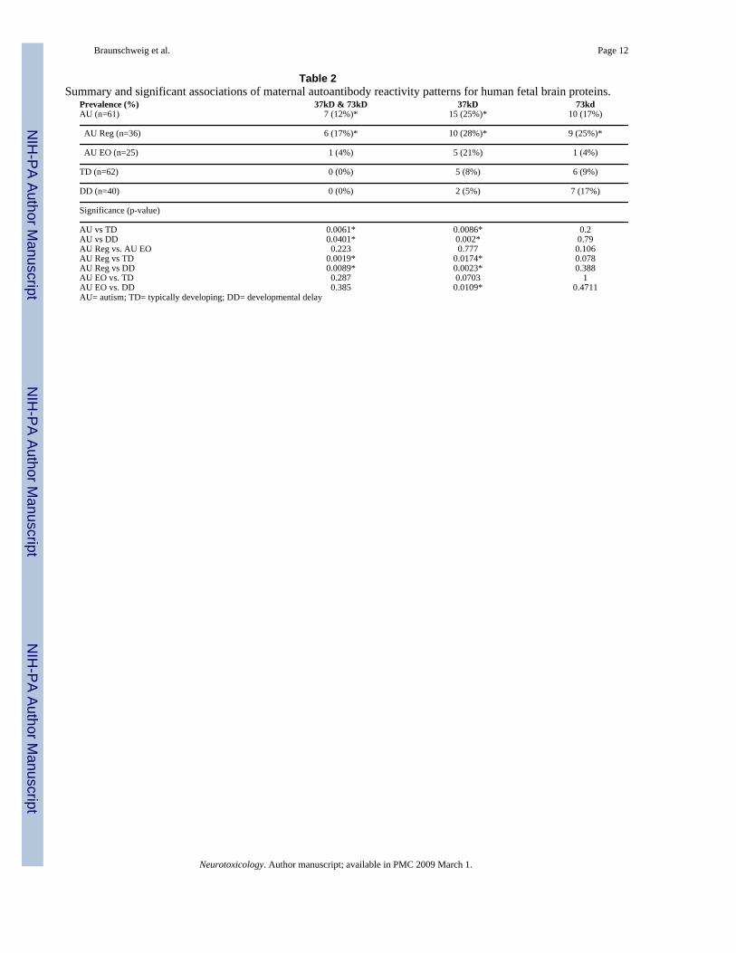
Table 2Summary and significant associations of maternal autoantibody reactivity patterns for human fetal brain proteins.
Prevalence (%) 37kD & 73kD 37kD 73kdAU (n=61) 7 (12%)* 15 (25%)* 10 (17%)
AU Reg (n=36) 6 (17%)* 10 (28%)* 9 (25%)*
AU EO (n=25) 1 (4%) 5 (21%) 1 (4%)
TD (n=62) 0 (0%) 5 (8%) 6 (9%)
DD (n=40) 0 (0%) 2 (5%) 7 (17%)
Significance (p-value)
AU vs TD 0.0061* 0.0086* 0.2AU vs DD 0.0401* 0.002* 0.79AU Reg vs. AU EO 0.223 0.777 0.106AU Reg vs TD 0.0019* 0.0174* 0.078AU Reg vs DD 0.0089* 0.0023* 0.388AU EO vs. TD 0.287 0.0703 1AU EO vs. DD 0.385 0.0109* 0.4711AU= autism; TD= typically developing; DD= developmental delay
Neurotoxicology. Author manuscript; available in PMC 2009 March 1.
Copyright © 2022 FDOKUMEN